Embed Size (px)
Citation preview
Citation :Egypt. Acad. J. Biolog. Sci. ( D-Histology and histochemistry) Vol.11(1)pp15-32(2019)
Egypt. Acad. J. Biolog. Sci., 11(1): 15- 32 (2019)
Egyptian Academic Journal of Biological Sciences
D. Histology & Histochemistry
2090 – 0775
www.eajbsd.journals.ekb.eg
The Enhancing Effect of Chamomile on Histological and Immunohistochemical
Alterations in Diabetic Rats
Nahla H. El-shaer 1, and Amany E. Nofal2
1-Zoology Department, Faculty of Science, Zagazig University, Egypt.
2-Zoology Department, Faculty of Science, Menoufia University, Egypt.
E.Mail.: [email protected]
INTRODUCTION Nowadays, diabetes is more seen in society, and show prevalence in about 382
million people around all the world (Shi and Hu 2014). It is also considered as a
metabolic disorder, which is characterized by many, disorders such as hyperglycemia,
glucosuria and negative nitrogen balance these all due to inhibition of insulin
secretion, which occurs in beta cells inside pancreas, and desensitization of insulin
receptors to the insulin. Vats et al. (2005) found that, it is the most prevalent disease
in the world, which affects 25% of the population, afflicts 150 million people, and is
predicted to rise to 300 million by 2025. In addition, it caused different disorders like
retinopathy, neuropathy, cardiovascular diseases, peripheral vascular insufficiencies,
ulcers and amputations (Wrighten et al., 2009).
Many experimental diabetic models of animals used and for the past 30 years
these drugs, alloxan, streptozotocin (STZ, 2-deoxy-2-(3-(methyl-3- nitrosoureido)-D-
glucopyranose), high-fat diet-fed and nicotinamide are for establishing it (Islam and
ARTICLE INFO ABSTRACT Article History
Received: 15/1/2019
Accepted: 20/3//2019
_________________
Keywords:
Chamomile,
Diabetes Mellitus,
streptozotocin,
caspase-3
Background:Herbal medicine showed an important role in diabetes
and other disorders treatment. Using natural products in animal
diabetic models is still unclear so that in this study we aimed to
investigate the possible effect of Chamomile extract on the diabetic
dysfunctions-induced by streptozotocin (STZ) through the
histopathological and immunohistochemical point of view. Result: Our experiment was carried out on 4 rat groups, five rats in
each to study the effect of Chamomile water extract on STZ -induced
diabetes .The animals were classified into four groups. A negative
control group (G1). A positive diabetic control group (G2) STZ (45
mg/kg body weight in 0.1 M citrate buffer, pH 4.5) was injected
intraperitoneally to rats after all overnight fasting (all animals exhibited
hyperglycemia within 36 to 48 hr). Chamomile treatment group (G3)
normal rats administered water extract of Chamomile, orally at a dose
of 200 mg/kg body weight for 21 successive days. Diabetic-
Chamomile treated group (G4) diabetic rats received 1 ml water extract
of Chamomile for 21 days 36 h post- STZ injection.
Conclusion: Histopathological investigations of our result of pancreas,
kidneys, spleen and liver besides pancreatic anti-insulin and caspase-3
monoclonal antibody marker were carried out, the collected data
revealed protective, and enhancement effect of Chamomile against
STZ- induced diabetes in rats.
Nahla H. El-shaer 1,and Amany E. Nofal2
16
Loots 2009). Streptozotocin also still
the most commonly used as an agent to
induce diabetes in rats and mice
(Ngubane et al., 2015), and its cause a
decrease in the plasma insulin levels in
animal models after Streptozotocin
administrated which is used for
diabetes induction (Patel et al., 2015).
Streptozotocin-antibiotic produced
naturally from Streptomyces
achromogenes, which cause impairing
for glucose oxidization, and thus
causing reduction in insulin
biosynthesis, secretion and
abnormalities of B-cells, it induced
hyperglycemia and widely used in the
experimental model by screening the
activity of hypoglycemic agents
(Szkudelski, 2001).
Beltramini et al. (2006) found that,
hyperglycemia arise because of b-islet
cells irreversible destruction in
pancreas by Streptozotocin which
leads to a reduction of insulin
secretion, and also the generated
reactive oxygen species (ROS) and the
subsequent increase of local oxidative
stress, DNA methylation, and protein
modification all are considered as the
pathophysiological mechanisms of
STZ-induced diabetes. Ramesh and
Pugalendi (2006) found that the
antioxidant agents acted against STZ-
induced diabetes because they are
made a diminishing for the oxidative
stress by inhibiting ROS generation
and lipid peroxidation. ROS is also
involved in the etiology and
pathogenesis of diabetes and the
development of diabetic complications
(Altan et al., 2006).
In spite of the use of many
hypoglycemic agents, diabetes and its
health complications are still an
important medical problem. And also it
is considered of the drugs that have
limited efficacy and certain adverse
effects, which causing hypoglycemia at
higher doses, liver problems, lactic
acidosis, and diarrhea. Anorexia, brain
atrophy and fatty liver in many chronic
treatments (Weidmann et al., 1993).
Many investigations found that
herbal drugs effect on the treatment of
diabetic mellitus (Pari et al., 1999).
Present anti-diabetic agents for
diabetes is still not made a complete
cure. Insulin therapy is the only
satisfactory approach in diabetic
mellitus, despite it has several
drawbacks like insulin resistance
(Piedrola et al., 2001). The efficacy of
herbal medicine is the low incidence of
side effects, and also the low cost
(Manal, 2012).
Hyperglycemia long-run
exposure caused oxidative stress and
reduced the efficiency of the
endogenous antioxidant defense
system by the production of several
reducing sugars (through glycolysis
and the polyol pathway) (Robertson
and Harmon, 2006). The reducing
sugars produced during glycolysis
react easily with lipids and proteins
(nonenzymatic glycation reaction),
which caused an increase in the
production of ROS (Jay et al., 2006).
On the other hand, it has been found
that these ROS generation contributes
to streptozotocin (STZ) and thus
induced destruction of pancreatic b-
cells (Szkudelski, 2001).
Chamomile tea (Matricaria
chamomile L.) is widely used from the
native old world and also well known
as a medicinal plant (Astin et al.,
2000) .Chamomile tea is prepared from
dried flowers and also has been used as
herbal medicine in Europe. Especially,
it also has been used in the treatment
of many inflammations, irritations, and
many pains such as skin diseases,
wounds, eczema, ulcers, gout,
neuralgia, and rheumatic pains. On the
other hand, many studies found that
Chamomile plant extract suppressed
the growth of human cancer cells
The Enhancing effect of Chamomile on Histological and Immunohistochemical alterations in diabetic rats
17
(Srivastava and Gupta, 2007). The
ameliorative effect of Chamomile on
hyperglycemia and diabetic
complications induced suppression of
blood sugar levels, increased liver
glycogen storage, and also the
inhibition of sorbitol in the human
erythrocytes (Kato et al ., 2008).
The pharmacological activity of
Chamomile showed an independent
effect on insulin secretion (Eddouks et
al., 2005). Also, different studies
showed its protective effect on
pancreatic beta cells which leads to
diminishing hyperglycemia-related
oxidative stress (Cemek et al., 2008).
So that many studies are required to
evaluate the useful effect of
Chamomile in managing diabetes.
MATERIALS AND METHODS
Collection and Extraction of Plant
Material:
The fresh leaves of the plant
Matricaria chamomile L (Chamomile)
were obtained from the local herbal
market present in Egypt. Then 10 gm
of plant leaves of Chamomile was
taken and soaked in 100 ml of boiled
water in a glass jar, then shaken and
stirred for 4 hr. The container content
was left overnight at room temperature
and then was filtered through double
filter paper (Double Rings filter paper
102, 11.0 cm). Finally, the filtrates
were concentrated by using a vacuum
pump rotary evaporator to afford a
greenish mass of leaves extracts
(manal, 2012).
Experimental Animals:
The experiment was conducted on
adult male albino rats with the weights
of 150 - 200gm obtained from Animal
House of Faculty of Science,
Menoufia University, Egypt. All rats
were fed normally in Labe a chow
food containing (16% protein, 66%
carbohydrate, 8% fats and water).
They were housed at a (12:12) hrs.
Light and Dark cycle at 240C ± 3ºC
and with relative humidity (60-70) %.
The guidelines followed for the animal
experiment were approved by
experimental procedures was approved
by the animal ethical committee in
accordance with the guide for the care
and use of laboratory animals with
approval No (MUFSS/F/HI/2/19).
Experimental Design and Treatment
Schedule:
The experiment was carried out on
4 groups, five rats in each group, to
study the effect of plant water extract
on STZ -induced diabetes as follows:
1-The Negative Control Group (G1): Healthy control rats, received distilled
water ad-libitum.
2-The Positive Diabetic Control
Group (G2): Received a freshly
prepared solution of Streptozotocin or
STZ (45 mg/kg body weight in 0.1 M
citrate buffer, pH 4.5) by injection
intraperitoneally to rats after overnight
fasting (deprived of food for 16 h but
allowed free access to water). STZ
injected animals exhibited
hyperglycemia within 36 to 48 hr
(Olfat, 2012). Rats having
hyperglycemia with fasting blood
glucose (FBG) values of 250 mg/dl or
above were considered for the study.
3-Chamomile Treatment Group
(G3): Normal rats administered water
extract of Chamomile, orally at a dose
of 200 mg/kg body weight (dosage
determined earlier, (Seelinger et al,
2008), for 21 successive days.
4-Diabetic-Chamomile Treated
Group (G4): After induction of
diabetes, rats received 1 ml water
extract of Chamomile for 21 days 36 hr
post- STZ injection. 200 mg/kg/day
Chamomile (Tavafi et al., 2011) was
given daily for 21days to the diabetic
rats, using gastric channel (Makino et
al., 2002).
No detectable irritation or adverse
effect such (respiratory distress,
abnormal locomotion or catalepsy) was
observed in any animals after
Chamomile administration.
Nahla H. El-shaer 1,and Amany E. Nofal2
18
Study Protocol and Collection of
Samples:
In the present study increase
consumption of drinking water by rats
insure induction of diabetes mellitus
(DM) , and after confirmation of
diabetic status , a blood drop was taken
from the distal end of the tail of fasted
animals and applied to a blood strip
and analyzed using a blood glucose
monitoring device (Acu-Check,
Performance, Germany) for both
control and diabetic animals, and FBG
(Fasting blood glucose) level was
measured in every 7th day using blood
strips over the course of the treatment,
and rats receive injection of STZ
exhibiting hyperglycemia with (FBS)
250 mg/dl. And from the 1st day (3rd
day of STZ-injection) of Chamomile
extract administration to diabetic rats
(Mallick et al., 2007). On the 21st day
of extract administration, all the
animals were anesthetized (Nesdonal
50 mg/kg, i.p.), the pancreas was
removed for the histological and
immunohistochemical analysis.
Kidney, liver, heart and spleen were
excised immediately and thoroughly
washed in saline and used for
histological experiments.
Histological Study:
After blood sampling, the kidney,
spleen, pancreas and liver were
removed with minimum handling and
fixed in 10% neutral buffered formalin
for 24 hr, washed the specimen in tap
water then dehydrated through serial
dilutions of alcohol (ethyl and absolute
ethyl) were used always for
dehydration. Specimens were cleared
in hydrocarbon (xylene), infiltrated in
paraffin at 56 degrees in an oven, and
embedded outside the oven. Paraffin
bees wax tissue blocks were prepared
for sectioning at 4 microns by slide
using microtome. The obtained tissue
sections were collected on glass slides,
deparaffinized and stained by
hematoxylin and eosin stains for
histopathological examination through
the electric light microscope (Suvarna
et al. , 2018).Then all slides were
viewed by using Labomed , Labo
America, Inc. USA microscope and
images were captured by a digital
camera (Sony DSC_S5000). The
severity of inflammation was recorded
using a grading scale of 0 to 4, as
follows:
0 = indistinguishable from
controls.
1 = minimal, ≤25% of cells
affected.
2 = mild, 25% ≥ 50% of cells
affected.
3 = moderate, 50% ≥ 75% of
cells affected.
4 = marked, 75% cells affected.
Scale bars were measured by
using Image-J software.
Immunohistochemical Study:
Principle The standard immunohistochemical
methods were adopted (Eissa and
Shoman, 1998). The tissue sections
were routinely microwave-treated to
unmistaken the epitopes of antigen
(Cattoreti et al., 1992). The
demonstration of antigen in tissues by
immunostaining is a two-step process.
The first step is binding of the primary
antibody to insulin and Caspase 3
(Biogenex Laboratories, San Ramón,
CA, U.S.A.), followed by visualization
of this reaction by a secondary or link
antibody to which are attached
different enzyme systems. The primary
antibody determines the specificity of
the reaction; whereas, the secondary
antibody, with its linked enzyme,
causes amplification of the reaction,
hence, increase of the sensitivity of the
test. The Biotin-Streptavidin (BSA)
system was used to visualize the
markers (Hsu et al., 1981).
Diaminobenzidine (DAB) was used as
chromogen since it allows a permanent
preparation. Hematoxylin counterstain
was done using Mayer's hematoxylin
The Enhancing effect of Chamomile on Histological and Immunohistochemical alterations in diabetic rats
19
(Hx). The specimens were mounted
using mounting medium Canada
balsam and examined by a microscope
(Olympus DP 71), the percentages of
the relative staining areas were
measured by using Image-J software.
RESULTS
1. Histopathologic Finding:
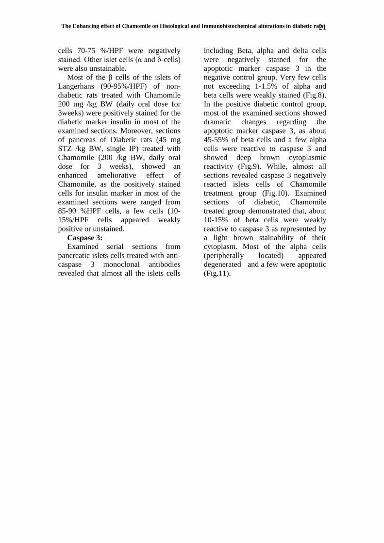
Pancreas:
Histological examination of
pancreas sections of negative control
group rats showed normal islets cells
with prominent beta and alpha cells
beside normal pancreatic parenchymal
and stromal cells. The positive diabetic
control group showed congested
pancreatic blood vessels, focal
degeneration of the pancreatic acini
and cystic dilatation of pancreatic
ducts were seen. Some of the islets
cells showed decrease cellular
population with degeneration and
necrosis of some of the beta cells.
Other islets cells showed a normal
population of cells including alpha,
beta and delta cells. The chamomile
treatment group showed normal
pancreatic tissue with apparently
normal islets and their cellular
contents. In Diabetic, Chamomile
treated group most of the pancreatic
islets were large in size or of normal
sizes with apparently normal active
cellular contents. In some sections, the
pancreatic blood vessels were mild to
moderately congest with mild
interstitial lymphocytic infiltration.
Some of the pancreatic ducts were
cystically dilated and filled with
secretion (Fig.1& 2).
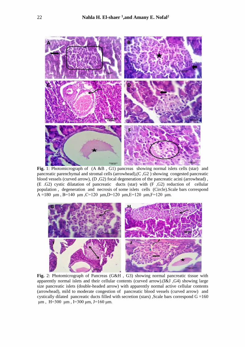
Liver: Liver sections of the negative
control group showed normal
histomorphology with preserved
hepatic cords, central veins, portal area
and stromal structures. Positive
diabetic control group revealed mild
portal congestion and mixed
inflammatory infiltrates with round
cells infiltration central‐portal fibrous
septa. The chamomile treatment group
showed normal hepatic parenchyma,
central veins, sinusoids and portal
triads. Diabetic, Chamomile treated
group revealed apparently normal
histomorphological structures (Fig.3).
Kidney:
Histological examination of a
kidney of the negative control group
showed normal nephron units with
preserved glomerular and tubular
structures with a normal appearance of
a tuft of capillaries, and glomerular
basement membrane surrounded by a
double-walled epithelial capsule. The
positive diabetic control group showed
shrinkage of the Malpighian corpuscles
and mild vacuolated of epithelial lining
renal tubules also dilated congested
vascular spaces. Chamomile treatment
group showed normal nephron
histomorphology and, there is no
histopathological alterations were
observed. Diabetic, Chamomile treated
group revealed apparently normal
nephron histomorphology (Fig.4).
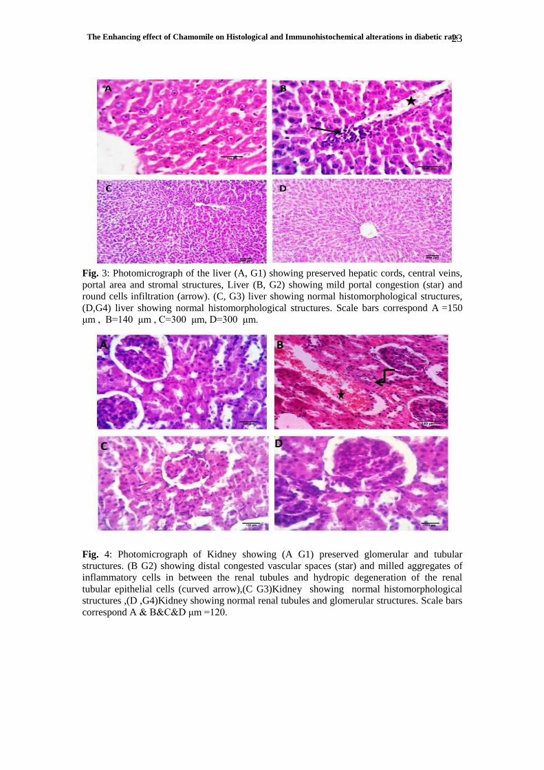
Heart:
Rats of the negative control group
showed normal cardiac muscles with
preserved coronary, intermuscular
blood vessels, normal cardiomyocytes
and Purkinje fibers. Positive diabetic
control group revealed interstitial and
perivascular edema beside
degenerative changes in some
cardiomyocytes. Chamomile treatment
group revealed normal cardiac
parenchymal and stromal structures
with mild congestion of the coronary
and intermuscular blood vessels.
Diabetic, Chamomile treated group
showed apparently normal
cardiomyocytes with mild congestion
of the coronary and intermuscular
blood vessels (Fig.5).
Spleen: Spleen sections of the negative
control rats showed normal white pulp
lymphoid structures, red pulp
sinusoidal, and reticuloendothelial
Nahla H. El-shaer 1,and Amany E. Nofal2
20
structures. The positive diabetic
control group showed a decrease in the
size of white pulp and congestion
inside red pulp. The chamomile
treatment group showed preserved
white pulp, red pulp histomorphology
keeping the normal lymphoid
population, sinusoidal and
reticuloendothelial structures. Diabetic,
Chamomile treated group showed
normal structures with mild to
moderate reactivity of the white pulp
lymphocytes and increased infiltration
of mature lymphocytes in the red pulp
(Fig.6).
Table (1): Histopathological scoring obtained by using light microscopic analysis of
pancreas, Liver, Kidney, Heart and Spleen in Tissue sections of all treatment groups
2. Immunohistochemical Findings:
Insulin:
Immuno-histochemical examination
of non-diabetic non treated rats for
detection of insulin marker revealed
active positive cytoplasmic stainability
of almost all β cells of islets of
Langerhans, the stained cells appeared
large , rounded with centrally located
nuclei and granular orange-red to
brown cytoplasm . Alpha and delta
cells were negatively stained. Alpha
cells (α-cells) were smaller in size,
round or ovoid in shape and arranged
peripherally and delta cells (δ-cells)
were the smallest, rod shape and were
randomly distributed inside the islets.
While, the pancreas of diabetic rats (45
mg STZ/kg BW (Single IP)) showed a
decrease on positively reacted and
stained cells with insulin diabetic
marker (25-30% /HPF), the remaining
The Enhancing effect of Chamomile on Histological and Immunohistochemical alterations in diabetic rats
21
cells 70-75 %/HPF were negatively
stained. Other islet cells (α and δ-cells)
were also unstainable.
Most of the β cells of the islets of
Langerhans (90-95%/HPF) of non-
diabetic rats treated with Chamomile
200 mg /kg BW (daily oral dose for
3weeks) were positively stained for the
diabetic marker insulin in most of the
examined sections. Moreover, sections
of pancreas of Diabetic rats (45 mg
STZ /kg BW, single IP) treated with
Chamomile (200 /kg BW, daily oral
dose for 3 weeks), showed an
enhanced ameliorative effect of
Chamomile, as the positively stained
cells for insulin marker in most of the
examined sections were ranged from
85-90 %HPF cells, a few cells (10-
15%/HPF cells appeared weakly
positive or unstained.
Caspase 3:
Examined serial sections from
pancreatic islets cells treated with anti-
caspase 3 monoclonal antibodies
revealed that almost all the islets cells
including Beta, alpha and delta cells
were negatively stained for the
apoptotic marker caspase 3 in the
negative control group. Very few cells
not exceeding 1-1.5% of alpha and
beta cells were weakly stained (Fig.8).
In the positive diabetic control group,
most of the examined sections showed
dramatic changes regarding the
apoptotic marker caspase 3, as about
45-55% of beta cells and a few alpha
cells were reactive to caspase 3 and
showed deep brown cytoplasmic
reactivity (Fig.9). While, almost all
sections revealed caspase 3 negatively
reacted islets cells of Chamomile
treatment group (Fig.10). Examined
sections of diabetic, Chamomile
treated group demonstrated that, about
10-15% of beta cells were weakly
reactive to caspase 3 as represented by
a light brown stainability of their
cytoplasm. Most of the alpha cells
(peripherally located) appeared
degenerated and a few were apoptotic
(Fig.11).
Nahla H. El-shaer 1,and Amany E. Nofal2
22
Fig. 1: Photomicrograph of (A &B , G1) pancreas showing normal islets cells (star) and
pancreatic parenchymal and stromal cells (arrowhead),(C ,G2 ) showing congested pancreatic
blood vessels (curved arrow), (D ,G2) focal degeneration of the pancreatic acini (arrowhead) ,
(E .G2) cystic dilatation of pancreatic ducts (star) with (F ,G2) reduction of cellular
population , degeneration and necrosis of some islets cells (Circle).Scale bars correspond
A =180 μm , B=140 μm ,C=120 μm,D=120 μm,E=120 μm,F=120 μm.
Fig. 2: Photomicrograph of Pancreas (G&H , G3) showing normal pancreatic tissue with
apparently normal islets and their cellular contents (curved arrow).(I&J ,G4) showing large
size pancreatic islets (double-headed arrow) with apparently normal active cellular contents
(arrowhead), mild to moderate congestion of pancreatic blood vessels (curved arrow) and
cystically dilated pancreatic ducts filled with secretion (stars) ,Scale bars correspond G =160
μm , H=300 μm , I=300 μm, J=160 μm.
The Enhancing effect of Chamomile on Histological and Immunohistochemical alterations in diabetic rats
23
Fig. 3: Photomicrograph of the liver (A, G1) showing preserved hepatic cords, central veins,
portal area and stromal structures, Liver (B, G2) showing mild portal congestion (star) and
round cells infiltration (arrow). (C, G3) liver showing normal histomorphological structures,
(D,G4) liver showing normal histomorphological structures. Scale bars correspond A =150
μm , B=140 μm , C=300 μm, D=300 μm.
Fig. 4: Photomicrograph of Kidney showing (A G1) preserved glomerular and tubular
structures. (B G2) showing distal congested vascular spaces (star) and milled aggregates of
inflammatory cells in between the renal tubules and hydropic degeneration of the renal
tubular epithelial cells (curved arrow),(C G3)Kidney showing normal histomorphological
structures ,(D ,G4)Kidney showing normal renal tubules and glomerular structures. Scale bars
correspond A & B&C&D μm =120.
Nahla H. El-shaer 1,and Amany E. Nofal2
24
Fig. 5: Photomicrograph of Heart (A, G1) showing normal cardiac muscles (Curved arrow)
with preserved coronary blood vessels (star). (B & C, G2) Heart showing interstitial and
perivascular edema (star) beside degenerative changes in some cardiomyocytes (arrowheads).
(D, G3) Heart showing normal cardiac parenchymal (arrowhead) with mild congestion of
intermuscular blood vessels. (E,G4)heart showing apparently normal cardiomyocytes with
mild congestion of coronary blood vessels (arrow).Scale bars correspondA =120 μm , B=200
μm , C=120 μm, D=110 μm,E=120 μm.
Fig. 6: Photomicrograph of spleen (A, G1) Spleen showing normal white pulp (star), red pulp
and Sinusoidal structures (arrowhead). (B,G2) Spleen showing decrease in the size of white
pulp and congestion inside red pulp (star),(C, G3)Spleen sections showing preserved white
(star) and red pulp structures (closed arrows). (D,G4) Spleen showing moderate reactivity of
the white pulp (star) and increased infiltration of mature lymphocytes in the red pulp (closed
arrow).Scale bars correspond A =120 μm , B=200 μm , C=120 μm, D=110 μm, E=120
μm.
The Enhancing effect of Chamomile on Histological and Immunohistochemical alterations in diabetic rats
25
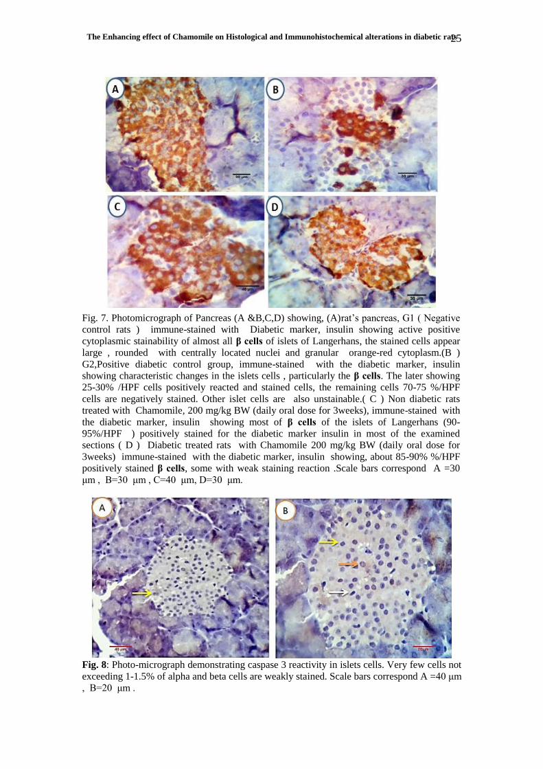
Fig. 7. Photomicrograph of Pancreas (A &B,C,D) showing, (A)rat’s pancreas, G1 ( Negative
control rats ) immune-stained with Diabetic marker, insulin showing active positive
cytoplasmic stainability of almost all β cells of islets of Langerhans, the stained cells appear
large , rounded with centrally located nuclei and granular orange-red cytoplasm.(B )
G2,Positive diabetic control group, immune-stained with the diabetic marker, insulin
showing characteristic changes in the islets cells , particularly the β cells. The later showing
25-30% /HPF cells positively reacted and stained cells, the remaining cells 70-75 %/HPF
cells are negatively stained. Other islet cells are also unstainable.( C ) Non diabetic rats
treated with Chamomile, 200 mg/kg BW (daily oral dose for 3weeks), immune-stained with
the diabetic marker, insulin showing most of β cells of the islets of Langerhans (90-
95%/HPF ) positively stained for the diabetic marker insulin in most of the examined
sections ( D ) Diabetic treated rats with Chamomile 200 mg/kg BW (daily oral dose for
3weeks) immune-stained with the diabetic marker, insulin showing, about 85-90% %/HPF
positively stained β cells, some with weak staining reaction .Scale bars correspond A =30
μm , B=30 μm , C=40 μm, D=30 μm.
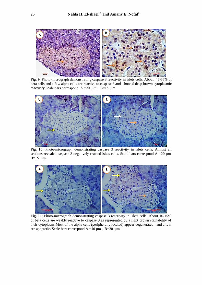
Fig. 8: Photo-micrograph demonstrating caspase 3 reactivity in islets cells. Very few cells not
exceeding 1-1.5% of alpha and beta cells are weakly stained. Scale bars correspond A =40 μm
, B=20 μm .
Nahla H. El-shaer 1,and Amany E. Nofal2
26
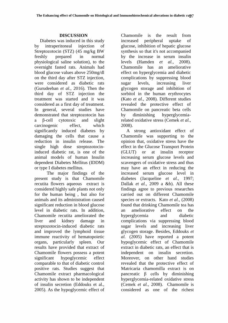
Fig. 9: Photo-micrograph demonstrating caspase 3 reactivity in islets cells. About 45-55% of
beta cells and a few alpha cells are reactive to caspase 3 and showed deep brown cytoplasmic
reactivity.Scale bars correspond A =20 μm , B=18 μm
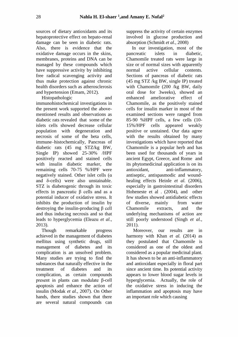
Fig. 10: Photo-micrograph demonstrating caspase 3 reactivity in islets cells. Almost all
sections revealed caspase 3 negatively reacted islets cells. Scale bars correspond A =20 μm,
B=15 μm
Fig. 11: Photo-micrograph demonstrating caspase 3 reactivity in islets cells. About 10-15%
of beta cells are weakly reactive to caspase 3 as represented by a light brown stainability of
their cytoplasm. Most of the alpha cells (peripherally located) appear degenerated and a few
are apoptotic. Scale bars correspond A =30 μm , B=20 μm.
The Enhancing effect of Chamomile on Histological and Immunohistochemical alterations in diabetic rats
27
DISCUSSION
Diabetes was induced in this study
by intraperitoneal injection of
Streptozotocin (STZ) (45 mg/kg BW
freshly prepared in normal
physiological saline solution), to the
overnight fasted rats. Animals had
blood glucose values above 250mg/dl
on the third day after STZ injection,
were considered as diabetic rats
(Gurudeeban et al., 2016). Then the
third day of STZ injection the
treatment was started and it was
considered as a first day of treatment.
In general, several studies have
demonstrated that streptozotocin has
a β-cell cytotoxic and slight
carcinogenic effect, which
significantly induced diabetes by
damaging the cells that cause a
reduction in insulin release. The
single high dose streptozotocin-
induced diabetic rat, is one of the
animal models of human Insulin
dependent Diabetes Mellitus (IDDM)
or type I diabetes mellitus.
The major findings of the
present study is that Chamomile
recutita flowers aqueous extract is
considered highly safe plants not only
for the human being , but also for
animals and its administration caused
significant reduction in blood glucose
level in diabetic rats. In addition,
Chamomile recutita ameliorated the
liver and kidney damage in
streptozotocin-induced diabetic rats
and improved the lymphoid tissue
immune reactivity of hematopoietic
organs, particularly spleen. Our
results have provided that extract of
Chamomile flowers possess a potent
significant hypoglycemic effect
comparable to that of diabetic control
positive rats. Studies suggest that
Chamomile extract pharmacological
activity has shown to be independent
of insulin secretion (Eddouks et al.,
2005). As the hypoglycemic effect of
Chamomile is the result from
increased peripheral uptake of
glucose, inhibition of hepatic glucose
synthesis so that it's not accompanied
by the increase in serum insulin
levels (Hamden et al., 2008).
Chamomile has an ameliorative
effect on hyperglycemia and diabetic
complications by suppressing blood
sugar levels, increasing liver
glycogen storage and inhibition of
sorbitol in the human erythrocytes
(Kato et al., 2008). Different studies
revealed the protective effect of
Chamomile on pancreatic beta cells
by diminishing hyperglycemia-
related oxidative stress (Cemek et al.,
2008).
A strong antioxidant effect of
Chamomile was supporting to the
opinion that, oxidative stress have the
effect in the Glucose Transport Protein
(GLUT) or at insulin receptor
increasing serum glucose levels and
scavengers of oxidative stress and thus
may have an effect in reducing the
increased serum glucose level in
diabetes (Jacqueline et al., 1997;
Dallak et al., 2009 a &b). All these
findings agree to previous researches
carried out on different Chamomile
species or extracts. Kato et al., (2008)
found that drinking Chamomile tea has
an ameliorative effect on the
hyperglycemia and diabetic
complications via suppressing blood
sugar levels and increasing liver
glycogen storage. Besides, Eddouks et
al. (2005) have reported a potent
hypoglycemic effect of Chamomile
extract in diabetic rats, an effect that is
independent on insulin secretion.
Moreover, on other hand studies
revealed that the protective effect of
Matricaria chamomilla extract is on
pancreatic β cells by diminishing
hyperglycemia-related oxidative stress
(Cemek et al., 2008). Chamomile is
considered as one of the richest
Nahla H. El-shaer 1,and Amany E. Nofal2
28
sources of dietary antioxidants and its
hepatoprotective effect on hepato-renal
damage can be seen in diabetic rats.
Also, there is evidence that the
oxidative damage occurs in the skins,
membranes, proteins and DNA can be
managed by these compounds which
have suppressive activity by inhibiting
free radical scavenging activity and
thus make protection against chronic
health disorders such as atherosclerosis
and hypertension (Emam, 2012).
Histopathologic and
immunohistochemical investigations in
the present work supported the above-
mentioned results and observations as
diabetic rats revealed that some of the
islets cells showed decrease cellular
population with degeneration and
necrosis of some of the beta cells,
immune–histochemically, Pancreas of
diabetic rats (45 mg STZ/kg BW,
Single IP) showed 25-30% /HPF
positively reacted and stained cells
with insulin diabetic marker, the
remaining cells 70-75 %/HPF were
negatively stained. Other islet cells (α
and δ-cells) were also unstainable.
STZ is diabetogenic through its toxic
effects in pancreatic β cells and as a
potential inducer of oxidative stress. It
inhibits the production of insulin by
destroying the insulin-producing β cell
and thus inducing necrosis and so that
leads to hyperglycemia (Eleazu et al.,
2013).
Though remarkable progress
achieved in the management of diabetes
mellitus using synthetic drugs, still
management of diabetes and its
complication is an unsolved problem.
Many studies are trying to find the
substances that naturally effective in the
treatment of diabetes and its
complication, as certain compounds
present in plants can modulate β-cell
apoptosis and enhance the action of
insulin (Modak et al., 2007). On Other
hands, there studies shown that there
are several natural compounds can
suppress the activity of certain enzymes
involved in glucose production and
absorption (Schmidt et al., 2008)
In our investigation, most of the
pancreatic islets in diabetic,
Chamomile treated rats were large in
size or of normal sizes with apparently
normal active cellular contents.
Sections of pancreas of diabetic rats
(45 mg STZ /kg BW, single IP) treated
with Chamomile (200 /kg BW, daily
oral dose for 3weeks), showed an
enhanced ameliorative effect of
Chamomile, as the positively stained
cells for insulin marker in most of the
examined sections were ranged from
85-90 %HPF cells, a few cells (10-
15%/HPF cells appeared weakly
positive or unstained. Our data agree
with the results obtained by many
investigations which have reported that
Chamomile is a popular herb and has
been used for thousands of years in
ancient Egypt, Greece, and Rome and
its phytomedicinal application is on its
antioxidant, anti-inflammatory,
antiseptic, antispasmodic and wound-
healing effects Heinle et al. (2006),
especially in gastrointestinal disorders
Hoheneste et al . (2004), and other
few studies showed antidiabetic effects
of diverse, mainly from water
Chamomile extracts, and the
underlying mechanisms of action are
still poorly understood (Singh et al.,
2011).
Moreover, our results are in
harmony with Khan et al. (2014) as
they postulated that Chamomile is
considered as one of the oldest and
considered as a popular medicinal plant.
It has shown to be an anti-inflammatory
and antioxidant especially in floral part
since ancient time. Its potential activity
appears to lower blood sugar levels in
hyperglycemia. Actually, the role of
the oxidative stress in inducing the
inflammation and apoptosis may have
an important role which causing
The Enhancing effect of Chamomile on Histological and Immunohistochemical alterations in diabetic rats
29
histological deteriorations since the
STZ-induced diabetic rats and their
results exhibited a remarkable increase
in the expression of
immunohistochemically-detected pro-
inflammatory 331 cytokine, TNF-α and
apoptotic markers including P53 and
caspase-3 (Osama et al., 2019).
In the present study it was indicated
that, the expression caspase-3 and
insulin were remarkably increased in
diabetic rats and were decreased as a
result of treatment with Chamomile,
and this proved the fact that the use of
Chamomile with other plant extracts
induced a more synergistic enhancing
effects of the aqueous extracts of
Chamomile and oregano and made it
useful in treating hyperglycemia and
related diabetic complications. The
mixture of Chamomile and Oregano
extracts produced a medical product
with a potent anti-diabetic activity
(Rajagopalan et al., 2016). Finally, our
results showed high expression of the
apoptotic marker caspase 3 Further
studies are necessary to elucidate the
exact molecular mechanism involved in
anti-diabetic action of Chamomile.
Conclusions Chamomile extract used as popular
medicinal plant many studies indicate
it's anti-inflammatory and antioxidant
activity, this study showed a remarkable
antdiabetic effect through elevation of
histological parameter in kidney, liver
and immunohistochemical parameter
pancreas against STZ- induced
diabetes in rats.Altan N., Sepici-
Dinc¸el A., Koca C. (2006). Diabetes
mellitus and oxidative stress.Turk J
Biochem 31:51–56
REFERENCES
Astin J.A., Pelletier K.R., Marie A. and
Haskell W.L. (2000).
Complementary and alternative
medicine use among elderly
persons: One-year analysis of
Blue Shield medicare supplement.
J. Gerontol. A Biol. Sci. Med.
Sci, 55(1): 4-9.
Beltramini M ., Zambenedetti P., Raso
M., IbnlKayat M.I., Zatta P.
(2006) .The effect of Zn(II) and
streptozotocin administration in
the mouse brain. Brain Res
1109:207–218
Cattoretti G., Becker M.H.G., Key G.,
Duchrow M., Schluter C., Galle J.
and Gerdes J. (1992).Monoclonal
antibodies against recombinant
parts of the Ki-67 .antigen (MIB 1
and MIB 3) detect proliferating
cells in microwave-processed
formalinfixed paraffin sections. J
Pathol 168: 357–363.
Cemek M., Kaga S., Simsek N.,
Buyukokuroglu M.E., Konuk M.
(2008). Antihyperglycemic and
antooxidative potential of
Matricaria chamomilla L. in
streptozotocin-induced diabetic
rats. J. Nat Med. 62:284–293.
Dallak, M., M. Al-Khateeb, M. Abbas,
R. Elessa and F. Al-Hashem
(2009a). In vivo, acute,
normohypoglycemic,
antihyperglycemic, insulinotropic
actions of orally administered
ethanol extract of Citrullus
colocynthis (L.) schrab pulp. Am.
J. Biochem. Biotechnol., 5: 118-
125. DOI:
10.3844/ajbbsp.2009.118.125
Dallak, M., N. Bashir, M. Abbas,
R.E.M. Haidara and M. Khalil,(
2009b.) Concomitant down
regulation of glycolytic enzymes,
upregulation of gluconeogenic
enzymes and potential
hepatonephro- protective effects
following the chronic
administration of the
hypoglycemic, insulinotropic
citrullus colocynthis pulp extract.
Am. J. Biochem. Biotechnol., 5:
153-161. DOI:
10.3844/ajbbsp.2009.153.161.
Nahla H. El-shaer 1,and Amany E. Nofal2
30
Eddouks M, Lemhadri A, Zeggwah
NA, Michel JB. (2005) .Potent
hypoglycaemic activity of the
aqueousextract of chamaemelum
nobile in normal and
streptozotocin-induced diabetic
rats. Diabetes ResClin Pract;
67:189–195.
Eissa, S. and S. Shoman (1998)
.Markers of invasion and
metastasis and markers of tumor
proliferation and apoptosis in:
Tumors markers. Chapman and
Hall, Lippincott- Raven Publisher
Inc., London, UK, p. 131-153.
leazu CO, Iroaganachi M, Eleazu KC
(2013). Ameliorative Potentials of
Cocoyam (Colocasia esculenta L.)
and Unripe Plantain (Musa
paradisiaca L.) on theRelative
Tissue Weights of Streptozotocin-
Induced Diabetic Rats.J Diabetes
Res, (160964):8.
doi:10.1155/2013/1609640.
Emam, M.A., (2012). Comparative
evaluation of antidiabetic activity
of Rosmarinus officinalis L. and
Chamomile recutita in
streptozotocin induced diabetic
rats. Agric. Biol. J. N. Am., 3:
247-252. DOI:
10.5251/abjna.2012.3.6.247.252
Gurudeeban S, Kaliamurthi S,
Thirugnanasambandam R.(2016).
Positive Regulation of Rhizophora
mucronata PoirExtracts on Blood
Glucose and Lipid Profile in
Diabetic Rats. Herb Med.
Hamden, K., S. Carreau, S. Lajmi, D.
Aloulou and D. Kchaou (2008).
Hyperglycaemia, stress oxidant,
liver dysfunction and histological
changes in diabetic male rat
pancreas and liver: Protective
effect of 17 beta-estradiol.
Steroids, 73: 495-501. DOI:
10.1016/j.steroids.2007.12.026.
Heinle H, Hagelauer D, Pascht U,
Kelber O, Weiser D (2006)
.Intestinal spasmolytic effects of
STW 5 (Iberogast) and its
components. Phytomedicine 13
Suppl 5: 75-79.
Hohenester B, Rühl A, Kelber O,
Schemann M (2004). The herbal
preparation STW5 (lberogast) has
potent and region-specific effects
on gastric motility.
Neurogastroent Motil 16: 7
Hsu, H.M. et. al. (1981). Am. J. Clin.
Pathol. 75:734
Islam MS, Loots du T. (2009)
.Experimental rodent models of
type 2 diabetes: a review. Methods
and Findings in Experimental and
Clinical Pharmacology. ;
31(4):249–261.
Jacqueline, M.S., L. Jongsoon and F.P.
Paul, (1997). Tumor necrosis
factor-α-induced insulin resistance
in 3T3-L1 adipocytes is
accompanied by a loss of insulin
receptor substrate-1 and GLUT4
expression without a loss of
insulin receptor-mediated signal
transduction. J. Biol. Chem., 272:
971-976. DOI:
10.1074/jbc.272.2.971
Jay D, Hitomi H, Griendling KK
(2006) .Oxidative stress and
diabetic cardiovascular
complications. Free Radic Biol
Med 40:183–192
Kato, A., Minoshima, Y., Yamamoto,
J., Adachi, I., Watson, A. A., and
Nash, R. J. (2008) Protective
effects of dietary Chamomile tea
on diabetic complications. J.
Agric. Food Chem. 56:8206–
8211.
Khan SS , Najam R , Anser H , Riaz
B , Alam N. (2014). Chamomile
tea: herbal hypoglycemic
alternative for conventional
medicine, Pak J Pharm Sci. 2014
Sep; 27(5 Spec no):1509-14.
Makino T, Ono T, Liu N, Nakamura T,
Muso E, Honda G. (2002).
Suppressive effects of rosmarinic
acid on mesangio proliferative
The Enhancing effect of Chamomile on Histological and Immunohistochemical alterations in diabetic rats
31
glomerulonephritis in rats.
Nephron. 92:898-904.
Mallick C, Chatterjee K, GuhaBiswas
M, Ghosh D. (2007).
Antihyperglycemic effects of
separate and composite extract of
root of Musa paradisiaca and leaf
of Coccinia Indica in
Streptozotocin induced diabetic
male albino rat, Afr. J. Trad.
CAM, 4: 362-371.
Manal, A.E. (2012) .Comparative
evaluation of antidiabetic activity
of Rosmarinus officinalis L. and
Chamomile recutita in
streptozotocin induced diabetic
rats. Agric. Biol. J. N. Am. 3(6):
247-252.
Modak M., Dixit P., Londhe J.,
Ghaskadbi S., Devasagayam
T.P.(2007) . Indian herbs and
herbal drugs used for the
treatment of diabetes. J. Clin.
Biochem. Nutr. ; 40:163–173.
Ngubane PS, Hadebe SI, Serumula
MR, Musabayane CT.(2015). The
effects of transdermal insulin
treatment of streptozotocin-
induced diabetic rats on kidney
function and renal expression of
glucose transporters. Renal
Failure. ; 37(1):151–159.
Olfat A. K , Kholoud S. R , Enas N.
D,Hanan S. A and Najla O. A
2012 .Antidiabetic activity of
Rosmarinus officinalis and its
relationship with the antioxidant
property. African Journal of
Pharmacy and Pharmacology Vol.
6(14), pp. 1031 - 1036, 15 April,
2012
Osama M. A, Tarek M. A, Mohamed
A. Abdel Gaid, Ahmed A. Elberry
2019: Effects of enalapril and
paricalcitol treatment on diabetic
nephropathy and renal expressions
of TNF-α, P53, Caspase-3 and
Bcl-2 in STZ-induced diabetic
rats. bioRxiv preprint CC-BY 4.0
International license.
.doi: https://doi.org/10.1101/5771
06.
Pari L and Uma MJ. (1999)
.Hypoglycemic effect of Musa
sapientum L. in alloxan-induced
diabetic rats. Journal of
Ethnopharmacology; 68: 321–325.
Patel SB, Santani D, Patel V, Shah
M.(2015). Anti-diabetic effects of
ethanol extract of Bryonia
laciniosa seeds and its saponins
rich fraction in neonatally
streptozotocin-induced diabetic
rats. Pharmacognosy Research;
7(1):92–99.
Piedrola G, Novo E, Escober F,
Garcia-Robles R. (2001). White
blood cell count and insulin
resistance in patients with
coronary artery disease. Annual
Endocrinology (Paris) 62, 7–10.
Rajagopalan P.,. Elbessoumy A.A.,
Mahmoud A.E.(2016)
.Chamomile and Oregano extracts
synergistically exhibits anti
hyperglycemic, anti
hyperlipidaemic and renal
protective effects in Alloxan
induced Diabetic rats. Canadian
Journal of Physiology and
Pharmacology
Ramesh B, Pugalendi KV (2006)
.Antioxidant role of umbelliferone
in STZ-diabetic rats. Life Sci
79:306–310
Robertson RP, Harmon JS (2006)
.Diabetes, glucose toxicity, and
oxidative stress: a case of double
jeopardy for the pancreatic islet
beta cell. Free Radic Biol Med
41:177–184
Schmidt B., Ribnicky D.M., Poulev A.,
Logendra S., Cefalu W.T., Raskin
I. (2008) .A natural history of
botanical therapeutics.
Metabolism. ; 57:S3–S9. doi:
10.1016/j.metabol.2008.03.001
Seelinger G, Merfort I, Schempp CM.
(2008) .Antioxidant, anti-
inflammatory and antiallergic
Nahla H. El-shaer 1,and Amany E. Nofal2
32
activities of luteolin. Planta Med.
74: 1667-1677.
Shi Y, Hu FB. (2014) .The global
implications of diabetes and
cancer. The Lancet. ;
383(9933):1947–1948.
Singh O, Khanam Z, Misra N,
Srivastava MK (2011) Chamomile
(Matricaria chamomilla. p. L.): An
overview. Pharmacogn Rev 5: 82-
95.
Srivastava JK and Gupta S. (2007)
.Antiproliferative and apoptotic
effects of Chamomile extract in
various human cancer cells. J.
Agric. Food Chemistr . 55: 9470–
9478.
Suvarna, K.; Layton, C.; Bancroft, J.
D. (2018): Bancroft's Theory and
Practice of Histological
Techniques, 8th Edition, London,
Elsevier; 672.
Szkudelski T. (2001). The mechanism
of alloxan and streptozotocin
action in B cells of the rat
pancreas. Physiol Res 50:536–
546
Tavafi M, Ahmadvand H, Khalatbari
A, Tamjidipoor A. (2011)
.Rosmarinic acid ameliorates
diabetic nephropathy in
uninephrectomized diabetic rats.
Iran. J. Basic Med. Sci. 14 (3):
275-283.
Vats RK, Kumar V, Kothari A, Mital
A, Ramachandran U. (2005).
Emerging targets for diabetes.
Curr Science; 88: 241-247...
Weidmann P, Boehlen LM and DE
(1993) .Courten M. Pathogenesis
and treatment of hypertension
associated with diabetes mellitus.
American Heart Journal; 125:
1498–1513.
Wrighten SA, Piroli GG, Grillo CA,
Reagan LP.(2009) . A look inside
the diabetic brain: Contributors to
diabetes-induced brain aging.
Biochimica ET Biophysica Acta. ;
1792:444–453.