Embed Size (px)
Citation preview
Loyola University Chicago Loyola University Chicago
Loyola eCommons Loyola eCommons
Master's Theses Theses and Dissertations
1972
The Formation and Constriction of the Trapping Rings in the The Formation and Constriction of the Trapping Rings in the
Nematophagous Fungus Dactylaria Brochopaga Dreschler Nematophagous Fungus Dactylaria Brochopaga Dreschler
Walter Thomas Rudek Loyola University Chicago
Follow this and additional works at: https://ecommons.luc.edu/luc_theses
Part of the Biology Commons
Recommended Citation Recommended Citation Rudek, Walter Thomas, "The Formation and Constriction of the Trapping Rings in the Nematophagous Fungus Dactylaria Brochopaga Dreschler" (1972). Master's Theses. 2599. https://ecommons.luc.edu/luc_theses/2599
This Thesis is brought to you for free and open access by the Theses and Dissertations at Loyola eCommons. It has been accepted for inclusion in Master's Theses by an authorized administrator of Loyola eCommons. For more information, please contact [email protected].
This work is licensed under a Creative Commons Attribution-Noncommercial-No Derivative Works 3.0 License. Copyright © 1972 Walter Thomas Rudek
The Formation and Constriction of the Trapping
Rings in the Nematophagous Fungus
Pactylaria brochopaga Dreschler
by
Walter Thomas Hudek
A Thesis Submitted to the Faculty of the
Graduate School of Loyola University
in Partial Fulfillment of the
Requirements for the Degree of
Master of Science
June 1972
ACKNOWLEL'GEMENTS
The author wishes to express his gratitude to his
advisor, Dr. Benedict J. Jaskoski of Loyola University,
for his valuable guidance, patience and understanding
throughout the course of this work and for his assistance
in the preparation of this manuscript. Sincere appreciation
is also extended to Dr. Leon L. Gershbein of Northwest
Institute For Medical Research for his ~elp and permission
1n making available the facilities and equipment at
Northwest Hospital Clinical Laboratory.
My speci~l thanks arc to my wife, Cathy, for her
patience and encouragement throughout the course of my
graduate studies and for her unselfish help in typing this
manuscript.
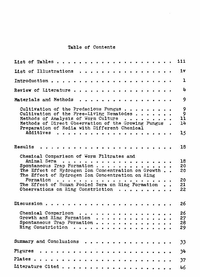
Table of Contents
List of Tables • • • • • • • • • • • • • •
List of Illustrations • • • • • • • • • • • •
Introduction • • • • • • • • • • • • • •
Review of Literature • • • • • • • • • • • •
Materials and Methods • • • • • • • • • • • • •
Cultivation of the Predacious Fungus • • • • Cultivation of the Free-Living Nematodes • Methods of Analysis of Worm Culture • Methods of Direct Observation of the Growing Preparation of Media with Different Chemical
Additives • • • • • • • • • • • •
• • • •
• • Fungus
• • • • •
Results • • • • • • • • • •
Chemical Comparison of Worm Filtrates and Animal Sera • • • • • • • • • • • •
Spontaneous Trap Formation • • • • The Effect of Hydrogen Ion Concentration on Growth • The Effect of Hydrogen Ion Concentration on Ring
Formation • • • The Effect of Human Pooled Sera on Ring Formation • Observations on Ring Constriction • • • •
Discussion • • • • •
Chemical Comparison • Growth and Ring Formation Spontaneous Trap Formation • Ring Constriction •
• • •
• • • • •
• • • • •
• • • • • •
• • • • • • • • • • • • • •
• • • • • • • • •
• • •
•
• •
•
Summary and Conclusions • • • • • • • • • • • • • • • •
Figures
Plates • • • • • • • • • • • • • • • • • • • • •
• • • • • • • • • • • • • • • • • • • Literature Cited • • • • • • • • • • • • • • • •
iii
iv
1
4
9
9 9
11 14
15
18
18 20 20
20 21 22
26
26 27 28 29
33
34
37 46
Table
1.
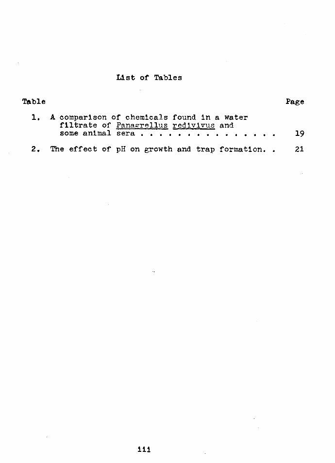
List of Tables
A comparison of chemicals found in a water filtrate of Pana~rellus redivivus and some animal sera • • • • • • • • • • • •
Page
• • • 19
2. The effect of pH on growth and trap formation. • 21
111
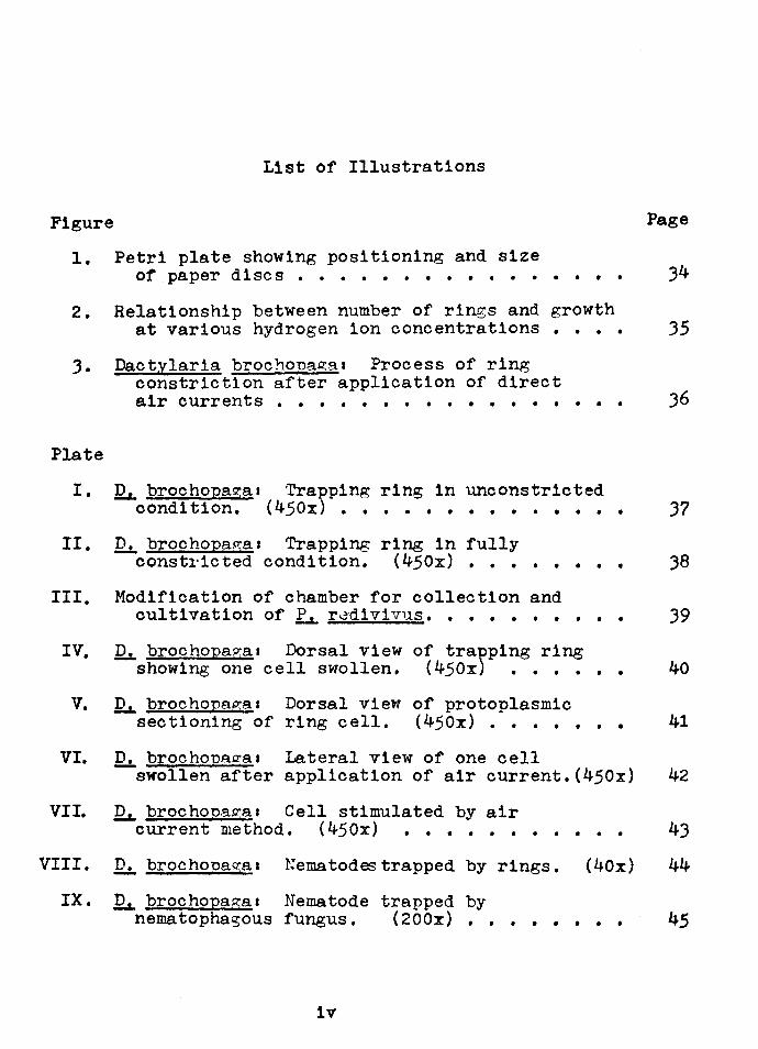
List of Illustrations
Figure Page
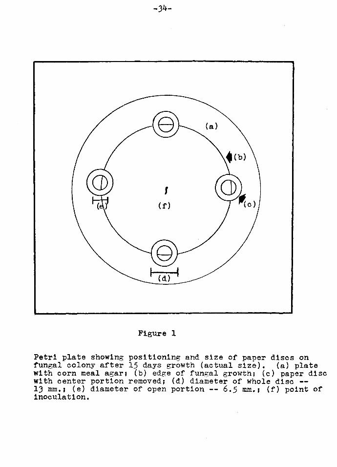
1. Petri plate showing positioning and size of paper discs • • • • • • • • • • • • • • • • 34
2. Relationship between number of rings and growth
3.
Plate
at various hydrogen ion concentrations • • • • 35
Dactylaria brochona~a• Process of ring constriction after application of direct air currents • • • • • • • • • • • • • • • • • 36
I. I2.i., brochopa~a1 Trapping ring in unconstricted condition. (450x) • • • • • • • • • • • • • • 37
II.
III.
IV.
v.
D. brochopa~a1 Trapping ring in fully const1·ic ted condition. ( 450x) • • • • • • • •
Modification of chamber for collection and cultivation of L.. rddivivus ••••••• • • •
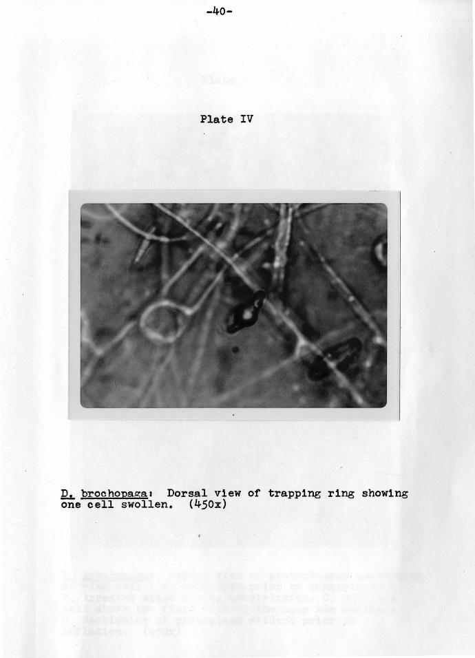
Qs,. brochopaga1 Dorsal view of tra~ping ring showing one cell swollen. (450x) • • • •
~ brochopa~a1 Dorsal view of protoplasmic sectioning of ring cell. (450x) • • • • •
• •
• •
38
39
40
41
VI. Qs,. brochopaga1 lateral view of one cell
VII.
swollen after application of air current.(450x) 42
~ brochona~a1 Cell stimulated by air current method. ( 450x) • • • • • • • • • • • 43
VIII. D. brochonaga1 Nematodes trapped by rings. (40x) 44
IX. ~ brochopaga1 Nematode trapped by nematophagous fungus. (200x) •• • • • • • • 45
iv
INTRODUCTION
Predacious fungi are a group of heterogeneous organisms
that prey on microscopic animals. They attack these animals
in a variety of ways. Some are endoparasitic, others trap
their prey by means of adhesive secretions on the mycelia
and yet others modify their mycelium to form traps or "snares".
There are approximately 50 different species of nemato
phagous fungi. Most of these belong to the order Moniliales
of the class Dueteromycetes. Some do, however, belong to
the class Phycomycetes and there has been at least one re
ported in the class Basidiomycetes. Although they are
taxonomically diverse, the nematode trapping fungi are
ecologically a natural group united by their adaptation of
the predacious habit.
These fungi capture and digest nematodes, independent
of the latter's specific or generic position. The only
limiting factor is the size of the nematode. Usually only
those nematodes measuring less than 800 to 1000 microns in
length and less than 40 to 50 microns ln width are suitable
as prey.
In the order Moniliales, the nematophagous fungi exhibit
-1-
-2-
five different types of trapping mechanisms. In '!1Y.£ traps
are formed only in the presence of the nematodes. However,
1n vitro Roubaud and Deschiens (1939) observed that traps
were induced by a wide variety of animal substances.
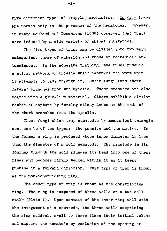
The five types of traps can be divided into two main
categories, those of adhesion and those of mechanical en
tanglement. In the adhesive trapping, the fungi produce
a sticky network of mycelia which captures the worm when
it attempts to pass through it. Other fungi form short
lateral branches from the mycelia. These branches are also
coated with a glue-like material. Others exhibit a similar
method of capture by forming sticky knobs at the ends of
the short branches from the mycelia.
Those fungi which trap nematodes by mechanical entangle
ment can be of two types& the passive and the active. In
the former a ring is produced whose inner diameter is less
than the diameter of a soil nematode. The nematode in its
journey through the soil plunges its head into one of these
rings and becomes firmly wedged within it as it keeps
pushing in a forward direction. This type of trap is known
as the non-constricting ring.
The other type of trap is known as the constricting
ring. The ring is composed of three cells on a two cell
stalk (Plate I). Upon contact of the inner ring wall with
the integument of a nematode, the three cells comprising
the ring suddenly swell to three times their initial volume
and capture the nematode by occlusion of the opening of

-3-
the ring (Plate ID. No matter how hard the nematode attempts
to escape its fate is inevitable. Microscopic filaments
from the inside of the ring penetrate the body and the
contents are quickly absorbed and utilized by the fungus.
The fungus, Dactylaria brochopaga Dreschler, which was used
1n this study was of this final type.
The objectives of this investigation were the followings
1. To determine what a water filtrate of the nematode,
Pana.grellus red1v1vus (L.) Goodey, had in common with
various animal sera since both the filtrate and the sera
elicit ring formation in the nematophagous fungi.
2. To determine what effect several different chemicals
had on the growth and trap formation of the fungus.
J. To determine the effect of another type of mechani
cal stimulus on ring constriction. In this way, perhaps,
the constricting mechanism could be better understood.

REVIEW OF LITERATURE
Zopf (1888) recorded the first observation of a
nematophagous fungus trapping nematodes when he described
the action of the adhesive networks of Arthrobotrys
oligospora. Since that time many investigators have
approached this phenomenon in a variety of different ways.
Couch (1937) studied the trapping mechanism of Dactylella
bembicoides. He found that by lowering the pH of the
media with phosphoric acid, he could induce spontaneous
trap formation. Couch (1937) also found that ring con
striction was brought about by use of distilled water at
any temperature from 33°c to 75°c. A hot scapel held
close to the fungus produced this same response.
Muller (1958) working with Arthrobotrys dactyloides
and Dactylella doedycoides slowed down the process of ring
constriction to nearly 10 seconds by treatment with various
concentrations and temperatures of sucrose solutions.
Muller (1958) also investigated the effect of other
stimuli on the constricting rings. He found that mechanical
stimulation of the cells of the ring caused the rings to
close. Thus, he confirmed the observations of Comandon and
-4-
-5-
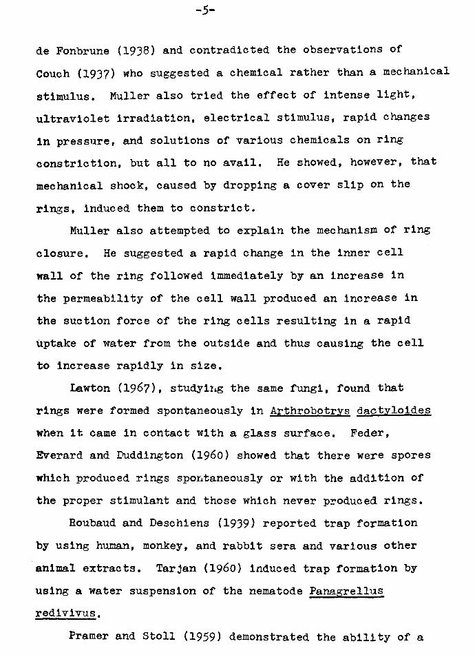
de Fonbrune (1938) and contradicted the observations of
Couch (1937) who suggested a chemical rather than a mechanical
stimulus. Muller also tried the effect of intense light,
ultraviolet irradiation, electrical stimulus, rapid changes
in pressure, and solutions of various chemicals on ring
constriction, but all to no avail. He showed, however, that
mechanical shock, caused by dropping a cover slip on the
rings, induced them to constrict.
Muller also attempted to explain the mechanism of ring
closure. He suggested a rapid change in the inner cell
wall of the ring followed immediately by an increase in
the permeability of the cell wall produced an increase in
the suction force of the ring cells resulting in a rapid
uptake of water from the outside and thus causing the cell
to increase rapidly in size.
I.awton (1967), studyir~ the same fungi, found that
rings were formed spontaneously in Arthrobotrys dactyloides
when it came in contact with a glass surface. Feder,
Everard and Duddington (1960) showed that there were spores
which produced rings spo~taneously or with the addition of
the proper stimulant and those which never p~oduced rings.
Roubaud and Deschiens (1939) reported trap formation
by using human, monkey, and rabbit sera and various other
animal extracts. Tarjan (1960) induced trap formation by
using a water suspension of the nematode Panagrellus
redivivus.
Framer and Stoll (1959) demonstrated the ability of a
-6-
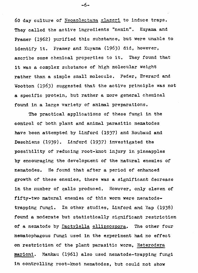
60 day culture of Neoanlectana glaseri to induce traps.
They called the active ingredients "nemin". Kuyama and
Framer (1962) purified this substance, but were unable to
identify it. Framer and Kuyama (1963) did, however,
ascribe some chemical properties to it. They found that
it was a complex substance of high molecular weight
rather than a simple small molecule. Feder, Everard and
Wootton (1963) suggested that the active principle was not
a specific protein, but rather a more general chemical
found in a large variety of animal preparations.
The practical applications of these fungi in the
control of both plant and animal parasitic nematodes
have been attempted by Linford (1937) and Roubaud and
Deschiens (1939). Linford (1937) investigated the
possibility of reducing root-knot injury in pineapples
by encouraging the develop~ent of the natural enemies of
nematodes. He found that after a period of enhanced
growth of these enemies, there was a significant decrease
in the number of galls produced. However, only eleven of
fifty-two natural enemies of this worm were ne:natode
trapping fungi. In other studies, Linford and Yap (1938)
found a moderate but statistically significant restriction
of a nematode by Dactylella ellipsospora. The other four
nematophagous fungi used in the experiment had no effect
on restriction of the plant parasitic worm, Heterodera
mar1on1. Mankau (1961) also used nematode-trapping fungi
1n controlling root-knot nematodes, but could not show
-7-
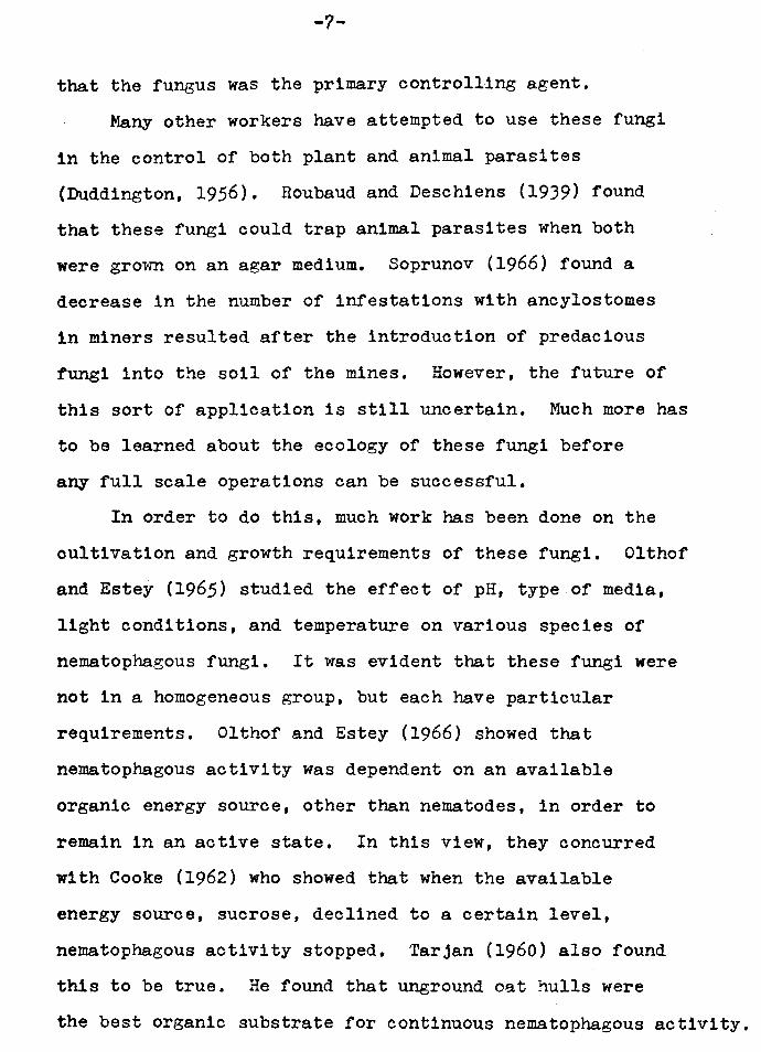
that the fungus was the primary controlling agent.
Many other workers have attempted to use these fungi
1n the control of both plant and animal parasites
(Duddington, 1956). Roubaud and Deschiens (1939) found
that these fungi could trap animal parasites when both
were gro~m on an agar medium. Soprunov (1966) found a
decrease in the number of infestations with ancylostomes
in miners resulted after the introduction of predacious
fungi into the soil of the mines. However, the future of
this sort of application is still uncertain. Much more has
to be learned about the ecology of these fungi before
any full scale operations can be successful.
In order to do this, much work has been done on the
cultivation and growth requirements of these fungi. Olthof
and Estey (1965) studied the effect of pH, type of media,
light conditions, and temperature on various species of
nematophagous fungi. It was evident that these fungi were
not in a homogeneous group, but each have particular
requirements. Olthof and Estey (1966) showed that
nematophagous activity was dependent on an available
organic energy source, other than nematodes, in order to
remain in an active state. In this view, they concurred
with Cooke (1962) who showed that when the available
energy source, sucrose, declined to a certain level,
nematophagous activity stopped. Tarjan (1960) also found
this to be true. He found that unground oat hulls were
the best organic substrate for continuous nematophagous activity.
-8-
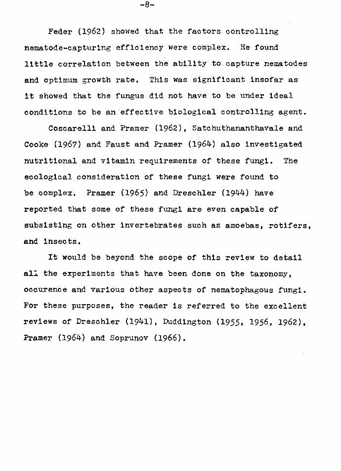
Feder (1962) showed that the factors controlling
nematode-capturing efficiency were complex. He found
little correlation between the ability to capture nematodes
and optimum growth rate, This was significant insofar as
it showed that the fungus did not have to be under ideal
conditions to be an effective biological controlling agent.
Coscarelli and Framer (1962), Satchuthananthavale and
Cooke (1967) and Faust and Framer (1964) also investigated
nutritional and vitamin requirements of these fungi. The
ecological consideration of these fungi were found to
be complex. Framer (1965) and Dreschler (1944) have
reported that some of these fungi are even capable of
subsisting on other invertebrates such as amoebas, rotifers,
and insects.
It would be beyond the scope of this review to detail
al~ the experiments that have been done on the taxonomy,
occurence and various other aspects of nematophagous fungi.
For these purposes, the reader is referred to the excellent
reviews of Dreschler (1941), Duddington {1955, 1956, 1962),
Pramer (1964) and Soprunov (1966).
MATERIALS AND METHODS
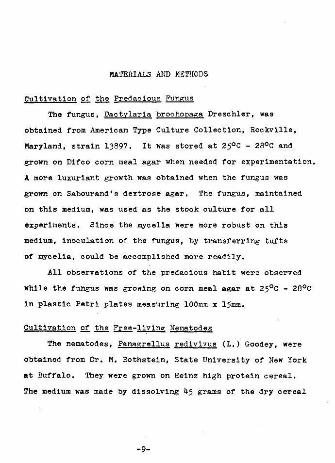
Cultivation of the Predacious Fungus
The fungus, Dactylaria brochopaga Dreschler, was
obtained from American Type Culture Collection, Rockville,
Maryland, strain 13897. It was stored at 25°c - 28°c and
grown on Difeo corn meal agar when needed for experimentation.
A more luxuriant growth was obtained when the fungus was
grown on Sabourand's dextrose agar. The fungus, maintained
on this medium, was used as the stock culture for all
experiments. Since the mycelia were more robust on this
medium, inoculation of the fungus, by transferring tufts
of mycelia, could be accomplished more readily.
All observations of the predacious habit were observed
while the fungus was growing on corn meal agar at 25°c - 28°c
in plastic Petri plates measuring lOOmm x 15mm.
Cultivation of the ~-living Nematodes
The nematodes, Panagrellus redivivus (L.) Goodey, were
obtained from Dr. M. Rothstein, State University of New York
at Buffalo, They were grown on Heinz high protein cereal.
The medium was made by dissolving 45 grams of the dry cereal
-9-
-10-
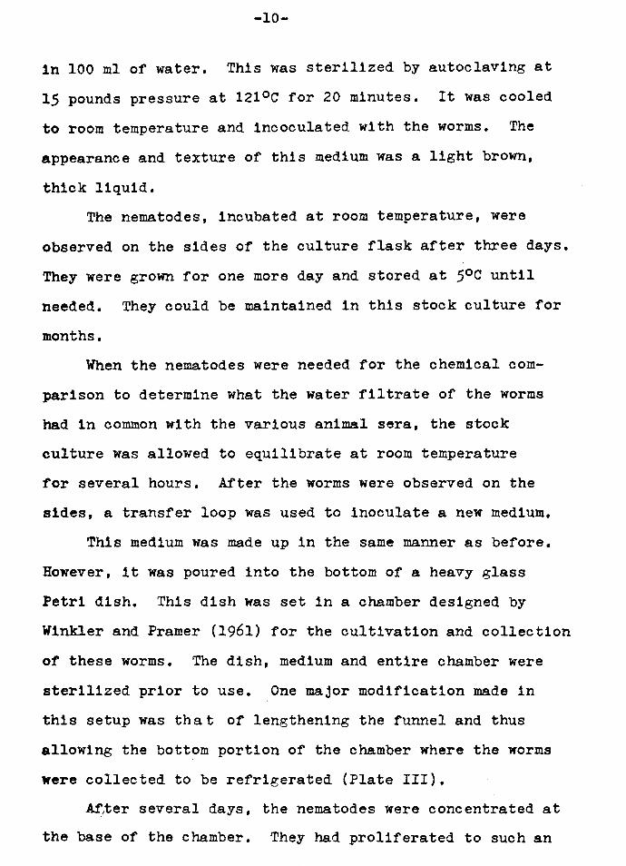
in 100 ml of water. This was sterilized by autoclaving at
15 pounds pressure at 121°c for 20 minutes. It was cooled
to room temperature and incoculated with the worms. The
appearance and texture of this medium was a light brown,
thick liquid.
The nematodes, incubated at room temperature, were
observed on the sides of the culture flask after three days.
They were grown for one more day and stored at 5oc until
needed. They could be maintained in this stock culture for
months.
When the nematodes were needed for the chemical com
parison to determine what the water filtrate of the worms
had in common with the various animal sera, the stock
culture was allowed to equilibrate at room temperature
for several hours. After the worms were observed on the
sides, a transfer loop was used to inoculate a new medium.
This medium was made up in the same manner as before.
However, it was poured into the bottom of a heavy glass
Petri dish. This dish was set in a chamber designed by
Winkler and Framer (1961) for the cultivation and collection
of these worms. The dish, medium and entire chamber were
sterilized prior to use. One major modification made in
this setup was that of lengthening the funnel and thus
allowing the bottom portion of the chamber where the worms
were collected to be refrigerated (Plate III).
~~er several days, the nematodes were concentrated at
the base of the chamber. They had proliferated to such an

-11-
extent that they came over the sides of the dish and into
the water. Since they were heavier than water, they
slowly sank to the lowest possible point in the chamber.
Thus, they were collected at the base of the stopcock in
an inactive state due to the decreased temperature.
Approximately 6,000-8,000 worms were collected daily for
the first few days. After this time, the water in the
chamber became cloudy and the chamber was disassembled.
The worms that were collected daily were washed three
times with distilled water and put in 4 ml of distilled
water. This yielded approximately 75-100 worms per drop
of water suspension. This solution was left at room
temperature for either under 24 hours, over 24 hours,
or a homogenate was made.
The reason for varying the time allowed for the worms
to remain at room temperature was to determine if upon
death of the worms any of the chemical values would rise
significantly. Since the activating substance causing the
rings to appear was produced by the worms, their decomposing
bodies should have yielded greater amounts.
Methods 2f. Analrsis Qf ~ Culture
The water suspensions of worms were analyzed at
Northwest Hospital clinical laboratory for the following
tests• glucose, urea nitrogen, uric acid, total protein,
globulin, albumin, cholesterol, alkaline phosphatase,
phosphorus, bilirubin, protein bound iodine, sodium, potassium,
chloride, calcium, bicarbonate, creatin1ne, glutamic oxalacetic

-12-
transaminase, glutamic pyruvic transaminase, lactic dehy
drogenase, creatine phosphokinase, leucine aminopeptidase,
amylase, lipase and protein electrophoresis. Since it was
of interest to determine what these cultures had in common
with animal sera, all tests were performed just as they
would have been with serum. Lawton (1957) showed that a
1/50 dilution of horse serum had the greatest activity as
compared to other concentrations of the serum. Therefore,
the amounts of any of the chemicals in the water suspensions
were expected to be small.
However, even though these values were expected to be
rather small, micromethods were not employed since it was
of interest only to determine how these water suspensions
were comparable in range to serum. As a result, standard
clinical procedures were used.
The twenty-five different chemical tests were run on
the distilled water suspensions. Before this fluid was
analyzed, the worms were removed by centrifugation. The
tests were performed manually and also on an autoanalyzer.
In fact, the majority of the runs were performed alongside
the regular serum samples, controls and blanks. In this
way, the results were comparable in reliability and
accuracy as were those reported by this hospital laboratory.
An autoanalyzerl was used in determination of eleven
of the tests. The other fourteen tests were performed
1The Hycel Mark X model was used. All procedures performed on this analyzer are modifications of accepted clinical laboratory procedures.

-13-
manually at all times.2 These particular tests were selected
s1nce they covered a wide range of biological constituents.
When a summary of results was obtained and the major
components in the water suspensions were determined, a
synthetic solution approximating these values was prepared.
This was tested for the same chemicals as were the worm
cultures and the results were noted. This was applied to
the growing fungus to determine if this solution would
produce rings. The only major ingredient that was omitted
was a protein element since a specific protein has not yet
been identified (Framer, 1964) and since there is some
question whether or not a specific protein is indeed the
activating agent (Feder, Everard and Wootton, 1963).
Human pooled sera which are normally used as controls
1n the laboratory were also used to produce ring formation.
Versatol, Monitrol I and rr3 are the trade names of these
controls. They were applied to the fungus in a similar
concentration and the number of rings were counted after
24 hours.
2These procedures are the common clinical laboratory methods. Coleman Jr. Model 6C and Lietz Model M spectraphotometers were used. An IL flame photometer was used to determine the sodium and potassium levels. Oxford titrators were used to determine CO-, ca++ and c1- ions. 3 ,
3Versatol was obtained from General Diagnostics Division, Warner-Chilcott Laboratories, Morris Plains, N.J. Monitrol I and II were obtained from Dade Division, American Hospital Supply Corporation, Miami, Florida.
-14-
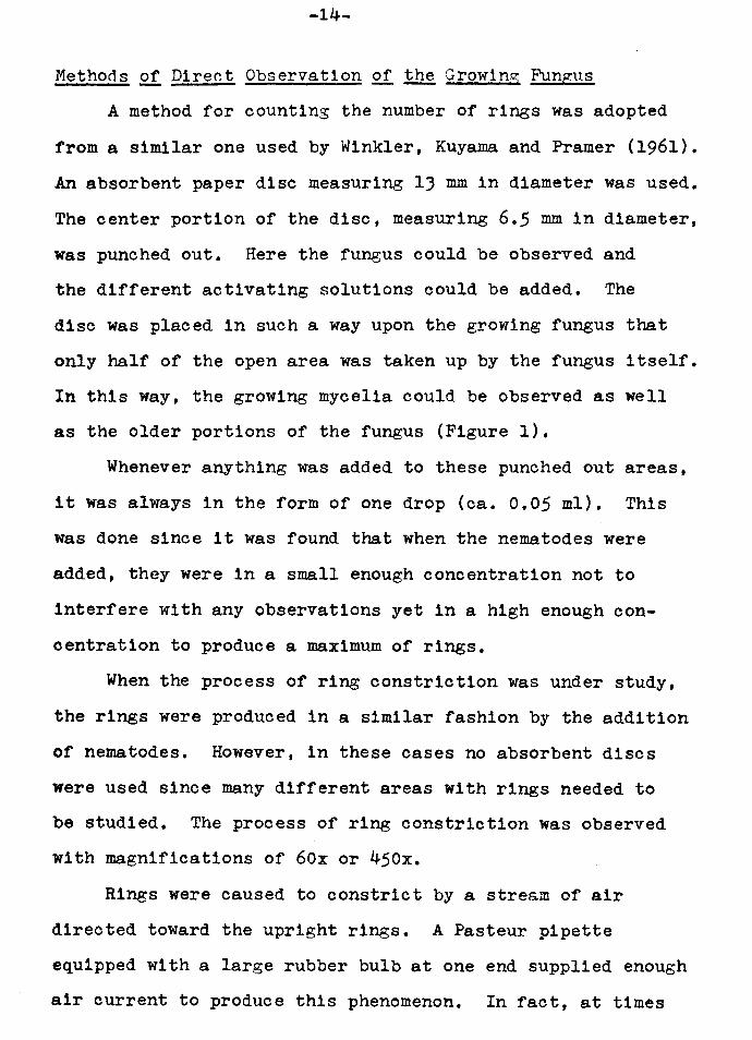
Methods of Direct Observation of the Growing, Fungus
A method for counting the number of rings was adopted
from a similar one used by Winkler, Kuyama and Framer (1961).
An absorbent paper disc measuring lJ mm in diameter was used.
The center portion of the disc, measuring 6.5 mm in diameter,
was punched out. Here the fungus could be observed and
the different activating solutions could be added. The
disc was placed in such a way upon the growing fungus that
only half of the open area was taken up by the fungus itself.
In this way, the growing mycelia could be observed as well
as the older portions of the fungus (Figure 1).
Whenever anything was added to these punched out areas,
it was always in the form of one drop (ca. 0.05 ml). This
was done since it was found that when the nematodes were
added, they were in a small enough concentration not to
interfere with any observations yet in a high enough con
centration to produce a maximum of rings.
When the process of ring constriction was under study,
the rings were produced in a similar fashion by the addition
of nematodes. However, in these cases no absorbent discs
were used since many different areas with rings needed to
be studied. The process of ring constriction was observed
with magnifications of 60x or 450x.
Rings were caused to constrict by a stre~m of air
directed toward the upright rings. A Pasteur pipette
equipped with a large rubber bulb at one end supplied enough
air current to produce this phenomenon. In fact, at times
-15-
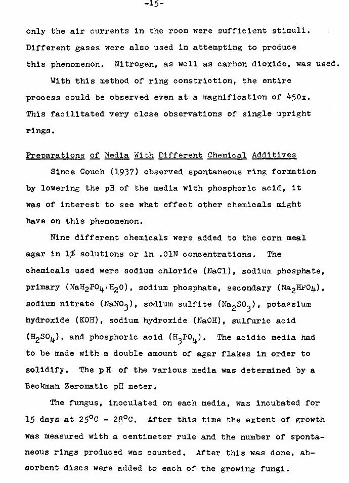
only the air currents in the room were sufficient stimuli.
Different gases were also used in attempting to produce
this phenomenon. Nitrogen, as well as carbon dioxide, was used.
With this method of ring constriction, the entire
process could be observed even at a magnification of 450x.
This facilitated very close observations of single upright
rings.
Preparations of Media With Different Chemical Additives
Since Couch (1937) observed spontaneous ring formation
by lowering the pH of the media with phosphoric acid, it
was of interest to see what effect other chemicals might
have on this phenomenon.
Nine different chemicals were added to the corn meal
agar in 1% solutions or in .OlN concentrations. The
chemicals used were sodium chloride (NaCl), sodium phosphate,
primary (NaH2P04°H20), sodium phosphate, secondary (Na2H:t>04),
sodium nitrate (NaNo3), sodium sulfite (Na2so3), potassium
hydroxide (KOH), sodium hydroxide (NaOH), sulfuric acid
CH2S04), and phosphoric acid (H3Po4 ). The acidic media had
to be made with a double amount of agar flakes in order to
solidify. The pH of the various media was determined by a
Beckman Zeromat1c pH meter.
The fungus, inoculated on each media, was incubated for
15 days at 25°c - 28°c. After this time the extent of growth
was measured with a centimeter rule and the number of sponta
neous rings produced was counted. After this was done, ab
sorbent discs were added to each of the growing fungi.
-16-
Controls and water blanks were also set up. The nematodes
were then added. One drop of a 4 ml water suspension of the
worms, after it was mixed, was added. This yielded approxi
mately 75-100 worms per open area of disc. After 60 hours
a count of the number of rings produced in each area was
taken.
Since the area of the open disc was very large and the
number of rings was in the thousands, it was impossible to
to count the rings separately. Therefore, a random count
was made.
A reticule was placed in the eyepiece of the microscope.
It was divided into 85 squares which totally covered the
area of the field. Three fields were observed and each
time the rings in five adjoining squares were counted.
The average was taken and multiplied by 17. This gave the
total number of rings in the field under view. When the
number of rings were counted manually under the field of
view, no greater than a 10% fluctuation was found from
what was counted randomly. This was done at a magnification
of 6ox. At this magnification, the field under view had
a diameter of 1.7 mm. Therefore, if the area of the field
was determined and this was divided by the area of the open
disc, one would be able to determine a factor which when
multiplied by the number of rings in one field would give
the total number of rings for that entire area. Since
the same number of worms were used to stimulate the
activity in the other media as well, one could obtain a

-17-
relationship between the pH, the amount of growth as well
as the nematode-trapping ability of the fungus on the same
base media with different chemical additives.
RESULTS
Chemical Comparison of Worm Filtrates and Animal Sera
The results of the comparison of the various chemicals
found are summarized in Table 1. Twelve of the twenty-five
chemical tests yielded results. The following could not
be demonstrated• alkaline phosphatase, creatine phospho
kinase, leucine aminopeptidase, protein bound iodine, amylase,
lipase, chloride, bicarbonate, calcium, acid phosphatase,
creatinine, and cholesterol. Values for paper electrophoresis
showed only 100 to 150 mg% of protein while the rest of the
strip was blank. Slight values for bilirubin were con
sistently observed but this was probably due to the
turbidity of the water filtrates. It is unlikely that any
bilirubin was present in the samples.
The water filtrate used in these trials was a sample
that had worms in it for more than 24 hours. The homogenate
samples differed only in the concentration of these chemicals.
The concentrations were usually greater in these samples.
The samples of water filtrates in which the worms were kept
for under 24 hours differed only in having a smaller con
centration of each of the chemicals present.
-18-
-19-
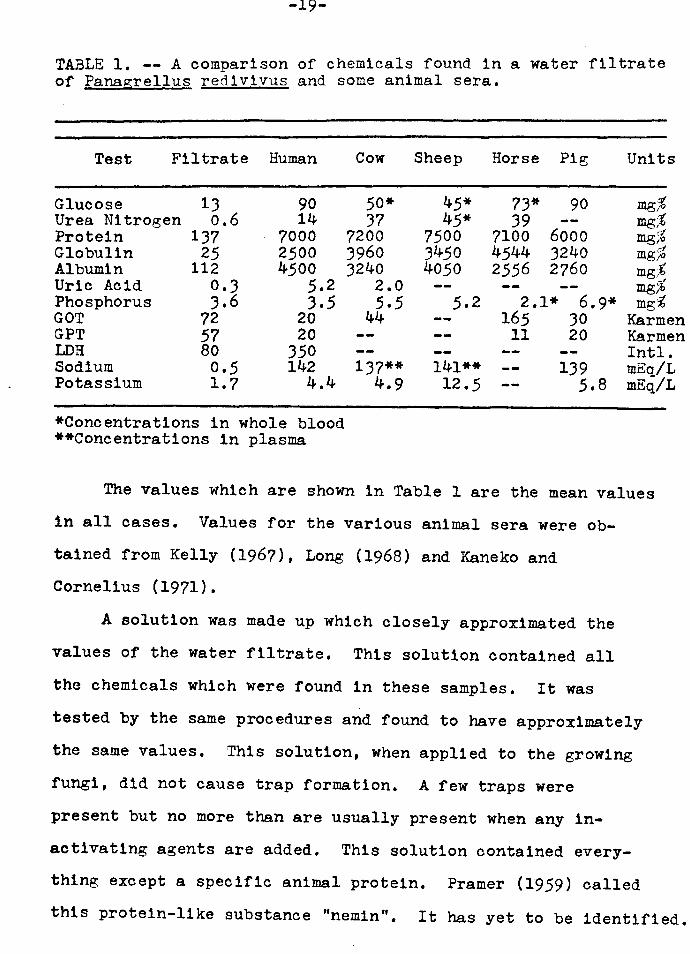
TABLE 1. -- A comparison of chemicals found in a water filtrate of Panagrellus red1v1vus and some animal sera.
Test Filtrate Human
Glucose Urea Nitrogen Protein Globulin Albumin Uric Acid Phosphorus GOT GPT LDH Sodium Potassium
13 o.6
137 25
112 0.3 3.6
72 57 80 0.5 1.7
90 14
7000 2500 4500
5.2 3.5
20 20
350 142
4.4
*Concentrations in whole blood **Concentrations in plasma
Cow
50* 37
7200 3960 3240
2.0 5.5
44
137** 4.9
Sheep
45* 45*
7500 3450 4050
5.2
141** 12.5
Horse Pig
73* 90 39
7100 6000 4544 3240 2556 2760
2.1* 165 11
6.9* 30 20
139 5.8
Units
mg,% mg;t mg% mg,% mg,:€ mg,% mg%
Ka.rm en Karmen Intl. mEa/L mEq/L
The values which are shown in Table 1 are the mean values
in all cases. Values for the various animal sera were ob-
tained from Kelly (1967), Long (1968) and Kaneko and
Cornelius (1971).
A solution was made up which closely approximated the
values of the water filtrate. This solution contained all
the chemicals which were found in these samples. It was
tested by the same procedures and found to have approximately
the same values. This solution, when applied to the growing
fungi, did not cause trap formation. A few traps were
present but no more than are usually present when any in-
activating agents are added. This solution contained every
thing except a specific animal protein. Pramer (1959) called
this protein-like substance "nemin". It has yet to be identified.

-20-
Spontaneous Trap Formation
There was no significant difference in any of the media
with the chemical additives as far as spontaneous trap
formation was concerned. Even upon lowering the pH to
4.0!.J there was no significant change in the ~umber of rings
produced. However, when the fungus was grown on an agar that
had peptone incorporated in it, such as Sabouraud's dextrose
agar, there was always a greater number of spontaneous rings
produced than on a medium without peptone (Soprunov, 1966).
Preliminary studies done to find a suitable medium for growth
of these fungi showed isolated areas on the media with
peptone where spontaneous rings were abundant. It is for
this reason that corn meal agar was used for experimentation
rather than Sabouraud's.
The Effect of Hydrogen I2.!1 Concentration Q!1 Growth
The growth of the fungus on the various media supple
mented with different chemicals was correlated with the pH
of that medium. It did not matter a great deal what the
chemical at a certain pH was, rather the hydrogen ion
concentration played the more important role. The optimum
pH for Dactylaria brochona~a has been recorded as 6.o - 6.5
by Olthof and Estey (1965). In these experiments, it was
found to be within this range. Table 2 illustrates the
effect of pH on radial growth.
~ Effect £! Hydrogen Ion Concentration 2!1 Rin~ Formation
The formation of the trapping rings was also affected
-21-
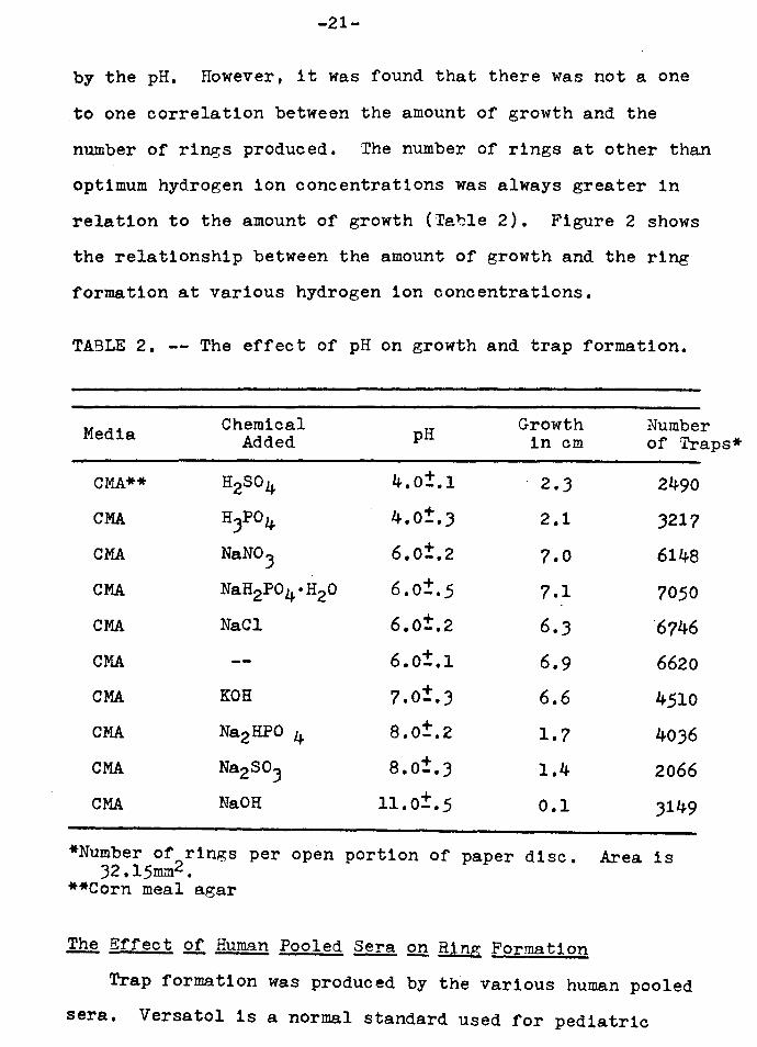
by the pH. However, it was found that there was not a one
to one correlation between the amount of growth and the
number of rings produced. The number of rings at other than
optimum hydrogen ion concentrations was always greater in
relation to the amount of growth (Teble 2). Figure 2 shows
the relationship between the amount of growth and the ring
formation at various hydrogen ion concentrations.
TABLE 2. -- The effect of pH on growth and trap formation.
Media
CMA**
CMA
CMA
CMA
CMA
CMA
CMA
CMA
CMA
CMA
Chemical Added
H2S04
H3Po4
NaN0.3
NaH2P04°H20
NaCl
KOH
Na2HPO 4
Na.230.3
Na.OH
pH
4.0±.1
4.0±.3
6.0±.2
6.0±.5
6.0±.2
6.0±.1 + 7.0-.J
8.0±.2
8.0±.3
11.0±.5
Growth in cm
2 • .3
2.1
7.0
7.1
6 • .3
6.9
6.6
1.7
1.4
0.1
Number of Traps*
2490
J217
6148
7050
"6746
6620
4510
40J6
2066
J149
*Number of 2rings per open portion of paper disc. Area is 32.15mm •
**Corn meal a.gar
~ Effect .Qf. Human Pooled ~ Q!! Ring Formation
Trap formation was produced by the various human pooled
sera. Versatol 1s a normal standard used for pediatric
-22-
cases. Monitrol II is a high standard used for abnormally
high adult values and Monitrol I is a normal standard
with values approximating a normal human serum. It was
found that the Versatol had the greatest ability to
produce ri~..gs while the Monitrol I was less effective.
The Monitrol II was least effective.
It should be noted, however, that even though there was
a varying degree of ring production by using the different
sera, they all had a profound effect upon the trap
production. These sera are readily available from
scientific supply laboratories and can be stored for
weeks after rehydration without losing their trap forming
ability. They are consistent from one stock to the next
and can, therefore, be used most effectively in
quantitative studies on the formation of trapping rings.
Observations 2!l Ring Constriction
With the methods employed, the ring constriction process
was observed very closely. Several unique observations
were made. Primarily, it was found that direct contact
with the rings was not necessary for the rings to close.
Simply by blowing air on the rings the mechanism of ring
constriction could be initiated. It was also observed that
a single air mixture or gas did not have exclusive ability
to produce constriction since both carbon dioxide and
nitrogen had the same effect as did normal air.
On one occasion a series of plates was noted to have
the rings constrict when the cover of the plate was removed.
-23-
Two possible explanations were proposed. One was that the
decrease in humidity in the plate after the removal of
the cover caused this phenomenon or alternately it was
caused by the air currents in the room. The first
hypothesis was discounted when it was observed that in a
constructed chamber where no air currents were allowed,
no constriction took place. However, when this same plate
was taken out of the current-free chamber, many of the rings
were seen to be constricted. It should be emphasized
that this series of plates had a very young fungal growth
and that these rings were formed spontaneously. All the
other observations of ring constriction were made on
fifteen day old plates that had rings produced by various
stimulating agents.
The process of ring constriction was slowed down by
the air current method. It was observed to take as long
as 5 seconds for one of the cells of the ring to completely
swell. It should be noted that the majority of rings
caused to constrict by this method did not have all three
of the cells swollen. Usually only one cell, the uppermost
or middle one of a vertical ring, was swollen. The other
two were virtually undisturbed. Observations of the
process were made simpler by this fact since the uppermost
cell could be focused upon more readily (Plate IV).
All rings were not stimulated by the air current method.
Normally, only those perpendicular to the surface of the
agar showed a response. Rings embedded within the fluid
-24-
film above the agar were not affected since they could not
be stimulated by the air currents. However, even in
nature where nematodes cause rings to constrict, many
rings lay dormant, appearing as though they had lost their
ability to constrict even when a nematode enters one of them!
With this method all possible variations of ring
constriction were seen. Rings with none, one, two or all
of the cells swollen were common throughout the field of
observation. Rings with one cell swollen were most common.
Upon initiation of the process of ring constriction by
the air current method, it was noted that a change within
the ring cells occurred. Immediately prior to the actual
swelling, two to three distinct areas could be seen within
the cell (Plate V}. One of the areas would start to swell
gradually. At a certain point, it suddenly flowed into
the next area causing that one to swell in a similar
fashion. Finally the entire cell of the ring would be
swollen. Occasionally, after prolonged activation with
the air currents, the other two cells would also swell
in a similar manner.
The majority of the time only one of the cells was
observed to swell. Upon careful observation of the swollen
cell, it was found that it was not quite as large as the
cells observed in a ring that had all three of the cells
swollen by mechanical irritation. The possibility that
this method of ring constriction stimulated only a part of the entire process was considered likely.

-25-
Numerous trials of this process were observed and various
modifications of the air currents were tried. It was found
that while the air was hitting the ring, the ring was in a
compressed condition. Only after the application of the
air current was the ring constriction seen. The pressure of
the air current from the pipette was enough to hold back
the swelling process. In fact, even after the cell was
swollen, it could be returned to its original unswollen
shape by directing more air at it. However, when the air
current was once again halted, the ring swelled. In no
case was the application of more air able to reverse this
process to where the ring stayed in its initial unswollen
position.
DISCUSSION
Chemical Comparison
It is evident that not all the constituents of human
or animal sera are needed to elicit the production of
trapping rings. Twelve of twenty-five different chemicals
normally found in the serum were consistently isolated
in the water filtrates of P. red1vivus. Of the twelve,
nine were inactive in the concentrations reported. Since
the protein fractions were the only constituents that were
not attempted, it is clear that they were involved as
activating agents. This fact concurred with Kuyama and
Pramer (1961) and Lawton (1967) who observed that proteins
were associated with trap forming activity.
It is evident also that the presence of the stimulating
substances needs only to be in very small concentrations.
Winkler, Kuyama and Framer (1961) showed that 112000 dilu
tions of pooled horse and pig sera elicited the response.
Iawton (1957) found that a 1150 dilution of horse serum
had the greatest trap producing ability. This would bring
the concentration of the protein down to approximately
142 mg%, only a 5 mg% difference from the value reported for
-26-
-27-
the water filtrate.
Feder, Everard and Wootton (1963) speculated as to
whether or not a specific protein could be involved rather
than a "general chemical". It is clear from the data that
even in the water filtrates a specific protein similar,
if not identical, to the active proteins in animal sera is
found. The other general chemicals found in the various
animal sera elicit no response when tested. It is interesting
to note, however, that only animal proteins caused this
response. Deschiens and Lamy (1942) used aqueous extracts
of various vegetables and grains, but found no stimulatory
response.
Growth fil'.19. Ill.ng Formation
As can be seen from Figure 2, the slopes of the lines
representing growth are steeper than those representing
ring formation. These lines were drawn to coincide with
the mean values of growth and ring formation respectively.
At the optimum pH, the fungus grew well and produced the
greatest number of rings in all cases. However, at pH
values away from the optimum where the pH fell below 5.0
or rose above 7.0, the growth was more substantially
affected than was the production of rings. Even at a pH
of 11.0, the number of rings produced far surpassed what
would have been expected for such a scant growth. If growth
and ring formation were equally dependent on hydrogen ion
concentration, then one would expect very few rings at this
adverse pH. However, if pH had a more pronounced effect on
-28-
growth and a lesser effect on ring formation, one would
expect more rings to be produced in relation to growth at
adverse conditions than would be produced in relation to
growth at optimum conditions.
Feder (1962) stated that "there seems to be little
correlation between the ability to capture nematodes and
the optimum growth rate of the capturing fungus •••• " If
one were to equate the number of rings produced by the
fungus and the nematode capturing efficiency of that fungus,
then it is evident that Feder's relationship is seen in
Figure 2. This is important since it clearly showed that
the fungus does not have to be under ideal conditions to
exhibit the predacious mode of life. Even under adverse
conditions, which commonly occur in nature, the trap
forming fungus can still capture nematodes. Satchuthananthavale
and Cooke (1967) showed that ring-forming fungi require
nematodes to supplement the bulk of their essential nitro
genous needs. Cooke (1964) showed that this type of fungus
was also more advanced toward a predacious mode of life since
it lost much of its saprophytic ability. Obviously then, the
fungus would be in a precarious situation if rings were only
produced in abundance under optimum conditions.
Spontaneous Trap Formation ,, Spontaneous ring formation was not exhibited for a
variety of reasons. The first could have been due to the
fact that the pH was not low enough. Although Couch (1937)
assumed that the trapping rings appear in an acidic medium,
-29-
he did not present any data on buffering or pH of the
medium. His medium was a maltose peptone agar. It was
found by Soprunov (1966) that peptone had an effect on
spontaneous ring formation. Perhaps, the peptone, along
with the acidic media, worked in a synergistic way to
produce an abundance of rings for Couch. On an acidified
corn meal agar without peptone, Couch's experiment could
not be repeated.
Ring Constriction
Muller (1958) observed a rearrangement of the proto
plasm within the ring cell. In the cells.stimulated by
the air current method, this was also found. A distinct
separation of the protoplasm into two or three areas was
observed (Plate V). In fact, Muller showed the ring cell
protoplasts to be surrounded by a membrane, although he
did not enumerate how many of these sections there were.
He showed this in observing that Brownian movement of
protoplasmic granules was restricted to enclosed areas.
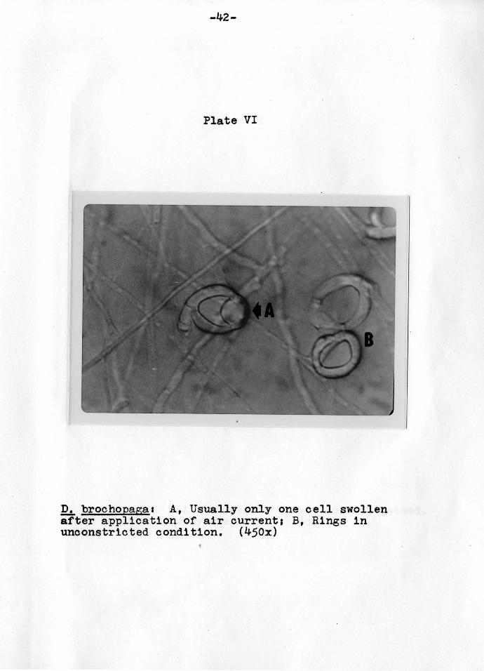
Upon initiation of inflation, these areas coalesced with
one another causing only one of the cells to swell (Plate VI).
The stimulus which caused this change in the protoplasm
was similar to the mechanical stimulus of the nematode
brushing the interior of the ring. This pressure stimulus
caused a response in the cell wall. Muller (1958) stated
that it was almost certain that the cell wall played a
passive role in cell inflation, being pushed out by the
pressure from the inside of the cell. This pressure being
-30-
caused by the increased volume of water brought into the
cell due to the increased permeability of the cell wall,
The increased permeability caused a rapid suction of water
from the outside of the cell. Since water surrounds the
ring, it entered at all angles, This accounted for the
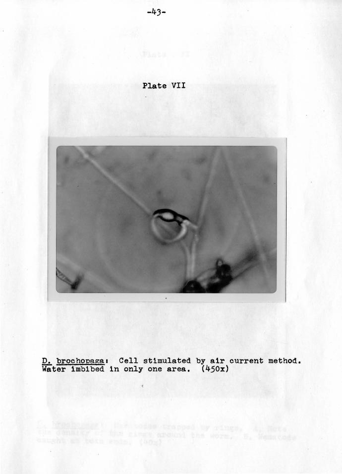
rapidity of the constricting mechanism, However, I observed
that the cells of the rings most often caused to inflate
by a gentle stream of air were not surrounded by water,
but were in upright positions above the fluid film on the
agar (Plate VII), The entire process cannot then be explained
by Muller's theory since his theory would not explain why
these rings in the dry conditions would increase in volume,
Couch (1937) suggested that water was imbibed by
the stalk and this caused the tremendous volume increase,
This alone would not account for the rapidity of the
mechanism, If imbibition was the mechanism, then why would
the middle or central cell, the one found by Higgins and
Pramer (1967) to be furthest from the stalk, inflate first?
It would, thus, seem that Couch's theory could not be
utilized completely either,
From the observations made on the cells inflated by
the air current method and from the observations of Couch (1937)
and Muller (1958), a new theory to explain all phenomenon
observed to date was proposed, It involves applying the
basic concepts of both the theories of Couch and Muller.
As Couch stated, there is a rearrangement of the protoplasm
of the stiruulated cell. This response is brought about
-31-
by the stimulation of the cell wall. The rearrangement is
in such a manner that it requires more volume per unit area.
This would cause vacant areas within the cell as the
proteins moved apart, and would be the stimulus for the
imbibition of the water through the stalk. Evidence that
the water must come via this route can be seen when these
cells are in dry conditions. The other two cells are not
affected by this passage of water since they have not yet
had their cell wall stimulated, nor protoplasm rearranged
(Plate VII).
The cells stimulated in this fashion are smaller than
cells stimulated by other means. This would suggest that
1mbibition is only part of the entire process in nature.
This is conceivable since the imbibition a.lone could not
account for the rapidity of the phenomenon. However, it
could account for the slow inflation that was observed
when cells in dry conditions were stimulated by the
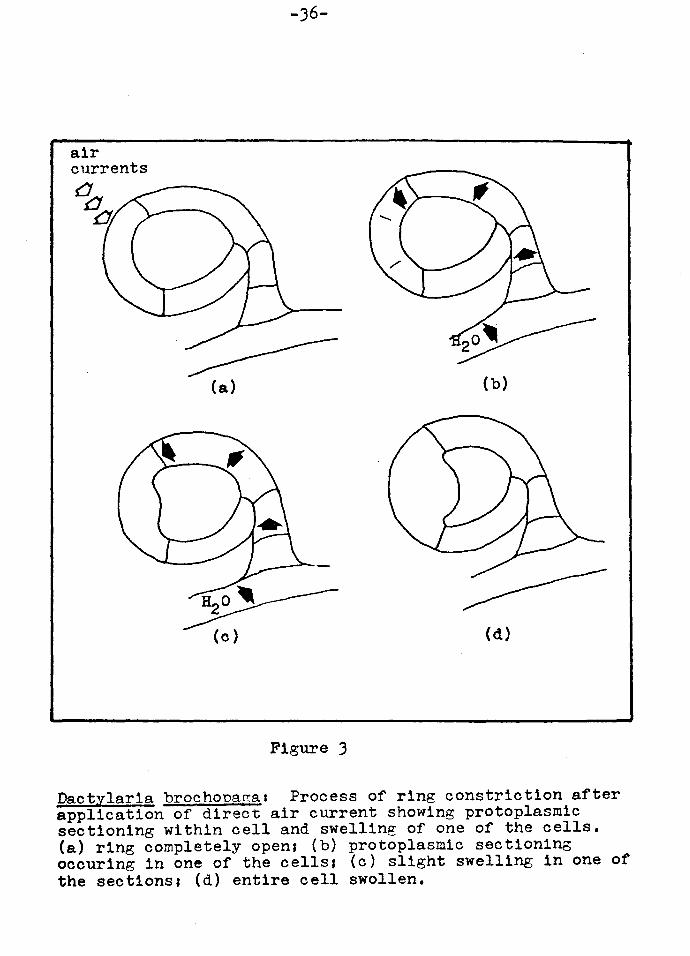
air current method. This process is diagrammed in
Figure 3.
My explanation along with Couch's and Muller's might
very well be an explanation for the entire process. The
process of imbibition is no doubt a part of the natural
process, but is overshadowed by the tremendous intake of
water when the cells are submerged in it. Muller (1958)
has stated that Couch's theory is not plausible on its
own and that the rearrangement of the molecules of the cell
cannot account for the sudden trebling of the cell volume.
-32-
He notes the work of Eyring, Johnson and Gensler (1946)
which showed that although the unfolding of protein chains
is accompanied by an increase in the absorption of
these proteins, there is an increase in the molecular
volume of only about 1%. However, it ls not proposed that
this is the entire process which causes the cell to inflate.
On the contrary, it is only a part and can be seen only
if water does not surround the cell.
Couch's theory alone did not explain the rapidity
of the mechanism, while Muller's theory did not explain
the mechanism in dry conditions. Yet, if the basic
concepts of both theories are applied, all observed
phenomena can be explained.

SUMMARY AND CONCLUSIONS
The formation and constriction of the trapping rings
in the nematophagous fungus, Dactylaria brochouaga were
investigated. The following conclusions were made1
1. Although twelve of twenty-five chemical con
stituents of serum were also found in a water filtrate of
Panagrellus redivivus, only a protein was necessary as the
stimulatory agent.
2. Various human pooled lyophilized sera promoted
trap formation.
J. Peptone in the medium promoted spontaneous trap
formation.
4. Different hydrogen ion concentrations tested
had no effect on spontaneous trap formation, while pH
values, other than the optimum, had a more pronounced
effect on growth than on trap formation.
5. A method to initiate and slow down the process of
ring constriction was found. It consisted of directing
air currents toward upright rings.
LOYOLA UN1 v c."·"''' ' uok'.ARY
-JJ-
-34-
Figure 1
Petri plate showing positioning and size of paper discs on fungal colony after 15 days growth (actual size). (a) plate with corn meal agar1 (b) edge of fungal growth1 (c) paper disc with center portion removed; (d) diameter of whole disc --13 mm.1 (e) diameter of open portion -- 6.5 mm.1 (f) point of inoculation.

-~"' 0 o-o growth 0 8 ct ,... rings - X-·X en Cl gx c 7 ·-... A. Cl 0 c 0. 6 0. C'CI ... - ~ - ., 0 5 ' ... ' Q)
' J:I ' E ' ::I ' z 4 x ', ~ ' ' I -E '
l...J
(,) 3 ., x \.}\
I - ' >. ' c I ' 0 I ' 0 2 I x ' (,) I ' - I 0
' 0 I 0 . ... I ' . Q) 1 I ' - ., Q) I E
., C'CI I ., c I 0 .,
I I I I I I I I I I •
pH 2 3 4 5 6 7 8 9 10 1 1 12
Figure 2 The effect of pH on growth and ring formation
air currents
-36-
:=----------(a)
~ (o)
Figure 3
(b)
(d)
Dactylarla brochona~as Process of ring constriction after application of direct air current showing protoplasmic sectioning within cell and swelling of one of the cells. (a) ring completely open; (b) protoplasmic sectioning ocour1ng in one of the cells; (c) slight swelling in one of the sections1 (d) entire cell swollen.
-37-
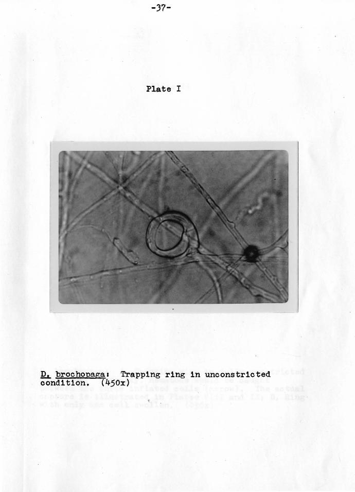
Plate I
!h. brochopagaa Trapping ring in unconstricted condition. (450x)
-38-
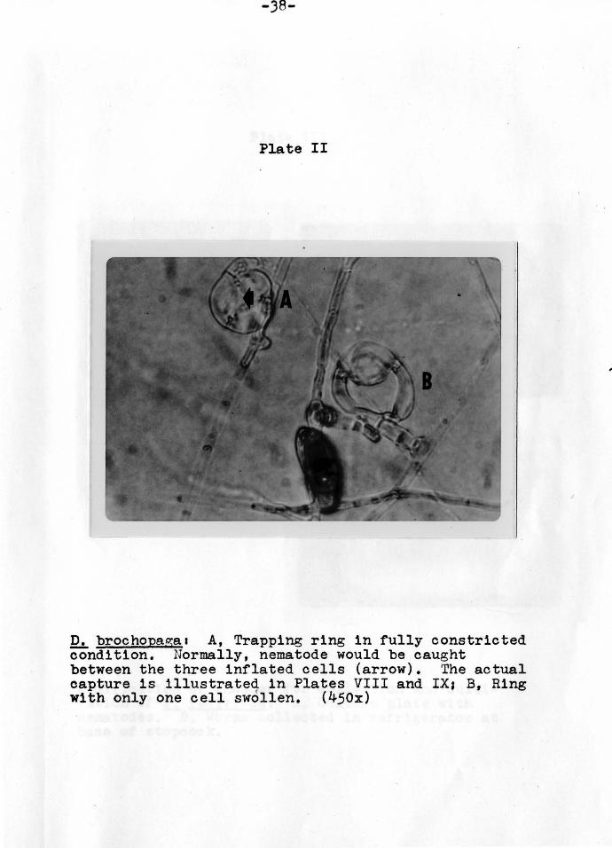
Plate II
D. brochopa~a1 A, Trapping ring in fully constricted condition. Normally, nematode would be caught between the three inflated cells (arrow). The actual capture is illustrated in Plates VIII and IX; B, Ring with only one cell swollen. (450x)
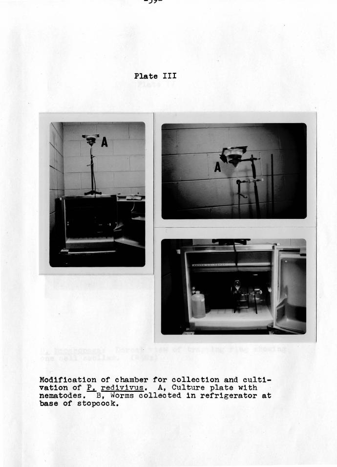
Plate III
Modification of chamber for collection and cultivation of ~ redivivus. A, Culture plate with nematodes. B, ~forms collected in refrigerator at base of stopcock.
-40-
Plate IV
12L brochopaga1 Dorsal view of trapping ring showing one cell swollen. (450x)

-41-
Plate V
~ brochopagaa Dorsal view of protoplasmic sectioning of ring cell. A, Condition prior to constrictions B, Arrested stage during constriction, C, Only the cell above the fluid film of the a gar was swolleni D, Sectioning of protoplasm evident prior to inflation. (450x)
-42-
Plate VI
D. brochopaga1 A, Usually only one cell swollen after application of air current1 B, Rings in unconstricted condition. (450x)
-43-
Plate VII
D. brochopagaa Cell stimulated by air current method. Water imbibed 1n only one area. (450x)
-44-
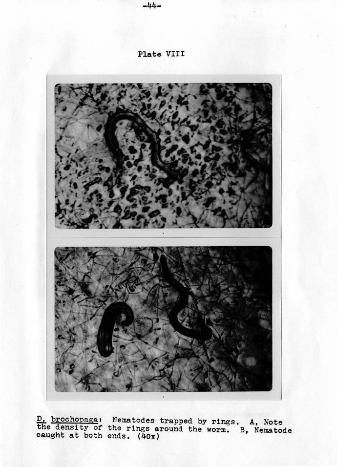
Plate VIII
D. brochopaga1 Nematodes trapped by rings. A, Note the density of the rings around the worm. B, Nematode caught at both ends. (40x)
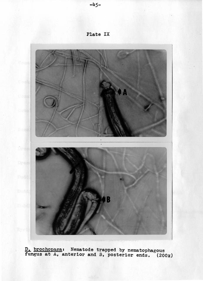
-45-
Plate IX
~ brochopaga1 Nematode trapped by nematophagous fungus at A, anterior and B, posterior ends. (200x)

Literature Cited
Comandon, J. and P. De Fonbrune. 1938. Recherches experimentales sur les champlgnons predateurs du sol. C.R. Soc. Biol., Paris 1291619-625,
Cooke, R, 1962, The ecology of nematode trapping fungi in the soil. Ann. Appl. Biol. 501507-513,
Cosarelli , 1.'1, and D. Pramer. Arthrobotrys conoides.
1962. Nutrition and growth of J, Bacteriol, 84160-64,
Couch, J,N, 1937. The formation and operation of the traps in the nema.tode-cat~hing fungus Da.ctylella bemb1co1des Dreschler. J, Elisha Mitchell Sci. Soc, 5Ja301-J09.
Deschiens, R. and L. La.my. 1942. Sur les facteurs determinants des pieges chez les Hyphomycetes predateurs de Nematodes. C.R. Acad, Sci., Paris 2151450-452.
Dreschler, Charles, 1941. Predaceous fungi. Biol, Rev. 161265-290,
Dreschler, Charles. 1944. captures springtails,
A species of Arthrobotrys that Mycologia 36:382-399,
Duddington, C,L, 1955. Fungi that attack microscopic animals, Bot. Rev. 211377-439,
Duddington, C,L, 1956. The predaceous fungi1 Zoopagalis and Moniliales, Biol. Rev, Jl1152-19J.
Duddington, C,L. 1962. Predaceous fungi and the control of eelworms, p. 151-200, In J.D. earthy and C.L. Duddington (ed.), Viewpoints 1!! Biology, vol. 1. Butterworth and Co,, London,
Eyring, H,, F. Johnson and R. Gensler. 1946. Pressure and reactivity of proteins, with particular reference to invertase. J, phys. Chem. 50145)-464,
-46-

-47-
Faust, M.A. & D. Framer. 1964. Nutrition and growth of Dactylella ellipsospora. Life Sci. 31141-143.
Feder, W.A. 1962. A comparison of Nematode capturing efficiences of five Dactylella species at four temperatures. Hycopathol. Mycol. Appl. 19199-104.
Feder, W.A., C.O.R. Everard and C.L. Duddington. 1960. Heterocaryotic nature of ring formation in the predaceous fungus pactylella doedycoides. Science 1311922-9
Feder, W.A., C.O.R. Everard and L.M.O. Wootton. 1963. Sensitivity of several species of the nematophagous fungus Dactylella to a morphogenic substance derived from freeliving nematodes. Nematologica 9149-54.
Higgins, M.L. and D. Framer. 1967. Fungal morphogenisis1 Ring formation and closure of Arthrobotrys dactyloides. Science 1551345-346.
Kaneko, J.J. and C.E. Cornelius. 1971. Clinical Biochemistry of Domestic Animals. Vol. 1, p. 320. Academic Press, New York.
Kelly, W.R. 1967. Veterinary Clinical Diagnosis. p. 219-228. The Williams & Wilkins Company, Baltimore.
Kuyama, Shimpei and David Framer. 1962. Purification and properties of a protein having nem!.n activity. Biochim. Biophys. Acta 561631-632.
Iawton, J .R. 1957. ·rhe formation of constricting rings in nematode-catching hyphomycetes grown in pure culture. J. Exp. Bot. 8150-54.
Iawton, J.R. 1967. The formation and closure of the constricting rings in two nematode-catching hyphomycetes. Trans. Brit. Mycol. Soc. 501195-205.
Linford, M.B. 1937. Stimulated activity of natural enemies of nematodes. Sciences 851123-124.
Linford, M.B. and F. Yap. 1938. Root-knot injury restricted by a nematode-trapping fungus. Phytopathology 28114-15.
'Long, Cyril (ed.). 1968. Biochemists' Sandbook. p. 839-885. D. Van Nostrand Company, Inc., New Jersey.
Mankau, R. 1961. The use of nematophagous fungi to control rootknot nematodes. Phytopathology 501645.
Muller, H.G. 1958. The constr1ct1n~ ring mechanisms of two predaceous hyphomycetes. I'rans. Bit. Mycol. Soc. 411J41-J64.

-48-
Olthof, T. and R.H. Estey. 1965. Relation of some environmental factors to growth of several nematophagous hypomycetes. Can. J • .Microbiol. 111939-946.
Olthof, T. and R.H. Estey. 1966. Carbon and nitrogen levels of a medium in relation to growth and nematophagous activity of Arthrobotrys oligospora.. Nature 20911158.
Pramer, David. 1964. Nematode-trapping fungi. Science 1441382-388.
Pramer, David. 1965. Fungal parasites of insects and nematodes. Bacteriol. Rev. 291382-387.
Pramer, David and Shimpei Kuyama. 1963. Nemin and the nematode-trapping fungi. Bacteriol. Rev. 271282-292.
Pramer, David and N.R. Stoll. 1959. Nemin1 A morphogenic substance causing trap formation by predaceous fungi. Science 1291966-967.
Pram.er, David and E.J. Winkler. 1961. A chamber for culturing and collecting the nematode Panagrellus redivivus. Nature 1921472-473.
Roubaud, E. and R. Deschiens. 1939. Sur les agents de formation des dispositifs de capture chez les Hypomycetes predateurs de Nematodes. C.R. Acad. Sci•, Paris 209•77-79.
Satchuthananthavale, v. and R.C. Cooke. 1967. Nitrogen nutrition of some nematode-trapping fungi. Trans. Brit. Mycol, Soc. 50:423-428.
Soprunov, F.F. 1966. Predacious hyphom~rcetes and their application in the control of ~atho~enic nerri'atoaes. Academy of SCiences of the TUrk11en, SSR.
Tarjan, A.C. 1960. Induction of traps of nematophagous fungi using Panagrellus redivivus. Nature 1851779-780.
Tarjan, A.C. 1960, Predaceous activity and growth of nematophagous fungi on various organic substances. Phytopathology 501577.
Winkler, E.J,, s. Kuyama and D. Pramer. 1961. A nemin assay procedure. Nature 1911155-156.
Zopf, w. 1888. Zur kenntnis der infections -- Krankheiten neiderer thiere und pflanzen. Nova Acta Leop. Carol. 521314-376.
APPROVAL SHEET
The thesis submitted by Walter Thomas Rudek has been
read and approved by the director of the thesis.
Furthermore, the final copies have been examined by
the director and the signature which appears below verifies
the fact that any necessary changes have been incorporated,
and that the thesis is now given final approval.
The thesis is therefore accepted in partial fulfillment
of the requirements for the degree of Master of Science.
Date Signature of Advisor