Embed Size (px)
Citation preview
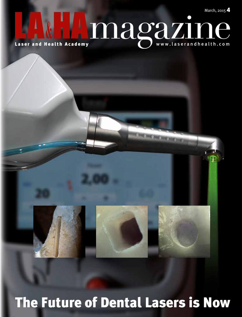
The Future of Dental Lasers is Now
March, 2015 4
L a s e r a n d H e a l t h A c a d e m y www . l a s e r a n d h e a l t h . c o m

3March 2015 42 LA&HAmagazine
Editorial
Dentistry has entered a new era
In proclaiming 2015 as the International Year of Light and Light-based Technologies, the UN has recognized the importance of rais-ing global awareness about how light-based technologies promote sustainable development and provide solutions to global challenges in energy, education, agriculture and health.
Lasers have indeed transformed medicine and dentistry in particular. It may sound as a bold statement, yet the future of dental lasers has truly arrived — today. The latest generation of dental lasers has revo-lutionized dentistry with treatments that are faster, more effective and more satisfying to patients and practitioners than ever before.
LA&HA, the Laser and Health Academy, has for many years served as a platform for exchanging ideas, leading to the development of new applications and value in using laser technology. It is our pleasure and privilege to introduce you to the latest state–of–the–art dental la-sers that are making the modern treatment process unrecognizable in comparison to the way classical treatments have long been conducted.
In this LA&HA magazine we present how lasers have moved the boundaries in the dental treatment practice, through in–depth reports from LA&HA members around the globe, interviews with some of the top experts in the field, and through a selection of clinical treatment guides. We also give you an inside peek into one of Fotona’s dental laser prodigies, LightWalker®, winner of the ‘red dot’ design award, together with a special interview with Fotona management.
Welcome to the new era!
Dr. Branka Korosec General Secretary of the Slovenian technology platform Fotonika21PUBLISHING
The Journal of the Laser and Health Academy is an international peer reviewed journal that follows new trends and progress proven practice in laser technology, in the use of lasers in medicine and in related sciences.
RESEARCH
LA&HA® collaborates with industry, medical professionals and universities on projects aimed at developing new and improving existing laser applications
EDUCATION
LA&HA® serves as a platform for continuous education, with a focus on practical instruction and the demonstration of laser techniques and procedures, delivered through a variety of workshops and seminars by experienced lecturers.
INTEGRATING KNOWLEDGE
TRANSFORMING LIFE
www.laserandhealth.com
Oglas LAHA Mag.pdf 1 18. 02. 15 08:46

5March 2015 44 LA&HAmagazine
NewsEXHIBITING IN ROME
Fotona’s Laser Technology Making Inroads in Implantology
Fotona and its local partner Emmeci 4 s.r.l. exhibited at the 23rd annual EAO (Europe-an Association for Osse-ointegration)
Congress in Rome, an event which attracted more than 3,000 delegates from around the world.
During the congress, an exceptionally high lev-el of interest was observed among implantologists from numerous countries, all of whom were eager to harness the power of Fotona’s laser technology for a range of applications, from preparing implant beds to sinus lifting. Interest was particularly high in the company’s X–Runner® digitally controlled handpiece, the world’s first “automatic” dental handpiece that provides higher precision and con-trol over ablation in both hard and soft tissues.
AT WFLD CONGRESS
WFLD Paris Hotspot: Laser DentistryFotona took part in WFLD congress in Paris, an occasion which saw excep-tionally high international participation
and record interest in the company’s dental laser systems.
There were numerous reports about success-ful uses of LightWalker due to its high precision, VSP technology, special fiber tip shapes, and new treatment methods such as NightLase®. Lectures at WFLD covered all fields of dentistry, although a major focus was on implantology.
According to the lecture of Peter Fahlstedt, a dental practitioner from Sweden, peri–implantitis presents an increasing threat to oral health: 12 mil-lion implants are placed every year (1/3 by gener-al dentists) and after 5–8 years, 30–40% of patients may develop peri–implantitis (at least 3.6 million patients per year).
Dr. Fahlstedt also added that his treatments with Fotona’s Er:YAG dental lasers promote fast healing and regeneration due to a number of reasons, including: • selectivity in the removal of granulation tissue
from alveolar bone and connective tissue • bactericidal effects at the surgical site, including
lipopolysaccharides (endotoxin) without chemi-cals
• better cleaning of implant surfaces (elimination of biofilm).
LA&HA SYMPOSIUM
Record Attendance at 4th International LA&HA Symposium
More than 350 attendees were present at the fourth annual Laser and Health Acade-my (LA&HA) Symposium,
which took place on Friday, May 23rd at the Austria Trend Hotel in Ljubljana, Slovenia.
During the symposium a total of 43 clinical lecturers from around the world presented the latest innovations and applications in the field of medical laser technology. The lecture topics were grouped into parallel sessions according to three main sub-ject categories, Lasers in Dentistry was one of them.
In the dental program, participants were high-ly impressed with the hard– and soft–tissue treat-ment capabilities provided by digitally controlled laser handpieces, as well as with the latest update on revolutionary PIPS method and treatments with QSP mode.
In the evening, all LA&HA guests were invited to attend a special gala dinner hosted by Fotona as part of its 50th anniversary celebration.
The Laser and Health Academy (LA&HA®) is a non–profit organization dedicated to the promo-tion of research, education and publishing in the field of laser medicine. LA&HA actively collabo-rates with industry, medical professionals and uni-versities on projects aimed at the development and improvement of laser applications. Additionally, LA&HA serves as a platform for continuous educa-tion in the medical laser community with numerous professional workshops offered worldwide on a va-riety of medical laser topics.
LIGHTWALKER AT S
LightWalker AT S — better visibility in oral surgery
Fotona’s LightWalker AT S laser sys-tems are now being offered with a green laser pointer for enhanced visibility, along with a larger touch screen for easier oper-ation.
Dental and maxillofa-cial surgeons have expressed keen interest in
a green pointer option, which makes their use of ti-pless handpieces more comfortable during soft–tissue surgical procedures. Using a green point-er beam with a dental laser shows the exact cutting line that the Er:YAG laser beam will perform. Even in the exceptionally high brightness of the treat-ment area, a green pilot beam clearly indicates the path of the surgical cut.
QSP™
LightWalker’s QSP™ Mode Superiority Confirmed
Four years after the first introduction of LightWalker’s unique QSP™ (Quantum Square Pulse) mode, many
reports have surfaced about its superior effective-ness in cutting even the hardest dental tissues and its unmatched precision in soft–tissue surgery. With QSP, procedures are faster and also more comfort-able, especially at higher pulse energies.
A detailed study at the University of Ljubljana in Slovenia compared the ablation efficacy of dif-ferent erbium laser pulse duration modes. The high-est ablation efficacy was measured with QSP mode, due to its significant reduction of undesirable ef-fects associated with laser beam scattering and ab-sorption in the debris cloud.
Research studies at Bezmialem Vakif University in Istanbul, Turkey, concerning microleakage and secondary bacterial contamination through filling borders, have provided further support for the effectiveness of LightWalker’s QSP mode and the exceptional quality of filling margins prepared with QSP.
At the Department of Oral surgery at the Uni-versity of Zagreb, the QSP mode for cutting and ab-lation of soft tissue was shown to be an effective, pleasant, and highly successful treatment modali-ty for oral surgery. Excellent coagulation and preci-sion of surgeries was reported, and no recurrence of lesions nor other potential complications were ob-served.
X–RUNNER
X–Runner + HDS = the perfect combination for high precision!
Dental treat-ments that require the removal of a large amount of hard dental tissue, such as a deep or broad surface area, become more precise, easy and elegant
with the help of Fotona’s X–Runner®, a unique laser handpiece that offers automatic guidance as well as adjustable spot size and shape. With the new ultra–high–precision HDS (High Density Scanning) mode, X–Runner® now assures even smoother edges and deeper ablation depths at the same parameters.
X–Runner® allows practitioners to perform treatments involving otherwise unattainable pat-terns. Automatic guidance of the laser beam allows higher repetitions to be used, and consequently pro-cedures are significantly faster. Surface treatments like preparations for veneer bonding and ortho-dontic brackets in hard tissue, as well as lesion re-moval and deepithelization in soft tissue, can be performed with a significantly higher degree of pre-cision. X–Runner®, which works in conjunction with the LightWalker AT S system, Fotona’s latest generation of dental lasers, is an ideal tool when-ever deep or extended cuts need to be made in hard or soft dental tissue. Since it’s a laser–based hand-piece, all of the other well–known laser benefits still apply, such as non–contact, vibration–free, low–pain, minimally invasive treatments.

7March 2015 46 LA&HAmagazine
8“Modern dentistry without a laser is simply not modern dentistry.” Interview with Fotona management. Lasers are playing an in-creasingly important role in modern dentistry and have achieved their original goal of replacing and supple-menting mechanical tools with more precise and less–invasive optical technology.
14Highest–perfor-mance, best–made lasers in the world. Red Dot Award for Light-Walker®
With the prestigious red dot design award, Fotona has further strengthened its position as a manufacturer of exceptionally powerful, high–quality, user–friendly and professionally designed medical laser systems.
34Laser: Efficiency and ‘Fun Factor’ In-creased. Interview with German Dentists Frank Herdach, DDS, Alexandra Deutsch, DDS, and Alexan-der Kelsch, DDS»More and more patients who are afraid of dental treatments come to us espe-cially on account of laser treatments.«
LA&HA MAGAZINE Issue 4 March, 2015
PUBLISHER LA&HA Laser&Health Academy Editorial Office Stegne 3 SI–1000 Ljubljana Slovenia
T +386 1 500 91 56 F +386 1 500 92 00 E [email protected] W www.laserandhealth.com
EDITOR Dr. Branka Korosec
EDITORIAL Dr. Branka Korosec William Wagner Edita Krajnovic Marusa Bertoncelj Sasa Gnezda
ART DIRECTOR AND DESIGNER Zvone Kosovelj
PHOTOS Milan Grbovic Marusa Bertoncelj Mateja Princic LA&HA archives
FOTONA archives Modri Atelje d.o.o. Dreamstime Personal archives
PRINTED BY Birografika Bori d.o.o. Linhartova cesta 1 1000 Ljubljana Slovenia
DISCLAIMER This edition of LA&HA Magazine is sponsored by the Slovenian technology platform Fotonika21 and Fotona. The intent of this edition is to promote research activities and the importance of lasers in dentistry during the UN General Assembly's »International
Year of Light and Light–based Technologies« (IYL 2015) and to highlight Fotona's products as an example of these research activities. Certain articles in this issue are republished from LA&HA magazine
issue 2013/1. This issue is intended for distribution outside of the USA. Please check with Fotona to find out whether a specific product or indication has been approved or cleared to be marketed and sold in your country.
40 IN–DEPTHX–Runner®: Hold the Future of Laser Dentistry in Your HandsThe newest and most innovative handpiece for oral hard– and soft–tissue removal from Fotona is the X–Runner®, an ideal accessory for the company’s LightWalker AT S laser.
24Better, Stronger and Longer–Lasting Res-torations. Interview with Prof. Dr. Aslihan Usumez DDS, PhD, Bezmialem Va-kif University, Department of Prosthodontics, Istanbul»The quality of treatments in a dental clinic using a la-ser will forever surpass the quality in the same clinic before using the laser.«
26Magic in Everyday Dental Practice. In-terview with Hong Kong dentist Dr. Seto Siu Keung, BDS»Once a dentist starts treat-ing patients with a laser, he will most likely enjoy his everyday practice more than ever before.«
28The Best Treatment Platform Possible. Interview with Maryland dentist Steven Pohlhaus, DDS, FAGD»From a personal perspec-tive, I would emphasize that after working with the LightWalker, I cannot imag-ine working again without a dental laser.«
20 EXPERTS
The ‘Magic Beam’ Changed my Career as an Orthodontist! Interview with Prof. Dr. Carlo Fornaini, MD, DDS, University of Parma»Digitally controlled handpieces will be a great opportunity for dentists. It allows for reduced operating times, greater control of the depth of ablation, and a pre–defined treatment area.«
42 High Finesse? Low problem!Fast, minimally invasive treat-ments requiring high finesse are finally possible thanks to Quantum Square Pulse™ (QSP™) mode Erbium dental laser technology.
44 Laser induced photoacoustics: a root cause revolutionThe photon–induced photoacoustic method represents a revolutionary solution for cleaning and disinfect-ing the root canal system, reaching almost 100% bacterial reduction.
46 Lower heat, more precise cutting and faster healingA recent study of the performance of an Er:YAG laser compared to a surgical drill for osteotomy treat-ment in oral surgery proved beyond doubt that Er:YAG treatment in bone surgery at specific parameters (MAX mode, Fotona) assures lower heat generation, precise cutting, rapid osseous healing and osteoinduction.
50 TREATMENTGUIDESEr:YAG — Your First Choice in Cavity Preparation
Orthodontic Treatment: Stress Gone With the Light
Taking endo–perio treatments to a whole new level
QSP Mode for Fascinating Results in Soft–Tissue Surgery
3 EDITORIALThe denstry has entered a new era
Inside Out: Impressive Development in Facial Tightening
Double treatment power with TwinLight® therapy
The TwinLight approach to peri–implantitis
NightLase®: Creating a Wonderland for Patients
Contents
30A Smarter Way of Treating Patients and Building Your Prac-tice. Interview with Dr. Kresimir Simunovic, DMD, MSc»Actually, the future is now. The new LightWalker handpiece brings a new dimension into the laser–assisted therapeutic tissue approach.«

9March 2015 48 LA&HAmagazine
“Modern dentistry without a laser is simply not modern dentistry.”Interview with Jeffrey W. Jones, CEO of Fotona Holdings, LLC, and dr. Matjaz Lukac, CEO of Fotona d.d. By William Wagner & Edita Krajnovic
F otona is a leading global dental and medical laser company, with facilities
located in the USA (Fotona, LLC) and in Eu-rope (Fotona, d.d.). We sat down with Fotona management to discuss the company’s vision for the future of the dental lasers market.
What you believe are Fotona’s key competitive strengths in the laser industry?
J. Jones: Fotona is built upon a solid foundation of cus-tomers, partners, employees, quality products and glob-al leadership. Based in the US and the EU, with corporate headquarters in San Clemente, California and Ljublja-na, Slovenia, Fotona’s business philosophy is to contin-uously choose perfection to meet the needs of a high-ly demanding marketplace. The company has been in the business of making lasers since 1964, just four years af-ter the invention of the first laser. This makes it one of the most experienced, if not the most experienced laser com-pany in the world. These strong roots, accompanied with Fotona’s long–term focus on research and development, represent the company’s major strengths and provide a solid foundation for sustainable growth in the medical la-ser technology industry. These same core strengths also give us the know–how and confidence to strategically commit ourselves to offer medical practitioners no less than highest performance, best made lasers in the world.
How has laser technology evolved during the years you’ve been working in the industry?
Dr. Lukac: The huge potential of lasers in dentistry was recognized almost immediately after the invention of the first laser. However, the technological challenges were such that it has taken several decades before dental lasers have fulfilled, and recently even surpassed, the early ex-pectations of the dental and medical community. Foto-na has been one of the pioneers in this development. For
example, we led the way by introducing Variable Square Pulse Technology, which has given practitioners great-er control over the intensity and extent of any laser treat-ment to a degree that far exceeds what is possible using standard scalpels or drills.
Even though lasers were invented several decades ago, it is still considered to be an exciting and new tech-nology, and I attribute this widely held perception to the fact that the laser is so different and unique compared to other technologies that it has inspired a continuous flow of innovation and technological developments till this day, and will undoubtedly continue to do so for quite some time to come.
From a practitioner’s perspective, what are some of the unique advantages that lasers can now offer the dental profession?
Dr. Lukac: As a result of the dramatic technological ad-vances in recent years, lasers are playing an increasing-ly important role in modern dentistry and have achieved their original goal of replacing and supplementing me-chanical tools with more precise and less–invasive opti-cal technology. Dental lasers enhance and improve upon classical procedures and, as opposed to classical tools such as burrs or scalpels, offer a much wider range of treatment protocols and greater precision of control. With classical tools, the effect on the patient’s tissue is con-trolled mainly through tactile pressure on the dentist’s hand. With a laser, however, the dentist can precisely ad-
just and optimize the speed, finesse and thermal depth of any treatment at the touch of a button.
Even more importantly, lasers enable new proce-dures that are simply not possible or even imaginable us-ing classical dental tools. And laser treatments are also friendlier to patients and dentists. So the unique advan-tages of laser technology really speak for themselves, and the laser is the way to go, not just in dentistry, but in med-icine in general. Laser light allows a practitioner to work selectively on different tissues, and in a minimally inva-sive, contact–free manner. Laser light is also “weight-less”, and can thus be moved and positioned effortlessly in 3D space, making it an ideal match with the latest rev-olutionary technologies in medicine such as intelligent robotics.
What new developments or technological break-throughs in laser dentistry can we expect to see from Fotona this year?
J. Jones: This year Fotona will be introducing some of the company’s most advanced technological achievements.
Our latest generation dental laser, the LightWalker® AT S with a green pilot beam, which improves the dentist’s abil-ity to see the laser spot clearly during surgical procedures, will be shown to the public and professionals with some exciting new features, such as novel procedures for diffi-cult–to–treat conditions like peri–implantitis, a QSP–opti-mized procedure for oral surgery, a new straight–tip hand-piece for Er:YAG, and exciting new uses for our improved digitally controlled X–Runner handpiece. We will also announce a new biomodulation Nd:YAG handpiece with a versatile collimated flat–top beam pro-file, which allows faster and homogenous irradiation with-out the risk of undesired thermal effects. As a recent study concluded, Nd:YAG laser appears to be an ideal wave-length for biomodulation because its photons can penetrate deeper tissue structures. The new straight–tip handpiece will enable the dentist to use natural movements of the hand, just like a pencil, re-sulting in improved clinical results and greater satisfaction of both the practitioner and the patient. We also greatly im-proved the X–Runner’s pattern algorithms to create truly precise cutting shapes and lines.
Lasers are playing an increasingly important role in modern dentistry and have achieved their original goal of replacing and supplementing mechanical tools with more precise and less–invasive optical technology.
Fotona has been following a different approach, which is based on a disciplined focus on the highest performing laser technologies, and an almost fanatical commitment to the quality and reliability of our products.

11March 2015 410 LA&HAmagazine
In terms of global competition, where do you see the future of the dental laser industry head-ing and how well is Fotona positioned for the fu-ture?
Dr. Lukac: We are aware that Fotona is not the only com-pany that has recognized the great potential of laser tech-nology in medicine and dentistry. There are several ap-proaches to the ever–increasing global competition in the laser industry, one of which is consolidation. Fotona has been following a different approach, which is based on a disciplined focus on the highest performing laser technologies, and an almost fanatical commitment to the quality and reliability of our products.
J. Jones: Our belief is that dental practitioners around the world will be as excited as we are with the recent techno-logical breakthroughs in laser dentistry. Modern dentist-ry without a laser is simply not modern dentistry. With Fotona’s LightWalker system, every dentist can finally “walk the light”. A
X–RUNNER®: ADVANCED HANDPIECE TECHNOLOGY
In 2013, Fotona introduced a major breakthrough in laser dentistry, a digitally controlled handpiece for dental lasers with instantly adjustable spot size and shape. The new X–Runner® handpiece adds to the precision of laser treatments by helping the practitioner to guide the laser beam swiftly and accurately across the surface of treated tissues. What makes it very unique and practical is that by pressing a button on the screen, the size and shape of the treatment zone can be changed, unlike classical treatments where the dentist needs to switch between drills and saws of different sizes. It is essentially robotics on a miniature scale.

13March 2015 412 LA&HAmagazine

15March 2015 414 LA&HAmagazine
T he LightWalker® dental laser, devel-oped by the Slovenian company Foto-
na, one of the leading global manufacturers of medical lasers, was awarded one of the world's largest and most distinguished design awards, the “red dot award: product design” for com-bining innovation, technological perfection and excellent design. The red dot award is considered one of the most distinguished inter-national quality seals for exceptional design.
“The aesthetics of dental accessories play an important role. Because dental rooms are small and each piece is very noticeable, we decided to focus our efforts on de-veloping not only the best–made laser, but also the most beautiful one,” explains dr. Marko Marincek, director of development at Fotona.
With the introduction of the LightWalker on the mar-ket, Fotona’s R&D department, led by dr. Marincek,
Highest–performance, best–made lasers in the world!*
By Mateja PrincicRed Dot Award for LightWalker®
caused a revolution in dentistry. The system offers little–or–no–pain treatment of soft and hard tissues, with fast-er healing, bloodless and sutureless soft–tissue surgery, effective periodontal treatments, safe and efficient end-odontic treatments and numerous cosmetic procedures.
It is notable that Fotona designed the LightWalk-er in collaboration with two different designers — the Slovenian industrial designer Bojan Klancar and the in-ternationally recognized Italian design agency Creano-va. “Collaboration with two different designers was not an easy job at all. Both of them had excellent ideas and I served as a moderator between. We sat down together for hours and hours developing the design that we ultimate-ly decided for, and as you can see, the results turned out excellent and users are all highly satisfied,” explains dr. Marincek.
Applications for LightWalkerLightWalker can be used for everything from oral surgery to cosmetic TouchWhite™ tooth whitening, offering the highest standard of dental treatment and simplicity of use. It allows for an extensive range of
THE RESULTS SPEAK FOR THEMSELVES: THE LIGHTWALKER HAS REALLY EXCEEDED OUR EXPECTATIONS!!Dr. Marianne Degerstrom, Tannklinikken in Narvik, Norway
“Surgery with the LightWalker is fantastic and post–op there is no pain or swelling. We also use the Nd:YAG for endo and we are excited to soon start with PIPS®. It is almost like we do not believe the results we are seeing and the LightWalker has really exceeded our expectations! The patients are very positive towards this treatment as well and they accept laser treatment in a much higher degree compared to conventional therapy. I believe it has got to do with the different sound and non contact approach. The local newspaper found out about our LightWalker very quickly which resulted in a very positive article and new patients!
At first it felt a little bit confusing with completely new terminology, but after our 3–day course at ILSD in Stockholm, we felt very comfortable in offering this treatment to our patients. The three day course consisted of both theoretical and practical parts and it really gave us a great start. I already see so many clinical benefits. We have used the laser on different perio cases with excellent results. We are also very pleased with results in various carious treatments and abrasion defects where we have not needed to use anaesthetics so far.”

17March 2015 416 LA&HAmagazine
fidence and success, bringing in extra practice income along the way. The specially designed handpieces allow for easy access to hard–to–reach places and prevent cross contamination.Because of LightWalker’s widest range of pulse dura-tions, the spectrum of possible applications is virtually unlimited. In particular, Fotona’s unmatched pulsewidth technology provides a virtually limitless parameter range for hard–tissue ablation options.
“With its reputation for developing and manufactur-ing strictly high–performance laser systems for the glob-al market and by maintaining a consistent marketing and communications strategy, Fotona has established itself as a recognized and respected global brand. And with the prestigious 2012 red dot design award, Fotona has fur-ther strengthened its position as a manufacturer of excep-tionally powerful, high–quality, user–friendly and pro-fessionally designed medical laser systems, explains Dr. Matjaz Lukac, Fotona d.d. CEO. A
presets for 40 different applications, such as intra–oral soft–tissue surgery, removal of fibroma, leukoplakia, and also selected dermatology and plastic surgery indications (skin resurfacing, skin tags). “More and more dentists around the world are nowadays deciding to offer simple dermatological services, and LightWalker provides this capability,” dr. Marincek said.It is obvious that laser dentistry is gentler, so procedures are quicker and simpler, and there is often no need for anesthetic. LightWalker has one of the most comprehen-sive lists of clinical applications of any dental laser in the world. With the availability of both tipped and tipless handpieces, easy–to–follow treatment protocols, and touch–of–a–button treatment settings, practitioners are able to perform every dental treatment with greater con-
The red dot award was LightWalker’s third prestigious international quality recognition. In 2011 the Pride Institute awarded the laser system the “Best of Class Technology Award”, and Dentistry Today, America’s leading clinical news magazine for dentists, recognized LightWalker as one of the “TOP 100 dental products of the year.”
*Fotona was founded in 1964, only four years after the invention of the very first laser. Today Fotona is one of the most experienced developers of high–technology laser systems, recognized as a world leader in the design, manufacture, and support of advanced laser systems for dentistry, dermatology, surgery, gynecology and other areas of medicine. Fotona is a company committed to designing, manufacturing and delivering the highest performance, best made laser systems in the world. Stringent testing of all components and in–house production of its medical and dental laser systems ensures that the company’s products are of the highest quality, reliability and durability.

19March 2015 418 LA&HAmagazine
Experts

21March 2015 420 LA&HAmagazine
Experts
The ‘magic beam’ changed my career as an orthodontist
By Zala Kerle
Interview with Prof. Dr. Carlo Fornaini, MD, DDS, University of Parma
ABOUT DR. CARLO FORNAINI
Prof. Fornaini is an eminent researcher and lecturer in the field of lasers in oral applications and dentistry. He currently holds a research position at the University of Nice Sophie Antipolis where he also coordinates the EMDOLA, European Master degree in Oral Laser Applications program. He is a faculty member at the Dental School of the Faculty of Medicine and Surgery of the University of Parma, which runs EMDOLA program, a scientific committee member of several international and national laser dentistry organizations and has lectured and published numerous times on various topics within laser dentistry. He currently practices laser dentistry in his own private practice in Fiorenzuola d’Arda (Italy) with a particular focus on pediatric dentistry. Prof. Fornaini is a LA&HA Expert Clinical Lecturer.
H ow did you decide to become a dentist, and what influenced you to start using
a laser?
Prof. Fornaini: Back when I was a university student (probably in the Middle Ages!) there still did not exist a dental school in my region, so I had to take a degree in Medicine and Surgery and then to specialize in Dentistry. This path has had a great influence in my daily practice, as I also frequently use my laser to treat vascular and der-matological diseases. I’ve always been technology–ori-ented, also in my private life, so when I heard about this new “magic beam” more than twelve years ago, I decided to look into it. And this rendezvous totally changed my working life, stimulating new areas of research, both fun-damental and clinical, and generating new enthusiasm to-ward my job.
I think that laser utilization should be considered a new specialization of medicine, since it is one of the few fields where it is still possible to make major advance-ments in research — for most other fields it seems as if everything important has already been discovered. Un-fortunately, the other side of the coin is that now, de-spite my age, I work a lot more than before, but also with greater enthusiasm, so it is not a burden!
What kind of treatments do you routinely per-form with your Fotona laser, and what do you see are the main benefits with using a laser?
Prof. Fornaini: I think that today it is possible to use a la-ser in nearly all dental treatments. About the only pro-cedure that I do not do with a laser is crown preparation. But I use my Fotona laser in about 75% of my daily prac-tice and find it invaluable, especially due to the fact that the device offers a combination of two complementa-ry wavelengths (1064 nm + 2940 nm) which provide the possibility of “360° utilization”. I have described this concept in several papers, as sometimes I find it very in-teresting and useful to employ both wavelengths in the
different steps of the same treatment, i.e. in the exposure of a retained tooth or to re–contour the gingiva during a composite restoration.
But, to strictly answer to the question, I use my Light-Walker in conservative treatments, for surgery of soft and hard tissues, perio, endo, ortho, prosthetics, bleaching and even for intra–oral metal welding. And last but not least, I like to use the laser for the treatment of perioral tissues: it is always wonderful, after a complex oral rehabilitation, to improve the aesthetics of a patient’s lips or to eliminate wrinkles — it is the “icing on the cake”.

23March 2015 422 LA&HAmagazine
I use my LightWalker in conservative treatments, for surgery of soft and hard tissues, perio, endo, ortho, prosthetics, bleaching and even for intra–oral metal welding.
I’m sure that this manner of working with Er:YAG will eventually replace the current practice of working strictly with the classic handpiece.
You have published numerous academic articles on dentistry. What are some of the topics that you have recently been work-ing on?
Prof. Fornaini: In the past several years I’ve been very busy on the topic of intra–oral laser weld-ing with Fotona lasers, with several “in vitro”, “ex vivo” and “in vivo” tests. I published ten papers on this matter. But my recent publications also regard Er:YAG surgery in soft tissues (i.e. oral lichen pla-nus) and hard tissues (tori mandibularis and maxil-laris) and also in conservative dentistry (i.e. restorations of traumatically fractured permanent incisors). Also very interesting, for its originality, was a study on custom-er satisfaction with Er:YAG conservative treatments, in which an 11–item questionnaire was given to 100 pa-tients, with the results indicating a very high level of sat-isfaction (90 – 100%).
What is your impression of Fotona’s new X–Runner® dental handpiece, and where do you find it to be the most helpful at your practice?
Prof. Fornaini: Several years ago I began conducting tests with a modified Fotona dermatological scanner on human extracted teeth. The reason for doing this was that I thought, and still believe, that digitally controlled hand-pieces will be a great opportunity for dentists. It allows for reduced operating times, greater control of the depth of ablation, and a pre–defined treatment area.
I believe that there are many clinical situations where in-stantly adjustable treatment shape and size may be of great benefit, and it should be considered as a signifi-cant upgrade to the classic handpiece during every mo-ment of daily practice. In fact, even though it is possible to change from the X–Runner’s digitally controlled auto-mated modality to the classical handpiece modality with only a touch of the screen, I prefer to utilize the automat-ed modality in nearly every clinical situation: from or-thodontics to surgery and from conservative to pediat-ric dentistry. I’m sure that this manner of working with Er:YAG will eventually replace the current practice of working strictly with the classic handpiece. A
Also very interesting, for its originality, was a study on customer satisfaction with Er:YAG conservative treatments, in which an 11–item questionnaire was given to 100 patients, with the results indicating a very high level of satisfaction (90–100%).
instantly adjustable spot size and shapeprecise coverage of large areaslightweight, ergonomic designfor Fotona LightWalker AT laser systems
The first digitally controlled dental laser handpiece
X-Runner®
The universe at your fingertips.9273
4/3

25March 2015 424 LA&HAmagazine
Better, stronger and longer–lasting restorations
W hen did you first become interested in laser dentistry and what inspired you to
make it the focus of your academic research?
Prof. Dr. Usumez: It began back in 1999 when I was working at the Oklahoma University Health Sciences Center. One day I attended a lecture by Charles Arcoria, who was in Oklaho-ma City speaking about dental lasers, and this topic immediate-ly caught my interest. During my PhD, I planned to perform a study on dental lasers, and then decided to base my PhD thesis on a specific laser topic — about the etching of enamel surfac-es and the bonding of Porcelain Laminate Veneers, which was later published in the Journal of Prosthetic Dentistry.
You’ve conducted some studies on the bond strength and microleakage of dental composites. Can you tell us something about how lasers may influence these factors with typical cavity preps?
Prof. Dr. Usumez: Firstly, when working with lasers on den-tal hard tissues, it is essential to choose the right parameter set-tings. This is the most important factor that will influence the final results, although other factors such as water spray will influence the results as well. We can also say that when per-formed in the right way, you will certainly achieve exception-ally good results in terms of bond strength and low microleak-age between composite and hard dental tissues, and this will increase your level of proficiency with the Er:YAG laser for cavity preparation.
From your research, how do hard–tissue treatments with LightWalker’s QSP Er:YAG mode compare to la-ser treatments using standard Er:YAG?
Prof. Dr. Usumez: We did several research projects with the QSP mode of LightWalker. I can say that we achieved out-standing results for the etching of enamel and the bond strength of orthodontic brackets to enamel. In another study, we also achieved especially good results for the etching of dentin.
Interview with Prof. Dr. Aslihan Usumez DDS, PhD, Bezmialem Vakif University, Department of Prosthodontics, Istanbul, Turkey, on New Research on Bond Strength and Microleakage
From studying atomic force microscopic pictures, we realized that the surface was perfect for bonding. Readers can find more details of this study in the one of the upcoming issues of the Journal of Orthodontics.
In your opinion, how would you summarize the main benefits of choosing a laser system that also in-cludes a second complementary wavelength, such as Nd:YAG?
The quality of treatments in a dental clinic using a laser will forever surpass the quality in the same clinic before using the laser.
When performed in the right way, you will certainly achieve exceptionally good results in terms of bond strength and low microleakage between composite and hard dental tissues.
Prof. Dr. Usumez: Being a prosthodontist as well as a laser dentist, I can list several advantages of a second complemen-tary wavelength such as Nd:YAG. With the Nd:YAG laser I can perform: hypersensitivity treatment of dentin before or af-ter crown cementation, gingival troughing before taking an im-pression, bleaching of enamel, soft–tissue surgeries with fast healing and without bleeding, treatment of hyperpigmented gingiva, and fast wound healing in mucosa and also aphthous lesions.
I would further add some specific applications for the prosthodontic area like intraoral welding of alloys as well as applications in the treatment of temporomandibular joint disor-ders. I can shortly summarize that the quality of treatments in a dental clinic using a laser will forever surpass the quality in the same clinic before using the laser. A
Experts
By Anisa Faganelj
ABOUT PROF. DR. ASLIHAN USUMEZ
Dr. Usumez is a 1996 graduate of Hacettepe University Faculty of Dentistry. In 1997 she started her PhD education in Prosthodontics and completed her PhD thesis “Evaluation of bonding Porcelain Laminate Veneers to acid etched or Er,Cr:YSGG laser etched teeth surfaces” in 2001. She was appointed as “Assistant Professor” in 2003, as “Associate Professor” in 2005 and as “Professor” in 2010. She completed her MSc in “Lasers in Dentistry” in RWTH Aachen University in 2012. She was awarded as the “Young Scientist of 2008” by The Turkish Dental Association. She has published over 60 scientific articles in journals, received oral and poster presentations awards and travel stipends from international congresses. She is currently the head of the Department of Prosthodontics in Bezmialem Vakif University, Faculty of Dentistry, Istanbul. She is married and has 2 children.

27March 2015 426 LA&HAmagazine
Magic in everyday dental practice
W hat was your first contact with a dental laser?
Dr. Seto: My first contact was thanks to my friend, Dr. Johnny Wong, who had been using Nd:YAG lasers since early 90’s. On one occasion he had asked for my help to videotape a cavity preparation with an new Er:YAG la-ser, which was a demonstration unit. Later, when I stud-ied acupuncture, an instructor had explained the thera-peutic uses of a laser to me in Chinese. After I finished that course, I volunteered to treat some elderly people in a social welfare center, where I witnessed firsthand the power of lasers in clinical treatment. Now I understand that this was purely the effect of LLLT (Low Level Laser Therapy), but at the time laser treatments appeared to me as something magical.
When I first learned that there was master course of-fered at the Aachen Dental Laser Center, I immediately applied, and since then I’ve learned many more fascinat-ing details and have truly become ‘addicted’.
What do you appreciate the most about working with a laser?
Dr. Seto: I appreciate that it is based on simple physics, and that there are always new applications with lasers. It seems there is unlimited potential, and it always enhanc-es the clinical results over conventional dentistry.
Interview with Hong Kong dentist Dr. Seto Siu Keung, BDS, on fast and effective procedures in dental surgery
With lasers, we can broaden the scope of many services provided, and some procedures are not only possible but are indeed quite simple.
Once a dentist starts treating patients with a laser, he will most likely enjoy his everyday practice more than ever before.
In cavity preparations, the need for local anesthesia is very much reduced and the laser avoids unnecessary pul-pal exposure due to its selectivity characteristics in caries removal. However, the operator should be very familiar with the different parameters and laser settings to cope with each situation.
With periodontal treatments, patients are highly pleased with the minimal post–operative discomfort fol-lowing laser treatments. With lasers, we can broaden the scope of many services provided, and some procedures such as gingival depigmentation, lip depigmentation, frenectomy or crown lengthening, are not only possible but are indeed quite simple.
What is your major indication?
Dr. Seto: Basically every discipline in general dentist-ry, i.e. cavity prep, periodontal treatment, oral surgery, and conservative dentistry, but my favorite is endodon-tic treatments. I will no longer do a root canal treatment without the assistance of a laser. When you fully under-stand the power of lasers in canal disinfection, you will
be much more confident in performing endodontic treat-ments. I was very impressed by a case in which I had per-formed a root canal treatment in a lower premolar with the use of Er:YAG to assist irrigation. I could see that there were a total of five portal openings after obtura-tions, however, they were not visible in my pre–op X–rays. To be frank, discovering apical delta or accessory canals was not very common before I began using lasers in my endodontic procedures.
Do you still think of the laser as a magical tool?
Dr. Seto: The laser is truly a magical tool, but it does takes time and commitment to learn the necessary knowledge and practice to developed the same speed, or even faster, compared to conventional mechani-cal methods. From a patient’s perspective, comfort and clinical outcome are what matter the most. But the prac-titioner’s perspective is also important. In my opinion, once a dentist starts treating patients with a laser, he will most likely enjoy his everyday practice more than ever before. A
ABOUT DR. SETO SIU KEUNG
Dr. Seto obtained his Bachelor of Dental Surgery degree (HK) in 1992 from the University of Hong Kong. After several years in general dental practice he obtained his Diploma of General Dental Practice (UK) from the Royal College of Surgeons of England in 1996. He has also enriched himself in Dental Radiology and gained a Post–Graduate Diploma in Dental Surgery (HK) in 1999 and an MSc (London) in 2001. Dr. Seto then switched to the cutting edge of technology, where he obtained his MSc (Lasers in Dentistry) with distinction from the RWTH Aachen University, Germany, in 2007. He is currently a part–time Clinical Lecturer at the University of Hong Kong’s Faculty of Dentistry. Dr. Seto is a member of the World Federation of Laser Dentistry, Vice President of the LOC for the WFLD Congress 2008 Hong Kong and academic co–worker of AALZ – Aachen Dental Laser Center of RWTH Aachen University, Germany. Dr. Seto is a LA&HA Expert Clinical Lecturer.
Experts
By Zala Kerle

29March 2015 428 LA&HAmagazine
The best treatment platform possible
D r. Steven Pohlhaus, DDS, FAGD from Maryland, USA, has been using a Light-
Walker laser system for quite some time now, and is, overall, more than satisfied with its benefits. He recently shared his experiences as one of the main instructors at the Academy of Clinical Technology (ACT), a three day intensive LightWalker training session in Las Vegas, Nevada.
Interview with Maryland dentist Steven Pohlhaus, DDS, FAGD on the advanced capabilities of LightWalker lasers in the field of dentistry
Dr. Chad Edwards instructing on LightWalker functions
Drs Cho Nguyen & Lewandowski working in vitro
Dr. Steven Pohlhaus
LightWalker’s PHAST™ technology allows me to perform less invasive endo efficiently and more effectively than traditional methods. This advanced system has also allowed me to perform many more root canals in my practice rather than referring these cases to specialists.
ABOUT DR. STEVEN POHLHAUS
Dr. Steven Pohlhaus, DDS, FAGD from Linthicum, Maryland, has been practicing dentistry for over twenty years and laser dentistry since 2004. He has devoted his career to introducing his patients and colleagues to the benefits of lasers. Dr. Pohlhaus has been lecturing on the topic of dental lasers since 2005 and is a trainer for Tecnology4Medicine’s “Laser Essentials” course for new owners of the LightWalker Laser. He is a member of the faculty at the University of Maryland Dental School in the Department of Oncology and Diagnostic Sciences.
The LightWalker allows me to rapidly and efficiently cut tooth structure, performing the large majority of my operative dentistry and cavity preparations without using a high speed drill and without having to give shots.
Experts
By Keith Bateman
In what ways has working with the LightWalker laser system transformed your daily experience as a dentist?
Dr. Pohlhaus: The LightWalker allows me to rapidly and efficiently cut tooth structure, performing the large ma-jority of my operative dentistry and cavity preparations without using a high speed drill and without having to give shots. Patients appreciate the lack of a drill and the reduced need for local anesthetics, and I and my staff ap-preciate the ability to perform minimally invasive den-tistry on a daily basis. One of the unexpected benefits of the LightWalker is being able to quickly remove ve-neers. From a personal perspective, I would emphasize that after working with the LightWalker, I cannot imag-ine working again without a dental laser.
How would you describe your experience in us-ing LightWalker for performing endodontic treatments?
Dr. Pohlhaus: LightWalker’s PHAST™ technology al-lows me to perform less invasive endo efficiently and more effectively than traditional methods. This advanced system has also allowed me to perform many more root canals in my practice rather than referring these cases to specialists. The many technical and clinical advantages of LightWalker have given me the confidence that I am doing the best endo treatment possible.
Are you also performing periodontal treatments as well?
Dr. Pohlhaus: Since implementing the LightWalker into my practice we have significantly increased the treatment of periodontal disease. The unique capabilities of the Lightwalker’s dual Nd:YAG and Er:YAG wavelengths provide the ability to comprehensively attack pathogens, and the photobiomodulation or LLLT effects of these two wavelengths work together to effectively treat this wide-spread disease.
How would you summarize the advantages of Lightwalker’s advanced technology in a nut-shell?
Dr. Pohlhaus: The precise pulse characteristics of the LightWalker allow me to pristinely cut dentin and enamel with amazing speed. LightWalker’s PHAST™ technol-ogy is the combination of specific, unique advanced de-velopments in dental laser technology. These include in-dustry leading pulse durations, pulse shape, and preferred wavelengths effectively delivered to target tissues, com-bined with advanced and proven clinical protocols devel-oped by leading visionary dentists. A

31March 2015 430 LA&HAmagazine
A smarter way of treating patients and building your practiceInterview with Dr. Kresimir Simunovic, DMD, MSc
Experts
By Sasa Gnezda
Y ou have been involved in laser dentistry since the early 1990’s. How would you
compare the art of laser dentistry back then with the way things are now today?
Dr. Simunovic: Just two words: totally different! In the early 1990’s we already had an efficient, but unfortu-nately anecdotal–based approach to laser dentistry. From this promising start the emerging field moved forward through many years of experimental approaches, leading to extraordinary and objective clinical outcomes. Today, we are living and working in a very privileged era of al-most completely evidence–based laser–assisted dentistry, with an exceptionally wide application field. The scien-tific background and technology have progressed signifi-cantly in the past decade, with major impacts on our clin-ical applications, representing a true historical milestone. I consider it to be a totally new and exciting point of view for everyday clinical experience in the dental profession.
Today, there are no alternatives in dental medicine that are more efficient than the laser for oral hard– and soft–tissue removal and for decontamination. The har-mony between settings, the fundamental play of pulse durations and the combination of two leading wave-lengths, Er:YAG and Nd:YAG, offer a unique biological, minimally invasive approach to soft and hard oral tissue treatments.
You have given many presentations around the world on the topic of laser dentistry. What would you say are some of the most common misconceptions that dentists have about using lasers in dentistry?
Dr. Simunovic: The need for an investment in addition-al basic knowledge and a completely new and different perception of tactile and visual feedback create some de-gree of insecurity in dentists who are not yet experienced with a laser. Questions we often have to deal with include
“Why should I change my in–office treatment protocols, which have worked very well in past decades?”
The goal of our presentations and workshops is to show a different way of treatment with laser dentistry. Once our colleagues commit to taking their first steps, they never go back. Seriously!
From a business perspective, how would you make the case that it’s a smart financial decision for a dentist to invest in a laser system?
Dr. Simunovic: The decision is inherently smart, but it has to be considered as a long–term investment, both fi-nancially and in terms of personal education. This aspect is often the primary obstacle that has to be discussed and
ABOUT DR. KRESIMIR SIMUNOVIC
Dr. Simunovic is a graduate from the Faculty of Dentistry at the University of Zurich, Switzerland. After practicing general dentistry for 2 years in private practice he joined Zurich University’s Faculty of Dentistry, focusing his studies on the effect of CO2 laser in hard dental tissues and common restorative materials. He received his Doctorate Degree from the same faculty in 1991. The following year he became an assistant at the Department of Oral Dental Surgery, being mainly responsible for radiotherapy and laser therapy patients. In 1997 he established his own dental office focusing mainly on laser–assisted general and aesthetic dentistry, periodontology and oral dental surgery. He is a Board Member for Dentistry of EMLA, an international associate member of the Chicago Dental Society, and member of various Swiss dental societies, among which the Swiss Society of Oral Laser Application. Dr. Simunovic is a LA&HA Expert Clinical Lecturer.
The harmony between settings, the fundamental play of pulse durations and the combination of two leading wavelengths, Er:YAG and Nd:YAG, offer a unique biological, minimally invasive approach to soft and hard oral tissue treatments.

33March 2015 432 LA&HAmagazine
redefined. Dental office devices of this investment lev-el require an almost immediate financial return from the point of view that most of our colleagues are very often both clinicians and entrepreneurs at the same time. Start-ing with a laser means, at first, a greater investment in time at chair side and in personal and team education, but with the benefit of receiving better, long–lasting profit and an enduring personal and professional enthusiasm in the near future.
What are some of the features of your Fotona LightWalker system that you appreciate the most?
Dr. Simunovic: The LightWalker generation represents a remarkable, and indeed a historical step forward in sci-ence and technology for laser–assisted dentistry. The er-gonomic benefits, due to the completely new and easy–to–maneuver OPTOflex articulated arm, the interactive adjustable panel with fast menu access and easy, com-plete clinical guidance, and the choice of ready–to–use Nd:YAG fibers for both sizes at the same time, are tru-ly unique features, which allow for comfortable and ef-ficient chair–side work, fully focusing on the patient’s need, considered as a pillar of evidence–based dentistry.
The improved quality of pulses, including QSP, and the extended range of settings, allow an even more pre-cise and energetically optimized approach to treating tis-sue, as in PIPS™, at very low, almost athermal energy
The future is now. The new LightWalker digitally controlled handpiece (X–Runner) brings a new dimension into the laser–assisted therapeutic tissue approach.
levels, and in the extended TwinLight® protocols for end-odontics and periodontology, as well as in other emerg-ing protocols such as TouchWhite™ for bleaching and snoreplasty.
Where do you see the future headed with dental laser technology?
Dr. Simunovic: Actually, the future is now. The new LightWalker digitally controlled handpiece (X–Runner) brings a new dimension into the laser–assisted therapeu-tic tissue approach. It allows a faster, extremely precise and accurate ablation for more extensive hard– & soft–tissue preps, and marks the beginning of a new era of im-plant surgery, from complete guided implant settings in the near future to surgical release and maintenance.
Looking slightly further ahead, my father, one of the pioneers in LLLT (Low Level Laser Therapy), and I are both looking forward to more improved and evidence–based photobiomodulation and analgesia procedures with both Er:YAG and Nd:YAG. A
Clean and fully intact dentinal tubules after laser treatment.
Journey into a new dental experience with speed, precision and great results. Visit www.fotona.com today!
The universe at your fingertips.
The highest technology dental laser system
8889
7/25
Unmatched simplicity of use: Balanced and weightless OPTOflex® arm Nd:YAG handpiece detection system Quantum Square Pulse technology for fast minimally invasive treatments X-RunnerTM - the first digitally controlled Er:YAG dental laser handpiece
Supreme clinical results: TwinLight® Perio Treatments TwinLight® Endo Treatments No-sutures soft-tissue surgery Patient-friendly conservative dentistry Pre-sets for over 40 applications

35March 2015 434 LA&HAmagazine
Laser: Efficiency and ‘Fun Factor’ IncreasedInterview with German Dentists Frank Herdach, DDS, Alexandra Deutsch, DDS and Alexander Kelsch, DDS
ABOUT DR. ALEXANDRA DEUTSCH
Dr. Alexandra Deutsch graduated from the Eberhard Karls University of Tübingen, Germany, at the Center for Oral and Maxillofacial Surgery. She worked for four years as assistant dentist in specialized practice covering the entire range of orthodontic treatments in Stuttgart. Her post–graduate training was in Orthodontics with a focus on invisible dental corrections, aesthetic orthodontics.
Dr. Deutsch is a certified Laser Safety Officer of the German Society for Laser Dentistry, with expert knowledge in health service facilities and a special emphasis on applications of laser technology in dentistry. She also worked for eight years as a medical technician in the University of Würzburg’s Clotten microbiological laboratory in Freiburg and in the bacteriology laboratory in Herman. Dr. Deutsch is a member of various German and international societies.
ABOUT DR. FRANK HERDACH
Dr. Herdach received his license to practice dentistry from the Eberhard Karls University of Tübingen, Germany, where he spent five years as a research assistant at the University’s Center for Dental, Oral and Maxillofacial Surgery. He is a certified implantologist and endodontist and has completed 3 years of postgraduate training with the German Society of Prosthetic Dentistry and Biomaterials to qualify as a specialist in Prosthodontics DGPRO.
Dr. Herdach is also an investigator for clinical trials in STZ–DCTC Tübingen and the Robert Bosch Hospital in Stuttgart. He has published articles on topics including emergency dental medicine, laser dentistry, Cerec 3D, implantology, and prophylaxis. He is a member of the German societies for Dental, Oral and Maxillofacial Surgery (DGZMKK), Oral Implantology (DGI), Laser Dentistry (DGL) and others.
Experts
By Sasa Gnezda
H ow did you decide to buy a Fotona laser?
Dr. Herdach&Deutsch: We were invited to a laser work-shop in a dental office. During the event the dentist intro-duced the laser and some real treatments using the laser. Everybody was impressed by the effects the laser caused in the different tissues. Finally a lecture about the eco-nomics of the device closed this very impressive event.
We decided immediately to buy our first laser on the same day.
Dr. Kelsch: For years I was not really interested in dental lasers. I therefore was not really aware of the latest im-pressive developments. In the end, my sales representa-tive Mr. Marcus Dahlinger had to slightly push me to at-tend a first laser workshop, where I was able to receive all necessary information.

37March 2015 436 LA&HAmagazine
ABOUT DR. ALEXANDER KELSCH
Dr. Kelsch received his degree in dentistry from the University of Heidelberg, Germany, in 1995 and opened his own dental practice in Karlsruhe–Neureut in 1998. He has been active in laser dentistry since 2011 and a dedicated user of Fotona’s LightWalker laser system since 2012, acquiring a second LightWalker for his practice in the following year. Dr. Kelsch is also a trainer and lecturer in the fields of laser dentistry and implantology. He conducts regular workshops throughout Germany as well as at his private practice in Karlsruhe–Neureut.
Dr. Kelsch: “The efficiency of my daily routine has significantly been improved and we were able to considerably reduce the amount of appointments with complex treatments.”
I first began using a diode laser unit, but soon had to ad-mit that I was fascinated by the application areas and pos-sibilities of the LightWalker. Finally, I could not resist and had to place an order!
What do you see as the main benefits with using a laser?
Dr. Herdach&Deutsch: The main benefit is the improve-ment of several treatments, e.g. the non–contact remo-val of dental hard tissues and bone, the immediate coag-ulation, and the improvement of healing. The effect of decontamination during an endodontic treatment, or the possibility of immediately performing an impression af-ter uncovering an implant, are very important advantages in our daily routine.
Dr. Kelsch: To keep it brief: with my laser everything is much easier! The efficiency of my daily routine has sig-nificantly been improved and we were able to consider-ably reduce the amount of appointments with complex treatments.
Additionally, the LightWalker has shown the great-est effects on me and my team. Out of all dental units and devices I have used throughout the past 17 years in my dental practice, the LightWalker has definitely shown the most positive effect on the “fun factor”.
From the first day on, my staff was thrilled by the amount of treatment opportunities offered by the two wave lengths.
As a consequence of this enthusiasm, my team is highly motivated to explain and recommend the benefits of a laser treatment to our patients.
How has your everyday work changed with LightWalker? And how well do patients accept the new form of treatment?
Dr. Herdach&Deutsch: More and more patients who are afraid of dental treatments come to us especially on ac-count of laser treatments. Some patients who have not seen a dentist for many years overcome their resistance and visit us because they have heard or read about our dental laser office. We can see a very high level of accep-tance among patients. As a result these patients bring oth-er patients to our office and the laser is used more and more. After a few years, we decided to buy a second and a third laser system – since we are two dentists, our laser treatments overlapped often and we lost time with wait-ing for the laser to be free.
Dr. Kelsch: After a short introductory period, using the LightWalker became a daily routine. This unfortunate-ly led to another problem: very often the patient was seat-ed in the wrong room (the one not being equipped with a LightWalker) which forced us to re–arrange the appoint-ments.
The only solution to this problem was the purchase of an additional LightWalker. Now, after just one year, we are able to use the laser right when we need it. Our pa-tients highly appreciate this new flexibility.
Furthermore, the laser has extended the range of treatments offered in our practice – referrals to other spe-cialists have become less frequent. In the end, our enthu-siasm for laser dentistry is positively received by our pa-tients. Of course this has also had a significant effect on the increased revenue of the practice.
Do you meet with other “laser dentists”? Do you still learn new laser procedures?
Dr. Kelsch: In the past, trainings were a necessary evil. Now, my attitude has totally changed. Henry Schein hereby offers a unique training program, which has now been expanded to a quality circle.
I personally regard other laser users as particular-ly cooperative and helpful among each other. As a conse-quence, all events are very interesting, giving me new as-pects and ideas for alternative treatment methods that I can implement into my practice routine.
Laser dentistry is alive and remains very exciting! A

39March 2015 438 LA&HAmagazine
In–depth

41March 2015 440 LA&HAmagazine
I n the field of dentistry, the vision of devel-oping a digitally controlled laser handpiece
has long been seen as an ideal means to enable a significantly higher degree of speed and pre-cision with laser treatments.
With the increased power and performance of modern dental lasers, it was inevitably a question of how soon the first digitally controlled dental handpiece would emerge to take advantage of these advanced capabilities. Handheld Er:YAG laser scanners have been used for many years in the field of dermatology, where they have proven exceptionally effective for a wide range of skin treatments that demand highly precise surface ablation.
The X–Runner® allows for both precise and extensive tissue removal, defined by the choice between three differ-ent geometrical shapes: a circle, rectangle and hexagon. These can be highlighted as full ablation areas or only as borders (as a means to carve out just the margins in order to maintain the full integrity of the inner area). The extent of ablation can be incrementally adjusted between 1 to 6 mm, depending on the geometry, with a range of from one to 99 successive passes. The X–Runner® also includes a handy
X–Runner®: Hold the Future of Laser Dentistry in Your Hands
time–saving feature: with a simple change of output shape settings it can also perform as a regular non–contact H02 handpiece.
A Versatile Handpiece For an Extended Range of Indications With X–Runner®, many remarkable advantages can be no-ticed in daily in–office applications – in the preparation of cavities, veneers and partial or full crowns, in oral surgery, especially for soft–tissue management, in orthodontics for the bracket bonding procedure and in implantology for im-plant release.
The digitally controlled laser handpiece is pushing the boundaries of dentistry and opening up many new treat-ment possibilities. Forward thinking dental practitioners will be sure to notice that the future of laser dentistry is al-ready here today, and it is small enough to hold comfortably in their hand. A
In–depth
The newest and most innovative handpiece for oral hard– and soft–tissue removal from Fotona is the X–Runner®, an ideal accessory for the company’s LightWalker AT S laser. By Dr. Kresimir Simunovic
Our first experiences with the X–Runner® handpiece provided us with fascinating insights into new, powerful and innovative aspects of Er:YAG laser–assisted dentistry.
VENEER PREPARATION
Laser source Er:YAG, 2940 nm (LightWalker AT S, Fotona)Pulse mode QSPEnergy 150 mJFrequency 15 HzHandpiece X–Runner
IMPLANT RELEASELaser source Er:YAG, 2940 nm (LightWalker AT S, Fotona)Pulse mode LPEnergy 225 mJFrequency 20 HzHandpiece X–RunnerX–RUNNER® HANDPIECE IN ACTION: TWO DIFFERENT CLINICAL CASES WITH ROUTINE INDICATIONS IN
LASER–ASSISTED DENTISTRY.
CASE 1: Veneer preparationAn extended and fast mode preparation was performed with the X–Runner® using the predefined veneer prep setting on the LightWalker, followed by a final surface modification. There was no need for local anesthesia. The finished surfaces were bonded instantly, the impression taken, and a couple of days later the lab veneers were integrated into the patient’s smile.
CASE 2: Implant releaseAfter the healing period, the soft tissue above an osseointegrated implant was removed by multiple passes, following the preset circular shape and size of the ablation area. A healing abutment was fixed on the fully uncovered implant after the impression was taken. The surgery was performed without need for local anesthesia.
Figs. 1–3: Extensive surface preparation with X–Runner®, followed by adhesive in–office protocol
Figs. 4–5: Before and after pics of the veneer case on the upper incisors
Figs. 1–4: Before, during and after the implant release procedure

43March 2015 442 LA&HAmagazine
High finesse? Low problem!
R ecently, the range of treatment pa-rameters of Variable Square Pulse
(VSP) Er:YAG lasers has been significantly extended. [1] With the latest proprietary Quantum Square Pulse (QSP) technolo-gy, minimally invasive treatments that require extremely high finesse have now been made possible. With high finesse it is meant that the tissue is treated with high spatial precision and with small or moder-ate pulse energy and short duration laser pulses at high repetition rates.
Extremely high finesse of laser treatment is required, for example, when making hard tissue surface modi-fications before applying composite fillings. High fi-nesse is also desirable when making fine cuts with controlled bleeding into the soft tissue.
Similarly to achieving high ablation speeds, ob-taining high treatment finesse has represented a sig-nificant technological challenge. This is due to the fact that short pulses of low energy have suboptimal effi-ciency and are extremely difficult to generate at suffi-ciently high repetition rates.
In the QSP mode, a longer laser pulse is divided, i.e. quantized, into several short pulses (pulse quan-ta) that follow each other at an optimally fast rate. This enables the QSP mode to deliver short, high finesse pulses with the efficiency of long duration laser puls-es without sacrificing the precision that is provided by short duration pulses.
Fig. 1: a) Standard laser pulse; b) QSP pulse: a long laser pulse is quantized into several pulslets (pulse quanta).
One of the major advantages of the QSP mode is that it sig-nificantly reduces the undesirable effects of laser beam scattering and absorption in the debris cloud during hard tissue ablation. Namely, when an ablative laser light pulse is directed onto the tissue an ablation of the tissue starts that leads to the emission of ablated particles above the tis-sue surface, forming a debris cloud (Fig. 2).
The influence of beam scattering on the precision of hard–tissue ablation can be seen in Fig. 3, which shows laser ab-lated craters in enamel and dentin at two Er:YAG pulse du-rations. As a result of scattering, the ablated cavities do not have well defined edges. This effect is more pronounced at higher pulse energies and longer pulse durations.
Pulse energy 400 mJ, pulse duration 300 µsec
Fig. 2: Formation of a typical debris cloud.One of the major advantages of the QSP mode is that it significantly reduces the undesirable effects of laser beam scattering and absorption in the debris cloud during hard tissue ablation.
In–depth
Fast, minimally invasive treatments requiring high finesse are finally possible thanks to Quantum Square Pulse™ (QSP™) mode Erbium dental laser technology. By Dr. Evgeniy Mironov
Standardlong laser pulse
QSP pulseshort laser pulse quanta
QSP pulseshort laser pulse quanta
Standardlong laser pulse
Pulslet spacing Pulslet spacing
a)
b)Pulse repetition
In order to avoid the effects of scattering, the pulse dura-tion should be shorter than the time required for the ablation cloud to develop. At the same time, when using the QSP la-ser pulse technology, the pulslet spacing should be longer than the debris cloud decay time. This ensures that the sec-ond pulslet does not encounter any cloud remains from the previous pulslet (Fig. 4).
With the QSP mode a compromise is found, whereby the temporal pulse spacing between pulslets is longer than the cloud decay time and shorter than the inversion population remaining time. A sufficiently short temporal pulslet spac-ing is required because there is some inversion population of the laser energy status remaining after the end of the la-
QSP pulse quantum Ablation cloud
Fig. 3: The influence of beam scattering on hard–tissue ablation.
Fig. 4: Pulslet spacing with QSP mode.
Short pulses Long pulses ser pulse. In cases where the pumping for the second pulslet starts early enough, the threshold is reduced as the laser has already been pre–pumped from the previ-ous pump pulse. This ensures an enhancement of las-ing efficiency without significantly compromising the quality of laser ablation.Clinical benefits from the new QSP mode are easily recognizable [2, 3]. The margins of preparations for fill-ings or for surface modification are clearer and sharp-er than with any other operational mode used to date. This is of primary importance when working close to the pulp or near the gingiva. QSP is also a safe and re-liable mode in class II cavity preparations where the neighboring teeth should be kept intact.
According to SEM micrographs, QSP–treated surfaces appear to have the high quality required for high bond strength [4], in addition to being free of a smear layer. The dentin surface appears clean, regu-lar and flat with wide–open tubules with no difference between inter–tubular and peri–tubular dentin. The enamel surface also appears clean and homogeneous with a well–defined micro–roughness.
As well as being an optimal mode for procedures that require high finesse (i.e. tissue treated with high spatial precision and with small or moderate pulse en-ergy and short–duration laser pulses at high repetition rates), the QSP mode also guarantees a high speed with the procedure [5].The speed of cavity prepara-tions is increased by a factor of up to 1.75 when com-pared to “single” (non–quantized) laser pulses at the same total energy setting. Since the QSP mode con-sists of a series of optimally spaced super–short puls-es, it can be viewed also as a super–short pulse mode “on steroids”. Speed of preparation is important in pe-diatric dentistry and with anxious patients, and QSP mode is the method of choice if we require short prepa-ration times without sacrificing finesse. Also, the noise level generated with this mode is lower than in other currently available laser operating modes, which nota-bly increases the level of comfort of the procedure.
In conclusion, the QSP mode excels in preparation of dental hard tissues. Working in QSP mode allows the dentist to perform procedures with an unprecedented level of finesse without sacrificing speed, and with the added advantage of decreasing the noise level of the procedures. A
References:1. Gutknecht N, Lukač M, Marinček M, Perhavec T, Kažič M, A Novel Quantum Square Pulse (QSP) Mode Erbium Dental Laser. JLA&HA Vol. 2011, No.1: 15–21.2. Mironov E, Mironova Z, Quantum Square Pulse Er:YAG Lasers in Clinical Practice, Int.. Mag. of Laser dentistry 3/2012: 34–373. Mironov E. Clinical Experience with a Quantum Square Pulse (QSP) Er:YAG Laser. JLA&HA Vol. 2012, No.1: 80–85.4. Lukac M, Malej Primc N, Pirnat S, Quantum Square Pulse Er:YAG Lasers for Fast and Precise Hard Dental Tissue Preparation, J LA&HA Vol. 2012, No 1: 14–21.5. Lukac M, Suhovrsnik T, Filipic C, Minimally Invasive Cutting of Enamel with QSP Mode Er:YAG Laser. J Laser Dent 2014;22(1):28–35.

45March 2015 444 LA&HAmagazine
T he removal of vital and necrotic pulp tissue, mi-croorganisms and their toxins, and the preven-
tion of reinfection through a hermetic coronal and api-cal seal, are essential for endodontic success. Clinical experience and research have shown that the use of endodontic irrigants results in ineffective irrigation [Haapasalo, 2010]. Also, currently used instrumen-tation techniques left 35% or more of the canals’ sur-face area unchanged [Peters, 2001] and only partially removed vital and necrotic tissues from the entrance of lateral canals and apical ramifications, leaving adja-cent tissue inflamed, or infected and associated with periradicular disease [Ricucci and Siqueira, 2010].
The main problem of irrigation in endodontics is the fluid–dynamics properties of irrigants in the con-fined canal space. Because of the inherent taper seen within the canal morphology, deep penetrations of irrigants are more difficult because of the absence of turbulence over much of the canal volume [Gulabiva-la, 2010]. Both irrigant penetration and biofilm removal may be improved through canal fluid agitation using a close fitting instrument, sonic or ultrasonic activation, or laser. Consequently, the efficacy of NaOCl depends on the means by which free chlorine ions are readily available at the target tissue site.
Comparing passive ultrasonic irrigation (PUI) and laser–activated irrigation (LAI) it was found that tis-sue dissolution was more pronounced after the use of LAI with sodium hypochlorite and an Erbium:YAG
Laser induced photoacoustics: a root cause revolution
(2940 nm) laser. [Macedo 2010]. Laser–activated irri-gation by the PIPS™ technique was found to generate tremendous turbulence and 3D streaming within the root canals [DiVito and Olivi, 2011]. Laser–activation of NaOCl (PIPS™ technique — Fotona Er:YAG laser) with in vitro infected specimens generated more negative bacterial samples and left less apical bacteria/biofilm than ultrasonic activation (PUI) [Peters, 2011].
Another study confirmed that the combination of Er:YAG laser (PIPS™ technique — Fotona, LightWalk-er laser) and 6% sodium hypochlorite produced 100% elimination of Enterococcus faecali from ex vivo infect-ed root canals [Jaramillo, 2011]. Also Laser–activa-
Laser–activated irrigation by the PIPS™ technique was found to generate tremendous turbulence and 3D streaming within the root canals.
In–depth
The photon–induced photoacoustic method represents a revolutionary solution for cleaning and disinfecting the root canal system, reaching almost 100% bacterial reduction. By Prof. Giovanni Olivi
Fig. 1: PIPS™ method
tion of EDTA (PIPS™ technique–Fotona Er:YAG laser) of chemomechanically prepared root canals resulted in more cleaning of the root–canal walls and a higher quantity of open tubules in comparison with the tradi-tional irrigation method [DiVito, 2012].
The fact that the PIPS™ photon–induced photo-acoustic steaming effectively travels 3–dimensionally in the root canal spaces also makes it advantageous as a treatment modality for removing biofilms associated with periodontal pockets that are in difficult–to–ac-cess furcation areas and interproximal vertical defects [DiVito and Lloyd 2012]. A
References:
Haapasalo M, Shen Y, Qian W, Gao Y, Irrigation in endodontics, Dent Clin North Am. 2010; 54(2):291–312.Peters OA, Schonenberger K, Laib A, Effects of four Ni–Ti preparation techniques on root canal geometry assessed by microcomputed tomography, Int.
Endod. J 2001; 34(3):221–30.Ricucci D, Siqueira JF, Fate of the tissue in lateral canals and apical ramifications in response to pathologic conditionas and treatment procedures, J
Endod 2010; 36: 1–15.Gulabivala K, Ng YL, Gilbertson M, Eames I, The fluid mchanics of root canal irrigation, Physiol Meas. 2010; 31(12): R49–84.Macedo RG, Wesselink PR, Zaccheo F, Fanali D, van der Sluis LW, Reaction rate of NaOCl in contact with bovine dentine: effect of activation, exposure
time, concentration and pH, Int Endod J 2010; 43: 1108–15.DiVito E, Colonna MP, Olivi G, The Photoacoustic Efficacy of an Er:YAG Laser with Radial and Stripped Tips on Root Canal Dentin Walls: An SEM Evalu-
ation, J Laser Dent 2011: 19(1): 156–161.Peters O, Bardsley S, Fong J, Pandher G, DiVito E, Disinfection of Root Canals with Photon–initiated Photoacustic Streaming, J Endod, 37, 1008–1012,
2011. Jaramillo D, Aprecio RM, Angelov N, Di Vito E, McClammy TV, Efficacy of photon induced photoacoustic streaming (PIPS™) on root canals infected with
Enterococcus faecalis: A pilot study, Endod Practice 2011; Vol 5, no 3:28–32.DiVito E, Peters O.A, Olivi G, Effectiveness of the erbium:YAG laser and new designed radial and stripped tips in removing the smear layer after root
canal instrumentation, Lasers Med Sci 2012; 27:273–280.DiVito E, Lloyd A, Er:YAG laser for 3–dimensional debridement of canal systems: use of photon–induced photoacoustic streaming, Dent Today 2012;
31(11): 122, 124–7.
Fig. 3: Fotona LightWalker screen with PIPS™ preset
Fig. 2: Root canal dentinal walls irrigated with 17% EDTA and PIPS™

47March 2015 446 LA&HAmagazine
Lower heat, more precise cutting and faster healing
In–depth
Arecent study of the performance of an Er:YAG laser compared to
a surgical drill for osteotomy treatment in oral surgery proved beyond doubt that Er:YAG treatment in bone surgery at specific parameters (MAX mode, Fotona) assures lower heat generation, precise cut-ting, rapid osseous healing and osteoinduc-tion. Compared to conventional mechanical drills and saws, it provides non–contact and low–vibration intervention, bacterio-stasis, less traumatization and decreased bleeding.
The Fotona MAX mode is currently the fastest Er:YAG dental laser ablation mode available. Scanning elec-tron microscopy analysis and chemical and crystal-lographical changes of the bone tissue after Er:YAG MAX mode laser ablation and drilling were reported at the EAO Annual Scientific Meetings [2, 3]. The com-
Er:YAG Advantages … Compared to Surgical Drill …
Excellent cutting efficiency 2.6 times more bone tissue removed
Short preparation time only 17% of drilling timeLower temperature 3.8°C lower final temperature
Regular shapes with clear, sharp edges Decreased risk for infection caused by bony particles which remain after drill treatment
No smear layer Increased adhesion of blood elements at the start of the healing process
plete study was published recently in the Journal for Oral and maxillofacial surgery [3] and in the Photomed-icine and Laser Surgery [4].
Overcoming delayed healing and infectionThe aim of the studies was to find out if it is possible to avoid the disadvantages associated with the conven-tional drill, such as extensive heat deposition, a necrotic surface zone, injury of the bone cells, and consequently, delayed healing, infections due to fragments left on the bone surface, and mechanical traumatization.
Holes for fixation screws were performed in 4.6 mm thick bone blocks from porcine ribs using a 1.0 mm wide surgical pilot drill (15000 rpm) and an Er:YAG laser (1000 mJ, 20 W, MAX mode, Fotona). The tem-perature during the preparation, the removed bone volume, and the time required for the preparation were compared in the study. The cortical and spongiose sur-faces of the specimens were examined microscopically and histologically.
The results, which speak for themselves, are sum-marized below:
Superiority of Er:YAG MAX mode over classical drill for osteotomiesBy Asst. Prof. Dr. Dragana Gabric
Optical microscopic observations (10x) of the cortical appearance are shown in figures 1 and 2.
Fig 1: Laser preparation (MAX mode, Fotona) Fig 2: Drill preparation.
Fig 6: Spongiose bone (drill); organic tissue between bone trabecules.
References1. Gabrić Pandurić D, Bago I, Katanec D, Žabkar J, Miletić I, Anić I, Comparison of Er:YAG Laser and Surgical Drill for Osteotomy in Oral Surgery: An
Experimental Study, J Oral Maxillofac Surg 70:2515–2521, 2012.2. Gabrić Pandurić D, Anić I, Žabkar J, Katanec D, Sušić M, Musić S, Scanning electron microscopy analysis of bone removal using Er:YAG and sur-
gical drill, Poster presented at the 20th Annual Scientific Meeting of the European Association of Osseoinegration, 2011, Greece.3. Gabrić Pandurić D, Musić S, Molčanov K, Katanec D, Bago I, Anić I, Chemical and crystallographical analysis of bone tissue after laser and drill
osteotomies, Clinical Oral Implants Research, Vol 23, S7, 60–60, 2012.4. Gabrić Pandurić D, Bago Jurić I, Musić S, Molčanov K, Sušic M, Anić I, Morphological and Ultrastructural Comparative Analysis of Bone Tissue
After Er:YAG Laser and Surgical Drill Osteotomy, Photomedicine and laser surgery, Vol 32, 7, 401–408, 2014.
All photos courtesy of the authors.
Fig 3: Laser groove; well–defined edges and surface.
Fig 4: Drill groove; irregular edges with hair–like appearance.
Fig 5: Spongiose bone (laser); empty spaces between trabecules due to vaporisation of the organic tissue.
The SEM pictures (figs. 3–6) show the dif-ferences between the bone surfaces pro-duced by the Er:YAG laser and the drill.
It was concluded that Er:YAG treatment in bone surgery at the specific parameters (MAX mode, Fotona) assures lower heat generation, precise cutting, rapid osseous healing and osteoinduction. Compared to conventional mechanical drills and saws, it provides non–contact and low–vibration intervention, bacteriostasis, less traumati-zation and decreased bleeding. A
There was no change in the chemical composition of bone tissue and no thermal modification of hydroxyap-atite crystals after Er:YAG ablation.
Compared to conventional mechanical drills and saws, it provides non–contact and low–vibration intervention, bacteriostasis, less traumatization and decreased bleeding.

49March 2015 448 LA&HAmagazine
Treatment Guides

51March 2015 450 LA&HAmagazine
E r:YAG laser energy is highly absorbed by water molecules, rapidly heating a small
volume. The vaporization of the water creates high subsurface pressure and leads to an explosive re-moval of the surrounding mineral. The water con-tent in carious tissue is higher than in healthy tis-sue, so for the same settings, the laser ablation rate will be higher in carious tissue than in healthy tissue. As we move in to dentin, we lower the en-ergy and frequency settings, since ablation is fast-er in dentin because of its higher water content. Even lower parameters are required for the final modification to create a retentive surface for the filling material.
No Need For Acid EtchingA 35–year–old female visited our dental clinic com-plaining about sensitivity to cold food. After exam-ination we noticed the need to replace four old fill-ings and make two new for the neighboring teeth. The most sensitive was tooth 46 (see Figs. 1–7). We removed the old filling with MAX mode and switched to QSP for caries removal. The cavity ap-peared very deep, with bleeding in the interdental area, so we proceeded with Nd:YAG for hemosta-sis. At the end, we irradiated all of the dentin and enamel with QSP for surface modification, without the need for acid etching. A
Er:YAG – Your First Choice in Cavity Preparation
OLD FILLING REMOVAL
Laser source: Er:YAG (LightWalker AT S, Fotona)
Wavelength: 2940 nmMode: MAXEnergy: 1000 mJFrequency: 20 HzHandpiece: H02–N
All patients were pleasantly surprised with the use of the laser and felt very happy and comfortable during the procedure.
Conservative
The Er:YAG laser is the first choice and an ideal tool for performing any cavity preparation without anesthesia and with great precision and safety.By Dr. Antonis Kallis
CARIES REMOVAL
Laser source: Er:YAG (LightWalker AT S, Fotona)
Wavelength: 2940 nmMode: QSPEnergy: 200 mJFrequency: 10 HzHandpiece: H02–N
HEMOSTASIS
Laser source: Nd:YAG (LightWalker AT S, Fotona)
Wavelength: 1064 nmMode: MSPEnergy: 5 WFrequency: 30 HzHandpiece: R21–C3
SURFACE MODIFICATION
Laser source: Er:YAG (LightWalker AT S, Fotona)
Wavelength: 2940 nmMode: QSPEnergy: 120 mJFrequency: 10 HzHandpiece: H02–N
Fig. 1: Tooth 46 before procedure Fig. 2–3: Old filling removal with MAX and QSP modes
Fig. 7: Tooth 46 after procedure
Figs. 4–5: Hemostasis with Nd:YAG Fig. 6: Surface modification with QSP mode

53March 2015 452 LA&HAmagazine
P roper conditioning of the enam-el surface is necessary for the
bonding of orthodontic brackets to teeth. There has been extensive re-search to find an alternative condition-ing method to replace acid etching, with the aim to overcome its main dis-advantage – the potential for produc-ing decalcification.
Er:YAG laser has become recog-nized as an effective and safe alter-native. Laser etching is a painless method that does not involve either vi-bration or heat.
An in vitro study from the Universi-ty of Parma, based on strength analysis,
Orthodontic Treatment: Stress Gone With the Light By Prof. Carlo Fornaini
All patients reported absolutely no stress during the procedure.
ENAMEL PREPARATION
Laser source Er:YAG, 2940 nm (LightWalker AT, Fotona)
Pulse duration QSP
Energy 80 mJ, defocusedFrequency 18 HzHandpiece X–Runner
showed the same effects with Er:YAG irradiation alone as with acid etch-ing. This was obtained by using a spe-cial QSP mode (Fotona, Ljubljana). With this mode a specific surface roughness is achieved with micro–fissures that are ideal for resin penetration. The surface produced by laser irradiation is also acid resistant, which reduces the sus-ceptibility to caries attack. Additionally, the laser method is important in the pre-vention of decalcification zones around the brackets, particularly in patients with scanty oral hygiene.
In line with the concept of mod-ern minimally invasive dentistry, seve-
ral techniques have been proposed to prepare a very small surface of enamel. With the introduction of the X–Runner digitally–controlled handpiece (Foto-na, Ljubljana) it is very simple to auto-matically irradiate an area equivalent to the bracket surface.
Additionally, Er:YAG laser is also used for debonding of ceramic and me-tallic brackets, eliminating the prob-lems associated with conventional bracket removal techniques, such as enamel tear outs, bracket failures and pain. A
DEBONDING
Laser source Er:YAG, 2940 nm (LightWalker AT, Fotona)
Pulse duration MSP
Energy 80 mJFrequency 10 HzHandpiece H14–N, chiseled fiber tip
Fig. 1: Laser irradiation.
Fig. 2: Bonding
procedure completed.
Fig. 3: X–Runner,
Fotona.
Orthodontics
Taking endo–perio treatments to a whole new level
Parameters:
Laser source: Er:YAG
Wavelength: 2940 nmPulse duration: SSPEnergy: 15 mJFrequency: 15 Hz
Fig. 1: Pre–op: Before the PIPS™ treatment
Fig. 2: Post–op: Immediately after the PIPS™ treatment
Fig. 3: 1 month post–op
Fig. 4: 4 months post–op
Endodontics
A patient asked for the option to save her teeth that were scheduled for extraction by another dentist. The low-
er–left first and second molars had high mobility (grade 2), were necrotic, with significant probing depths in the buccal aspect. The teeth were diagnosed for endo–perio treatment.
Difficulties with this case included complex radicular anatomy, long anatomical measurements (26 and 27 mm re-spectively for #36 and 37) and the presence of a deep verti-cal bone loss in the buccal aspect. After scaling and root plan-ning, the teeth were scheduled for root–canal therapy.
Before each treatment the PIPS™ technique was applied into the periodontal pockets of each tooth for refining the de-bridement, removal of biofilm from the root surfaces and pock-et disinfection. The root–canal treatments were performed using PIPS™–specific irrigation protocols with 5% NaOCl and 17% EDTA. The canals were obturated with a flowable resin sealer (Endoreze Ultradent, South Jordan, UT–USA) and gutta–percha points. A final treatment of the pockets using PIPS™ for disinfection was performed after completing each root canal therapy to remove any extruded sealer or residu-al biofilm. No post–op symptoms were reported and the mo-bility of the teeth progressively disappeared up to grade 0. The follow up X–rays performed after 1 and 4 months showed healing in progress for both the teeth.
LightWalker AT laser device with contact H14–C hand-piece and PIPS™ fiber tip was used for the treatment. A
No post–op symptoms were reported and the mobility of the teeth progressively disappeared up to grade 0.
A difficult clinical case involving endo–perio treatment demonstrates the effectiveness of the photon induced photoacoustic streaming techniqueBy Prof. Giovanni Olivi
Figs. 4–6: Debonding procedure.

55March 2015 454 LA&HAmagazine
M any situations in oral surgery require the removal of pathological, changed or
healthy soft tissue. There are many treatments for oral lesions, including the use of various medica-tions, change of lifestyle, surgical excision, cryo-surgery or laser ablation and excision. Classical excision with a scalpel is performed under local or general anesthesia, depending on the size of the lesion and the general health of the patient, and the treatment can be fairly invasive, with a lengthy post–operative recovery perio.
Lasers have long been studied as high–poten-tial surgical tools due to their coagulative proper-ties and reduced edema and pain. The Er:YAG la-ser in particular appears to be a very promising tool for excision and ablation in the oral cavity. The prin-ciple of ablation is completely the same, whether for oral lesion removal, gum hyperpigmentation re-moval or the ablation of healthy soft tissue as a nec-essary step in the treatment process.
X–Runner: Precision is His Middle NameThe recently introduced X–Runner digitally con-trolled dental handpiece provides the possibility to guide the Er:YAG laser beam automatically in a required shape and dimension. Such precise cov-erage of large areas is highly appreciated by sur-geons and dentists.
When bloodless surgery is preferred, the la-ser’s LP and VLP modes (with longer pulse du-rations) allow for a greater thermal effect. In this case, part of the energy is used for effective abla-tion and the rest for coagulation of the surround-ing soft tissue, observed as a slight whitish tissue color around the ablated area.
In the following presented cases, a newer QSP mode was used, by which the ablation is very pre-cise and heals even faster than with the LP mode, due to the minimally invasive delivery of short, low–energy pulses of high frequency. A
QSP Mode for Fascinating Results in Soft–Tissue Surgery
After the treatment of leukoplakia and hyperpigmentation with Er:YAG, a perfect healing process with no recurrence was observed.
Surgery
I ncreasingly deep nasolabial folds and perioral wrinkles are the most significant signs of facial skin
aging. Facial skin rejuvenation has long been the most highly desired aesthetic procedure. The demand for no–downtime procedures has guided researchers to devel-oped newer non–invasive procedures based on thermal effects that result in collagen remodeling.
A novel non–invasive method that combine high ef-ficacy with minimal downtime and minimal chance of side effects was introduced using the Er:YAG laser in a non–ablative SMOOTH mode.
Shrinking up to 30% of the tissue volumeThe intraoral treatment with Er:YAG enables tissue heating to well–controlled temperature levels between 45°C and 65°C in the upper layers of the dermis (around 500 µm deep), resulting in immediate shrinkage of the tissue, which can be as large as 30% of the tissue vol-ume. An additional effect is achieved by the mechani-cal pull of deeper tissue layers following the shrinkage of upper, photo–thermally processed tissue layers. The long lasting tightening effect can be attributed to neo–collagenesis, which further contributes to the improve-ment of thickness and elasticity in the facial skin.
Immediate Wrinkles ReductionIn the following case, 9 patients with different grades of perioral wrinkle severity were treated with Er:YAG SMOOTH mode. The first step of the treatment includ-ed tightening of the cheeks, diminishing of the nasolabi-al fold and lifting of the labial commissure. The second step involved a raising of the philtrum and cupid bow lip augmentation. Six passes were delivered to the in-traoral area, each shot in one spot with no overlapping.
A significant reduction of perioral wrinkles was ob-served in all treated patients. The reduction of wrinkles was detected immediately after the intraoral treatment (a result of immediate collagen remodeling), with no ad-verse effect such as additional erythema or edema. The therapy was very well tolerated by all patients, with no discomfort. A
INTRAORAL FACIAL TIGHTENING
Laser source: Er:YAG (LightWalker AT S, Fotona)
Wavelength: 2940 nmPulse duration: SMOOTH
Inside Out: Impressive Deve–lopment in Facial TighteningBy Dr. Adrian Gaspar and Dr. Gustavo Alfaro Gasti
This method is non–invasive, safe and very effective with no down time or adverse effects.
Facial aesthetics
LEUKOPLAKIA REMOVAL
Laser source: Er:YAG (LightWalker AT S, Fotona)
Wavelength: 2940 nmMode: QSP
Energy: 120 mJFrequency: 20 HzHandpiece: X–Runner (4 mm x 4 mm, rectangle)
DEHYPERPIGMENTATION
Laser source: Er:YAG (LightWalker AT S, Fotona)
Wavelength: 2940 nmMode: QSPEnergy: 120 mJFrequency: 20 HzHandpiece: X–Runner (3 mm diameter, circle)
Fig. 1: Lingual Leukoplakia – before. Fig. 2: Lingual Leukoplakia – immediately after the treatment with X–Runner.
Fig. 1: Overview of the two–step procedure showing the treated area inside the mouth.
Fig. 2: Reduction of the nasolabial folds in a 39–year–old–woman after treatment with Er:YAG in non–ablative SMOOTH mode.
Fig. 3: Significant reduction of perioral wrinkles achieved with five sessions of intraoral Er:YAG treatment. Before (left) and 60 days after (right) the treatment.
CASE 1: Leukoplakia removalIn the following case of 30–year–old female patient with leukoplakia (7 mm x 4 mm), a total of 8 X–Runner passes were necessary to ablate the lesion completely. There was a complete absence of bleeding and no observation of any thermal effects. No analgesic was prescribed.
CASE 2: DepigmentationHyperpigmentation of the gingiva is caused by melanin pigmentation and represents an aesthetic problem. In the case of the following 32–year–old female patient, the treatment was paused after each pass to examine the depth of ablation. A light water spray was used during the procedure. The gingiva healed in 5 days.
By Asst. Prof. Dr. Dragana Gabric
Fig. 3: Hyperpigmenta-tion – before.
Fig. 4: Hyperpigmenta-tion – during the treat-ment with X–Runner.
Fig. 5: 5 Days after the treatment.

57March 2015 456 LA&HAmagazine
A patient visited our office on 19 January 2010 with a buccal perio abscess on the lateral left inferior incisor
with suppuration and bleeding, elevated mobility and pain. A TwinLight® perio procedure was performed with subsequent re–establishment of full function, normal physiological mobili-ty and an absence of inflammation or infection.
On 19 July a relapse with endo involvement as a buc-cal combined perio/endo abscess was resolved with a double TwinLight® endo & perio treatment, followed by a second endo session on 3 August and laser–assisted filling on 12 August. The situation has been stable since 12 August 2010, with the patient on a three–month recall (Fig.1). Results of follow–up re-calls and x–rays show clear new bone and soft–tissue regenera-tion, pocket reduction and disappearance of any inflammation. Full functionality is restored. A
Results of follow–up recalls and x–rays show clear new bone and soft–tissue regeneration, pocket reduction and disappearance of any inflammation.
Double treatment power with TwinLight® therapyBy Dr. Kresimir Simunovic
Periodontics
19 Jan 2010 29 Jul 2010
12 Aug 2010 21 Dec 2011
12 Nov 2012
TwinLight® perio parameters for pocket decontami-nation (Fig. 3a):Laser source: Nd:YAG
Wavelength: 1064 nmMode: MSPPower: 2–4 WFrequency: 20 HzHandpiece: R21–C3
Fig. 1: X–ray follow ups from first visit to latest recall
Fig. 3a: Nd:YAG step
TwinLight® endo parameters (PIPS™ protocol) for laser–assisted mechanical root–canal opening and instrumentation, cleansing and first decontamina-tion (Fig. 2a):Laser source: Er:YAG
Wavelength: 2940 nmMode: SSPEnergy: 10 mJFrequency: 15 HzHandpiece: H14–CWater/Air Spray Settings: none
Fig. 2a: Er:YAG step
TwinLight® endo parameters for final decontamina-tion (Fig. 2b):Laser source: Nd:YAG
Wavelength: 1064 nmMode: MSPPower: 1.5 WFrequency: 15 HzHandpiece: R21–C2cycles per root canal: 3 to 5
TwinLight® perio parameters for debridement (Fig. 3b):Laser source: Er:YAG
Wavelength: 2940 nmMode: SPEnergy: 50 mJFrequency: 40 HzHandpiece: H14–C with Varian 500/14Water/Air Spray Settings: 5/2
TwinLight® perio parameters for final decontamina-tion (Fig. 3c):Laser source: Nd:YAG
Wavelength: 1064 nmMode: VLPPower: 2 – 4 WFrequency: 20 HzHandpiece: R21–C3
Fig. 3c: Nd:YAG step
Fig. 2b: Nd:YAG step
Fig. 3b: Er:YAG step

59March 2015 458 LA&HAmagazine
T he most beneficial usage of the Er:YAG laser in implantology is
for treatment of peri–implantitis; with Er:YAG, it is possible to clean the gran-ulation tissues, both on the bone sur-face and implant surface. This is done through decontamination of the site, which is the main purpose of peri–im-plantitis treatments.
In this case, a removable pros-thetic with two ball attachments was planned. Due to the patient’s request the implants were immediately load-ed, which most probably is the reason for the resorption seen around the im-plant on the right lower jaw (Fig. 1). The site was directly accessed to clean the granulation tissue and disinfect the implant surface with Er:YAG laser, while deep disinfection and biomodu-lation were executed with Nd:YAG la-ser (Fig. 2). The defect was augmented with synthetic bone substitute.
After 3 years of follow up with very good healing (Fig. 3), the patient de-
The TwinLight approach to peri–implantitis
In addition to being safe, both wavelengths are known to promote healing by disinfecting and biomodulating the tissue.
Implantology
Peri–implantitis is one of the major complications in implantology. As the number of dental implants being placed increases, reported cases of peri–implantitis are becoming more frequent. By Dr. Ilay Maden and Dr. Zafer Kazak
SettingsDegranulation: Er:YAG, 160 mJ, 10 HZ, LP, 1.3 mm cylindrical tip, H14–C handpiece, W/A: 6/4.
Implant surface disinfection: Er:YAG, 80 mJ, 10 HZ, MSP, non–contact, H02–C handpiece, W/A: 6/4.To create bleeding spots on the bone: Er:YAG, 160 mJ, 15 Hz, non–contact, H02–C handpiece, W/A: 6/4.Deep disinfection of the bone (no lasing of the implant with Nd:YAG):
Nd:YAG, 1.5 W, 15 Hz, MSP, non–contact, R21–C3 handpiece, 300 µm fiber.
Biomodulation: Nd:YAG, 0.5 W, 10 HZ, VLP, non–contact, R21–C3 handpiece, 300 µm fiber.
manded a fixed prosthetic, which was delivered with an additional place-ment of implants in both jaws. X–rays taken 5 years after the peri–implan-titis treatment can be seen in Fig. 4. Two more implants were placed distal-ly when the patient could afford more treatments after a year.
There are a number of advantages of using lasers in this type of case. One of them is that there is no mechanical, chemical or any other means of trau-ma while removing the granulation tis-sue around the implant – neither to the implant nor to the bone tissue. In addi-tion to being safe, both wavelengths are known to promote healing by dis-infecting and biomodulating the tis-sue. The erbium laser targets the water content to remove the granulation tis-sue selectively, due to its long chosen pulse duration and lower peak power while ablating the microorganisms on the surface of the bone. Shorter pulses are used on the surface of the implant
to avoid thermal effects, but with low-er energies, so as to not have a too high peak power and thereby damage the surface. With short pulses and high-er peak power (higher energy), we can create bleeding spots on the bone to improve healing of the augmentation material.
The penetration of Nd:YAG thro–ugh bone helps the achievement of deep disinfection and biomodulation. Care should be taken to avoid lasing the implant surface with Nd:YAG because the absorption is high in titanium and could cause a rise in temperature. It is also important to have a fast, sweeping motion with high suction to avoid heat accumulation on one spot. Too much bleeding would block the penetration of the Nd:YAG laser. Nd:YAG can also be used on the incision line, vestibular, the oral side of the surgical site and ex-tra orally after suturing, and bi–daily for faster and better healing, with less pain and swelling. A
Fig. 1a: Pre–op X–ray Fig. 1c: Pre–op Clinical
Fig. 2a: De–granulation and disinfection of the implant surface with Er:YAG
Fig. 2b: Deep disinfection and biomo–dulation on the bone with Nd:YAG
Fig. 3a: 3 years Post–op X–ray Fig. 3b: 3 years Post–op X–ray zoomed
Fig. 4a: 5 years Post–op X–ray Fig. 4b: 5 years Post–op Clinical
Fig. 1b: Pre–op X–ray zoomed

61March 2015 460 LA&HAmagazine
The NightLase® method is quick and easy to perform, minimally invasive, doesn’t require any anesthesia or post–operative therapy and has a very high success rate in producing a positive change in sleep patterns.
NIGHTLASE
Laser source: Er:YAG (LightWalker AT, Fotona)
Wavelength: 2940 nmFrequency: 10 Hz
S noring is a common problem that occurs when the flow of air through the mouth and nose is physical-
ly obstructed. Studies estimate that 45% of men and 30% of women snore on a regular basis and that just about every-one snores occasionally.
New Er:YAG Laser Treatment of Sleep–disordered BreathingDue to numerous limitations, high treatment risks, side ef-fects and the low treatment success rates of classical non-surgical and surgical procedures, many people decide not to treat their snoring problems. However, since snoring can cause many health complications, it may result in a life threatening disorder and eventually in premature death.
Recently, a new minimally invasive and more effective method for the treatment of snoring and apnea, known as NightLase®, was presented. The method uses Er:YAG laser light for non–ablative thermal heating of the treated areas, which causes shrinkage of the collagen fibers and subse-quently opens up the air flow in the mouth and nose and de-creases snoring and apnea problems.
In the presented case, a patient with anatomic charac-teristics of Mallampati Class IV underwent NightLase treat-ment. A nonablative tightening of the anterior pillar, soft
NightLase®: Creating a Wonderland for Patients
By Dr. Katarina Svahnström
palate and uvula with the lower part of the hard palate, pos-terior pillars and tonsils and lateral and bottom of the tongue was performed three times in a period of 45 days. The num-ber of delivered treatment pulses was 15000.
90% Success Rate After Three TreatmentsThe outcome of the treatment was very beneficial for the pa-tient and the success rate after three treatments was 90%. The patient also reported that he breathed much easier, be-ing more alert and focused. Compared to more aggressive surgical and also nonsurgical methods, NightLase achieved significantly better results without side effects or risk for the patient.
Examination prior to the treatment and good anamnesis is essential for achieving a positive result. In addition to this, we also have to consider the exclusion criteria. For example, in cases where the patient is overweight, a dietician can be very beneficial in helping to solve the snoring problem. A
Sleep medicine
Fig. 2: Patient’s mouth before treatment (Class 4).
Fig. 3: Immediately after the first treatment (Class 2).
Fig. 4: After three treatments (Class 1).
NightLaseSnoring and Apnea Treatment
9453
1/1
ww
w.fo
tona
.com
The Medical Power of Light
• Non-invasive• Increases the quality of a patient’s sleep • Extremely easy for any doctor or dentist to perform• Lessen the effects of snoring and sleep apnea• Safe and patient-friendly treatment
Fig. 1: Mallampati score
Class 1 Class 2 Class 3 Class 4
LAHA Trainings oglas.pdf 1 4.3.13 15:11