Embed Size (px)
Citation preview
RESEARCH ARTICLE Open Access
The genomic profiling and MAMLD1expression in human and canines withCushing’s diseaseAndrew Wang1,2, Stewart G. Neill3, Scott Newman4, Marianna A. Tryfonidou5, Adriana Ioachimescu6,7,Michael R. Rossi8, Björn P. Meij5 and Nelson M. Oyesiku6,7*
Abstract
Background: Cushing’s disease (CD) is defined as hypercortisolemia caused by adrenocorticotropic hormone(ACTH)-secreting pituitary adenomas (corticotroph PA) that afflicts humans and dogs. In order to map commonaberrant genomic features of CD between humans and dogs, we performed genomic sequencing andimmunostaining on corticotroph PA.
Methods: For inclusion, humans and dog were diagnosed with CD. Whole exome sequencing (WES) wasconducted on 6 human corticotroph PA. Transcriptome RNA-Seq was performed on 6 human and 7 dogcorticotroph PA. Immunohistochemistry (IHC) was complete on 31 human corticotroph PA. Corticotroph PA werecompared with normal tissue and between species analysis were also performed.
Results: Eight genes (MAMLD1, MNX1, RASEF, TBX19, BIRC5, TK1, GLDC, FAM131B) were significantly (P < 0.05)overexpressed across human and canine corticotroph PA. IHC revealed MAMLD1 to be positively (3+) expressed inthe nucleus of ACTH-secreting tumor cells of human corticotroph PA (22/31, 70.9%), but absent in healthy humanpituitary glands.
Conclusions: In this small exploratory cohort, we provide the first preliminary insights into profiling the genomiccharacterizations of human and dog corticotroph PA with respect to MAMLD1 overexpression, a finding ofpotential direct impact to CD microadenoma diagnosis. Our study also offers a rationale for potential use of thecanine model in development of precision therapeutics.
Keywords: Cushing’s disease, Pituitary adenomas, Dog and man sequencing, MAMLD1
BackgroundCorticotrope pituitary adenomas (PA) are benign tu-mors of the anterior pituitary gland that secrete ex-cessive amounts of adrenocorticotropin hormone(ACTH), causing Cushing’s disease (CD) [1]. The pre-cursor polyprotein to ACTH, proopiomelanocortin
(POMC), is synthesized within residential anterior pi-tuitary corticotropic cells [2]. When ACTH is se-creted, zona fasciculata cells within the adrenal cortexrelease cortisol, which exerts diverse physiological sys-temic effects [3, 4]. Mortality is increased up to 5times in CD compared with the general population,but significantly improves after achievement of nor-mocortisolemia by treatment [5, 6].CD treatment options are diverse. Surgery is the first
choice treatment and removal of the tumor results in re-mission in 75–90% of patients with a 35% biochemical
© The Author(s). 2021 Open Access This article is licensed under a Creative Commons Attribution 4.0 International License,which permits use, sharing, adaptation, distribution and reproduction in any medium or format, as long as you giveappropriate credit to the original author(s) and the source, provide a link to the Creative Commons licence, and indicate ifchanges were made. The images or other third party material in this article are included in the article's Creative Commonslicence, unless indicated otherwise in a credit line to the material. If material is not included in the article's Creative Commonslicence and your intended use is not permitted by statutory regulation or exceeds the permitted use, you will need to obtainpermission directly from the copyright holder. To view a copy of this licence, visit http://creativecommons.org/licenses/by/4.0/.The Creative Commons Public Domain Dedication waiver (http://creativecommons.org/publicdomain/zero/1.0/) applies to thedata made available in this article, unless otherwise stated in a credit line to the data.
* Correspondence: [email protected] of Neurosurgery, Emory University School of Medicine, Atlanta,GA , USA7Department of Medicine, Emory University School of Medicine, Atlanta, GA,USAFull list of author information is available at the end of the article
Wang et al. BMC Endocrine Disorders (2021) 21:185 https://doi.org/10.1186/s12902-021-00845-z
recurrence [4, 7–10]. The remainder have either smalltumors not identified at surgery or nonresectable tumorsthat invade the cavernous sinus [11]. Other options in-clude radiotherapy and bilateral adrenalectomy, howeverthese procedures have risks of permanent cortisol andaldosterone deficiency along with potentiation of aggres-sive tumor growth and inevasible behavior (CorticotrophTumor Progression) [7]. Finally, while medical treatmentoptions have expanded to include somatostatin receptorligands, cortisol synthesis inhibitors, glucocorticoid re-ceptor blockers, and dopamine agonists, long-term safetydata continues to be lacking [12–15]. Taken together,many of the available compounds target the adrenalgland or suppress ACTH secretion, but not the sourceof the secreting PA, therefore there is a strong need forfurther research on this topic.Because CD is a rare tumor (incidence of 4:1,000,000
[16, 17]), research has yielded a limited number of dys-functional genes for diagnostic or treatment purposes.While genes (POMC, t-box transcription factor 19(TBX19), ubiquitin carboxyl-terminal hydrolase 8(USP8), ubiquitin carboxyl-terminal hydrolase 48(USP48) and cyclin-dependent kinase 5 and abelsonmurine leukemia viral oncogene homolog 1 enzyme sub-strate 1 (CABLES1)) associated to PA have been identi-fied [2, 18–27], preclinical studies suggest that onlyUSP8 PA mutation and TBX19 expression may respondto epidermal growth factor receptor (EGFR) inhibitionand cyclin-dependent kinase (CDK) inhibitors, respect-ively [18–21]. With limited results, a broader catalogueof PA mutations are needed to explore a wider variety oftreatment diagnosis options.Dogs may be viable models for targeted interventions
in CD. Dogs share an 84% genetic overlap with humans,and they naturally develop Cushing’s disease from theintermediate pituitary lobe with an incidence of 1500:1,000,000 and recurrence after hypophysectomy of 27%,analogous to humans [28–32]. A canine disease modelhas advantages over transgenic mouse models, particu-larly since dogs share similar environmental exposuresand stressors to humans [29]. However, while TBX19expression were demonstrated in dog PA, USP8 muta-tions were not identified as frequently mutant [33]. Thisprompted us to investigate a more complete catalogue ofgenomic aberrations that may lead to dysfunctional pro-tein expression observed in CD and could potentiallylead to the development of targeted pharmaceuticalinterventions.Using both human and dog corticotroph PA, our ob-
jective was to describe the common aberrant genomicfeatures of CD between humans and dogs. We profiledgenetic expression using whole transcriptome sequen-cing of both species. Whole exome sequencing (WES)was also performed on the human corticotroph PA and
an identified target was confirmed by immunohisto-chemistry when compared to normal human pituitarytissue. Across experimentation, corticotroph PA werecompared with normal tissue, and when possible be-tween species analysis were also performed. Further-more, although genetic studies have been previouslyconducted in humans with CD [15, 18, 19, 24, 34–37],our results provide a broad overview of the genetic pro-files between human and dog corticotroph PA.
MethodsFor eligibility, human and dogs were required to have aCD diagnosis. Genomic sequencing and immunostainingwere performed on corticotroph PA. Across experimen-tation, corticotroph PA were compared with normal tis-sue, and when possible between species analysis werealso performed.
Human cohortThirty-seven CD African American patients (average 43+/− 12 years old) with intermediate anterior sized PA(avg lateral 1.16 +/− 0.44 cm x AP 0.88 +/− 0.29 cm xcranial 0.90 +/− 0.49 cm) were recruited through the De-partment of Neurosurgery at Emory University Hospital.In all cases, the diagnosis of Cushing’s disease entailedthe following steps: 1. two or more abnormal screeningtests (including lack of suppression to low-dose dexa-methasone, higher than normal urinary free cortisollevels and/or higher than normal late-night salivary cor-tisol levels), 2. ACTH-dependent hypercortisolism (i.e.high or high-normal ACTH level) and 3. localizationtests supporting autonomous pituitary ACTH secretion(cortisol suppression by more than 50% or below 5 mcg/dL after to high-dose dexamethasone and/or ACTHstimulation after CRH test). In addition, depending onpituitary MRI findings, selected patients underwent pe-trosal sinus sampling which indicated a baseline central-to-periphery ACTH ratio > 2 and/or post-CRH ratio > 3).For all cases, pathology reports showed immunopositivestaining for ACTH, and negative immunostaining fortumor protein p53 (TP53), follicle-stimulating hormone(FSH), thyroid stimulating hormone (TSH), luteinizinghormone (LH), growth hormone (GH), and E3 ubiquitinprotein-ligase (MIB-1) proliferation index < 3%. Duringmicrosurgical transsphenoidal hypophysectomy, the PAtissue was removed with no complications.In total, 39 human specimens were utilized (37 corti-
cotroph PA, 2 normals). For WES and RNA-seq, frozenspecimens were available from 6 African American pa-tients (1 male, 5 females) and normal human pituitaryspecimens from 1 male (ND01199–09) and 1 female(ND01218–06) were purchased from the Coriell Biore-pository (Camden, NJ). For IHC, paraffin specimens
Wang et al. BMC Endocrine Disorders (2021) 21:185 Page 2 of 11
were available from 31 African American patients with 1normal anterior pituitary tissue.”
Dog cohortCD Dogs from canine client owners were recruitedthrough the Department of Clinical Sciences of Com-panion Animals at the Utrecht University, theNetherlands. Suspicion of hypercortisolism was raisedbased on the characteristic clinical signs including poly-phagia, polydipsia/polyuria, skin atrophy, thin hair coat,calcinosis cutis, truncal obesity (pot belly), depression,and exercise intolerance. Serum chemistry showed typ-ical changes: alkaline phosphatase, alanine aminotrans-ferase and total cholesterol were commonly increased incanine Cushing’s disease.Preliminary diagnosis of pituitary-dependent hyper-
cortisolism was based on increased urinary corticoid-to-creatinine ratios (UCCRs) in the first two morningurinary samples collected at home and more than50% suppression of the UCCR in the third urine sam-ple in the oral high dose (0.1 mg/kg) dexamethasonesuppression test [32]. CD was further confirmed bymeasurement of elevated plasma ACTH concentra-tion, visualization of symmetrically enlarged adrenalglands by ultrasonography, and visualization of pituit-ary gland enlargement with computed tomography(CT) or MRI. During microsurgical transsphenoidalhypophysectomy in client-owned dogs, the PA tissuewas removed with no complications. Immediatelyafter collection, specimens of pars distalis PA tissuewere fixed in 4% neutral buffered formaldehyde, em-bedded in paraffin, and consecutive sections wereused for histology and immunohistochemistry forACTH, α-melanocyte-stimulating hormone (MSH),and GH [32]. Representative adenoma tissue sampleswere snap-frozen and stored in liquid nitrogen untilanalysis after histology confirmed a basophilic aden-oma with ACTH immunostaining.Pituitary specimens from 6 healthy beagle dogs and
7 client-owned dogs with CD that underwent hypoph-ysectomy were sequenced. The cohort of client-owneddog patients included different breeds (i.e., Jack Rus-sell Terrier, Scottish Shepherd, English Springer Span-iel, Mixed breed, French Bulldog, Chesapeake BayRetriever, and American Staffordshire Terrier), 3 fe-male (of which two castrated) and 4 male (of whichone castrated) dogs, with a median age of 8.2 years(range 5.6–10.7 years). All owners consented to thehypophysectomy as treatment for CD. As control tis-sue, anterior lobes of normal pituitary glands wereobtained from 6 healthy Beagle dogs euthanized inunrelated experiments (all female; age range 1.6–1.8years).
Nucleic acid extraction and sequencingDNA and RNA were extracted from 6 intermediate sizedfrozen human PA tissues using E.Z.N.A. Ⓡ kits (OmegaBio-tek, Norcross, GA). RNA was extracted from the ca-nine healthy and tumor pituitary samples using the miR-CURY cell and plant RNA isolation kit according to themanufacturer’s instruction including an on-columnDNAse digestion (Qiagen, RNAse free DNAse, 79,254).Specimen quantity and quality were assessed usingQubit and Agilent Bioanalyzer.
Human exome sequence analysisHuman genomic DNA was extracted from fresh-frozenPA and normal anterior pituitary as described above.Whole human exome libraries were prepared using theAgilent SureSelect Human All Exon (version 5) permanufacturer’s protocols. Libraries were paired-end se-quenced at 50x coverage using and Illumina HiSeq 2000.FASTQ files were aligned to the human hg19 referencegenome using BWA 0.7.5.a and duplicate reads were re-moved with Picard tools (Version 1.1.1) [32, 38]. De-duplicated aligned (BAM) files were used for copy num-ber estimation using Control-FREEC with recommendedsettings for exome sequencing [39]. Normalized FREECoutput values were segmented with DNACopy [40].Copy number assessments for 6 PA samples werematched to one normal. Human PA specimen 1, had amatched normal sample and mutations were called usingVarscan2 in somatic mode under standard parameters[41]. Mutations in the other 5 samples were called usingVarscan2 without a matched normal. Human PA speci-men 1 was also analyzed with Mutect (version 1.1.4)using standard parameters following probabilistic indelrealignment and base recalibration using GATK (version3.3.0) [42]. GISTIC-like analysis using cghMCR wereused to identify potential regions of focal gain and loss[43]. Identified single nucleotide variants (SNV) werecompared to somatic variants in the COSMIC database(version 81) and gnomAD database [38, 44]. All pre-dicted mutations were annotated using ANNOVAR [45].
RNA sequence analysisTotal RNA was processed using the Illumina TruSeqRNA kit and paired-end sequenced (2 × 75) at 100Mreads per specimen on a HiSeq 2000 instrument. FASTQfiles were aligned to the hg19 human or CanFam3 refer-ence genomes using Tophat 2.0.1 using standard param-eters [46]. RefSeq, CanFam3 and Ensembl transcriptswere quantified using Cufflinks (version 2.0.1) [47]. Hu-man RefSeq transcripts were also quantified usingHTSeq (version 0.6.1) [48]. Mutations were called in thecanine and human samples using Varscan2 as describedabove. Identification of differentially expressed genes (>2 fold change) between tumor and normal tissue by
Wang et al. BMC Endocrine Disorders (2021) 21:185 Page 3 of 11
simple hierarchical clustering of normalized fragmentsper kilobase of transcript per million mapped reads(FPKM) values were assessed separately for human anddog Cushing samples and dichotomized into over orunder gene expression. With the feasible utilization ofIHC for conformational protein upregulation, onlyshared overexpressed genes were analyzed.
ImmunohistochemistryImmunostaining was performed on 5 μm formalin fixedparaffin embedded (FFPE) sections of 31 corticotrophPA samples and 1 normal anterior pituitary tissue. Sec-tions were deparaffinized in xylene and tissue was hy-drated by a descending ethanol sequence. Afterrehydration, sections were incubated with 3% H2O2 toinactivate endogenous peroxidases and blocked with 1%BSA for 10 min. Due to limited tissue quantity, a ran-dom choice generator (SOCR) selected four of the eightcross-species significantly overexpressed genes (stained -TBX19, RASEF, MAMLD1, MNX1; non-stained -BIRC5, TK1, GLDC, FAM131B) as well as POMC,USP8, and ubiquitin carboxyl-terminal hydrolase 48(USP48) were selected from reference studies [2, 18, 19,49]. In total, seven genes were selected for staining.Antibodies against TBX19 (Sigma Aldrich, HPA072686,rabbit 1:2000), MAMLD1 (Sigma Aldrich, HPA003923,rabbit 1:250), MNX1 (Sigma Aldrich, ABN174, rabbit 1:500), RASEF (Sigma Aldrich, WH0158158M1, mouse 1:200), USP8 (Sigma Aldrich, HPA004869, rabbit 1:100),USP48 (Sigma Aldrich, HPA030046, rabbit 1:100), andPOMC (Abcam, AB210605, rabbit 1:8000) were used.The antibodies were then incubated at 4 °C overnightand visualized with diaminobenzidine. After staining, 2tissues were fully exhausted. Image analysis was per-formed under a light microscope at × 400 magnification.Immunopositivity was graded by two independent re-viewers (AW, SGN) with a 0 to 3+ scoring system:
(0) no immunostaining.(1) mild positive: weak immunostaining of less than30% of tumor cells.(2) positive: complete membranous staining and eitheruniform or weak in at least 50% of tumor cells.(3+) strong positive: uniform intense nuclear and/orcytoplasmic staining in at least 80% of tumor cells.
Grading disagreements were reexamined and reas-sessed collectively by the same reviewers.
Statistical analysisDifferentially expressed transcripts were converted tonormalized fragments per kilobase million (FPKM)values using tools described above [47, 48]. Average logtransformed FPKM values for both humans and dog
transcripts are expressed with standard deviations. Com-parisons of each subset of genes between ACTH secret-ing PA and normal pituitary tissues across either thehuman or dog groups were considered statistically sig-nificant when P < 0.05 in a one-tail Student T-test (i.e.overexpression tail). Immunohistochemistry results wereexpressed as proportions, mean, standard deviations(SD) and median. Scoring agreements were expressed asa weighted Cohen’s Kappa coefficient. With no censoredor missing data, a sensitivity analysis were performed be-tween stained and non-stained genes. Stata/IC (v15.0;StataCorp LP, College Station, TX, USA) statistical soft-ware was used for all analyses.
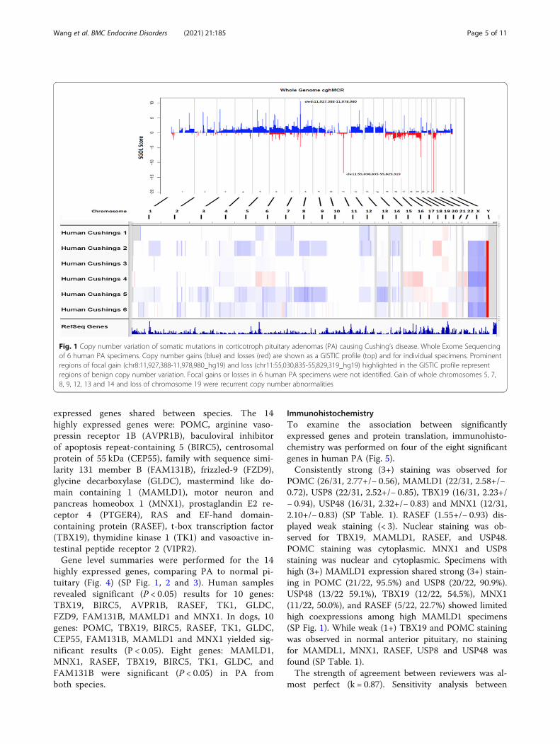
ResultsWhole exome sequencingTo identify recurrent somatic mutations and copy num-ber abnormalities in corticotroph PA, whole-exome se-quencing was performed in human PA.No specimens with mutations of USP8 were identified.
Two human specimens with USP48 (NM_032236) SNVwere found. Human PA specimen 1 had a USP48 c.1245G > A, p.M415I (COSM904151) SNV and specimen 6had a USP48 c.1243 A > G, p.M415V (COSM25000)SNV.Two human PA (3 and 4) had guanine nucleotide-
binding protein G(q) subunit alpha (GNAQ) c.286A > T,p.T96S substitutions.Copy number analysis using the whole exome sequen-
cing data was also performed to ascertain recurrentgains of whole chromosomes or chromosome arms(Fig. 1). Human PA specimens 2 and 5 had similar pat-terns with gain of 1q, 5, 7, 8q, 9, 12, 13 and 14. Speci-men 1 shared gain of 7, 12 and 14 with specimens 2 and5. Loss of 19 was shared by specimens 1, 3 and 6. Nofocal gains or losses enriched in PA in non-benign copynumber variations were observed.
RNA-sequencingGene expression between Cushing’s tumor and normaltissue was assessed in human and dogs.Unsupervised hierarchical clustering showed the nor-
mal pituitary specimens clustered separately from thePA specimens for both species (Fig. 2). Heterogeneity ofgene expression was found in individual PA specimensin both species, and a subset of genes that were over orunder expressed relative to normal pituitary in both spe-cies organisms clustered (Fig. 2).Differential gene expression showed 517 overex-
pressed and 1670 underexpressed genes in humanPA, and 127 overexpressed and 953 underexpressedgenes in dog PA relative to normal pituitary tissues(Fig. 3). Intersection of the human and dog gene listsshowed 14 overexpressed genes and 239 under
Wang et al. BMC Endocrine Disorders (2021) 21:185 Page 4 of 11
expressed genes shared between species. The 14highly expressed genes were: POMC, arginine vaso-pressin receptor 1B (AVPR1B), baculoviral inhibitorof apoptosis repeat-containing 5 (BIRC5), centrosomalprotein of 55 kDa (CEP55), family with sequence simi-larity 131 member B (FAM131B), frizzled-9 (FZD9),glycine decarboxylase (GLDC), mastermind like do-main containing 1 (MAMLD1), motor neuron andpancreas homeobox 1 (MNX1), prostaglandin E2 re-ceptor 4 (PTGER4), RAS and EF-hand domain-containing protein (RASEF), t-box transcription factor(TBX19), thymidine kinase 1 (TK1) and vasoactive in-testinal peptide receptor 2 (VIPR2).Gene level summaries were performed for the 14
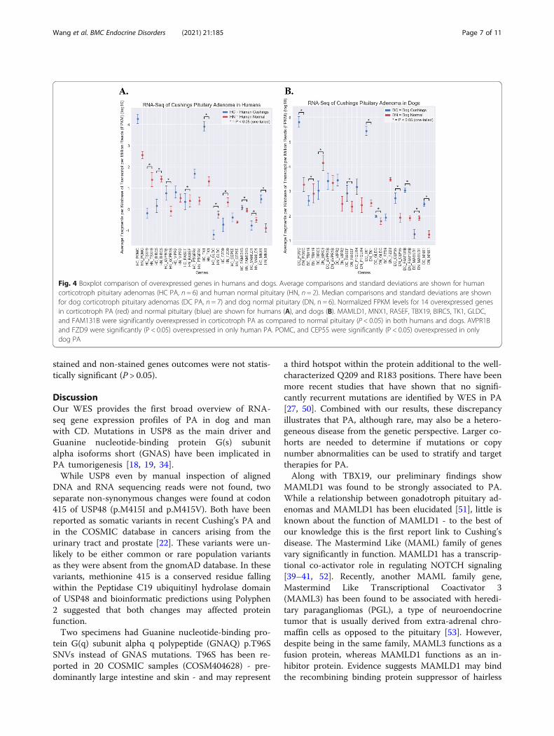
highly expressed genes, comparing PA to normal pi-tuitary (Fig. 4) (SP Fig. 1, 2 and 3). Human samplesrevealed significant (P < 0.05) results for 10 genes:TBX19, BIRC5, AVPR1B, RASEF, TK1, GLDC,FZD9, FAM131B, MAMLD1 and MNX1. In dogs, 10genes: POMC, TBX19, BIRC5, RASEF, TK1, GLDC,CEP55, FAM131B, MAMLD1 and MNX1 yielded sig-nificant results (P < 0.05). Eight genes: MAMLD1,MNX1, RASEF, TBX19, BIRC5, TK1, GLDC, andFAM131B were significant (P < 0.05) in PA fromboth species.
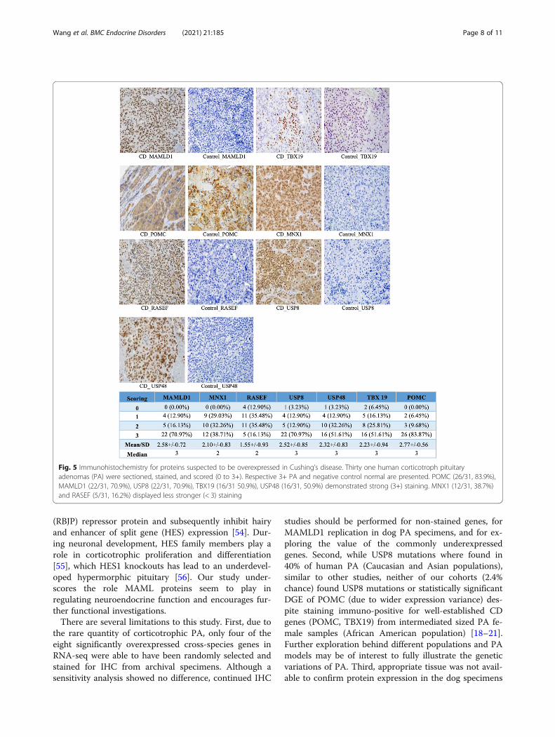
ImmunohistochemistryTo examine the association between significantlyexpressed genes and protein translation, immunohisto-chemistry was performed on four of the eight significantgenes in human PA (Fig. 5).Consistently strong (3+) staining was observed for
POMC (26/31, 2.77+/− 0.56), MAMLD1 (22/31, 2.58+/−0.72), USP8 (22/31, 2.52+/− 0.85), TBX19 (16/31, 2.23+/− 0.94), USP48 (16/31, 2.32+/− 0.83) and MNX1 (12/31,2.10+/− 0.83) (SP Table. 1). RASEF (1.55+/− 0.93) dis-played weak staining (< 3). Nuclear staining was ob-served for TBX19, MAMLD1, RASEF, and USP48.POMC staining was cytoplasmic. MNX1 and USP8staining was nuclear and cytoplasmic. Specimens withhigh (3+) MAMLD1 expression shared strong (3+) stain-ing in POMC (21/22, 95.5%) and USP8 (20/22, 90.9%).USP48 (13/22 59.1%), TBX19 (12/22, 54.5%), MNX1(11/22, 50.0%), and RASEF (5/22, 22.7%) showed limitedhigh coexpressions among high MAMLD1 specimens(SP Fig. 1). While weak (1+) TBX19 and POMC stainingwas observed in normal anterior pituitary, no stainingfor MAMDL1, MNX1, RASEF, USP8 and USP48 wasfound (SP Table. 1).The strength of agreement between reviewers was al-
most perfect (k = 0.87). Sensitivity analysis between
Fig. 1 Copy number variation of somatic mutations in corticotroph pituitary adenomas (PA) causing Cushing’s disease. Whole Exome Sequencingof 6 human PA specimens. Copy number gains (blue) and losses (red) are shown as a GISTIC profile (top) and for individual specimens. Prominentregions of focal gain (chr8:11,927,388-11,978,980_hg19) and loss (chr11:55,030,835-55,829,319_hg19) highlighted in the GISTIC profile representregions of benign copy number variation. Focal gains or losses in 6 human PA specimens were not identified. Gain of whole chromosomes 5, 7,8, 9, 12, 13 and 14 and loss of chromosome 19 were recurrent copy number abnormalities
Wang et al. BMC Endocrine Disorders (2021) 21:185 Page 5 of 11
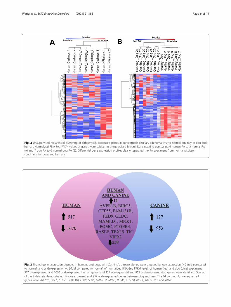
Fig. 2 Unsupervised hierarchical clustering of differentially expressed genes in corticotroph pituitary adenoma (PA) vs normal pituitary in dog andhuman. Normalized RNA-Seq FPKM values of genes were subject to unsupervised hierarchical clustering comparing 6 human PA to 2 normal PA(A) and 7 dog PA to 6 normal dog PA (B). Differential gene expression profiles clearly separated the PA specimens from normal pituitaryspecimens for dogs and humans
Fig. 3 Shared gene expression changes in humans and dogs with Cushing’s disease. Genes were grouped by overexpression (> 2-fold comparedto normal) and underexpression (< 2-fold compared to normal) of normalized RNA-Seq FPKM levels of human (red) and dog (blue) specimens.517 overexpressed and 1670 underexpressed human genes, and 127 overexpressed and 953 underexpressed dog genes were identified. Overlapof the 2 datasets demonstrated 14 overexpressed and 239 underexpressed genes between dog and man. The 14 commonly overexpressedgenes were: AVPR1B, BIRC5, CEP55, FAM131B, FZD9, GLDC, MAMLD1, MNX1, POMC, PTGER4, RASEF, TBX19, TK1, and VIPR2
Wang et al. BMC Endocrine Disorders (2021) 21:185 Page 6 of 11
stained and non-stained genes outcomes were not statis-tically significant (P > 0.05).
DiscussionOur WES provides the first broad overview of RNA-seq gene expression profiles of PA in dog and manwith CD. Mutations in USP8 as the main driver andGuanine nucleotide-binding protein G(s) subunitalpha isoforms short (GNAS) have been implicated inPA tumorigenesis [18, 19, 34].While USP8 even by manual inspection of aligned
DNA and RNA sequencing reads were not found, twoseparate non-synonymous changes were found at codon415 of USP48 (p.M415I and p.M415V). Both have beenreported as somatic variants in recent Cushing’s PA andin the COSMIC database in cancers arising from theurinary tract and prostate [22]. These variants were un-likely to be either common or rare population variantsas they were absent from the gnomAD database. In thesevariants, methionine 415 is a conserved residue fallingwithin the Peptidase C19 ubiquitinyl hydrolase domainof USP48 and bioinformatic predictions using Polyphen2 suggested that both changes may affected proteinfunction.Two specimens had Guanine nucleotide-binding pro-
tein G(q) subunit alpha q polypeptide (GNAQ) p.T96SSNVs instead of GNAS mutations. T96S has been re-ported in 20 COSMIC samples (COSM404628) - pre-dominantly large intestine and skin - and may represent
a third hotspot within the protein additional to the well-characterized Q209 and R183 positions. There have beenmore recent studies that have shown that no signifi-cantly recurrent mutations are identified by WES in PA[27, 50]. Combined with our results, these discrepancyillustrates that PA, although rare, may also be a hetero-geneous disease from the genetic perspective. Larger co-horts are needed to determine if mutations or copynumber abnormalities can be used to stratify and targettherapies for PA.Along with TBX19, our preliminary findings show
MAMLD1 was found to be strongly associated to PA.While a relationship between gonadotroph pituitary ad-enomas and MAMLD1 has been elucidated [51], little isknown about the function of MAMLD1 - to the best ofour knowledge this is the first report link to Cushing’sdisease. The Mastermind Like (MAML) family of genesvary significantly in function. MAMLD1 has a transcrip-tional co-activator role in regulating NOTCH signaling[39–41, 52]. Recently, another MAML family gene,Mastermind Like Transcriptional Coactivator 3(MAML3) has been found to be associated with heredi-tary paragangliomas (PGL), a type of neuroendocrinetumor that is usually derived from extra-adrenal chro-maffin cells as opposed to the pituitary [53]. However,despite being in the same family, MAML3 functions as afusion protein, whereas MAMLD1 functions as an in-hibitor protein. Evidence suggests MAMLD1 may bindthe recombining binding protein suppressor of hairless
Fig. 4 Boxplot comparison of overexpressed genes in humans and dogs. Average comparisons and standard deviations are shown for humancorticotroph pituitary adenomas (HC PA, n = 6) and human normal pituitary (HN, n = 2). Median comparisons and standard deviations are shownfor dog corticotroph pituitary adenomas (DC PA, n = 7) and dog normal pituitary (DN, n = 6). Normalized FPKM levels for 14 overexpressed genesin corticotroph PA (red) and normal pituitary (blue) are shown for humans (A), and dogs (B). MAMLD1, MNX1, RASEF, TBX19, BIRC5, TK1, GLDC,and FAM131B were significantly overexpressed in corticotroph PA as compared to normal pituitary (P < 0.05) in both humans and dogs. AVPR1Band FZD9 were significantly (P < 0.05) overexpressed in only human PA. POMC, and CEP55 were significantly (P < 0.05) overexpressed in onlydog PA
Wang et al. BMC Endocrine Disorders (2021) 21:185 Page 7 of 11
(RBJP) repressor protein and subsequently inhibit hairyand enhancer of split gene (HES) expression [54]. Dur-ing neuronal development, HES family members play arole in corticotrophic proliferation and differentiation[55], which HES1 knockouts has lead to an underdevel-oped hypermorphic pituitary [56]. Our study under-scores the role MAML proteins seem to play inregulating neuroendocrine function and encourages fur-ther functional investigations.There are several limitations to this study. First, due to
the rare quantity of corticotrophic PA, only four of theeight significantly overexpressed cross-species genes inRNA-seq were able to have been randomly selected andstained for IHC from archival specimens. Although asensitivity analysis showed no difference, continued IHC
studies should be performed for non-stained genes, forMAMLD1 replication in dog PA specimens, and for ex-ploring the value of the commonly underexpressedgenes. Second, while USP8 mutations where found in40% of human PA (Caucasian and Asian populations),similar to other studies, neither of our cohorts (2.4%chance) found USP8 mutations or statistically significantDGE of POMC (due to wider expression variance) des-pite staining immuno-positive for well-established CDgenes (POMC, TBX19) from intermediated sized PA fe-male samples (African American population) [18–21].Further exploration behind different populations and PAmodels may be of interest to fully illustrate the geneticvariations of PA. Third, appropriate tissue was not avail-able to confirm protein expression in the dog specimens
Fig. 5 Immunohistochemistry for proteins suspected to be overexpressed in Cushing’s disease. Thirty one human corticotroph pituitaryadenomas (PA) were sectioned, stained, and scored (0 to 3+). Respective 3+ PA and negative control normal are presented. POMC (26/31, 83.9%),MAMLD1 (22/31, 70.9%), USP8 (22/31, 70.9%), TBX19 (16/31 50.9%), USP48 (16/31, 50.9%) demonstrated strong (3+) staining. MNX1 (12/31, 38.7%)and RASEF (5/31, 16.2%) displayed less stronger (< 3) staining
Wang et al. BMC Endocrine Disorders (2021) 21:185 Page 8 of 11
and contrary to humans canine CD rise predominatelyfrom intermediate pituitary lobe as opposed to the anter-ior. However, 31 archival human PA provided strongevidence that co-overexpression of MAMLD1 andTBX19 occurred in approximately 70.9% of PA. Finally,although transcriptome analysis (Affymetrix arrays) onhuman PA has been complete [57, 58] and no functionalMAMLD1 assays were complete, our current investiga-tion was neither a discovery cohort for direct compari-son nor a functional assessment study, respectively.Rather, our cohort aimed to broadly profile the somaticaberrations of PA between dog and man.In spite of these genetic heterogeneities, differentially
expressed genes were identified that were common be-tween human and dog PA. Our study not only highlightsand contributes to the growing complex understandingsof genetic mechanism of CD, but also underscores the po-tential clinical implications of shared mechanisms be-tween dog and man. Since MAMLD1 appears to stainexclusively for corticotrophic tumor cells as compared toTBX19 (may stain both normal and tumor corticotrophiccells), MAMLD1 staining may be helpful in diagnosingCD, particularly in microadenomas with aberrant in symp-tomology and potentially recurrent when tissue quantity islimited. This may improve CD clinical outcomes and helpbetter characterize different hormone secreting pituitarytumors. Our results also suggests that, because MAMLD1expression appears absent in normal pituitary, targeted in-hibition of MAMLD1 may be a potential potent targetedstrategy for inhibiting the growth of corticotroph PA thatmerits further functional exploration [7]. Furthermore,dog PA may provide suitable veterinary cohorts to testnovel diagnostic and therapeutic approaches that may re-duce CD burden in both dog and man.
ConclusionWe highlight the first preliminary insights into profilingthe genomic characterizations of human and dog corti-cotroph PA with respect to MAMLD1 overexpression, afinding of potential direct impact to CD microadenomadiagnosis. Our study also offers a rationale for potentialuse of the canine model in development of precisiontherapeutics.
AbbreviationsCD: Cushing’s Disease; PA: pituitary adenomas; MAMLD1: Mastermind LikeDomain Containing 1; WES: Whole Exome Sequencing; RNA-Seq : Wholetranscriptome shotgun sequencing; IHC: Immunohistochemistry
Supplementary InformationThe online version contains supplementary material available at https://doi.org/10.1186/s12902-021-00845-z.
Additional file 1: Table S1. Post-agreement ImmunohistochemistryChemistry Scoring. Individualized immunohistochemistry scoring (0 to
3+) of Cushing’s pituitary adenoma human specimens from 7 antibodies(k = 0.87). Fig. S1. Difference in Magnitude of RNA-seq T-test Results. Nor-malized averaged FPKM values and standard errors are shown for genessuspected to be overexpressed by RNA-seq in human corticotroph pituit-ary adenomas (HC PA, n = 6) and dog corticotroph pituitary adenomas(DC PA, n = 7). Fig. S2. GeneCards® Summary Characteristics of HighlyExpressed Genes in Humans and Dogs with Cushing’s disease. Fig. S3.Theoretical Schematic Representation of the Highly Expressed Genes inHumans and Dogs with Cushing’s disease.
AcknowledgementsWe appreciate the support of Emory University, Winship – Pathology Corelabservices, specifically Dianne Alexis, MPH, who supported section histologyand staining.
Authors’ contributionsAW - drafting of the manuscript; acquisition of data; analysis andinterpretation of data; and critical revision of the manuscript. SGN -acquisition of data; analysis and interpretation of data; and critical revision ofthe manuscript. SN - acquisition of data; analysis and interpretation of data;and critical revision of the manuscript. MAT - technical, or material support;study concept and design; acquisition of data; and critical revision of themanuscript. AI - critical revision of the manuscript. MRR - acquisition of data;analysis and interpretation of data; and critical revision of the manuscript.BPM - technical, or material support; study concept and design; acquisitionof data; and critical revisionf of the manuscript. NMO - principal investigator;technical, or material support; study concept and design; acquisition of data;analysis and interpretation of data; critical revision of the manuscript; andstudy supervisor. All authors had access to the study data and reviewed andapproved the final manuscript.
FundingAl Lerner Chair Award, Department of Neurosurgery, Emory University Schoolof Medicine [AW, NMO], supported experimentation expenses.
Availability of data and materialsWe are currently analyzing the data from a different perspective andplanning a related study. Therefore, the data and material are not shared inthe current state. However, the datasets used and/or analyzed during thecurrent study are available from the corresponding author on reasonablewritten request. After the conclusion of the final study, all sequencing datawill be made available in NCBI SRA database.
Declaration
Ethics approval and consent to participateThis study was submitted and approved by the review and ethics committeeof each institution.This study protocol for the acquisition of human samples was approved bythe Emory University Institutional Review Board (IRB00045827). Written andverbal informed consent was obtained from the participating patientspreoperatively, and all human participant research were performed inaccordance with relevant guidelines and regulations.The study protocol for the acquisition of all dog samples was approved bythe Ethics Committee on Animal Experimentation of Utrecht University, theNetherlands, in accordance to the 3R (replacement, reduction, andrefinement of animal experimentation)-policy (DEC2012.III.02.017). Writtenand verbal informed consent was obtained from all client owners, andcanine treatment adhered to the legal and institutional animal welfareguidelines. All live vertebrae experiments were performed in accordancewith relevant guidelines and regulations, and released to the client owners.
Consent for publicationAll authors had access to the study data and reviewed and approved thefinal manuscript. All authors report no conflict of interests.
Competing interestsAll authors declare that they have no competing interests.
Wang et al. BMC Endocrine Disorders (2021) 21:185 Page 9 of 11
Author details1David Geffen School of Medicine, University of California, Los Angeles, LosAngeles, CA, USA. 2College of Medicine, Charles R. Drew University ofMedicine and Science, Los Angeles, CA, USA. 3Department of Pathology andLaboratory Medicine, Emory University School of Medicine, Atlanta, GA, USA.4Department of Computational Biology, St. Jude Children’s Research Hospital,Anchorage, TN, USA. 5Department of Clinical Sciences of CompanionAnimals, Faculty of Veterinary Medicine, Utrecht University, Utrecht,Netherlands. 6Department of Neurosurgery, Emory University School ofMedicine, Atlanta, GA , USA. 7Department of Medicine, Emory UniversitySchool of Medicine, Atlanta, GA, USA. 8Department of Genetics and GenomicSciences, Icahn School of Medicine at Mount Sinai, New York City, NY, USA.
Received: 1 September 2020 Accepted: 20 August 2021
References1. Lamberts SW, de Lange SA, Stefanko SZ. Adrenocorticotropin-secreting
pituitary adenomas originate from the anterior or the intermediate lobe inCushing’s disease: differences in the regulation of hormone secretion. J ClinEndocrinol Metab. 1982;54(2):286–91 https://doi.org/10.1210/jcem-54-2-286.
2. Slominski A, Wortsman J, Luger T, Paus R, Solomon S. Corticotropinreleasing hormone and proopiomelanocortin involvement in the cutaneousresponse to stress. Physiol Rev. 2000;80(3):979–1020 https://doi.org/10.1152/physrev.2000.80.3.979.
3. Aguilera G. Regulation of pituitary ACTH secretion during chronic stress.Front Neuroendocrinol. 1994;15(4):321–50. https://doi.org/10.1006/frne.1994.1013.
4. Valassi E, Biller BMK, Swearingen B, Pecori Giraldi F, Losa M, Mortini P, et al.Delayed remission after transsphenoidal surgery in patients with Cushing’sdisease. J Clin Endocrinol Metab. 2010;95(2):601–10 https://doi.org/10.1210/jc.2009-1672.
5. Clayton RN, Raskauskiene D, Reulen RC, Jones PW. Mortality and morbidityin Cushing’s disease over 50 years in stoke-on-Trent, UK: audit and meta-analysis of literature. J Clin Endocrinol Metab. 2011;96(3):632–42 https://doi.org/10.1210/jc.2010-1942.
6. Valassi E, Tabarin A, Brue T, Feelders RA, Reincke M, Netea-Maier R, et al.High mortality within 90 days of diagnosis in patients with Cushing’ssyndrome: results from the ERCUSYN registry. Eur J Endocrinol. 2019;181(5):461–72 https://doi.org/10.1530/EJE-19-0464.
7. Nieman LK, Biller BMK, Findling JW, Murad MH, Newell-Price J, Savage MO,et al. Treatment of Cushing’s syndrome: an Endocrine Society clinicalpractice guideline. J Clin Endocrinol Metab. 2015;100(8):2807–31 https://doi.org/10.1210/jc.2015-1818.
8. Mampalam TJ. Transsphenoidal microsurgery for Cushing disease: a reportof 216 cases. Ann Intern Med. 1988;109(6):487–93 https://doi.org/10.7326/0003-4819-109-6-487.
9. Trainer PJ, Lawrie HS, Verhelst J, Howlett TA, Lowe DG, Grossman AB, et al.Transsphenoidal resection in Cushing’s disease: undetectable serum cortisolas the definition of successful treatment. Clin Endocrinol. 1993;38(1):73–8https://doi.org/10.1111/j.1365-2265.1993.tb00975.x.
10. Patil CG, Prevedello DM, Lad SP, Vance ML, Thorner MO, Katznelson L, et al.Late recurrences of Cushing’s disease after initial successful transsphenoidalsurgery. J Clin Endocrinol Metab. 2008;93(2):358–62 https://doi.org/10.1210/jc.2007-2013.
11. Guiot G, Derome P. Surgical problems of pituitary adenomas. In: KrayenbühlH, Brihaye J, Loew F, et al., editors. Advances and technical standards inneurosurgery. Vienna: Springer Vienna; 1976. p. 3–33. https://doi.org/10.1007/978-3-7091-7080-9_1.
12. Fleseriu M. Medical treatment of Cushing disease: new targets, new hope.Endocrinol Metab Clin N Am. 2015;44(1):51–70 https://doi.org/10.1016/j.ecl.2014.10.006.
13. Feelders RA, de Bruin C, Pereira AM, Romijn JA, Netea-Maier RT, Hermus AR,et al. Pasireotide alone or with cabergoline and ketoconazole in Cushing’sdisease. N Engl J Med. 2010;362(19):1846–8 https://doi.org/10.1056/NEJMc1000094.
14. Hamrahian AH, Yuen KCJ, Hoffman AR, AACE Neuroendocrine And PituitaryScientific Committee (2014) AACE/ACE disease state clinical review: medicalManagement of Cushing Disease. Endocr Pract 20:746–757. https://doi.org/10.4158/EP14147.RA.
15. Bilodeau S, Vallette-Kasic S, Gauthier Y, Figarella-Branger D, Brue T, BertheletF, et al. Role of Brg1 and HDAC2 in GR trans-repression of the pituitaryPOMC gene and misexpression in Cushing disease. Genes Dev. 2006;20(20):2871–86 https://doi.org/10.1101/gad.1444606.
16. Molitch ME. Diagnosis and treatment of pituitary adenomas: a review.JAMA. 2017;317(5):516–24. https://doi.org/10.1001/jama.2016.19699.
17. Steffensen C, Bak AM, Rubeck KZ, Jørgensen JOL. Epidemiology of Cushing’ssyndrome. Neuroendocrinology. 2010;92(Suppl 1):1–5 https://doi.org/10.1159/000314297.
18. Ma Z-Y, Song Z-J, Chen J-H, Wang YF, Li SQ, Zhou LF, et al. Recurrent gain-of-function USP8 mutations in Cushing’s disease. Cell Res. 2015;25(3):306–17https://doi.org/10.1038/cr.2015.20.
19. Reincke M, Sbiera S, Hayakawa A, Theodoropoulou M, Osswald A,Beuschlein F, et al. Mutations in the deubiquitinase gene USP8 causeCushing’s disease. Nat Genet. 2015;47(1):31–8 https://doi.org/10.1038/ng.3166.
20. Vallette-Kasic S, Figarella-Branger D, Grino M, Pulichino AM, Dufour H, GrisoliF̧, et al. Differential regulation of proopiomelanocortin and pituitary-restricted transcription factor (TPIT), a new marker of normal andadenomatous human corticotrophs. J Clin Endocrinol Metab. 2003;88(7):3050–6 https://doi.org/10.1210/jc.2002-021934.
21. Liu N-A, Araki T, Cuevas-Ramos D, Hong J, Ben-Shlomo A, Tone Y, et al.Cyclin E-mediated human proopiomelanocortin regulation as a therapeutictarget for Cushing disease. J Clin Endocrinol Metab. 2015;100(7):2557–64https://doi.org/10.1210/jc.2015-1606.
22. Sbiera S, Perez-Rivas LG, Taranets L, Weigand I, Flitsch J, Graf E, et al. Drivermutations in USP8 wild-type Cushing’s disease. Neuro-oncology. 2019;21(10):1273–83 https://doi.org/10.1093/neuonc/noz109.
23. Perez-Rivas LG, Theodoropoulou M, Ferraù F, Nusser C, Kawaguchi K,Stratakis CA, et al. The gene of the ubiquitin-specific protease 8 is frequentlymutated in adenomas causing Cushing’s disease. J Clin Endocrinol Metab.2015;100(7):E997–1004 https://doi.org/10.1210/jc.2015-1453.
24. Hernández-Ramírez LC, Gam R, Valdés N, Lodish MB, Pankratz N, BalsalobreA, et al. Loss-of-function mutations in the CABLES1 gene are a novel causeof Cushing’s disease. Endocr Relat Cancer. 2017;24(8):379–92 https://doi.org/10.1530/ERC-17-0131.
25. Neou M, Villa C, Armignacco R, Jouinot A, Raffin-Sanson ML, Septier A, et al.Pangenomic classification of pituitary neuroendocrine tumors. Cancer Cell.2020;37(1):123–34 e5. https://doi.org/10.1016/j.ccell.2019.11.002.
26. Salomon MP, Wang X, Marzese DM, Hsu SC, Nelson N, Zhang X, et al. TheEpigenomic landscape of pituitary adenomas reveals specific alterations anddifferentiates among acromegaly, Cushing’s disease and endocrine-inactivesubtypes. Clin Cancer Res. 2018;24(17):4126–36 https://doi.org/10.1158/1078-0432.CCR-17-2206.
27. Bi WL, Horowitz P, Greenwald NF, Abedalthagafi M, Agarwalla PK, GibsonWJ, et al. Landscape of genomic alterations in pituitary adenomas. ClinCancer Res. 2017;23(7):1841–51 https://doi.org/10.1158/1078-0432.CCR-16-0790.
28. de Bruin C, Meij BP, Kooistra HS, Hanson JM, Lamberts SWJ, Hofland LJ.Cushing’s disease in dogs and humans. Horm Res. 2009;71(Suppl 1):140–3https://doi.org/10.1159/000178058.
29. Karlsson EK, Lindblad-Toh K. Leader of the pack: gene mapping in dogs andother model organisms. Nat Rev Genet. 2008;9(9):713–25. https://doi.org/10.1038/nrg2382.
30. Krzywinski M, Schein J, Birol I, Connors J, Gascoyne R, Horsman D, et al.Circos: an information aesthetic for comparative genomics. Genome Res.2009;19(9):1639–45. https://doi.org/10.1101/gr.092759.109.
31. Lindblad-Toh K, Wade CM, Mikkelsen TS, et al. Genome sequence,comparative analysis and haplotype structure of the domestic dog. Nature.2005;438(7069):803–19. https://doi.org/10.1038/nature04338.
32. van Rijn SJ, Galac S, Tryfonidou MA, et al. The influence of pituitary size onoutcome after Transsphenoidal Hypophysectomy in a large cohort of dogswith pituitary-dependent Hypercortisolism. J Vet Intern Med. 2016;30(4):989–95. https://doi.org/10.1111/jvim.14367.
33. Hanson JM, Mol JA, Leegwater PA, Bilodeau S, Drouin J, Meij BP. Expressionand mutation analysis of Tpit in the canine pituitary gland and corticotrophadenomas. Domest Anim Endocrinol 2008;34(3):217–222. doi: https://doi.org/10.1016/j.domaniend.2007.03.002. Epub 2007 May 21. PMID: 17544240.
34. Fragoso MCBV, Domenice S, Latronico AC, Martin RM, Pereira MAA, ZerbiniMCN, et al. Cushing’s syndrome secondary to adrenocorticotropin-independent macronodular adrenocortical hyperplasia due to activating
Wang et al. BMC Endocrine Disorders (2021) 21:185 Page 10 of 11
mutations of GNAS1 gene. J Clin Endocrinol Metab. 2003;88(5):2147–51.https://doi.org/10.1210/jc.2002-021362.
35. Stratakis CA, Tichomirowa MA, Boikos S, Azevedo MF, Lodish M, Martari M,et al. The role of germline AIP, MEN1, PRKAR1A, CDKN1B and CDKN2Cmutations in causing pituitary adenomas in a large cohort of children,adolescents, and patients with genetic syndromes. Clin Genet. 2010;78(5):457–63. https://doi.org/10.1111/j.1399-0004.2010.01406.x.
36. Albani A, Theodoropoulou M, Reincke M. Genetics of Cushing’s disease. ClinEndocrinol. 2018;88(1):3–12. https://doi.org/10.1111/cen.13457.
37. Hernández-Ramírez LC, Stratakis CA. Genetics of Cushing’s syndrome.Endocrinol Metab Clin N Am. 2018;47(2):275–97. https://doi.org/10.1016/j.ecl.2018.02.007.
38. Karczewski KJ, Francioli LC, Tiao G, Cummings BB, Alföldi J, Wang Q, et al.The mutational constraint spectrum quantified from variation in 141,456humans. Nature. 2020). https://doi.org/10.1038/s41586-020-2308-7;581(7809):434–43.
39. Boeva V, Popova T, Bleakley K, Chiche P, Cappo J, Schleiermacher G, et al.Control-FREEC: a tool for assessing copy number and allelic content usingnext-generation sequencing data. Bioinformatics. 2012;28(3):423–5. https://doi.org/10.1093/bioinformatics/btr670.
40. Olshen AB, Bengtsson H, Neuvial P, Spellman PT, Olshen RA, Seshan VE.Parent-specific copy number in paired tumor-normal studies using circularbinary segmentation. Bioinformatics. 2011;27(15):2038–46. https://doi.org/10.1093/bioinformatics/btr329.
41. Koboldt DC, Zhang Q, Larson DE, Shen D, McLellan MD, Lin L, et al. VarScan2: somatic mutation and copy number alteration discovery in cancer byexome sequencing. Genome Res. 2012;22(3):568–76. https://doi.org/10.1101/gr.129684.111.
42. Cibulskis K, Lawrence MS, Carter SL, Sivachenko A, Jaffe D, Sougnez C, et al.Sensitive detection of somatic point mutations in impure andheterogeneous cancer samples. Nat Biotechnol. 2013;31(3):213–9. https://doi.org/10.1038/nbt.2514.
43. Zhang J, Feng B. cghMCR: find chromosome regions showing commongains/losses. R package version 1.48.0 2020.
44. John G Tate, Sally Bamford, Harry C Jubb, Zbyslaw Sondka, David M Beare,Nidhi Bindal, Harry Boutselakis, Charlotte G Cole, Celestino Creatore,Elisabeth Dawson, Peter Fish, Bhavana Harsha, Charlie Hathaway, Steve CJupe, Chai Yin Kok, Kate Noble, Laura Ponting, Christopher C Ramshaw,Claire E Rye, Helen E Speedy, Ray Stefancsik, Sam L Thompson, Shicai Wang,Sari Ward, Peter J Campbell, Simon A Forbes, COSMIC: the Catalogue OfSomatic Mutations In Cancer, Nucleic Acids Research, Volume 47, Issue D1,08 January 2019, Pages D941–D947, https://doi.org/10.1093/nar/gky1015
45. Wang K, Li M, Hakonarson H. ANNOVAR: functional annotation of geneticvariants from high-throughput sequencing data. Nucleic Acids Res. 2010;38(16):e164. https://doi.org/10.1093/nar/gkq603.
46. Trapnell C, Pachter L, Salzberg SL. TopHat: discovering splice junctions withRNA-Seq. Bioinformatics. 2009;25(9):1105–11. https://doi.org/10.1093/bioinformatics/btp120.
47. Trapnell C, Roberts A, Goff L, Pertea G, Kim D, Kelley DR, et al. Differentialgene and transcript expression analysis of RNA-seq experiments withTopHat and cufflinks. Nat Protoc. 2012;7(3):562–78. https://doi.org/10.1038/nprot.2012.016.
48. Anders S, Pyl PT, Huber W. HTSeq--a Python framework to work with high-throughput sequencing data. Bioinformatics. 2015;31(2):166–9. https://doi.org/10.1093/bioinformatics/btu638.
49. Chen J, Jian X, Deng S, Ma Z, Shou X, Shen Y, et al. Identification ofrecurrent USP48 and BRAF mutations in Cushing’s disease. Nat Commun.2018;9(1):3171. https://doi.org/10.1038/s41467-018-05275-5.
50. Bi WL, Greenwald NF, Ramkissoon SH, Abedalthagafi M, Coy SM, Ligon KL,et al. Clinical identification of oncogenic drivers and copy-numberalterations in pituitary tumors. Endocrinology. 2017;158(7):2284–91. https://doi.org/10.1210/en.2016-1967.
51. Qi J, Ni W. Attenuation of MAMLD1 expression suppresses the growth andmigratory properties of Gonadotroph pituitary adenomas. Pathol Oncol Res.2020). https://doi.org/10.1007/s12253-019-00615-2”;26(2):937–46.
52. Nam Y, Sliz P, Song L, Aster JC, Blacklow SC. Structural basis forcooperativity in recruitment of MAML coactivators to notch transcriptioncomplexes. Cell. 2006;124(5):973–83. https://doi.org/10.1016/j.cell.2005.12.037.
53. Fishbein L, Leshchiner I, Walter V, Danilova L, Robertson AG, Johnson AR,et al. Comprehensive molecular characterization of Pheochromocytoma and
Paraganglioma. Cancer Cell. 2017;31(2):181–93. https://doi.org/10.1016/j.ccell.2017.01.001.
54. Fukami M, Wada Y, Okada M, Kato F, Katsumata N, Baba T, Morohashi KI,Laporte J, Kitagawa M, Ogata T (2008) Mastermind-like domain-containing 1(MAMLD1 or CXorf6) transactivates the Hes3 promoter, augmentstestosterone production, and contains the SF1 target sequence. J Biol Chem283:5525–5532. M703289200, 9. https://doi.org/10.1074/jbc.
55. Monahan P, Rybak S, Raetzman LT. The notch target gene HES1 regulatescell cycle inhibitor expression in the developing pituitary. Endocrinology.2009;150(9):4386–94. https://doi.org/10.1210/en.2009-0206.
56. Goldberg LB, Aujla PK, Raetzman LT. Persistent expression of activatednotch inhibits corticotrope and melanotrope differentiation and results indysfunction of the HPA axis. Dev Biol. 2011;358(1):23–32. https://doi.org/10.1016/j.ydbio.2011.07.004.
57. Evans C-O, Young AN, Brown MR, Brat DJ, Parks JS, Neish AS, et al. Novelpatterns of gene expression in pituitary adenomas identified bycomplementary deoxyribonucleic acid microarrays and quantitative reversetranscription-polymerase chain reaction. None. 2001;86(7):3097–107. https://doi.org/10.1210/jcem.86.7.7616.
58. Morris DG, Musat M, Czirják S, et al. Differential gene expression in pituitaryadenomas by oligonucleotide array analysis. Eur J Endocrinol. 2005;153(1):143–51. https://doi.org/10.1530/eje.1.01937.
Publisher’s NoteSpringer Nature remains neutral with regard to jurisdictional claims inpublished maps and institutional affiliations.
Wang et al. BMC Endocrine Disorders (2021) 21:185 Page 11 of 11