Embed Size (px)
Citation preview
Biochimica et Biophysica Acta, 706 (1982) 179-187 179 Elsevier Biomedical Press
BBA31279
THE HELICAL CONFORMATIONS OF 14- AND 16-RESIDUE FRAGMENTS OF SUZUKACILLIN, A MEMBRANE CHANNEL-FORMING POLYPEPTIDE
M.IQBAL and P. BALARAM *
Molecular Biophysics Unit, Indian Institute of Science, Bangalore 560 012 (India)
(Received February 2nd, 1982)
Key words: Suzukacillin fragment; Helical peptide; Oligopeptide conformation; Membrane channel; NMR
The suzukacillin fragments, Boc-Ala-Aib-Aib-Gln-Aib-Leu-Aib-Gly-Leu-Aib-Pro-Vai-Aib-Aib-OMe (14), Boc-Ala-Aib-Aia-Aib-Aib-Gin-Aib-Leu-Aib-Gly-Leu-Aib-Pro-Val-Aib-Aib-OMe (16G) and the completely apolar 16-residue peptide in which the glutamine residue has been replaced by alanine (16A) have been studied by 270 MHz IH-HMR, in C2HC!3 and (C2H3)2SO solution. Intramolecularly hydrogen-bonded NH groups have been identified by temperature and solvent dependence of chemical shifts. Peptides 14 and 16A adopt folded 310 helical conformations stabilized by 11 and 13 hydrogen bonds, respectively. In peptide 16G there are 12 intramolecular hydrogen bonds, with the glycine NH being solvent-exposed, in contrast to 14 and 16A
The 3~0 helix [1] is a structural feature found over short segments in protein crystal structures [2]. Incipient 310 helical segments may serve as nucleation sites for peptide folding [3]. The only poly-peptide for which a 310 helical structure has been proposed is poly-a-aminoisobutyric acid [4]. Recent structural studies have established a tend- ency for a-aminoisobutyric acid (Aib)-containing peptides to form Type III fl-turn structures [5-7], which on repetition generate a 310 helix. Recent interest in the conformation of a-aminoisobutyric acid-containing peptides, stems from the extensive occurrence of this residue in alamethicin [8], suzukacillin [9] (Fig. 1) and related membrane channel-forming peptides [10]. Studies in this laboratory on synthetic alamethicin [5,11] and suzukaciUin fragments [12-14] suggest that these hydrophobic polypeptides adopt highly folded rodlike 310 helical structures. These proposals have
*To whom correspondence should be addressed. Abbreviation: Aib, ot-aminoisobutyric acid.
0167-4838/82/0000-0000/$02.75 © 1982 Elsevier Biomedical Press
been based on NMR and X-ray analyses of short segments, together with infrared and CD studies of longer peptides. Since helical peptide conforma- tions are stabilized by intramolecular hydrogen bonds, 1H-NMR techniques, which allow delinea- tion of hydrogen-bonded NH groups, have proved most useful in the analysis of these systems. How- ever, the application of ~H-HMR requires the ob- servation of well-resolved amide NH resonances in oligopeptides, a condition that becomes difficult to satisfy in the study of moderately large peptides, at readily accessible magnetic field strengths. In our studies so far, we have provided a detailed analysis of the amide NH resonances of the 1-10 [12] and 11-21 [13,14] fragments of suzukacillin. In the present report we examine the larger 14- (Boc-Ala-Aib-Aib-Gln-Aib-Leu-Aib-Gly-Leu-Aib- Pro-Val-Aib-Aib-OMe, 14) and 16- (Boc-Als-Aib- Ala-Aib-Aib-Gln-Aib-Leu-Aib-Gly-Leu-Aib-Pro- Val-Aib-Aib-OMe, 16G) residue central fragments of suzukacillin and the related 16-residue peptide (Boc-Ala-Aib-Ala-Aib-Aib-Ala-Aib-Leu-Aib-Gly- Leu-Aib-Pro-Val-Aib-Aib-OMe, 16A ) and present
180
evidence for 3:0 helical folding in these sequences. The results support a structure incorporating five turns of a 3:0 helix stabilized by 12 intramolecular hydrogen bonds, for the 6-21 fragment of suzukacillin.
Experimental section
All peptides were synthesized by solution phase procedures as described for alamethicin I [15]. Peptides were characterized by 270 MHz :H-NMR spectra and found to be homogeneous by TLC on silica gel. ~H-NMR studies were carried out on a Bruker WH-270 FT-NMR spectrometer at the Bangalore NMR Facility as reported earlier [11,12]. Spectral widths of 3012 Hz were used and the data after Fourier transformation were stored in 8K memory locations, yielding a digital resolution of 0.367 Hz/point. All chemical shifts are expressed as 8(ppm) downfield from tetramethylsilane. Solvent titration and variable temperature mea- surements were performed at a peptide concentra- tion of 10 mg/ml as described earlier [12,13].
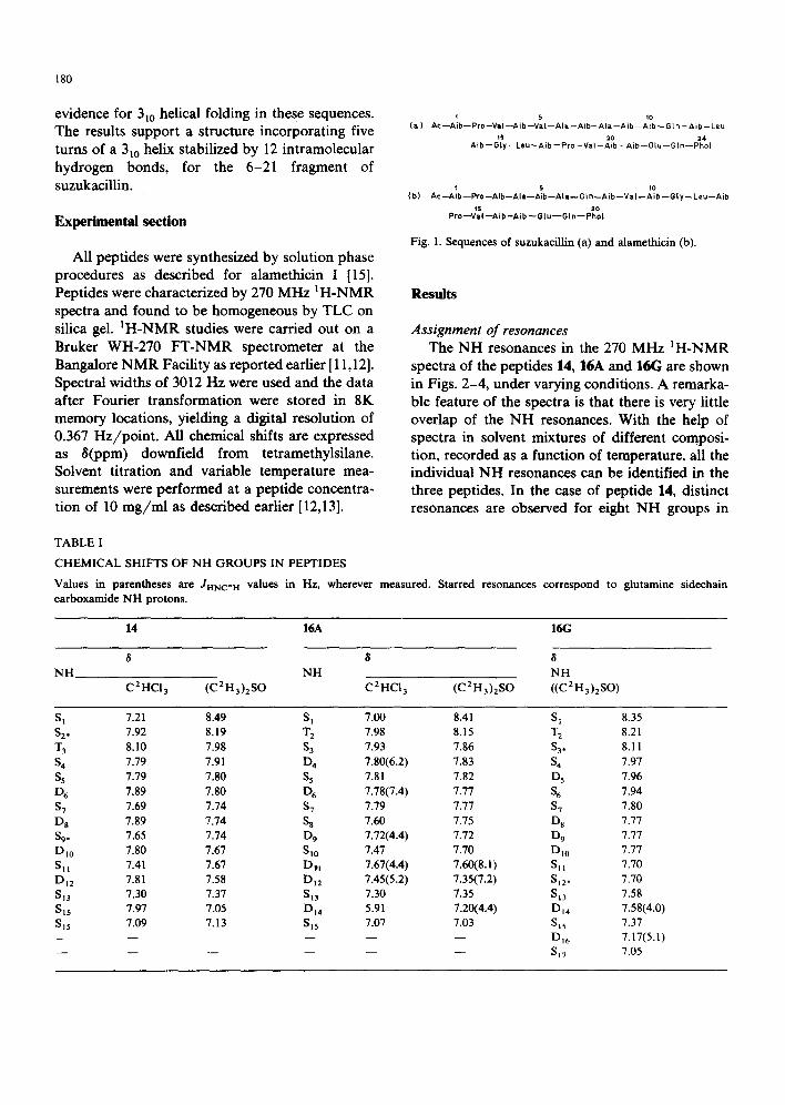
'~ 5 1o (a ) A c - - A i b - - P r o - - V a I - - A i b - - V a I - - A l a - - A i b - - A l a - - A i b - - A i b - - G t n - - A i b - - L e u
ts 20 24 A ib - - G ly- - Leu ~ A i b - - P ro - -Va I - -A ib - -A ib - -G lu - -G In ~ P h o l
(b) t 5 t0
A c - - A i b - - P r o - - A i b - - A l a - - A i b - - A t a - - G I n - - A i b - - V a I - - A i b - - G l y - - L e u - - A i b
15 20 P r o - - V a I - - A i b - A i b - - G I u - - G I n - - P h o L
Fig. 1. Sequences of suzukacillin (a) and alamethicin (b).
Results
Assignment of resonances The NH resonances in the 270 MHz 1H-NMR
spectra of the peptides 14, 16A and 1613 are shown in Figs. 2-4, under varying conditions. A remarka- ble feature of the spectra is that there is very little overlap of the NH resonances. With the help of spectra in solvent mixtures of different composi- tion, recorded as a function of temperature, all the individual NH resonances can be identified in the three peptides. In the case of peptide 14, distinct resonances are observed for eight NH groups in
TABLE I
C H E M I C A L SHIFTS OF N H G R O U P S IN PEPTIDES
Values in parentheses are Jmqc,H values in Hz, wherever measured. Starred resonances correspond to glutamine sidechain carboxamide N H protons.
14 16A 16(;
8 8 N H NH
C2HC13 (C2H3)2SO C2HCI3 (C2H3)2SO
8 N H ((C2 H 3)2SO)
S I 7.21 8.49 S 1 7.00 8.41 S 1 8.35 $2. 7.92 8.19 T 2 7.98 8.15 T 2 8.21 T 3 8.10 7.98 S 3 7.93 7.86 $3o 8.11 S 4 7.79 7.91 D 4 7.80(6.2) 7.83 S 4 7.97 S 5 7.79 7.80 S 5 7.81 7.82 D 5 7.96 D 6 7.89 7.80 D 6 7.78(7.4) 7.77 S 6 7.94 S 7 7.69 7.74 S 7 7.79 7.77 S 7 7.80 D s 7.89 7.74 S s 7.60 7.75 D s 7.77 $9. 7.65 7.74 D 9 7.72(4.4) 7.72 D 9 7.77 Dl0 7.80 7.67 Si0 7.47 7.70 Dlo 7.77 Sit 7.41 7.67 Dpt 7.67(4.4) 7.60(8.1) Sll 7.70 D j 2 7.81 7.58 D 12 7.45(5.2) 7.35(7.2) S 12 * 7.70 $13 7.30 7.37 S~3 7.30 7.35 Si3 7.58 Sl5 7.97 7.05 DI4 5.91 7.20(4.4) DI4 7.58(4.0) S15 7.09 7.13 SIs 7.07 7.03 SIs 7.37 . . . . . DI6 7.17(5.1) . . . . . . SiT 7.05

181
C2HCI3 and two intense sets of overlapping reso- nances are observed at approx. 7.9 8 and 7.7 8, corresponding to three and four NH protons, re- spectively. In (C2H3)2SO, eight distinct NH reso- nances and an overlapping set of peaks centred at approx. 7.74 8 are observed. Using spectra in C2HC13/(CEH3)2SO mixtures and by varying the temperature in the pure solvents all 15 NH groups (13 backbone and two sidechain) in 14 can be identified. (See Fig. 1 for representative spectra). The various resonances are labelled as S, (singlets), D, (doublets) and T, (triplets), where the subscript
n refers to the order of appearance from lowfield in (C2H3)2SO.
The glycine-NH (I"3) is unambiguously assigned to the only triplet resonance. The Ala(1)-NH (Dn4) is assigned to the highest-field NH resonance in C2HC13, since the position of the urethane NH in this solvent has been established in a variety of peptides [11-13, 16, 17]. Its position in (C2H3)2SO was determined on the basis of solvent titration experiments. The Ala(1)-NH appears as a broad resonance under many conditions and a doublet is not clearly discernable. The other four doublets
D6,SJD 8
I I , I I I I 9.0 8.5 8-0 6 is7 7.5 7.0 6.5
S D6 $9 515 s~ 4. ss\~/~ " ,s1~ s~3
$2 T3 D12
I I J I I I 9.0 8.5 8-0 6 7.5 7.0 6.5
- , I I I I I 8.5 8.0 7.5 8 7.0 6.5
s 01o,O12 s -'I s15 D6,~ )~ Is7 $13J I
. . . . " L . , , . . A , J ' u V L , _ _ . . . . _ _
I I l I I I 8.5 8.0 7. 5 ?.0 6.5 6.0 &
Fig. 2. Partial 270 MHz IH-NMR spoetra of peptid¢ 14 (NH region). (a) C2HCI3, 293 K; (b) C2HCI3 +(C2H3)2SO (7:3), 293 K; (c) (C2H3)2SO, 293 K. Peptide concentration 0.0071 M.
182
can be assigned to the valine, glutamine and two leucine residues. Of the nine singlets, seven arise from a-aminoisobutyric acid residues, which lack a hydrogen at C% while two are due to the gluta- mine sidechain carboxamide group. The latter, S 2 and S 9, were identified on the basis of their broad- ening at elevated temperatures, as compared to other singlets, in (C2H3)2SO. This behaviour of glutamine sidechain protons has been noted earlier [13, 14, 16]. The chemical shifts of the various reso- nances are summarized in Table I.
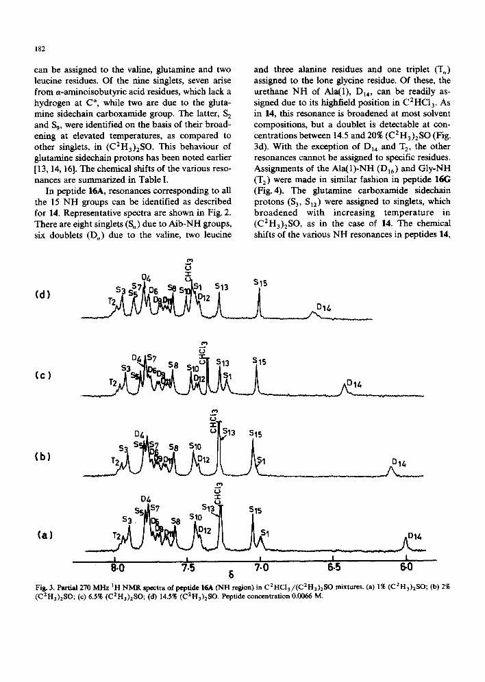
In peptide 16A, resonances corresponding to all the 15 NH groups can be identified as described for 14. Representative spectra are shown in Fig. 2. There are eight singlets (S,) due to Aib-NH groups, six doublets (D,) due to the valine, two leucine
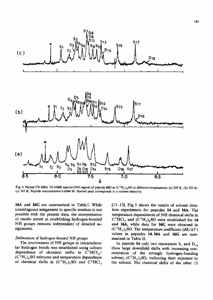
and three alanine residues and one triplet (T,) assigned to the lone glycine residue. Of these, the urethane NH of Ala(1), DI4 , carl be readily as- signed due to its highfield position in C2HC13 . As in 14, this resonance is broadened at most solvent compositions, but a doublet is detectable at con- centrations between 14.5 and 20% (C 2 H 3)2 SO (Fig. 3d). With the exception of D~4 and T 2, the other resonances cannot be assigned to specific residues. Assignments of the AIa(1)-NH (Dl6) and Gly-NH (T2) were made in similar fashion in peptide 16(; (Fig. 4). The glutamine carboxamide sidechain protons (S 3, Si2 ) were assigned to singlets, which broadened with increasing temperature in (C2H3)2SO, as in the case of 14. The chemical shifts of the various NH resonances in peptides 14,
(d)
L)
o . s,?o. y2"..= S~ It ,ub / = ~ D 1 2 / i
(c)
trt ¢.3
°-41 s7 s ̂ ~ s13 sis
~::r_ D41 Ullt~13 S15
T 2 D12 $1
.D U
D4 1" U. ssdS~ sL3I sis
s3 " sa slo "fl l
I I I I '0 7.5 7.0 6.5 6.0
6 Fig. 3. Partial 270 MI-Iz IH NMR spectra of peptide 16A (NH resion) in C2HCI3/(C2H3)2SO mixtures. (a) 1% (C2H3)2SO; (b) 270 (C2H3)2SO; (c) 6.5~ (C2H3)2S0; (d) 14.5% (C2H3)2SO. Peptide concentration 0.0066 M.
S['o a
S3 r~DlO 513 D9 S17
) I / 1"2 ~lv} A / I I l 11,.°1~ / /
I , I , I , I , I , ,
183
I D8 S" D5 /[D9 .
(a)
I , !
8.5 8-0 7.5 7.0 B-5 6
Fig. 4. Partial 270 MHz IH-NMR spectra (NH region) of peptide 16G in (C2Ha)2SO at different temperatures. (a) 295 K; (b) 325 K; (c) 345 K. Peptide concentration 0.0064 M. Starred peak corresponds to a solvent impurity.
16A and 16G are summarized in Table I. While unambiguous assignment to specific residues is not possible with the present data, the interpretation of results aimed at establishing hydrogen-bonded NH groups remains independent of detailed as- signments.
Delineation of hydrogen-bonded NH groups The involvement of NH groups in intramolecu-
lar hydrogen bonds was established using solvent dependence of chemical shifts in C2HC13/ (C2H3)2SO mixtures and temperature dependence of chemical shifts in (C2H3)2SO and C2HC13
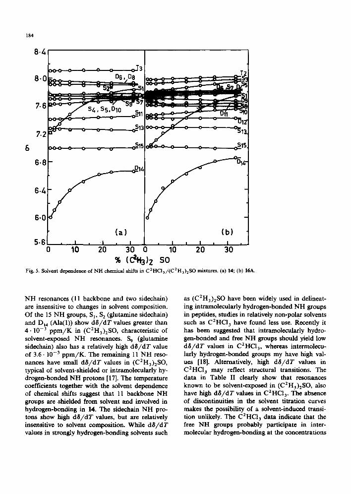
[ 11-13]. Fig. 5 shows the results of solvent titra- tion experiments for peptides 14 and 16A. The temperature dependences of NH chemical shifts in C2HC13 and (C2H3)2SO were established for 14 and 16A, while data for 16G were obtained in (C 2 H3 )2 SO. The temperature coefficient (d 8 / d T) values in peptides 14, 16A and 16G are sum- marized in Table II.
In peptide 14 only two resonances S, and D{4 show large downfield shifts with increasing con- centration of the strongly hydrogen-bonding solvent, (C2H3)2SO, indicating their exposure to the solvent. The chemical shifts of the other 13
184
&
8./-,
3 T 2 8.0 D6, De
7.~
7.2
6.8
6./-,
6.0
B
(a) (b) 5 " 6 . i . i , I , I i I , I.
0 10 20 30 0 10 20 30 % ( ~ ) z so
Fig. 5. Solvent dependence of NH chemical shifts in C2HC13/(C2H3)2SO mixtures. (a) 14; (b) 16A.
NH resonances (11 backbone and two sidechain) are insensitive to changes in solvent composition. Of the 15 NH groups, S I, S 2 (glutamine sidechaln) and Di4 (Ala(1)) show dS/dT values greater than 4 .10 -3 p p m / K in (C2H3)2SO, characteristic of solvent-exposed NH resonances. S 9 (glutamine sidechain) also has a relatively high dS/dT value of 3.6.10 -3 ppm/K. The remaining 11 NH reso- nances have small dS/dT values in (C2H3)2SO, typical of solvent-shielded or intramoleculady hy- drogen-bonded NH protons [17]. The temperature coefficients together with the solvent dependence of chemical shifts suggest that 11 backbone NH groups axe shielded from solvent and involved in hydrogen-bonding in 14. The sidechain NH pro- tons show high dS/dT values, but are relatively insensitive to solvent composition. While d$/dT values in strongly hydrogen-bonding solvents such
as (C2H3)2SO have been widely used in delineat- ing intramoleculaxly hydrogen-bonded NH groups in peptides, studies in relatively non-polar solvents such as C2HC13 have found less use. Recently it has been suggested that intramoleculaxly hydro- gen-bonded and free NH groups should yield low dO/dT values in C2HC13, whereas intermolecu- laxly hydrogen-bonded groups my have high val- ues [18]. Alternatively, high dS/dT values in C2HC13 may reflect structural transitions. The data in Table II clearly show that resonances known to be solvent-exposed in (C2H3)2SO, also have high dS/dT values in C2HC13 . The absence of discontinuities in the solvent titration curves makes the possibility of a solvent-induced transi- tion unlikely. The C2HC13 data indicate that the free NH groups probably participate in inter- molecular hydrogen-bonding at the concentrations

TABLE II
T E M P E R A T U R E COEFFICIENTS OF N H G R O U P S IN PEPTIDES
Starred resonances correspond to glutamine sidechain carboxamide N H protons.
185
14 16A 16G
dS/dT(ppm/K)×(lO 3)
C2HC13 (CEH3)2SO
N H dS/dT(ppm/K)× 103 )
C2HC13 (C2H3)2SO
N H dS/dT(ppm/K)( X 10 3)
(C2H3)2SO
S 1 6.0 6.0 S 1 5.4 5.9 S2. 4.6 5.1 T 2 3.7 3.8 T 3 2.3 1.6 S 3 2.2 1.2 S 4 2.2 2.0 D 4 1.9 1.2 S 5 2.2 1.0 S 5 2.2 1.7 D 6 1.8 2.2 D 6 1.7 1.6 S 7 1.5 1.2 S 7 1.6 1.6 D s 1.8 1.2 S 8 1.2 0.9 S 9. 4.2 3.6 D 9 0.8 2.2 Dto 3.2 1.8 Si0 2.0 2.2 Sll 1.6 2.9 Dil 3.2 2.8 DI2 3.2 2.9 Di2 1.8 0.5 513 1.2 3.2 813 1.4 2.9 Dr4 1 i.7 6.5 Di4 8.4 6.6 Sis 1.0 2.0 Sis 0.7 1.9
S I 5.7 T 2 5.2 S 3. 5.3 S 4 2.5 D s 2.5 S 6 2.5 S 7 0.1 D s 2.0 0 9 2.0 D Io 2.0 S N 3.0 St2 3.2 Si3 2.1
Dl4 2.4 St5 3.0 DI6 7.0 SI7 2.0
used. Association of the peptide Boc-Gln-Aib-Leu- Aib-Gly-Leu-Aib-Pro-Val-Aib-Aib-OMe (11-21 fragment of suzukacillin) has been demonstrated to occur in C2HC13 via NH groups, which are not involved in intramolecular hydrogen bonds [14].
In peptide 16A only resonances S~ and O i 4
(Ala(1)-NH) have high dS/dT values in ( C 2 H 3 ) 2 S O ( > 4 . 1 0 - 3 ppm/K) a n d s h o w signifi- cant solvent-dependence of chemical shifts (Table II and Fig. 5). All the other 13 NH groups have dS/dT values less than 4 . 10 -3 p p m / K in (C2H3)2SO and also show very little solvent-sensi- tivity of chemical shifts. This suggests that 13 NH groups in 16A probably participate in intramolecu- lar hydrogen bonds.
In peptide 16G, of the 17 NH resonances (15 backbone and two sidechain) Di6 (Ala(1)-NH), S l and one sidechain NH (Sa) have high dS/dT values i n ( C 2 H 3 ) 2 S O , characteristic of free NH groups. 12 backbone NH groups and one sidechain NH (S~2) have low dS/dT values. Interestingly the GIy-NH (T2) has a much higher dS/dT value in peptide 16G, as compared to peptide 14 and 16A.
Discussion
The results presented above favour the follow- ing conclusions: (i) Peptide 14 adopts a folded conformation in solution, stabilized by eleven intramolecular hy- drogen bonds. Together with the known stereo- chemical preferences of a-aminoisobutyric acid re- sidues for Type III fl-turn or incipient 3~0 helical structures [10], the data suggest that 14 is folded into a long stretch of 310 helix stabilized by eleven 4 ~ 1 hydrogen bonds. (ii) Peptide 16A also folds into a ~al0 helix stabi- lized by 13 intramolecular hydrogen bonds. (iii) Peptide 16G, which corresponds to the 6-21 fragment of suzukacillin, is also folded into a helical conformation stabilized by 12 hydrogen bonds involving backbone NH groups. In contrast to peptide 16A, the Gly-NH in 16G does not appear to participate in an intramolecular hydro- gen bond in (C2H3)2SO solution.
Earlier studies from this laboratory have pro- vided evidence for the 3~o helical folding of the

186
(a) Bo - o M ,
(b) Boc- Gln --Aib -- L e ~ ~ A i b -- Pro--Val - - Aib--Aib --OMe
( c ) B o c ' A l a - - " - - " b - - A i b - A l a - A i b - L e u - A i b - G l y - - r e u - A i b - - P r o - - V a t - A i b - - A i b - O M e
Fig. 6. Schematic representation of the hydrogen-bonding pattern in suzukacillin fragments and 16A. (a) 1-I 0 [12]; (b) 11-21 [13]; (c) 16A. Arrows represent NH to CO hydrogen bonds.
1-10 and 11-21 fragments of suzukacillin [12,13]. The present report establishes that these confor- mations are maintained over longer segments. Both the 8-21 suzukacillin fragment 14 and the apolar 16-residue peptide 16A favour 310 helical struc- tures. In the latter, five complete turns of a 3~0 helix are fully accommodated. The hydrogen- bonding schemes proposed for 16A, together with those postulated for shorter fragments of suzuka- cillin are schematically illustrated in Fig. 6. In the 6-21 fragment 16G there appears to be a loss of the hydrogen bond involving the Gly-NH, leading to a high dg/dT value for this group in (C2Ha)2SO. Peptides 16A and 16(; differ solely at residue 6. The presence of the carboxamide group in 16G may conceivably affect the conformation either by sidechain backbone hydrogen-bonding [13] or by favouring intermolecular association. Aggregation of peptides stabilized by sidechain hydrogen-bonding groups could also lead to the observed differences between 16A and 16G. How- ever, a detailed NMR study of the aggregation of the smaller 11-21 suzukacillin fragment revealed that peptide association is unimportant in (C2H3)2SO at the concentrations employed [14]. Further, helical a-aminoisobutyric acid peptides aggregate in apolar solvents such as C2HC13, at concentrations used in NMR studies, without sig- nificant disruption of the conformation of individ- ual molecules [14].
In the suzukacillin sequence, the proline residue at position 18 interrupts the chain of successive 4 - , 1 hydrogen bonds, leaving the Gly-CO group free, in a 3~0 helical structure. In both alamethicin and suzukacillin sequences there is the possibility
of some conformational flexibility at the Gly-Leu- Aib-Pro segment [11]. The possibility of expansion of the 3t0 helix into an a-helix in this region has already,been considered from infrared studies of these fragments [6]. It may be noted that there are very small differences in 0, ~ values for the 3 ~o and a-helical conformations. These structures can be distinguished largely on the basis of their hydro- gen-bonding patterns. Distorted conformations lying between these regular structures also explain the observed data. Transitions from the 4--, 1 hy- drogen-bonded to 5--, 1 hydrogen-bonded struc- tures in the central part of 16G could lead to exposure of the Gly-NH. In longer sequences the additional hydrogen bond present in the 3~0 helix would not lend very much additional stabilization as compared to the a-helix. It is quite likely that, while the 3~0 helix is maintained over shorter seg- ments, expansion to an a-helix must certainly be considered in the longer sequences. Resonance S~, which is solvent-exposed in all three pqatides, may be assigned to the Aib(2)-NH, which is not hydro- gen-bonded in either 3~0 or a-helical conforma- tions.
The presence of L-amino acids in these se- quences would favour chain-folding into right- handed helical conformations [19]. The JHNC°H values for all the NH doublets that can be mea- sured are less than 8 Hz (Table I), which is com- patible with ,/, ~ - 4 0 to - 7 0 °, required for right- handed 310 or a-helical conformations [20]. The results of this study, together with earlier reports on small fragments, demonstrate that the apolar 1-21 segment of suzukacillin must adopt a largely 3,10 or a-helical conformation. The close homology
of their sequences suggests tha t this conclus ion is
a lso val id for a lamethic in . These s tudies emphas ize the ut i l i ty of highf ie ld
N M R studies in the de l inea t ion of hydrogen- b o n d e d N H groups, even in ra ther long acyclic pep t i de sequences, which a d o p t s te reochemical ly r igid conformat ions . The ava i lab i l i ty of higher magne t i c field s t rengths should pe rmi t de ta i led analys is of the comple t e sequences of m e m b r a n e channe l - fo rming po lypep t ides , by p rov id ing ade- qua te spect ra l resolut ion. I t is, however, c lear that the a - a m i n o i s o b u t y r i c ac id con ta in ing channel- formers favour p r e d o m i n a n t l y 3 l0 or a -hehca l con- format ions . Such helices canno t a c c o m m o d a t e pas- sage of ca t ions th rough the helix inter ior , as p ro- posed in the mode l s for the g ramic id in A trans- m e m b r a n e channel [21]. M e m b r a n e channels must arise b y aggregat ion of apo la r helices in the case of the a - a m i n o i s o b u t y r i c ac id con ta in ing po ly- pep t ides [10]. S t ruc tura l inves t iga t ions of such p e p t i d e aggragates should be va luab le in develop- ing de ta i led molecu la r mechan i sms for the mem- b r a n e act ivi ty of suzukaci l l in , a lameth ic in and re- l a ted po lypep t ides .
Acknowledgements
This research was s u p p o r t e d by a g ran t f rom the D e p a r t m e n t of Science and Technology. M.I. thanks the U G C for award of a Teacher Fe l low- ship. P.B. is the rec ip ient of a U G C Career Award .
References
1. Donohue, J. (1953) Proc. Natl. Acad. Sci. U.S.A. 39, 470- 478
187
2 Dickerson, R. E. and Geis, I. (1969). The structure and Action of Proteins; Harper and Row, New York.
3 Venkatachalapathi, Y. V. and Balaram, P. (1979)) Nature 281, 83-84
4 Malcolm, B. R. (1977) Biopolymers 16, 2591-2592 5 Nagaraj, R., Shamala, N. and Balaram, P. (1979) J. Am.
Chem. Soc. 101, 16-20 6 Rao, C. P., Nagaraj, R., Rao, C. N. R. and Balaram, P.
(1980) Biochemistry 19, 425-431 7 Shamala, N., Nagaraj, R. and Balaram, P. (1978) J. Chem.
Soc. Chem. Commun. 996-997 8 Pandey, R. C., Carter Cook J., Jr. and Rinehart, K. L. Jr.
(1977) J. Am. Chem. Soc. 99, 8469-8483 9 Jung, G., Konig, W. A., Liebfritz, D., Ooka, T., Janko, J.
and Boheim, G. (1976) Biochim. Biophys. Acta 433, 164- 181
10 Nagaraj, R. and Balaram, P.(1981) Acc. Chem. Res. 14, 356-362
11 Nagaraj, R. and Balaram, P. (1981) Biochemistry 20, 2828- 2835
12 lqbal, M. and Balaram, P. (1981) J. Am. Chem. Soc. 103, 5548--5552
13 Iqbal, M. and Balaram, P. (1981) Biochemistry 20, 4866- 4871
14 Iqbal, M. and Balaram, P. (1981) Biochemistry 20, 7278- 7284
15 Nagaraj, R. and Balaram, P. (1981) Tetrahedron 37, 1263- 1270
16 Zanacchi, R. M. and Moore, W. J. (1980) Aust. J. Chem. 33, 1505-1510
17 Hruby, V. J. (1974) in Chemistry and Biochemistry of Amino Acids, Peptides and Proteins (Weinstein, B., ed.), Vol. 3, pp.l-188, Dekker, New York
18 Stevens, E. S., Sugawara, N., Bonora, G. M. and Toniolo, C. (1980) J. Am. Chem. Soc. 102, 7048-7050
19 Ramachandran, G. N. and Sasisekharan, V. (1968) Adv. Protein Chem. 23, 283-437
20 Bystrov, V. F. (1976) Prog. NMR Spectrosc. 10, 41-81 21 Urry, D. W. (1971) Proc. Natl. Acad. Sci. U.S.A. 68,
672-676