Embed Size (px)
Citation preview
N E W S A N D V I E W S
NATURE MEDICINE VOLUME 13 | NUMBER 6 | JUNE 2007 673
It is important that we explore how this new regulatory pathway might be used in a therapeutic setting. One difficulty is that altering retinaldehyde levels might also affect retinoic acid levels. For example, we might consider directly providing retinaldehyde, but in addition to potential difficulties in tis-sue distribution and pharmacoavailability, long-term treatment might result in higher retinoic acid concentrations. In contrast, inhibiting retinaldehyde catabolism might decrease retinoic acid. In both cases, a careful evaluation of the retinoic acid–mediated con-sequences on cellular activities will be needed. Given the data presented, it seems likely that retinaldehyde concentrations are tightly con-trolled. Low retinaldehyde in the fat tissue of
obese mice arises from the imbalanced lev-els of its metabolic enzymes compared with those in lean mice1. How these enzyme levels are controlled in the adipose tissue will have to be explored, starting with the question of whether an imbalance in enzyme expression ratio is a cause or a consequence of obesity.
Metabolic syndrome is a highly complex disease that challenges both the fundamen-talist, who still hopes for a single causal mechanism, and the clinician, who dreams of a single molecule that would treat all the abnormalities associated with the syndrome. There is no reason to think that retinalde-hyde resolves these issues. Rather, the paper by Ziouzenkova et al.1 opens a new avenue of investigation, offering one exciting entry
point into the complexity of nuclear receptor networks and their diverse and multifaceted natural ligands.
COMPETING INTERESTS STATEMENTThe author declares no competing financial interests.
1. Ziouzenkova, O. et al. Nat. Med. 13, 695–702 (2007).
2. Desvergne, B. Vitam. Horm. 75, 1–32 (2007).3. Avram, M.M., Avram, A.S. & James, W.D. J. Am.
Acad. Dermatol. 56, 472–492 (2007).4. Bonet, M.L., Ribot, J., Felipe, F. & Palou, A. Cell.
Mol. Life Sci. 60, 1311–1321 (2003).5. Schwarz, E.J., Reginato, M.J., Shao, D., Krakow,
S.L. & Lazar, M.A. Mol. Cell. Biol. 17, 1552–1561 (1997).
6. Yamauchi, T. et al. J. Clin. Invest. 108, 1001–1013 (2001).
7. Shug, T.T., Berry, D.C., Shaw, N.S., Travis, S.N., & Noy, N. Cell 129, 723–733 (2007).
8. Yang, Q. et al. Nature 436, 356–362 (2005).
The hydrogen highway to reperfusion therapyKatherine C Wood & Mark T Gladwin
Hydrogen gas debuts as a selective antioxidant with explosive potential as cytoprotective therapy for ischemia-reperfusion injury and stroke.
The authors are at the Vascular Medicine Branch,
Intramural Research Division, National Heart, Lung
and Blood Institute, National Institutes of Health,
Bethesda, Maryland 20892, USA.
e-mail: [email protected]
Just when we thought we had exhausted our tool kit of therapeutic gases, Ohsawa et al.1
provide evidence that inhaled hydrogen gas (H2) has antioxidant and antiapoptotic activi-ties that protect the brain against ischemia-reperfusion injury and stroke1.
During the ischemic phase of thrombo-embolic stroke, a blood clot travels to and lodges in the distal blood vessels in the brain, blocking blood flow to the oxygen-starved tissue for a period of hours. This is followed by the reperfusion phase, when the blood clot is broken down by natural or pharmacological means and blood flow is restored. Although restoration of blood flow is critical, the reintroduction of molecular oxygen triggers a cytotoxic cascade during which reactive oxygen species are gener-ated by the mitochondria. This burst of reactive oxygen species irrevocably drives downstream signaling networks that lead to cellular necrosis and apoptosis. For both stroke and myocardial infarction, there are now highly successful approaches to restore blood flow to the ischemic tissue. So far,
however, we have completely failed to relieve this pathological cascade of oxidative dam-age after reperfusion injury. In this issue, Ohsawa et al.1 report that highly diffusible hydrogen gas can target intracellular sources of reactive oxygen species and dose-depend-ently inhibit reperfusion-induced oxidative damage.
Numerous studies have consistently dem-onstrated a burst of reactive oxygen species on restoration of blood flow after a stroke2,3. Reactive oxygen species, such as superoxide, have been suggested to be the primary activa-tor of the mitochondrial permeability transi-tion pore, a large multiprotein conductance channel4. The opening of this channel causes a loss of membrane potential, mitochondrial swelling with membrane rupture, cyto-chrome C release and apoptotic cell death.
After ischemic damage to the mitochon-drial electron transport chain, there is inef-ficient transfer of electrons to molecular oxygen, leading to the generation of super-oxide. What’s more, activation of superox-ide-producing enzymes, such as xanthine oxidase and NADPH oxidase, following isch-emia-reperfusion injury raises superoxide levels even higher. The central role of reac-tive oxygen species in reperfusion injury has been further demonstrated in recent studies showing that inhibitors of mitochondrial
respiratory complexes I and III prevent reperfusion reactive oxygen species genera-tion and improve cellular viability5–7.
The lightweight gas diatomic hydrogen (H2), a major component of interstellar space and the fuel that sustains the stars, is rare on Earth. Hydrogen gas directly and violently reacts with oxidizing elements such as chlor-ine and fluorine and is highly flammable, a property evident in the 1937 Hindenburg zeppelin fire and its use as propellant fuel for the space shuttle. Hydrogen gas is highly diffusible and reacts with hydroxyl radical to produce water8.
Ohsawa et al. set out to see if hydrogen gas could be used as a therapeutic mitochondrial antioxidant to neutralize oxidative stress after ischemia-reperfusion injury1. To induce the production of reactive oxygen species, the authors treated cultured cells with a mito-chondrial respiratory complex I inhibitor or subjected them to oxygen or glucose depriva-tion. After oxidative damage, cells underwent pathological mitochondrial depolarization, ATP depletion, DNA oxidation, lipid peroxi-dation, and cellular necrosis and apoptosis. When dissolved in the media, hydrogen gas dose-dependently prevented these events and improved cell viability.
These studies also indicated that hydrogen gas could reach subcellular compartments
©20
07 N
atur
e P
ublis
hing
Gro
up
http
://w
ww
.nat
ure.
com
/nat
urem
edic
ine
N E W S A N D V I E W S
674 VOLUME 13 | NUMBER 6 | JUNE 2007 NATURE MEDICINE
such as the nucleus and mitochondria. This is particularly important, as the latter is the primary site of generation of reactive oxygen species after reperfusion and is notoriously difficult to target. Biochemical experiments using fluorescent probes and electron para-magnetic resonance spectroscopy spin traps indicated that hydrogen gas may selectively scavenge the hydroxyl radical. The authors propose this as a unique cytoprotective path-way that specifically quenches the hydroxyl radical while preserving other reactive oxygen and nitrogen species important in signaling.
To test the efficacy of hydrogen gas therapy during oxidative stress, Ohsawa et al. used a rat model of stroke, with middle cerebral artery ligation and reperfusion1. Inhalation of 2% hydrogen gas limited the stroke vol-ume if given before the reperfusion phase of injury. This effect was comparable to that of FK506, an immunosuppressive and neuro-
protective drug used in preclinical trials for the treatment of cerebral infarct8. Hydrogen gas treatment also reduced brain tissue lipid peroxidation and DNA oxidation, findings that were also noted in cultured cells chal-lenged with reactive oxygen species. The decrease in reperfusion damage improved long term neurological function, such as thermoregulation and weight maintenance, at one week, implying that hydrogen gas can protect cells in vivo.
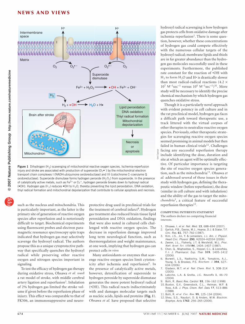
Many antioxidants or enzymes that scav-enge reactive oxygen species limit cytotox-icity after ischemia and reperfusion9. In the presence of catalytically active metals, however, detoxification of superoxide to hydrogen peroxide by superoxide dismutase generates the more potent hydroxyl radical (•OH). This radical reacts indiscriminately with and damages molecular targets such as nucleic acids, lipids and proteins (Fig. 1). Ohsawa et al. have proposed that selective
Intermembranespace
Superoxidedismutase
Lipid peroxidationDNA oxidation
Thiyl radical formationMitochondrialdepolarization
Necrosisor
apoptosis
Matrix
O2 O2• –
• OH + Fe3+ + OH–
H2 + • OH H2O + H•
Fe2+ + H2O2
O2O2• –
H2
H+ H+
I III
Mitochondrion
Brain after stroke
Figure 1 Dihydrogen (H2) scavenging of mitochondrial reactive oxygen species. Ischemia-reperfusion injury and stroke are associated with production of superoxide (O2•–) by the mitochondrial electron transport chain complexes I (NADH:ubiquinone oxidoreductase) and III (cytochrome C–coenzyme Q oxidoreductase). Superoxide dismutase forms hydrogen peroxide (H2O2) from superoxide. In the presence of catalytically active metals, such as Fe2+ or Cu+, hydrogen peroxide breaks down to hydroxyl radical (•OH). Hydrogen gas (H2) reduces •OH to H2O, thereby preventing the lipid peroxidation, DNA oxidation, thiyl radical formation and mitochondrial depolarization that contribute to cellular apoptosis and necrosis.
Kim
Cae
sar
hydroxyl radical scavenging is how hydrogen gas protects cells from oxidative damage after ischemia-reperfusion1. There is some ques-tion, however, whether these concentrations of hydrogen gas could compete effectively with the numerous cellular targets of the hydroxyl radical; membrane lipids and thiols are in far greater abundance than the hydro-gen gas molecules successfully used in these experiments. Furthermore, the published rate constant for the reaction of •OH with H2 to form H2O and H• is drastically slower than most radical-radical reactions (4.2 ×10⁷ M–1sec–1 versus 109 M–1sec–1)10. More study will be necessary to identify the precise chemical mechanism by which hydrogen gas quenches oxidative stress.
Though it is a particularly novel approach with evident potency in cell culture and in the rat preclinical model, hydrogen gas faces a difficult path toward therapeutic use, a track littered with the virtual corpses of other therapies to neutralize reactive oxygen species. Previously, other therapeutic strate-gies for scavenging reactive oxygen species seemed promising in animal models but then failed in human clinical trials11. Challenges facing any successful reperfusion therapy include identifying the dose, duration and site at which an agent will be optimally effec-tive. Of particular importance is targeting the site of reactive oxygen species genera-tion, such as the mitochondria11. Ohsawa et al. addressed several of these issues in their study with hydrogen gas, defining the thera-peutic window (before reperfusion), the dose (similar in cell culture and with inhalation) and the ability of the gas to target the mito-chondria1, a critical feature of successful reperfusion therapies12.
COMPETING INTERESTS STATEMENTThe authors declare no competing financial interests.
1. Ohsawa, I. et al. Nat. Med. 13, 688–694 (2007). 2. Garlick, P.B., Davies, M.J., Hearse, D.J. & Slater, T.F.
Circ. Res. 61, 757–760 (1987).3. Kim, J.S., Jin, Y. & Lemasters, J.J. Am. J. Physiol.
Heart Circ. Physiol. 290, H2024–H2034 (2006).4. Zweier, J.L., Flaherty, J.T. & Weisfeldt, M.L. Proc.
Natl. Acad. Sci. USA 84, 1404–1407 (1987).5. Chen, Q., Moghaddas, S., Hoppel, C.L. & Lesnefsky,
E.J. J. Pharmacol. Exp. Ther. 319, 1405–1412 (2006).
6. Burwell, L.S., Nadtochiy, S.M., Tompkins, A.J., Young, S. & Brookes, P.S. Biochem. J. 394, 627–634 (2006).
7. Gladwin, M.T. et al. Nat. Chem. Biol. 1, 308–314 (2005).
8. Labiche, L.A. & Grotta, J.C. NeuroRx 1, 46–70 (2004).
9. Bolli, R. Basic Res. Cardiol. 93, 156–162 (1998).10. Buxton, G.V., Greenstock, C.L., Helman, W.P. &
Ross, A.B. J. Phys. Chem. Ref. Data 17, 513–886 (1988).
11. Bolli, R. et al. Circ. Res. 95, 125–134 (2004).12. Sheu, S.S., Nauduri, D. & Anders, M.W. Biochim.
Biophys. Acta 1762, 256–265 (2006).
©20
07 N
atur
e P
ublis
hing
Gro
up
http
://w
ww
.nat
ure.
com
/nat
urem
edic
ine