Embed Size (px)
Citation preview

1
The impact of mutations near
the AUG codon of precore region on hepatitis B e antigen production
Ahn, Sang Hoon
Department of Medical Science
The Graduate School, Yonsei University

2
The impact of mutations near the AUG codon of precore region on
hepatitis B e antigen production
Directed By Professor Kim, Kyung Hwan
The Doctoral Dissertation submitted to
the Department of Medical Science,
the Graduate School, Yonsei University
in partial fulfillment of the requirements for the
degree of Doctor of Philosophy of Medical Science
Ahn, Sang Hoon
June, 2003

3
This certifies that the Doctoral Dissertation of
‘Ahn, Sang Hoon’ is approved.
(Supervisory committee, Chairman)
(Supervisory committee)
(Supervisory committee)
(Supervisory committee)
(Supervisory committee)
The Graduate School of Yonsei University
June, 2003

4
Acknowledgements
I am pleased to acknowledge a number of people whose
good work and kindness have enabled me to complete this
thesis. First of all, I have to express my sincere appreciation
to Prof. Kyung Hwan Kim, thesis director, who encouraged
and supported me to write this thesis successfully. He helped
me with patience, confidence, generosity, critical advice, and
personal relationship, so that I would never forget his grace.
I would like to thank specially to readers as supervisory
committee of this thesis, Prof. Young Myoung Moon and
Kwang-Hyub Han, who read my thesis proposal and offered
important suggestions and advices. I also want to express my
deep gratitude to Prof. Hyeyoung Kim and Min Goo Lee,
who gave me kind comments and review.
I am grateful to Shuping Tong, who always helped me out
of troubles in performing experiment in the laboratory and to
Anna Kramvis for clinical samples and data.
I am indebted to my wife. If there were no support of her

5
encouragement, care and endless love, it would have been
impossible that this thesis have come to light.
I am dedicating this thesis to my parents, younger brother
and his wife who fully understood and supported me with
patience and generosity.
Ahn, Sang Hoon
6
Table of contents
Abstract .................................................................................. 1
I. Introduction........................................................................ 4
II. Materials and Methods .................................................... 10
1. Subjects ........................................................................ 10
2. Sequencing ................................................................... 10
3. The parental HBV clones and site-directed mutants for
transfection................................................................... 11
(1) Serum samples and DNA extraction .......................12
(2) PCR and cloning......................................................12
(3) Site-directed mutagenesis....................................... 13
(4) Tandem dimer construction .................................... 14
4. Transfection and analysis of viral replication/gene
expression..................................................................... 15
(1) Transfection ............................................................ 15
(2) Isolation of core-particle–related HBV DNA ........ 15
(3) Southern blot hybridization of HBV DNA............. 16
(4) HBsAg and HBeAg assay ...................................... 17
5. Core protein mediated trans-complementation of HBV
replication..................................................................... 17
7
6. In vitro transcription/translation................................... 19
7. Statistical Analysis ....................................................... 19
III. Results ............................................................................ 20
1. The SA mutations are present throughout the course of
infection 20
2. The SA mutations reduced HBeAg production in the
context of replicating HBV genome ............................ 23
3. The SA mutation reduced HBeAg expression to a
similar extent as the core promoter mutations ............. 29
4. The reduction in HBeAg expression by the SA
mutations was independent of HBx function............... 29
5. The SA mutations produce leaky scanning of the
precore AUG codon ..................................................... 32
6. The replication defect of a core-minus HBV genome
can be rescued by precore mRNA containing the triple
SA mutations ................................................................ 35
IV. Discussion ............................................................37
V. Conclusions ...........................................................43
References .................................................................45
Abstract (in Korean) ....................................................55
8
List of figures
Figure 1. HBeAg biosynthesis and South African variations
near the precore AUG codon ................................... 9
Figure 2. Results of one transfection experiment with 4B-
based constructs in Huh7 cells ................................. 25
Figure 3. Summary of the effects of SA mutations on
HBeAg expression from different genetic
backgrounds (2A vs 4B) and two hepatoma cell lines
(Huh7 vs HepG2) ................................................... 27
Figure 4. Comparison of SA double mutations with the
1762T/1764A core promoter mutations (mu1) on
HBeAg expression ................................................. 30
Figure 5. Effect of change in HBx function on HBsAg and
HBeAg expression in transfected HepG2 cells...... 31
Figure 6. Molecular evidence for a leaky scanning
mechanism with respect to translation from the
precore mRNA of MT1 and MT3 variants ............ 34
Figure 7. Functional evidence for a leaky scanning
mechanism with respect to translation from the
precore mRNA of MT1 and MT3 variants ............ 36

9
List of tables
Table 1. Prevalence of 1809-1812 mutations in black South
Africans .................................................................. 22

10
Abstract
The impact of mutations near the AUG codon of
precore region on hepatitis B e antigen production
Ahn, Sang Hoon
Department of Medical Science
The Graduate School, Yonsei University
<Directed by Professor Kim, Kyung Hwan>
Black South African (SA) carriers of hepatitis B virus (HBV)
seroconvert from hepatitis B e antigen (HBeAg) to antibody against
HBeAg(anti-HBe) much earlier than do Asian patients. Since
majority of South African HBV strains harbor double or triple
nucleotide changes immediately upstream of the precore AUG,
the translational initiation codon for HBeAg, their impact on
HBeAg expression was investigated by site-directed
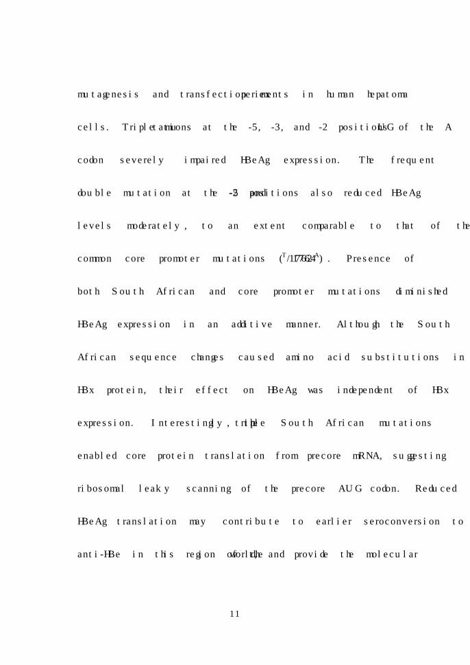
11
mutagenesis and transfection experiments in human hepatoma
cells. Triple mutations at the -5, -3, and -2 positions of the AUG
codon severely impaired HBeAg expression. The frequent
double mutation at the -5 and -2 positions also reduced HBeAg
levels moderately, to an extent comparable to that of the
common core promoter mutations (1762T/1764A). Presence of
both South African and core promoter mutations diminished
HBeAg expression in an additive manner. Although the South
African sequence changes caused amino acid substitutions in
HBx protein, their effect on HBeAg was independent of HBx
expression. Interestingly, the triple South African mutations
enabled core protein translation from precore mRNA, suggesting
ribosomal leaky scanning of the precore AUG codon. Reduced
HBeAg translation may contribute to earlier seroconversion to
anti-HBe in this region of the world, and provide the molecular
12
basis for a previously unexplained clinical event associated with
viral clearance.
Key Words: hepatitis B virus, hepatitis B e antigen, South African,
precore/core promoter mutation, AUG codon, ribosomal leaky
scanning, seroconversion.
13
The impact of mutations near the AUG codon of
precore region on hepatitis B e antigen production
<Directed by Professor Kim, Kyung Hwan>
Ahn, Sang Hoon
Department of Medical Science
The Graduate School, Yonsei University
I. Introduction
Hepatitis B virus (HBV) is the prototype member of
hepadnaviridae, a group of hepatotropic enveloped DNA viruses.
The HBV genome consists of a partially double-stranded 3.2kb
DNA molecule arranged in a relaxed circular conformation. It
contains four overlapping reading frames for the polymerase (P
gene), core protein and HBeAg (pre-C/C gene), large, middle,
and small surface proteins (pre-S/S gene), and the X protein (X
14
gene). These proteins are expressed from 3.5 kb pregenomic
RNA (core and polymerase proteins), 3.5 kb precore RNA
(Hepatitis B e antigen), 2.4 kb subgenomic RNA (large envelope
protein), 2.1 kb subgenomic RNA (middle and major envelope
proteins), and 0.7 kb subgenomic RNA (X protein).1
HBV infection may lead to a spectrum of liver diseases,
ranging from acute self-limited hepatitis, an asymptomatic
carrier state to chronic hepatitis progressing to liver cirrhosis
and hepatocelluar carcinoma. HBV related liver diseases are the
outcome of complex interplay between the virus and its host.
The ever-changing host environment selects for outgrowth of
different viral variants. During the last decade, with the advent
of polymerase chain reaction (PCR) technology, genetic
variability of the HBV genome has gained increasing recognition
and attention. Major HBV variants have been identified that
survive among the host hepatitis B e antibody (anti-HBe)
immunity [precore mutants unable to express hepatitis B e Ag
and core promoter mutants with reduced HBeAg expression],
HBV vaccine (vaccine escape mutants), and Lamivudine

15
treatment (YMDD motif mutants). However, with a few
exceptions, the functional consequences of these prevalent and
hence important mutations remain poorly characterized.
Hepatitis B e antigen (HBeAg) was discovered three
decades ago2 and corresponds to an alternative translational
product of the core gene (Figure 1A). It is translated from the
AUG initiation codon upstream of the core gene on the precore
messenger RNA (mRNA), generating a product with an additional
29 amino acid residues on the amino terminus. This 25 k daltons
precore/core fusion protein (p25) is post-translationally modified
by signal peptidase and basic endopeptidase during the secretory
pathway to generate a final circulating 15-17 k daltons HBeAg.
HBeAg may promote immune tolerance during perinatal infection
and alter host immune response against core protein.3 The anti-
HBe immune response plays a critical role in the clearance of
HBV during natural infection. Thus, seroconversion from HBeAg
to anti-HBe is usually associated with marked drop in viremia
and normalization of aminotransferase levels. Continued
expression of HBeAg at this stage of infection may be

16
detrimental to viral survival.
Two types of HBeAg variants, with reduction and
termination of HBeAg expression, emerge sequentially at the
late stages of HBV infection. Reduction in HBeAg expression by
the core promoter mutants is a result of declined precore mRNA
transcription,4-7 while abolition of HBeAg expression in the
precore mutants is achieved by nonsense or frameshift mutations
in the precore region.8-10 Both types of HBeAg variants are
adaptive variants selected by the host anti-HBe immune
pressure.
A unique feature of South African (SA) black carriers of
HBV is that HBeAg expression is lost very early during the
course of infection; only 5% are HBeAg positive in adulthood
compared to a rate of 40% or higher found in other
hyperendemic areas of the world such as Southeast Asia. The
clinical impact of rapid HBeAg seroconversion on disease
progression is still unclear. Recently, partial sequences of HBV
genomes isolated from Black South African patients were
17
reported. Of particular interest was the finding that 80% of SA
HBV strains harbor double or triple point mutations at nucleotide
1809, 1811, and 1812 of the HBV genome, the -5, -3, and -2
positions of the precore translation initiation codon,11 which
might impair HBeAg expression as a result of sub-optimal
translation initiation.12-14 This hypothesis was formally tested in
the present study by site-directed mutagenesis and transfection
experiments. If proven this, this finding may solve the mystery
of accelerated seroconversion as found in South African patients.
It may also reveal third mechanism whereby the virus can down-
regulate HBeAg expression.
18
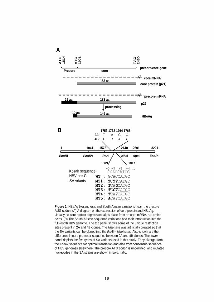
A
B
precore/core gene
precore mRNA
core mRNA
Precore core
AT
G18
14
TA
G24
50
AT
G19
01183 aa
149 aa
183 aa
processing10 aa
29 aa
HBeAg
p25
core protein (p21)
EcoRI
1 26011572 2140 3221
RsrII NheI ApaI EcoRI
Kozak sequenceHBV pre-CSA vriants
CCACCATGGWT : GCACCATGCMT1: TCTTCATGCMT2: TCAGCATGCMT3: TCCTCATGCMT4: TCATCATGCMT5: ACATCATGC
+1 +4 nt-3-5
1041
EcoRV
1809 1817
1753 1762 1764 17662A: T A G C 4B: C T A T
Figure 1. HBeAg biosynthesis and South African variations near the precoreAUG codon. (A) A diagram on the expression of core protein and HBeAg. Usually no core protein expression takes place from precore mRNA. aa: amino acids. (B) The South African sequence variations and their introduction into the full-length HBV genome. The top panel shows some of the unique restriction sites present in 2A and 4B clones. The NheI site was artificially created so that the SA variants can be cloned into the RsrII – NheI sites. Also shown are the difference in core promoter sequence between 2A and 4B clones. The lower panel depicts the five types of SA variants used in this study. They diverge from the Kozak sequence for optimal translation and also from consensus sequence of HBV genomes elsewhere. The precore ATG codon is underlined, and mutated nucleotides in the SA strains are shown in bold, italic.
19
II. Materials and Methods
1. Subjects
Blood was obtained, with informed consent, from 45 black
Africans. Eighteen were HBsAg positive asymptomatic carriers
with normal serum alanine aminotransferase levels. The HBeAg
status of these patients is summarized in Table 1. Five patients
suffered from acute hepatitis. Alanine aminotransferase levels in
these patients ranged between 799 IU/L and 4747 IU/L. One
patient with fulminant hepatitis was a female aged 32 years with
an alanine aminotransferase level of 979 IU/L. In addition 21
children were studied. Fifteen of the children with biopsy proven
HBV membranous nephropathy were index cases in a previous
study15 and 6 were family members of these index cases. All had
normal alanine aminotransferase levels.
2. Sequencing
DNA was extracted from the serum samples using the
QIAamp DNA Mini Kit (Qiagen, Valencia, CA, USA) according to
the protocol provided by the manufacturer. In order to sequence
20
the region upstream of the precore translational start codon the
region was amplified by nested PCR using primers 1687(+) and
2498R(-) in the first round and 1730(+) and 2043R(-) in the
second round.16 Sequencing of PCR products was performed
manually using the Sequenase PCR product sequencing kit
(Roche Diagonstics Co. Indianapolis, IN, USA) or automatically
using Big DyeTM Terminator v3.0 Cycle sequencing kit (Applied
Biosystems, Foster city, CA, USA) and the 377 DNA sequencer
(Applied Biosystems, Foster city, CA, USA).
3. The parental HBV clones and site-directed mutants for
transfection
Because the majority of SA isolates of HBV belong to
genotype A,11,17,18 we used two well-characterized HBV genomes
of this genotype to create the SA mutations (Figure 1B). Clone
2A had wild-type core promoter sequence, thus mimicking viral
genomes in the early HBeAg-positive phase of infection. Clone
4B was a high replicating core promoter mutant representing
viral genomes in the late stage of infection. Use of both HBV
constructs allowed us to examine the effect of the SA mutations

21
alone and in combination with core promoter mutations, which
occur at the late stage of infection.11
(1). Serum samples and DNA extraction
Serum samples are collected from HBeAg positive chronic
carriers infected with same genotype A. The serum samples are
diluted in TEN buffer and digested at 37oC for 2 hrs with
Proteinase K (0.5 mg/ml) in the presence of SDS (0.5%). DNA is
extracted with phenol/ chloroform/ isoamyl alcohol (25:24:1) and
precipitated with ethanol. Purified DNA is resuspended in water.
(2). PCR and cloning
Full-length HBV genomes are PCR amplified according to
the method of Gunther et al19 and cloned into pUC18 Vector. To
facilitate the cloning of PCR products, both primer sequences are
modified at the 5’ end such that the sense primer contained just
HindIII site (5' - CCG GAA AGC TTA TGC TCT TCT TTT TCA
CCT CTG CCT AAT CAT C - 3', underlined) while the antisense
primer contained SacI site only (5' - CCG GAG AGC TCA TGC
TCT TCA AAA AGT TGC ATG GTG CTG GTG - 3', underlined).
22
Forty cycles of amplification are performed with the Expand high
fidelity PCR system (Roche Diagonstics Co. Indianapolis, IN,
USA) using conditions specified by Gunther et al.19 The PCR
products are cloned into the HindIII/SacI sites of pUC18 Vector.
(3). Site-directed mutagenesis
Point mutations were generated by the PCR-based
overlap extension method,20 using the high fidelity PCR system
(Roche Diagonstics Co. Indianapolis, IN, USA) and 25 cycles of
amplification for each PCR reaction. To further reduce the
possibility of unwanted PCR errors, an artificial NheI site was
introduced into position 2140 of both 2A and 4B genomes. The
NheI mutations did not alter core protein sequence, and the
corresponding dimers did not differ from the original 2A or 4B
clones in terms of viral replication or gene expression (data not
shown). The PCR products containing the SA mutations were
digested with the RsrII/NheI restriction enzymes to replace the
cognate fragment (position 1572 - 2140) in wild-type 2A and 4B
clones. The entire PCR derived region was subsequently
sequenced to confirm the presence of desired mutations and lack
23
of unwanted ones. For comparison of HBeAg expression with the
common core promoter mutants, a 2A-based construct with
1762T1764A core promoter mutations (mu1) was employed. To
evaluate the impact of hepatitis B virus X protein (HBX) function
on HBeAg expression, a C to G change was introduced into
position 1706 of the 2A constructs with and without the SA
mutations, creating a TAG stop codon in the HBX gene.
(4). Tandem dimer construction
For HBV replication and gene expression to proceed
under the control of endogenous core promoter, each construct
was converted into a tandem dimer. To make head-to-tail
tandem dimers, the HBV DNA insert will be released from
pUC18 vector by SapI/BglI double digestion and circulized with
T4 DNA ligase. Sequently, HBV DNA is linearized with EcoRI
and ligated with EcoRI digested, dephosphorylated pUC18 DNA
at insert: vector ratio of 15~20:1. Tandem HBV dimer screened
with an oligonucleotide probe spanning the tail-to-head junction
of HBV dimer as previously described.21 The 30-mer
oligonucleotide covered the EcoR I site of HBV genome and had
24
the sequence 5' – GGC CAT GCA GTG GAA TTC CAC WRC
YTT CCA - 3' (W=A+T; R=A+G, Y=C+T).
4. Transfection and analysis of viral replication/gene
expression
(1). Transfecton
The dimeric HBV DNA constructs were transfected into
Huh7 and HepG2 human hepatoma cells using a calcium
phosphate transfection kit (Promega, Madison, WI, USA), using
3.6 μg or 5.4 μg DNA per 6-cm dish. Cells were harvested and
culture supernatant was collected at day 5 posttransfection. In
order to reduce experimental variability, all the constructs were
tested at least five times.
(2). Isolation of core particle– related HBV DNA
HBV replication was monitored by Southern blot analysis
of HBV DNA associated with intracellular core particles.22 HBV
core particles will be obtatined from an aliquot of transfected
cells basically as described.22 Cells are lysed with 400µl of
buffer containing 10 mM Hepes, pH 7.5, 100 mM NaCl, 1 mM
25
EDTA, and 1% NP 40. The lysate is supplemented with 10 mM
CaCl2, 12 mM MgCl2, and digested at 37oC for 15 min with DNase
I (2 u) and mung bean nuclease (30 u) to degrade transfected
HBV DNA. Core particles are precipitated with 150µl of 26%
PEG solution (1.2 M NaCl, 60 mM EDTA, 30% sucrose, 26%
PEG), resuspended in 100µl of solution containing 10 mM Tris,
pH 7.5, 6 mM MgCl2, and 8 mM CaCl2, and digested with 2 u of
DNase I and 3 u of mung bean nuclease at 37oC for 10 min to
further remove transfected DNA. After addition of 270µl of
protease digestion buffer (25 mM Tris, pH 7.5, 10 mM EDTA,
100 mM NaCl, 0.5% SDS), samples are digested at 37oC for 1 to
2 hr with proteinase K (0.5 mg/ml). DNA is extracted with phenol
and precipitated with ethanol. Purified DNA is subjected to
Southern blot analysis with a highly pure full-length HBV probe
(obtained by two rounds of PCR amplication).
(3). Southern blot hybridization of HBV DNA
HBV DNA was resolved in 1.0% agarose gels, transferred
to nylon membranes (Hybond N+; Amersham Pharmacia Biotech,
Little Chalfont, United Kingdom) by Southern blotting, and

26
hybridized with an alkaline phosphatase– labeled wild-type full-
length HBV DNA probe obtained by two rounds of PCR
amplification. Gel electrophoresis was either performed in the
absence of ethidium bromide or in its presence both in the gel
and in the running buffer, which greatly accelerated migration of
the single stranded HBV genome.
(4). HBsAg and HBeAg assay
The HBsAg secreted to culture supernatant is measured
by an enzyme immunoassay (Auszyme kit; Abbott Laboratories,
Abbott Park, IL, USA) using the protocol provided by the
manufacturer. The samples were diluted 3 or more times with
phosphate-buffered saline to avoid signal saturation. For those
samples with OD at 492nm greater than 3, a two fold serial
dilution is performed until the OD value fell within the readable
range. Secreted HBeAg is detected by radioimmunoassay (EIA
kit; DioSorin, Stillwater, MN, USA). The monoclonal antibody for
HBeAg detection did not cross-react with Hepatitis B core
antigen (HBcAg).

27
5. Core protein mediated trans-complementation of HBV
replication
A replication deficient mutant of clone 4B was generated
by a C to G change at position 2044, creating a TGA nonsense
mutation in the core gene. As the potential source of the core
protein, the entire precore/core gene of wild-type 4B and SA
mutants (MT1, MT3) was amplified by PCR, using sense primer
5'- GGA GGC TCG AGG CAT AAA TTG GTC TGC GCA CC -3'
(XhoI site underlined), and antisense primer 5'- AAA GCG AAT
TCA AGT TTC CCA CCT TAT GAG TCC -3' (EcoRI site
underlined). The PCR products were cloned into the XhoI/EcoRI
sites of the pcDNA3.1 Zeo(-) vector, which contains a
cytomegalovirus promoter to drive eukaryotic gene expression.
When transcribed, the 5’ end HBV sequence in the mRNA would
correspond to the 5’ end of the authentic precore mRNA. As a
positive control of core protein expression, the core gene of
wild-type 4B genome was amplified using the same antisense
primer but a different sense primer 5'- AGC ACC TCG AGA CTT
TTT CAC CTC TGC CTA ATC ATC -3' (XhoI site underlined).
The precore and core constructs (3.6 μg DNA) were separately

28
co-transfected with the tandem dimer of core-minus 4B genome
(3.6 μg DNA). Viral genome replication was analyzed from
lysates of cells harvested at day 5 after transfection.
6. In vitro transcription / translation
The above-mentioned PCR primers were used to amplify
the entire precore/core gene or the core region alone, using 2A-
based constructs as templates. The PCR products were cloned
into the XhoI/EcoRI sites of pBluescript SK vector. Coupled
transcription/translation was performed at 30oC using a
commercial kit (Promega, Madison, WI, USA) with rabbit
reticulocyte lysates, [35S]-methionine, and T7 polymerase. An
aliquot of translation product was electrophoresed through a
12% polyacrylamide gel, and treated with enlightening solution
(PerkinElmer, Boston, Massachusetts). Dried gels were exposed
to X-ray films at – 80oC.
7. Statistical Analysis
The difference of the HBeAg/HBsAg levels of the wild-
type and SA mutants was determined by Student′s t-test. A value
of p < 0.05 was taken to be statistically significant. All of
29
statistical analysis was performed using Window-SPSS release
10.0 for personal computer.
III. Results
1. The SA mutations are present throughout the course
of infection
Many individuals described in a previous study had
seroconverted to anti-HBe and contained core promoter
mutations. A subset of these patients had developed
hepatocellular carcinoma.11 Thus, the possibility that sequence
changes at 1809-1812 of the HBV genome represent adaptive
change under immune pressure, similar to that observed with
core promoter and precore mutations, could not be excluded. For
this reason, HBV isolates from 6 acute hepatitis patients (one
with fulminant hepatitis), 21 HBV infected children, and 18
asymptomatic carriers, most at the HBeAg-positive phase of
infection, were sequenced. As shown in Table 1, all 6 acute HBV
patients contained HBV with the 1809T1812T double mutation. In
addition, 90% (19/21) of the isolates from children were found to
30
harbor the 1809T1812T double mutation. This prevalence is
similar to that reported in our previous study.11 Among the 18
asymptomatic carriers, 11 were infected with the SA subtype
A’ of genotype A,17 2 with conventional genotype A, and 5 with
genotype D. All the genotype D and genotype A as well as 3 of
the genotype A’ isolates had wild-type sequence preceding the
precore AUG codon. For the remaining genotype A’ isolates, 5
contained the 1809T1812T double mutation and 3 contained the
1809T1812G double mutation. These latter 3 individuals, but none
of the other 15 asymptomatic carriers, harbored 1762T1764A
core promoter mutations. Thus, the 1809-1812 mutations are
highly prevalent in HBV genotype A’ infected South Africans
with acute infection, during childhood, and at the HBeAg-
positive phase of infection. These findings suggest the 1809-
1812 mutations as stable traits of genotype A’ isolates in South
Africa, rather than adaptive mutations under anti-HBe immune
pressure.

31
Table 1. Prevalence of 1809-1812 mutations in black South
Africans.
1 Some samples not available for HBeAg assay
2 One case with fulminant hepatitis
3 Fifteen with membranous nephropathy
4 Five infected with genotype D, 2 with genotype A, and 3 with A’
5 All contained 1762T1764A core promoter mutations
1809-1812
sequence
No. of
patients
Age
range
M / FHBeAg1
+ / -
WT 0 - - Acute
hepatitis2 1809T1812T 6
18 – 32
3 / 3 3 / 0
WT 2 2 / 0 1 / 1
Children3
1809T1812T 19
4 – 16
17 / 2 14 / 5
WT 104
10 / 0
6 / 0
1809T1812T 5 5 / 0 3 / 1
Asymptomatic
carriers
1809T1812G 35
20 - 40
3 / 0 2 / 0

32
2. The SA mutations reduced HBeAg production in the
context of replicating HBV genome
Five types of naturally occurring SA mutations identified in
a previous study11 were introduced into two HBV genomes of the
same genotype (2A and 4B; Figure 1B). Of these, MT1
(1809T1811T1812T) and MT3 (1809T1811C1812T) contained triple
mutations at the -5, -3, and -2 positions relative to the
adenosine of the precore AUG codon, while MT2 (1809T1812G),
MT4 (1809T1812T), and MT5 (1809A1812T) had double mutations
at the -5 and -2 positions. An HBV clone (construct 5.4)
defective in genome replication and HBeAg expression owing to
a single nucleotide deletion in the core gene was included as a
negative control. Following transfection of the tandem dimers of
these constructs into Huh7 and HepG2 human hepatoma cells,
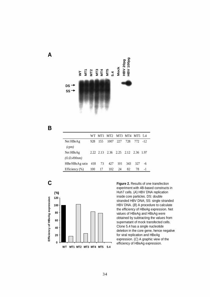
HBeAg and HBsAg expression was analyzed. Figure 2 shows
results of one typical experiment of 4B-based constructs in
Huh7 cells. The various constructs did not differ significantly in
the levels of intracellular core particle-associated DNA, or
secreted HBsAg (panels A and B). These results suggest similar
transfection efficiencies of the constructs because the mutations

33
preceding the precore translation initiation codon are not
expected to modulate HBsAg expression.
HBeAg expression was greatly reduced by the MT1 and
MT3 mutations, and also appears diminished by the MT4 and
MT5, but not MT2 mutations (Figure 2B). To precisely evaluate
the effects of different SA mutations and to minimize the
contribution of variations in transfection efficiencies, we used
the ratio of net HBeAg / net HBsAg values as an indicator of the
efficiency of HBeAg expression (the net value is equal to the
value of the sample minus the value obtained from mock
transfected cells). Assuming a 100% efficiency of HBeAg
expression by the parental 4B clone, the efficiencies for the
MT1, MT2, MT3, MT4, and MT5 mutants were 17%, 102%, 24%,
82%, and 78% in this particular experiment (Figure 2B and 2C).
34
WT
MT1
MT4
MT5
5.4
MT2
MT3
Moc
k
HB
V 2
0pg
HB
V 1
00pg
DSSS
A
B
(%)
0
20
40
60
80
100
120
WT MT1 MT2 MT3 MT4 MT5 5.4
Eff
icie
ncy
of H
BeA
gex
pres
sion
WT MT1 MT2 MT3 MT4 MT5 5.4Net HBeAg 928 155 1007 227 728 772 -12(cpm)
Net HBsAg 2.22 2.13 2.36 2.25 2.12 2.36 1.97 (O.D.490nm)HBe/HBsAg ratio 418 73 427 101 343 327 -6Efficiency (%) 100 17 102 24 82 78 -1
C Figure 2. Results of one transfectionexperiment with 4B-based constructs in Huh7 cells. (A) HBV DNA replication inside core particles. DS: double stranded HBV DNA; SS: single stranded HBV DNA. (B) A procedure to calculate the efficiency of HBeAg expression. Net values of HBeAg and HBsAg were obtained by subtracting the values from supernatant of mock transfected cells. Clone 5.4 has a single nucleotide deletion in the core gene, hence negative for viral replication and HBeAgexpression. (C) A graphic view of the efficiency of HBeAg expression.
35
Figure 3A summarizes results of transfection experiments
using 2A-based constructs in HepG2 cells. The MT1 and MT3
triple mutations suppressed HBeAg expression to 8% and 28% of
the wild-type levels, respectively, while the MT4 and MT5
double mutations reduced HBeAg expression to 74% and 72% of
the wild-type levels, respectively. Similar results were obtained
in Huh7 cells (Figure 3C and D). Transfection of Huh7 cells with
4B-based constructs resulted in a similar trend in the reduction
of HBeAg levels except that the HBeAg levels were much lower
relative to those produced by 2A-based constructs (Figure 3C).
The 1753C/1762T/1764A/1766T quadruple core promoter
mutation in clone 4B is known to greatly reduce HBeAg
expression (data not shown). These results are compatible with
the concept that the core promoter mutations and the 1809-1812
mutations affect HBeAg biosynthesis at different levels, and
hence their effects may be additive.

36
B
4B constructs, HepG2 cells
0
20
40
60
80
100
120
140(%)
MT1 MT2 MT3WT MT4 MT5
**
**E
ffic
ien
cy o
f H
BeA
gex
pre
ssio
n
A
2A constructs, HepG2 cells
0
20
40
60
80
100
120
140
MT1 MT2 MT3WT MT4 MT5
(%)
*
*
*
*
Eff
icie
ncy
of
HB
eAg
exp
ress
ion
C D
0
20
40
60
80
100
120
MT1 MT2 MT3WT MT4 MT5
4B constructs, Huh7 cells
(%)
Eff
icie
ncy
of
HB
eAg
exp
ress
ion
* *
Eff
icie
ncy
of
HB
eAg
exp
ress
ion
0
20
40
60
80
100
120
140
2A constructs, Huh7 cells
MT1 MT2 MT3WT MT4 MT5
(%)
*
*
††

37
E
0200400600800
100012001400160018002000
MT1 MT2 MT3WT MT4 MT5 MT1 MT2 MT3WT MT4 MT5
2A 4B
Huh7 cells
HB
eAg
/ HB
sAg
ratio
Figure 3. Summary of the effects of SA mutations on HBeAg expression from different genetic backgrounds (2A vs. 4B) and two hepatoma cell lines (Huh7 vs. HepG2). The results are shown in HBeAg / HBsAg ratios for panel E, and as percentiles of the wild-type genome for panels A to D. Statistical differences between the wild-type and SA mutations are shown. * : p <.001, † : p <.05 ( vs. the wild-type clone 2A or 4B)
* **
*
††

38
3. The SA mutation reduced HBeAg expression to a
similar extent as the core promoter mutations
Of the five types of SA mutations tested in this study, the
MT4 (1809T1812T) double mutation was the most common,
accounting for more than 80% of the isolates found in South
Africa11 (Table 1). The effect of this double mutation on HBeAg
expression was compared with that of the most common core
promoter mutations (1762T/1764A, construct mu1). The MT4
mutant suppressed HBeAg expression to 67% of the wild-type
level, while mu1 produced 70% of the wild-type level (Figure 4).
Thus, the effect of the 1809T1812T mutation was similar to that
of the core promoter mutations.
4. The reduction in HBeAg expression by the SA
mutations was independent of HBx function
Because the HBX gene overlaps with the core promoter
and 5’ precore region, mutations around precore AUG codon
could modify C-terminal sequence of HBx. Indeed, all five SA
mutations studied here induced double amino acid substitutions
in the HBx (Figure 5C, inset). To establish whether sequence

39
0
20
40
60
80
100
120
WT MT2 MT4 MT5 mu1
(%)
Eff
icie
ncy
of H
BeA
gex
pres
sion
Figure 4. Comparison of SA double mutations with the 1762T/1764A core promoter mutations (mu1) on HBeAgexpression. All the constructs shown here are 2A-based, and experiments were performed in Huh7 cells. The relative HBeAg expression efficiencies were 92%, 67%, 68%, and 70% for MT2, MT4, MT5, and mu1, respectively.

40
WT MT1 MT3 WT MT1 MT3
X+ X-
0100200300400500600700800900
1000
Net
HB
eAg
leve
ls (c
pm)
B
WT MT1 MT3 WT MT1 MT3
X+ X-
00.20.40.60.81.01.21.41.6
Net
HB
sAg
leve
ls (O
.D.4
90n
m)
A
(%)
0
20
40
60
80
100
120
WT MT1 MT3 WT MT1 MT3
X+ X-
C
Eff
icie
ncy
of H
BeA
gex
pres
sion
The X proteinCodon 146 147WT : Ala ProMT1 : Ser SerMT2 : Ser AlaMT3 : Ser SerMT4 : Ser SerMT5 : Thr Ser
Figure 5. Effect of changes in HBx function on HBsAg and HBeAg expression in transfected HepG2 cells. HBX-intact (X+) and HBX-defective (X-) versions of WT, MT1, and MT3 constructs in 2A background were analyzed. (A) HBsAg levels. (B) HBeAglevels. (C) Efficiency of HBeAg expression. Amino acid changes in HBx are shown in the inset of panel C. Inactivation of HBX function reduced both HBsAg and HBeAgexpression for all the three constructs, but the MT1 and MT3 constructs continued to express much less HBeAg than their WT counterpart. DS: double stranded HBV DNA; SS: single stranded HBV DNA.
41
changes of HBx were responsible for the reduction in HBeAg
expression, we abolished HBx expression by a nonsense
mutation upstream of the SA mutations. Only the MT1 and MT3
mutants were studied, as they displayed the greatest reduction in
HBeAg expression. Experiments in Huh7 cells did not reveal any
effect of HBX gene inactivation on HBsAg or HBeAg expression
(data not shown). In HepG2 cells HBX-minus mutants expressed
roughly half the amount of HBsAg and HBeAg as compared with
their HBX-competent counterparts (Figure 5A and 5B). However,
HBx-minus MT1 and MT3 constructs still produced far less
HBeAg than the HBx-minus WT clone (Figure 5C), suggesting
that amino acid changes in HBx were not responsible for the
inhibition of HBeAg expression.
5. The SA mutations produce leaky scanning of the
precore AUG codon
According to the scanning model, translation of most eukaryotic
mRNAs is initiated by binding of the 40S ribosomes to the 5’ cap
structure, followed by scanning for the AUG with an optimal
surrounding sequence.23 Translation from this AUG codon will
prevent initiation from other downstream AUG codons. The

42
precore AUG has a favorable context for translational initiation
and thus only HBeAg, but no core protein, is expressed from the
intact precore mRNA.24-26 Because the critical -3 position of
precore AUG12,13 is mutated in the triple mutants from A to T
(MT1) or C (MT3) (Figure 1B), reduced translational initiation
was probably responsible for diminished HBeAg expression. In
such instances, continued scanning of ribosomes down towards
the core AUG codon may result in core protein expression. To
test this hypothesis, we cloned the entire precore/core sequence
of 2A, and 2A-based MT1 and MT3 constructs into the
pBluescript vector, and performed in vitro
transcription/translation experiments using T7 polymerase.
While the precore/core sequence of wild-type 2A produced only
p25 (primary translation product of the precore/core gene and
the precursor to HBeAg, Figure 1A), the MT1 and MT3
constructs produced a 21 k daltons protein (p21)
indistinguishable from core protein synthesized from core mRNA
(Figure 6). In addition, the amount of p25 expressed from the
MT1 and MT3 constructs was greatly reduced. These results
confirmed a leaky scanning mechanism of the precore AUG in

43
WT
MT
3
MT
1
WT
p25 p21
precore core
Figure 6. Molecular evidence for a leaky scanning mechanism with respect to translation from the precore mRNA of MT1 and MT3 variants. In vitro transcription and translation of the precore and core mRNA were performed in rabbit reticulocyte lysates. MT1 and MT3 produced less 25 k daltons protein (p25, HBeAg precursor) than the wild-type control and generated some 21 k daltons core protein (p21).
44
the triple SA mutants as the explanation for reduced HBeAg
expression.
6. The replication defect of a core-minus HBV genome
can be rescued by precore mRNA containing the triple SA
mutations
A genetic complementation experiment was designed to
confirm p21 expressed from the precore mRNA of MT1 and MT3
as the authentic core protein. During natural HBV infection the
pregenomic RNA serves both as mRNA for core and polymerase
expression, and as pregenome to be encapsidated into newly
formed core protein particles, where replication takes place.1
For transfection experiments in cell lines, it is possible to ablate
core or polymerase protein expression from pregenomic RNA via
a nonsense mutation. When a plasmid DNA capable of generating
such a pregenomic RNA is co-transfected with another plasmid
encoding the missing protein (core or polymerase), viral DNA
replication will be rescued.27 Introduction of a nonsense mutation
into the core gene of clone 4B abolished viral replication, which
could be rescued by co-transfection with the 4B core gene

45
DS
SS
precore core
CM
+MT1
CM
+MT3
CM
CM
+WT
CM
+WT
Moc
k
HB
V 2
0 p
g
HB
V 5
pg
WTd
imer
Figure 7. Functional evidence for a leaky scanning mechanism with respect to translation from the precore mRNA of MT1 and MT3 variants. Complementation of HBV replication by core protein provided in trans. The entire precore/core gene was amplified from 4B-based wild-type and MT1, MT3 constructs, and cloned into the pcDNA3.1 Zeo(-) vector. The core gene of 4B was also cloned into the same vector as a positive control. These constructs were co-transfected with the core-defective 4B dimer (CM). Viral replication was rescued by 4B core mRNA, and also by the precore mRNA harboring MT1 and MT3 mutations.
46
cloned into the pcDNA vector (Figure 7). Interestingly, co-
transfection with the entire precore/core sequence also rescued
the replication defect of the 4B dimer if the MT1 or MT3
mutations were present (Figure 7), thus confirming core protein
production from the precore mRNA of the triple SA mutants.
IV. Discussion
The sequence around the precore initiation codon is
conserved in all the eight known genotypes as 5’ -
AGCACCAUGC - 3’ (nt 1808 – 1817).28-31 This sequence
conforms to the optimal context for translational initiation, the
so-called Kozak sequence: 5’ - GCCA/GCCAUGG - 3’ .13, 23 In
contrast, many South African HBV strains, which belong to a
subgroup of genotype A,11,17,18 harbor point mutations
immediately upstream of the precore AUG codon.11 Such HBV
variants have also been found occasionally in other parts of the
world.32-34 The most prevalent South African variant contained
double nucleotide substitutions: 5’ - TCATCATGC - 3’
(mutated nucleotides in italics, MT4 in Figure 1B), but variants
47
with triple nucleotide substitutions have also been detected, such
as 5’ - TCTTCATGC - 3’ (MT1) and 5’ - TCCTCATGC - 3’
(MT3).11 Site-directed mutagenesis followed by transfection
experiments demonstrated several fold reduction in HBeAg
expression by the MT1 and MT3 mutants (Figure 3). The
common MT4 mutation and another double mutation (MT5)
displayed 20% or more reduction in HBeAg expression, while the
MT2 double mutation exhibited slightly enhanced HBeAg
expression. Taken together, these results confirm the critical
importance of an adenosine or guanine at the -3 position for
translation efficiency,13 and also suggest a modulating effect of
changes in the -2 position.
The effect of the South African mutations on HBeAg
expression was independent of that of the core promoter
mutations, but the two sets of mutations had an additive effect
(Figure 3E). Thus, while the core promoter mutations in clone 4B
drastically reduced HBeAg expression, addition of the South
African mutations further reduced HBeAg expression to a similar
extent observed in the 2A background. The clinical significance
48
of this finding is that development of core promoter mutations in
the SA variants, which does occur in this group of HBV strains,11
will further reduce HBeAg expression. All the South African
mutations caused double amino acid changes in the HBx (Figure
5), a viral transcriptional transactivator.36 However, the three
mutants with identical amino acid substitutions in HBx (MT1,
MT3, and MT4) produced quite different levels of HBeAg upon
transfection into either HepG2 or Huh7 cells (Figure 3).
Introduction of a stop codon into the HBx gene upstream of the
1809-1812 mutations did not have any effect on HBeAg or
HBsAg expression in Huh7 cells, which is consistent with the
observation that the HBX gene had a limited effect on HBV
replication in this particular cell line.37 On the other hand,
blocking HBx expression in HepG2 cells reduced both HBsAg
and HBeAg expression by about 50% (Figure 5A and 5B). This is
in agreement with a previous study that showed that HBx
expression is critical for HBV genome replication in HepG2
cells.38 Nevertheless, the degrees in the reduction of HBsAg and
HBeAg expression in MT1 and MT3 were similar to those found
in the WT construct, which argues against the importance of

49
amino acid changes in the HBx on HBV gene expression.
According to the scanning model of mammalian gene
translation, the 40S ribosomal subunits bind initially at the
capped 5’ terminus and scan down the mRNA sequence until an
AUG with an optimal context is encountered, where translation is
initiated.23 When the sequence context of the first AUG is sub-
optimal, a fraction of the 40S ribosomes will continue scanning
and initiate translation from a downstream AUG codon (leaky
scanning). The precore AUG codon has an optimal context for
translational initiation and prevents core protein expression.24-26
If the reduction in HBeAg expression by the South African
mutations is mediated by sub-optimal context for initiation, then
a substantial fraction of the 40S ribosomes will pass the precore
AUG and initiate translation from the core AUG. This hypothesis
was confirmed in this study by the in vitro transcription and
translation experiments of the entire precore/core gene, which
produces an mRNA species with a similar structure as the 5’
end of the authentic precore mRNA. Indeed, we were able to
observe reduced expression of p25 (HBeAg precursor) from the
50
MT1 and MT3 constructs, and detect de novo p21 expression
from these two constructs but not from the wild-type 2A (Figure
6). The identity of this molecule as authentic core protein was
confirmed by a functional assay, where precore mRNA
containing the triple mutations rescued the replication defect of a
core-minus 4B dimer mutant (Figure 6). However, we could not
demonstrate above findings with double point mutations (MT2,
MT4, MT5) using in vitro transcription/translation and trans-
complementation experiments (Data was not shown). Probably
these methods were not appropriate to detect smaller amount of
HBeAg reduction than triple point mutations.
What is the functional consequence of reduced HBeAg
expression? Black Africans predominantly contract HBV
horizontally between the ages of 1 and 5 years and seroconvert
to anti-HBe at a much earlier age than Southeast Asian patients.
We hypothesize that reduced HBeAg expression as a result of
the novel mutations upstream of the precore translation start
codon will facilitate seroconversion to anti-HBe. In support of
this concept, all patients with MT1 and MT3 mutations in a
51
previous study were already seroconverted to anti-HBe
positive.11 Although the most common MT4 mutation reduced
HBeAg production to about 20-30% of wild-type virus and
substantially less than the triple mutations (Figure 3), the
reduction is comparable to that found with the common core
promoter mutations (1762T/1764A) (Figure 4). It should be
emphasized that both core promoter and precore mutations arise
around or following seroconversion to anti-HBe, and therefore
may be the result of the anti-HBe immune pressure rather than
playing a direct role in seroconversion. On the other hand, the
1809-1812 mutations are present in children and at the acute
stage of HBV infection (Table 1), and therefore may have a
major effect in accelerating seroconversion in this population.
In summary, we have identified a novel class of mutations
immediately preceding the precore AUG codon of South African
HBV isolates that are present throughout infection. These
HBeAg variants reduce HBeAg expression by a leaky scanning
mechanism, and may facilitate the early HBeAg seroconversion
seen in South African carriers of the virus, which is accompanied

52
by a striking reduction in viral replication and improvement in
the necro-inflammatory activity of the liver.
V. Conclusions
Black South African (SA) carriers of hepatitis B virus
(HBV) have been seroconverted from HBeAg to anti-HBe much
earlier than do Asian patients. Since majority of SA HBV strains
harbor point mutations immediately upstream of the precore
AUG codon, the effect on HBeAg expression was investigated in
this study.
The results as below were obtained:
1. The SA sequence changes were easily detectable in the
acute, HBeAg positive phase of infection, suggesting
they were not induced by immune pressure.
2. The SA mutations, especially the triple mutations,
reduced HBeAg production in the context of replicating
HBV genome.
3. The common MT4 double mutations reduced HBeAg

53
expression to a similar extent as the common core
promoter mutations at 1762/1764.
4. Reduction of HBeAg expression by the SA mutations
was additive with core promoter mutations, independent
of X protein function, and mediated by ribosomal leaky
scanning.
5. The SA strains of HBV thus represent a novel class of
HBeAg variant which express less HBeAg due to sub-
optimal context for translational initiation.
6. Combined with the clinical data, it is tempting to
propose that SA mutations contribute to the accelerated
seroconversion in South African patients.
South African mutations enabled core protein translation
from precore mRNA, suggesting ribosomal leaky scanning of the
precore AUG codon. Reduced HBeAg translation may contribute
to earlier seroconversion to anti-HBe in this region of the world,
and provide the molecular basis for a previously unexplained
clinical event associated with viral clearance.
54
References
1. Ganem D, Schenider R. Hepatitis B virus. In: Knipe D. and
Howley P, eds. Fields Virology. 4th ed. New York: Raven,
2001:2923-2970.
2. Magnius L, Espmark J. New specificities in Australia
antigen positive sera distinct from the Le Bouvier
determinants. J Immunol 1972;109:1017-1021.
3. Milich D, Jones J, Hughes J, Price J, Raney A, McLachlan,
A. Is a function of the secreted hepatitis B e antigen to
induce immunologic tolerance in utero? Proc Natl Acad
Sci USA 1990;87:6599-6603.
4. Okamoto H, Tsuda F, Akahane Y, Sugai Y, Yoshiba M,
Moriyama K et al. Hepatitis B virus with mutations in the
core promoter for an e antigen-negative phenotype in
carriers with antibody to e antigen. J Virol
1994;68:8102-8110.
55
5. Buckwold V, Xu Z, Chen M, Yen T, Ou J. Effects of a
naturally occurring mutation in the hepatitis B virus basal
core promoter on precore gene expression and viral
replication. J Virol 1996;70:5845-5851.
6. Moriyama K, Okamoto H, Tsuda F, Mayumi M. Reduced
precore transcription and enhanced core-pregenome
transcription of hepatitis B virus DNA after replacement
of the precore-core promoter with sequences associated
with e antigen-seronegative persistent infections.
Virology 1996;226:269-280.
7. Scaglioni P, Melegari M, Wands J. Biological properties of
hepatitis B viral genomes with mutations in the precore
promoter and precore open reading frame. Virology
1997;233:374-381.
8. Carman W, Jacyna M, Hadziyannis S, Karayiannis P,
McGarvey M, Makris A et al. Mutation preventing

56
formation of the hepatitis B e antigen in patients with
chronic hepatitis B infection. Lancet 1989;2:588-591.
9. Tong S, Li J, Vitvitski L, Trepo C. Active hepatitis B
virus replication in the presence of anti-HBe is
associated with viral variants containing an inactive pre-
C region. Virology 1990;176:596-603.
10. Santantonio T, Jung M, Miska S, Pastore G, Will H.
Prevalence and type of pre-C HBV mutants in anti-HBe
positive carriers with chronic liver disease in a highly
endemic area. Virology 1991;183:840-844.
11. Baptista M, Kramvis A, Kew MC. High prevalence of
1762T 1764A mutations in the basic core promoter of
hepatitis B virus isolated from Black Africans with
hepatocellular carcinoma compared with asymptomatic
carriers. Hepatology 1999;29:946-953.
12. Kozak M. Point mutations define a sequence flanking the
57
AUG initiator codon that modulates translation by
eukaryotic ribosomes. Cell 1986;44:283-292.
13. Kozak M. At least six nucleotides preceding the AUG
initiator codon enhance translation in mammalian cells. J
Mol Biol 1987;196:947-950.
14. Kramvis A, Kew MC. The core promoter of hepatitis B
virus. J Viral Hepatitis 1999;6:415-427.
15. Bhimma R, Coovadia H, Ramjee G, Kramvis A, Adhikari M,
Kew MC et al. Characterization of proteinuria in
asymptomatic family members and household contacts of
children with hepatitis B virus-associated membranous
nephropathy. Am J Kidney Dis 2001;37:125-133.
16. Owiredu W, Kramvis A, Kew MC. Molecular analysis of
hepatitis B virus genomes isolated from black African
patients with fulminant hepatitis B. J Med Virol
2001;65:485-492.

58
17. Bowyer SM, van Staden L, Kew MC, Sim JG. A unique
segment of the hepatitis B virus group A genotype
identified in isolates from South Africa. J Gen Virol
1997;78:1719-1729.
18. Kramvis A, Weitzmann L, Owiredu W, Kew M. Analysis of
the complete genome of subgroup A’ hepatitis B virus
from South Africa. J Gen Virol 2002;83:835-839.
19. Gunther S, Li B, Miska S, Kruger D, Meisel H, Will H. A
novel method for efficient amplification of whole hepatitis
B virus genomes permits rapid functional analysis and
reveals deletion mutants in immunosupressed patients. J
Virol 1995;69:5437-5444.
20. Ho S, Hunt H, Horton R, Pullen J, Pease L. Site-directed
mutagenesis by overlap extension using the polymerase
chain reaction. Gene 1989;77:51-59.
59
21. Tong S, Li J, Vitvitski L, Benjelloun S, Trepo C. Rapid
screening for bacterial colonies harbouring tandem
hepatitis B virus sequences by an oligonucleotide probe.
J Virol 1991;32:109-114.
22. Tong S, Li J, Vitvitski L, Trepo C. Replication capacities
of natural and artificial precore stop codon mutants of
hepatitis B virus: relevance of pregenome encapsidation
signal. Virology 1992;191:237-245.
23. Kozak M. Initiation of translation in prokaryotes and
eukaryotes. Gene 1999;234:187-208.
24. Weimer T, Salfeld J, Will H. Expression of the hepatitis B
virus core gene in vitro and in vivo. J Virol
1987;61:3109-3113.
25. Jean-Jean O, Levrero M, Will H, Perricaudet M, Rossignol
J. Expression mechanism of the hepatitis B virus (HBV) C
gene and biosynthesis of HBeAg antigen. Virology

60
1989;170:99-106.
26. Tong S, Brotman S, Li J, Pascal D, Prince A, Trepo C. In
vitro and in vivo replication capacity of the precore
region defective hepatitis B virus variants. J Hepatol
1991;13(Suppl. 4): S68-S73.
27. Lavine J, Hirsch R, Ganem D. A system for studying the
selective encapsidation of hepadnavirus RNA. J Virol
1989;63:4257-4263.
28. Okamoto H, Tsuda F, Sakugawa H, Sastrosoewignjo R,
Imai M, Miyakawa Y et al. Typing hepatitis B virus by
homology in nucleotide sequence: comparison of surface
antigen subtypes. J Gen Virol 1988;69:2575-2583.
29. Norder H, Courouce AM, Magnius LO. Complete genomes,
phylogenetic relatedness, and structural proteins of six
strains of the hepatitis B virus, four of which represent
two new genotypes. Virology 1994;198:489-503.
61
30. Stuyver L, De Gendt S, Van Geyt C, Zoulim F, Fried M,
Schinazi R et al. A new genotype of hepatitis B virus:
complete genome and phylogenetic relatedness. J Gen
Virol 2000;81:67-74.
31. Arauz-Ruiz P, Norder H, Robertson B, Magnius L.
Genotype H: a new Amerindian genotype of hepatitis B
virus revealed in Central America. J Gen Virol
2002;83:2059-2073.
32. Estacio R, Chavez C, Okamoto H, Lingao A, Reyes M,
Domingo E et al. Nucleotide sequence of a hepatitis B
virus genome of subtype adw isolated from a Philippino:
comparison with the reported three genomes of the same
subtype. J Gastro Hepatol 1988;3:215-222.
33. Kidd-Ljunggren K, Oberg M, Kidd A. The hepatitis B
virus X gene: analysis of functional domain variation and
gene phylogeny using multiple sequences. J Gen Virol
62
1995;76:2119-2130.
34. Kidd-Ljunggren K, Oberg M, Kidd A. Hepatitis B virus X
gene 1751 to 1764 mutations: implications for HBeAg
status and disease. J Gen Virol 1997;78:1469-1478.
35. Laskus T, Rakela J, Steers J, Wiesner RH, Persing D.
Precore and contiguous regions of hepatitis B virus in
liver transplantation for end-stage hepatitis B.
Gastroenterology 1994;107:1774-1780.
36. Koike K, Takada S. Biochemistry and functions of
hepatitis B virus X protein. Intervirology 1995;38:89-99.
37. Blum H, Zhang Z, Galun E, von Weizsacker F, Gerner B,
Liang T, Wands J. Hepatitis B virus X protein is not
central to the viral life cycle in vitro. J Virol
1992;66:1223-1227.
38. Michael J, Bouchard M, Wang L, Schneider R. Calcium
63
signaling by HBx protein in hepatitis B virus DNA
replication. Science 2001;294:2376-2378.

64
국문요약
Precore영역 번역개시 코돈 (AUG codon) 주위의 돌연변이가
B형 간염 바이러스 e 항원 (HBeAg) 발현에 미치는 영향
아시아와 아프리카 B형 간염 환자들은 대부분 수직감염이라는
같은 경로로 감염된다. 아시아인들과 달리 아프리카 흑인 환자들은
성인에 이르면 95%정도까지 B형 간염바이러스 e 항원(HBeAg)에
서 항체(anti-HBe)로 혈청전환을 하는데 그 기전은 알려진 바 없다.
최근 남아프리카 흑인 환자들의 염기서열에서 80%이상이 precore
영역 번역개시 코돈 주위의 돌연변이가 일어났다고 보고된 바 있다.
본 연구는 아프리카 흑인들의 돌연변이가 HBeAg 발현에 미치는 영
향과 기전을 규명하여 혈청전환이 빠르게 일어나는 이유를 알아보
고자 하였다.
항원발현과 복제능의 변화를 조사하기 위하여 남아프리카 환자
들에서 보여지는 5가지의 precore영역 번역개시 코돈 주위 돌연변
이들을 같은 genotype A의 B형 간염 바이러스에 삽입시키고
trans-complementation과 in vitro translation 실험을 통해 기전을
규명하였다. 또한 45명의 남아프리카 흑인 환자들의 혈청을 조사,
비교하였다.
1. Precore영역 번역개시 코돈 주위의 염기서열 변이들은 HBeAg
발현을 억제시켰고 번역개시 코돈으로부터 – 3 위치의 변이가
가장 중요하였다.
2. 남아프리카 흑인 환자들에서 가장 흔한 1809T1812T 변이는 – 3
위치의 변이가 없지만 기존에 알려진 가장 흔한 core promoter

65
변이(1762T/1764A)와 비슷한 정도로 HBeAg 발현을 억제시켰다.
3. Precore영역 번역개시 코돈 주위의 변이들은 core promoter변
이가 동반되었을 때 상합적인 영향을 보여주었고 B형 간염 바이
러스 X 단백질의 변화와는 무관하였다.
4. In vitro translation과 Trans-complementation실험은 각각 분자
생물학적과 기능적으로 precore영역 번역개시코돈 주위의 변이
들이‘ ribosomal leaky scanning’ 이란 새로운 기전으로
HBeAg 발현을 억제하는 것을 입증하였다.
5. 남아프리카의 precore영역 번역개시 코돈 주위의 변이들은 주로
소아 또는 급성기의 간염에서 나타나 이 변이가 면역압력에 의
해 나중에 발생한 것이 아니라 감염 초기부터 존재하였다는 것
을 알 수 있었다.
이상의 결과에서 남아프리카의 precore영역 번역개시 코돈 주
위의 염기서열 변이들은 ribosomal leaky scanning 기전으로
HBeAg 발현을 억제하였다. 이는 남아프리카인이 B형 간염바이러스
의 수직감염 후 HBeAg 혈청전환이 빠르게 일어나는 분자생물학적
근거로 생각된다.
핵심되는 말 : B형 간염 바이러스, B형 간염 바이러스 e 항원,
남아프리카인, precore 및 core promoter 변이종, 번역개시코돈,
리보좀 리키스캔, 혈청전환