Embed Size (px)
Citation preview
The Impact of Surgery on Survival After Progression of Glioblastoma: A Retrospective Cohort Analysis of a Contemporary Patient Population
CitationSastry, Rahul. 2017. The Impact of Surgery on Survival After Progression of Glioblastoma: A Retrospective Cohort Analysis of a Contemporary Patient Population. Doctoral dissertation, Harvard Medical School.
Permanent linkhttp://nrs.harvard.edu/urn-3:HUL.InstRepos:40621373
Terms of UseThis article was downloaded from Harvard University’s DASH repository, and is made available under the terms and conditions applicable to Other Posted Material, as set forth at http://nrs.harvard.edu/urn-3:HUL.InstRepos:dash.current.terms-of-use#LAA
Share Your StoryThe Harvard community has made this article openly available.Please share how this access benefits you. Submit a story .
Accessibility
1
The impact of surgery on survival after progression of glioblastoma: a retrospective cohort analysis
of a contemporary patient population
Rahul A. Sastry, BS;2 Ganesh M. Shankar, MD, PhD;2 Elizabeth R. Gerstner, MD;1 William T. Curry,
MD2
1Pappas Center for Neuro-Oncology, Massachusetts General Hospital Cancer Center, Harvard Medical
School, Boston, MA
2Department of Neurosurgery, Massachusetts General Hospital, Harvard Medical School, Boston, MA
Correspondence should be addressed to:
William T. Curry, MD
Stephen E. and Catherine Pappas Center for Neuro-Oncology
Massachusetts General Hospital
55 Fruit Street, Yawkey 9E
Boston, MA 02114
Mailstop: Y9E
Phone: 617-726-3779
Fax: 617-726-3365
Email: [email protected]
Acknowledgements: The authors would like to thank Alona Muzikansky, MS, for biostatistical review of
the manuscript.
Funding: Departmental
Conflict of Interest: None
Unpublished Papers Cited: None
Presentations: E-poster, AANS Annual Meeting, April 22-26, 2017, Los Angeles, CA
Total Word Count: 2550
2
Abstract
Background: Despite updated management of glioblastoma (GB), progression is virtually
inevitable. Previous data suggest a survival benefit from resection at progression; however,
relatively few studies have evaluated the role of surgery in the context of contemporary GB
treatment and widespread bevacizumab use.
Objective: The purpose of this study is to evaluate outcomes following surgical resection in
patients with progressive GB since 2008.
Methods: The records of all patients who underwent biopsy or resection of GB between January
1, 2008, and December 31, 2015, were retrospectively identified and reviewed to identify 368
patients with progressive GB. Median survival and 95% confidence intervals were generated
with the Kaplan-Meier method. Multivariate analysis, which controlled for age, Karnofsky
Performance Status (KPS), extent of resection, adjuvant chemotherapy and radiation, tumor
location, and tumor multifocality, of post-progression survival was carried out using a Cox
proportional hazards model.
Results: Of 368 patients with progressive disease, 77 (20.9%) underwent resection at first
documented progression. The median post-progression survivals for patients who did and did not
undergo resection at this time were 12.8 and 7.0 months, respectively. In multivariate analysis,
KPS ≥ 70 at progression (HR 0.438), receipt of bevacizumab at first progression (HR 0.756), and
receipt of chemotherapy at first progression (HR 0.644) were associated with increased post-
progression survival.
Conclusions: Surgery for progressive GB may not improve post-progression survival in the
context of contemporary maximal non-surgical therapy. Further investigation is necessary to
elucidate what role, if any, bevacizumab has in prolonging post-progression survival in patients
with progressive GB.
Running Title: Surgery for progressive glioblastoma
Keywords: glioblastoma; neurosurgery; neuro-oncology; bevacizumab; progressive
glioblastoma
3
Importance
Despite advancements, the prognosis for patients with glioblastoma remains poor. Progression is
nearly inevitable. Although previous studies have suggested a benefit of surgery for patients with
progressive disease, relatively few have re-assessed this benefit in the context of modern
treatment regimens and, specifically, widespread use of bevacizumab at progression. We
conducted a large retrospective single-institution analysis in a contemporary series of patients,
many of whom received bevacizumab for progressive disease. Our multivariate analysis suggests
that undergoing craniotomy for resection of progressive glioblastoma is not associated with
increased post-progression survival. Maximal non-surgical treatment may be a more reasonable
choice than surgery for prolongation of life in patients with progressive GB.
4
Introduction
Glioblastoma (GB) is the most common primary malignancy of the central nervous
system1. Standard therapy for newly diagnosed GB consists of surgical resection followed by
concurrent involved field radiotherapy and temozolomide and subsequent adjuvant
temozolomide2, 3. The prognosis for patients with GB remains poor, with median overall survival
of 14-17 months from time of diagnosis3-5. Options for nearly inevitable disease progression
include resection, systemic chemotherapy, radiation therapy, or clinical trial enrollment. Among
these options, two interventions are currently approved by the Food and Drug Administration
(FDA) for progressive GB: bevacizumab, which is now commonly used in the United States in
this population, and delivery of low energy alternating electric fields via the Tumor Treating
Fields (Optune, Novocure Ltd, St. Helier, Jersey) device.
As quality of life for patients with newly diagnosed or progressive GB has improved over
the last two decades, resection at progression has become an increasingly frequent choice and is
performed on 20-30% of patients with progressive disease6, 7. Surgery at progression may extend
life, obtain tissue for diagnostic confirmation, allow entrance into a clinical trial, or improve
symptoms by relieving mass effect. There is also a risk, however, of incurring new post-
operative deficits, which may reduce quality of life, diminish survival, or delay subsequent
treatment options. The majority of data suggest that there is a survival benefit associated with
resection at progression8-10, with increasing benefit associated with greater extent of resection
(EOR)9, 11-16. However, many of the patients included in these series were diagnosed and initiated
treatment prior to the currently accepted standards of treatment at diagnosis and prior to the
current widespread availability of bevacizumab for progression17. In fact, recent studies have
suggested that when the initial disease is managed according to current standards of treatment,
resection at progression does not offer a survival benefit over non-surgical therapy18-20. To date,
only three studies have evaluated resection at progression in the context of bevacizumab use at
progression12, 18, 21. By reviewing a large contemporary series of GB patients treated at a single
institution, we sought to update our understanding of which patients with progressive disease
benefit from resection.
Materials and Methods
Population
5
This study was approved by the Institutional Review Board of Massachusetts General
Hospital (MGH). We retrospectively identified all patients who received care at the MGH
Pappas Center for Neuro-Oncology and who underwent craniotomy for biopsy or resection of
newly diagnosed GB between January 1, 2008 and December 31, 2015. GB pathology was
confirmed in each case by neuropathologists in accordance with the 2007 World Health
Organization (WHO) classification system. Patients with the pathologic diagnosis of gliosarcoma
were included. Patients with both primary and secondary GBs were included in this study.
Patients who underwent surgery or received treatment at other medical centers were included as
long as adequate documentation (patient notes, pathologic specimens, peri-operative imaging)
was available for review. In total, 563 patients met these criteria (Supplemental Figure 1).
Data Collection
All relevant data available in the health record system were reviewed in June 2016. Data
collection included patient age at diagnosis, patient gender, date of initial pathologic diagnosis of
GB, date of initial surgery, EOR at initial surgery, peri-operative KPS (recorded as ≥ 70 or <
70)3, 22, adjuvant radio- and chemo-therapy, and clinical trial enrollment. We also recorded the
dates at which patient tumors were observed to progress, whether the tumor was multifocal or in
an eloquent location at progression, date(s) and type of surgery at the time of observed
progression, EOR for each craniotomy after initial progression, post-progression treatments, and
date of death or last follow-up.
Date of initial diagnosis was defined as the first surgery at which the diagnosis of WHO
Grade IV was established regardless of prior surgery for low-grade glioma. EOR was assessed
via retrospective review of radiology, neuro-oncology, radiation oncology, and brain tumor
board assessments of peri-operative imaging. Gross total resection (GTR) was defined as
complete removal of contrast-enhancing disease on gadolinium-enhanced T1-weighted magnetic
resonance imaging (MRI). Any non-biopsy resection not considered to be a GTR was considered
to be a sub-total resection (STR). Eloquent location at progression was defined by assessment of
location of contrast-enhancing tumor in eloquent cortex (motor/supplementary motor cortex,
primary somatosensory cortex, Broca’s and Wernicke’s area, and primary visual cortex) with
intraoperative language-/motor-mapping or with pre-operative imaging and concurrent
symptoms at presentation; in the brainstem; adjacent to or infiltrating the ventricles; or adherent
6
to major blood vessels23. Multifocality was defined as multiple foci of contrast-enhancement on
MRI. Peri-operative KPS was assessed by neuro-oncologists or radiation oncologists at peri-
operative consultations. If KPS was not formally recorded, a score was retrospectively assessed
based on chart review. Date of progression was retrospectively identified as the imaging date at
which the patient’s neuro-oncologist, neuro-radiologist, or the institutional brain tumor board felt
the evidence supported progression. For cases in which radiographic diagnosis of progressive
disease was equivocal, the date of diagnostic biopsy or surgery was recorded as the date of
progression. An operation was only considered to be a resection at progression if the patient
underwent a craniotomy for non-biopsy resection with confirmed post-operative pathologic
diagnosis of progressive GB. As such, patients who underwent either stereotactic biopsy or
craniotomy for debulking of subsequently confirmed pathologic diagnosis of pseudoprogression
were considered to have not undergone resection for progression. In total, 368 patients had
documented progression per these criteria and were included in all subsequent analyses
(Supplementary Figure 1). Resection, radiotherapy, cytotoxic chemotherapy, and bevacizumab
for progression were recorded as binary variables after initial surgery and at first progression24.
Deaths were recorded regardless of cause.
Statistical Analysis
Statistical analysis was conducted with MATLAB (MathWorks, Natick, MA). Fisher’s
exact test was used to compare binary variables, the Chi-square test was used to compare
categorical variables, and the two-sample t-test was used to compare continuous variables. In
order to accurately model the effects of post-progression treatment decisions and to address the
inherent time bias that arises from prolonged pre-progression survival in patients who may also
be better re-resection candidates25, we used post-progression survival, instead of overall survival,
as our primary outcome measure. Median survival and 95% confidence intervals were generated
with the Kaplan-Meier method. Multivariate analysis was carried out using a Cox proportional
hazards method for post-progression survival. Standard censoring was utilized for patients who
were lost to follow up. Only variables that satisfied the proportional hazards assumption, as
determined by examination of scaled Schoenfeld residuals, were included in the model; as such,
clinical trial status and radiation at first progression were excluded26. Thirteen variables were
included in the model: age at diagnosis, KPS at diagnosis, extent of resection at initial resection,
7
post-operative radiation, post-operative temozolomide, time to first progression, eloquence at
first progression, multifocal disease at first progression, KPS at first progression, number of
resections at first progression, extent of resection at first progression, bevacizumab at first
progression, and other chemotherapy at first progression. EOR at initial diagnosis (GTR, STR,
biopsy), EOR at first progression (GTR, STR, no resection), and number of resections at
progression (2+ re-resections, 1 re-resection, no re-resection) were treated as categorical
variables with more than 2 levels. In order to avoid over-estimation of significance and other
sources of bias, variable selection methods were not used27. As there are more than 10 patients
per included variable, the model is not at risk of being overfit27-29. Hazard ratios and 95%
confidence intervals were generated for each variable in the model. All statistical tests used a
significance level of p ≤ 0.05. Results are reported in accordance with the Strengthening the
Reporting of Observational Studies in Epidemiology (STROBE) guidelines30.
Results
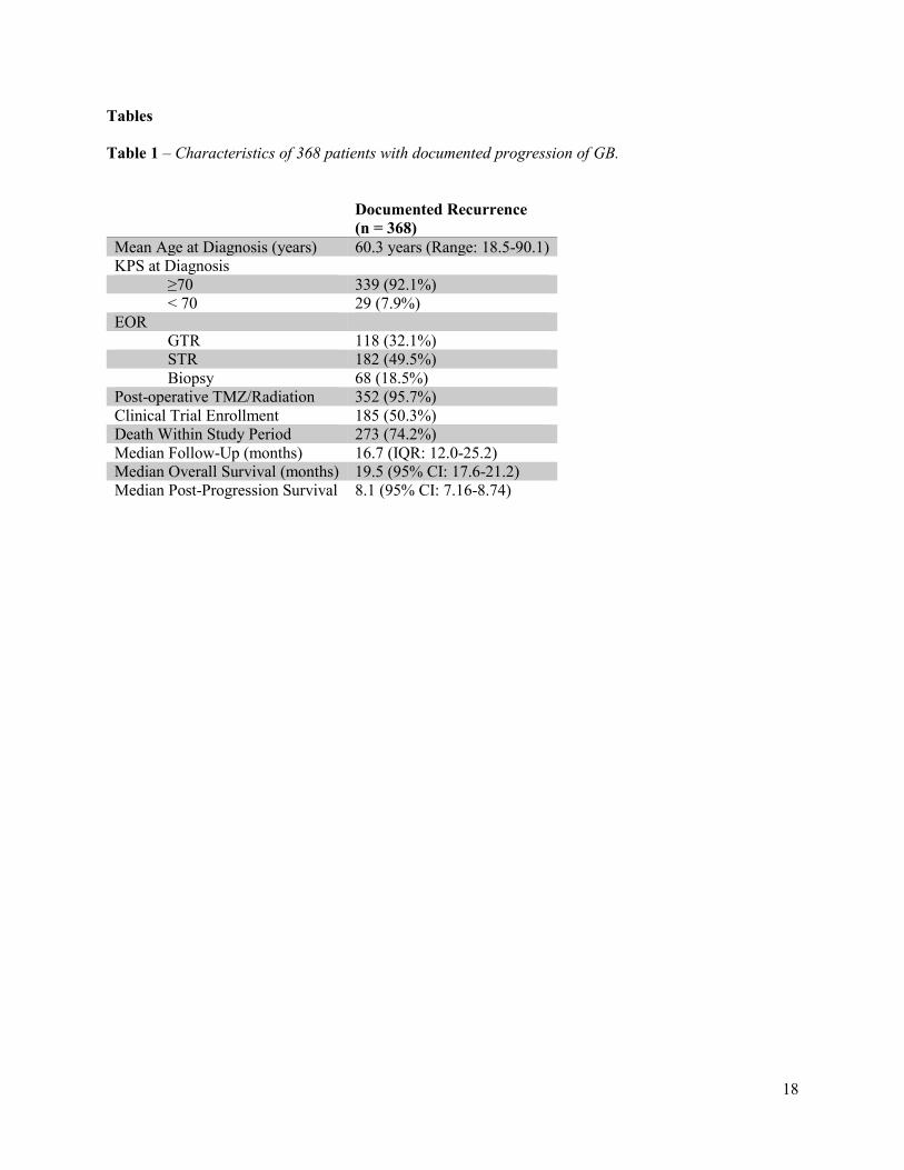
The characteristics of the overall patient population are summarized in Table 1. Two
hundred and seventy-three patients (74.2%) of patients died within the study period. The median
follow-up period was 16.7 months. Of the 368 patients with documented progression, 118
(32.1%) underwent GTR at initial surgery, 182 (49.5%) underwent STR at initial surgery, and 68
(18.5%) underwent biopsy at initial surgery. Three hundred and fifty-two (95.7%) patients
underwent subsequent treatment radiotherapy and temozolomide. One hundred and eighty-five
(50.3%) of patients were enrolled in a clinical trial at some point during their disease course. The
median overall survival for the entire patient cohort was 19.5 months (95% CI: 17.6-21.2
months, Figure 1A). The median post-progression survival for the entire patient cohort was 8.1
months (95% CI: 7.16-8.74 months, Figure 1B).
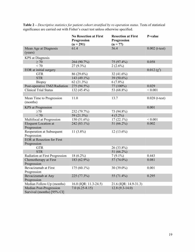
We collected demographic data to better understand the baseline differences between
patients who underwent resection at first progression (n=77 patients, 20.9%) and those who did
not (n=291, 79.1%) (Table 2). Patients who underwent resection for progression were
significantly younger at initial presentation (56.4 years vs. 61.4 years, p = 0.002) and underwent
a greater proportion of GTRs (41.6% vs. 29.6%, p = 0.01) at initial resection. Patients who
underwent resection for progression experienced progression 2.7 months later than those who did
not (13.7 months vs 11.0 months, p = 0.03). Patients who underwent resection for progression
8
presented with higher KPS at progression (94.8% KPS ≥ 70 vs. 79.7%, p = 0.001) and were less
likely to have either a multifocal (22.1% vs. 51.6%, p < 0.001) or eloquently situated tumor
(66.2% vs 83.1%, p = 0.002) at progression. A substantially smaller proportion of patients who
underwent resection at first progression received bevacizumab at first progression (39.0% vs.
60.1%, p = 0.001), though there was no difference in the proportion of patients who received
bevacizumab at any time for first or subsequent progressions (71.4% vs. 77.3%, p = 0.30). Rates
of radiotherapy at first progression (9.1% vs. 6.2%, p = 0.44) and chemotherapy at first
progression (74.0% vs. 62.9%, p = 0.08) did not differ significantly between these groups,
however. Among the 77 patients who underwent resection of progressive GB, 12 (13.6%) had
further resections. At the time of resection for first progression, GTR was achieved in 26 (33.8%)
and STR was achieved in 51 (66.2%) patients. Notably, 11 patients (3.8%) who did not undergo
resection at first progression eventually underwent resection for a subsequent progression. None
of these 11 patients underwent multiple reoperations for progression. Patients who underwent
craniotomy for resection for first progression of GB had increased median post-progression
survival (12.8 months vs. 7.0 months) and median follow-up (21.6 vs. 16.0 months) when
compared to patients who did not.
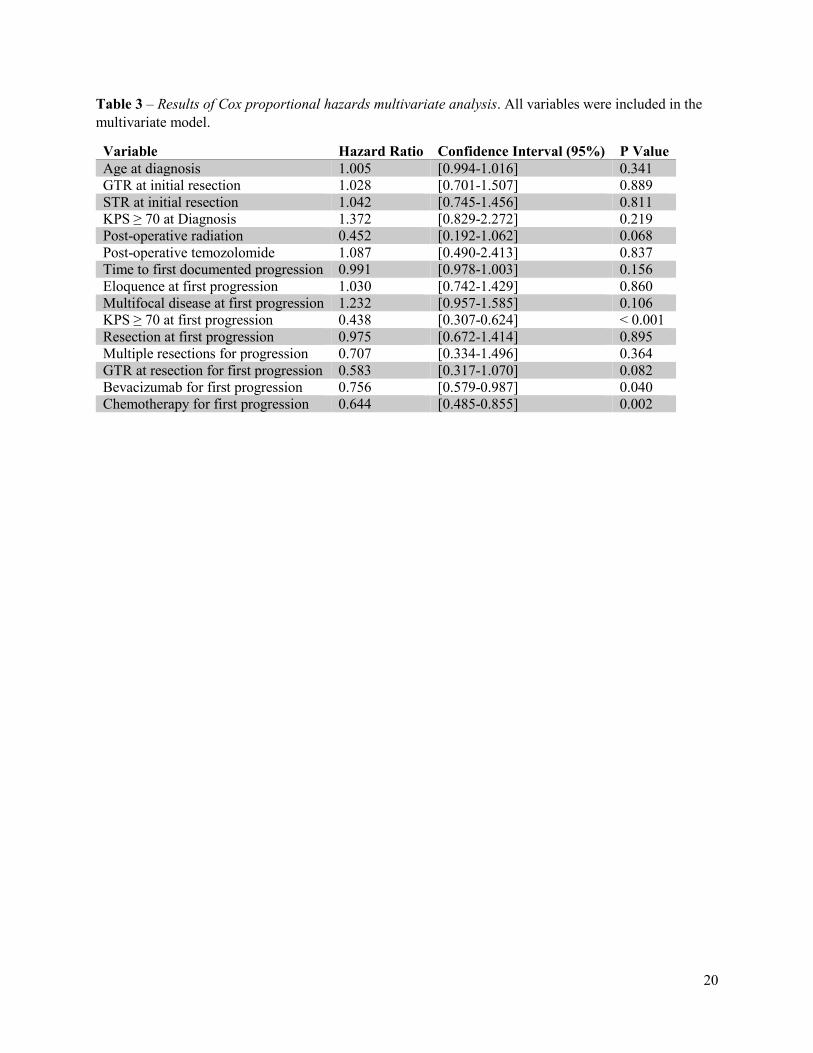
Multivariate Analysis
We performed a multivariate analysis to identify those that were significantly associated
with increased post-progression survival. Three variables were identified: KPS ≥ 70 at
progression (HR 0.438, 95% CI 0.307-0.624, p < 0.001), bevacizumab at first progression (HR
0.756, 95% CI: 0.579-0.987, p = 0.04), and cytotoxic chemotherapy at first progression (HR
0.644, 95% CI 0.485-0.855, p = 0.002). Notably, in this multivariate analysis, extent of resection
achieved at initial operation, whether a patient underwent resection at the time of progression,
extent of resection at time of surgery for progression, and the number of post-progression
resections were not significantly associated with increased post-progression survival, although
extent of resection at time of resection did trend toward significance. Results from select
variables of interest are briefly summarized in Table 3. Kaplan-Meier curves of selected
variables are presented in Figure 2.
Discussion
9
Key Results
We sought to reassess the survival benefit of resection for progressive GB in a patient
population in which the use of adjuvant chemoradiation following initial resection was high
(95.7%) and in which the use of bevacizumab at progression was more widespread (76.1% of all
identified) than has been reported in any previous study of this topic12, 18, 21. The results of this
study suggest that, when controlling for several potentially confounding factors, resection of
progressive GB is not significantly associated with prolonged post-progression survival even if a
GTR is achieved. In addition, our analysis identified KPS ≥ 70 at first progression, use of
bevacizumab at first progression, and use of chemotherapy at first progression as being
significantly associated with improved post-progression survival.
Interpretation
The finding of our study that resection of progressive GB does not significantly prolong
post-progression survival is at odds with some previous analyses. Notably, although Chaichana
et al previously concluded that resection for progressive GB and the number of resections were
both associated with improved overall survival in a case-control analysis9, the low overall
survival of single-resection patients (6.8 months) and low proportion of patients receiving both
radiation (65%) and temozolomide (27%) following initial surgery limit the relevance of results
to the current era of GB treatment. More recent studies have suggested that resection at
progression is valuable if a GTR is achieved or if post-progression EOR exceeds initial EOR8, 11-
13, 16. Our results are concordant with the conclusions reached by Ortega, et al. who determined
that repeat resection was not significantly associated with increased overall survival in a
contemporary patient population that was commonly treated with bevacizumab for progression18.
Our findings expand on these data by including a larger and more heterogeneous population,
considering more variables in our model, and examining post-progression survival instead of
overall survival.
Taken together, these results suggest that resection for progression may have offered a
clearer survival benefit before the widespread adoption of aggressive initial resection followed
by concurrent chemoradiation. Despite the fact that resection for progressive GB may not be life-
extending, there are still indications for resection at progression, such as to debulk tumor mass to
10
palliate symptoms, to minimize steroid dependence, to acquire tissue for molecular analysis, and
to allow enrollment in clinical trials.
Interestingly, our model identifies post-progression bevacizumab and cytotoxic
chemotherapy as being significantly associated with improved post-progression survival. The
significant survival benefit attributed to bevacizumab is at odds with findings from recent data
from the yet unpublished EORTC 26101 study which suggest that use of bevacizumab in
combination with lomustine, despite initial promise in early trials, improves only progression-
free survival and not overall survival31-34. This discordance may be a result of divergent
treatment combinations, dosages, or durations utilized at our practice as compared to the EORTC
26101 protocol. Of course, with any retrospective analysis, there may be an unidentified source
of confounding that has contributed to this result. Further study of the role of bevacizumab for
progressive GBM will likely be necessary to determine which, if any, subsets of patients with
progressive GB benefit from bevacizumab.
Limitations and Generalizability
As with any retrospective analysis, this study has limitations. A number of patients were
lost to follow-up. Furthermore, we did not consider patients who underwent biopsy or resection
of pseudoprogression as having undergone resection of progressive disease; however, these
procedures undoubtedly carry their own benefits and risks of morbidity and mortality. Molecular
characteristics of the tumor, notably including IDH1 mutation status and MGMT methylation
status, were not included in our analysis as testing results were not routinely available for every
patient throughout the study time period. While we have identified three variables as being
independently associated with post-progression survival, it is possible that these are surrogate
markers for other, as yet unidentified, features of these tumors or their hosts. Finally, the
generalizability of our results may be limited by its retrospective nature and by variations in
management of GB at other centers, and constantly evolving treatment options for GB.
Conclusions
Despite maximal treatment, progression of GB is a near inevitability. A variety of
treatment options, including surgery, chemotherapy, radiation, and clinical trial enrollment, are
available to patients at progression. In the context of contemporary treatment approaches and

11
recent widespread adoption of bevacizumab for progressive GB, we sought to reevaluate the role
of resection for progressive GB. Our multivariate analysis suggests that undergoing resection of
progressive GB is not associated with post-progression survival. Our findings also support the
idea that maximal medical therapy may be a more reasonable choice than surgery for managing
GB progression.
12
References
1. Ostrom QT, Gittleman H, Fulop J, et al. CBTRUS Statistical Report: Primary Brain and Central
Nervous System Tumors Diagnosed in the United States in 2008-2012. Neuro Oncol. Oct
2015;17 Suppl 4:iv1-iv62.
2. Nabors LB, Portnow J, Ammirati M, et al. Central Nervous System Cancers, Version 1.2015. J.
Natl. Compr. Canc. Netw. Oct 2015;13(10):1191-1202.
3. Stupp R, Mason WP, van den Bent MJ, et al. Radiotherapy plus concomitant and adjuvant
temozolomide for glioblastoma. N. Engl. J. Med. Mar 10 2005;352(10):987-996.
4. Stummer W, Reulen HJ, Meinel T, et al. Extent of resection and survival in glioblastoma
multiforme: identification of and adjustment for bias. Neurosurgery. Mar 2008;62(3):564-576;
discussion 564-576.
5. Gilbert MR, Dignam JJ, Armstrong TS, et al. A randomized trial of bevacizumab for newly
diagnosed glioblastoma. N. Engl. J. Med. Feb 20 2014;370(8):699-708.
6. Weller M, van den Bent M, Hopkins K, et al. EANO guideline for the diagnosis and treatment of
anaplastic gliomas and glioblastoma. Lancet Oncol. Aug 2014;15(9):e395-403.
7. Montemurro N, Perrini P, Blanco MO, Vannozzi R. Second surgery for recurrent glioblastoma: A
concise overview of the current literature. Clin. Neurol. Neurosurg. Mar 2016;142:60-64.
8. Suchorska B, Weller M, Tabatabai G, et al. Complete resection of contrast-enhancing tumor
volume is associated with improved survival in recurrent glioblastoma-results from the
DIRECTOR trial. Neuro Oncol. Apr 2016;18(4):549-556.
9. Chaichana KL, Zadnik P, Weingart JD, et al. Multiple resections for patients with glioblastoma:
prolonging survival. J. Neurosurg. Apr 2013;118(4):812-820.
10. Stark AM, Nabavi A, Mehdorn HM, Blomer U. Glioblastoma multiforme-report of 267 cases
treated at a single institution. Surg. Neurol. Feb 2005;63(2):162-169; discussion 169.
11. Ringel F, Pape H, Sabel M, et al. Clinical benefit from resection of recurrent glioblastomas:
results of a multicenter study including 503 patients with recurrent glioblastomas undergoing
surgical resection. Neuro Oncol. Jan 2016;18(1):96-104.
12. Oppenlander ME, Wolf AB, Snyder LA, et al. An extent of resection threshold for recurrent
glioblastoma and its risk for neurological morbidity. J. Neurosurg. Apr 2014;120(4):846-853.
13. Brandes AA, Bartolotti M, Tosoni A, et al. Patient outcomes following second surgery for
recurrent glioblastoma. Future Oncol. Apr 2016;12(8):1039-1044.
14. Yong RL, Wu T, Mihatov N, et al. Residual tumor volume and patient survival following
reoperation for recurrent glioblastoma. J. Neurosurg. Oct 2014;121(4):802-809.
15. Quick J, Gessler F, Dutzmann S, et al. Benefit of tumor resection for recurrent glioblastoma. J.
Neurooncol. Apr 2014;117(2):365-372.
16. Bloch O, Han SJ, Cha S, et al. Impact of extent of resection for recurrent glioblastoma on overall
survival: clinical article. J. Neurosurg. Dec 2012;117(6):1032-1038.
17. Johnson DR, Leeper HE, Uhm JH. Glioblastoma survival in the United States improved after
Food and Drug Administration approval of bevacizumab: a population-based analysis. Cancer.
Oct 1 2013;119(19):3489-3495.
18. Ortega A, Sarmiento JM, Ly D, et al. Multiple resections and survival of recurrent glioblastoma
patients in the temozolomide era. J. Clin. Neurosci. Feb 2016;24:105-111.
19. Clarke JL, Ennis MM, Yung WK, et al. Is surgery at progression a prognostic marker for
improved 6-month progression-free survival or overall survival for patients with recurrent
glioblastoma? Neuro Oncol. Oct 2011;13(10):1118-1124.
20. Nava F, Tramacere I, Fittipaldo A, et al. Survival effect of first- and second-line treatments for
patients with primary glioblastoma: a cohort study from a prospective registry, 1997-2010. Neuro
Oncol. May 2014;16(5):719-727.
21. Woernle CM, Peus D, Hofer S, et al. Efficacy of Surgery and Further Treatment of Progressive
Glioblastoma. World Neurosurg. Aug 2015;84(2):301-307.
13
22. Curran WJ, Jr., Scott CB, Horton J, et al. Recursive partitioning analysis of prognostic factors in
three Radiation Therapy Oncology Group malignant glioma trials. J. Natl. Cancer Inst. May 5
1993;85(9):704-710.
23. Chang EF, Smith JS, Chang SM, et al. Preoperative prognostic classification system for
hemispheric low-grade gliomas in adults. J. Neurosurg. Nov 2008;109(5):817-824.
24. Hamza MA, Mandel JJ, Conrad CA, et al. Survival outcome of early versus delayed bevacizumab
treatment in patients with recurrent glioblastoma. J. Neurooncol. Aug 2014;119(1):135-140.
25. Goldman DA, Panageas KS. Letter to the Editor: Biases in estimation of overall survival in
patients who underwent repeat resection of glioblastoma. J. Neurosurg. Jun 3 2016;0(0):1-2.
26. Schoenfeld D. Partial residuals for the proportional hazards regression model. Biometrika.
1982;69(1):239-241.
27. Harrell FE, Jr., Lee KL, Matchar DB, Reichert TA. Regression models for prognostic prediction:
advantages, problems, and suggested solutions. Cancer Treat. Rep. Oct 1985;69(10):1071-1077.
28. Benedetti JK, Liu PY, Sather HN, Seinfeld J, Epton MA. Effective Sample-Size for Tests of
Censored Survival-Data. Biometrika. 1982;69(2):343-349.
29. Harrell F. Regression modeling strategies: with applications to linear models, logistic and
ordinal regression, and survival analysis: Springer; 2015.
30. von Elm E, Altman DG, Egger M, et al. Strengthening the Reporting of Observational Studies in
Epidemiology (STROBE) statement: guidelines for reporting observational studies. BMJ. Oct 20
2007;335(7624):806-808.
31. Friedman HS, Prados MD, Wen PY, et al. Bevacizumab alone and in combination with irinotecan
in recurrent glioblastoma. J. Clin. Oncol. Oct 1 2009;27(28):4733-4740.
32. Taal W, Oosterkamp HM, Walenkamp AM, et al. Single-agent bevacizumab or lomustine versus
a combination of bevacizumab plus lomustine in patients with recurrent glioblastoma (BELOB
trial): a randomised controlled phase 2 trial. Lancet Oncol. Aug 2014;15(9):943-953.
33. van den Bent M, Gorlia T, Bendszus M, et al. EH1.3EORTC 26101 phase III trial exploring the
combination of bevacizumab and lomustine versus lomustine in patients with first progression of
a glioblastoma. Neuro Oncol. 2016;18(suppl_4):iv1-iv2.
34. Vredenburgh JJ, Desjardins A, Herndon JE, 2nd, et al. Bevacizumab plus irinotecan in recurrent
glioblastoma multiforme. J. Clin. Oncol. Oct 20 2007;25(30):4722-4729.
14
Figures
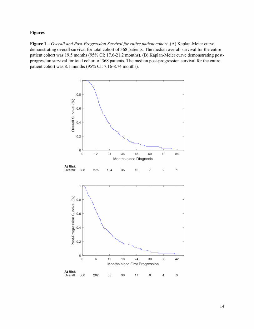
Figure 1 – Overall and Post-Progression Survival for entire patient cohort. (A) Kaplan-Meier curve
demonstrating overall survival for total cohort of 368 patients. The median overall survival for the entire
patient cohort was 19.5 months (95% CI: 17.6-21.2 months). (B) Kaplan-Meier curve demonstrating post-
progression survival for total cohort of 368 patients. The median post-progression survival for the entire
patient cohort was 8.1 months (95% CI: 7.16-8.74 months).
15
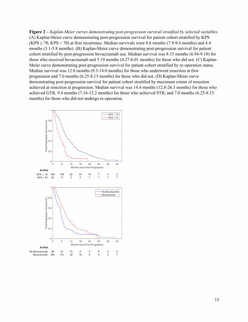
Figure 2 – Kaplan-Meier curves demonstrating post-progression survival stratified by selected variables.
(A) Kaplan-Meier curve demonstrating post-progression survival for patient cohort stratified by KPS
(KPS ≥ 70, KPS < 70) at first recurrence. Median survivals were 8.6 months (7.9-9.4 months) and 4.4
months (3.1-5.8 months). (B) Kaplan-Meier curve demonstrating post-progression survival for patient
cohort stratified by post-progression bevacizumab use. Median survival was 8.15 months (6.94-9.10) for
those who received bevacizumab and 5.19 months (4.27-6.01 months) for those who did not. (C) Kaplan-
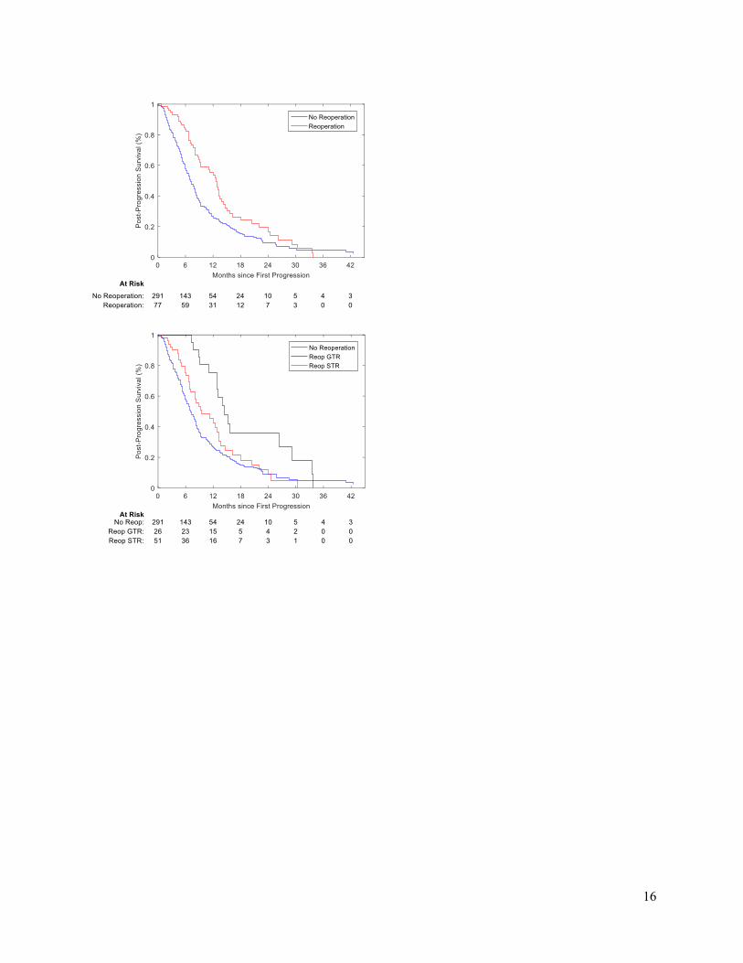
Meier curve demonstrating post-progression survival for patient cohort stratified by re-operation status.
Median survival was 12.8 months (9.3-14.0 months) for those who underwent resection at first
progression and 7.0 months (6.25-8.15 months) for those who did not. (D) Kaplan-Meier curve
demonstrating post-progression survival for patient cohort stratified by maximum extent of resection
achieved at resection at progression. Median survival was 14.4 months (12.8-26.3 months) for those who
achieved GTR, 9.4 months (7.16-12.2 months) for those who achieved STR, and 7.0 months (6.25-8.15
months) for those who did not undergo re-operation.
16
17
Supplementary Figure 1 – Schematic of patient selection
563 patients with documented
GB between January 2008 and
December 2015
368 patients with radiographic
or biopsy-proven progressive
GB
195 patients without known
progressive GB
88 patients with 1 or more
resections at progression 280 patients without resection at
progression
18
Tables
Table 1 – Characteristics of 368 patients with documented progression of GB.
Documented Recurrence
(n = 368)
Mean Age at Diagnosis (years) 60.3 years (Range: 18.5-90.1)
KPS at Diagnosis
≥70 339 (92.1%)
< 70 29 (7.9%)
EOR
GTR 118 (32.1%)
STR 182 (49.5%)
Biopsy 68 (18.5%)
Post-operative TMZ/Radiation 352 (95.7%)
Clinical Trial Enrollment 185 (50.3%)
Death Within Study Period 273 (74.2%)
Median Follow-Up (months) 16.7 (IQR: 12.0-25.2)
Median Overall Survival (months) 19.5 (95% CI: 17.6-21.2)
Median Post-Progression Survival 8.1 (95% CI: 7.16-8.74)
19
Table 2 – Descriptive statistics for patient cohort stratified by re-operation status. Tests of statistical
significance are carried out with Fisher’s exact test unless otherwise specified.
No Resection at First
Progression
(n = 291)
Resection at First
Progression
(n = 77)
P-value
Mean Age at Diagnosis
(years)
61.4 56.4 0.002 (t-test)
KPS at Diagnosis
≥ 70 264 (90.7%) 75 (97.4%) 0.058
< 70 27 (9.3%) 2 (2.6%)
EOR at initial surgery 0.012 (χ2)
GTR 86 (29.6%) 32 (41.6%)
STR 143 (49.1%) 39 (50.6%)
Biopsy 62 (21.3%) 6 (7.8%)
Post-operative TMZ/Radiation 275 (94.5%) 77 (100%) 0.029
Clinical Trial Status 132 (45.4%) 53 (68.8%) < 0.001
Mean Time to Progression
(months)
11.0 13.7 0.028 (t-test)
KPS at Progression 0.001
≥70 232 (79.7%) 73 (94.8%)
< 70 59 (21.3%) 4 (5.2%)
Multifocal at Progression 150 (51.6%) 17 (22.1%) < 0.001
Eloquent Location at
Progression
242 (83.1%) 51 (66.2%) 0.002
Reoperation at Subsequent
Progression
11 (3.8%) 12 (13.6%)
EOR at Resection for First
Progression
GTR 26 (33.8%)
STR 51 (66.2%)
Radiation at First Progression 18 (6.2%) 7 (9.1%) 0.443
Chemotherapy at First
Progression
183 (62.9%) 57 (74.0%) 0.081
Bevacizumab at First
Progression
175 (60.1%) 30 (39.0%) 0.001
Bevacizumab at Any
Progression
225 (77.3%) 55 (71.4%) 0.295
Median Follow-Up (months) 16.0 (IQR: 11.3-24.5) 21.6 (IQR: 14.9-31.3)
Median Post-Progression
Survival (months) [95% CI]
7.0 (6.25-8.15) 12.8 (9.3-14.0)
20
Table 3 – Results of Cox proportional hazards multivariate analysis. All variables were included in the
multivariate model.
Variable Hazard Ratio Confidence Interval (95%) P Value
Age at diagnosis 1.005 [0.994-1.016] 0.341
GTR at initial resection 1.028 [0.701-1.507] 0.889
STR at initial resection 1.042 [0.745-1.456] 0.811
KPS ≥ 70 at Diagnosis 1.372 [0.829-2.272] 0.219
Post-operative radiation 0.452 [0.192-1.062] 0.068
Post-operative temozolomide 1.087 [0.490-2.413] 0.837
Time to first documented progression 0.991 [0.978-1.003] 0.156
Eloquence at first progression 1.030 [0.742-1.429] 0.860
Multifocal disease at first progression 1.232 [0.957-1.585] 0.106
KPS ≥ 70 at first progression 0.438 [0.307-0.624] < 0.001
Resection at first progression 0.975 [0.672-1.414] 0.895
Multiple resections for progression 0.707 [0.334-1.496] 0.364
GTR at resection for first progression 0.583 [0.317-1.070] 0.082
Bevacizumab for first progression 0.756 [0.579-0.987] 0.040
Chemotherapy for first progression 0.644 [0.485-0.855] 0.002