Embed Size (px)
Citation preview
i
The Importance of Keratinized Gingiva Surrounding Dental
Implants
By
Anita Desai
Juniata College, 2008
Howard University College of Dentistry, 2012
University of Pittsburgh School of Dental Medicine Department of Periodontics, 2015
Submitted to the Graduate Faculty of
University of Pittsburgh School of Dental Medicine in partial fulfillment
of the requirements for the degree of
Masters of Dental Science
University of Pittsburgh
2015
i i
UN IVERS ITY OF P ITTSBURGH
SCHOO L OF DENTAL MED ICINE
This thes i s was p resented
b y
Ani ta Desai
I t was de fended on
May12, 2015
and approved b y
Al i Seyedain , DMD , MDS, Ass i s tan t Professo r , Di rector of
Undergraduate Per iodont i cs , Depar tment of
Per iodont i cs and Preventa t ive Dent i s t ry
Edward Hein r ichs , DDS, Ass i s t an t Pro fes sor Per iodont ics Depar tment
Thes i s Di rector : Pouran Fami l i , DMD, MDS, MPH, PHD, P rofessor ,
Depar tme nt o f Per iodont i cs and Prevent ive Dent i s t r y
i i i
Copyr ight © by Anita Desai
2015
i v
The Importance of Kerat in ized Gingiva Surrounding Dental
Implants
Ani ta Desai , DDS, MDS
Univers i t y o f P i t t sburgh , 2015
Purpose: The purpose of t h i s s tud y was to det ermine i f ke ra t in ized
gingiva has an e f fec t on the success o f implants .
Materia ls and Methods: S ix ty-n ine implants were used in th i s s tud y.
The amount o f ke ra t in ized gingiva was measured and d iv ided in to two
groups ; l ess t han 2mm and great er t han 2mm. The amoun t of
kera t in ized g ingiva was compared to c l i n ica l paramete rs such as
b leeding upon p robing, redness , and pocket depths to det ermine
whether implant success was re l a t ed to the amount o f ke ra t in ized
gingiva .
Resul ts : Chi square and regres s ion analys i s were used to anal yze the
dat a . Al l implant s surv ived independent of the amount of kera t in ized
gingiva . Pat i en ts wi th les s than 2mm of kera t in ized gingiva d i sp layed
increased b l eeding upon probing and redness , which was s t a t i s t i ca l l y
s igni f i cant (p=0.023) , i nd i cat ing inc reased in f lammat ion due to l ack
of ke ra t in ized g ingiva .
Conclus ion: Amount of kera t in ized gingiva d id not af fec t the success
ra t e o f implant s . However , implants wi th les s than 2mm of
v
kerat in ized g ingiva exhib i ted increased b leeding upon p rob ing,
redness , and inf l ammat ion , which may cont r ibute t o la t e r f a i lu re .
v i
TABLE OF CONTENTS
1.0 INTRODUCTION . . . . . . . . . . . . . . . . . . . . . . . . . . . . . . . . . . . . . . . . . . . . . . . . . . . . . . . . . . . . . . . . . . . . . 1
2.0 REVIEW OF THE LITERATURE . . . . . . . . . . . . . . . . . . . . . . . . . . . . . . . . . . . . . . . . . . . . . 3
2 .1 SURGI CAL TECHNIQUES . . . . . . . . . . . . . . . . . . . . . . . . . . . . . . . . . . . . . . . . . . . . . . . . . . . . . . . . . . . 3
2 .2 NEED F OR MINIMAL AMOUNT OF KERATI NI ZED GI NGIVA . . 4
2 .3 DENTAL IMP LANT ANATOMY . . . . . . . . . . . . . . . . . . . . . . . . . . . . . . . . . . . . . . . . . . . . . . . . . . . 5
2 .4 IMP LANT-MUCOSA INTERF ACE . . . . . . . . . . . . . . . . . . . . . . . . . . . . . . . . . . . . . . . . . . . . . . . 7
2 .5 IMP LANT SUP P ORTED RESTORATIONS . . . . . . . . . . . . . . . . . . . . . . . . . . . . . . . . . . . 8
2 .6 HYPOTHESIS AND P URP OSE . . . . . . . . . . . . . . . . . . . . . . . . . . . . . . . . . . . . . . . . . . . . . . . . . . . . . 9
3.0 MATERIALS AND METHODS . . . . . . . . . . . . . . . . . . . . . . . . . . . . . . . . . . . . . . . . . . . . . . . 10
4.0 RESULTS . . . . . . . . . . . . . . . . . . . . . . . . . . . . . . . . . . . . . . . . . . . . . . . . . . . . . . . . . . . . . . . . . . . . . . . . . . . . . . . 12
5.0 DISCUSSION . . . . . . . . . . . . . . . . . . . . . . . . . . . . . . . . . . . . . . . . . . . . . . . . . . . . . . . . . . . . . . . . . . . . . . . . . 19
6.0 CONCLUSION . . . . . . . . . . . . . . . . . . . . . . . . . . . . . . . . . . . . . . . . . . . . . . . . . . . . . . . . . . . . . . . . . . . . . . . 21
7.0 BIBLIOGRAPHY . . . . . . . . . . . . . . . . . . . . . . . . . . . . . . . . . . . . . . . . . . . . . . . . . . . . . . . . . . . . . . . . . . . . 22
v i i
LIST OF TABLES
TABLE 1 . KE R ATI NIZ E D G I NG IV A R E SE AR CH C ASE RE PO RT . . . . . 15
TABLE 2 . PO C KE T ING V S KE RA TINIZ E D G ING I V A . . . . . . . . . . . . . . . . . . . . . 16
TABLE 3 . BLE E DI NG V S KE RAT INIZ E D G ING I V A . . . . . . . . . . . . . . . . . . . . . . . . 17
TABLE 4 . RE G RE S SIO N F INA L M O DE L . . . . . . . . . . . . . . . . . . . . . . . . . . . . . . . . . . . . . . . . 18
1
1.0 INTRODUCTION
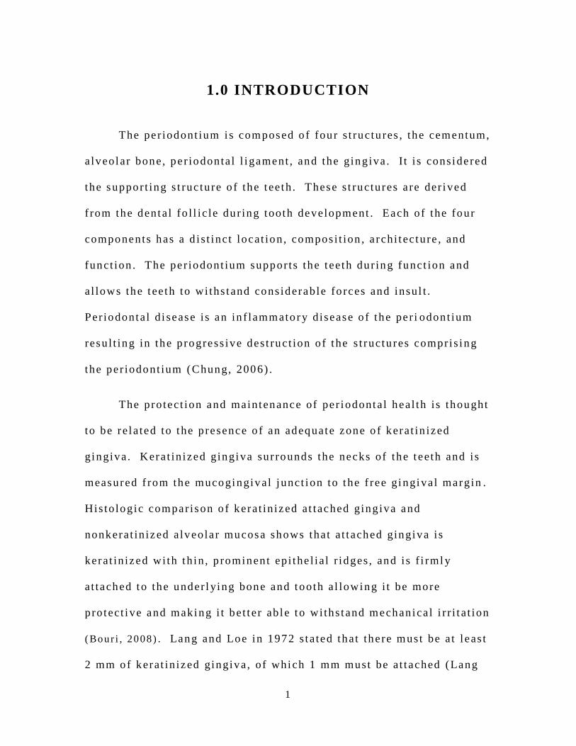
The per iodont ium i s composed o f fou r s t ructu res , the cementum,
a lveolar bone, pe r iodonta l l i gament , and the gingiva . I t i s cons idered
the suppor t ing s t ruc ture o f the t ee th . These s t ructures a re der ived
f rom the denta l fo l l i c le du r ing tooth development . Each of the four
components has a d i s t inct locat ion , composi t ion , a rchi t ec ture , and
funct ion . The per iodont ium suppor t s the t ee th du r ing funct ion and
a l lows the t ee th to wi ths t and cons iderable fo rces and insul t .
Per iodonta l d i sease i s an in f lammato r y d isease o f the pe r i odont ium
resul t ing in the p rogress ive des t ruct ion of t he s t ructu res compris ing
the pe r iodont ium (Chung, 2006) .
The pro tect ion and main t enance of pe r iodonta l heal th i s thought
to be re l a ted to t he presence o f an adequate zone o f kera t in ized
gingiva . Kera t in ized gingiva sur rounds the necks of the t ee th and i s
measured f rom the mucogingival j unct ion to t he f ree gingival margin .
His to logic compar i son o f kera t in ized a t tached gingiva and
nonkerat in ized a lveolar mucosa shows that a t t ached gingiva i s
kera t in ized wi th th in , p rominent ep i the l ia l r idges , and i s f i rml y
a t tached to the under l ying bone and tooth a l lowing i t be more
pro t ect ive and making i t be t t er ab l e t o wi ths t and mechanical i r r i t a t ion
(Bour i , 2008) . Lang and Loe in 1972 s t a ted that the re must be a t l eas t
2 mm of kera t in ized gingiva , of which 1 mm must be a t t ached (Lang
2
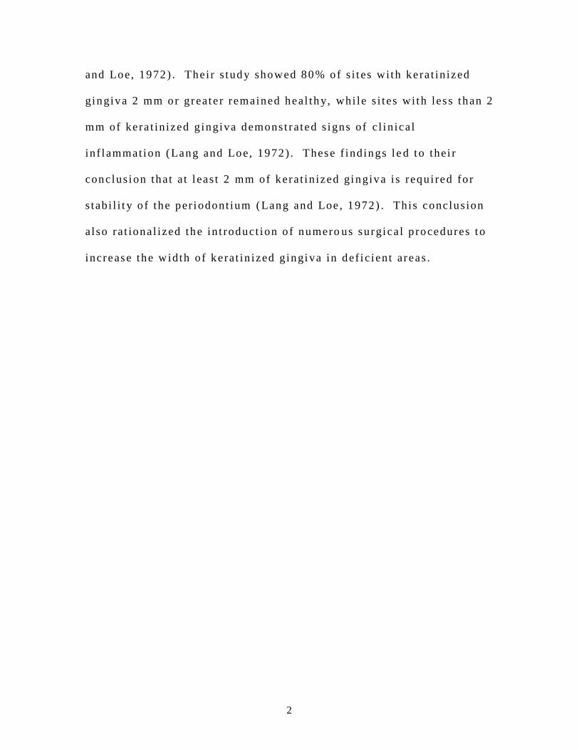
and Loe, 1972) . Thei r s tud y showed 80% of s i t es wi th ke ra t in ized
gingiva 2 mm or great e r r emained heal th y, whi le s i t es wi th les s than 2
mm of ke ra t in ized g ingiva demonst ra ted s igns of c l i n ica l
inf l ammat ion (Lang and Loe, 1972) . These f indings l e d to thei r
conclus ion that a t l eas t 2 mm of kera t in ized gingiva i s r equi red for
s tab i l i t y o f the per iodont ium ( Lang and Loe , 1972) . This conclus ion
a l so ra t ional ized the in t roduct ion o f numero us surgi cal p rocedures t o
increase the width of ke ra t in ized g ingiva in def i c i en t areas .
3
2.0 REVIEW OF THE LITERATURE
2.1 SURGICAL TECHNIQUES
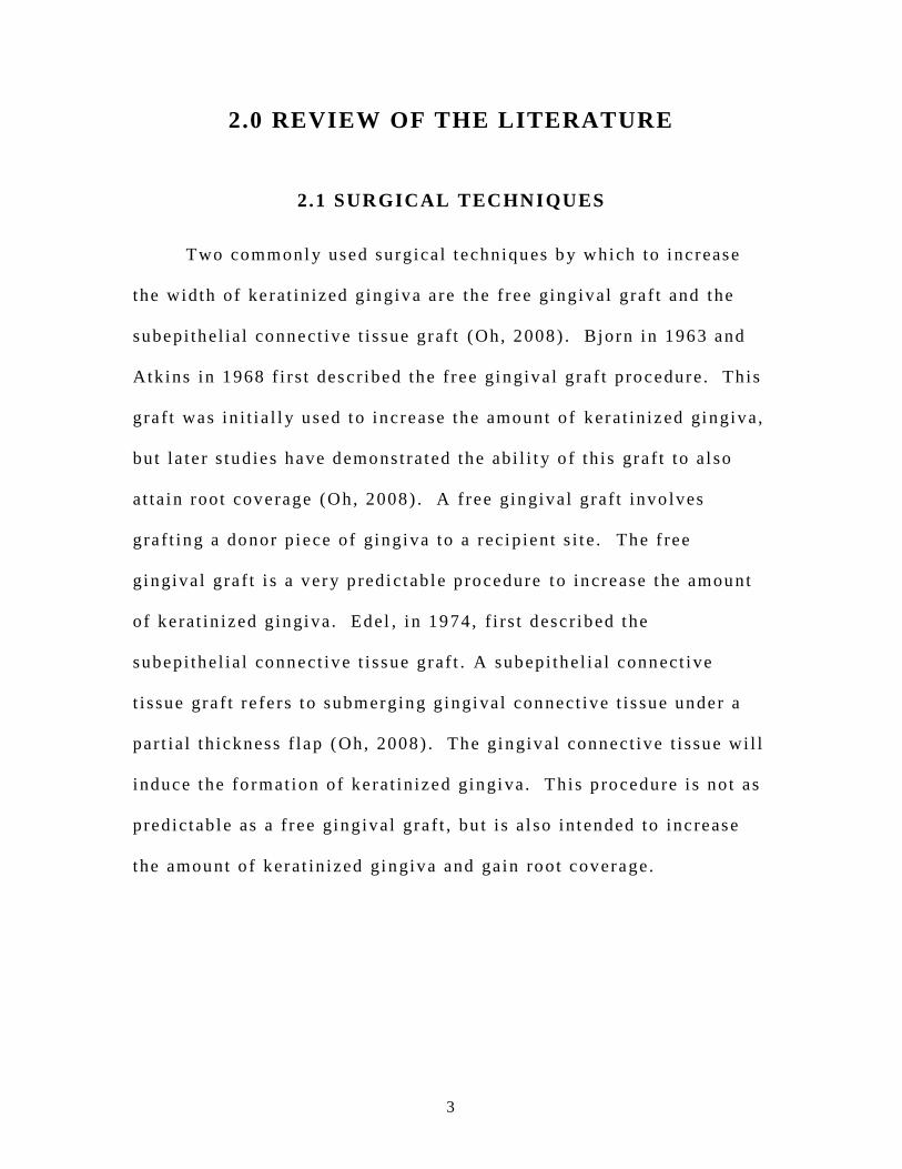
Two commonl y used surg ical t echniques b y which to i nc rease
the width of ke ra t in ized gingiva a re the f ree gingival gra f t and the
subepi thel ia l connect ive t i s sue graf t (Oh, 2008) . Bjorn in 1963 and
Atkins in 1968 f i r s t desc r ibed the f ree gingival gra f t p rocedure . This
gra f t was in i t i a l l y used to increase the amount o f kera t in ized gingiva ,
but l a te r s tudies have demonst ra t ed the ab i l i t y o f th i s gra f t to a l so
a t ta in root coverage (Oh, 2008) . A f ree gingival graf t invo lves
gra f t ing a donor p i ece of gingiva to a r ec ip ien t s i t e . The f ree
gingival gra f t i s a ver y p redic t ab l e p rocedure to increase the amount
of ke ra t in ized g ingiva . Edel , i n 1974, f i r s t desc r ibed the
subepi thel ia l connect ive t i s sue graf t . A subepi thel i a l connect ive
t i s sue gra f t r e fe rs t o submerging g ingival connect ive t i s sue under a
par t ia l t h ickness f lap (Oh, 2008) . The gingival connect ive t i s sue wi l l
induce the fo rmat ion of ke ra t in ized gingiva . This p rocedure i s not a s
predic t ab l e as a f r ee gingival gra f t , bu t i s a l so in t ended to inc rease
the amount o f kera t in ized gingiva and gain root coverage.
4
2.2 NEED FOR MINIMAL AMOUNT OF KERATINIZED
GINGIVA
Later s tudies chal l enged th i s concept o f the need for a min imal
amount o f kera t in ized gingiva , and have shown tha t b y con t ro l l ing
inf l ammat ion wi th adequate ora l h ygiene , pe r iodonta l s t ab i l i t y can be
main t a ined wi th a lmost no kera t in ized gingiva . According to
Wenns t rom, a minimal amount o f kera t in ized gingiva does not
necessa r i l y l ead to gingiva l recess ion and in f lammat ion (Wenns tom,
2012) . He s t a ted that the na rrow zone of ke ra t in ized g ingiva located
ap ical l y to an a rea of recess ion i s the resu l t o f r ecess ion , not the
cause (Wenns tom, 2012) . Some l a te r s tudies s ta t e t ha t even in areas
of minimal ke ra t in i zed gingiva , p roper p laque cont ro l t echniques can
prevent gingival r eces s ion and so f t t i s sue inf l ammat ion .
An except ion to th i s was in tee th wi th subgin gival r es to ra t ions .
There was a s igni f i cant a ssocia t ion between subgi ngival res tora t ions
and g ingival i nf l ammat ion in areas o f minimal ke ra t in ized gingiva
(Bour i , 2008) . S t e t ler concluded that subgingival r es to ra t ions p l aced
on tee th su rrounded b y l ess t han 2 mm of ke ra t in ized g ingiva
demonst ra t ed an inc reased gingival index (S te t l e r , 1986) . According
to S te l er in 1986, g ingiva l gra f t ing i s r ecommended in a reas where
subgingival margins wi l l be p laced i f t he width of ke ra t in i zed gingiva
i s l ess than 5 mm (Ste t le r , 1986) . The ra t ionale behind th i s i s tha t
the ke ra t in ized g ingiva wi l l p rovide a p ro tect ive ba rr i e r agains t
5
in f l ammat ion and a t tachment loss (S t e t l er , 1986) . A s imi la r s tud y b y
Lindhe and Er i csson demonst ra t ed an increase in p laque and bact e r ia l
in f i l t ra t e in a reas where subgingival res tora t ions were p laced wi th
minimal ke ra t in ized gingiva (Greens te in , 2011) .
2.3 DENTAL IMPLANT ANATOMY
In 1978, Dr . Branemark p resented the t i t an ium root – fo rm
implant (Abraham, 2014) . This d i scover y was made accident l y whi l e
s tud ying b lood f low in rabbi t f emurs (Abraham, 2014) . He p laced
t i t an ium chambers i n thei r bone and no t iced that over t ime the
t i t an ium became r ig id l y f ixat ed to the bone and was not ab l e to be
removed (Abraham, 2014) . This was l a ter t e rmed b y Branemark as
osseo in tegra t ion , and was de f ined as a “d i rec t s t ructu ra l and
funct ional connect ion between o rdered , l iv ing bone, and the sur face
of a load car r ying implant” (Abraham, 2014) . Severa l d i f ferent t ypes
of implant s were l a t er i n t roduced and the use of denta l implants fo r
rep l acement of miss ing tee th began to d ramat i ca l l y increase
(Abraham, 2014) . As the use of denta l implants rep l acing natu ra l
dent i t i on becomes increas ingl y the s t andard o f ca re , the amount o f
kera t in ized g ingiva sur rounding denta l implants to opt imize gingiv al
heal th a l so comes in to ques t ion . Due to the s t ructura l and anatomical
d i f fe rences between implants and natura l t ee th , the same concepts
cannot be appl ied to implants (Lin , 2013) . Implants are more

6
suscept ib l e t o the development of i nf l ammat ion and subsequent bone
loss in t he presence of p laque accumula t ion and bacte r i a l i n f i l t r a t ion
due to severa l fac to rs (Lin , 2013) . The implant to mucosa in te r face i s
d i f fe ren t f rom the in ter face between na tura l t ee th and mucosa (Lin ,
2013) . Whi l e the junct ional ep i t hel ium ends a t a s imi l ar d i s tance to
the bone c res t in bo th tee th and denta l implants , the gingival f ibe r
or ien ta t ion i s d i f ferent (Lin , 2013) . The gingival f ibe rs of natu ra l
t ee th run in a perpendicula r conf igu ra t ion , whereas the gingival f ibe rs
of implant s run in a pa ra l l e l conf igura t ion to t he implant and do not
a t tach to the implan t sur face crea t ing a much weaker me chanical
a t tachment compared to natu ra l t ee th (Lin , 2013) . This weaker
a t tachment inc reases the suscept ib i l i t y to bact er i a l i n f i l t ra t ion
leading to gingival inf l ammat ion and bone loss around the implant . I f
the sur face o f the implant i s contaminated b y bact er i a , an
inf l ammator y response i s t r i ggered in t he connect ive t i s sue (Paiva ,
2012) . Unl ike the per iodonta l l i gamen t around natu ra l t ee th , the bone
sur rounding the implant cannot o rganize a de fense mechanism agains t
infect ion (Paiva , 2012) . There fo re the ap i cal ex tens ion of the
inf l ammator y inf i l t r a te a round implant s seems to resu l t f rom the
or ien ta t ion of t he supra -a lveolar pe r i - implant f ibe rs (Paiva , 2012) .
7
2.4 IMPLANT-MUCOSA INTERFACE
As s t a ted b y Bour i in 2008 nar row zones o f ke ra t in ized g ingiva
are l ess r es i s t an t to insu l t a long the implant -mucosa in t e r face . In the
presence o f an inf l ammato r y response , implants p l aced in a reas wi th
nar row zones o f ke ra t in ized gingiva have an inc reased suscept ib i l i t y
to t i s sue b reakdown and showed ea r l i er loss o f a t t achment (Bour i ,
2008) . Greens t e in , in a l i t e ra ture rev iew, s imi l ar l y s t a t ed that a
nar row zone o f ke ra t in ized gi ngiva , l es s than 2 mm, was as soci a ted
wi th inc reased in f l ammat ion , p l aque accumulat ion , and recess ion o f
the gingiva , u l t imate l y resu l t ing in t i s sue des t ruct ion (Greens t e in ,
2011) . Wider zones of ke ra t in ized gingiva may of fer more res i s tance
to the forces o f mas t ica t io n and f r ic t i onal contact tha t occurs dur ing
ora l h ygiene procedures and may c reat e an envi ronment tha t i s l e ss
suscept ib l e t o t i s sue b reakdown in t he presence o f in f l ammat ion
(Bour i , 2008) .
8
2.5 IMPLANT SUPPORTED RESTORATIONS
Also , t he implant - suppor ted res to ra t ion i s o f ten located
subgingival l y. As s ta ted b y Valderhaug and Bi rkel and the
subgingival p l acement o f t he res to ra t ion was associ a ted wi th a
s igni f i cant l y inc reased ra te of inf l ammat ion and a t tachmen t loss ,
especi a l l y in areas wi th minimal ke ra t in ized gingiva (Chung, 2006) .
An adequate b io log ic width i s fundamenta l t o the success of implant s .
The b io logic width around implants ranges f rom 3 -4 mm (Esper ,
2012) . I t i s composed o f j unct ional ep i thel ium and connec t ive t i s sue .
P ros thet ic r es to ra t ions ex tending subgingival l y requi re a width of a t
l eas t 5 mm of kera t in ized gingiva (Esper , 2012) . These t ypes of
res tora t ions o f ten fac i l i t a t e the accumulat ion o f p l aque bacte r i a and
gingival in f l ammat ion b y impinging on b io logic width (Esper , 2012) .
According to Abrahamsson in 1996 , a cer t a in width o f kera t in ized
gingiva i s r equi red to promote an adequate ep i thel ia l a nd connect ive
t i s sue a t t achment ; o therwise bone reso rp t ion can occur in an a t t empt
to es t ab l i sh an adequate b io logic width around denta l implants
(Wenns t rom, 2012) .
9
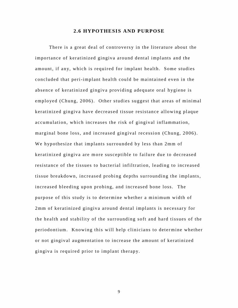
2.6 HYPOTHESIS AND PURPOSE
There i s a great dea l of cont rovers y in the l i t e ra tu re about the
impor t ance of kera t in ized gingiva around denta l implants and the
amount , i f an y, which i s r equi red for implant heal th . Some s tudies
concluded that per i - implant heal th cou ld be main ta ined even in the
absence of ke ra t in i zed gingiva p rovid ing adequate ora l h ygiene i s
emplo yed (Chung, 2006) . Other s tudies sugges t t ha t a reas of minimal
kera t in iz ed g ingiva have decreas ed t i s sue res i s tance a l lowing p l aque
accumulat ion , which inc reases the r i sk of g ingival i nf l ammat ion ,
marginal bone loss , and increased ging ival recess ion (Chung, 2006) .
We h ypothes ize t ha t implants su r rounded b y l ess than 2mm of
kera t in ized g ingiva are m ore suscept ib l e to f a i lu re due to decreased
res i s tance o f the t i s sues to bacte r ia l in f i l t ra t i on , l eading to inc reased
t i s sue breakdown, i ncreased p robing depths su r rounding the implants ,
increased b l eeding upon probing, and increased bone loss . The
purpose of th i s s tudy i s to det e rmine whether a minimum width of
2mm of kera t in ized gingiva around denta l implants i s necessar y for
the heal th and s t ab i l i t y of the su rround ing so f t and hard t i s sues of the
per iodont ium. Knowing th i s wi l l he lp c l in i c ians to det e r mine whether
or not gingival augmenta t ion to increase the amount o f ke ra t in ized
gingiva i s r equi red pr io r to implant the rap y.
10
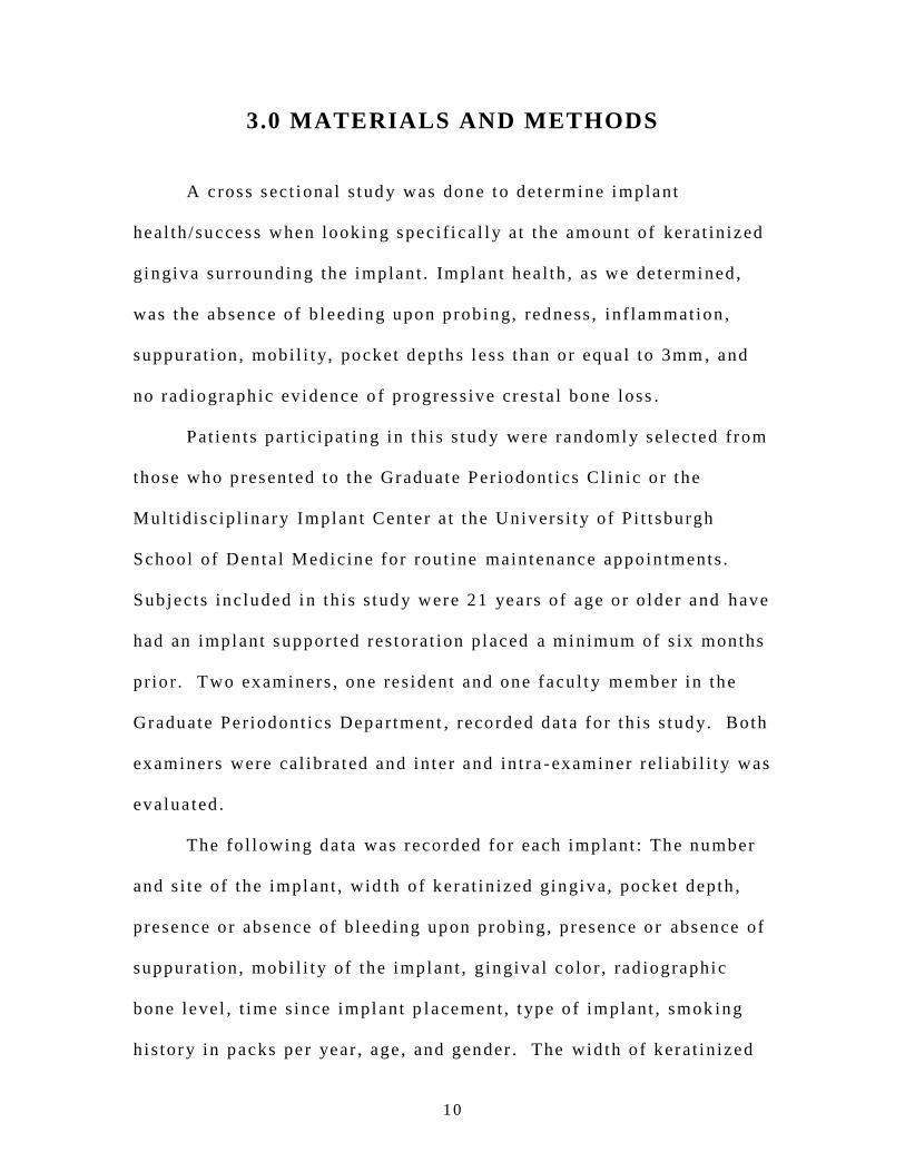
3.0 MATERIALS AND METHODS
A cross sect ional s tud y was done to det ermine implant
heal th / success when looking speci f i ca l ly a t the amount of kera t in ized
gingiva su r rounding the implant . Implant heal th , as we det ermined ,
was the absence o f b l eeding upon p robing, redness , i n f l ammat ion ,
suppurat ion , mobi l i t y, pocket depths l e ss than o r equal to 3mm , and
no rad iograph ic ev idence o f p rogres s ive cres t a l bone loss .
Pat ien ts par t i c ipat ing in t h i s s tud y were randoml y se l ec ted f rom
those who p resented to the Graduate Per iodont i cs Cl in ic o r the
Mul t id i sc ip l inar y Implant Cente r a t the Univers i t y o f P i t t sburgh
School of Denta l Medic ine for rout ine main t enance appoin tments .
Subjects inc luded in th i s s tud y were 21 years o f age o r o lder and have
had an implant suppor ted res tora t ion p l aced a minimum of s ix months
pr io r . Two examiners , one res ident and one facul t y member i n the
Graduate Per iodont i cs Depar tment , reco rded dat a for t h i s s tud y. Both
examiners were ca l ibra t ed and in t e r and in t ra -examiner r e l i ab i l i t y was
evalua ted .
The fo l lowing data was recorded fo r each implant : The number
and s i te of the implant , wid th o f kera t in ized gingiva , pocket depth ,
presence o r absence of b leeding upon p robing , p resence o r absence of
suppurat ion , mobi l i t y of the implant , g ingiva l co lo r , rad iographic
bone level , t ime s ince implant p l acement , t ype o f implant , smok ing
h is tor y in packs pe r year , age , and gender . The wid th o f kera t in ized
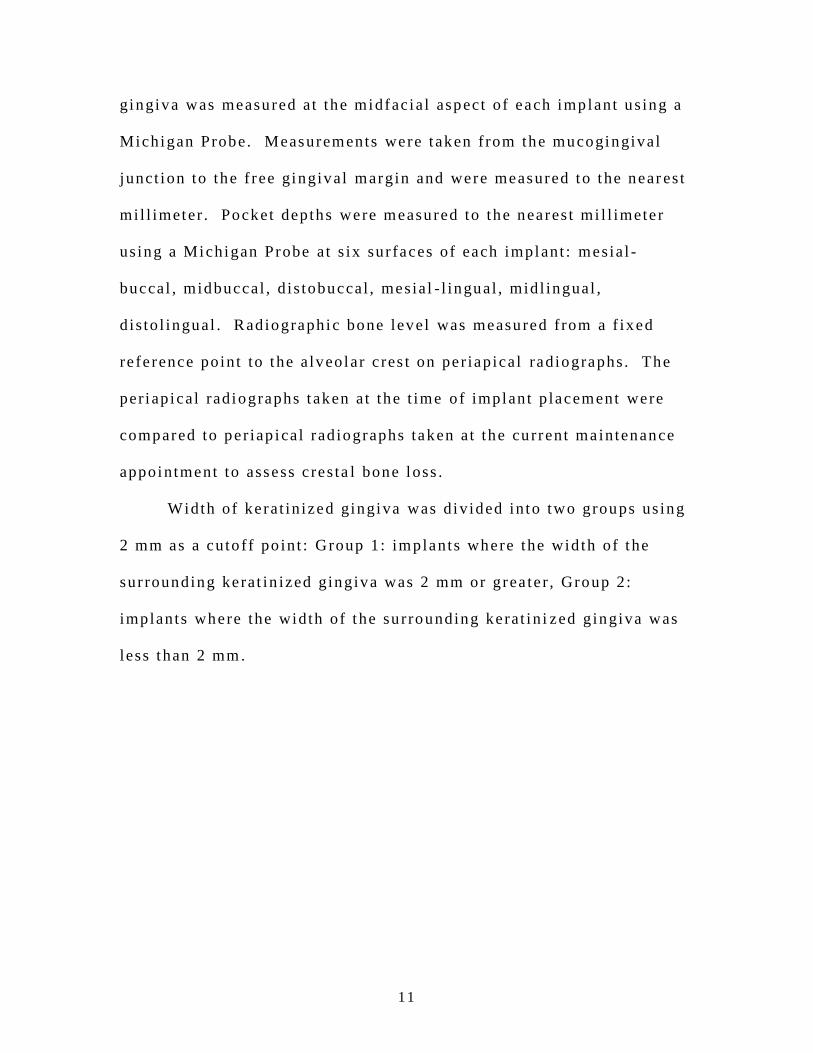
11
gingiva was measured a t the midfaci a l aspect of each implant us ing a
Michigan Probe. Measurements were t aken f rom the mucogingival
junct ion to the f ree gingival margin and were measured to t he near es t
mi l l imeter . Pocket depths were measured to the neares t mi l l imete r
us ing a Michigan Probe a t s ix sur faces of each implant : mes ia l -
buccal , midbuccal , d i s tobuccal , mes ia l - l ingual , midl ingual ,
d i s to l ingual . Radiographic bone level was measured f rom a f ixed
re fe rence poin t to t he a lveolar c res t on per iap ical rad iographs . The
per i ap i cal rad iographs t aken a t the t ime o f implant p lacement were
compared to pe r iap i cal r ad iographs taken a t the cu rrent main tenance
appoin tment to assess c res ta l bone loss .
Width of ke ra t in ized gingiva was d iv ided in to two groups us ing
2 mm as a cu to ff po in t : Group 1 : implants where the wid th of t he
sur rounding kera t in ized gingiva was 2 mm or grea te r , Group 2 :
implants where the width of t he su rrounding kera t in i zed g ingiva was
less t han 2 mm .
12
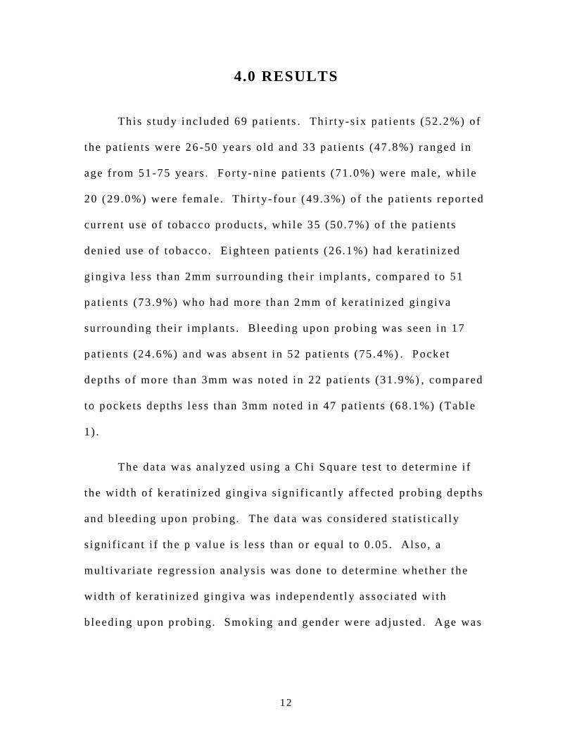
4.0 RESULTS
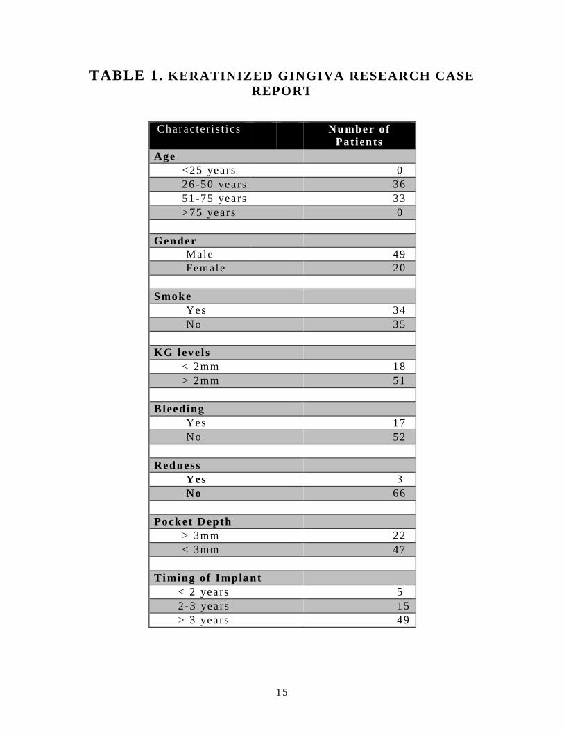
This s tud y included 69 pat i en ts . Thi r t y-s ix pat i en ts (52 .2%) of
the pat i en ts were 26 -50 years o ld and 33 pat ien ts (47 .8%) ranged in
age f rom 51 -75 yea rs . For t y-n ine pat i en ts (71 .0%) were male , whi le
20 (29 .0%) were female . Thi r t y - four (49 .3%) of t he pat ien ts r epor t ed
cur rent u se of tobacco p roduct s , whi l e 35 (50 .7%) o f the pat ien ts
denied use of tobacco . Eighteen pat i en ts (26 .1%) had kera t in ized
gingiva l ess t han 2mm sur rounding the i r implants , compare d to 51
pat i en ts (73 .9%) who had more than 2 mm of ke ra t in ized gingiva
sur rounding thei r implants . Bleeding upon probing was seen in 17
pat i en ts (24 .6%) and was absent i n 52 pat i en ts (75 .4%) . Pocket
depths o f more than 3mm was noted in 22 pat ien ts (31 .9%) , compared
to pockets depths l e ss than 3mm noted in 47 pat i en ts (68 .1%) (Table
1) .
The dat a was anal yzed us ing a Chi Square t es t t o dete rmine i f
the width of ke ra t in iz ed gingiva s igni f i cant l y af fec t ed probing depths
and b leeding upon probing. The dat a was cons idered s ta t i s t i ca l l y
s igni f i cant i f the p value i s l es s than o r equal to 0 .05 . Al so , a
mul t iva r i a te r egress ion anal ys i s was done to dete rmine whether t he
width of ke ra t in ized gingiva was independent l y as socia t ed wi th
b leeding upon p robing. Smoking and gender were ad jus ted . Age was
13
not d i s t r ibuted wel l enough to be used in the model . Other var i ab l es
were not s igni f icant and were not inc luded in the f inal model .
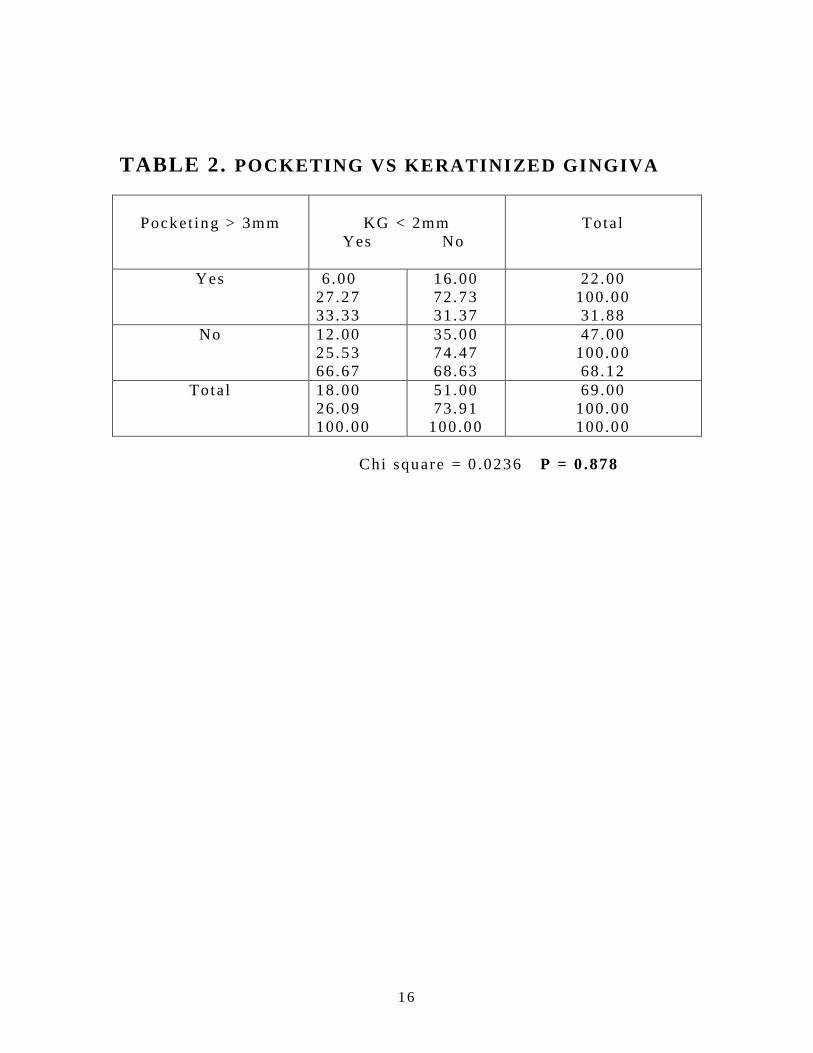
Chi Square anal ys i s was done to evaluat e the as soci a t ion
between the amount of kera t in i zed gingiva and pocket depths .
S ta t i s t i ca l anal ys i s of the dat a fo r pocket depths shows that there i s
no s igni f icant as soci a t ion between pocket ing and amount of
kera t in ized gingiva ; ind icat ing that a l ack of ke ra t in ized gingiva does
not r esu l t in great e r p ocket depths (p = 0 .878) (Table 2 ) .
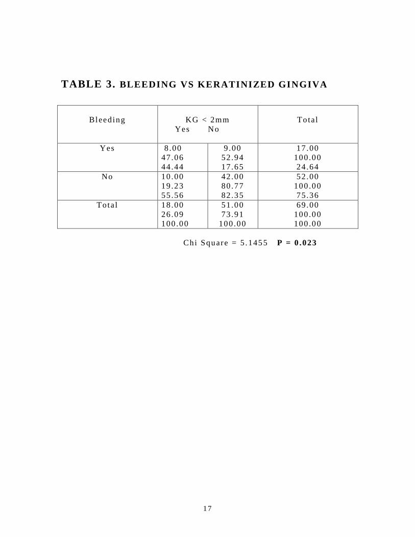
S ta t i s t i ca l anal ys i s of the dat a for b l eeding upon probing shows
that t he re i s a s t a t i s t i ca l l y s igni f i cant a ssoci a t ion between the amount
of ke ra t in ized g ingiva and b leeding upon probing (p = 0 .023) .
E ight y- two percen t of the implants wi th les s than 2mm of kera t in ized
gingiva exper i enced b leeding upon p rob ing as compared to 56% of t he
implants wi th kera t in ized gingiva great er t han 2mm (Table 3) .
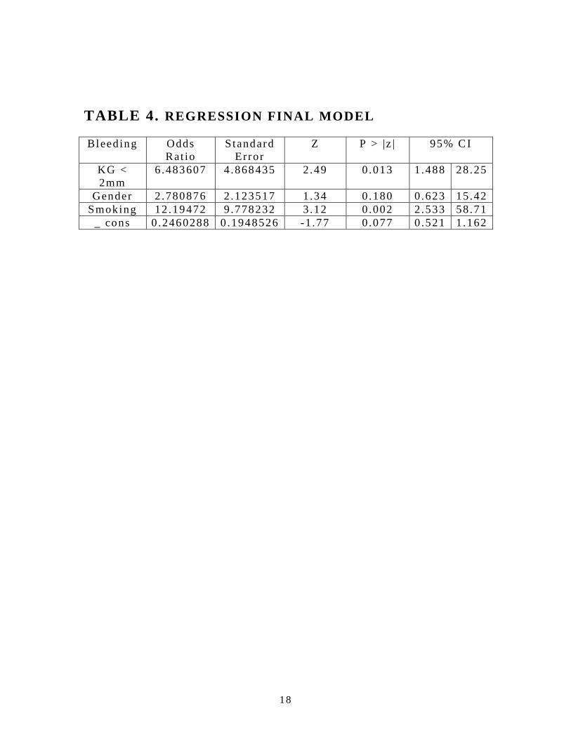
A logis t ic r egress ion model was pe r formed, and i t was ad jus ted
for smoking and gender . S ince age was not d i s t r i buted wel l enough,
i t was not used in t he model . Var iab les such as , suppurat ion ,
mobi l i t y o f t he implant , t iming of implant p l acement , and width of
the implant were no t s igni f icant and were not i nc luded in t he f ina l
model .
Table 4 showed implants wi th l ess than 2mm of kera t in ized
gingiva are 6 .5 t imes more l ike l y to exper i ence b l eeding upon p robing
14
than those implant s wi th great e r than 2mm of ke ra t in ized g ingiva
(OR=6.5 ) . The f ina l mode l was ad jus ted fo r smoking a nd gender .
15
TABLE 1 . KERATINIZED GINGIVA RESEARCH CASE
REPORT
Characte r i s t i cs
Number of
Pat i ents
Age
<25 years 0
26 -50 years 36
51 -75 years 33
>75 years 0
Gender
Male 49
Female 20
Smoke
Yes 34
No 35
KG levels
< 2mm 18
> 2mm 51
Bleeding
Yes 17
No 52
Redness
Yes 3
No 66
Pocket Depth
> 3mm 22
< 3mm 47
Timing of Implant
< 2 years 5
2 -3 years 15
> 3 years 49
16
TABLE 2. POCKETING VS KERATINIZED GINGIVA
Pocket ing > 3mm
KG < 2mm
Yes No
Tota l
Yes
6 .00
27 .27
33 .33
16 .00
72 .73
31 .37
22 .00
100.00
31 .88
No
12 .00
25 .53
66 .67
35 .00
74 .47
68 .63
47 .00
100.00
68 .12
Tota l
18 .00
26 .09
100.00
51 .00
73 .91
100.00
69 .00
100.00
100.00
Chi square = 0 .0236 P = 0 .878
17
TABLE 3. BLEEDING VS KERATINIZED GINGIVA
Bleeding
KG < 2mm
Yes No
Tota l
Yes
8 .00
47 .06
44 .44
9 .00
52 .94
17 .65
17 .00
100.00
24 .64
No
10 .00
19 .23
55 .56
42 .00
80 .77
82 .35
52 .00
100.00
75 .36
Tota l
18 .00
26 .09
100.00
51 .00
73 .91
100.00
69 .00
100.00
100.00
Chi Square = 5 .1455 P = 0 .023
18
TABLE 4. REGRESSION FINAL MODEL
Bleeding Odds
Rat io
S tandard
Error
Z P > |z | 95% C I
KG <
2mm
6.483607 4 .868435 2 .49 0 .013 1 .488 28 .25
Gender 2 .780876 2 .123517 1 .34 0 .180 0 .623 15 .42
Smoking 12 .19472 9 .778232 3 .12 0 .002 2 .533 58 .71
_ cons 0 .2460288 0 .1948526 -1 .77 0 .077 0 .521 1 .162
19
5.0 DISCUSSION
The need fo r ke ra t in ized gingiva around denta l implants has
been a cont rovers ia l top i c . Severa l s tudies have sugges t ed that a
minimal width of kera t in ized gingiva a round implants i s necessar y for
heal th and s tab i l i t y of the implant , whi l e o the r s tudies ha ve fa i led to
demonst ra t e the need fo r minimal width .
The resu l t s of th i s s tud y sugges t tha t implants su rrounded b y
less t han 2mm of ke ra t in i zed g ingiva have an inc reased amount o f
b leeding upon p robing. Bleeding upon probing i s a c l in i ca l ind i cat ion
of ac t ive inf l ammat ion . P ro longed in f l ammat ion around denta l
implant s can resu l t in subsequent a t t achment loss and bone los s ,
u l t imate l y l eading to fa i lu re of the implant .
As s t a ted b y Lang and Loe, the minimum width fo r heal th y
kera t in ized t i s sue surrounding the t ee th i s 2mm (Lang and Loe , 1972) .
This concept has been ca r r ied over to per i - implant ke ra t in i zed t i s sue .
However , s evera l s tudies have chal l enged th i s concept as i t pe r ta ins
to tee th and a l so implants , and have s ta ted that a minimum width of
kera t in ized g ingiva i s not requi red p rov ided adequate o ra l h ygiene i s
main t a ined . Cox and Zarb in 1987 conducted a s tud y in which they
found that 80% of t he implant s evalua t ed had no kera t in ized gingiva
but had heal th y per i - implant t i s sue (Cox and Zarb , 1987) . S imi lar l y,
Esper i n 2012 showed no s t a t i s t i ca l l y s igni f i cant d i f fe rence between
20
bleeding upon p robing and p l aque cont ro l and the width of kera t in ized
gingiva (Esper , 2012) .
Whi le the absence o f kera t in ized gingiva around denta l implants
does not necessar i l y cause pe r i - implan t d i sease , main t a in ing
met icu lous ora l h yg iene in a reas of min imal kera t in ized g ingiva i s
d i f f i cu l t because mobi le mucosa i s more suscept ib l e t o in f l ammator y
changes (Ten Bruggencate , 1991) . P roper o ra l h ygiene may be bet te r
fac i l i t a ted in a reas of adequate kera t in i zed gingiva (Salv i and Lang,
2004) .
Heal th y kera t in ized gingiva around den ta l implants r esu l t s in
more p redic t ab l e success and ma in t enance o f the implant , and a l so
resu l t s in an improved es thet i c outcome. Kerat in ized g ingiva
provides s t ab i l iza t ion to t he pe r iodont ium, p ro t ect s the t ee th and
implants f rom mast i ca tor y and ex ternal t rauma, and p rovides a ba rr i e r
to inf l ammato r y in f i l t ra t e (Paiva , 2012) . Whi l e the sample s ize in
th i s s tud y i s l imi ted , we fee l tha t implants should have a minimum
amount o f 2mm of kera t in ized g ingiva to main ta in heal th . We bel ieve
that recons t ruct ion of the ke ra t in ized g ingiva in de f ic i en t areas us ing
techniques such as t he f ree gingival gra f t o r the subepi thel i a l
connect ive t i s sue graf t should be employed pr ior t o implant
p lacement .
21
6.0 CONCLUSION
Despi te l imi ted dat a in t h i s s tud y, we concluded that r egard less
of the amount o f ke ra t in ized g ingiva present , implant p l acement was
successfu l . However , implants wi th le ss than 2mm of kera t in ized
gingiva exhib i ted incr eased b l eeding upon p robing, which i s a c l in i ca l
s ign o f in f l ammat ion . Pers i s t en t in f lammat ion a round an implant may
poss ib l y cont r ibute to la t er f a i lu re . These f indings may war rant
gingival augmenta t ion p r ior to implan t p lacement i n areas where
minimal ke ra t in ized gingiva ex is t s to p reven t fu ture fa i l ure . Fur the r
s tudies may be needed to conf i rm the f indings f rom th i s s tud y due to
the smal l sample s i ze .
22
7.0 BIBLIOGRAPHY
Abraham CM. A br i ef h i s to r ica l pe rspect ive on denta l implants , the i r
sur face coat ings and t r ea tment s . Open Dent J . 2014 May 16;8 :50 -5 .
doi : 10 .2174/1874210601408010050. eCol lec t ion 2014 . PubMed
PMID: 24894638; PubMed Cent ra l PMCID: PMC4040928.
Adib rad , M, e t a l . S igni f i cance of t he Width of Kerat in ized Mucosa
on the Heal th S t a tus of the Suppor t ing Tissue Around Implants
Suppor t ing Overden tures . Jour nal of Oral Implanto logy. 2009; 35: no 5
Bour i A J r , Bis sada N, Al -Zahrani MS, Faddoul F, Nouneh I . Width o f
kera t in ized g ingiva and the heal th s t a tus of the suppor t ing t i s sues
around denta l implants . In t J Oral Max i l lofaci a l Implants . 2008 Mar -
Apr ; 23 (2) : 323-6 .
Coatoam GW, Behrents RG, Bis sada NF. The width o f ke ra t in ized
gingiva dur ing o r thodont i c t rea tme nt : i t s s igni f icance and impact on
per iodonta l s ta tus . J Per iodonto l . 1981 Jun;52(6 ) :307 -13 .
Chung D, Oh T, Shotwel l J , Misch , C , Wang, H. S igni f i ance of
kera t in ized mucosa in main tenance o f denta l implants wi th d i f fe ren t
sur faces . Journal of Per iodonto lo gy 2006; 77 (8 ) :1410 -20
Cox JF, Zarb GA. The longi tudina l c l i n ica l e f f i cacy o f
osseo in tegra t ed den ta l implan ts : a 3 - year repor t . In t J Ora l
Max i l lofac Implant s . 1987 Spr ing;2 (2 ) :91 -100.
Esper LA, Ferre i ra SB J r , Kaizer Rde O, de Almeida AL. The ro l e o f
kera t in ized mucosa in pe r i - implant heal th . Cle f t Pala te Craniofac J .
2012 Mar;49(2 ) :167 -70 . doi : 10 .1597/09 -022. Epub 2011 Mar 20 .
Greens te in G , Cava l laro J . The c l in i ca l s igni f icance of kera t in ized
gingiva around denta l implants . Compend Cont in Educ Den t . 2011
Oct ;32(8) :24 -31; qu iz 32 , 34 . Review.
Lang NP, Loe H. The re l a t ionship be twee n the width o f kera t in ized
gingiva and g ingiva l heal th . J Per iodon ta l 1972; 43:623 -627
Lin , Guo -Hao e t a l . The S igni f icance o f Kerat in ized Mucosa on
Implant Heal th : A Sys t emat ic Review. J Per iodonta l 2013; 84:1755 -
1767
Lin GH, Chan H L, Wang HL. The s ign i f icance o f kera t in i zed mucosa
on implant heal th : a s ys t emat i c rev iew. J Per iodonto l . 2013
23
Dec;84(12) :1755 -67 . doi : 10 .1902/ jop .2013.120688. Epub 2013 Mar 1 .
Review.
Oh SL. At t ached gingiva: h i s to logy and surg ical augmenta t ion . Gen
Dent . 2009 Ju l -Aug;57(4 ) :381 -5; quiz 386-7 .
Paiva RBM, Mendonça JAG, Zenóbio EG. Per i - implant t i s sues heal th
and i t s associa t ion to the gingival phenot ype . Denta l P ress Implanto l .
2012 Oct -Dec;6 (4) :104 -13 .
Salv i GE, Lang NP. Diagnos t i c pa rameters fo r moni to r ing per i -
implant condi t ions . In t J Oral Max i l lofac Implants . 2004;19
Suppl :116 -27 . Review.
S te t le r KJ , Bis sada NF. S igni f i cance o f the width o f kera t in ized
gingiva on the per iodonta l s t a tus of t ee th wi th submarginal
res tora t ions . J Per iodonto l . 1987 Oct ;58(10) :696 -700.
Wenns t röm J L, Derks J . Is t he re a need fo r kera t in ized mucosa around
implants to main ta in heal th and t i s sue s tab i l i t y? Cl in Oral Implants
Res . 2012 Oct ;23 Suppl 6 :13 6-46. doi : 10 .1111/ j .1600 -
0501.2012 .02540.x . Review.