Embed Size (px)
Citation preview
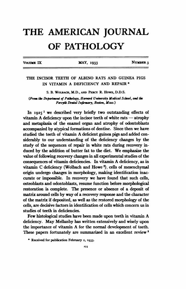
THE AMERICAN JOURNAL
OF PATHOLOGY
VOLUME EX MAY, I933 NuMBER 3
THE INCISOR TEETH OF ALBINO RATS AND GUINEA PIGSIN VITAMIN A DEFICIENCY AND REPAIR *
S. B. WoIBAc, M.D.,Am PEac R. HowE, DD.S.
(From the Departet of Pathl, Hawd UxiwesiyMal School, axd thFors%h Det Infirmary, Bostox, Mass.)
In 1925 1 we describeed very briefly two outstanding effects ofvitamin A deficiency upon the incsor teeth of white rats- atrophyand metaplasia of the enamel organ and atrophy of odontoblastsaccompanied by atypical formations of dentine. Since then we havestudied the teeth of vitamin A deficient guinea pigs and added con-siderably to our understanding of the deficiency changes by thestudy of the sequences of repair in white rats during recovery in-duced by the addition of butter fat to the diet. We emphasize thevalue of following recovery changes in all experimental studies of theconsequences of vitamin deficiences. In vitamin A deficiency, as invitamin C deficiency (Wolbach and Howe e,clls of mesenchymalorigin undergo changes in morphology, making identification inac-curate or impossible. In recovery we have found that such cells,osteoblasts and odontoblasts, resume function before morphologicalrestoration is complete. The presence or absence of a deposit ofmatrix around cells by way of a recovery response and the characterof the matrix if deposited, as well as the restored morphology of thecels, are decisive factors in identification of cells which concem us instudies of teeth in deficiencies.Few histological studies have been made upon teeth in vitamin A
deficiency. May Mellanby has written extensively and wisely uponthe importance of vitamin A for the normal development of teeth.These papers fortunately are summarized in an excellent review3
* Received for publication February 2, 1933.
275
WOLBAC3H AND HOWE
and present definite proof that defective or absent dentine andenamel formations are consequences of vitamin A deficiency. In herstudies of human and experimental animal material, undecalcifiedground sections were employed so that cell responses and histologicalsequences could not be followed. Marshall,4 without giving his-tological details, attributed the formation of "pulp stones" (calcifi-cation in the pulp) in white rats to vtamin A deficency. Coolidge 5mentioned and illustrated irregularities of the dentine and the for-mation of osteoid masses free in the pulp or attached to the dentinein one white rat "of several hundred." The diet of this rat was notgiven and he gave no histological details, but as the illustrations arethose of conditions constant in vitamin A deficency, we believe thatintentionally or inadvertently this rat's diet was deficent in vita-min A. More recently, Shibata 6 reported as consequences of vita-min A deficiency in white rats "abnormal formation of the enamel,dentin and cementum; degenerative changes such as atrophy andabnormality in enamel organ dental pulp and root membrane tis-sues; retardation of the eruption of the incisors; and the nges inthe chemical compositions such as the decrease of phosphorous andcalcium, increase of water magnesium, etc." Shibata refers to earlierpapers of his but does not give the references so that we could notconsult them for the histological descriptions which the above-men-tioned paper lacks. It seems probable that many of the details weshall describe were seen by Shibata.Our endeavor, as part of a program to achieve specific histopath-
ological characterizations of the avitaminoses, has been to follow thesequences due to A deficiency in the teeth for the purpose of ascer-taining the initial or direct (specific) effects, as contrasted with in-direct or non-specific effects such as may be the consequences ofsecondary general disturbances in metabolism. Accordingly sectionsof decalcified skulls were used. Fixation was either in Zenker's liquidor Io per cent formalin. Five per cent nitric acid in 70 per cent al-cohol was used for decalcfying. Celloidin sections stained withhematoxylin and eosin were routinely used, but various special stainsfor connective tissue and reticulin were employed when indicated.The diet used for the rats was that descrbed by us in 1925.
Whether or not the absence of vitamins C, D, and E was of impor-tance we cannot say. We adhered to it because (i) soDrbutic effectscannot be produced in rats; (2) vitamin D is not necessary to prevent
276
INCISOR TEETH IN VITAMIN A DEFICIENCY AND REPAIR 277
rickets in rats with properly balanced inorganic salts in their dietswhen kept under proper hygienic conditions; (3) we know of noeffects of vitamin E deficiency outside of the testes in the male andthe products of conception in the female; and (4) of greatest im-portance, the addition of vitamin A alone sufficed to bring abouthistological recovery.The diet used for the guinea pigs was that described by us in I928.7
We varied the amount of orange juice in an attempt to find anamount which would prevent scorbutus and yet allow an A de-ficiency. Twenty cc. of orange juice daily is sufficient to preventhistological effects of vitamin A deficiency in any organ in guineapigs of 300 to 500 gm. weight. Eight cc. daily does not prevent theepithellal metaplasia of vitamin A deficiency. Two cc. daily is suffi-cient to prevent scorbutic lesions in bones. The majority of ourguinea pigs received 4 to 8 cc. of orange juice daily. This we empha-size because certain findings in the teeth of guinea pigs which werereported by Hojer 8 are apparently duplicated in guinea pigs onvitamin A deficient diet plus 4 to 8 cc. of orange juice daily.Our studies were made from frontal plane sections through the
skull at three or more levels each, including the upper incisor teeth.Owing to the curvature of the incisor tooth the frontal plane sectionsdo not give true cross-sections, except in the midportion of thetooth. Allowance must be made for this in viewing the illustrationsbecause the sections through anterior and posterior portions of thetooth cut the labial and lingual walls obliquely. The lower incisorswere studied in sections at the level of the first molars in rats withdifferent degrees of the deficiency. In guinea pigs and in rats duringrecovery the lower jaws were sectioned at three levels, one throughthe first molars, and two posterior to that.The following tables serve to indicate the scope of our material.
THE INCISOR TEETH OF RATS
The gross changes have not been accurately followed by us.Growth of the teeth seems to be commensurate with the growth ofthe skull, which shares in the marked retardation of the skeleton asa whole. The teeth lose the normal orange pigmentation and acquirea chalky-white appearance, due we believe to loss of the enamelwhich is the pigmented part, and to change in composition of the
278 WOLBACH AND HOWE
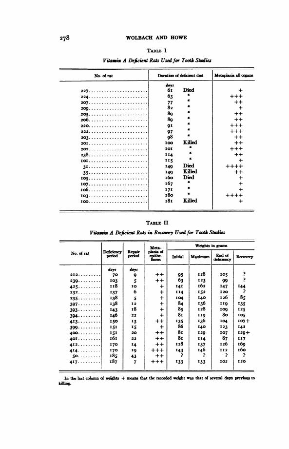
TABLE 1
Viamix A Deficiext Rats Used for Tooth Studies
No.of rat Dumaim ofdect dnt Metaplasia al orgs
'as227 .. .. 6i Died +224 .63 +++207 .77 ++209 .82 +
205 .89 ++20 .89 £ ++
220. 91 +++22 .97 +++203 .98 ++2010.10KIlOed ++202.. 101 ++
238 .114 £ ++101. I5 +
31 .149 Died ++++
35.1I49 Kied ++5 I..6o Died +
107 .67 +1 0 6 ......................171£+
103. 80 ++++IO.. KIilled +
TABL II
Viamix A Deficien Rats ix Recoy Usedfor Tooth Studies
Met&- Weights in grmsNo. ci rat Deficxy Re pNo.onLpperiod eriod l Moginj End ofRci a
days days212 ...... ......709++ 95 128I 05 ?
239.......,, 103 5 ++ 63 123 99 ?
425......... II8 10 + 141 162 147 144232.........137 6 + 114 152 120 ?235.1....... I38 5 + 104 140 126 85397......... 138 12 + 84 136 II9 135393--------. 143 I8 + 85 128 lO9 125394-.1...... I46 22 + 8I "I9 80 OS413 ........ I15 13 ++ 135 136 I14 107+399., I 5 + 86 140 123 142400....... 151 20 ++ 8I 129 107 129+401......... i6i6 22 ++ 8 I I4 87 117412.........170 14 ++ 128 137 126 I69414,,,,,,. 170 19 +++ I43 146 112 160So......... I85 43 ++ ? ? ? ?
417 ........ 1837 7 +++ 133 133 102 120
In the last colmn oi weights + meas that the reixded I t was that of seveal days previoU to
}iln.
INCISOR TEETH IN VITAMIN A DEFICIENCY AND REPAIR 279
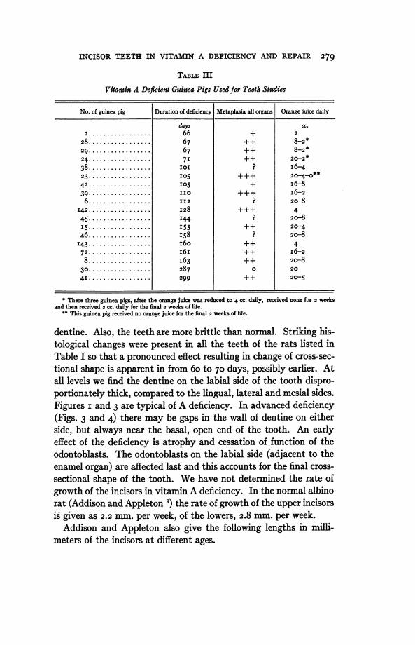
TABLE III
Vitamin A Deficient Guinea Pigs Used for Tooth Studies
No. of guinea pig Duration of deficiency Metaplasia all organs Orange juice daily
days cc.2................. 66 + 2
28 ................. 67 ++ 8-2*29 ................. 67 ++ 8-2*24 ................. 7I ++ 20-2*386-4................. I I ? i6423 ................. 5I0 +++ 2040**42 ................. I05 + I6-839 ................. IIO +++ I6-26................. 112 ? 20-8
142 .............1.... I28 +++ 445 ................. I44 ? 20-8I5 .............1.... I53 ++ 20-446 ................. I58 ? 20-8143 ................. I60 ++ 472 ............6..... O ++ I6-28................. I63 ++ 2o-830 ............... 287 0 2041 ................. 299 ++ 20-5
* These three guinea pigs, after the orange juie was reduced to 4 cc. daily, received none for 2 weeksand then received 2 CC. daily for the final 2 weeks of life.
** This guinea pig received no orange juice for the final 2 weeks of life.
dentine. Also, the teeth are more brittle than normal. Striking his-tological changes were present in all the teeth of the rats listed inTable I so that a pronounced effect resulting in change of cross-sec-tional shape is apparent in from 6o to 70 days, possibly earlier. Atall levels we find the dentine on the labial side of the tooth dispro-portionately thick, compared to the lingual, lateral and mesial sides.Figures i and 3 are typical of A deficiency. In advanced deficiency(Figs. 3 and 4) there may be gaps in the wall of dentine on eitherside, but always near the basal, open end of the tooth. An earlyeffect of the deficiency is atrophy and cessation of function of theodontoblasts. The odontoblasts on the labial side (adjacent to theenamel organ) are affected last and this accounts for the final cross-sectional shape of the tooth. We have not determined the rate ofgrowth of the incisors in vitamin A deficiency. In the normal albinorat (Addison and Appleton 9) the rate of growth of the upper incisorsis given as 2.2 mm. per week, of the lowers, 2.8 mm. per week.Addison and Appleton also give the following lengths in milli-
meters of the incisors at different ages.
WOLBACH AND HOWE
Age uppe ns LouI ci'23 daYy ........... .............. 12.8 I8.I41 .........................15.0 21.7I weeks........1................ 1.3 25.51I5 . ................ 2M32645 months .........*.*.*. 23-3 29-48 . 23.7 29.910 . ..................... 26.2 31.3
If in vitamin A deficency the rate of growth of the incisor teeth inrdation to the growth of the skeleton as a whole is approximatelythat of the normal animal, it is probable, and we believe certain, thatthe entire tooth is replaced through growth during dietary periodscovered by our observation. This must be kept in mind in studyingsections taken at different lengths from the formative end of thetooth.The enamel organ which extends for the entire length of the incisor
tooth on its labial surface (elsewhere only about i mm. from thebasal formative end) early undergoes atrophy and metaplasia (Fig.2) and consists finally of atrophic remnants of the epithelial papillaeand squamous cells replacing the ameloblasts and stratum inter-medium (see Fig. 5 for contrast). The anterior part of the enamelorgan is first affected but finally the whole length is involved, indud-ing the basal formative end (Rat 3I, Table I). At the basal endthere is continuous renewal of the enamel organ and in many ratswith marked metaplasia in many organs practically normal struc-tures were found here, though with distorted relationships, owing topressure deformity of the tooth because of defective or absent den-tine formation. According to Addison and Appleton the ameloblaststravel forward as the incisor grows and it is a fact of interest that fora brief period these cells, as well as the rest of the enmel organ, mayretain normal structural appearances. The effect of the deficiency ismost apparent after the ameloblasts have reached the region wherethey are normally most active in enamel formation. Toward theocclusal end, though still within the area of functional enamel organ,the metaplasia may become complete and as a result the enamel or-gan may be replaced by an epidermis-like structure with superficialkeratinized cells. In middle and posterior parts we have not seen thisdegree of metaplasia. In the atrophy of the enamel organ (Fig. 5) theepithelial papillae are affected first. These structures shrink in sizebecause of atrophy of cells and the surrounding connective tissuesbecome less vascular. The ameloblasts diminish in size, become
280
IWCSOR TEETH IN VITAI A DEFICIENCY AND REPAIR 28I
coarsely granular and finally disapear. Cell outlines are lost andthe ameloblast layer becomes represented by a layer of granular ma-terial and pyknotic nudei, and then disappears. Cells presumablyof the stratum intermedium persist as flat cells, usually two rowsdeep, until in very late stages in the anterior half of the tooth thesedevelop numerous layers of flat cells, the most superficial of whichoccasionally become keratinized. Very rarely, in contrast to its oc-currence in guinea pigs, after complete loss of ameloblasts globularformations of calcified material develop in the connective tissue ad-jacent to atrophied papillae (Fig. 6). This calcification followingatrophy of the structure whose function it is to receive and segregatecalcium salts from the blood stream, invites speculations regardingthe possibilities of selective permeabilities of the ies supply-ing the epithelial papillae.The sequences determined by study of rats in varying degrees of
the defidency are supported by the sequences of repair during re-covery induced by feeding butter fat. The first evidence of recoveryin the enamel organ is the plumping of cells of the epithelial papillae.This is accompanied by engorgement of capillaries and a less denseappearance of the connective tissue immediately adjacent to thepapillae. The earliest response was in Rat 417 (Table II) after 7days of treatment. Subsequently, and in a very few days, the sur-face cells, presumably of stratum intermedium origin, becomecuboidal in shape and continue to assume more and more the appear-ance of ameloblasts (Rats 397 and 399, Table II, for example). Inrats responding favorably to treatment, as shown by rapid gain ofweight, the enamel organ is practically normal in appearance on theigth day (Rat 414, Table II).The details of chnges in the pulp and odontoblasts are more difl-
cult to follow than those in the enamel organ. Siultaneously withchanges in the enamel organ occurs atrophy of the odontoblasts but,as we have said above, the odontoblasts on the labial side (adjacentto the enamel organ) retain their morphology and function for longperiods. This accounts for the extraordinary thickness of the den-tine on the labial side, as contrasted with the remainder of the tooth.It is naturally a pertinent suggestion that the enamel organ, eventhough undergoing atrophy and separated by a wall of dentine andenamel, still exerts an influence upon the maintenance of the odonto-blasts comparable to that in the fetus and at the basal formative end.
WOLBACH AND HOWE
Conversely, one may argue that all of the changes in the odonto-blasts are secondary to atrophy of the enamel organ and withdrawalof its influence. At the basal formative end the odontoblasts undergoearly atrophy, except on the convex or labial surface.The atrophy of odontoblasts, with the exception of those opposite
the enamel organ, proceeds rapidly and is complete usually whengeneral histological changes in many organs are well established, andtherefore in as short a period as 6o to 7o days. All cells resemblingodontoblasts disappear and the intemal surface of the dentine be-comes bounded by cells that are indistinguishable from other con-nective tissue cells of the pulp until extreme thinning of the dentinehas resulted (Fig. 7).During the process of atrophy, odontoblasts, and rarely capillaries
that supply them, become incorporated into the dentine becausesome cells remain functional for a longer time than others and bycontinuing to form it surround non-functioning cells with dentine.This detail is most strikingly seen when the odontoblasts on thelabial side finally begin to atrophy (Fig. 8).
In rare instances, not accounted for by us, cells resembling osteo-blasts in size and shape were present on the inner surface of the den-tine and had deposited a thin interrupted layer of bone (secondarydentine?).
In the depths of the pulp small areas of osteoid tissue were fre-quently found in rats with fully developed deficency. Going overthe whole series of rats we worked out the sequences of pulp ossifica-tion as follows: first, minute spherical globules of hyaline matrix ap-pear between the pulp cells and smultaneously narrow zones ofsimilar material form about occasional capillaries; these deposits in-crease and incorporate pulp cells which assume the morphology ofosteoblasts, and finally cells in contact with the periphery acquirethe morphology of osteoblasts and presumably function as such.
In consequence of the atrophy of odontoblasts, as the tooth con-tinues to grow, the dentine near the formative end becomes verythin, often sharply folded or pleated, or entirely absent (Flgs. 3, 9,io and I3). Associated with these late consequences of the deficencyare aggregations of cells of two types, each responsible for the for-mation of masses of dentine (dentides) continuous with or separatedfrom the tooth wall. In every instance where our sections passedthrough or near to the basal formative end, when these cell aggrega-
282
INCISOR TEETH IN VITAMIN A DEFICIENCY AND REPAIR 283
tions were present we found strikingly defective dentine formationassociated with atrophic odontoblasts and much atrophied amelo-blasts in Hertwig's sheath (the enamel organ at the basal formativeend).
In some instances columnar cells were enclosed within folds of den-tine and the relationships were such as to indicate positively thatthese cells were incorporated ameloblasts. The morphology indi-cated also that these cells were ameloblasts because they were cylin-drical and radially arranged in contrast to the shrunken odontoblastson the opposite side of the dentine. In regions of complete absenceof the dentine at or dose to the formative end there were frequently,in the gaps, gland-like dusters of cells that we are also forced tointerpret as of enamel organ origin. Such gland-like formations alsolie under the thin abnormal dentine (without canaliculi) at consider-able distances from the basal end (Figs. II, 12 and I5). One type ofcell aggregate then we regard as of enamel organ origin and due tothe fact that cells (presumably ameloblasts because, as we shl showbelow, they do not survive in repair) become incorporated by thepleating of a very thin and atypical dentine at the formative end.The other type of cell aggregate is proliferative in type and con-
sists of cells derived from the pulp that forms in regions of ex-tremely thin or totally absent dentine. These cells are short, plump,basic staining, and resemble transitional forms between mesen-chymal cells and osteoblasts. Their presence is accompanied bydeposition of atypical dentine (osteodentine) upon the tooth wall(Figs. 3, 9, Io and ii), although in very severe deficiency they formno matrix at all. These cell aggregates we regard as reparative inongin, comparable to callus formation in bone, and indicative thateven in extreme vitamin A defidency proliferative responses on thepart of connective tissues may be energetic. We have further proofof this in the repair of wounds made for experimental purposes. Allof our decisions regarding origins of cells were made after carefulstudy of the histology of repair during recovery induced by sub-stituting butter fat for lard in the diet.An outline of the sequences observed in recovery is therefore indi-
cated. As we have descrbed above, the restoration of the enamelorgan takes place first at the basal formative end. Very promptlythe odontoblasts in juxtaposition to the enamel organ at this end re-cover and by the 7th day (Rat 417, Table II) in the region of Hert-
WOLBACH AND HOWE
wig's sheath they have assumed normal morphology and function, asshown by the deposition of predentine with naliculi. The pre-dentine calcifies rapidly. Folds in pleated dentine fill in rapidly.Excess atypical dentine, so-called secondary dentine or osteodentine,the product of the proliferative pulp cell aggregates and denticles ofameloblast inclusion onrgin grow in size by the deposition of dentineupon them (Figs. i6, I7 and i8). The ultimate fate of these "den-tides" we have not followed, but it seems reasonably certain thatthey continue to increase in size while advancing forward as thetooth grows.The reappearance of columnar odontoblasts is represented in all
stages by our series of repair experiment rats. It takes place first atthe basal formative end and works forward, first upon the labial sideof the tooth, presumably in response to the recovery of the enamelorgan. Later it extends throughout the tooth. Predentine forma-tion is active before the odontoblasts have acquired columnar shapesComplete morphological recovery was almost achieved in ig days inRat 414, which showed a very satisfactory increase of weight inresponse to butter fat in the diet. There is little to record about thesequences of odontoblast recovery. The changes observed at differ-ent periods of recovery were quite like those to be seen in the forma-tion of osteoblasts in granulation tissue in the repair of bone. Con-nective tissue cells in contact with the dentine become plumper andmore basic in staining reaction. For a brief period they are polyhe-dmal in shape and processed like young osteoblasts. Soon they be-come aligned more or less parallelly with their long diameters obliqueor perpendicular to the dentine, but before there is any regularity inarrangement or uniformity in shape, predentine is deposited upon theold dentine. The sequences may be arranged in four stages: (i) in-crease of size, or at least of cytoplasm oDncentrated about the nu-cleus; (2) assumption of osteoblast-like morphology; (3) evidences offunction and polarity, as evidenced by directional deposit of pre-dentine; and finally, (4) further growth in size and acquisition of thenormal shape.
Various stages of odontoblast recovery may be seen in one toothbecause, as stated above, the recovery begins first opposite thenamelorgan, i. e. at the basal end and labial surface. We have thushad ample opportunity in our recovery series to feel secure in com-
284
INCISOR TEETH IN VITAMN A DEFICIENCY AND REPAIR 285
paring the recovery or reappearance of odontoblasts to the sequencesseen in bone repair from fibroblast to osteoblast. Our observationslead us to regard, from the morphological viewpoint, the odontoblastas a polarized osteoblast.The recovery sequences permit no doubt regarding the nature of
the proliferative cell aggregates. They are of pulp connective tissueongin. The other types of cell aggregate in gland-like formation wehave already concluded were of enamel organ onrgin. Again the mor-phological evidence is supplied by the sequences seen in recovery.The first change in recovery is an increase in size of the cells, some ofwhich undergo mitosis (Fig. 12). The pulp cells in contact with thesegland-like formations respond, as do those in conact with the dentine,and proceed to deposit predentine. We thus get the picture of epi-thelial cells in the pulp surrounded by dentine, in turn surroundedby odontoblasts. This is shown in low power at A and B in Figure13 and at higher power in Figure 14. The enlosed epithelial cellsundergo atrophy and disappear (Fig. i9). The last stage of degen-eration is like that of the ameloblasts in the deficiency. The cellsbreak up into coarse granules and the residue becomes calcified;pyknotic nuclei can be seen in dentides of this origin for some timeafter calcification of the cell debris (Fig. i8).
Excessive local formation of atypical dentine (denticles) is pro-duced in vitamin A deficency by focal proliferative responses ofpulp cells. Such deposits are insigificant, but increase in the re-Covery period commensurate with rate and extent of recovery. An-other type of excess dentine formation or denticle is initiated by thepresence of cells of the enamel organ that become incorporated byfolds of dentine or through total gaps in the dentine. In repair thesecells stimulate the formation of odontoblasts from the adjacent pulpand hence dentide formation. We thus have brought new expen-mental evidence to support the prevailing opinion (Orban 1') thatcells of Hertwig's sheath may be carried forward into the pulp andstimulate the formation of odontoblasts from pulp cells, and con-sequently the formation of denticles. Fridrichovsky n described inhurman teeth and in rats' teeth (diet not stated) folds of dentine suchas were of common occurrence in our series. These he regarded asdue to developmental irregularities of Hertwig's sheath. He ex-pressed the opinion that such folds could carry enamel organ cells
WOLBACH AND HOWE
forward, and thus accounted for one source of dentide formation.Naturally he did not consider deficiency disease as a possible causeof the mecnism.
THE INcISOR TEETH oF GuNEA PIGS
ID general the effect of vtamin A deficiency upon the incisor teethof guinea pigs is similar to that in rats. There are some quantitativedifferences and some apparent contradictions appear to our interpre-tation of events from the teeth of rats. The outstanding difference isthe rapidity and severity of the enamel orga n ges. The apparentcontradiction is exhibited in the prolonged persistence of the mor-phology and function of odontoblasts after marked degrees of enamelorgan atrophy.The durations of the deficent diet in the experiments with
guinea pigs were on the whole shorter than those with rats, inasmuchas at the beginning the majority of the guinea pigs received sufficentorange juice to afford very considerable amounts of vitamin A.Twenty cc. of orange juice daily is completely protective againsthistological vitamin A effects in all tissues, except occasionally in therespiratory mucosa in the nares. Sixteen cc. daily is nearly protec-tive. Therefore, the durations given in Table Ill are not those ofcompletely deficient diets. For example, Guinea pig 38 receivedI6 cc. of orange juice daily for 56 days, 8 cc. daily for 2I days and4 cc. daily for 24 days. Guiea pig I5 received 2o cc. for 6 days, 8 cc.for go days and 4 cc. for 57 days. Guinea pig 42 recived I6 cc. for55 days, 8 cc. for 50 days; Guinea pig 39 received I6 cc. for 63 days,4 cc. for 7 days and 2 CC. for 4o days. Guinea pig 6 received 20 CC. for7o days and 8 cc. for 39 days. Gulinea pig 45 received 2o cc. for 69days and 8 cc. for 75 days. Guinea pig 46 received 20 CC. for 28 daysand8cc. for 130days. Guineapig8received 20CC. for 52 daysand8 cc. for II days. Guinea pig 41 received 20 CC. for 209 days, IO cc.for 15 days and 5 cc. for 75 days.
Gross evidence of scorbutus was absent in each of the guinea pigsin Table III. Histological evidence was found in the costochondraljunctions of Guinea pig 23 which received no orange juice during thelast two weeks of the experiment. These changes were so slight as tosuggest that the retardation of growth secondary to the A deficiencywas a factor, inasmuch as there was the typical athreptic type of
286
INCISOR TEETH IN VITAMIN A DEIICIENCY AND REPAIR 287
costochondral junction. In Guinea pigs 24, 28 and 29 there weremicroscopic evidences of repaired scorbutus in the teeth, such as wedescribed in 1926.2 The scorbutic factor, as possibly responsible forthe changes we found in our vitamin A defident guinea pigs, may bedismissed because of the fact that all of the changes we are describingwere found in guinea pigs that had never received less than 8 cc. oforange juice daily and that exhibited no gross or microscopic evi-dences of scorbutus. This we emphI beause Hojer 8 has de-scribed and illustrated changes involving odontoblasts and dentine,with the production of osteoid or bony formations in the pulp andupon the dentine in latent or partial scorbutus. As our own workwith scorbutus has been upon animals completely deprived of vita-min C we have as yet no basis for criticizing Hojer's conclusions.Such premises as we now have incline us to believe that in partialvitamin C deficiency Hojer's results would be duplicated. Both vita-min A and vitamin C deficiencies affect odontoblasts, though bydifferent routes, but the effect upon dentine formation may be thesame.
ThE ENAMEL ORGAN
The first demonstrable effect of the deficency upon the incisorteeth is in the enamel organ. As in the case of the rat the externallayer atrophies first. Tne ameloblasts become smaller and shorter.In rapidly progressing deficency they finally undergo granular de-generation and dintegrate. Usually low cuboidal cells persist forlong periods and we are not able to say positively whether they areatrophied ameloblasts or cels of the stratum intermedium, thoughwe believe they are derived from the latter, inauch as often thesecells have horizontal diameters nearly double the vertical, and it isimprobable that atrophied cells should undergo such a change.Eventually the enamel organ completely disappears, unlike whatoccurs in rats. Before the atrophy is completsglobules of calcifiedmatrix appear in the outer layer of the enamel organ between the in-conspicuous epithelial papillae and within the epithelium itself.These globules increase in size and number, extending into the adja-cent connective periodontal membrane. Similar calcified depositsform above the atrophic remains of the enamel epithelium and en-close epithelial cells (Figs. 2o and 21). When large, these calcifieddeposits take on the appearance of bone (Figs. 22 and 23), because
WOLBACH AND HOWE
of the inorporated cells. However, the cells do not have the appear-ance of osteoblasts. They are without processes and in the largestplaques are surrounded by dear spaces reminiscent of crtiagerather than bone. Neither do these plaques become surrounded byosteoblasts. They resemble cementum rather than other examples ofpathological bone formations, such as we are familiar with. They areoften bounded on the deep side by cuboidal epithelial cells thatlaterally are continuous with atrophic cells of the enamel organ.The plaques increase by accretion. Globules of matrix continue tobe deposited from the adjacent connective tissue.
TME ODONTOBLASTS
Atrophy of the odontoblasts occurs late. In no guinea pigs of ourseries was there complete cewation of dentine formation; alwaysthere were on the labial side a normal-appearing layer of odonto-blasts and evidences of functional activity; on mesial and lateralsides only partial atrophy was found and hence the cross-sectionalshape peculiar to the rat's incisors was not seen in the guinea pig.As in the case with rats the atrophy of odontoblasts appeared firston the lingual side of the tooth and never before advanced atrophyof the enamel organ had occurred. A very striking difference fromoccurrences in rat teeth is that coincdent with the atrophy of odon-toblasts, spiculesof a modified dentine form (Fig. 24). This material,at first uncalcified and similar to predentine in all respects exceptthat it contains no naliculi, becomes finally calcified and con-tinues to grow by peripheral deposit. In long-ontinued deficiences,when these dentine-like deposits extend deeply into the pulp, theybecome surrounded by osteoblasts which they incorporate as theygrow; further increase in size is like that of growth of osteoid tissue(Fig. 25).The earliest formation of these amorphous dentine processes we
found in regions of most marked atrophy of the odontoblasts andalways upon the lingual side of the tooth. The initial stage we foundto be a deposit of predentine-like matrix between atrophied odonto-blasts that had lost their columnar form. It was evident that func-tional activity continued, but polarity of the odontoblasts was lost.In early formations but few cells are found endosed in matrix. Theoutgrowths for a period are surrounded by other atrophic cells of the
288
INCISOR TEETH IN VITAMIN A DEFICIENCY AND REPAIR 289
odontoblast layer which retain their polarity. Finally, polarity islost and the cells concemed in further growth are indistinguishablefrom osteoblasts.
The Pulp: The pulp practically unnged. The onlyossification we found in our series took origin in the manner de-sbed above as a continuation of growth of dentinoid proces.The Cementum: As a rule in well establishedA deficency there are
local in eases in thickness of the cementum, for the narrow, deeplycalcified border surrounding the dentine is to be regarded as cemen-turn. In fact the only cementum resembling bone we have seen inconnection with the incisors of guinea pigs has been in A deficiencyanimals. The formation of cementum of appreciable thickness andenclosing cells is always found first upon the lingual side, but mayextend on lateral and mesial sides forward to the enamel organ regionas an irregular layer (Flg. 24). Frequently sharply circumscribedcementum formations were found, perhaps warranting applicationof the term cementide (Fig. 26).
DISCUSSION
Three facts are of assistance in interpreting vitamin A deficencyeffects upon incisor teeth of rodents: (I) the enamel organ moves for-ward with the growth of the tooth so that the ameloblasts maintainthroughout the same relative position to caldfied structures; (2) thedentine increases in thickness progressively toward the anterior(occdusal) end, indicating activity of the odontoblasts extendingnearly to the end of the tooth; and (3) the occlusal end of the pulpchamber is being constantly filled in by bony material (osteoden-tine), indicating a normal tendency on the part of pulp cells to pro-duce osteoid matrix and ceation of a polar deposition of matrix onthe part of the odontoblasts.
Fact i is of assistance in explaining why the earliest and maximumatrophy of the enamel organ is found at some distance from the for-mative end as a requirement of time for the deficiency to take effect.Fact 2, as dentine is not resorbed in consequence of the deficiency,exlains the greater thickness of dentine for long periods in the de-ficency toward the occdusal end. Fact 3 warrants the assumptionthat the deposit of osteodentine following atrophy of odontoblastsis an adaptation or premature stmulation of a normal sequence.
WOLBACH AND HOWE
Thne enamel organ nges we have described in rats and guineapigs arep consequences of vitamin A defidency common tomany epitllal organs. The more rapidly produced and pro-nounced canges in guinea pigs, as compared with rats, are in con-formity with the changes in other organs in these two animals (Wol-bach and Howe 1).We prefer to interpret some effects upon odontoblasts as second-
ary to e el organ atrophy, assuming that the latter, in teeth thatare continuously growng, exerts an influence upon the former, assuggested by the early sequences in tooth formation and throughoutlife at the basal formative end of rodents' indsors. This interpreta-tion is not supported by the facts that odontoblasts persist afterdisappearance of the enamel-forming organ in all stationary per-manent teeth of animals, and that in inacsor teeth of rodents theenamel-forming organ is continued forward only upon the labialside. Nevertheless, our observations show that enamel organ at-rophy precedes atrophy and depolaization of odontoblasts. Thefact that in rats odontoblasts remai morphologically and function-ally active on the labial side, in apposition to the enamel organ., longafter complete disappearance upon other surfaces, and that in thissituation their eventual atrophy only follows enamel organ atrophy,dnds the above interpretation. The finer sequences attendingatrophy of the odontoblasts on the labial side, which we have care-fully followed, here and there an odontoblast ceasing to depositmatrix upon its external pole, with continued deposition centrif-ugally, support strongly the argument that even though the odonto-blast may be idividually physiologically independent of the enamelorgan, architecturaily it is dependent. We have come to regard theodontoblast as a polarized osteoblast and the enamel organ as thepolarizing agent. In guinea pigs the same changes occur, though inless evident degree. The persstence of normal odontoblasts uponthe labial side is more pronounced than in rats. Odontoblast atrophyis less striking, yet ilustrates more conspicuously in the sequences wehave descrbed the effects of the depolarization of odontoblasts, asshown by the extensions into the pulp of the osteodental processes(dentides) built up by these depolarized cells.Late vitamin A deficiency effects in bones, as described by Pap-
penheimer,l2 and completely confirmed by us in unpublished studies,indude complete cessation of growth. It may, therefore, be argued
290
INCISOR TEET IN VITAMI A DEFICIENCY AND REPAIR 291
that effects similar to those in bone should be expected in odonto-blastic activities and that both are of the same nature. This we mayadmit without affecting the rWle of the enamel epithelium in the de-termination of the order of events. More direct attack based uponexperimentally produced lesions of the enamel organ is needed tosettle these problems.The formation of bone in rats in the pulp proceeds, we have
learned, as does the ossification in membranous bone formation,through direct ange in character of the intercellular matrix, pre-ceded by finely granular calcium deposition. Calcification in and ad-jacent to the atrophied mel organ, followed by ossification, inguinea pigs may be regarded as a phenomenon having similar patho-genesis. In both tances the cells directly responsible for theelaboration of spcific calcified structures, dentine and enamel, arepartly or completely inactive, a fact that invites speculations alongthe lines of a continued extracapillary delivery of calcium and phos-phorous compounds to the tissues, and therefore to questions of dif-ferentiated permeability of capillary endothelium. The high mineralcontent of enamel and its rapid growth rate present problems con-cerning concentration that make such speculations attractive.We have presented two oringins of osteodentine excrescences, or
dentides, one occurring in both rats and guinea pigs by the depolari-zation of odontoblasts, and in the rat accompanied by a proliferativereparative response of pulp cells; the other, only in rats, induced byinclusions of ameloblasts in folds of recently formed dentine, late inthe deficency, dose to the formative end. These solutions as toorign could not have been achieved without having for study manystages of repair.We have observed no changes in the exceedingly thin calcified
zone surrounding the dentine in rats which by some morphologists isregarded as cementum. In guinea pigs definite cementum over-growths (cementides) were frequently seen on the lingual side of theteeth in consequence of the deficency, but we can offer no explana-tion except the suggestion that they formed in response to structuralweakness of the bony socket as a part of the general athreptic skele-tal effect of late vitamin A deficiency.The application of our observations to human teeth obviously
must be restricted to the formative period and particularly to teethof the second dentition. Our inference, based upon studies of experi-
WOLBACH AND HOWE
mental scorbutus,2 and rickets (unpublished), is that vtamin Adeficiency is the most important because of its effect upon theenamel organ and because recogition of this deficiency is alwayslate and will continue to be so, unless other criteria than oculareffects are looked for by pediatrins Defective enamel formation,ossification of the pulp, dentide and cementidle formation are allpossible consequences of vitamin A deficiency in humans. Studies ofunerupted teeth from infants in vitamin A deficiency, obtained post-mortem, are being made by Boyle,'3 and he has recorded observa-tions that indicate we may safely apply some of our observations tothe humnan.
SUMMARY ANI CONCLUSIONS
I. The initial effect of vtain A deficency upon incisor teeth ofrats and guinea pigs is upon the enamel organ. The ameloblasts re-spond earliest by atrophy, then the remainder of the organ atrophies;finally metaplasia and calcification, and, in the guinea pig, ossifica-tion occur.
2. Atrophy and depolarization of odontoblasts follow enamel or-gan chnges. The odontoblasts survive longest on the side (labial)where in apposition to the enamel orgn and in long-continued ex-periments gross deformities in the incisors of rats resulted from ab-sent or deficient dentine formaton.
3. Two types of dentide formation are described, one built up bydepolarized odontoblasts, the other by indusions of ameloblasts bythe folding of imperfectly formed dentine at the formative end of thetooth.
4. Defective enamel formation and other poorly understood con-ditions in teeth, such as denticles, pulp bone and cementides, mayreasonably be regarded in the hman as vitamin A deficiency possi-bilities.
5. Our observations indicate that in the incisor teeth of rodentsthe odontoblasts throughout life are influenced by the enamel organ.
6. As in other morphological problems concerning vitamin de-ficiences, study of the sequences of repair was essential. We em-phasize the importance of two types of control material, the normaland progressive stages in repair.
292
INCISOR TEETH IN VITAMIN A DEFICIENCY AND REPAR 293
7. Our observations indicate that vitamin A deficency is the mostimportant of the known vitamin defidencies in its effect upon toothformation.
REFERENCES
I. Wolbch, S. B., and Howe, P. R Tissue changes following deprivation offat soluble A vitamin. J. Exper. Med., 1925, 42, 753-777.
2. WolbaCh, S. B., and Howe, P. R. Intrcllular substances in experimentalscorbutus. Arck. Path. b Lab. Med., i926, I, i-24.
3. Mellanby, May. The influence of diet on the structure of the teeth. Physi.Rev., 1928, 8, 545-577.
4. Marshall, J. A. Dental caries and pulp sequelae resulting from experi-mental diets. J. Am. DeWtal Assoc., I927, 4,3-37.
5. Coolidge, E. D. Heterotopic ossification in the pulp: a contnbution to re-search on the biology of the dental pulp. J. Am. Dental Assoc., 1929, I6,821434.
6. Shibata, M. Effects of lack of vitamins on development of teeth. Japa-xese J. Exper. Med., 1931, 9, 21-32.
7. Wolbach, S. B., and Howe, Percy R. Vitamin A deficiency in the guinea-pig. Arch. Palk., 1928, 5, 239-253.
8. H6jer, J. A. Studies in scurvy. Acfa. Paediat., I924, SuppL 3.9. Addison, W. H. F., and Appleton, J. L., Jr. The structure and growth of
the inisor teeth of the albino rat. J. Morphol., 1915, 26, 43-96.Io. Orban, B. Dental Histology and Embryology. P. Blaliston's Son & Co.,
Inc., 1929, Ed. 2.
ii. Fririchovsky, J. Zur Histologie der Deniel Ztschr. f. Somatologi,1927, 25, 124-157.
12. Hess, A. F., McCann, G. F., and Pappenheimer, A. M. Expmentalrickets in rats. H. The failure of rats to develop rickets on a diet de-ficient in vitamine A. J. BioS. Chem., 1921, 47, 395-410.
13. Boyle, Paul E. Manifestatis of vitamin A deficency in a human toothgerm. Report of a cas J. Dedal Research, 1933, 13, No. i.
DESCRIPTION OF PLATES
PLATE 38
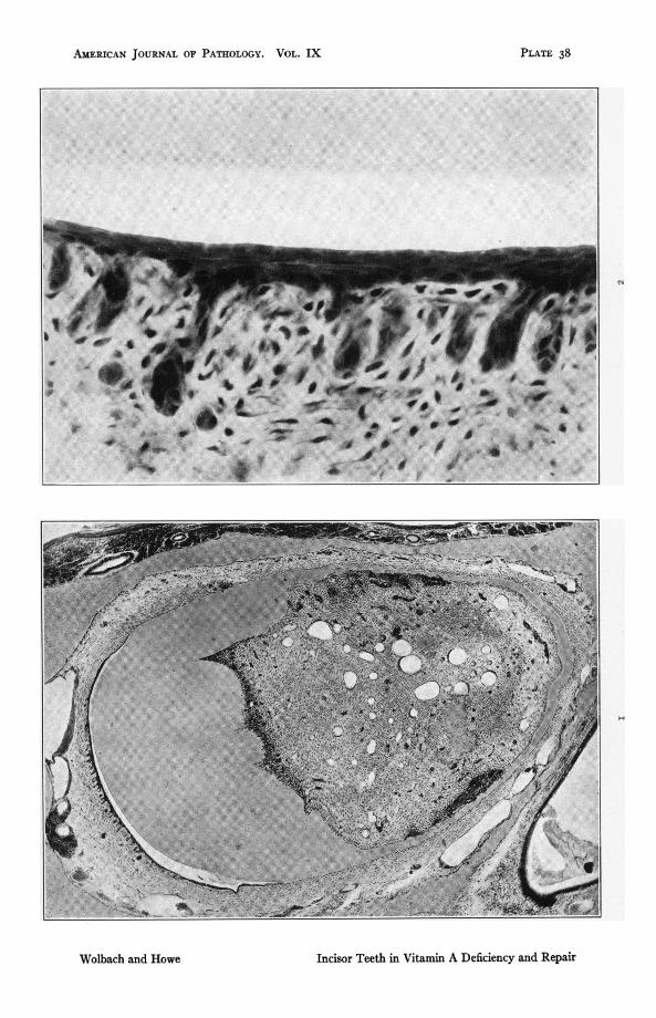
Fig. i. Rat 201. Upperincisor,typicalofAdeflciencyintherat. ioidaysonvitamin A deficent diet. Atrophy and metaplasia of the enamel organ.Odontoblasts still present on labial side where the dentine is abnormallythik. Note thinn and folding of dentine on both sides of tooth wherethere are also many large areas of cellular proliferation derived from pulpcells and possibly from odontoblasts. Osteoid formation present in pulp.x46.2.
FIG. 2. Rat 201. High power of enamel orga Same preparation as Fig. i.ioI days on vitamin A deficent diet. Note great atrophy of the epithelialpaplae, absence of aloblasts and the several layers of flattened cellsupon the surface. x 588.
AMERICAN JOURNAL OF PATHOLOGY. VOL. IX
C.
Incisor Teeth in Vitamin A Deficiency and Repair
PLATE 38
Wolbach and Howe
PLAIE 39
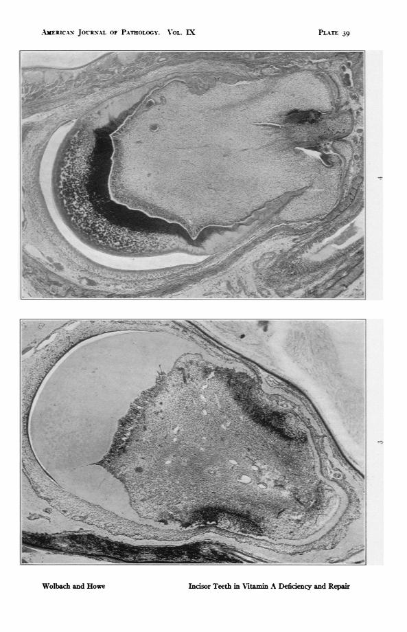
FIG. 3. Rat 205. Upper incisor. 89 days on vitamin A deficient diet. Atrophyand metaplasia of enamel organ. Columnar odontoblasts still present onlabial side. On mesial and lateral sides are gaps in the dentine which iselsewhere thin and much folded. Note the large areas of proliferativeresponse composed of cells resembling osteoblasts. x 46.2.
FIG. 4. Rat 412. Upper incisor. 170 days on vitamin A deficient diet folowedby I4 days with addition of butter fat. There is restoration of the enamelorgan and heavy calcification of the dentine. The layer of recoveringodontoblasts and newly deposited dentine (predentine) shows clearly onboth sides, also folding and gaps of the dentine with gross distortion of theshape of the tooth. On the mesial side there are several groups of amelo-blasts incorporated in folds of the dentine and further embedded by dentineformed during repair. x 46.2.
AbERICAP%N JOURNAL OF PATHOLOGY. VOL. EX
Inisor Teeth in Vitamin A Deficiency and Repair
PL-xE 39
Wolbach and Howe
PLATE 40
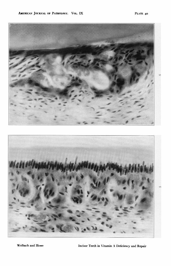
FIG. ,. The enamel organ of rat fed upon the control diet in which butter fatreplaced the lard of the deficient diet. x 588.
FIG. 6. Rat 235. Upper incisor. I38 days on vitamin A deficient diet followedby 5 days with addition of butter fat. Shows extreme atrophy of enamelorgan with globular deposits of calcified material in and adjacent to theatrophic remains of the epithehal papillae. x 588.
AEiJc.AN JoLRs.L oF PATHOLOGY. VOL LX
dw .4dW
*_do. .4-
me 4!I _w _w _w.a0 - 4
oncisor Teeth in V-itamin A Deficiency and Repair
,.a
PIATE 40
4W. -Aw- 410 p- 49P-,%-. 4%. ip 4w w
*4 .0 oft 111%..-
-w illde
--MP AIP'OpOP .0010, 0-1
_.d. 4ow
Wolbach and Howe
PLATE 4I
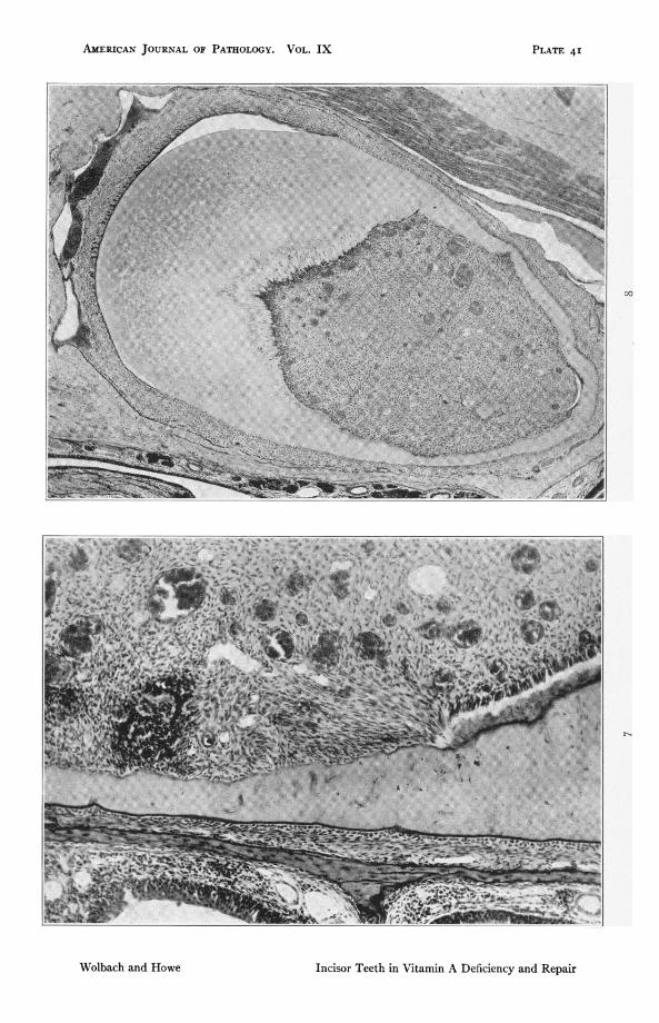
FIG. 7. Rat 412. Upper incisor at junction of mesial and labial sides. i, Odayson vitamin A deficient diet followed by IA davs with addition of butter fat.The preparation illustrates the differences in behavior of the odontoblastsof the labial side from those on mesial and lateral sides. The odontoblastsof the first show deposition of dentine under influence of restored diet. Onthe mesial wall the dentine is bounded by connective tissue-like cells.though earlv repair is shown in places by traces of dentine deposit betweencells. some of which are arranged perpendicularly to the dentine. Note alsonuclei of cells incorporated in the dentine. x I47.
FIG. S. Rat 235. Upper incisor. I38 days on A deficient diet followed by 5 dayswith addition of butter fat. Shows typical shape of vitamin A deficiencv ratincisor. Complete atrophy with metaplasia of the enamel organ. Completeatrophy of odontoblasts except on the labial side where the atrophy isnearly complete. Continued function of some cells has resulted in the in-corporation of others by the most recently deposited dentine. x 46.2.
AMERICAN JOURNAL OF PATHOLOGY. VOL. IX
Wolbach and Howe Incisor Teeth in Vitamin A Deficiency and Repair
PLATE 4I

PLATE 42
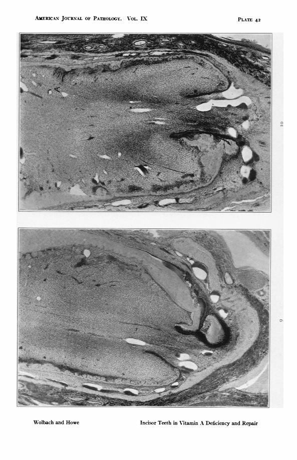
FIGS. g and io. Rat 103. Sections of both upper incisors through formativeends. i8o days on vitamin A deficient diet. Fig. g shows particularly wellthe folding of the dentine which makes possible the inclusion of amelo-blasts. Both photographs show dusters of proliferated odontoblasts and inFig. io the gland-like formations due to ameloblasts free in the pulp at A.For high power see Figs. ii and 12. X 46.2.
AIERIc-A JouRNAL OF PATHOLOGY. VOL. LX
Incisor Teeth in Vitamin A Deficiency and Repair
Pi--tTE 42
Wolbach and Howe

PLATE 43
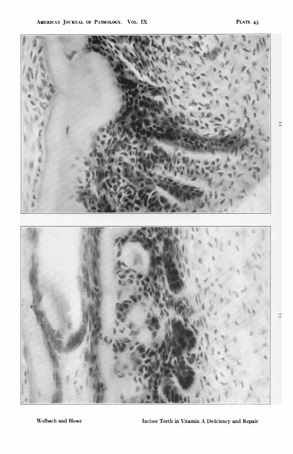
YIGS. ii and I2. High power details of Fig. io showing inclusions of atrophiedameloblasts in the pulp. In Fig. ii osteodentine is being formed by pulpcells external to the inclusions of ameloblasts. x ,88.
AMERICAN JOURNAL OF PATHOLOGY. VOL. EX
Incisor Teeth in V-itamin A Deficiency and Repair
I
I
PLATE 43
7-
2-
Wolbach and Howe
PL-TE 44
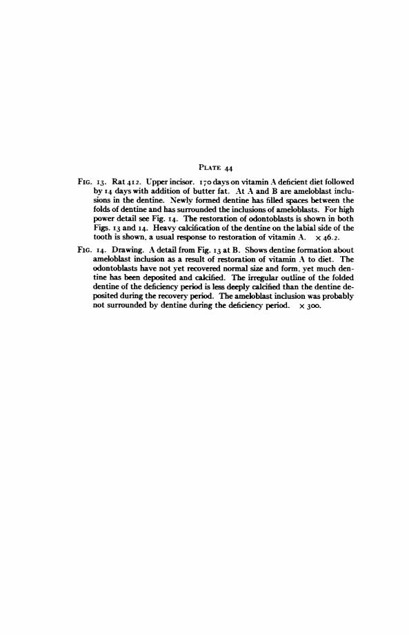
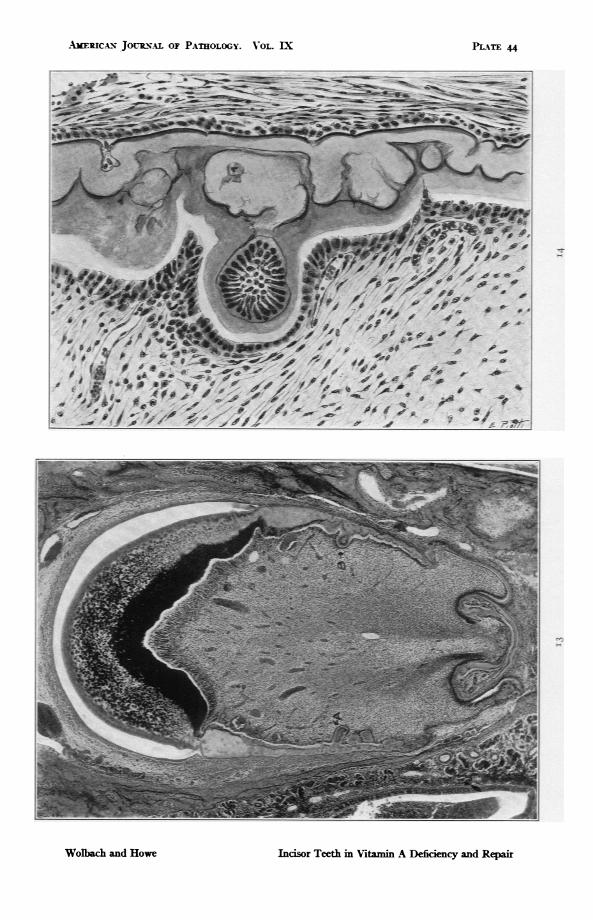
FIG. 13. Rat 412. Upper incisor. 170O days on vitamin A deficient diet followedby I4 days with addition of butter fat. At A and B are ameloblast indu-sions in the dentine. Newly formed dentine has filled spaces between thefolds of dentine and has surrounded the indusions of ameloblasts. For highpower detail see Fig. I4. The restoration of odontoblasts is shown in bothFigs. 13 and 14. Heavy calcification of the dentine on the labial side of thetooth is shown, a usual response to restoration of vitamin A. x 46.2.
FIG. 14. Drawing. A detail from Fig. 13 at B. Shows dentine formation aboutameloblast inclusion as a result of restoration of vitamin A to diet. Theodontoblasts have not yet recovered normal size and form, yet much den-tine has been deposited and calcified. The irregular outline of the foldeddentine of the deficency period is less deeply calcified than the dentine de-posited during the recovery period. The ameloblast indusion was probablynot surrounded by dentine during the deficiency period. x 300.
AMERICAIN JOURNAL OF PATEOLOGY. VOL. IX
Incisor Teeth in Vitamin A Defidcency and Repair
PLATE 44
Wolbach and Howe
PLATE 45
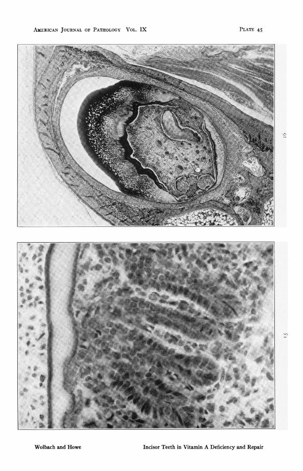
FIG. I . Rat 417. Upper insor. i87 days on vitamin A deficent diet folowedbyv days with addition of butter fat. To show gland-like formation ofameloblast indusions in pulp in recovery from atrophy. Note mitoticfigure. x 588.
FIG. i6. Rat 412. Lower incisor. I7Odays on vitamin A defiaent diet followedby 14 days with addition of butter fat. To show growth of dentides in con-sequence of restored diet. Heavy calcum deposits in old dentine. x 46.2.
AMERICAN JOURNAL OF PATHOLOGY VOL. IX
Incisor Teeth in Vitamin A Deficiency and Repair
If)
PLATE 4 5
Wolbach and Howe
PLATE 46
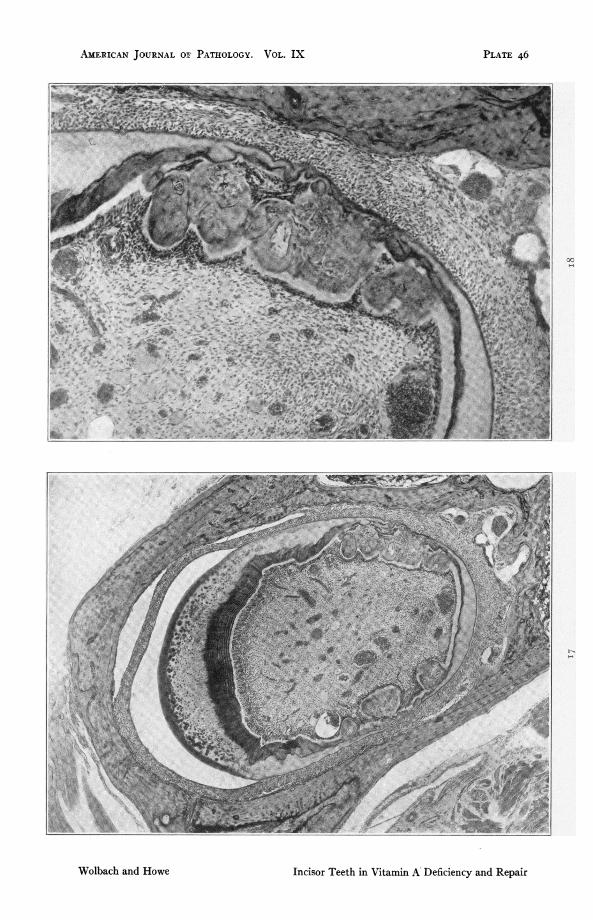
FIG. I 7. Rat 401. Upper incisor. i6i days on vitamin A deficient diet followedby 22 days with addition of butter fat. Denticles of both types showinglarge deposits of dentine formed since restoration of the diet. x 46.2.
FIG. i8. High power detail of Fig. I 7 showing that the odontoblasts, thoughfunctionally active, are not yet completely restored in morphology. Inclu-sions of ameloblasts have disappeared. x 147.
AMLERICAN JOURNAL or; PATHOLOGY. VOL. IX
00
Incisor Teeth in Vitamin A Deficiency and Repair
PLATE 46
Wolbach and Howe
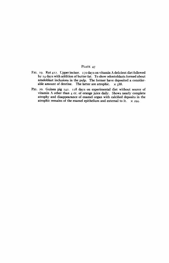
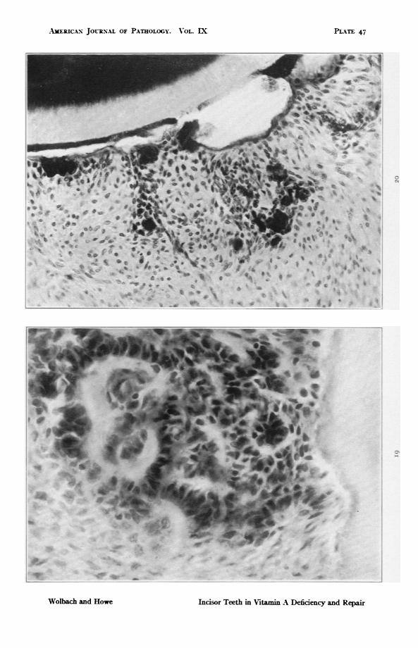
PLATE 47
FIG. i9. Rat 412. Upper incisor. 17Odas on vitamin A deficient diet followedby 14 days with addition of butter fat. To show odontoblasts formed aboutameloblast indusions in the pulp. The former have deposited a consider-able amount of dentine. The latter are atrophic. x 588.
FIG. 20. Guinea pig 142. 128 days on experimental diet without source ofvitamin A other than 4 cc. of orange juice daily. Shows nearly completeatrophy and disappearance of enamel organ with calcified deposits in theatrophic remains of the enamel epithelium and external to it. X 294.
AMXERICAN JO?-RNAL OF PATHOLOGY. AOL. IX
PI
V~~~i
.I.w*4. is&AW_ vii
Indsor Teeth in Vitamin A Deficiency and Repair
i 'I- 1-
PLAsTE 47
Wolbach and Howe

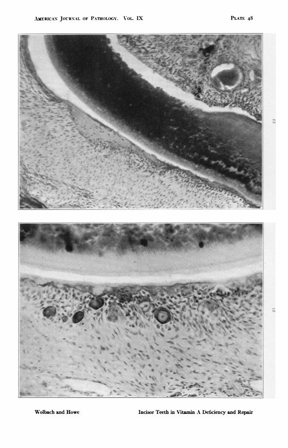
PLATE 48
FIG. 2I. Guinea pig 142. Lower incisor. Extreme atrophy and calcification ofenamel organ. X 294.
F1G. 22. Guinea pig 38. 9I days on experimental diet without source of Nita-min A other than orange juice, of which it received 20 CC. daily for 56 days,8 cc. daily for 2I days and 4 CC. daily for 24 days. Ossification of enamelorgan. x 147.
AMERICAN JOURNAL OF PATHOLOGY. VOL. EI PLATE 48
Inisor Teeth in Vitamin A Defidency and RepairW'olbach and Howe

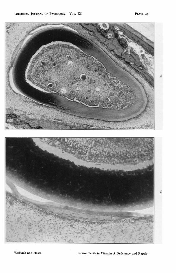
PLATE 49
FIG. 23. Guinea pig 38. Same preparation as Fig. 22. Another region. At eachend of the bony plaque and extenal to it, remains of the enamel epitheliumcan be seen. x 147.
FIG. 24. Guinea pig 38. Lower power. Same preparation used for Figs. 22 and23. Shows ossification of the enamel organ, cementides, and on the lingualside, numerous ingrowths of osteodentine. x 46.2.
AMERICA-N JOIR-NAL OF PATHOLOGY. VOL LP
* W
,,,> f ow
Incisor Teeth in V-itamin A Deficiency and Repair
PLATE 49
$ 1 --, 1.
.1 . il, IL
'iIIZ *I
39. " -*.,I ....
W'olbJach and Howe
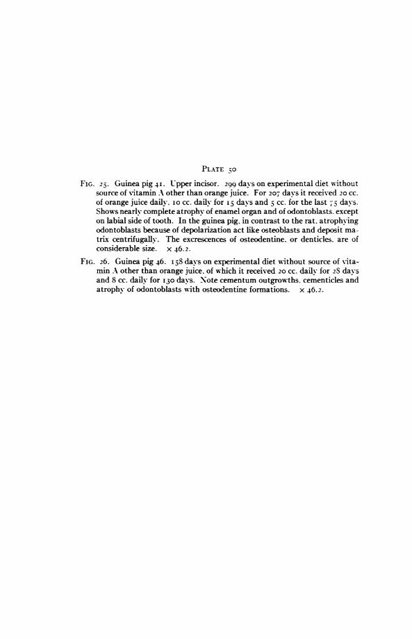
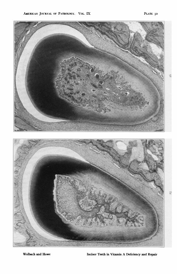
PLATE 50FIG. 25. Guinea pig 4I. Upper incisor. 299 days on experimental diet without
source of vitamin A other than orange juice. For 207 days it received 20 CC.of orange juice dailv. io cc. dailv for I davs and D cc. for the last 75 days.Shows nearly complete atrophy of enamel organ and of odontoblasts. excepton labial side of tooth. In the guinea pig. in contrast to the rat. atrophyingodontoblasts because of depolarization act like osteoblasts and deposit ma-trix centrifugallv. The excrescences of osteodentine. or denticles. are ofconsiderable size. x 46.2.
FIG. 26. Guinea pig 46. I,8 days on experimental diet without source of vita-min A other than orange juice. of which it received 20 cc. dailv for 28 davsand 8 cc. daily for 130 days. Note cementum outgrowths. cementicles andatrophy of odontoblasts with osteodentine formations. x 46.2.
AMERICAN JOuRN-L OF PATHOLOGY. VOL. IX
Incisor Teeth in Vitamin A Deficiency and Repair
PL-A,T 50
Wolbach and Howe