Embed Size (px)
Citation preview

Pergamon
0021-9290(95)00144-l
J. Biomechanics, Vol. 29, No. 6, pp. 723-733, 1996 Copyright 0 1996 Elsevier Science Ltd. All rights reserved
Printed in Great Britain 1X21-9290/96 $15.00 + .OO
THE INFLUENCE OF MUSCLES ON KNEE FLEXION DURING THE SWING PHASE OF GAIT
Stephen J. Piazza* and Scott L. Delpt
*Department of Mechanical Engineering, Northwestern University and tDepartments of Biomedical Engineering and Physical Medicine & Rehabilitation, Northwestern University, and Sensory Motor Performance Program,
Rehabilitation Institute of Chicago, IL 60611, U.S.A.
Abstract-Although the movement of the leg during swing phase is often compared to the unforced motion of a compound pendulum, the muscles of the leg are active during swing and presumably influence its motion. To examine the roles of muscles in determining swing phase knee flexion, we developed a muscle-actuated forward- dynamic simulation of the swing phase of normal gait. Joint angles and angular velocities at toe-off were derived from experimental measurements, as were pelvis motions and muscle excitations. Joint angles and joint inoments resulting from the simulation corresponded to experimental measurements made during normal gait. Muscular joint moments and initial joint angular velocities were altered to determine the effects of each upon peak knee flexion in swing phase. As expected, the simulation demonstrated that either increasing knee extension moment or decreasing toe-off knee flexion velocity decreased peak knee tlexion. Decreasing hip flexion moment or increasing toe-off hip flexion velocity also caused substantial decreases in peak knee flexion. The rectus femoris muscle played an important role in regulating knee flexion; removal of the rectus femoris actuator from the model resulted in hyperflexion of the knee, whereas an increase in the excitation input to the rectus femoris actuator reduced knee flexion. These findings confirm that reduced knee flexion during the swing phase (stiff-knee gait) may be caused by overactivity of the rectus femoris. The simulations also suggest that weakened hip flexors and stance phase factors that determine the angular velocities of the knee and hip at toe-off may be responsible for decreased knee flexion during swing phase. Copyright 0 1996 Elsevier Science Ltd.
Keywords: Gait; Swing phase; Knee flexion; Dynamic simulation; Rectus femoris.
INTRODUCTION
The beginning of swing phase is marked by flexion of the hip, knee, and ankle of the swing limb that draws the toe up and away from the ground as the limb moves forward. Knee flexion is especially important to toe clearance; without sufficient knee flexion in swing phase, the toe of the swing limb will strike the ground. Gage (1990) re- ported that knee flexion of approximately 60” is neces- sary to ensure toe clearance. Winter (1992) reported that mean toe clearance is only 1.29 cm in normal swing and that toe clearance is particularly sensitive to changes in knee angle.
The motion of the swing leg is often likened to the unforced swinging of a compound pendulum. The low level of activity in the leg muscles during swing (relative to stance) supports this characterization of swing phase as a ‘ballistic’ motion. In a simulation of swing phase, Mochon and McMahon (1980) found a range of initial segment angular velocities for which toe clearance was achieved without applied forces or moments representing the actions of muscles. Mena et al. (1981) also found that
Received inJina1 form 5 September 1995. Address correspondence to: Scott L. Delp, Ph.D., Sensory
Motor Performance Program, Rehabilitation Institute of Chicago (Room 14061, 345 East Superior Street, Chicago, IL 60611, U.S.A.
a near-normal swing could be simulated in the absence of moments applied to the thigh and shank segments. McGeer (1990) analyzed and built two-legged ‘passive dynamic’ machines with knees that are able to walk down slight slopes without forces or moments applied to represent the actions of muscles. Although these exam- ples suggest that the swing leg is not muscle-driven, it is reasonable to expect that muscles do affect the motions of swing. The muscles of the leg exhibit stereotypical patterns of activity during swing and presumably gener- ate forces that affect limb motion.
Knee flexion may be influenced by muscles that cross the knee and by muscular moments produced at other joints (via dynamic coupling). For example, Perry (1987) and Kerrigan et al. (1991) have theorized that a hip flexion moment not only flexes the hip but also flexes the knee in normal swing phase. Yamaguchi and Zajac (1990) found that hip flexion moment contributed to knee flexion through dynamic coupling in a computer simula- tion of human walking. In some pathologies, normal knee flexion seems to be prevented by the actions of muscles. For instance, patients with cerebral palsy who walk with decreased knee flexion in swing phase (termed stiff-knee gait) have difficulty achieving toe clearance without compensating by circumducting the hip or vault- ing on the stance limb (Sutherland and Davis, 1993). Stiff-knee gait is frequently attributed to the knee-extend- ing action of spastic quadriceps-especially the rectus femoris-during swing (Perry, 1987; Sutherland et al.,
123

124 S. J. Piazza and S. L. Delp.
1990; Damron et al., 1993). However, the role of hip flexion moment (or its absence) in producing knee flexion during the swing phase of either normal of stiff-knee gait is presently unknown.
The ways in which individual muscles contribute to the motions of gait are not well known. Speculation about the function of a muscle during gait is often based only upon its activation pattern, anatomical position, and the concurrent motion of the spanned joint(s). However, pre- vious studies have shown that it is also important to consider limb dynamics in analyses of muscle function (Hollerbach and Flash, 1982; Zajac and Gordon, 1989). Muscles have the potential to accelerate joints they do not cross and biarticular muscles may produce joint accelerations that oppose their joint moments; an analy- sis of muscle function that ignores limb dynamics does not allow for such possibilities. Information about indi- vidual muscles is difficult to obtain when muscle actions are modeled by moments applied to limb segments (Mena et al., 1981) or about joints (Onyshko and Winter, 1980). However, an accurate dynamics simulation of swing phase in which muscle forces are applied to the skeleton may be used to interpret the functional roles of individual muscles.
We therefore developed a muscle-actuated, dynamic simulation of swing phase to test the following hypothe- ses: (1) muscles play a role in producing normal flexion of the knee joint in early swing phase; and (2) knee flexion in early swing phase may be inhibited by a decrease in muscular hip flexion moment. We specifically investi- gated the role of rectus femoris because overactivity of this muscle is thought to cause stiff-knee gait. Analysis of the rectus femoris is complex because it potentially in- creases knee flexion through its hip flexion moment and potentially decreases knee flexion through its knee exten- sion moment.
METHODS
A model of the lower extremity and its muscles was developed to simulate swing phase dynamics. Experi- mentally derived muscle excitations and pelvis motions were input to the simulation, and computation of muscle forces and the motions of the swing limb were based upon these inputs. Details of the formulation of the model and of the swing phase simulation are given below.
Five segments were represented in the lower extremity model: the pelvis, thigh, patella, shank, and foot of the right leg (Fig. 1). Inertial properties for the thigh, shank, and foot segments (see Appendix) were specified using the regression equations of McConville et al. (1980) for a 180 cm, 75 kg male. The joints connecting the segments permitted motions in only the sagittal plane. The hip and ankle joints were modeled as frictionless revolutes, but the knee joint model included both the rolling and sliding of the femoral condyles on the tibia1 plateau and the patellofemoral kinematics (both of which depended only upon knee flexion angle), as described by Delp et al. (1990). Translation and tilt of the pelvis were prescribed
throughout the simulation, leaving only three degrees of freedom: flexionextension of the hip, knee, and ankle.
Forces representing muscle forces were applied to the segments throughout simulation of the swing phase. Each muscle (and its tendon) was modeled by an actuator that was characterized by four unique parameters: opti- mum fiber length I,, maximum isometric force F,, penna- tion angle, and tendon slack length (Zajac, 1989). The values used for these parameters and for the coordinates of muscle attachment sites on the segments were defined by Delp et al. (1990). All muscles were assumed to obey the same normalized force-velocity and force-length curves, and all muscles were assumed to have a max- imum shortening velocity of lo& s-i, as suggested by Zajac (1989) for muscles of mixed fiber type. The passive tension produced by each musculotendon actuator was determined by an exponential force-length curve which specified that passive tension was generated when the actuator’s muscle fibers were stretched beyond 1, and that passive force equal to F, was developed when fibers were stretched to 1.51,.
The input to each musculotendon actuator was a time- varying ‘neural excitation’ signal, u(t), that determined muscle activation, a(t), via first-order activation dynam- ics (Zajac, 1989):
da z = (u - a)(k1u + u (1)
where the activation and deactivation time constants were determined by
and
1 7
act = k,
1 7deact = -.
k2
(21
(3)
The activation time constant, 7ac,, was chosen to be 12 ms (Zajac, 1989) and the deactivation time constant, rdeact, was chosen to be twice the value of the activation time constant, or 24 ms. The value of r&act was chosen arbit- rarily, but the simulation was not sensitive to this choice; doubling or halving the value of r&act had very little effect on simulation output.
The formulation of a Hill-type model (Schutte, 1992) was used to represent musculotendon contraction dy- namics. The equation governing contraction dynamics was of the form
(4)
where &,, is muscle fiber length, Z,,,$ is the total length of the musculotendon actuator, andf, is the force-velocity relation. Thus, the time derivative of the fiber length (the fiber velocity) was found by first calculating the fiber force given the fiber length, musculotendon length, and activation, and then inverting the force-velocity curve.
Angle-dependent moments represented the effects of nonmuscular structures that cross the simulated hip and knee. Specifically, a hip flexion moment was applied

The influence of muscles on knee flexion during the swing phase of gait 125
PATELIA
Fig. 1. Schematic drawing of the lower extremity model. All motions of the’model were constrained to the sagittal plane. The model included 11 muscles: (1) iliacus, (2) psoas, (3) gluteus maximus, (4) rectus femoris, (5) adductor longus, (6) combined hamstrings muscles, (7) short head of biceps femoris, (8) combined vasti muscles, (9) gastrocnemius, (10) soleus, (11) pretibial group: combined tibialis anterior, extensor hallicus longus, and extensor digitorum longus, and (12) tibialis posterior. Muscle geometry is distorted for purposes of illustration. Inset: Hip angle was defined as the angle between the long axis of the thigh and the perpendicular of the line connecting the ASIS and PSIS. Knee angle was defined as the supplement of the angle formed by the long axes of the thigh and shank. Ankle angle was given by the angle formed by the long axis of the shank and the line perpendicular to the plantar surface of the foot. Anterior pelvic tilt, hip
flexion, knee flexion, and ankle dorsiflexion each corresponded to a positive joint angle.
during initial swing when the hip was most extended, and a knee flexion moment was applied during late swing as the knee reached full extension. These moments were assumed to be exponential functions of joint angle:
hip: M~“(CIn)
Xlexp[ - O.lll(8u + 9.9611, Bn < 20”, =
0 OH > 200, t5)
knee: MiM (0,)
30.2 exp [ - 0.207(& - 0.030)], BK < 30”, = 0 8, > 30”, @)
where the nonmuscular hip and knee moments MzM and MgM are in N m, and the hip and knee angles, OH and OK, are in degrees. Audu and Davy (1985) proposed relation-
ships representing the moments generated by uniarticu- lar muscles and nonmuscular tissues that were derived by fitting double-exponential curves to measurements re- ported by Hatze (1976). Our hip and knee moment rela- tions were derived by fitting single-exponential relations to the curves of Audu and Davy with the passive uniar- titular muscle moments subtracted. However, we found that deriving MH NM in this fashion resulted in total (mus- cular and nonmuscular) hip flexion moments that were much larger than those measured during normal gait (Winter, 1991); for this reason we scaled Mfl” to 75% of the value reported by Audu and Davy (1985).
We used the musculoskeletal model to simulate the swing phase (Fig. 2). The excitation inputs to the simula- tion were derived from the averaged intramuscular EMG (expressed as a percentage of maximum EMG) collected by Perry (1992) during normal gait. These experimental

726 S. J. Piazza and S. L. Delp.
excitations u(t)
1..‘.“.;..1
activations a(t)
A muscular joint moments
I? T
lj joint angular accelerations tl sdt
f 0 joint angutarvetcdties 1
I I
0 joint angles
Fig. 2. Block diagram for the simulation of swing phase. Muscle activations were determined from muscle excitation inputs (approximated by EMG). The force generated by each musculotendon actuator was a function of its activation, length, and velocity. Total applied joint moments were calculated by multiplying muscular forces by moment arms and adding the angle-dependent non-muscular moments. The equations of motion were integrated in time to determine the kinematic output of the simulation. Hip joint center translation and pelvic tilt were prescribed from measured gait analysis data. Values for initial joint angles
and angular velocities were also derived from experimental measurements.
EMG data were reported as functions of the gait cycle; to obtain excitations as functions of time, we assumed the duration of normal swing to be 0.42 s (Perry, 1992). The excitation signals used as input to the simulation were determined by approximating step functions to the ex- perimental EMG measurements (Fig. 3); the use of step functions made it possible to vary the excitation inputs by varying at most three parameters (height, width, and time of onset).
Motions of the pelvis were prescribed throughout the simulation as functions of time based upon experimental data. We measured the gait kinematics of ten healthy subjects (age range = 6.9-24.7 yr; mean = 12.6 yr) using a Vicon (Oxford Metrics; Oxford, England) motion measurement system. A fifth-order polynomial was fit to the mean of the measured pelvic tilt angle and was used
to prescribe pelvic tilt for the simulation, This function is given by
O,(t) = 9.58 + 5.32t - 19.6t2 + 358t3 - 1290t4 + 1210t’
(7)
where the units of t&.(t) are degrees and t, the time elapsed after toe-off, is in seconds. The values prescribed for toe-off joint angles and angular velocities (Table 1) were also calculated from measurements of normal gait for this subject pool. The horizontal (x) and vertical (y) displacements of the pelvis were also prescribed as func- tions of time. These functions were derived by fitting linear and sine functions to averaged measurements of the hip center trajectory made for a group of six healthy adult subjects (age range = 18-31 yr; mean = 24 yr;

The influence of muscles on knee flexion during the swing phase of gait 121
adductor longus
f 0.4 - 3? a I 0.3 -
3 .g 1 0.2 -
” g 0.1 -
hamstrings
iNA\
iliacus and psoas
vasti
i~,;;~~:‘-’ oil , , glu;,
0.0 0.1 0.2 0.3 0.4 0.5 0.0 0.1 0.2 0.3 0.4 0.5 time (s) time (s)
0.5
r rectus temoris
pretibial group
O.ooS 0.4 0.5 time (s)
Fig. 3. Excitation signals, u(t), input to the musculotendon actuators. Simulation inputs (solid lines) are step function approximations to the intramuscular EMG data (dashed lines) reported by Perry (1992). No excitation input was provided for soleus, gastrocnemius, or tibialis posterior. For actuators that represent combined muscles (hamstrings, vasti, and pretibial group), experimental EMG is shown for all of the constituent muscles. The iliacus and psoas actuators received the same excitation input. The thick dashed line shown for rectus femoris corresponds to the simulation performed with increased rectus femoris
excitation.
Table 1. Measured joint angles and angular velocities at toe-off.
Mean angle or angu- lar velocity Standard deviation
Parameter (deg or deg s-l) (deg or deg s-l)
Hip flexion angle - 1.2* 1.5 Knee flexion angle 39.5 10.2 Ankle dorsiflexion angle - 8.8+ 4.5 Hip flexion velocity 182 46 Knee flexion velocity 322 42 Ankle dorsiflexion velocity - 109+ 106
*Negative hip angle indicates extension. ‘Negative ankle angle indicates plantarllexion; negative ankle
dorsiflexion velocity indicates that the ankle is plantartlexing at toe-off.
height range = 172-185 cm; mean = 179 cm), and are given by
x(t) = l.l7t, (8)
y(t) = 0.026sin(8.4t - O.OSO),
where x(t) and y(t) are expressed in meters.
(9)
The simulation was implemented on a Silicon Graphics (Mountain View, CA) workstation using two dynamic simulation software packages: Dynamics Pipeline (Mus- culoGrapbics, Inc.; Evanston, IL) and SD/FAST (Symbolic Dynamics, Inc.; Mountain View, CA). The equations of motion for the model were integrated forward in time using SD/FAST, which employs a variable time step method based on a fourth-order Runge-Kutta-Merson step.
We performed a ‘one-at-a-time’ factorial analysis (Hogg and Ledolter, 1987) to determine the capability of each of the joint moments and initial angular velocities to diminish peak knee flexion in swing. Each joint angular velocity was varied from its normal swing simulation value by two standard deviations (Table 1). Muscular joint moments equal to twice the joint moment standard deviation were added to or subtracted from the normal simulation joint moment at each time step during the simulation. The standard deviations of the muscularjoint moments were approximated by averaging the standard deviations calculated by Winter (1991) over the duration of the swing phase. The change in peak knee flexion and the change in minimum toe height that resulted from each altered joint moment and initial angular velocity were determined.
A simulation of swing phase was performed with the rectus femoris actuator removed from the model, and another simulation was performed with a prolonged and exaggerated excitation input to the rectus femoris actuator. The purpose of these simulations was to clarify the role of the rectus femoris in producing normal knee flexion. For the latter simulation, the excitation input to the rectus femoris actuator continued through- out the swing phase at 30% of its maximum level (as opposed to 0.03 s and 5% for the normal simulation; see Fig. 3).
The equations of motion of the swing leg system were used to assess which factors contribute to angular accel- eration at the knee. A Lagrangian formulation was used to derive the equations of motion after a frictionless revolute was substituted for the knee joint. The equations were expressed in matrix form:
MH
+G+ -MK [ 1 , (10)
MA
where OH, &, and #A are the joint angles; MH, MK, and MA are the joint moments applied to the model by musculotendon actuators and non-muscular joint springs; x and y are the horizontal and vertical displace- ments of the pelvis relative to a ground-fixed reference frame; and the matrices M, C, V, P, and G depend upon joint angles and inertial parameters (see the appendix for a full account of the components of these matrices).
The mass matrix M was found to be full rank and thus invertible throughout the simulation. Premultiplying both sides of equation (10) by M-’ gives
[ ~~K]=M-lc[~]+M-~v[ cuff]
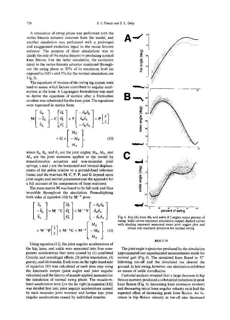
Fig. 4. Hip (A), knee (B), and ankle (C) angles versus percent of swing. Solid curves represent simulation output; dashed curves
[I M” with shading represent measured mean joint angles plus and . .
+M-‘P ’ Y
+ M-‘G + M-i [ 1 - MK (11) minus one standard deviation for normal swing.
.
MA
Using equation (1 l), the joint angular accelerations of RESULTS
the hip, knee, and ankle were separated into four com- The joint angle trajectories produced by the simulation ponent accelerations that were caused by (1) combined approximated our experimental measurements made for Coriolis and centrifugal effects, (2) pelvis translation, (3) normal gait (Fig. 4). The simulated knee flexed to 57” gravity, and (4) muscles. Each term on the right-hand side following toe-off and the simulated toe cleared the of equation (11) was calculated at each time step using ground. In late swing, however, our simulation exhibited the kinematic output (joint angles and joint angular an excess of ankle dorsiflexion. velocities) and the history of muscle-applied moments for Factorial analysis revealed that a large decrease in hip the simulation of normal swing phase. The muscle-re- flexion moment produced a substantial reduction in peak lated acceleration term [on the far right in equation (1 l)] knee flexion (Fig. 5). Increasing knee extension moment was divided first into joint angular accelerations caused and decreasing initial knee angular velocity each had the by each muscular joint moment and further into joint expected effect of decreasing peak knee flexion. An in- angular accelerations caused by individual muscles. crease in hip flexion velocity at toe-off also decreased
728 S. J. Piazza and S. L. Delp.

The influence of muscles on knee flexion during the swing phase of gait 129
decrease in peak knee flexion (deg) gait. Changes in initial ankle velocity and ankle moment
0 1 2 3 4 5 6 7 also affected peak knee flexion, but to a lesser extent.
increased hip flexion velocity
decreased ankle plantarflexion velocity
decreased ankle dorsiflexion moment
Fig. 5. Decrease in peak knee flexion corresponding to a two- standard-deviation change in each muscular joint moment and
toe-off joint angular velocity.
knee flexion in swing phase. Specifically, a two-standard- deviation change (in the direction indicated in Fig. 5) in the knee extension moment, initial knee flexion velocity, hip flexion moment, and initial hip flexion velocity de- creased peak knee flexion by 6.6,5.4,4.1, and 3.2”, respec- tively. These changes were considered important because they were accompanied by decreases in minimum toe height of 2.92,1.29,1.33, and 1.72 cm, respectively; all of which are at least as large as the 1.29 cm average min- imum toe clearance reported by Winter (1992) for normal
60.
50.
IO
--cl 0, !
Rectus femoris was found to play an important role in regulating knee flexion during swing phase. A simulation of swing performed with the rectus femoris actuator removed resulted in excessive knee flexion, suggesting that the knee-extending action of the rectus femoris in early swing is important for normal knee flexion (Fig. 6). Conversely, overactivity in rectus femoris inhibited knee flexion in the simulation; an increase in the excitation input to the rectus femoris actuator caused a decrease in knee flexion.
In early swing the muscles acted to brake the rapid toe-off knee flexion velocity; the net muscle-induced ac- celeration of the knee during this period was in the extension direction. The rectus femoris actuator produc- ed a knee extension acceleration prior to peak knee flexion, as did the passive vasti actuator (though the vasti-induced acceleration was only 50% of the acceler- ation induced by rectus femoris on average). Knee flexion acceleration in early swing was produced by the ac- tuators representing hip flexors, the biceps femoris (short head), the pretibial group, and the nonmuscular hip flexion moment [equation (5)]. Gravitational, coriolis, and centrifugal forces collectively caused a knee exten- sion acceleration throughout the swing.
We unexpectedly found that the gastrocnemius muscle produced a knee extension acceleration between 25 and 60% of swing while simultaneously producing a knee
0 time after toe-off (s)
Fig. 6. Knee angle versus time for simulations performed with the rectus femoris (RF) actuator removed and with increased and extended excitation of the rectus femoris. The dashed curve with shading represents measured mean joint angle plus and minus one standard deviation for normal swing. The solid curve within the shaded area represents the knee angle for a simulation performed with all musculotendon actuators intact and supplied with normal excitations. Removal of the rectus femoris both prolonged and exaggerated
knee flexion; increased rectus femoris activity decreased knee flexion.

730 S. J. Piazza and S. L. Delp.
Jlexion moment. Though the gastrocnemius was not ac- tive, it generated force during the simulation when it was passively stretched. Other biarticular actuators produced accelerations opposite in direction to their joint mo- ments: the hamstrings produced a hip flexion acceler- ation in mid-swing and rectus femoris produced a hip extension acceleration in early swing.
DISCUSSION
In this study, a muscle-actuated, dynamic simulation was developed to analyze how muscle actions affect flexion of the knee during the early part of the swing phase of normal gait, We found that normal knee flexion is determined by a large knee flexion velocity at toe-off which is tempered by the knee-extending action of the rectus femoris and vasti muscles. Lack of muscular hip Aexion moment was found to inhibit knee flexion in early swing.
The inputs and outputs of our simulation approxim- ated data reported in the literature. The initial joint angles used in the simulation were similar to those re- ported by Kadaba et al. (1990) at 62% of the gait cycle. Simulation joint angles (Fig. 4) compare favorably to data reported by Kadaba et al. (1990) and Perry (1992). We judged excessive ankle dorsiflexion and hip flexion in late swing to be tolerable because we were more con- cerned with accurately modeling early swing (the time of peak knee flexion). The sum of joint moments produced by muscles and nonmuscular joint springs in the simula- tion were similar to the swing phase joint moments reported by Winter (1991), who used an inverse-dynamic formulation to calculate muscular joint moments from measured joint angular kinematics (Fig. 7).
We tried to base our simulation on experimentally derived data wherever possible. Unfortunately, practical considerations dictated that we could not obtain all the experimental data we needed from a single source. We drew simulation parameters and input data from several sources in the literature as well as from our own measure- ments. Initial conditions for the simulation were deter- mined using data collected from a subject pool that included children over age seven; we believe that the inclusion of children’s data was justified by the finding of Sutherland et al. (1980) that an adult gait pattern is attained by age seven. However, it was necessary to measure pelvis translation in a different pool of adult subjects because we expected these data to be smaller in children.
By modeling the motions of only the swing leg, we have neglected to model explicitly the motions and mus- cular forces produced in other parts of the body. Mochon and McMahon (1980) found that inclusion of the stance limb was necessary to achieve times of swing that ap- proximated experimental measurements. However, the influences of such factors upon the motions of the swing leg were presumably accounted for in the present model by the prescribed pelvis translation [the third term from the right in equation (1111.
Fig. 7. Muscular hip (A), knee (B), and ankle (C) moments versus percent of swing. Solid curves represent simulation out- put; dashed curves with shading represent mean muscular joint moments plus and minus one standard deviation as calculated by Winter (1991) for the normal swing, based on an inverse- dynamic formulation. The abrupt changes in the hip and knee moments at approximately 65% of swing are caused by the
hamstrings actuator becoming active at that time.
The one-at-a-time factorial analysis that was employed neglects the effects of combined variation among the fac- tors. A more elaborate analysis would entail the covari- ation of all three initial joint angular velocities and all three joint moments. Such an analysis would require that 729 (36) simulations be run, as opposed to 13 (2.6 + 1) simulations for the one-at-a-time approach. However, a full factorial analysis would be helptil for understand- ing how factors combine to a&t peak knee flexion.
Our conclusion that the actions of museles are neces- sary to check the large knee flexion velocity normally

The influence of muscles on knee flexion during the swing phase of gait 731
Table 2. Comparison of knee flexion weakened hip flexors are also possible causes of stiff-knee velocities at toe-off. gait.
Initial knee flexion velocity
Source (degs-r)
Mena et al. (1981) 237 Kadaba et al. (1990) 327 Present study 322 Mochon and McMahon (1980)* 16&286
*Approximate range for which Mochon and McMahon (1980) reported that ‘ballistic’ swing is possible.
present in early swing (Fig. 7) is consistent with the find- ings of those who have analyzed above-knee prosthetic gait. Menkveld et al. (1981) and Hicks et al. (1985) found that the knee extension moment generated by a damped knee component is necessary to prevent excessive knee flexion. This conclusion contradicts the findings of Mena et al. (1981) who suggested that normal swing phase could be simulated in the absence of muscular knee moment. A comparison of toe-off knee flexion velocities (Table 2) explains this apparent contradiction. The initial knee flexion velocity used by Mena et al. (1981) is sub- stantially smaller than either the slope at 62% of the gait cycle of the averaged knee angle versus time curve re- ported by Kadaba et al. (1990) or the mean toe-off velo- city measured in the present study (237” s-l versus 327 or 322” s-l). Mochon and McMahon (1980) found that swing could occur without muscular moments only over a range of initial knee flexion velocities that excludes both of the values measured for normal gait; this further supports the conclusion that muscular action is necessary to affect a normal knee flexion pattern in most normal subjects. It is possible that the use of muscles to restrain knee flexion in early swing is not needed if toe-off knee flexion velocity is reduced, as occurs in slow walking. Murray et al. (1984) found that both rectus femoris activ- ity and toe-off knee flexion velocity (estimated from the slope of the reported mean knee angle trajectory) de- creased with walking speed, though peak knee flexion in swing did not. Patients who walk with stiff-knee gait have been found to walk at low speeds (Kerrigan et al., 1991) but are, perhaps. unable to duplicate the reduction in toe-off rectus fermoris activity that appears in the slow gait of unimpaired subjects.
Our finding that the gastrocnemius acts to extend the knee in swing was unexpected because the gastrocnemius passes posterior to the knee. Knee extension acceleration by a muscle that produces a knee tlexion moment is made possible by the coupled nature of the system dynamics (Zajac and Gordon, 1989). Gastrocnemius is a biarticular muscle that produces a knee flexion moment and an ankle plantarflexion moment. These moments produce opposing accelerations of the knee joint: knee flexion moment accelerates the knee in flexion and ankle plan- tarflexion moment accelerates the knee in extension. If the latter acceleration is larger than the former, a net knee extension acceleration results. The direction of the gastrocnemius-induced acceleration at the knee is deter- mined by the muscle moment arm at each joint and the components of the inverse of the inertia matrix [M- 1 in equation (ll)], which transforms joint moments into joint angular accelerations.
The actions of an individual muscle are often inferred from EMG recordings and from the moments generated by the muscle. We have concluded from the results of our muscle-actuated simulations that an accurate assessment of the function of a muscle at a particular time during gait must account for the force-generating properties of the muscle, the musculoskeletal geometry, and the coupled nature of the system dynamics. Although we have fo- cused on normal gait in this study, characterizing the accelerations caused by muscles during walking may also be helpful for understanding which muscles contribute to abnormal gait and for planning surgical procedures that alter the actions of muscles to correct pathological gait.
Acknowledgements-We are grateful to Carolyn Moore, Claudia Kelp-Lenane, Tony Weyers, and Steve Vankoski of the Gait Analysis Laboratory at Children’s Memorial Hospital in Chicago for providing us with gait data. We would also like to thank Abraham Komattu and Peter Loan for assistance with the computer implementation of the simulation, Lisa Schutte for discussions regarding the formulation of the muscle model, and MusculoGraphics, Inc. for its donation of musculoskeletal modeling software. This work was supported by NSF Grant BCS-9257229.
REFERENCES
Those who have attempted to identify the biomechani- cal determinants of stiff-knee gait have often cited only spasticity of the quadriceps during swing as the cause (Sutherland et al., 1990; Damron et al., 1993). We agree that the lack of swing phase knee flexion associated with stiff-knee gait might be caused by an increased knee extension moment that would accompany overactive quadriceps. The results of the present study, however, suggest that altered knee and hip flexion velocities at toe-off and decreased hip flexion moment may also con- tribute to decreased knee flexion (Fig. 5). Thus, the deter- minants of the toe-off knee and hip angular velocities and
Audu, M. L. and Davy, D. T. (1985) The influence of muscle model complexity in musculoskeletal motion modeling. J. Biomech. Engng. 107, 147-157.
Damron, T. A., Breed, A. L. and Cook, T. (1993) Diminished knee flexion after hamstring surgery in cerebral palsy: preva- lence and severity. J. Pediatric Orthop. 13, 188-191.
Delp, S. L., Loan, J. P., Hoy, M. G. Zajac, F. E., Topp, E. L. and Rosen, J. M. (1990) An interactive graphics-based model of the lower extremity to study orthopaedic surgical procedures. IEEE Trans. Biomed. Engng. 31, 757-767.
Gage, J. R. (1990) Surgical treatment of knee dysfunction in cerebral palsy. Gin. Orthop. Rel. Res. 253, 45-54.
Hatze, H. (1976) The complete optimization of a human motion. Mach. Biosci. Zs, 99-,135.
Hicks, R., Tashma2, S., Altman, R. F. and Gage, J. R. (1985) Swing phase control with knee friction in juvenile amputees, J. Orthop. Res. 3, 198-201.

732 S. J. Piazza and S. L. Delp.
Hogg, R. V. and Ledolter, J. (1987), Engineen’ng Statistics. Mac- millan, New York.
Hollerbach, J. M. and Flash, T. (1982) Dynamic interactions between limb segments during planar arm movement. Biol. Cybernet. 44, 67-77.
Kadaba, M. P., Ramakrishnan, H. K. and Wootten, M. E. (1990) Measurement of lower extremity kinematics during level walking. J. Orthop. Res. 8, 383-392.
KerriganyD. C., Gronley, J. and Perry, J. (1991) Stiff-legged gait in spastic paresis. Am. J. Phys. Med. Rehabil. 70, 294-300.
McConville, J. T., Churchill, T. D., Kaleps, I., Clauser, C. E. and Cuzzi, J. (1980) Anthropometric relationships of body and body segment moments of inertia. Technical Report AFAMRL-TR-80-119. Air Force Aerosnace Medical Re- search Laboratory, W’right-Patterson Air-Force Base, OH.
McGeer, T. (1990) Passive dynamic walking. Int. J. Robotics Res. 9, 62-82.
Mena, D., Mansour, J. M. and Simon, S. R. (1981) Analysis and synthesis of human swing leg motion duing gait and its clinical applications. J. Biomechanics 14, 823-832.
Menkveld, S., Mansour, J. M. and Simon, S. R. (1981) Mass distribution in prosthetics and orthotics: quantitative analysis of gait using a biomechanical model simulation. Transactions of the 27th Annual Meeting of the Orthopaedic Research Society, p. 228.
Mochon, S. and McMahon, T. A. (1980) Ballistic walking. J. Biomechanics 13,49-57.
Murray, M. P., Mollinger, L. A., Gardner, G. M. and Sepic, S. B. (1984) Kinematic and EMG patterns during slow, free, and fast walking. J. Orthop. Res. 2, 272-280.
Onyshko, S. and Winter, D. A. (1980) A mathematical model for the dynamics of human locomotion. J. Biomechanics 13, 361-368.
Perry, J. (1987) Distal rectus femoris transfer. Dew. Med. Child Neural. 29, 153-158.
Perry, J. (1992) Gait Analysis. SLACK, Thorofare, NJ. Schutte, L. M. (1992) Using musculoskeletal models to explore
strategies for improving performance in electrical stimulation- induced leg cycle ergometry, Stanford University, Ph. D. thesis.
Sutherland, D. H. and Davids, J. R. (1993) Common gait abnor- malities of the knee in cerebral palsy. Clin. Orthop. Rel. Res. 288, 139-147.
Sutherland, D. H., Olshen, R., Cooper, L. and Woo, S. L.-Y. (1980) The development of mature gait. J. Bone Jt Surg. 62A, 336353.
Sutherland, D. H., Santi, M. and Abel, M. F. (1990) Treatment of stiff-knee gait in cerebral palsy: a comparison by gait analysis of distal rectus femoris transfer versus proximal rectus release. J. Pediatric Orthop. 10, 433-441.
Winter, D. A. (1991) Biomechanics of Motor Control and Human Gait. Universitv of Waterloo Press, Waterloo, Ontario, Canada.
Winter, D. A. (1992) Foot trajectory in human gait: a precise and multifactorial motor control task. Phvs. Ther. 72. 45-56.
Yamaguchi, G. T. and Zajac, F. E. (1990) Restoring unassisted natural gait to paraplegics via functional neuromuscular stimulation: a computer simulation study. IEEE Trans. Bi- omed. Engng. 37, 886902.
Zajac, F. E. (1989) Muscle and tendon: properties, models, scaling, and application to biomechanics and motor control. Crit. Rev. Biomed. Engng. 17, 359411.
Zajac, F. E. and Gordon, M. E. (1989) Determining muscle’s force and action in multi-articular movement. Exert. Sport Sci. Rev. 17, 187-230.
APPENDIX
The components of the coefficient matrices in the matrix equation of motion [Equation (lo)] are
MI1 =Cr + 2C,cos& + 2C,cos& + 6,) + 2Cscos&,
MZ1 = C2 + C,cos& + C,cos(& + e,, + 2Cgcos&,
Ma1 = C3 + Cscos(& + 6,) + cgco&,
Miz = C2 + C,co& + Cscos(& + 6,) + 2cgco&,
Mz2 = Cz + 2C,cos&,
MS2 = C3 + C.+ose,,
M,s = Cs + Cscos(& + e,) + cacose,,
Mss = Cs + Cpcos&,,
M,, = Cs.
Cl, = 0, .̂ -
C2, = -&sin& - Cssm(& + B,), .- ̂ -
Czl = - cssm(flK + 0,) - CssmQ,,
Cl2 = Cssin& + Cssin(8, + t?,),
c22 = 0,
Cs2 = - CssinQ*,
Cl3 = &sin& + fiA) + CPsingA,,
Cz3 = Cgsini7A
CJj = 0.
VI1 = 2Cssin& + 2C,sin(& + 8,),
v*1 = 0,
Vsl = - 2CssinB,, .A -
VIZ = 2Cssm(0x + 6,) + 2Cssin8,, . -
V22 = 2CssmB,4,
v32 = 4
VI3 = 2Cssin($ + 8,) + 2Cssin9,,
- V23 = 2Cgsm8A,
v,, = 0.
P,, = c&ose, - CSCOS@” + &)
- c,cos(eH + e, + e,,,
Pzl = - cs~Os(eH + 8,) - C,COS(B~ + &
Pa, = - c,cos(e” + s,, + 8*),
Prs = - C4sin0, - Cssin(0, + &)
- C,sin(Bu + 8, + 8,),
+ e,,,
Pz2 = - Cssin(t% + &) - C7sin(0n + & + iIA),
PJ2 = - C,sin(Bn + & -t t?*).
G1 = - C,csin6, - C,, sin@, + &) ̂ ̂
- C,zsin(eH + e, + ed,
GZ = - C,rsin@n + 6,) - C,,sin(& + 8, + &),
Gs = - Cr2sin(Bn + & + &).

The influence of muscles on knee flexion during the swing phase of gait 733
The constants Ct-Cl2 depend on the inertial parameters of the segments and are given by
Cl = m& + %(l: + d,2) + m& + l,’ + d:)
Table Al. Segment inertial parameters
Parameter Symbol Value
C2 = msdi + m& + d:) + Is + IF,
C, =m&+ IF, Moments of inertia (I,,)
C5 = msds + m&, Lengths
C, = m&, Distances from prox- imal end to center of
9.74 kg 3.86 kg 0.99 kg
0.167 kgmZ 0.060 kgm* 0.005 kg mz
0.40 m 0.43 m
mass d-r 0.20 m 4 0.15 m 4 0.08 m
The hip flexion angle is represented by 0,. The adjusted knee Because all pelvis motions were prescribed, the values as- and ankle flexion aneles are defined bv: 8, = - 0, and signed for the pelvis inertial parameters did not influence the f?, = 0., + 36.5”; adjustments to 0x and 0, (which were defined simulation. Mass and moment of inertia were estimated for the to reflect commonly used clinical measures) were performed to patella (0.025 kg and 0.005 kgm’, respectively). The simulation write the equations more concisely. was not sensitive to these values; kinematic output was The acceleration due to gravity is given by g and the inertial similar when these estimates were either increased or decreased parameters of the segments are given in Table Al. fivefold.