Embed Size (px)
Citation preview

ORIGINAL PAPER
The influence of thermo-hygro-mechanical treatmenton the micro- and nanoscale architecture of wood cell wallsusing small- and wide-angle X-ray scattering
Juan Guo . Harald Rennhofer .
Yafang Yin . Helga C. Lichtenegger
Received: 28 February 2016 / Accepted: 29 May 2016 / Published online: 15 June 2016
� Springer Science+Business Media Dordrecht 2016
Abstract Tracking the changes of cellulose crystal-
lites upon thermo-hygro-mechanical treatment is
essential to understand the response of wood cell
walls to steam and compression. In this paper the
influence of Compression combined with Steam (CS)
treatment on wood cellulose crystallites and pores
structure of Chinese fir (Cunninghamia lanceolata)
was studied under different steaming temperatures and
compression ratios. Small-angle X-ray scattering and
wide-angle X-ray scattering were used to investigate
the changes of cellulose crystallites dimension, aspect
ratio, fibril diameter distribution, non-crystalline
fraction, the number of chains in each microfibril, as
well as the fractal dimension and size of pores in
response to CS treatment conditions. Results indicate
that the crystallinity increased due to CS treatment, but
did not show alteration with varying CS treatment
conditions, i.e. seemed nearly unaffected by higher
temperatures or compression ratio, both for earlywood
and latewood. The cellulose crystallite diameter
depended on processing parameters: it increased with
increasing treatment temperature. No considerable
differences were found for earlywood and latewood.
We interpret our findings as a rearrangement of
adjacent cellulose chains towards higher crystalline
perfection attributing to the increase in crystallinity.
The same effect allows a larger coherence length of
crystalline order and therefore features an increasing
cross-sectional dimension. In general we can state that
the CS treatment leads to higher crystallinity and more
perfectly arranged cellulose crystals, while it does not
greatly affect the microfibril diameter but rather the
amorphous regions of the microfibrils and the sur-
rounding hemicellulose and lignin.
Keywords Cellulose crystallites � Compression
combined with steam treatment � Small-angle X-ray
scattering �Wide-angle X-ray scattering � Earlywood �Latewood
Introduction
Wood is the most important natural and renewable
resource, while its utilization is restricted by the lack
of dimensional stability (i.e. changes of wood dimen-
sions following environmental changes, especially
moisture change), low resistance to decay and poor
durability. As an eco-friendly wood modification
J. Guo � Y. Yin (&)
Department of Wood Anatomy and Utilization, Research
Institute of Wood Industry, Chinese Academy of Forestry,
Beijing 100091, China
e-mail: [email protected];
H. Rennhofer � H. C. Lichtenegger (&)
Institute of Physics and Materials Science, Department of
Material Sciences and Process Engineering, University of
Natural Resources and Life Sciences (BOKU), Vienna,
Austria
e-mail: [email protected]
123
Cellulose (2016) 23:2325–2340
DOI 10.1007/s10570-016-0982-2

method, thermo-hygro-mechanical (THM) treatment
can significantly improve the physical properties of
wood, including the mechanical behaviour and size
stability (Navi and Heger 2004; Skyba and Schwarze
2009; Navi and Sandberg 2011; Sandberg et al. 2013).
A promising THM technique is Compression com-
bined with Steam (CS) treatment, as compressing
wood in the transverse direction under the steaming
environment allows to densify wood samples without
fractures. Anatomical, structural, chemical, microme-
chanical and hygroscopic properties of wood under CS
treatment have been intensely investigated (Inoue
et al. 1993; Ito et al. 1998; Navi and Girardet 2000;
Navi and Heger 2004; Yin et al. 2011; Guo et al.
2015). However, the response mechanism of wood cell
walls to CS treatment has not been fully understood,
due to the rather complicated chemical and structural
changes in the wood cell wall upon the steaming and
compression treatment.
The wood cell wall is principally composed of
cellulose microfibrils embedded in a matrix composed
of lignin and hemicelulloses (Fengel and Wegener
1984). Cellulose, the main biomacromolecule in
wood, is synthesized in plant cell walls as crystalline
nanosized fibrils, consisting of dozens of unidirec-
tionally aligned molecular chains (Wada et al. 2010).
These microfibrils contain both ordered and disor-
dered domains, where the disordered portion is often
referred to as amorphous. The structure, size and
arrangement of the cellulose fibrils and the proportion
of crystalline cellulose are key factors determining the
mechanical performance of wood materials (Reiterer
et al. 1999; Barnett and Bonham 2004; Burgert and
Fratzl 2009). In addition, the proportion of crystalline
cellulose and the arrangement of cellulose molecules
in the cellulose microfibrils influence the accessibility
to water or other molecules (Inagaki et al. 2010;
Kulasinski et al. 2015). Therefore, tracking the
changes of cellulose upon CS treatment can provide
important information for understanding the response
mechanism of wood cell wall to the CS treatment.
Several parameters describing the cellulose crystal-
lites inwood, i.e. crystallinity, crystalline dimensions and
arrangement of the crystallites correlate with the treat-
ment conditions, e.g. heat (Kim et al. 2001; Andersson
et al. 2005), steaming (Inagaki et al. 2010; Nishiyama
et al. 2014), or strain (Peura et al. 2006). Cellulose
molecules in the wood cell wall undergo different
cellulose degradation processes, reorientation and/or
co-crystallization processes under CS treatment. It was
reported that CS treated wood showed similar crys-
tallinity but increased regularity of the (200) lattice
distance compared with untreated wood (Dwianto et al.
1999). Nevertheless, the changes of cellulose crystallites
and pore structures in wood upon CS treatment have not
been fully characterized and understood.
Small-angle X-ray scattering (SAXS) as a method
capable of averaging over the volume in the X-ray
beam is one of the very few techniques that can
provide statistical information about the morphology,
porosity and specific surface area of materials on the
nanometre scale (Lichtenegger et al. 2002, 2003;
Rennhofer et al. 2014; Cheng et al. 2015). SAXS
patterns fromwood can be regarded as arising from the
electron density contrast in a two-phase composite,
cellulose fibrils in a hemicellulose–lignin matrix, and
with contributions from pores and other cavities at
very small scattering angles (Jakob et al. 1996). SAXS
has been used to investigate the microfibril structure,
diameter of cellulose element fibril and pore structures
in wood (Lichtenegger et al. 1999; Farber and
Lichtenegger 2001; Kennedy et al. 2007; Nishiyama
2009; Cheng et al. 2011; Fernandes et al. 2011). In
addition, Wide-angle X-ray scattering (WAXS) has
provided very important results on the crystallinity
and cellulose crystallite dimension in wood (Penttila
et al. 2010; French 2014; Song et al. 2014; Thomas
et al. 2013; Toba et al. 2013). Therefore, SAXS/
WAXS were chosen as appropriate techniques to
monitor the changes of cellulose crystalline structure
under CS treatment.
Softwood consists of much simpler wood cell types
as compared with hardwood. In the softwood, the main
wood cells are tracheids. Earlywood tracheids with a
thinner wall and wider lumen, and latewood tracheids,
with a much thicker wall and narrower lumen, can be
observed in an integrated growth ring (Panshin and
Zeeuw 1970). In our previous studies (Yin et al. 2011,
Guo et al. 2015), it is reported that cell walls in
earlywood and latewood undergo different biomacro-
molecules degradation and show different hygroscop-
icity and mechanical properties after steaming
treatment and CS treatment, deriving from their
different chemical and physical properties (Winandy
1994). Therefore, it is of interest to examine the
cellulose crystallites and the pore structure in the cell
wall of earlywood and latewood after CS treatment
and gain information that can be used to understand
2326 Cellulose (2016) 23:2325–2340
123

the degradation mechanisms in the individual cell
walls.
In this paper, to elucidate how the influences of CS
treatment on wood cellulose crystallites and the pore
structure proceed, CS treatment on Chinese fir (Cun-
ninghamia lanceolata), an important commercial
conifer species, was conducted under different steam-
ing temperature (140, 160 and 180 �C) and two
compression ratios (25 and 50 %). SAXS was used
to investigate the variations in the diameter of
cellulose crystallites, the fibril diameter distribution
and the pore structure of CS treated wood. WAXS was
used to study the changes of crystallinity and dimen-
sions of the cellulose crystallites, and the number of
chains in each microfibril during CS treatment. Our
results contribute to a deeper understanding of the
effect of steaming and compression on the cellulose
crystalline structure and also provide a scientific basis
for the development of eco-friendly and high value
added wood products.
Experimental
Material
Raw material
Chinese fir trees were harvested from a 28-aged
plantation located in Jiangxi Province of China. A tree
with good growth, health, stem straightness and vigor
was selected for this study. Mature wood was used as
the raw material for CS treatment. Small specimen
(dimensions 20 9 20 9 25 mm in the tangential (T),
radial (R), and longitudinal (L) directions, respec-
tively) containing an integrated 25th growth ring on
the surface of the specimen were cut from wood
samples (Fig. 1).
Compression combined with Steam (CS) treatment
CS treatment was conducted in a laboratory-scale
autoclave at Kyoto University. All treated specimens
were placed in the pre-heated autoclave and pressur-
ized steam was applied and regulated to the corre-
sponding prescribed temperature. Small specimens
were treated with a 25 % or 50 % radial compression
ratio (the percentage of the decrease in thickness as
compared to the initial thickness of the specimen) at
110 �C for 6 min followed by a steaming process at
different steam temperatures (140, 160 or 180 �C) for30 min, respectively (Fig. 1). The treated specimens
were then cooled down to room temperature inside the
autoclave and conditioned to an equilibrium moisture
content (EMC) of approximately 12 %, by storing in a
constant environment room maintained at a constant
20 �C, 65 % relative humidity for at least 20 days. For
simplicity, the wood specimens treated with a 25 %
radial compression ratio combined with steam treatment
at 140, 160 or 180 �C were labelled S14025, S16025 and
S18025, respectively, and the wood specimens treated
with a 50 % radial compression ratio combined with
steam treatment at 140, 160 or 180 �C were labelled
S14050, S16050 and S18050, respectively. The control wood
specimen without treatment was labelled Sun.
Sample preparation for SAXS/WAXS measurements
All slices of wood specimen were cut directly by hand
without softening treatment to avoid the influence of
water or other solvents on the structure of the wood
cell wall. The thickness of the wood specimen slices
was around 200 lm. For each treatment condition,
three different positions on the tracheids in the
earlywood and latewood of the 25th growth ring were
chosen for the SAXS/WAXS measurements,
respectively.
Instruments and methods
Small-angle X-ray scattering/wide-angle X-ray
scattering
Small-angle X-ray scattering (SAXS)/wide-angle
X-ray scattering (WAXS) measurements were carried
out using a Rigaku S-Max 3000 SAXS/WAXS system
equipped with a copper-target micro-focus X-ray tube
MicroMax-002? (45 kV, 0.88 mA), collimated
through three pinholes to achieve a beam diameter at
the sample position of 160 or 210 lm, respectively
and a Triton 200 2D multi wire gas-filled X-ray
detector (200 mm diameter of active area, spatial
resolution 195 lm). Taking the wavelength k of the
X-rays and the scattering angle 2h into account the
length of the scattering vector q is described by the
Bragg equation: q = 4p sin(h)/k.
Cellulose (2016) 23:2325–2340 2327
123

For SAXS measurements the sample to detector
distance was 520 and 1400 mm, respectively, for data
acquisition in a wider q-range from 0.007 to 0.7 A-1.
For WAXS measurements a Rigaku R-AXIS IV??
system (a 150 9 150 mm image plate) was used
26 mm away from the sample with a higher spatial
resolution of 100 lm and to access higher angles, than
the TRITON 200 detector in the setup would allow.
Scattering images were azimuthally averaged to
obtain the scattering intensity I(q) in dependence of
the scattering vector q and then background corrected
before further data evaluation.
For better statistics three different positions in the
25th growth ring for earlywood and latewood of each
sample were chosen, measured and evaluated. The
final results were averaged, thus values given in the
tables and displayed in the diagrams are mean values
of three individual measurements.
Crystallinity from WAXS
The crystallinity CrI of wood samples was determined
following the area method (Eq. 1) (Park et al. 2010).
CrI ¼ A1�10 þ A110 þ A102 þ A200
A1�10 þ A110 þ A102 þ A200 þ Aam� 100%
ð1Þ
where Ahkl denotes the peak areas of crystallographic
reflections in wood denoted by theMiller incides (hkl).
Hence the crystallinity CrI was calculated from areas
Ahkl of the reflections (1–10), (110), (102) and (200),
to the total area of both crystalline and amorphous
contributions Aam in the diffraction profile given by a
broad peak centered around 1.33 A-1. The peak fitting
program (Origin 8.0, OriginLab Corporation, UK) was
used to fit the measured data, assuming Gaussian
functions for all peaks.
Cellulose crystalline dimensions from WAXS
In addition to the above mentioned peaks (1–10),
(110), (102), (200) also the (004) was fitted with
Origin 8.0 (OriginLab Corporation, UK). The peak
positions of (1–10), (110), (102) and (200) were fixed
at 1.05, 1.15, 1.43 and 1.59 A-1 during the curve
fitting procedure. Also the widths of the (1–10), (110),
and (200) reflections were restricted to be the same
value during the curve fitting for each scattering
profile, since the values of the crystal sizes for these
reflections are almost identical in wood (Jakob et al.
1995).
The average dimension (Dhkl) of the cellulose
crystallites was calculated from the width of the Bragg
peaks using Scherrer’s formula (Eq. 2), with k the
wavelength of the X-rays, with the integral breath of
the corresponding diffraction peak, i.e. the full width
at half-maximum (FWHM) of a Gaussian peak, bi the
instrumental broadening and 2h the angle where the
diffraction peak occurred (Penttila et al. 2010):
Dhkl ¼0:89� k
cosHffiffiffiffiffiffiffiffiffiffiffiffiffiffiffiffiffiffiffiffiffiffiffiffiffiffiffiffiffi
FWHM2 � b2i
q ð2Þ
Dimension of element cellulose fibril by SAXS
The position of the first minimum of I(q) in the SAXS
curve of wood (see Fig. 6b) is defined by the diameter
Fig. 1 Schematic pictures
indicating the preparation of
samples for a the raw
material for Compression
combined with Steam (CS)
treatment; b the wood
specimen slice for SAXS/
WAXS measurements; c themicroscopy image of a wood
specimen slice with the
indications ‘‘L’’ and ‘‘E’’ at
example measurement
points for latewood and
earlywood in the 25th
growth ring, respectively
2328 Cellulose (2016) 23:2325–2340
123

of the cellulose fibrils (Jakob et al. 1995). Thus a fit of
the SAXS curve with a function describing the
scattering of fibrils allows determining the fibrillary
diameter. Considering a Gaussian distribution of fibril
diameters about a mean diameter D0 with a width ofr,the scattering curve is given by
I q;D0ð Þ �Z
1
0
I0
q� 4
J1q2� D
� �
q� D
� �2
� 1
rffiffiffiffiffiffi
2pp e�
D�Doð Þ2
2r2 dDþ Bgr
ð3Þ
where I0 is an intensity constant, q the scattering
vector, Bgr the background, J1 is the Bessel function of
the first kind and D the fibril diameter.
The fitting process was carried out using the
software Wolfram Mathematica 8 (Wolfram Research
Corporation, UK) in order to obtain values D0 and r.In order to speed up the fitting process, the integral was
approximated by a weighted sum for diameters D
between 1 and 5 nm.
Radii of gyration for pores in wood samples
The SAXS intensity at very low scattering angles was
attributed to the pore structure and evaluated in two
ways. For curves with straight slopes in the logarith-
mic plots at small q, the slope was determined in order
to obtain the fractal dimension from the relation
I(q) = A 9 q-m, with m the fractal dimension and
A being a constant.
For a small number of scattering curves a Guinier
region was detectable at small q and the Guinier radius
of pores could be determined according to (Eq. 4)
ln I qð Þð Þ ¼�R2
gq2
3þ ln I0ð Þ ð4Þ
with Rg the radius of gyration, which is proportional to
the mean pore radius in the wood samples.
Data statistical analysis
The Analysis of Variance (ANOVA), a particular form
of statistical hypothesis testing for comparing three or
more groups for statistical significance (Shapiro and
Wilk 1965, Yin et al. 2011; Guo et al. 2016), was also
applied to our data using an SAS program (SAS
Institute 9.0, USA). Least-significant-difference tests
were used. If the P value is less 0.05, it indicates that it
is significantly different at a significance level of 5 %.
Results
Results from the WAXS Measurements
The WAXS patterns of CS treated and untreated wood
samples showed the characteristic diffraction signal
for native cellulose (Fig. 2). The observed patterns are
anisotropic, since the elementary cellulose microfib-
rils are aligned parallel to each other and oriented
roughly in the vertical direction, which corresponds to
a small microfibril angle, i.e. the tilt angle of cellulose
fibers with respect to the cell axis (Fengel and
Wegener 1984). At such a low level of resolution it
is not possible to distinguish the Ia and Ib forms by
crystallographic means. Since the Ib form is dominant
in wood (Moon et al. 2011), for clarity all reflections
are indexed here on the Ib lattice.
The position of the (1–10) and (110) reflections was
found to be very close at around q = 1.13 A-1 but
were separable in the fit applied to the data. The
(1–10), (110) and (200) peaks did not show any shift
after CS treatment, neither for earlywood, nor for
latewood, indicating that the lattice constant remained
the same and no distortion took place under CS
treatment.
Crystallinity
The crystallinity CrI of the samples was evaluated
from the WAXS patterns as described with the peak
area method above. Figure 2 shows an example of a
typical scattering curve and the fit functions for the
individual peaks and the sum curve approximating the
data closely.
The results of the fit are given in Table 1 and the
crystallinity is displayed in Fig. 3. The crystallinity of
earlywood and latewood clearly increased due to CS
treatment with respect to the untreated samples. For
the untreated samples a value for CrI of 46 % was
calculated for earlywood and 49 % for latewood,
respectively, in agreement with previous studies
(Andersson et al. 2005; Xu et al. 2013). After CS
treatment, the CrI for both earlywood and latewood in
all samples increased to higher than 50 %, similar to
Cellulose (2016) 23:2325–2340 2329
123

Fig. 2 a WAXS pattern
from latewood in S18025indicating the different
Bragg peaks. b Example fit
to the data with the different
crystalline and the one
assumed amorphous
Gaussian peaks and the sum
curve of the fit functions.
The integration and
evaluation for (200) and
(004) were done in the
equatorial spectrum and the
meridional spectrum,
respectively
Table 1 Results of the WAXS evaluation for earlywood (EW) and latewood (LW): crystallinity CrI evaluated by the area method,
the crystallite size (D200 and D004) and the crystallite aspect ratio (D004/D200), which is the relation of the two
Sample Crystallinity CrI/% D200/A D004/A D004/D200
EW LW EW LW EW LW EW LW
Sun 46 (1) 49 (3) 29.0 (0.3) 31.6 (1.0) 197 (6) 185 (22) 6.8 (0.2) 5.8 (0.7)
S14025 51 (2) 55 (3) 32.8 (0.6) 33.5 (0.3) 226 (22) 197 (21) 6.9 (0.7) 5.9 (0.6)
S14050 54 (2) 52 (1) 32.9 (1.2) 33.5 (0.9) 176 (22) 162 (18) 5.4 (0.7) 4.8 (0.6)
S16025 51 (2) 55 (2) 33.7 (0.3) 34.4 (0.5) 234 (54) 300 (41) 6.9 (1.6) 8.7 (1.2)
S16050 53 (1) 53 (2) 35.0 (0.5) 35.1 (0.8) 203 (15) 242 (51) 5.8 (0.4) 6.9 (1.5)
S18025 50 (1) 56 (1) 38.2 (0.5) 38.1 (1.7) 240 (44) 292 (29) 6.3 (1.2) 7.7 (0.8)
S18050 52 (3) 50 (2) 37.1 (1.2) 37.2 (1.0) 239 (2) 255 (23) 6.4 (0.2) 6.9 (0.6)
The errors of the crystallinity, D200 and D004 are calculated from the standard deviation of three individual measurements (only two
measurements for the D004 values of S18025), respectively and accordingly the error of D004/D200. Errors are given in brackets
Fig. 3 The effect of CS treatments on the crystallinity in wood samples treated at a compression ratio of 25 % (left) and 50 % (right).
Filled square earlywood, open square latewood
2330 Cellulose (2016) 23:2325–2340
123

the variations in CrI after heating under moist
conditions (Bhuiyan et al. 2000; Ito et al. 1998).
Moreover, the crystallinity of latewood in CS treated
wood was still higher than that of earlywood, except
for the samples compressed by 50 %, where the
crystallinity of earlywood approached and partly
slightly exceeded that of latewood.
The two-factor ANOVA analysis was further used
to investigate the statistical significance for the
crystallinity derived from different CS treatments
and wood type, i.e. earlywood/latewood (Table 2).
Analysis showed that both CS treatment (P = 0.0014)
and the wood type, i.e. earlywood/latewood
(P = 0.0049) had a significant influence on the
crystallinity at a significance level of 5 %. While,
there was no considerable dependence on both the
treatment temperature and the compression ratio for
the crystallinity of CS treated wood, as shown in
Table 2 by the same symbol letter.
Cellulose crystalline dimensions in wood
The radial width of the (200) reflection in cellulose Ib,
has been widely used as a guide to the diameter of the
crystalline part of microfibrils (D200). The (004)
reflection is used to measure the length of the
crystalline domains (along the microfibrillar axis)
(D004) (Garvey et al. 2005).
The average dimensions (Dhkl) of cellulose crys-
tallites were calculated. The results are given in
Table 1 and displayed in Fig. 4. In the untreated wood,
the average D200 and D004 for earlywood were 29.0
and 197 A, respectively, and those for latewood were
31.6 and 185 A, respectively. With CS treatment, both
D200 and D004 in earlywood and latewood increased
(Fig. 4), as reported for woods after hydrothermal
treatment (Andersson et al. 2005; Inagaki et al. 2010);
for D200 (27.9 and 17.7 % increase for the earlywood
and latewood in S18050, respectively, compared with
that in untreated wood) and for D004 (21.1 and 38.1 %
increase for earlywood and latewood in S18050,
respectively, compared with that in untreated wood).
The cellulose crystalline dimensions in both early-
wood and latewood were comparable (Fig. 4).
In accordance with this, statistical analysis also
showed that CS treatment (P\ 0.0001 for D200 and
P = 0.0006 for D004) resulted in a significant increase
of the cellulose crystalline dimensions (Table 2). The
difference between the behavior of latewood and
earlywood with respect to the development of the
crystalline dimensions is, however, not significant—
D200 (P = 0.0429) and D004 (P = 0.2008). D200 and
D004 for 25 and 50 % compression ratio were similar.
Besides, statistical results showed that there was no
considerable difference of the crystallite aspect ratio
between CS treated wood and untreated wood (Fig. 5
and Table 2). This indicates that the increase of
cellulose crystallite dimension might proceed at a
similar rate for both the cross sectional dimension and
the longitudinal dimension. In addition, the influence
Table 2 Two-factor ANOVA results for the effects of earlywood/latewood and CS treatment conditions on the crystallinity,
cellulose crystallite dimensions D200 and D004, and crystallite aspect ratio (D004/D200), which is the relation of the two
Sun S14025 S14050 S16025 S16050 S18025 S18050
Crystallinity Earlywood (a) B A A A A A A
Latewood (b)
D200 Earlywood (a) D C C BC B A A
Latewood (b)
D004 Earlywood (a) CD CD D AB BC A BC
Latewood (a)
D004/D200 Earlywood (a) BC BC C A BC AB C
Latewood (a)
* a, b for wood type (earlywood and latewood): different letters indicate that there is a significant difference between the earlywood
and latewood at P\ 0.05 (least-significant-difference test)
A–E for treatment conditions: different letters indicate that there is a significant difference between different treatment conditions at
P\ 0.05 (least-significant-difference test)
Cellulose (2016) 23:2325–2340 2331
123

of earlywood or latewood on the aspect ratio was
insignificant (P = 0.3792).
Results from the SAXS measurements
A typical SAXS pattern is displayed in Fig. 6a. The
scattering curves obtained by integration and back-
ground subtraction are shown in Fig. 6c, d. First the
fibrillar diameter of the cellulose fibrils was deter-
mined by the fit with Eq. (3), as described above. In a
second step the fit function was subtracted from the
data in order to access the nano pores in the structure
under the simplifying assumption that pores and fibrils
are not correlated.
Cellulose fibrils
A typical fit function is displayed in Fig. 6b, the results
of the fit for the mean cellulose fibril diameter D0 and
the Gaussian diameter distribution r are listed in
Table 3 and displayed in Figs. 7 and 8. The mean
diameter of the fibrils of untreated wood was deter-
mined with 25 ± 2 and 25 ± 1 A for earlywood and
latewood, respectively. And their diameter distribution
Fig. 4 The effects of CS treatment on the cellulose crystalline
dimension (D200 and D004) in untreated wood samples and
treated at a compression ratio of 25 % (left) and 50 % (right).
Filled square: D200 for earlywood, open square D200 for
latewood, filled circle D004 for earlywood, open circle D004 for
latewood
Fig. 5 The changes of crystallite aspect ratio D004/D200 in untreated wood samples and treated at a compression ratio of 25 % (left) and
50 % (right). Filled square earlywood, open square latewood
2332 Cellulose (2016) 23:2325–2340
123

was 2.1 ± 0.7 and 1.4 ± 0.4 A, respectively. After CS
treatment, a slight increase of the mean diameter for
both earlywood and latewood, except the wood treated
at 140 �C, was found. Moreover, the fibril diameter
distribution also became broader, except for the wood
treated at 140 �C. For the sample treated at 140 �C, thediameter of fibrils and its distribution were the same as
those for untreated wood, respectively.
Statistical analysis showed that CS treatment
(P\ 0.0001) results in a significant increase of the
diameter of cellulose fibrils, except the wood treated at
a low steam temperature about 140 �C (Table 4). The
diameter of cellulose fibrils increased by 3.6 and
9.4 % for earlywood and latewood in S18050, compared
with that in untreated wood respectively (Table 3).
The fibril diameter for earlywood and latewood were
Fig. 6 a SAXS pattern of
the latewood in S18050. b TheSAXS curve fit results using
Eq. (3) and the Wolfram
Mathematica 8 software.
The dots are the measured
data of the scattering
intensity of earlywood in
S14025, whereas the line is
the corresponding curve
fitting profile. D0 = 24.5 A
and r = 2.3 A was the best
fit. Comparison of the SAXS
profiles for earlywood c andlatewood d in untreated
wood and CS treated wood
Table 3 Results of the SAXS evaluation for earlywood (EW) and latewood (LW): D0 is the mean cellulose fibril diameter and r the
Gaussian diameter distribution of the fibrils. m is the fractal dimension of the pores obtained by the power law
Sample D0/A r/A m
EW LW EW LW EW LW
Sun 25.0 (1.7) 24.5 (1.1) 2.1 (0.7) 1.4 (0.4) 3.77 (0.09) 3.42 (0.13)
S14025 23.9 (0.3) 23.9 (0.2) 2.1 (0.3) 2.1 (0.2) 3.38 (0.10) 3.31 (0.04)
S14050 24.1 (0.4) 24.1 (0.4) 2.1 (0.5) 2.1 (0.4) 3.61 (0.06) 3.58 (0.08)
S16025 26.3 (1.2) 27.3 (0.1) 2.8 (0.2) 2.8 (0.1) 3.55 (0.02) 3.41 (0.15)
S16050 26.2 (1.0) 26.4 (0.6) 2.9 (0.3) 3.0 (1.1) 3.67 (0.07) 3.38 (0.19)
S18025 27.8 (0.4) 27.8 (0.1) 2.6 (0.3) 2.6 (0.1) 3.17 (0.11) 2.99 (0.08)
S18050 25.9 (0.4) 26.8 (0.3) 3.3 (0.3) 3.7 (0.1) 3.20 (0.04) 3.31 (0.03)
The errors are calculated from the standard deviation of three individual measurements and are given in brackets
Cellulose (2016) 23:2325–2340 2333
123

Fig. 7 The changes of cellulose fibril diameter D0 evaluated via SAXS measurements in untreated wood samples and treated at a
compression ratio of 25 % (left) and 50 % (right). Filled square earlywood, open square latewood
Fig. 8 The changes of Gaussian diameter distribution of the fibrilr evaluated via SAXSmeasurements in untreated wood samples and
treated at a compression ratio of 25 % (left) and 50 % (right). Filled square earlywood, open square latewood
Table 4 Two-factor ANOVA results for the effects of earlywood/latewood and CS treatment conditions on the mean cellulose fibril
diameter (D0), Gaussian diameter distribution (r), and fractal dimension of the pores (m)
Sun S14025 S14050 S16025 S16050 S18025 S18050
D0 Earlywood (a) C C C AB B A B
Latewood (a)
r Earlywood (a) D CD CD B AB BC A
Latewood (a)
m Earlywood (a) A BC A AB A D C
Latewood (a)
A–E for treatment conditions: different letters indicate a significant difference between different treatment conditions at P\ 0.05
(least-significant-difference test)
* a, b for wood type (earlywood and latewood): different letters indicate a significant difference between the earlywood and latewood
at P\ 0.05 (least-significant-difference test)
2334 Cellulose (2016) 23:2325–2340
123

comparable (P = 0.3891). Besides, the diameter dis-
tribution is rather influenced by CS treatment
(P\ 0.0001) than by wood type—earlywood/latwood
(P = 0.9705). Furthermore, the diameter distribution
significantly increased with raising steaming temper-
ature while the compression ratio influenced only the
diameter distribution of cellulose fibrils at 180 �C.
Pore size
In the SAXS pattern of wood samples, the total
scattering intensity consists of contributions from the
cell wall (cellulose fibrils in a hemicellulose–lignin
matrix) and from pores and other cavities (Jakob et al.
1996). Depending on the way in which fiber and pore
structure is correlated, different approaches can be
chosen. In this paper we choose the simplest approx-
imation in assuming that the pores are not correlated
with the fibril arrangement. In this case both contri-
butions can be regarded as independent and contribute
as a sum to the full scattering profile. Following this
approach, we subtracted the microfibrillar contribu-
tion from the curve fit from the full scattering curve to
obtain the scattering curve of the pore structure. Only
three of these curves showed a Guinier region at small
q values that could be evaluated. The obtained radius
of gyration Rg of the pores in earlywood in S18025 and
latewood in S18025was 104 ± 1 A and Rg in latewood
in S16025 was 106 ± 1 A. In the other samples Rg may
be much larger and the Guinier region may therefore
be out of q-range accessible by our instrument.
Another explanation might be a very large pore size
distribution resulting in a smearing of the correspond-
ing signal and therefore vanishing of any discernible
Guinier region.
Pore structure
The fractal dimension of the porous structure is
accessible via the power law behavior of the scattering
curve in the low q range I(q) decays with q-m. The
exponent m was determined via a fit of the scattering
curves in the q-range of 0.009–0.1 A-1. The exponent
for untreated wood was 3.8 ± 0.1 and 3.4 ± 0.1 for
the earlywood and latewood samples, respectively.
Considerably different behavior for earlywood and
latewood was found. For earlywood all values for CS
treated samples were smaller compared to the
untreated samples. The 25 % compression had a
stronger effect than the 50 % compression, but both
show a clearly reduced m for CS treatment at 180 �C.Latewood only shows significant changes for the CS
treatment at 180 �C. After CS treatment at 180 �C, theexponent for both earlywood and latewood displayed a
significant decrease to 3.20 and 3.31 for earlywood
and latewood in S18050 respectively and comparable
low values for the S18025 sample.
An exponent of 4 usually is attributed to smooth
surfaces, while a decreasing value towards 3 indicates
rougher surfaces. Thus the CS treatment on earlywood
for all temperatures and for latewood only for 180 �Cfeatures an increase of surface roughness.
Discussion
We found that the cellulose crystallinity of cell walls
in both earlywood and latewood increased signifi-
cantly after CS treatment, similar to those variations
after heating under moist conditions (Bhuiyan et al.
2000; Ito et al. 1998). In addition, cellulose crystallite
dimensions increased significantly (Andersson et al.
2005; Inagaki et al. 2010). In accordance to this the
SAXS results also show a slight increase in the
microfibril diameter and the spread of the microfibril
diameter with increased treatment temperature. It has
to be mentioned that the SAXS evaluation of the
fibrillary diameter very much depends on the stability
of the fit of the first minimum of the Bessel function of
first kind. This could give rise to an additional
uncertainty in the fibrillary diameter values. But even
calculating a mean value from all fibrillary diameters
after CS treatment which is 25.7 A for the earlywood
and 26.1 A for the latewood an increase with respect to
the untreated sample value of 25.0 A is visible for both
wood types.
The diameter of cellulose fibrils calculated by
SAXS in untreated Chinese fir was peaked at a value
consistent with previous studies on Norway spruce
(Jakob et al. 1995). However, it was somewhat smaller
than the value obtained for D200 by WAXS. This may
be due to the above mentioned uncertainty in the
evaluation of the SAXS curve. Even a real discrepancy
in SAXS and WAXS diameters is conceivable due to
the different materials accessible to SAXS and
WAXS; SAXS depends on the contrast of the electron
density with the surrounding matrix and provides
information about the outline of the microfibrils, while
Cellulose (2016) 23:2325–2340 2335
123

WAXS originates from crystalline structures. In many
cases the crystalline dimensions as measured by
WAXS will therefore be smaller than the overall size
determined by SAXS, because the crystal dimension
determined from the Scherrer width is decreased by
any potential imperfections in the crystal (Jakob et al.
1995; Kennedy et al. 2007). This is in contrast to our
current results, where we observe somewhat larger
WAXS derived crystallite diameters than SAXS
derived fibril diameters. This may be due to uncer-
tainties in the Scherrer evaluation. It is known that
WAXS somewhat overestimates the crystallite size by
the application of the Scherrer formula. In addition,
the Scherrer dimension represents not the overall
diameter but the weighted-mean volume length in the
direction normal to the lattice plane concerned, and
thus depends on the shape of the microfibrils (Fernan-
des et al. 2011). Furthermore different fibril sections
along the fibril length may contribute to SAXS and
WAXS signal, respectively: highly or partially crys-
talline sections will yield the major contribution to the
WAXS signal while less ordered or amorphous
sections that may occur along the microfibril axis will
not contribute. These will be visible exclusively in
SAXS and may shift the mean diameter to a slightly
different value, depending on the exact fibril diameter
in such regions. CS treatment induces heat and stress
into the fibrillary wood cell wall structures, which
could allow misaligned cellulose sheets to realign and
for imperfections in the stacking to be optimized. This
would clearly increase the crystallinity as well as the
measured crystallite size, while the fibrillary diameter
determined by SAXS is much less affected, as
observed in our study.
In our previous study (Guo et al. 2015), it was
reported that the degradation of hemicellulose and
lignin in wood cell walls could be accelerated by
raising steaming temperature and/or compression
ratio. This might cause a relative increase of the
measured crystallinity, assuming that amorphous
hemicellulose and lignin contribute to the amorphous
scattering in the WAXS region of the cellulose crystal
peaks. However, in this paper, statistical results
showed that steaming temperature and compression
ratio had insignificant influences on the crystallinity.
This means that the increase of crystallinity after CS
treatment was mainly due to the increase or reordering
in the crystalline region of cellulose, but not from the
degradation of amorphous domains, which is
consistent with results reported by Ito’s group (Ito
et al. 1998). Besides, statistical results show that
earlywood/latewood have a significantly different
degree of crystallinity.
In contrast to the higher crystallinity for latewood
in untreated wood and CS treated wood at 25 %
compression, the crystallinity of earlywood in samples
compressed by 50 % approached that of latewood.
Also the crystallite dimensions for CS treated early-
wood and latewood with 50 % compression were
similar.
This may be due to alignment of cellulose chains in
wood cell wall under CS treatment, especially for the
higher compression ratio. Thus the disordered cellu-
lose chains that are more prominent in untreated
earlywood would end up in a similar state of order as
latewood after CS treatment with 50 % compression.
Regarding the changes of cellulose crystalline
dimensions under CS treatment, transverse contrac-
tion (Peura et al. 2006) or thermal expansion effects
(Nishiyama 2009; Wada et al. 2010) can be excluded,
since both would result in a change of d-spacing and
consequentially shift of peak positions. However, the
peak positions of (1–10), (110), (200) and (004) were
maintained during CS treatment. The d-spacing of the
(200) reflection for both untreated wood and CS
treated wood was 0.39 nm. The cross-sectional area of
one cellulose chain in the cellulose Ib lattice occupies
0.317 nm2 (Fernandes et al. 2011), therefore the
diameter of cellulose crystallites (D200) as determined
by our measurements corresponded to 24 chains and a
maximum of 36 chains in each microfibril for
untreated wood and CS treated wood (S18050), respec-
tively. This means that the increase of the cellulose
crystallites dimension can probably be attributed to the
rearrangement of cellulose molecules in the partially
detached chains and the adjacent microfibrils. It is
known that the glass transition temperature (Tg) of dry
cellulose was determined to be 220 �C (Salmen and
Back 1977), while it decreased in the presence of the
moisture (Szezesniak et al. 2008). Therefore, under CS
treatment, the movement of cellulose chains in the
amorphous domains is promoted. In addition, the
release of strains within the crystals may play an
important role (Sugiyama and Okano 1990), since the
relaxed monoclinic Ib form has a larger diameter of
cellulose crystallites. Since there are intermolecular
hydrogen bonding interactions along the [200] direc-
tion, both effects are sensitive to temperature rather
2336 Cellulose (2016) 23:2325–2340
123

than compression. This is compatible with our
results—the fibril diameter and its distribution showed
a significant increase after CS treatment (Scheme 1). It
should be mentioned that statistical results showed
that there was no significant difference for D0 (as
determined by SAXS) between CS treated wood at
140 �C and untreated wood, however significant
difference for D200. This indicates that the increase
of D200 in CS treated wood at 140 �C or higher
temperatures, might not be due to the rearrangement of
cellulose molecules in the partially detached chains
and the adjacent microfibrils, but rather a change in the
nature of surface chains. As indicated by Kennedy’s
work, the surface monolayer thickness is 0.4 nm when
the surface chains lie flat on the surface of the
microfibrils, while it is 0.563 nm when the surface
monolayer is a continuation of the crystalline lattice
(Kennedy et al. 2007). The increased value of D200 for
wood CS treated at 140 �C is consistent with this
finding. Our results thus hint towards a higher ordering
of the surface chains of the cellulose crystallites under
CS treatment at temperatures 140 �C or higher.
In contrast to the weaker intermolecular hydrogen
bonding interaction along the [200] direction, there are
the stronger glycosidic bonds in the longitudinal [004]
direction. Steaming treatment at low temperatures
Scheme 1 a Possible arrangements of cellulose chains in the
cross-section of an approximately square microfibril in Sun,
S14025, S14050, S16025, S16050, S18025 and S18050. With increasing
treatment temperature the adjacent molecules feature higher
order and thus contribute to the crystallinity. This is reflected in
a larger crystal diameter as measured by WAXS, while the
SAXS fibril diameter is hardly affected. bModel for the change
of the average crystalline regions in longitudinal direction. The
observed increased correlation length is reflected in the
increased dimension of D004. Since both ordered and unordered
domains are present along a cellulose fibril, it is assumed that the
ordered domains increase in length at the expense of the
unordered domains. The average cellulose crystallites dimen-
sions displayed in (a, b) correspond to the averaged values
measured in this study for both earlywood and latewood at each
treatment temperature. Note that the observed increase of crystal
dimensions as deduced from the decreased width of the WAXS
reflections can also partly be due to strain relaxation during heat
treatment or increased order within the crystals. The parameter
actually observed in WAXS is the coherence length in a given
crystallographic direction (i.e. the length over which the order is
maintained), which does not always concur with the crystal size
Cellulose (2016) 23:2325–2340 2337
123
should not affect the [004] direction verymuch. In fact,
our results showed that the longitudinal dimension of
cellulose crystallites only increased significantly for
steaming temperatures higher than 140 �C. This indi-cates that CS treatment at 140 �C mainly led to the
above described higher degree of orientation of the
surface chains of the cellulose crystallites, resulting in
an increase of D200 and at the same temperature
unchanged D004. CS treatment at higher temperatures,
enables rearrangement of cellulose molecules in the
partially detached chains and the adjacent microfibrils,
resulting in the increase of the crystallite dimension in
both the [200] and the [004] direction. In accordance
with this interpretation, the cellulose aspect ratio (D004/
D200) showed no considerable difference between CS
treated wood and untreated wood. Besides, cellulose
crystallite dimensions were mainly influenced by
steaming temperature rather than compression.
The surface fractal of the pore structure indicates a
development towards rougher surfaces for CS treat-
ment at 180 �C while the exponent of the power law
and therefore the pores are not affected at lower
temperatures, although there was also a small change
in earlywood at 140 �C and at 160 �C. The rougher
surface might be ascribed to the degradation of matrix
in the wood cell wall, which was proven by our
previous study. The hemicellulose and lignin were
degraded seriously under CS treatment at 180 �C (Guo
et al. 2015).
Conclusions
We investigated the influence of CS treatment on the
crystal and microfibrillar structure in earlywood and
latewood of Chinese fir. The crystallinity and the
dimensions of the cellulose crystallites in the [200]
and [004] directions were determined by WAXS, the
cellulose microfibril diameter with its Gaussian
diameter distribution and some pore size values were
determined by SAXS.
The crystallinity increased due to CS treatment, but
did not show alteration with varying CS treatment
conditions, i.e. seemed nearly unaffected by higher
temperatures or compression ratio, both for earlywood
and latewood. The cellulose crystallite dimension D200
and D004 was mainly affected by CS treatment and
depended on processing parameters: it increased with
increasing treatment temperature while compression
displayed insignificant influence on crystallite dimen-
sion. No considerable differences were found between
latewood and earlywood. SAXS measurements also
displayed only a very moderate increase of the fibril
diameter after CS treatment.
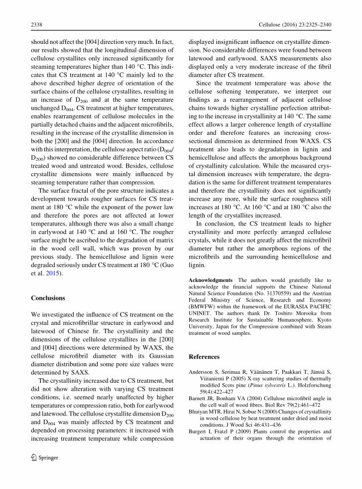
Since the treatment temperature was above the
cellulose softening temperature, we interpret our
findings as a rearrangement of adjacent cellulose
chains towards higher crystalline perfection attribut-
ing to the increase in crystallinity at 140 �C. The same
effect allows a larger coherence length of crystalline
order and therefore features an increasing cross-
sectional dimension as determined from WAXS. CS
treatment also leads to degradation in lignin and
hemicellulose and affects the amorphous background
of crystallinity calculation. While the measured crys-
tal dimension increases with temperature, the degra-
dation is the same for different treatment temperatures
and therefore the crystallinity does not significantly
increase any more, while the surface roughness still
increases at 180 �C. At 160 �C and at 180 �C also the
length of the crystallites increased.
In conclusion, the CS treatment leads to higher
crystallinity and more perfectly arranged cellulose
crystals, while it does not greatly affect the microfibril
diameter but rather the amorphous regions of the
microfibrils and the surrounding hemicellulose and
lignin.
Acknowledgments The authors would gratefully like to
acknowledge the financial supports the Chinese National
Natural Science Foundation (No. 31370559) and the Austrian
Federal Ministry of Science, Research and Economy
(BMWFW) within the framework of the EURASIA PACIFIC
UNINET. The authors thank Dr. Toshiro Morooka from
Research Institute for Sustainable Humanosphere, Kyoto
University, Japan for the Compression combined with Steam
treatment of wood samples.
References
Andersson S, Serimaa R, Vaananen T, Paakkari T, Jamsa S,
Viitaniemi P (2005) X-ray scattering studies of thermally
modified Scots pine (Pinus sylvestris L.). Holzforschung
59(4):422–427
Barnett JR, Bonham VA (2004) Cellulose microfibril angle in
the cell wall of wood fibres. Biol Rev 79(2):461–472
BhuiyanMTR, Hirai N, Sobue N (2000) Changes of crystallinity
in wood cellulose by heat treatment under dried and moist
conditions. J Wood Sci 46:431–436
Burgert I, Fratzl P (2009) Plants control the properties and
actuation of their organs through the orientation of
2338 Cellulose (2016) 23:2325–2340
123

cellulose fibrils in their cell walls. Integr Comp Biol
49(1):69–79
ChengW, Xing X,WangD, Zhang K, Cai Q,MoG, Chen Z,Wu
Z (2011) Small-angle X-ray scattering study on nanos-
tructural changes with water content in red pine, American
pine, and white ash. J Wood Sci 57:470–478
Cheng G, Zhang X, Simmons B, Singh S (2015) Theory, prac-
tice and prospects of X-ray and neutron scattering for lig-
nocellulosic biomass characterization: towards
understanding biomass pretreatment. Energy Environ Sci
8(2):436–455
Dwianto W, Morooka T, Norimoto M, Kitajima T (1999) Stress
relaxation of Sugi (Cryptomeria japonica D. Don) wood in
radial compression under high temperature steam. Holz-
forschung 53(5):541–546
Farber J, Lichtenegger HC (2001) Cellulose microfibirl angles
in a spruce branch and mechanical implications. J Mater
Sci 36(21):5087–5092
Fengel D, Wegener G (1984) Wood: chemistry, ultrastructure
reactions. Walter de Gruyter Press, Berlin and New York
Fernandes AN, Thomas LH, Altaner CM, Callow P, Forsyth VT,
Apperley DC, Kennedy CJ, Jarvis MC (2011) Nanostruc-
ture of cellulose microfibrils in spruce wood. PNAS
108(47):1195–1203
French AD (2014) Idealized powder diffraction patterns for
cellulose polymorphs. Cellulose 21:885–896
Garvey CJ, Parker IH, Simon GP (2005) On the interpretation of
X-ray diffraction powder patterns in terms of the nanos-
tructure of cellulose I fibres. Macromol Chem Phys
206(15):1568–1575
Guo J, Song K, Salmen L, Yin Y (2015) Changes of wood cell
walls in response to hygro-mechanical steam treatment.
Carbohyd Polym 115:207–214
Guo J, Guo X, Wang S, Yin Y (2016) Effects of ultrasonic
treatment during acid hydrolysis on the yield, particle size
and structure of cellulose nanocrystals. Carbohyd Polym
135:248–255
Inagaki T, Siesler HW, Mitsui K, Tsuchikawa S (2010) Dif-
ference of the crystal structure of cellulose in wood after
hydrothermal and aging degradation: a NIR spectroscopy
and XRD study. Biomacromolecules 11(9):2300–2305
Inoue M, Norimoto M, Tahanashi M, Rowell RM (1993) Steam
or heat fixation of compressed wood. Wood Fiber Sci
25(3):224–235
Ito Y, Tanahashi M, Shigematsu M, Shinoda Y (1998) Com-
pressive-molding of wood by high-pressure steam-treat-
ments. Part 2. Mechanism of permanent of fixation.
Holzforschung 52(2):217–221
Jakob HF, Fengel D, Tschegg SE, Fratzl P (1995) The ele-
mentary cellulose fibril in Picea abies: comparison of
transmission electron microscopy, small-angle X-ray
scattering, and wide-angle X-ray scattering results.
Macromolecules 28(26):8782–8787
Jakob HF, Tschegg SE, Fratzl P (1996) Hydration dependence
of the wood-cell wall structure in Picea abies. A small-
angle X-ray scattering study. Macromolecules
29(26):8435–8440
Kennedy CJ, Cameron GJ, Sturcova A, Apperley DC, Altaner C,
Wess TJ, Jarvis MC (2007) Microfibril diameter in celery
collenchyma cellulose: X-ray scattering and NMR evi-
dence. Cellulose 14:235–246
Kim D, Nishiyama Y, Wada M, Kuga S, Okano T (2001)
Thermal decomposition of cellulose crystallites in wood.
Holzforschung 55(5):521–524
Kulasinski K, Guyer R, Derome D, Carmeliet J (2015) Water
adsorption in wood microfibril-hemicellulose system: role
of the crystalline-amorphous interface. Biomacro-
molecules 16(9):2972–2978
Lichtenegger H, Reiterer A, Stanzl-Tschegg SE, Fratzl P (1999)
Variation of cellulose microfibril angles in softwoods and
hardwoods—a possible strategy of mechanical optimiza-
tion. J Struct Biol 128:257–269
Lichtenegger HC, Schoberl T, Bartl MH, Waite H, Stucky GD
(2002) High abrasion resistance with sparse mineraliza-
tion: copper biomineral in worm jaws. Science
298:389–392
Lichtenegger HC, Schoberl T, Ruokolainen JT, Cross JO, Heald
SM, Birkedal H, Waite HJ, Stucky GD (2003) Zinc and
mechanical prowess in the jaws of Nereis, a marine worm.
PNAS 100(16):9144–9149
Moon RJ, Martini A, Nairn J, Simonsen J, Youngblood J (2011)
Cellulose nanomaterials review: structure, properties and
nanocomposites. Chem Soc Rev 40(7):3941–3994
Navi P, Girardet F (2000) Effects of thermo-hydro-mechanical
treatment on the structure and properties of wood. Holz-
forschung 54(3):287–293
Navi P, Heger F (2004) Combined densification and thermos-
hydro-mechanical processing of wood. MRS Bull
29(5):332–336
Navi P, Sandberg D (2011) Thermo-hydro-mechanical pro-
cessing of wood. EPFL Press, Lausanne
Nishiyama Y (2009) Structure and properties of the cellulose
microfibril. J Wood Sci 55(4):241–249
Nishiyama Y, Langan P, O’Neill H, Pingali SV, Harton S (2014)
Structural coarsening of aspen wood by hydrothermal
pretreatment monitored by small- and wide-angle scatter-
ing of X-rays and neutrons on oriented specimens. Cellu-
lose 21:1015–1024
Panshin AJ, Zeeuw CD (1970) Textbook of wood technology:
structure, identification, properties, and uses of the com-
mercial woods of the United States and Canada. McGraw-
Hill series in forest resources, vol 1. McGraw-Hill College
Press, Ontario
Park S, Baker JO, Himmel ME, Parilla PA, Johnson DK (2010)
Cellulose crystallinity index: measurement techniques and
their impact on interpreting cellulase performance.
Biotechnol Biofuels 3:10
Penttila PA, Varnai A, Leppanen K, Peura M, Kallonen A,
Jaaskelainen P, Lucenius J, Ruokolainen J, Siika-aho M,
Viikari L, Serimaa R (2010) Changes in submicrometer
structure of enzymatically hydrolyzed microcrystalline
cellulose. Biomacromolecules 11(4):1111–1117
Peura M, Grotkopp I, Lemke H, Vikkula A, Laine J, Muller M,
Serimaa R (2006) Negative poisson ratio of crystalline
cellulose in kraft cooked Norway spruce. Biomacro-
molecules 7:1521–1528
Reiterer A, Lichtenegger H, Tschegg S, Fratzl P (1999)
Experimental evidence for a mechanical function of the
cellulose microfibril angle in wood cell walls. Philos Mag
A 79(9):2173–2184
Rennhofer H, Puchegger S, Pabisch S, Rentenberger C, Li C,
Siegel S, Steiger-Thirsfeld A, Paris O, Peterlik H (2014)
Cellulose (2016) 23:2325–2340 2339
123

The structural evolution of multi-layer graphene stacks in
carbon fibers under load at high temperature—a syn-
chrotron radiation study. Carbon 80:373–381
Salmen NL, Back EL (1977) The influence of water on the glass
phase transition temperature of cellulose. TAPPI
60(12):137–140
Sandberg D, Haller P, Navi P (2013) Thermo-hydro and ther-
mos-hydro-mechanical wood processing: an opportunity
for future environmentally friendly wood products. Wood
Mater Sci Eng 8(1):64–88
Shapiro SS, Wilk MB (1965) An analysis of variance test for
normality (complete samples). Biometrika 52(3–4):591–611
Skyba O, Schwarze FWMR (2009) Physical and mechanical
properties of thermos-hygro-mechanically (THM)-densi-
fied wood. Wood Res 54(2):1–18
Song K, Yin Y, Salmen L, Xiao F, Jiang X (2014) Changes in the
properties of wood cell walls during the transformation
from sapwood to heartwood. J Mater Sci 49(4):1734–1742
Sugiyama J, Okano T (1990) Transformation of Valonia cellu-
lose crystals by an alkaline hydrothermal treatment.
Macromolecules 23(12):3196–3198
Szezesniak L, Rachocki A, Tritt-Goe J (2008) Glass transition
temperature and thermal decomposition of cellulose pow-
der. Cellulose 15:445–451
Thomas LH, Forsyth VT, Sturcova A, Kennedy CJ, May RP,
Altaner CM, Apperley DC, Wess TJ, Jarvis MC (2013)
Structure of cellulose microfibrils in primary cell walls
from Collenchyma. Plant Physiol 161(1):465–476
Toba K, Yamamoto H, Yoshida M (2013) Crystallization of
cellulose microfibrils in wood cell wall by repeated dry-
and-wet treatment, using X-ray diffraction technique.
Cellulose 20:633–643
Wada M, Hori R, Kim U, Sasaki S (2010) X-ray diffraction
study on the thermal expansion behavior of cellulose Ib andits high-temperature phase. Polym Degrad Stabil
95(8):1330–1334
Winandy JE (1994) Wood properties. Encycl Agricult Sci
4:549–561
Xu F, Shi Y, Wang D (2013) X-ray scattering studies of ligno-
cellulosic biomass: a review. Carbohyd Polym 94:904–917
Yin Y, Berglund L, Salmen L (2011) Effect of steam treatment
on the properties of wood cell walls. Biomacromolecules
12(1):194–202
2340 Cellulose (2016) 23:2325–2340
123

本文献由“学霸图书馆-文献云下载”收集自网络,仅供学习交流使用。
学霸图书馆(www.xuebalib.com)是一个“整合众多图书馆数据库资源,
提供一站式文献检索和下载服务”的24 小时在线不限IP
图书馆。
图书馆致力于便利、促进学习与科研,提供最强文献下载服务。
图书馆导航:
图书馆首页 文献云下载 图书馆入口 外文数据库大全 疑难文献辅助工具