Embed Size (px)
Citation preview
Documenta Ophthalmologica99: 83–92, 1999.© 2000Kluwer Acadeic Publishers. Printed in the Netherlands.
The influence of visual input on the vestibulo-ocularreflex
JEROEN BOSMAN1,2, MARCEL P.M. TEN TUSSCHER2 and HERMANKINGMA 1
1University Hospital Maastricht, Department of Otorhinolaryngology and Head & NeckSurgery, P.O. Box 5800, 6202 AZ Maastricht, The Netherlands;2University HospitalMaastricht, Department of Ophthalmology, P.O. Box 5800, 6202 AZ Maastricht, TheNetherlands;1,2Institute Brain & Behaviour
Accepted 21 December 1999
Abstract. In order to stabilise a fixation target on the retina, eye movements have to com-pensate for head movements. During slow head movements visual feedback can control theseeye movements. During fast movements of the head, mainly the vestibulo-ocular reflex (VOR)controls eye movements, as visual feedback is too slow. However, visual feedback is an im-portant factor in controlling the VOR; e.g. the gain of the VOR depends on the distance ofthe target. This study investigates the influence of retinal image position during fast headmovements. The experiments were carried out in five human subjects using scleral searchcoils. The adaptation of each eye individually to a change of retinal position of a target wasexamined during head shaking. The change in visual input was carried out by placing Fresnelprisms of different strengths in front of both eyes, thus inducing a change in retinal imageposition without changing the retinal slip. The results show, that both eyes make the appro-priate corrections when the visual input changes, even during fast head-movements. Thesecorrections did not influence the gain of the VOR. From these results we conclude, that retinalimage position besides retinal slip has a major influence on the monocular eye movementseven at high head rotation frequencies.
Key words: eye movements, prism adaptation, stability of Gaze, vergence, VOR
Introduction
Human eyes tend to compensate for various head movements in order tomaintain a stable image on the retina. One of these movements is activehead rotation. Gaze stabilisation during active head rotation is accomplishedby the co-operation of visual, auditory, vestibular, proprioceptive [1, 2] andefference copy [3] mechanisms. In particular the visual and vestibular inputsare responsible for the co-ordination of compensatory eye movements, calledthe vestibulo-ocular reflex (VOR).
84
The efficiency of both inputs varies with head velocities (and frequencies).The vestibular system is effective at head rotation frequencies between 1–10Hz [4]. The visual system on the other hand can follow a target moving atlow frequepcies (0–2 Hz). At high frequencies (>2 Hz) ocular smooth pursuitbecomes less effective [5, 6]. At these frequencies the target is not perceivedclearly. This can be demonstrated by moving a finger in front of the eyes. Thefaster the movement, the more blurred the image becomes. Visual perceptionappears to be too slow at high target velocities. If one would hold the fingermotionless and shake the head, we can still see the finger clear up to shakefrequencies of 5 Hz.
Nevertheless visual input remains an important aspect in this experiment.Placing of the target closer to the head results in two things. First, a changeof image position occurs. To compensate for this a vergence eye movement isnecessary to maintain binocular vision. Second, the movement of the imageon the retina (the retinal slip) inpreases in size. To compensate for this slip,the eyes need to move faster than in the situation when the target is positionedin infinity. Therefore, a change of target position in depth during head shakingdemands for a change in VOR gain (ratio between velocity of eye and headmovement), However, are we able to change the gain during high-frequencyhead rotation? Do we perceive the position of the image? Do changes invergence and subsequent changes in gain occur as a result of a change inimage position only?
The role of retinal slip on the gain of the VOR has been pointed out earlierby Collewijn, Martins and Steinman [7]. Their conclusion is, that retinal slipis the main driving force for the control of the gain of the VOR. The aim ofthis study is to determine whether, besides retinal slip, retinal position alsoinfluences VOR gain during high-frequency head movements.
This study investigates compensatory eye movements during active headshaking. A change in position of the target by means of a Fresnel prism shouldcause a change in vergence angle and a slight change in VOR gain. If theposition is determined accurately by applying a different strength in prismfor each eye, the adaptation has to be different for each eye individually.
Methods
Apparatus
Horizontal and vertical eye movements and head movements were simultan-eously recorded using the scleral search coil technique [8] at 200 Hz. Headmovements were recorded using a 2D-coil attached to the forehead, connec-ted to a free torsion channel of the coil box. Vergence angle was defined as
85
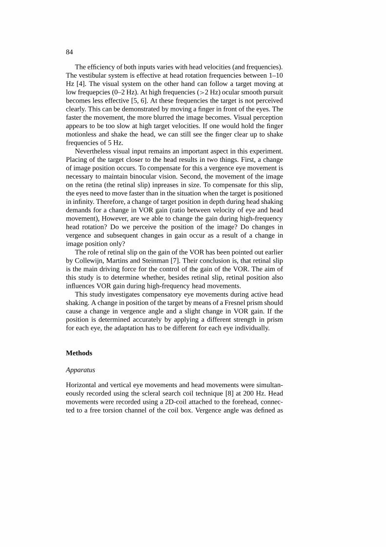
Figure 1. Set-up of the experiment. (a) Top view: displacement prism relocates the targetwithout enlarging or defocusing, a = vergence angle, (b) Frontal view: subject wearing glasseswith displacement prisms at the bottom looking through the prism, (c) Frontal view: after headtilt the subject is looking through the normal glass.
horizontal left eye minus right eye position. The subject sat on a stool withthe head free. Subjects were instructed to keep their heads in the centre ofthe coil box. This is necessary because the coil box only has a homogeneousmagnetic field in the centre: if the head moves outside a 30×30×30-cm area,non-linearities of the system became apparent. Linearity of the coil box wasverified using a calibration device.
Procedure
The subject fixated a word on a chart placed at 2 meters in front of thesubject. The word at this distance subtended an angle of approximately 1degree. The set-up of the experiment is schematically drawn in Figure 1.The subject yaw-rotated his head actively for 16 seconds while listening to arhythmic clicking sound. Each measurement was done twice. Three differenthead rotation frequencies were used: 0.5, 2 Hz and maximum frequency. Thesubject wore glasses on which two Fresnel prisms were mounted on the lowerhalf of the glasses. The glasses were firmly placed on the nose, to avoid anymovement. The prisms were two base-temporal displacement prisms (righteye 4D and left eye 1D). During the first experiment (prism-glass) the subject

86
initially looked through the prisms at the target, while shaking the head. After8 s the head was tilted slightly; thereby looking through normal glass at thetarget. This resulted in a vertical gaze angle displacement of approximately10 degrees. The amplitude of head shaking was within 10 degrees, varying foreach person and each trial. During fast head movements, the amplitude de-creased to 0.5–1 degree. The two prisms induce an optical displacement of thetarget, thereby demanding an immediate change of individual eye positions toregain binocular vision (see Figure 1a). The experiment was repeated (glass-prism); but now the subjects initially looked through the glass. Then, after 8s, they looked through the prisms. The whole procedure was repeated oncefor each subject. The results (horizontal and vertical eye and head positions)were stored on disc using the Nystagliner Eyemovement System (Toennies,Würzburg).
Subjects
The subjects were 27 (A), 39 (B), 24 (C) and 24 (E) year-old males and one27 year-old female (D), with no ocular dysfunction, 20/20 vision and normalstereopsis. Subject D was myopic in both eyes, this was corrected by−3.5glasses. All subjects had a dominant right eye. Prior to the experiment it wasestablished that subjects had single vision when looking through the prisms.The maximum frequency of head-shaking of subjects A and B was 4 Hz, sub-jects C, D and E’s maximum frequency reached up to 3 Hz. All experimentswere undertaken with the understanding and consent of the subjects.
Data analysis
Before experiments, eye movements were recorded without the glasses toshow any influences of head tilt on the eye movements. The subjects tiltedtheir heads slightly backward, as would resemble the glass-prism experiment.
The amount of eye position corrections made after head-tilt was scoredmanually. Because the prism strength was different for both eyes, each eyehad to make a specific change of position. A prism of 1 diopter results in achange of target direction of approximately 0.57◦. A 4D prism results in a2.2◦ change. Subjects wearing a 1D base-temporal (BT) prism left and a 4DBT prism right, should have an increase in vergence angle of approximately−2.8◦ in the first and 2.8◦ in the second experiment (see Figure 1a).
Finally, VOR gain values (velocity of eye versus head movements) werecalculated. First, data were split up in two parts; before and after head tilt. Inboth parts gain values were determined. The horizontal eye and head move-ments were differentiated and peak values (i.e. saccades) removed using a6-median filter. VOR gain values were calculated using a sinusoidal least-
87
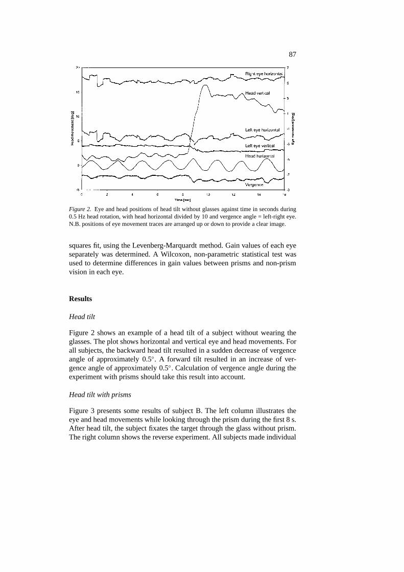
Figure 2. Eye and head positions of head tilt without glasses against time in seconds during0.5 Hz head rotation, with head horizontal divided by 10 and vergence angle = left-right eye.N.B. positions of eye movement traces are arranged up or down to provide a clear image.
squares fit, using the Levenberg-Marquardt method. Gain values of each eyeseparately was determined. A Wilcoxon, non-parametric statistical test wasused to determine differences in gain values between prisms and non-prismvision in each eye.
Results
Head tilt
Figure 2 shows an example of a head tilt of a subject without wearing theglasses. The plot shows horizontal and vertical eye and head movements. Forall subjects, the backward head tilt resulted in a sudden decrease of vergenceangle of approximately 0.5◦. A forward tilt resulted in an increase of ver-gence angle of approximately 0.5◦. Calculation of vergence angle during theexperiment with prisms should take this result into account.
Head tilt with prisms
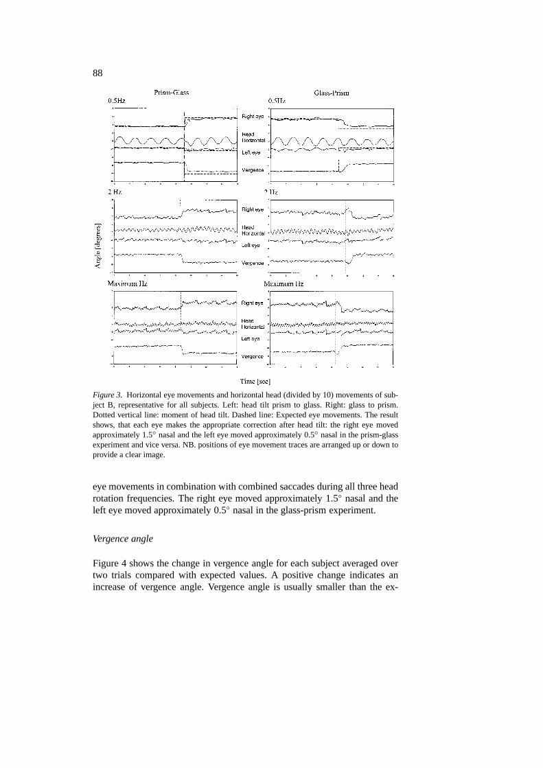
Figure 3 presents some results of subject B. The left column illustrates theeye and head movements while looking through the prism during the first 8 s.After head tilt, the subject fixates the target through the glass without prism.The right column shows the reverse experiment. All subjects made individual
88
Figure 3. Horizontal eye movements and horizontal head (divided by 10) movements of sub-ject B, representative for all subjects. Left: head tilt prism to glass. Right: glass to prism.Dotted vertical line: moment of head tilt. Dashed line: Expected eye movements. The resultshows, that each eye makes the appropriate correction after head tilt: the right eye movedapproximately 1.5◦ nasal and the left eye moved approximately 0.5◦ nasal in the prism-glassexperiment and vice versa. NB. positions of eye movement traces are arranged up or down toprovide a clear image.
eye movements in combination with combined saccades during all three headrotation frequencies. The right eye moved approximately 1.5◦ nasal and theleft eye moved approximately 0.5◦ nasal in the glass-prism experiment.
Vergence angle
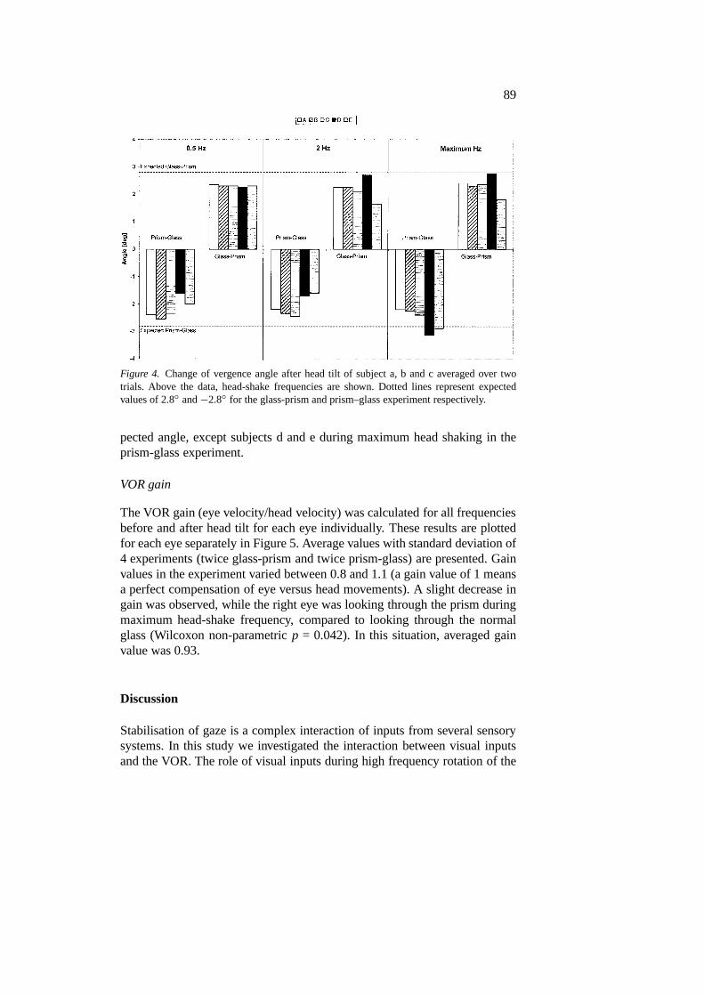
Figure 4 shows the change in vergence angle for each subject averaged overtwo trials compared with expected values. A positive change indicates anincrease of vergence angle. Vergence angle is usually smaller than the ex-
89
Figure 4. Change of vergence angle after head tilt of subject a, b and c averaged over twotrials. Above the data, head-shake frequencies are shown. Dotted lines represent expectedvalues of 2.8◦ and−2.8◦ for the glass-prism and prism–glass experiment respectively.
pected angle, except subjects d and e during maximum head shaking in theprism-glass experiment.
VOR gain
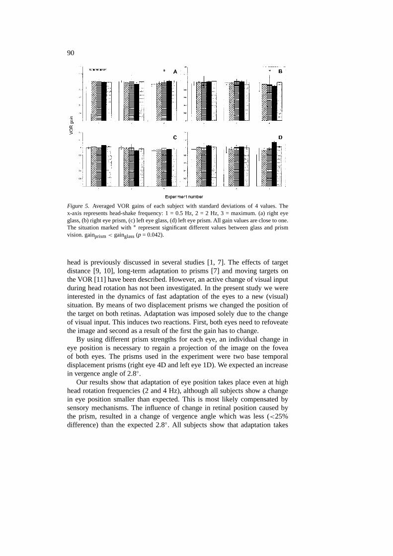
The VOR gain (eye velocity/head velocity) was calculated for all frequenciesbefore and after head tilt for each eye individually. These results are plottedfor each eye separately in Figure 5. Average values with standard deviation of4 experiments (twice glass-prism and twice prism-glass) are presented. Gainvalues in the experiment varied between 0.8 and 1.1 (a gain value of 1 meansa perfect compensation of eye versus head movements). A slight decrease ingain was observed, while the right eye was looking through the prism duringmaximum head-shake frequency, compared to looking through the normalglass (Wilcoxon non-parametricp = 0.042). In this situation, averaged gainvalue was 0.93.
Discussion
Stabilisation of gaze is a complex interaction of inputs from several sensorysystems. In this study we investigated the interaction between visual inputsand the VOR. The role of visual inputs during high frequency rotation of the
90
Figure 5. Averaged VOR gains of each subject with standard deviations of 4 values. Thex-axis represents head-shake frequency: 1 = 0.5 Hz, 2 = 2 Hz, 3 = maximum. (a) right eyeglass, (b) right eye prism, (c) left eye glass, (d) left eye prism. All gain values are close to one.The situation marked with∗ represent significant different values between glass and prismvision. gainprism< gainglass(p = 0.042).
head is previously discussed in several studies [1, 7]. The effects of targetdistance [9, 10], long-term adaptation to prisms [7] and moving targets onthe VOR [11] have been described. However, an active change of visual inputduring head rotation has not been investigated. In the present study we wereinterested in the dynamics of fast adaptation of the eyes to a new (visual)situation. By means of two displacement prisms we changed the position ofthe target on both retinas. Adaptation was imposed solely due to the changeof visual input. This induces two reactions. First, both eyes need to refoveatethe image and second as a result of the first the gain has to change.
By using different prism strengths for each eye, an individual change ineye position is necessary to regain a projection of the image on the foveaof both eyes. The prisms used in the experiment were two base temporaldisplacement prisms (right eye 4D and left eye 1D). We expected an increasein vergence angle of 2.8◦.
Our results show that adaptation of eye position takes place even at highhead rotation frequencies (2 and 4 Hz), although all subjects show a changein eye position smaller than expected. This is most likely compensated bysensory mechanisms. The influence of change in retinal position caused bythe prism, resulted in a change of vergence angle which was less (<25%difference) than the expected 2.8◦. All subjects show that adaptation takes
91
place one eye at a time, although correction saccades take place in botheyes simultaneously. This result supports the hypothesis of Dell’Osso [12]that there might be an individual ocular motor control of each eye, but alsoshows that a visual position change on the retina without a change in slip isperceived.
In this experiment we investigated the influence of both prisms on theVOR gain. If a subject looks through both prisms, the vergence angle in-creases approximately 2.8◦. This might be perceived as if the target is placed80 cm in front of the subject instead of 200 cm, although the size of thetarget remains the same. With a target at a distance of 200 centimetres, thegain should be approximately 1.04. At a distance of 80 centimetres it shouldbe approximately 1.12 [6]. This would mean an increase of VOR gain ofapproximately 0.1. Our data do not show this increase of gain, a slight de-crease can even be observed when the right eye looks through the prism,while the subjects shook their heads as fast as possible. Viire et al. [13] statethat the occurring VOR gain adjustments during fast head movements are toofast to be mediated by the pursuit or visually driven vergence systems. Theirexplanation for these adjustments is that the ocular motor system makes useof a fast non-visual estimate of the current target location relative to the head,called thetarget locator network(TLN). This network processes visual, audit-ory, proprioceptive and vestibular inputs and calculates the craniotopic targetposition. The visual input updates the network, which modulates the actionof the VOR [14]. How this update is accomplished has not been mentioned.We believe that, besides retinal slip, retinal image position also participatesin updating the VOR gain. In our experiment these updates presumably takeplace at the moment when head velocities are slow, i.e. when head movementschange direction.
References
1. Hine T, Thorn F. Compensatory eye movements during active head rotation for neartargets: effects of imagination, rapid head oscillation and vergence. Vis Res 1987; 27(9):1639–57.
2. Meiry JL. Vestibular and proprioceptive stabilization of eye movements. In: Bach-Y-RitaP, Collins CC, eds. The control of eye movements. New York: Academic Press, 1971:483–96.
3. Hine T. Effects of asymmetric vergence on compensatory eye movements during activehead rotation. J Vestibular Res 1991; 1: 357–71.7
4. Carpenter RHS. Movements of the eye. London: Psion Limited, 1988.5. Atkin A, Bender M. Ocular stabilisation during oscillatory head movements. Arch
Neurol 1986; 19: 559–66.6. Kasteel-van Linge A, Maas AJJ. Quantification of visuo-vestibular interaction up to 5.0
Hz in normal subjects. Acta Oto-Laryngol 1990; 110: 18–24.
92
7. Collewijn H, Martins AJ, Steinman RM. Compensatory eye movements during activeand passive head movements: fast adaption to changes in visual magnification. J Physiol1983; 340: 259–86.
8. Robinson DA. A method of measuring eye movement using a scleral search coil in amagnetic field. IEEE Trans Biomed Eng 1963; 10: 137–45.
9. Paige GD. The influence of target distance on eye movement responses during verticallinear motion. Exp Brain Res 1989; 77: 585–93.
10. Snyder LH, Lawrence DM, King WM. Changes in vestibulo-ocular reflex (VOR)anticipate changes in vergence angle in monkey. Vision Res 1992; 32(3): 569–75.
11. Das VE, Leigh RJ, Thomas CW, Averbuch-Teller LA, Zivotofsky AZ, Discenna AO,Dell’Osso LF. Modulation of HF VOR during visual tracking in humans. J Neurophysiol1995; 74(2): 624–32.
12. Dell’Osso LF. Evidence suggesting individual ocular motor control of each eye (muscle).J Vestibular Res 1994; 4: 335–45.
13. Viire E, Tweed D, Milner K, Vilis T. A re-examination of the gain of the VOR. JNeurophysiol 1986; 56: 439–50.
14. Vilis T. Interactions between the angular and translational components of the VOR. In:Sharpe JA, Barber HO, eds. The vestibulo ocular reflex and vertigo. New York: RavenPress, 1993: 117–24.
Address for correspondence: J. Bosman, University Hospital Maastricht, Department of Otorhinolaryngo-logy and Head & Neck Surgery, P.O. Box 5800, 6202 AZ Maastricht, The NetherlandsPhone: (+31)-43-3875586; Fax: (+31)-43-3875580