Embed Size (px)
Citation preview
TM
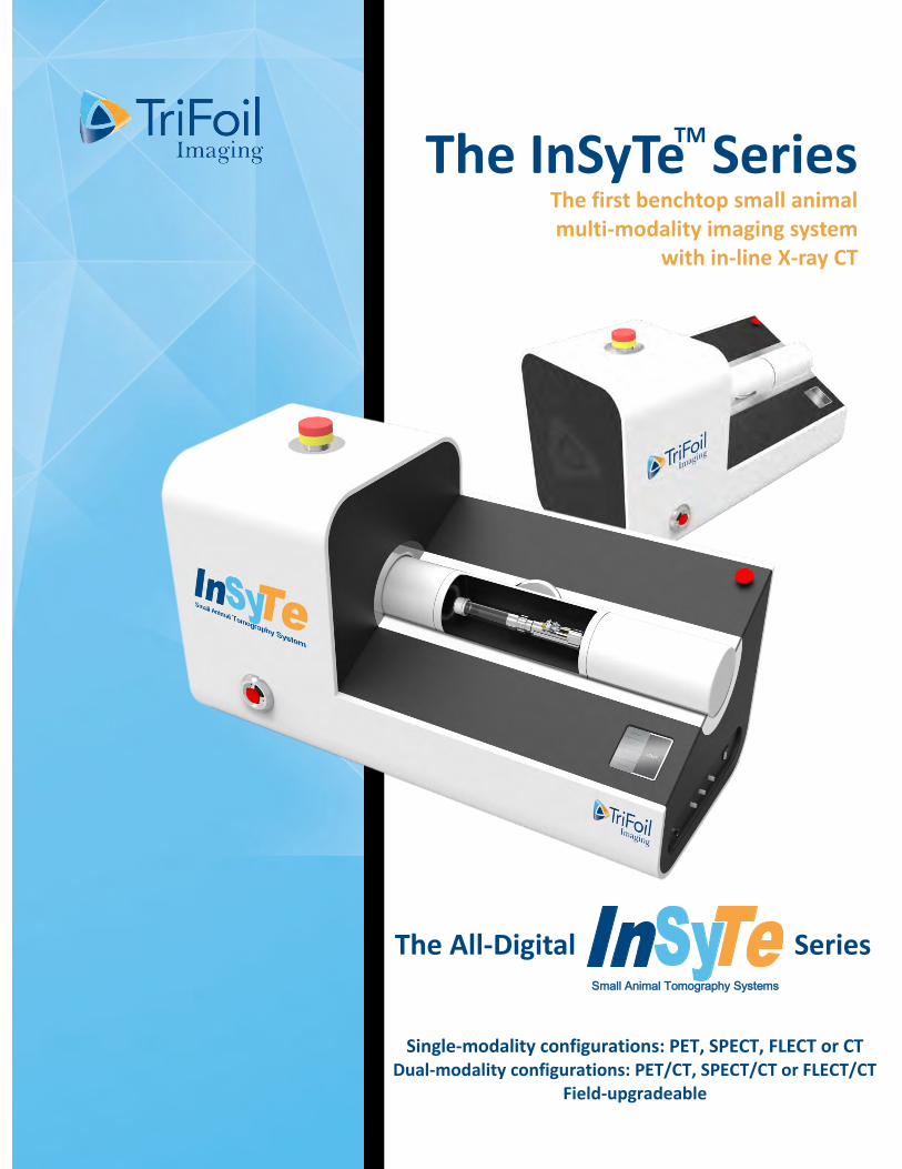
The InSyTe Series The first benchtop small animal multi-modality imaging system
with in-line X-ray CT
Single-modality configurations: PET, SPECT, FLECT or CT Dual-modality configurations: PET/CT, SPECT/CT or FLECT/CT
Field-upgradeable
The All-Digital Series Small Animal Tomography Systems
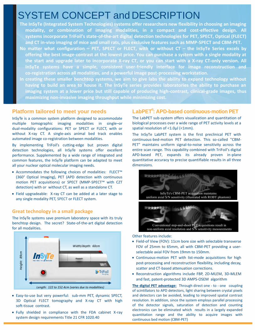
Length: 122 to 152.4cm (varies due to modalities)
Hei
gh
t: 8
9cm
Width/depth: 60cm
InSyTe's CBM-PET acquisition maintains uniform axial S/N sensitivity (illustrated with ROBY phantom)
Conventional step-and-shoot PET acquisitions result in non-uniform axial resolution and S/N sensitivity measurements
LabPET®: APD-based continuous-motion PET
The LabPET sub-system offers visualization and quantitation of biological processes over a wide range of PET activity levels at a spatial resolution of <1.0μl (<1mm).
The InSyTe LabPET system is the first preclinical PET with continuous-bed-motion PET detection. This so-called "CBM-PET” maintains uniform signal-to-noise sensitivity across the entire scan range. This capability combined with TriFoil’s digital APD-based PET, expands its already proven in-plane quantitative accuracy to precise quantifiable results in all three dimensions.
Other features include:
Field-of View (FOV): 11cm bore size with selectable transverse
FOV of 25mm to 65mm, all with CBM-PET providing a user-
selectable axial FOV from 19mm to 150mm.
Continuous-motion PET with list-mode acquisitions for high
post-processing and reconstruction flexibility, including decay,
scatter and CT-based attenuation corrections.
Reconstruction algorithms include FBP, 2D-MLEM, 3D-MLEM
and fast, patent-protected 3D AMPS-OSEM algorithm
The digital PET advantage: Through direct one - to - one coupling of scintillators to APD detectors, light sharing between crystal pixels and detectors can be avoided, leading to improved spatial contrast resolution. In addition, since the system employs parallel processing of the detector signals, saturation of detection and counting electronics can be eliminated which results in a largely expanded quantitation range and the ability to acquire images with continuous bed motion (CBM-PET)
SYSTEM CONCEPT and DESCRIPTION
The InSyTe (Integrated System Technologies) systems offer researchers new flexibility in choosing an imaging modality, or combination of imaging modalities, in a compact and cost-effective design. All systems incorporate TriFoil’s state-of-the-art digital detection technologies for PET, SPECT, Optical (FLECT) and CT in-vivo imaging of mice and small rats, plus exclusive features such as MMP-SPECT and CBM-PET.
No matter what configuration – PET, SPECT or FLECT, with or without CT – the InSyTe Series excels by offering the best image-contrast at the lowest price. You can purchase a system with a single modality at the start and upgrade later to incorporate X-ray CT, or you can start with a X-ray CT-only version. All InSyTe systems have a simple, consistent user-friendly interface for image reconstruction and co-registration across all modalities, and a powerful image post-processing workstation.
In creating these smaller benchtop systems, we aim to give labs the ability to expand technology without having to build an area to house it. The InSyTe series provides laboratories the ability to purchase an imaging system at a lower price but still capable of producing high-contrast, clinical-grade images, thus maximizing non-invasive imaging throughput while minimizing cost.
Platform tailored to meet your needs
InSyTe is a common system platform designed to accommodate multiple tomographic imaging modalities in single–or dual-modality configurations: PET or SPECT or FLECT, with or without X-ray CT. A single-axis animal bed track enables automated image co-registration between modalities.
By implementing TriFoil’s cutting-edge but proven digital detection technologies, all InSyTe systems offer excellent performance. Supplemented by a wide range of integrated and common features, the InSyTe platform can be adapted to meet all your nuclear optical molecular imaging needs.
Accommodates the following choices of modalities: FLECT™ (360° Optical Imaging), PET (APD detection with continuous motion PET acquisitions) or SPECT (MMP-SPECT™ with CZT detection) with or without CT, as well as a standalone CT.
Field upgradeable: X-ray CT can be added at a later stage to any single modality PET, SPECT or FLECT system.
Great technology in a small package The InSyTe systems save premium laboratory space with its truly benchtop design. The secret? State-of-the-art digital detection for all modalities.
Easy-to-use but very powerful: sub-mm PET, dynamic SPECT, 3D Optical FLECT tomography and X-ray CT with high soft-tissue contrast.
Fully shielded in compliance with the FDA cabinet X-ray system design requirements Title 21 CFR 1020.40
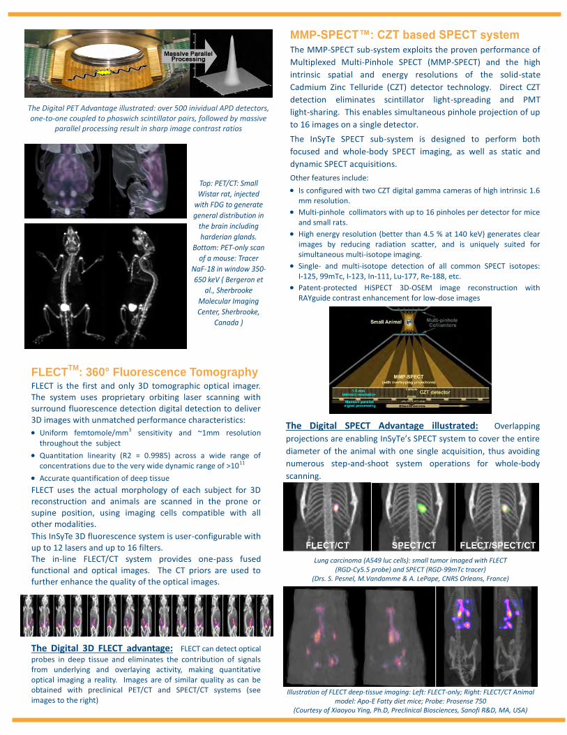
Top: PET/CT: Small
Wistar rat, injected
with FDG to generate
general distribution in
the brain including
harderian glands.
Bottom: PET-only scan
of a mouse: Tracer
NaF-18 in window 350-
650 keV ( Bergeron et
al., Sherbrooke
Molecular Imaging
Center, Sherbrooke,
Canada )
FLECTTM
: 360° Fluorescence Tomography FLECT is the first and only 3D tomographic optical imager. The system uses proprietary orbiting laser scanning with surround fluorescence detection digital detection to deliver 3D images with unmatched performance characteristics:
Uniform femtomole/mm3 sensitivity and ~1mm resolution throughout the subject
Quantitation linearity (R2 = 0.9985) across a wide range of concentrations due to the very wide dynamic range of >1011
Accurate quantification of deep tissue
FLECT uses the actual morphology of each subject for 3D reconstruction and animals are scanned in the prone or supine position, using imaging cells compatible with all other modalities.
This InSyTe 3D fluorescence system is user-configurable with up to 12 lasers and up to 16 filters. The in-line FLECT/CT system provides one-pass fused functional and optical images. The CT priors are used to further enhance the quality of the optical images.
The Digital 3D FLECT advantage: FLECT can detect optical
probes in deep tissue and eliminates the contribution of signals from underlying and overlaying activity, making quantitative optical imaging a reality. Images are of similar quality as can be obtained with preclinical PET/CT and SPECT/CT systems (see images to the right)
MMP-SPECT™: CZT based SPECT system The MMP-SPECT sub-system exploits the proven performance of
Multiplexed Multi-Pinhole SPECT (MMP-SPECT) and the high
intrinsic spatial and energy resolutions of the solid-state
Cadmium Zinc Telluride (CZT) detector technology. Direct CZT
detection eliminates scintillator light-spreading and PMT
light-sharing. This enables simultaneous pinhole projection of up
to 16 images on a single detector.
The InSyTe SPECT sub-system is designed to perform both
focused and whole-body SPECT imaging, as well as static and
dynamic SPECT acquisitions.
Other features include:
Is configured with two CZT digital gamma cameras of high intrinsic 1.6 mm resolution.
Multi-pinhole collimators with up to 16 pinholes per detector for mice and small rats.
High energy resolution (better than 4.5 % at 140 keV) generates clear images by reducing radiation scatter, and is uniquely suited for simultaneous multi-isotope imaging.
Single- and multi-isotope detection of all common SPECT isotopes: I-125, 99mTc, I-123, In-111, Lu-177, Re-188, etc.
Patent-protected HiSPECT 3D-OSEM image reconstruction with RAYguide contrast enhancement for low-dose images
The Digital SPECT Advantage illustrated: Overlapping
projections are enabling InSyTe’s SPECT system to cover the entire
diameter of the animal with one single acquisition, thus avoiding
numerous step-and-shoot system operations for whole-body
scanning.
The Digital PET Advantage illustrated: over 500 inividual APD detectors, one-to-one coupled to phoswich scintillator pairs, followed by massive
parallel processing result in sharp image contrast ratios
Lung carcinoma (A549 luc cells): small tumor imaged with FLECT (RGD-Cy5.5 probe) and SPECT (RGD-99mTc tracer)
(Drs. S. Pesnel, M.Vandamme & A. LePape, CNRS Orleans, France)
Illustration of FLECT deep-tissue imaging: Left: FLECT-only; Right: FLECT/CT Animal model: Apo-E Fatty diet mice; Probe: Prosense 750
(Courtesy of Xiaoyou Ying, Ph.D, Preclinical Biosciences, Sanofi R&D, MA, USA)
Specifications subject to change without notice; 17 June 2015 Copyright 2015. TriFoil Imaging All rights reserved.
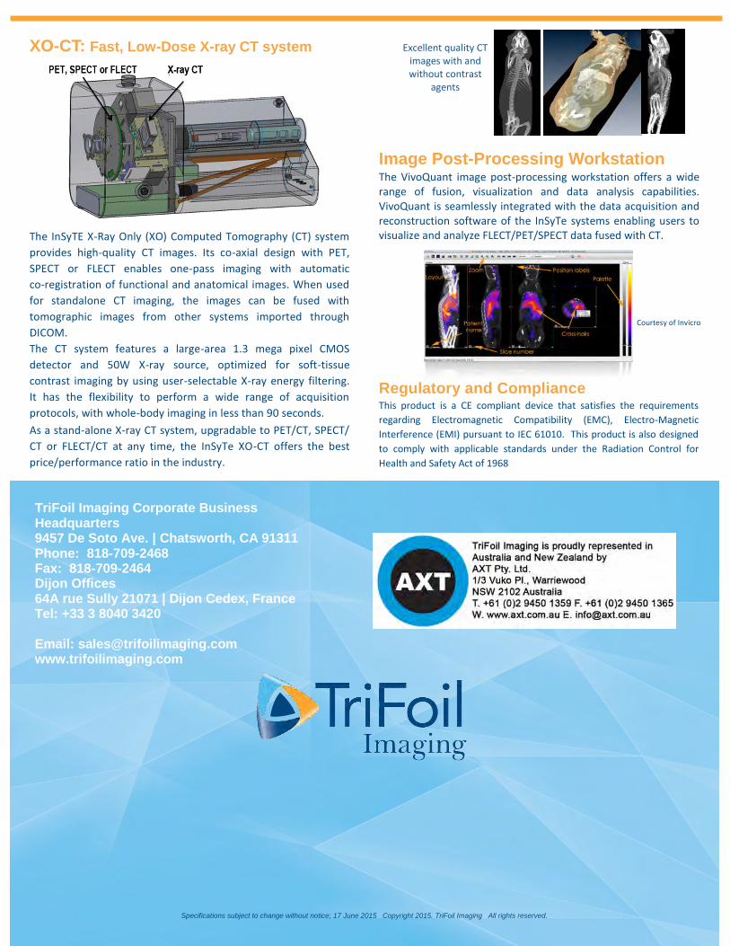
XO-CT: Fast, Low-Dose X-ray CT system
The InSyTE X-Ray Only (XO) Computed Tomography (CT) system
provides high-quality CT images. Its co-axial design with PET,
SPECT or FLECT enables one-pass imaging with automatic
co-registration of functional and anatomical images. When used
for standalone CT imaging, the images can be fused with
tomographic images from other systems imported through
DICOM.
The CT system features a large-area 1.3 mega pixel CMOS
detector and 50W X-ray source, optimized for soft-tissue
contrast imaging by using user-selectable X-ray energy filtering.
It has the flexibility to perform a wide range of acquisition
protocols, with whole-body imaging in less than 90 seconds.
As a stand-alone X-ray CT system, upgradable to PET/CT, SPECT/
CT or FLECT/CT at any time, the InSyTe XO-CT offers the best
price/performance ratio in the industry.
Regulatory and Compliance This product is a CE compliant device that satisfies the requirements
regarding Electromagnetic Compatibility (EMC), Electro-Magnetic
Interference (EMI) pursuant to IEC 61010. This product is also designed
to comply with applicable standards under the Radiation Control for
Health and Safety Act of 1968
Courtesy of Invicro
Image Post-Processing Workstation The VivoQuant image post-processing workstation offers a wide range of fusion, visualization and data analysis capabilities. VivoQuant is seamlessly integrated with the data acquisition and reconstruction software of the InSyTe systems enabling users to visualize and analyze FLECT/PET/SPECT data fused with CT.
TriFoil Imaging Corporate Business Headquarters 9457 De Soto Ave. | Chatsworth, CA 91311 Phone: 818-709-2468 Fax: 818-709-2464 Dijon Offices 64A rue Sully 21071 | Dijon Cedex, France Tel: +33 3 8040 3420
Email: [email protected] www.trifoilimaging.com
Excellent quality CT images with and without contrast
agents


![validation benchtop sterilizers db9804[1]](https://img.pdfslide.net/doc/110x75/55296ac4550346522e8b477a/validation-benchtop-sterilizers-db98041.jpg)