Embed Size (px)
Citation preview
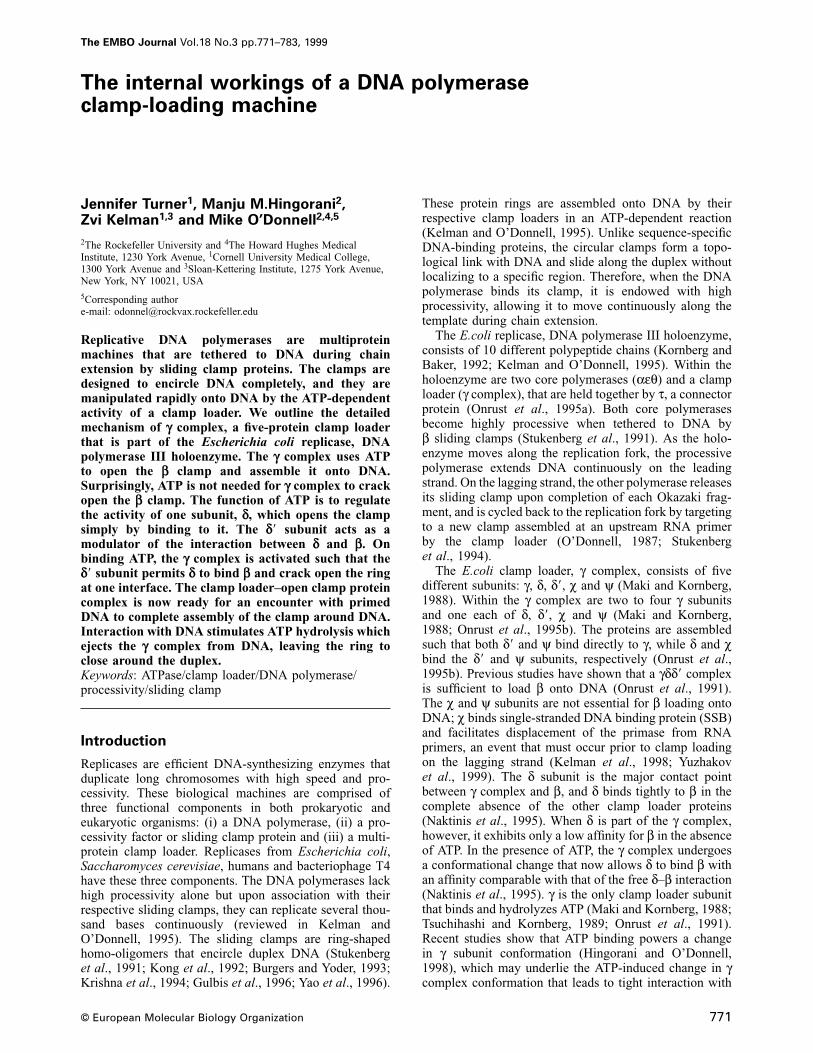
The EMBO Journal Vol.18 No.3 pp.771–783, 1999
The internal workings of a DNA polymeraseclamp-loading machine
Jennifer Turner1, Manju M.Hingorani2,Zvi Kelman1,3 and Mike O’Donnell2,4,5
2The Rockefeller University and 4The Howard Hughes MedicalInstitute, 1230 York Avenue, 1Cornell University Medical College,1300 York Avenue and 3Sloan-Kettering Institute, 1275 York Avenue,New York, NY 10021, USA
5Corresponding authore-mail: [email protected]
Replicative DNA polymerases are multiproteinmachines that are tethered to DNA during chainextension by sliding clamp proteins. The clamps aredesigned to encircle DNA completely, and they aremanipulated rapidly onto DNA by the ATP-dependentactivity of a clamp loader. We outline the detailedmechanism of γ complex, a five-protein clamp loaderthat is part of the Escherichia coli replicase, DNApolymerase III holoenzyme. The γ complex uses ATPto open the β clamp and assemble it onto DNA.Surprisingly, ATP is not needed for γ complex to crackopen the β clamp. The function of ATP is to regulatethe activity of one subunit, δ, which opens the clampsimply by binding to it. The δ� subunit acts as amodulator of the interaction between δ and β. Onbinding ATP, the γ complex is activated such that theδ� subunit permits δ to bind β and crack open the ringat one interface. The clamp loader–open clamp proteincomplex is now ready for an encounter with primedDNA to complete assembly of the clamp around DNA.Interaction with DNA stimulates ATP hydrolysis whichejects the γ complex from DNA, leaving the ring toclose around the duplex.Keywords: ATPase/clamp loader/DNA polymerase/processivity/sliding clamp
Introduction
Replicases are efficient DNA-synthesizing enzymes thatduplicate long chromosomes with high speed and pro-cessivity. These biological machines are comprised ofthree functional components in both prokaryotic andeukaryotic organisms: (i) a DNA polymerase, (ii) a pro-cessivity factor or sliding clamp protein and (iii) a multi-protein clamp loader. Replicases from Escherichia coli,Saccharomyces cerevisiae, humans and bacteriophage T4have these three components. The DNA polymerases lackhigh processivity alone but upon association with theirrespective sliding clamps, they can replicate several thou-sand bases continuously (reviewed in Kelman andO’Donnell, 1995). The sliding clamps are ring-shapedhomo-oligomers that encircle duplex DNA (Stukenberget al., 1991; Kong et al., 1992; Burgers and Yoder, 1993;Krishna et al., 1994; Gulbis et al., 1996; Yao et al., 1996).
© European Molecular Biology Organization 771
These protein rings are assembled onto DNA by theirrespective clamp loaders in an ATP-dependent reaction(Kelman and O’Donnell, 1995). Unlike sequence-specificDNA-binding proteins, the circular clamps form a topo-logical link with DNA and slide along the duplex withoutlocalizing to a specific region. Therefore, when the DNApolymerase binds its clamp, it is endowed with highprocessivity, allowing it to move continuously along thetemplate during chain extension.
The E.coli replicase, DNA polymerase III holoenzyme,consists of 10 different polypeptide chains (Kornberg andBaker, 1992; Kelman and O’Donnell, 1995). Within theholoenzyme are two core polymerases (αεθ) and a clamploader (γ complex), that are held together by τ, a connectorprotein (Onrust et al., 1995a). Both core polymerasesbecome highly processive when tethered to DNA byβ sliding clamps (Stukenberg et al., 1991). As the holo-enzyme moves along the replication fork, the processivepolymerase extends DNA continuously on the leadingstrand. On the lagging strand, the other polymerase releasesits sliding clamp upon completion of each Okazaki frag-ment, and is cycled back to the replication fork by targetingto a new clamp assembled at an upstream RNA primerby the clamp loader (O’Donnell, 1987; Stukenberget al., 1994).
The E.coli clamp loader, γ complex, consists of fivedifferent subunits: γ, δ, δ�, χ and ψ (Maki and Kornberg,1988). Within the γ complex are two to four γ subunitsand one each of δ, δ�, χ and ψ (Maki and Kornberg,1988; Onrust et al., 1995b). The proteins are assembledsuch that both δ� and ψ bind directly to γ, while δ and χbind the δ� and ψ subunits, respectively (Onrust et al.,1995b). Previous studies have shown that a γδδ� complexis sufficient to load β onto DNA (Onrust et al., 1991).The χ and ψ subunits are not essential for β loading ontoDNA; χ binds single-stranded DNA binding protein (SSB)and facilitates displacement of the primase from RNAprimers, an event that must occur prior to clamp loadingon the lagging strand (Kelman et al., 1998; Yuzhakovet al., 1999). The δ subunit is the major contact pointbetween γ complex and β, and δ binds tightly to β in thecomplete absence of the other clamp loader proteins(Naktinis et al., 1995). When δ is part of the γ complex,however, it exhibits only a low affinity for β in the absenceof ATP. In the presence of ATP, the γ complex undergoesa conformational change that now allows δ to bind β withan affinity comparable with that of the free δ–β interaction(Naktinis et al., 1995). γ is the only clamp loader subunitthat binds and hydrolyzes ATP (Maki and Kornberg, 1988;Tsuchihashi and Kornberg, 1989; Onrust et al., 1991).Recent studies show that ATP binding powers a changein γ subunit conformation (Hingorani and O’Donnell,1998), which may underlie the ATP-induced change in γcomplex conformation that leads to tight interaction with
J.Turner et al.
β (Naktinis et al., 1995; Hingorani and O’Donnell, 1998).Thus, the γ subunit transduces the energy from ATP toexpose the δ subunit for interaction with the β ring.
This report describes the mechanics of the γ complex-catalyzed process of β assembly onto DNA. We show thatthe energy of ATP binding powers the γ complex machineryto bind β and open the circular clamp at one interface.Once γ complex binds β, its ATPase activity is supresseduntil it binds DNA. The correct DNA substrate stimulatesATP hydrolysis, which is coupled to the release of γcomplex from β on DNA. On DNA, β reassumes itslowest free energy state and forms a closed ring, nowwith DNA passing through the center.
The molecular details that underlie the clamp loaderactivity have been explored and as a result, specificfunctions have been assigned to the three integral com-ponents of the γ complex: γ is the ‘motor’, δ is the ‘ringopener’ and δ� is the ‘modulator’. The γ subunits are theonly components of γ complex to interact with ATP, andtheir function is to drive ATP-induced conformationalchanges of other subunits in the clamp loader. Theδ subunit contains the intrinsic clamp-opening activity, asit can open the β ring in the complete absence of ATPand the other subunits. The δ� subunit binds δ and probablymodulates the ability of β to gain access to δ. The ATP-driven γ motor either moves or alters the δ� modulatorsuch that the δ clamp opener gains access to the β clampand opens the ring. This action requires only ATP bindingto the γ complex. The resulting clamp loader–open clampcomposite then binds primed DNA with high affinity.Upon interaction with primed DNA, two or three moleculesof ATP are hydrolyzed, resulting in closure of the β ringaround DNA and ejection of γ complex off the DNA andback into solution.
Results
The γ complex uses energy from ATP binding toopen the β ringThe β sliding clamp is composed of two crescent-shapedprotomers arranged in a tight head-to-tail dimer (Konget al., 1992). This circular protein clamp is transferredonto circular DNA by the ATP-dependent clamp-loadingactivity of the γ complex. How does γ complex assemblethe β ring around DNA? How is ATP used in this reaction?Is ATP binding sufficient, or is hydrolysis necessary?What is the role of DNA in this process? To beginaddressing these questions, we developed a novel ‘Cys-labeling’ assay to detect and characterize the ring-openingstep in clamp assembly.
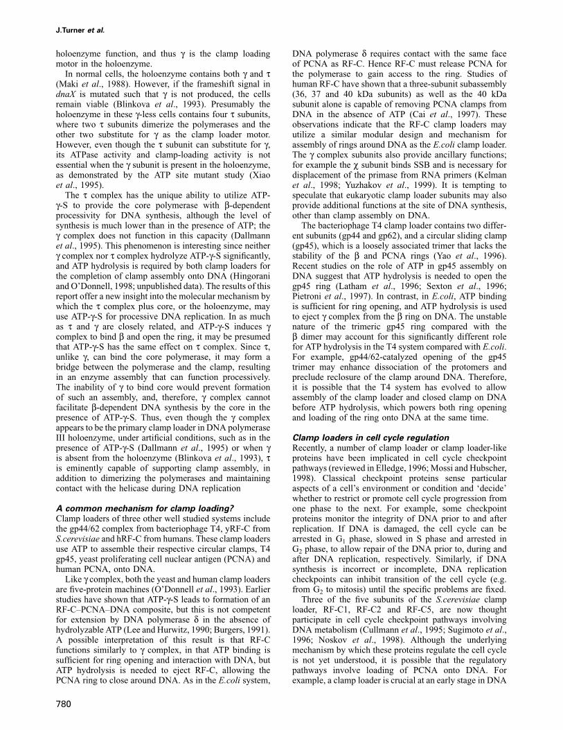
Leu273, a buried hydrophobic residue in the β dimerinterface, was substituted with cysteine (β crystal structure;Kong et al., 1992). This buried cysteine residue shouldnot be accessible to a thiol-reactive reagent, such as amaleimide, when the L273C-β ring is closed. When theinterface is opened, however, the buried cysteine shouldbecome accessible to the surface and reactive to maleimide.To follow this reaction, we synthesized a radioactivereagent by linking the N-terminus of a 32P-labeled peptidewith an N-hydroxysuccinimide (NHS) ester coupled tomaleimide (Figure 1). If the dimer interface opens, theinterface cysteine residue will react with the [32P]maleim-ide reagent, resulting in a radiolabeled β clamp. One other
772
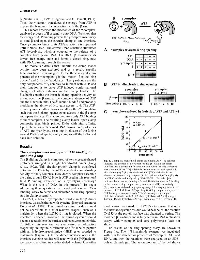
Fig. 1. γ complex opens the β clamp on binding ATP. The schemeindicates the position of a cysteine residue (S) within the dimerinterface that is accessible for reaction only when the ring is opened.The structure of the [32P]maleimide reagent used to label cysteine isalso shown. (A) β (3 μM) incubated with [32P]maleimide in theabsence or presence of γ complex (3 μM), primed oligoDNA (3 μM)or ATP (2 mM), and analyzed by SDS–PAGE. 32P-labeled β isindicated by an arrow, showing a 2- and 10-fold increase in β labelingin the presence of γ complex and γ complex � ATP, respectively.(B) γ complex-catalyzed ring opening assayed for varying times in thepresence of ATP (left) or ATP-γ-S (right). (C) γ complex-catalyzedATP hydrolysis compared with ATP-γ-S hydrolysis. γ complex(0.1 μM), incubated with β (0.2 μM), hydrolyzes ATP with a kcat �1.7/min (d) and hydrolyzes ATP-γ-S with a kcat � 4�10–3/min (j).
modification was made in L273C-β to ensure that onlythe interface cysteine residue would be labeled; the reactiveCys333 at the protein surface was changed to serine. Themodified β is a dimer and is fully active in DNA replicationassays with γ complex and core polymerase (data notshown).
The results of the ring-opening assay are shown inFigure 1A. The [32P]maleimide reagent was incubatedwith β in the absence or presence of γ complex, ATP andDNA, and then the reactions were analyzed on an SDS–polyacrylamide gel. The autoradiogram of the gel shows
Mechanics of clamp assembly on DNA
very low reactivity of β alone (Figure 1A, lane 1). Whenβ is mixed with γ complex, there is a 2-fold increase inβ labeling (Figure 1A, lane 2). However, when bothγ complex and ATP are present in the reaction, the β is10-fold more reactive with the [32P]maleimide (Figure1A, lane 4). The results indicate that in the presence ofATP, γ complex can open the β ring or at least changethe ring conformation enough to expose the cysteineresidue buried in the dimer interface. When DNA ispresent in the reaction, β is labeled to the same extent asin the absence of DNA (compare lanes 4 and 5), indicatingthat γ complex can open the β ring even in the absenceof DNA.
Next, the assay was used to examine whether ATP bindingor hydrolysis is required to stimulate β ring opening. Toseparate the effects of ATP binding from ATP hydrolysis,we substituted ATP with its analog, ATP-γ-S (Figure 1B).The turnover number for ATP hydrolysis catalyzed byγ complex (in the presence of β) is 1.5–2/min at 37°C(Figure 1C). Therefore, in a 10 min assay, each γ complexhydrolyzes 15–20 molecules of ATP. The γ complex bindsATP-γ-S with similar high affinity as it does ATP (Kd
ATP �2 μM and Kd
ATP-γ-S � 5 μM, respectively; Hingorani andO’Donnell, 1998), but the turnover number for ATP-γ-Shydrolysis is very low (4–6�10–3/min; see Figure 1C).Since γ complex catalyzes hydrolysis of only one ATP-γ-Severy 2–3 h, ATP-γ-S can be considered a non-hydrolyzableanalog of ATP within the time frame of the ring-openingassay.
The data shown in Figure 1B demonstrate that ATP andATP-γ-S facilitate γ complex-catalyzed ring opening tothe same extent. Therefore, ATP binding appears sufficientto drive γ complex-catalyzed opening of the β ring. Othernon-hydrolyzable ATP analogs such as AMP-PNP andAMP-PCP were also tested in the assay, but ring openingwas not detected (data not shown). However, this negativeresult may be explained by the fact that γ complex bindsthese analogs with much lower affinity than ATP (Kd
~1 mM versus 2 μM for ATP), and this binding energymay be insufficient to support ring opening (H.Xiao andM.O’Donnell, unpublished data).
Next, the ring-opening step was examined in greaterdetail to determine whether the clamp loader opens β atboth interfaces (e.g. monomerizes the dimer to assembleit around DNA), or if opening the β dimer at only oneinterface is sufficient to allow entry of DNA into the ring.
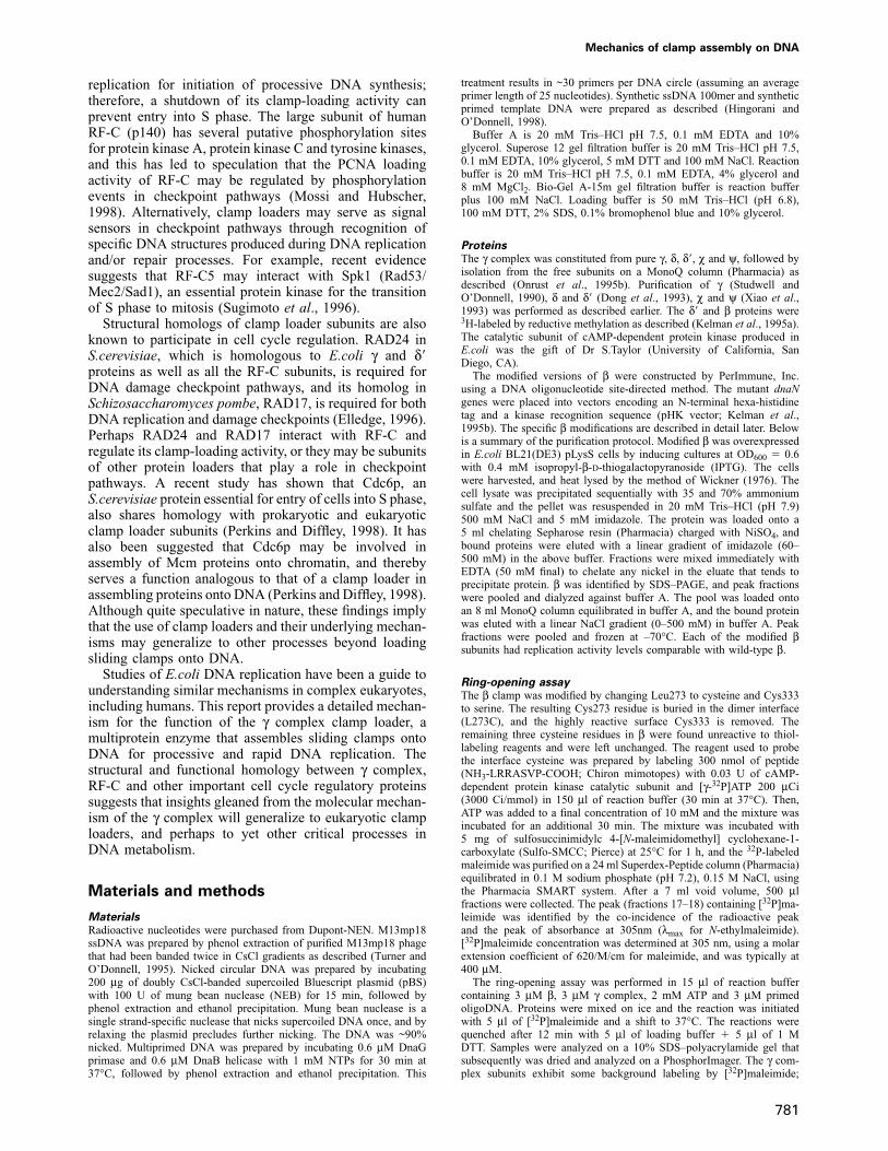
The γ complex need open only one interface of theβ dimer for clamp loadingTo determine whether γ complex needs to open one orboth interfaces of the β dimer for assembly of the clamparound DNA, we constructed a β dimer in which themonomers could be cross-linked at the dimer interface.Arg103 and Ile305, two residues close to each other buton opposing sides of the dimer interface (Cα atoms ~6 Åapart; β cyrstal structure; Kong et al., 1992), were substi-tuted with cysteine. Due to the head-to-tail arrangementof the β dimer, the two interfaces are identical. It wasexpected that oxidation would result in formation ofdisulfide cross-links between the cysteine residues, andyield β dimers covalently linked at one or both interfaces.
Initially, the modified β (βS–S) was 32P-labeled at anN-terminal kinase recognition site and analyzed on a
773
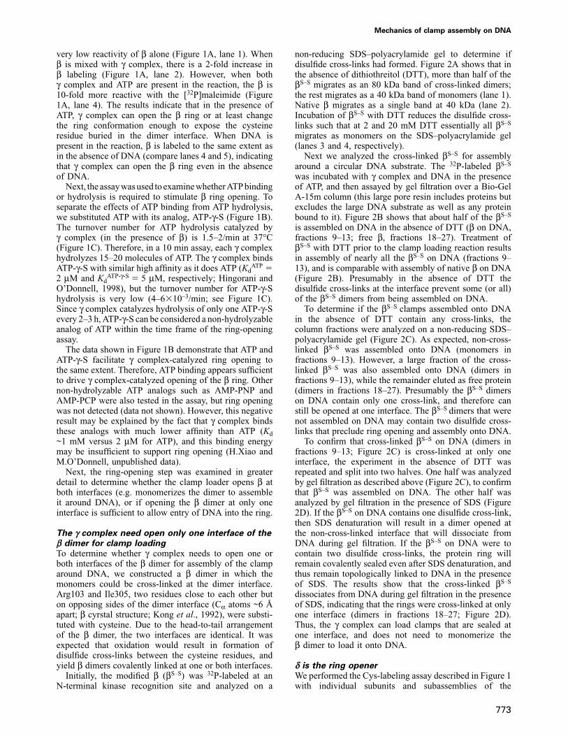
non-reducing SDS–polyacrylamide gel to determine ifdisulfide cross-links had formed. Figure 2A shows that inthe absence of dithiothreitol (DTT), more than half of theβS–S migrates as an 80 kDa band of cross-linked dimers;the rest migrates as a 40 kDa band of monomers (lane 1).Native β migrates as a single band at 40 kDa (lane 2).Incubation of βS–S with DTT reduces the disulfide cross-links such that at 2 and 20 mM DTT essentially all βS–S
migrates as monomers on the SDS–polyacrylamide gel(lanes 3 and 4, respectively).
Next we analyzed the cross-linked βS–S for assemblyaround a circular DNA substrate. The 32P-labeled βS–S
was incubated with γ complex and DNA in the presenceof ATP, and then assayed by gel filtration over a Bio-GelA-15m column (this large pore resin includes proteins butexcludes the large DNA substrate as well as any proteinbound to it). Figure 2B shows that about half of the βS–S
is assembled on DNA in the absence of DTT (β on DNA,fractions 9–13; free β, fractions 18–27). Treatment ofβS–S with DTT prior to the clamp loading reaction resultsin assembly of nearly all the βS–S on DNA (fractions 9–13), and is comparable with assembly of native β on DNA(Figure 2B). Presumably in the absence of DTT thedisulfide cross-links at the interface prevent some (or all)of the βS–S dimers from being assembled on DNA.
To determine if the βS–S clamps assembled onto DNAin the absence of DTT contain any cross-links, thecolumn fractions were analyzed on a non-reducing SDS–polyacrylamide gel (Figure 2C). As expected, non-cross-linked βS–S was assembled onto DNA (monomers infractions 9–13). However, a large fraction of the cross-linked βS–S was also assembled onto DNA (dimers infractions 9–13), while the remainder eluted as free protein(dimers in fractions 18–27). Presumably the βS–S dimerson DNA contain only one cross-link, and therefore canstill be opened at one interface. The βS–S dimers that werenot assembled on DNA may contain two disulfide cross-links that preclude ring opening and assembly onto DNA.
To confirm that cross-linked βS–S on DNA (dimers infractions 9–13; Figure 2C) is cross-linked at only oneinterface, the experiment in the absence of DTT wasrepeated and split into two halves. One half was analyzedby gel filtration as described above (Figure 2C), to confirmthat βS–S was assembled on DNA. The other half wasanalyzed by gel filtration in the presence of SDS (Figure2D). If the βS–S on DNA contains one disulfide cross-link,then SDS denaturation will result in a dimer opened atthe non-cross-linked interface that will dissociate fromDNA during gel filtration. If the βS–S on DNA were tocontain two disulfide cross-links, the protein ring willremain covalently sealed even after SDS denaturation, andthus remain topologically linked to DNA in the presenceof SDS. The results show that the cross-linked βS–S
dissociates from DNA during gel filtration in the presenceof SDS, indicating that the rings were cross-linked at onlyone interface (dimers in fractions 18–27; Figure 2D).Thus, the γ complex can load clamps that are sealed atone interface, and does not need to monomerize theβ dimer to load it onto DNA.
δ is the ring openerWe performed the Cys-labeling assay described in Figure 1with individual subunits and subassemblies of the
J.Turner et al.
Fig. 2. γ complex opens the β clamp at one interface. Cysteine residues were placed at the β dimer interface to design a clamp with disulfide cross-links at the interface (βS–S). (A) Native β and βS–S analyzed on a non-reducing SDS–polyacrylamide gel with no DTT, 2 mM DTT and 20 mM DTTin the reaction. (B) Gel filtration analysis of native [32P]β (d) and [32P]βS–S clamps assembled onto DNA by γ complex in the absence of DTT (s)and in the presence of DTT (u). The next scheme shows a mix of βS–S rings with two, one or zero cross-links at the interface, assembled on DNAand analyzed by gel filtration followed by non-reducing SDS–PAGE. (C) Analysis of βS–S from a clamp-loading reaction without DTT [as in (B)].(D) The same reaction analyzed with SDS present during gel filtration. Fraction numbers are indicated beneath the gels. The positions of non-cross-linked βS–S (monomers) and cross-linked βS–S (dimers) are indicated by arrows.
γ complex, to determine the minimal subunit requirementfor ring opening. The minimal subassembly that testedpositive for ring opening was the three-subunit γδδ�complex, which, like γ complex, needs ATP or ATP-γ-Sto crack open the β dimer interface (data not shown).Individual subunits or combinations of two subunits didnot give a detectable signal. This result implies that morethan two proteins are needed to detect ring opening in theCys-labeling assay, that γδδ� is sufficient for ring openingand that the χ and ψ subunits are not absolutely requiredfor the mechanics of this process. This conclusion isconsistent with earlier data showing that efficient stimula-tion of processive DNA polymerase activity requires the
774
simultaneous presence of all three subunits, γ, δ and δ�(Onrust et al., 1991). However, the Cys-labeling assay forring opening does not rigorously exclude the possibilitythat a smaller subassembly than γδδ�, or even just onesubunit, can crack open the dimer interface transiently.For example, the assay requires time for the [32P]maleimideto react with an exposed cysteine residue, and is likely toproceed more efficiently the longer the β dimer interfaceis held open. To investigate if any one subunit can openthe β ring at least transiently, we designed a more directassay to detect opening of the dimer interface. This assayis based on the following rationale: once β is assembledonto DNA, the β–DNA complex is very stable (t1/2 �1 h
Mechanics of clamp assembly on DNA
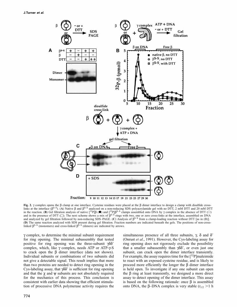
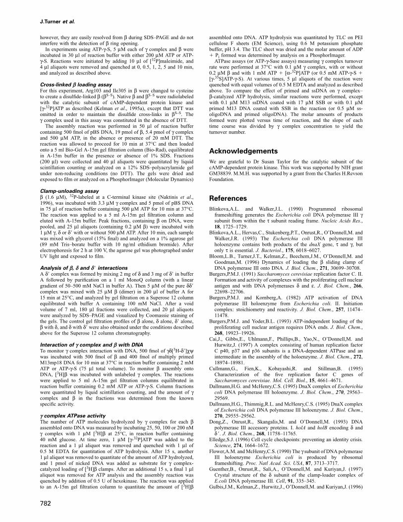
Fig. 3. A ring-unloading assay reveals that δ opens β. [32P]β clamps were assembled onto circular DNA and incubated with γ, δ or δ� in the absenceor presence of ATP and analyzed by agarose gel electrophoresis (scheme). (A) An autoradiogram of the agarose gel shows the [32P]β on DNAseparated from the [32P]β unloaded off DNA. (B) The gel stained with ethidium bromide shows that DNA is not degraded during the reaction.
at 37°C; Yao et al., 1996). However, if the dimer interfacewere to open even for a short time, the β ring could slipfree of DNA. The experiment described below examinesthe ring-opening activity of the γ, δ and δ� subunits byobserving the unloading of circular clamps from a circularDNA substrate.
In this experiment, 32P-labeled β clamps were assembledonto circular DNA by γ complex, and the [32P]β on DNAwas separated from free γ complex and free [32P]β by gelfiltration. The [32P]β–DNA complex was incubated furtherwith either γ, δ or δ�, in the absence or presence of ATP.Following this, the reactions were analyzed on an agarosegel to separate [32P]β trapped on DNA from the [32P]βreleased from DNA. The results in Figure 3A, show thatthe δ subunit by itself can release [32P]β from DNA, andthis process does not require ATP. The lack of ATPdependence is consistent with earlier evidence that δ bindsβ in the absence of ATP (Naktinis et al., 1995; noteneither δ nor β binds ATP). It also verifies that the [32P]βunloading (mediated by δ) observed in this assay is not aresult of a γ complex contaminant in the δ proteinpreparation, since γ complex requires ATP to release βfrom DNA (Naktinis et al., 1996). The gel was alsostained with ethidium bromide (Figure 3B), which showedthat the circular DNA remained intact during incubationwith each protein, including δ. The data confirm that βrelease from DNA is a result of ring opening and not dueto the ring sliding off linearized DNA, as the circularDNA substrate is not degraded during the assay.
These results show that clamp opening is actuallyperformed by only one subunit of the γ complex, and isthe result of a simple protein–protein interaction. It shouldbe noted that even though δ opens β enough to allow itto slip free from DNA, it cannot load β onto DNA. Thus,the γ and δ� subunits play a critical role in assembly ofthe open clamp around DNA. The following experimentsexamine how the δ� and γ subunits function in clamploading.
775
δ � and β compete for access to δPrevious studies identified δ as the only subunit of γcomplex with a detectable interaction with β (Naktiniset al., 1995). When δ is part of the γ complex, theinteraction with β is not favorable in the absence of ATP(Naktinis et al., 1995). In the presence of ATP, however,γ complex undergoes a conformational change and bindsβ with similar affinity as δ alone (Naktinis et al., 1995;Hingorani and O’Donnell, 1998). These observations sug-gest that δ is partially buried in the γ complex and thatthe ATP-induced conformational change in γ complexresults in exposure of δ for interaction with β. Whichsubunit of the γ complex is responsible for modulatingthe access of β to δ? Our previous studies have shownthat δ� binds stably to the δ subunit (Onrust and O’Donnell,1993). Therefore, we examined whether δ� might interferewith the interaction between δ and β.
In the experiment shown in Figure 4, we examined theinteractions between δ, δ� and β by gel filtration using aSuperose 12 sizing column. Figure 4A, B and C showsthe elution profiles of β (25 μM dimer), δ (5 μM) and δ�(5 μM), respectively. Figure 4D shows that β (25 μM)mixed with the δ subunit (5 μM) forms a δβ complex,and Figure 4E shows that a 1:1 mix of δ (5 μM) and δ�(5 μM) results in a stable δδ� complex. Finally, Figure 4Fshows that when β (25 μM) is added to purified δδ�complex (5 μM), the δ subunit interacts preferentiallywith β (to form δβ), resulting in the displacement of δ�.The δβ complex is favored over δδ� even when thereaction contains a 3-fold excess of δ� over β (datanot shown).
These protein–protein interaction studies indicate thatβ competes with δ� for interaction with δ, and providesome insight into how the γ complex subunits may workduring clamp loading. If, within the γ complex, δ� bindsδ and blocks access of β to δ, then the ATP-dependentchange in γ complex conformation may help remove theδ� block and favor δ–β interaction over the δ–δ� interaction.
J.Turner et al.
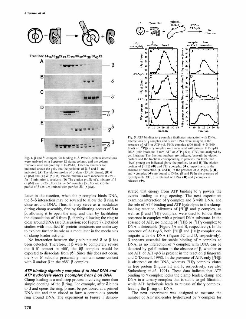
Fig. 4. β and δ� compete for binding to δ. Protein–protein interactionswere analyzed on a Superose 12 sizing column, and the columnfractions were analyzed by SDS–PAGE. Fraction numbers areindicated above the gels, and the positions of β, δ and δ� areindicated. (A) The elution profile of β alone (25 μM dimer), (B) δ(5 μM) and (C) δ� (5 μM). Protein mixtures were incubated at 25°Cfor 15 min prior to analysis. (D) The elution profile of a mixture of δ(5 μM) and β (25 μM), (E) the δδ� complex (5 μM) and (F) theprofile of β (25 μM) mixed with purified δδ� (5 μM).
Later in the reaction, when the γ complex binds DNA,the δ–β interaction may be severed to allow the β ring toclose around DNA. Thus, δ� may serve as a modulatorduring clamp assembly, first by facilitating access of δ toβ, allowing it to open the ring, and then by facilitatingthe dissociation of δ from β, thereby allowing the ring toclose around DNA (see Discussion; see Figure 7). Detailedstudies with modified δ� protein constructs are underwayto explore further its role as a modulator in the mechanicsof clamp loader activity.
No interaction between the γ subunit and δ or β hasbeen detected. Therefore, if β were to completely severethe δ–δ� contact in γδδ�, the δβ complex would beexpected to dissociate from γδ�. Since this does not occur,the γ or δ� subunits presumably maintain some contactwith δ and/or β in the γδδ�–β complex.
ATP binding signals γ complex–β to bind DNA andATP hydrolysis ejects γ complex from β on DNAClamp loading is a multistep process involving more thansimple opening of the β ring. For example, after δ bindsto β and opens the ring, β must be positioned at a primedDNA site and then closed to form a continuous proteinring around DNA. The experiment in Figure 1 demon-
776
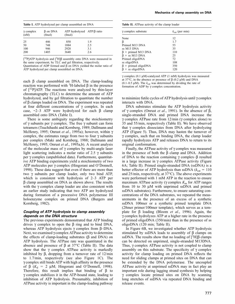
Fig. 5. ATP binding to γ complex facilitates interaction with DNA.Interactions of γ complex and β with DNA were assayed in thepresence of ATP or ATP-γ-S. [3H]γ complex (500 fmol) � β (500fmol) or [32P]β � γ complex were incubated with primed M13mp18DNA (400 fmol) and 2 mM ATP or ATP-γ-S at 37°C, and analyzed bygel filtration. The fraction numbers are indicated beneath the elutionprofiles and the fractions corresponding to proteins ‘on DNA’ and‘free’ protein are indicated above the profiles. (A and B) The elutionprofiles of [32P]β (d) and [3H]γ complex (r), respectively, in theabsence of nucleotide. (C and D) In the presence of ATP-γ-S, β (d)and γ complex (r) are bound to DNA. (E and F) In the presence ofhydrolyzable ATP, β is retained on DNA (d) and γ complex isreleased (r).
strated that energy from ATP binding to γ powers theevents leading to ring opening. The next experimentexamines interaction of γ complex and β with DNA, andthe role of ATP binding and ATP hydrolysis in the clamp-loading reaction. Mixtures of [3H]β and γ complex, aswell as β and [3H]γ complex, were used to follow theirpresence in complex with a primed DNA substrate. In theabsence of ATP, no binding of [3H]β or [3H]γ complex toDNA is detectable (Figure 5A and B, respectively). In thepresence of ATP-γ-S, both [3H]β and [3H]γ complex co-migrate with the DNA (Figure 5C and D, respectively).β appears essential for stable binding of γ complex toDNA, as no interaction of γ complex with DNA can bedetected by gel filtration in the absence of β, whether ornot ATP or ATP-γ-S is present in the reaction (Hingoraniand O’Donnell, 1998). In the presence of ATP, only [3H]βis observed on the DNA, whereas [3H]γ complex elutesas free protein (Figure 5E and F, respectively; see alsoStukenberg et al., 1991). These data indicate that ATPbinding to γ complex locks the clamp loader, clamp andDNA in a ternary complex that is stable to gel filtration,while ATP hydrolysis leads to release of the γ complex,leaving the β ring on DNA.
The next experiment was designed to measure thenumber of ATP molecules hydrolyzed by γ complex for
Mechanics of clamp assembly on DNA
Table I. ATP hydrolyzed per clamp assembled on DNA
γ complex β on DNA ATP hydrolyzed ATP/β ratio(nM) (fmol) (fmol)
25 446 850 1.950 748 1884 2.5100 946 2928 3.1200 1405 3640 2.6
[32P]ATP hydrolysis and [3H]β assembly onto DNA were measured inthe same experiment, by TLC and gel filtration, respectively.Quantitation of ADP formed and β on DNA yielded the molar ratio ofATP hydrolyzed per clamp assembled on DNA.
each β clamp assembled on DNA. The clamp-loadingreaction was performed with 3H-labeled β in the presenceof [32P]ATP. The reactions were analyzed by thin-layerchromatography (TLC) to determine the amount of ATPhydrolyzed, and by gel filtration to quantitate the numberof β clamps loaded on DNA. The experiment was repeatedat four different concentrations of γ complex. In eachcase, ~2–3 ATP were hydrolyzed for each β clampassembled onto DNA (Table I).
There is some ambiguity regarding the stoichiometryof γ subunits per γ complex. The free γ subunit can formtetramers (Tsuchihashi and Kornberg, 1989; Dallmann andMcHenry, 1995; Onrust et al., 1995a); however, within γcomplex, the estimates range from two to four γ subunitsper complex (Maki and Kornberg, 1988; Dallmann andMcHenry, 1995; Onrust et al., 1995a,b). A recent analysisof the molecular mass of γ complex by multi-angle laserlight scattering indicates a molar ratio of 2.5 γ subunitsper γ complex (unpublished data). Furthermore, quantitat-ive ATP binding experiments yield a stoichiometry of twoATP molecules per γ complex (Hingorani and O’Donnell,1998). These data suggest that even if there are more thantwo γ subunits per clamp loader, only two bind ATP,which is consistent with hydrolysis of 2–3 ATP perβ clamp assembled on DNA as shown above. The resultswith the γ complex clamp loader are also consistent withan earlier study indicating that two ATP are hydrolyzedduring formation of a processive DNA polymerase IIIholoenzyme complex on primed DNA (Burgers andKornberg, 1982).
Coupling of ATP hydrolysis to clamp assemblydepends on the DNA structureThe previous experiments demonstrated that ATP bindingdrives γ complex to open the β ring and to bind DNA,whereas ATP hydrolysis ejects γ complex from β–DNA.Next, we examined γ complex ATPase activity to determinethe effects of clamp-loading substrates (β and DNA) onATP hydrolysis. The ATPase rate was quantitated in theabsence and presence of β at 37°C (Table II). The datashow that the γ complex ATPase activity is markedlyinhibited by β, dropping from a turnover rate of 12/minto 1.7/min, respectively (see also Figure 1C). Theγ complex still binds ATP with high affinity in the presenceof β (Kd � 2 μM; Hingorani and O’Donnell, 1998).Therefore, this result implies that binding of β toγ complex stabilizes it in the ATP-bound state, leading toinhibition of ATP hydrolysis. Presumably, inhibition ofATPase activity is important in the clamp-loading pathway
777
Table II. ATPase activity of the clamp loader
γ complex substrate kcat (per min)
None 12β 1.7Primed M13 DNA 55ss M13 DNA 35β � primed M13 DNA 110β � ss M13 DNA 25Primed oligoDNA 72ss oligoDNA 108β � primed oligoDNA 350β � ss oligoDNA 120
γ complex (0.1 μM)-catalyzed ATP (1 mM) hydrolysis was measuredat 37°C, in the absence or presence of β (0.2 μM) and DNA(0.1–0.5 μM). The kcat was determined by dividing the rate offormation of ADP by γ complex concentration.
to minimize futile cycles of ATP hydrolysis until γ complexinteracts with DNA.
DNA substrates stimulate the ATP hydrolysis activityof γ complex (Onrust et al., 1991). In the absence of β,single-stranded DNA and primed DNA increase theγ complex ATPase rate from 12/min (γ complex alone) to35 and 55/min, respectively (Table II). We have observedthat γ complex dissociates from DNA after hydrolyzingATP (Figure 5). Thus, DNA may hasten the turnover ofγ complex, such that on binding DNA, the clamp loaderrapidly hydrolyzes ATP and releases DNA to return to itsoriginal conformation.
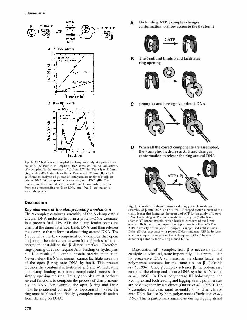
Finally, the ATPase activity of γ complex was measuredin the presence of both the β clamp and DNA. Additionof DNA to the reaction containing γ complex–β resultedin a large increase in γ complex ATPase activity (Figure6A; Table II). Primed single-stranded DNA (ssDNA) is abetter effector of ATP hydrolysis than ssDNA (kcat � 110and 25/min, respectively, at 37°C). The above experimentswere performed with 1 mM ATP in the reaction to ensuremaximum ATPase activity (γ complex Km for ATP rangesfrom 10 to 30 μM with unprimed ssDNA and primedssDNA substrates). Furthermore, to ensure saturating con-centrations of the DNA substrates, we repeated the meas-urements in the presence of an excess of a syntheticssDNA 100mer or a synthetic primed template DNA(28mer primer/100mer template), which serves as a tem-plate for β loading (Bloom et al., 1996). Again, theγ complex hydrolyzes ATP at a higher rate in the presenceof primed oligoDNA (350/min) than in the presence of ssoligoDNA (120 min; Table II).
In Figure 6B, we investigated whether ATP hydrolysisstimulated by ssDNA leads to assembly of β clamps onssDNA. The results show that no loading of [3H]β clampscan be detected on unprimed, single-stranded M13DNA.Thus, γ complex ATPase activity is not coupled to clampassembly on this substrate. The specificity of γ complexactivity for clamp loading on primed DNA reflects theneed for sliding clamps at primed sites on DNA that canbe extended by the DNA polymerase. The uncoupledATPase activity at unprimed ssDNA may in fact play animportant role during lagging strand synthesis by helpingγ complex locate primed sites on DNA by scanninglong stretches of ssDNA via repeated DNA binding andrelease events.
J.Turner et al.
Fig. 6. ATP hydrolysis is coupled to clamp assembly at a primed siteon DNA. (A) Primed M13mp18 ssDNA stimulates the ATPase activityof γ complex (in the presence of β) from 1.7/min (Table I) to 110/min(m), while ssDNA stimulates the ATPase rate to 25/min (j). (B) Agel filtration analysis of γ complex-catalyzed assembly of [3H]β onprimed DNA (m) compared with assembly on ssDNA (j). Thefraction numbers are indicated beneath the elution profile, and thefractions corresponding to ‘β on DNA’ and ‘free β’ are indicatedabove the profile.
Discussion
Key elements of the clamp-loading mechanismThe γ complex catalyzes assembly of the β clamp onto acircular DNA molecule to form a protein–DNA catenane.In a process fueled by ATP, the clamp loader opens theclamp at the dimer interface, binds DNA, and then releasesthe clamp so that it forms a closed ring around DNA. Theδ subunit is the key component of γ complex that opensthe β ring. The interaction between δ and β yields sufficientenergy to destabilize the β dimer interface. Therefore,ring-opening does not require ATP binding or hydrolysis,but is a result of a simple protein–protein interaction.Nevertheless, the δ ‘ring opener’ cannot facilitate assemblyof the open β ring onto DNA by itself. This processrequires the combined activities of γ, δ and δ�, indicatingthat clamp loading is a more complicated process thansimply opening the ring. Thus, γ complex must performseveral functions to complete the process of clamp assem-bly on DNA. For example, the open β ring and DNAmust be positioned correctly for topological linkage, thering must be closed and, finally, γ complex must dissociatefrom the ring on DNA.
778
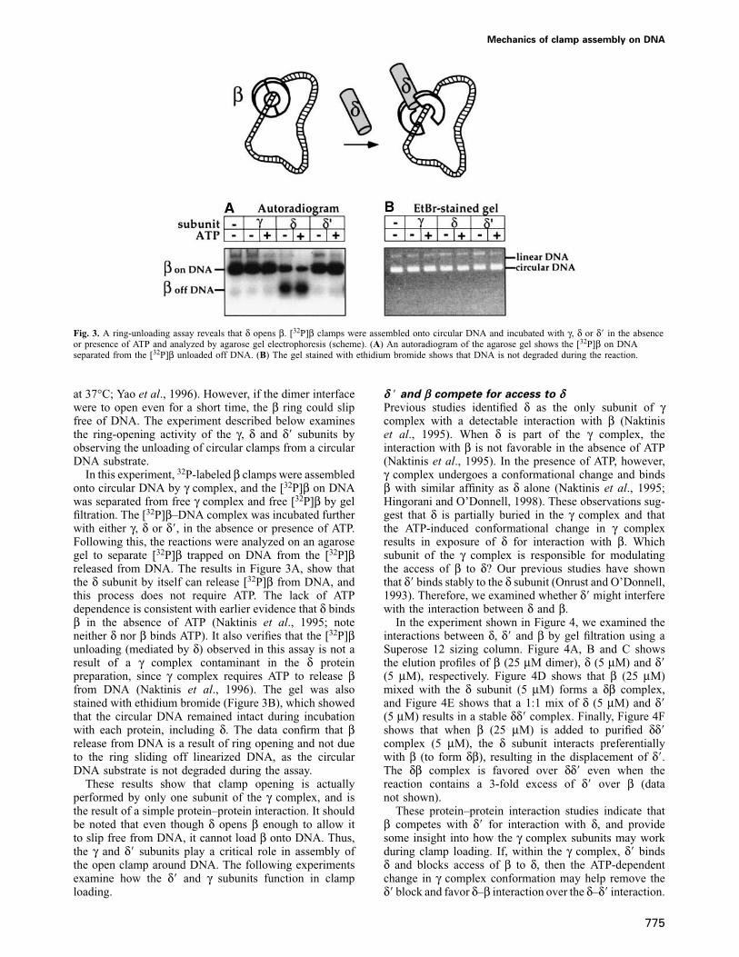
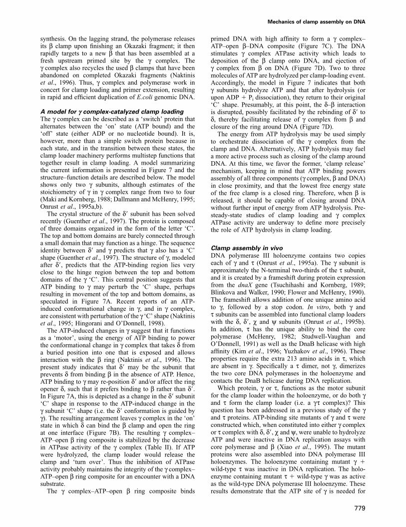
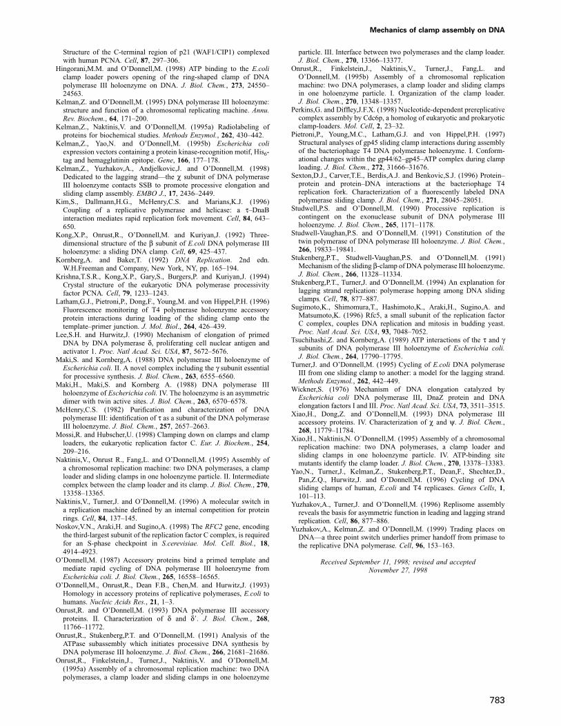
Fig. 7. A model of subunit dynamics during γ complex-catalyzedassembly of β onto DNA. (A) γ is the ‘C’-shaped motor subunit of theclamp loader that harnesses the energy of ATP for assembly of β ontoDNA. On binding ATP, a conformational change in γ affects δ�,another ‘C’-shaped protein, which leads to exposure of the δ ringopener. (B) δ binds β and opens the ring at one interface. (C) TheATPase activity of this protein complex is suppressed until it bindsDNA. (D) An encounter with primed DNA stimulates ATP hydrolysis,which is coupled to release of the β clamp and DNA. The open βdimer snaps shut to form a ring around DNA.
Dissociation of γ complex from β is necessary for itscatalytic activity and, more importantly, it is a prerequisitefor processive DNA synthesis, as the clamp loader andpolymerase compete for the same site on β (Naktiniset al., 1996). Once γ complex releases β, the polymerasecan bind the clamp and initiate DNA synthesis (Naktiniset al., 1996). In DNA polymerase III holoenzyme, theγ complex and both leading and lagging strand polymerasesare held together by a τ dimer (Onrust et al., 1995a). Theγ complex catalyzes rapid assembly of sliding clampsonto DNA for use by both polymerases (Yuzhakov et al.,1996). This is particularly significant during lagging strand
Mechanics of clamp assembly on DNA
synthesis. On the lagging strand, the polymerase releasesits β clamp upon finishing an Okazaki fragment; it thenrapidly targets to a new β that has been assembled at afresh upstream primed site by the γ complex. Theγ complex also recycles the used β clamps that have beenabandoned on completed Okazaki fragments (Naktiniset al., 1996). Thus, γ complex and polymerase work inconcert for clamp loading and primer extension, resultingin rapid and efficient duplication of E.coli genomic DNA.
A model for γ complex-catalyzed clamp loadingThe γ complex can be described as a ‘switch’ protein thatalternates between the ‘on’ state (ATP bound) and the‘off’ state (either ADP or no nucleotide bound). It is,however, more than a simple switch protein because ineach state, and in the transition between these states, theclamp loader machinery performs multistep functions thattogether result in clamp loading. A model summarizingthe current information is presented in Figure 7 and thestructure–function details are described below. The modelshows only two γ subunits, although estimates of thestoichiometry of γ in γ complex range from two to four(Maki and Kornberg, 1988; Dallmann and McHenry, 1995;Onrust et al., 1995a,b).
The crystal structure of the δ� subunit has been solvedrecently (Guenther et al., 1997). The protein is composedof three domains organized in the form of the letter ‘C’.The top and bottom domains are barely connected througha small domain that may function as a hinge. The sequenceidentity between δ� and γ predicts that γ also has a ‘C’shape (Guenther et al., 1997). The structure of γ, modeledafter δ�, predicts that the ATP-binding region lies veryclose to the hinge region between the top and bottomdomains of the γ ‘C’. This central position suggests thatATP binding to γ may perturb the ‘C’ shape, perhapsresulting in movement of the top and bottom domains, asspeculated in Figure 7A. Recent reports of an ATP-induced conformational change in γ, and in γ complex,are consistent with perturbation of the γ ‘C’ shape (Naktiniset al., 1995; Hingorani and O’Donnell, 1998).
The ATP-induced changes in γ suggest that it functionsas a ‘motor’, using the energy of ATP binding to powerthe conformational change in γ complex that takes δ froma buried position into one that is exposed and allowsinteraction with the β ring (Naktinis et al., 1996). Thepresent study indicates that δ� may be the subunit thatprevents δ from binding β in the absence of ATP. Hence,ATP binding to γ may re-position δ� and/or affect the ringopener δ, such that it prefers binding to β rather than δ�.In Figure 7A, this is depicted as a change in the δ� subunit‘C’ shape in response to the ATP-induced change in theγ subunit ‘C’ shape (i.e. the δ� conformation is guided byγ). The resulting arrangement leaves γ complex in the ‘on’state in which δ can bind the β clamp and open the ringat one interface (Figure 7B). The resulting γ complex–ATP–open β ring composite is stabilized by the decreasein ATPase activity of the γ complex (Table II). If ATPwere hydrolyzed, the clamp loader would release theclamp and ‘turn over’. Thus the inhibition of ATPaseactivity probably maintains the integrity of the γ complex–ATP–open β ring composite for an encounter with a DNAsubstrate.
The γ complex–ATP–open β ring composite binds
779
primed DNA with high affinity to form a γ complex–ATP–open β–DNA composite (Figure 7C). The DNAstimulates γ complex ATPase activity which leads todeposition of the β clamp onto DNA, and ejection ofγ complex from β on DNA (Figure 7D). Two to threemolecules of ATP are hydrolyzed per clamp-loading event.Accordingly, the model in Figure 7 indicates that bothγ subunits hydrolyze ATP and that after hydrolysis (orupon ADP � Pi dissociation), they return to their original‘C’ shape. Presumably, at this point, the δ–β interactionis disrupted, possibly facilitated by the rebinding of δ� toδ, thereby facilitating release of γ complex from β andclosure of the ring around DNA (Figure 7D).
The energy from ATP hydrolysis may be used simplyto orchestrate dissociation of the γ complex from theclamp and DNA. Alternatively, ATP hydrolysis may fuela more active process such as closing of the clamp aroundDNA. At this time, we favor the former, ‘clamp release’mechanism, keeping in mind that ATP binding powersassembly of all three components (γ complex, β and DNA)in close proximity, and that the lowest free energy stateof the free clamp is a closed ring. Therefore, when β isreleased, it should be capable of closing around DNAwithout further input of energy from ATP hydrolysis. Pre-steady-state studies of clamp loading and γ complexATPase activity are underway to define more preciselythe role of ATP hydrolysis in clamp loading.
Clamp assembly in vivoDNA polymerase III holoenzyme contains two copieseach of γ and τ (Onrust et al., 1995a). The γ subunit isapproximately the N-terminal two-thirds of the τ subunit,and it is created by a frameshift during protein expressionfrom the dnaX gene (Tsuchihashi and Kornberg, 1989;Blinkova and Walker, 1990; Flower and McHenry, 1990).The frameshift allows addition of one unique amino acidto γ, followed by a stop codon. In vitro, both γ andτ subunits can be assembled into functional clamp loaderswith the δ, δ�, χ and ψ subunits (Onrust et al., 1995b).In addition, τ has the unique ability to bind the corepolymerase (McHenry, 1982; Studwell-Vaughan andO’Donnell, 1991) as well as the DnaB helicase with highaffinity (Kim et al., 1996; Yuzhakov et al., 1996). Theseproperties require the extra 213 amino acids in τ, whichare absent in γ. Specifically a τ dimer, not γ, dimerizesthe two core DNA polymerases in the holoenzyme andcontacts the DnaB helicase during DNA replication.
Which protein, γ or τ, functions as the motor subunitfor the clamp loader within the holoenzyme, or do both γand τ form the clamp loader (i.e. a γτ complex)? Thisquestion has been addressed in a previous study of the γand τ proteins. ATP-binding site mutants of γ and τ wereconstructed which, when constituted into either γ complexor τ complex with δ, δ�, χ and ψ, were unable to hydrolyzeATP and were inactive in DNA replication assays withcore polymerase and β (Xiao et al., 1995). The mutantproteins were also assembled into DNA polymerase IIIholoenzymes. The holoenzyme containing mutant γ �wild-type τ was inactive in DNA replication. The holo-enzyme containing mutant τ � wild-type γ was as activeas the wild-type DNA polymerase III holoenzyme. Theseresults demonstrate that the ATP site of γ is needed for
J.Turner et al.
holoenzyme function, and thus γ is the clamp loadingmotor in the holoenzyme.
In normal cells, the holoenzyme contains both γ and τ(Maki et al., 1988). However, if the frameshift signal indnaX is mutated such that γ is not produced, the cellsremain viable (Blinkova et al., 1993). Presumably theholoenzyme in these γ-less cells contains four τ subunits,where two τ subunits dimerize the polymerases and theother two substitute for γ as the clamp loader motor.However, even though the τ subunit can substitute for γ,its ATPase activity and clamp-loading activity is notessential when the γ subunit is present in the holoenzyme,as demonstrated by the ATP site mutant study (Xiaoet al., 1995).
The τ complex has the unique ability to utilize ATP-γ-S to provide the core polymerase with β-dependentprocessivity for DNA synthesis, although the level ofsynthesis is much lower than in the presence of ATP; theγ complex does not function in this capacity (Dallmannet al., 1995). This phenomenon is interesting since neitherγ complex nor τ complex hydrolyze ATP-γ-S significantly,and ATP hydrolysis is required by both clamp loaders forthe completion of clamp assembly onto DNA (Hingoraniand O’Donnell, 1998; unpublished data). The results of thisreport offer a new insight into the molecular mechanism bywhich the τ complex plus core, or the holoenzyme, mayuse ATP-γ-S for processive DNA replication. In as muchas τ and γ are closely related, and ATP-γ-S induces γcomplex to bind β and open the ring, it may be presumedthat ATP-γ-S has the same effect on τ complex. Since τ,unlike γ, can bind the core polymerase, it may form abridge between the polymerase and the clamp, resultingin an enzyme assembly that can function processively.The inability of γ to bind core would prevent formationof such an assembly, and, therefore, γ complex cannotfacilitate β-dependent DNA synthesis by the core in thepresence of ATP-γ-S. Thus, even though the γ complexappears to be the primary clamp loader in DNA polymeraseIII holoenzyme, under artificial conditions, such as in thepresence of ATP-γ-S (Dallmann et al., 1995) or when γis absent from the holoenzyme (Blinkova et al., 1993), τis eminently capable of supporting clamp assembly, inaddition to dimerizing the polymerases and maintainingcontact with the helicase during DNA replication
A common mechanism for clamp loading?Clamp loaders of three other well studied systems includethe gp44/62 complex from bacteriophage T4, yRF-C fromS.cerevisiae and hRF-C from humans. These clamp loadersuse ATP to assemble their respective circular clamps, T4gp45, yeast proliferating cell nuclear antigen (PCNA) andhuman PCNA, onto DNA.
Like γ complex, both the yeast and human clamp loadersare five-protein machines (O’Donnell et al., 1993). Earlierstudies have shown that ATP-γ-S leads to formation of anRF-C–PCNA–DNA composite, but this is not competentfor extension by DNA polymerase δ in the absence ofhydrolyzable ATP (Lee and Hurwitz, 1990; Burgers, 1991).A possible interpretation of this result is that RF-Cfunctions similarly to γ complex, in that ATP binding issufficient for ring opening and interaction with DNA, butATP hydrolysis is needed to eject RF-C, allowing thePCNA ring to close around DNA. As in the E.coli system,
780
DNA polymerase δ requires contact with the same faceof PCNA as RF-C. Hence RF-C must release PCNA forthe polymerase to gain access to the ring. Studies ofhuman RF-C have shown that a three-subunit subassembly(36, 37 and 40 kDa subunits) as well as the 40 kDasubunit alone is capable of removing PCNA clamps fromDNA in the absence of ATP (Cai et al., 1997). Theseobservations indicate that the RF-C clamp loaders mayutilize a similar modular design and mechanism forassembly of rings around DNA as the E.coli clamp loader.The γ complex subunits also provide ancillary functions;for example the χ subunit binds SSB and is necessary fordisplacement of the primase from RNA primers (Kelmanet al., 1998; Yuzhakov et al., 1999). It is tempting tospeculate that eukaryotic clamp loader subunits may alsoprovide additional functions at the site of DNA synthesis,other than clamp assembly on DNA.
The bacteriophage T4 clamp loader contains two differ-ent subunits (gp44 and gp62), and a circular sliding clamp(gp45), which is a loosely associated trimer that lacks thestability of the β and PCNA rings (Yao et al., 1996).Recent studies on the role of ATP in gp45 assembly onDNA suggest that ATP hydrolysis is needed to open thegp45 ring (Latham et al., 1996; Sexton et al., 1996;Pietroni et al., 1997). In contrast, in E.coli, ATP bindingis sufficient for ring opening, and ATP hydrolysis is usedto eject γ complex from the β ring on DNA. The unstablenature of the trimeric gp45 ring compared with theβ dimer may account for this significantly different rolefor ATP hydrolysis in the T4 system compared with E.coli.For example, gp44/62-catalyzed opening of the gp45trimer may enhance dissociation of the protomers andpreclude reclosure of the clamp around DNA. Therefore,it is possible that the T4 system has evolved to allowassembly of the clamp loader and closed clamp on DNAbefore ATP hydrolysis, which powers both ring openingand loading of the ring onto DNA at the same time.
Clamp loaders in cell cycle regulationRecently, a number of clamp loader or clamp loader-likeproteins have been implicated in cell cycle checkpointpathways (reviewed in Elledge, 1996; Mossi and Hubscher,1998). Classical checkpoint proteins sense particularaspects of a cell’s environment or condition and ‘decide’whether to restrict or promote cell cycle progression fromone phase to the next. For example, some checkpointproteins monitor the integrity of DNA prior to and afterreplication. If DNA is damaged, the cell cycle can bearrested in G1 phase, slowed in S phase and arrested inG2 phase, to allow repair of the DNA prior to, during andafter DNA replication, respectively. Similarly, if DNAsynthesis is incorrect or incomplete, DNA replicationcheckpoints can inhibit transition of the cell cycle (e.g.from G2 to mitosis) until the specific problems are fixed.
Three of the five subunits of the S.cerevisiae clamploader, RF-C1, RF-C2 and RF-C5, are now thoughtparticipate in cell cycle checkpoint pathways involvingDNA metabolism (Cullmann et al., 1995; Sugimoto et al.,1996; Noskov et al., 1998). Although the underlyingmechanism by which these proteins regulate the cell cycleis not yet understood, it is possible that the regulatorypathways involve loading of PCNA onto DNA. Forexample, a clamp loader is crucial at an early stage in DNA
Mechanics of clamp assembly on DNA
replication for initiation of processive DNA synthesis;therefore, a shutdown of its clamp-loading activity canprevent entry into S phase. The large subunit of humanRF-C (p140) has several putative phosphorylation sitesfor protein kinase A, protein kinase C and tyrosine kinases,and this has led to speculation that the PCNA loadingactivity of RF-C may be regulated by phosphorylationevents in checkpoint pathways (Mossi and Hubscher,1998). Alternatively, clamp loaders may serve as signalsensors in checkpoint pathways through recognition ofspecific DNA structures produced during DNA replicationand/or repair processes. For example, recent evidencesuggests that RF-C5 may interact with Spk1 (Rad53/Mec2/Sad1), an essential protein kinase for the transitionof S phase to mitosis (Sugimoto et al., 1996).
Structural homologs of clamp loader subunits are alsoknown to participate in cell cycle regulation. RAD24 inS.cerevisiae, which is homologous to E.coli γ and δ�proteins as well as all the RF-C subunits, is required forDNA damage checkpoint pathways, and its homolog inSchizosaccharomyces pombe, RAD17, is required for bothDNA replication and damage checkpoints (Elledge, 1996).Perhaps RAD24 and RAD17 interact with RF-C andregulate its clamp-loading activity, or they may be subunitsof other protein loaders that play a role in checkpointpathways. A recent study has shown that Cdc6p, anS.cerevisiae protein essential for entry of cells into S phase,also shares homology with prokaryotic and eukaryoticclamp loader subunits (Perkins and Diffley, 1998). It hasalso been suggested that Cdc6p may be involved inassembly of Mcm proteins onto chromatin, and therebyserves a function analogous to that of a clamp loader inassembling proteins onto DNA (Perkins and Diffley, 1998).Although quite speculative in nature, these findings implythat the use of clamp loaders and their underlying mechan-isms may generalize to other processes beyond loadingsliding clamps onto DNA.
Studies of E.coli DNA replication have been a guide tounderstanding similar mechanisms in complex eukaryotes,including humans. This report provides a detailed mechan-ism for the function of the γ complex clamp loader, amultiprotein enzyme that assembles sliding clamps ontoDNA for processive and rapid DNA replication. Thestructural and functional homology between γ complex,RF-C and other important cell cycle regulatory proteinssuggests that insights gleaned from the molecular mechan-ism of the γ complex will generalize to eukaryotic clamploaders, and perhaps to yet other critical processes inDNA metabolism.
Materials and methods
MaterialsRadioactive nucleotides were purchased from Dupont-NEN. M13mp18ssDNA was prepared by phenol extraction of purified M13mp18 phagethat had been banded twice in CsCl gradients as described (Turner andO’Donnell, 1995). Nicked circular DNA was prepared by incubating200 μg of doubly CsCl-banded supercoiled Bluescript plasmid (pBS)with 100 U of mung bean nuclease (NEB) for 15 min, followed byphenol extraction and ethanol precipitation. Mung bean nuclease is asingle strand-specific nuclease that nicks supercoiled DNA once, and byrelaxing the plasmid precludes further nicking. The DNA was ~90%nicked. Multiprimed DNA was prepared by incubating 0.6 μM DnaGprimase and 0.6 μM DnaB helicase with 1 mM NTPs for 30 min at37°C, followed by phenol extraction and ethanol precipitation. This
781
treatment results in ~30 primers per DNA circle (assuming an averageprimer length of 25 nucleotides). Synthetic ssDNA 100mer and syntheticprimed template DNA were prepared as described (Hingorani andO’Donnell, 1998).
Buffer A is 20 mM Tris–HCl pH 7.5, 0.1 mM EDTA and 10%glycerol. Superose 12 gel filtration buffer is 20 mM Tris–HCl pH 7.5,0.1 mM EDTA, 10% glycerol, 5 mM DTT and 100 mM NaCl. Reactionbuffer is 20 mM Tris–HCl pH 7.5, 0.1 mM EDTA, 4% glycerol and8 mM MgCl2. Bio-Gel A-15m gel filtration buffer is reaction bufferplus 100 mM NaCl. Loading buffer is 50 mM Tris–HCl (pH 6.8),100 mM DTT, 2% SDS, 0.1% bromophenol blue and 10% glycerol.
ProteinsThe γ complex was constituted from pure γ, δ, δ�, χ and ψ, followed byisolation from the free subunits on a MonoQ column (Pharmacia) asdescribed (Onrust et al., 1995b). Purification of γ (Studwell andO’Donnell, 1990), δ and δ� (Dong et al., 1993), χ and ψ (Xiao et al.,1993) was performed as described earlier. The δ� and β proteins were3H-labeled by reductive methylation as described (Kelman et al., 1995a).The catalytic subunit of cAMP-dependent protein kinase produced inE.coli was the gift of Dr S.Taylor (University of California, SanDiego, CA).
The modified versions of β were constructed by PerImmune, Inc.using a DNA oligonucleotide site-directed method. The mutant dnaNgenes were placed into vectors encoding an N-terminal hexa-histidinetag and a kinase recognition sequence (pHK vector; Kelman et al.,1995b). The specific β modifications are described in detail later. Belowis a summary of the purification protocol. Modified β was overexpressedin E.coli BL21(DE3) pLysS cells by inducing cultures at OD600 � 0.6with 0.4 mM isopropyl-β-D-thiogalactopyranoside (IPTG). The cellswere harvested, and heat lysed by the method of Wickner (1976). Thecell lysate was precipitated sequentially with 35 and 70% ammoniumsulfate and the pellet was resuspended in 20 mM Tris–HCl (pH 7.9)500 mM NaCl and 5 mM imidazole. The protein was loaded onto a5 ml chelating Sepharose resin (Pharmacia) charged with NiSO4, andbound proteins were eluted with a linear gradient of imidazole (60–500 mM) in the above buffer. Fractions were mixed immediately withEDTA (50 mM final) to chelate any nickel in the eluate that tends toprecipitate protein. β was identified by SDS–PAGE, and peak fractionswere pooled and dialyzed against buffer A. The pool was loaded ontoan 8 ml MonoQ column equilibrated in buffer A, and the bound proteinwas eluted with a linear NaCl gradient (0–500 mM) in buffer A. Peakfractions were pooled and frozen at –70°C. Each of the modified βsubunits had replication activity levels comparable with wild-type β.
Ring-opening assayThe β clamp was modified by changing Leu273 to cysteine and Cys333to serine. The resulting Cys273 residue is buried in the dimer interface(L273C), and the highly reactive surface Cys333 is removed. Theremaining three cysteine residues in β were found unreactive to thiol-labeling reagents and were left unchanged. The reagent used to probethe interface cysteine was prepared by labeling 300 nmol of peptide(NH3-LRRASVP-COOH; Chiron mimotopes) with 0.03 U of cAMP-dependent protein kinase catalytic subunit and [γ-32P]ATP 200 μCi(3000 Ci/mmol) in 150 μl of reaction buffer (30 min at 37°C). Then,ATP was added to a final concentration of 10 mM and the mixture wasincubated for an additional 30 min. The mixture was incubated with5 mg of sulfosuccinimidylc 4-[N-maleimidomethyl] cyclohexane-1-carboxylate (Sulfo-SMCC; Pierce) at 25°C for 1 h, and the 32P-labeledmaleimide was purified on a 24 ml Superdex-Peptide column (Pharmacia)equilibrated in 0.1 M sodium phosphate (pH 7.2), 0.15 M NaCl, usingthe Pharmacia SMART system. After a 7 ml void volume, 500 μlfractions were collected. The peak (fractions 17–18) containing [32P]ma-leimide was identified by the co-incidence of the radioactive peakand the peak of absorbance at 305nm (λmax for N-ethylmaleimide).[32P]maleimide concentration was determined at 305 nm, using a molarextension coefficient of 620/M/cm for maleimide, and was typically at400 μM.
The ring-opening assay was performed in 15 μl of reaction buffercontaining 3 μM β, 3 μM γ complex, 2 mM ATP and 3 μM primedoligoDNA. Proteins were mixed on ice and the reaction was initiatedwith 5 μl of [32P]maleimide and a shift to 37°C. The reactions werequenched after 12 min with 5 μl of loading buffer � 5 μl of 1 MDTT. Samples were analyzed on a 10% SDS–polyacrylamide gel thatsubsequently was dried and analyzed on a PhosphorImager. The γ com-plex subunits exhibit some background labeling by [32P]maleimide;
J.Turner et al.
however, they are easily resolved from β during SDS–PAGE and do notinterfere with the detection of β ring opening.
In experiments using ATP-γ-S, 5 μM each of γ complex and β wereincubated in 30 μl of reaction buffer with either 200 μM ATP or ATP-γ-S. Reactions were initiated by adding 10 μl of [32P]maleimide, and4 μl aliquots were removed and quenched at 0, 0.5, 1, 2, 5 and 10 min,and analyzed as described above.
Cross-linked β loading assayFor this experiment, Arg103 and Ile305 in β were changed to cysteineto create a disulfide-linked β (βS–S). Native β and βS–S were radiolabeledwith the catalytic subunit of cAMP-dependent protein kinase and[γ-32P]ATP as described (Kelman et al., 1995a), except that DTT wasomitted in order to maintain the disulfide cross-links in βS–S. Theγ complex used in this assay was constituted in the absence of DTT.
The assembly reaction was performed in 50 μl of reaction buffercontaining 500 fmol of pBS DNA, 19 pmol of β, 5.4 pmol of γ complexand 500 μM ATP, in the absence or presence of 20 mM DTT. Thereaction was allowed to proceed for 10 min at 37°C and then loadedonto a 5 ml Bio-Gel A-15m gel filtration column (Bio-Rad), equilibratedin A-15m buffer in the presence or absence of 1% SDS. Fractions(200 μl) were collected and 40 μl aliquots were quantitated by liquidscintillation counting or analyzed on a 12% SDS–polyacrylamide gelunder non-reducing conditions (no DTT). The gels were dried andexposed to film or analyzed on a PhosphorImager (Molecular Dynamics)
Clamp-unloading assayβ (1.6 μM), 32P-labeled at a C-terminal kinase site (Naktinis et al.,1996), was incubated with 3.3 μM γ complex and 5 pmol of pBS DNAin 75 μl of reaction buffer containing 500 μM ATP for 10 min at 37°C.The reaction was applied to a 5 ml A-15m gel filtration column andeluted with A-15m buffer. Peak fractions, containing β on DNA, werepooled, and 25 μl aliquots (containing 0.2 μM β) were incubated with1 μM γ, δ or δ� with or without 500 μM ATP. After 10 min, each samplewas mixed with glycerol (15% final) and analyzed on a 1% agarose gel(89 mM Tris–borate buffer with 10 ng/ml ethidium bromide). Afterelectrophoresis for 2 h at 100 V, the agarose gel was photographed underUV light and exposed to film.
Analysis of β, δ and δ � interactionsA δ� complex was formed by mixing 2 mg of δ and 3 mg of δ� in bufferA followed by purification on a 1 ml MonoQ column (with a lineargradient of 50–500 mM NaCl in buffer A). Then 5 μM of the pure δδ�complex was mixed with 25 μM β (dimer) in 200 μl of buffer A for15 min at 25°C, and analyzed by gel filtration on a Superose 12 columnequilibrated with buffer A containing 100 mM NaCl. After a voidvolume of 7 ml, 180 μl fractions were collected, and 20 μl aliquotswere analyzed by SDS–PAGE and visualized by Coomassie staining ofthe gels. The control gel filtration profiles of β alone, δ alone, δ� alone,β with δ, and δ with δ� were also obtained under the conditions describedabove for the Superose 12 column chromatography.
Interaction of γ complex and β with DNATo monitor γ complex interaction with DNA, 500 fmol of γδ[3H-δ�]χψwas incubated with 500 fmol of β and 400 fmol of multiply primedM13mp18 DNA for 10 min at 37°C in reaction buffer containing 2 mMATP or ATP-γ-S (75 μl total volume). To monitor β assembly ontoDNA, [3H]β was incubated with unlabeled γ complex. The reactionswere applied to 5 ml A-15m gel filtration columns equilibrated inreaction buffer containing 0.2 mM ATP or ATP-γ-S. Column fractionswere quantitated by liquid scintillation counting, and the amount of γcomplex and β in the fractions was determined from the knownspecific activity.
γ complex ATPase activityThe number of ATP molecules hydrolyzed by γ complex for each βassembled onto DNA was measured by incubating 25, 50, 100 or 200 nMγ complex with 1 μM [3H]β at 25°C, in reaction buffer containing40 mM glucose. At time zero, 1 μM [γ-32P]ATP was added to thereaction and a 1 μl aliquot was removed and quenched with 1 μl of0.5 M EDTA for quantitation of ATP hydrolysis. After 15 s, another1 μl aliquot was removed to quantitate of the amount of ATP hydrolyzed,and 1 pmol of nicked DNA was added as substrate for γ complex-catalyzed loading of [3H]β clamps. After an additional 15 s, a final 1 μlaliquot was removed for ATP analysis and the assembly reaction wasquenched by addition of 0.5 U of hexokinase. The reaction was appliedto an A-15m gel filtration column to quantitate the amount of [3H]β
782
assembled onto DNA. ATP hydrolysis was quantitated by TLC on PEIcellulose F sheets (EM Science), using 0.6 M potassium phosphatebuffer, pH 3.4. The TLC sheet was dried and the molar amount of ADP� Pi formed was determined by analysis on a PhosphorImager.
ATPase assays (or ATP-γ-Sase assays) measuring γ complex turnoverrate were performed at 37°C with 0.1 μM γ complex, with or without0.2 μM β and with 1 mM ATP � [α-32P]ATP (or 0.5 mM ATP-γ-S �[γ-35S]ATP-γ-S). At various times, 5 μl aliquots of the reaction werequenched with equal volumes of 0.5 M EDTA and analyzed as describedabove. To compare the effect of primed and ssDNA on γ complex–β-catalyzed ATP hydrolysis, similar reactions were performed, exceptwith 0.1 μM M13 ssDNA coated with 17 μM SSB or with 0.1 μMprimed M13 DNA coated with SSB in the reaction (or 0.5 μM ss-oligoDNA and primed oligoDNA). The molar amounts of productsformed were plotted versus time of reaction, and the slope of eachtime course was divided by γ complex concentration to yield theturnover number.
Acknowledgements
We are grateful to Dr Susan Taylor for the catalytic subunit of thecAMP-dependent protein kinase. This work was supported by NIH grantGM38839. M.M.H. was supported by a grant from the Charles H.RevsonFoundation.
References
Blinkova,A.L. and Walker,J.L. (1990) Programmed ribosomalframeshifting generates the Escherichia coli DNA polymerase III γsubunit from within the τ subunit reading frame. Nucleic Acids Res.,18, 1725–1729.
Blinkova,A.L., Hervas,C., Stukenberg,P.T., Onrust,R., O’Donnell,M. andWalker,J.R. (1993) The Escherichia coli DNA polymerase IIIholoenzyme contains both products of the dnaX gene, τ and γ, butonly τ is essential. J. Bacteriol., 175, 6018–6027.
Bloom,L.B., Turner,J.T., Kelman,Z., Beechem,J.M., O’Donnell,M. andGoodman,M. (1996) Dynamics of loading the β sliding clamp ofDNA polymerase III onto DNA. J. Biol. Chem., 271, 30699–30708.
Burgers,P.M.J. (1991) Saccharomyces cerevisiae replication factor C. II.Formation and activity of complexes with the proliferating cell nuclearantigen and with DNA polymerases δ and ε. J. Biol. Chem., 266,22698–22706.
Burgers,P.M.J. and Kornberg,A. (1982) ATP activation of DNApolymerase III holoenzyme from Escherichia coli. II. Initiationcomplex: stoichiometry and reactivity. J. Biol. Chem., 257, 11474–11478.
Burgers,P.M.J. and Yoder,B.L. (1993) ATP-independent loading of theproliferating cell nuclear antigen requires DNA ends. J. Biol. Chem.,268, 19923–19926.
Cai,J., Gibbs,E., Uhlmann,F., Phillips,B., Yao,N., O’Donnell,M. andHurwitz,J. (1997) A complex consisting of human replication factorC p40, p37 and p36 subunits is a DNA-dependent ATPase and anintermediate in the assembly of the holoenzyme. J. Biol. Chem., 272,18974–18981.
Cullmann,G., Fien,K., Kobayashi,R. and Stillman,B. (1995)Characterization of the five replication factor C genes ofSaccharomyces cerevisiae. Mol. Cell. Biol., 15, 4661–4671.
Dallmann,H.G. and McHenry,C.S. (1995) DnaX complex of Escherichiacoli DNA polymerase III holoenzyme. J. Biol. Chem., 270, 29563–29569.
Dallmann,H.G., Thimmig,R.L. and McHenry,C.S. (1995) DnaX complexof Escherichia coli DNA polymerase III holoenzyme. J. Biol. Chem.,270, 29555–29562.
Dong,Z., Onrust,R., Skangalis,M. and O’Donnell,M. (1993) DNApolymerase III accessory proteins. I. holA and holB encoding δ andδ �. J. Biol. Chem., 268, 11758–11765.
Elledge,S.J. (1996) Cell cycle checkpoints: preventing an identity crisis.Science, 274, 1664–1672.
Flower,A.M. and McHenry,C.S. (1990) The γ subunit of DNA polymeraseIII holoenzyme Escherichia coli is produced by ribosomalframeshifting. Proc. Natl Acad. Sci. USA, 87, 3713–3717.
Guenther,B., Onrust,R., Sali,A., O’Donnell,M. and Kuriyan,J. (1997)Crystal structure of the δ subunit of the clamp-loader complex ofE.coli DNA polymerase III. Cell, 91, 335–345.
Gulbis,J.M., Kelman,Z., Hurwitz,J., O’Donnell,M. and Kuriyan,J. (1996)
Mechanics of clamp assembly on DNA
Structure of the C-terminal region of p21 (WAF1/CIP1) complexedwith human PCNA. Cell, 87, 297–306.
Hingorani,M.M. and O’Donnell,M. (1998) ATP binding to the E.coliclamp loader powers opening of the ring-shaped clamp of DNApolymerase III holoenzyme on DNA. J. Biol. Chem., 273, 24550–24563.
Kelman,Z. and O’Donnell,M. (1995) DNA polymerase III holoenzyme:structure and function of a chromosomal replicating machine. Annu.Rev. Biochem., 64, 171–200.
Kelman,Z., Naktinis,V. and O’Donnell,M. (1995a) Radiolabeling ofproteins for biochemical studies. Methods Enzymol., 262, 430–442.
Kelman,Z., Yao,N. and O’Donnell,M. (1995b) Escherichia coliexpression vectors containing a protein kinase-recognition motif, His6-tag and hemagglutinin epitope. Gene, 166, 177–178.
Kelman,Z., Yuzhakov,A., Andjelkovic,J. and O’Donnell,M. (1998)Dedicated to the lagging strand—the χ subunit of DNA polymeraseIII holoenzyme contacts SSB to promote processive elongation andsliding clamp assembly. EMBO J., 17, 2436–2449.
Kim,S., Dallmann,H.G., McHenry,C.S. and Marians,K.J. (1996)Coupling of a replicative polymerase and helicase: a τ–DnaBinteraction mediates rapid replication fork movement. Cell, 84, 643–650.
Kong,X.P., Onrust,R., O’Donnell,M. and Kuriyan,J. (1992) Three-dimensional structure of the β subunit of E.coli DNA polymerase IIIholoenzyme: a sliding DNA clamp. Cell, 69, 425–437.
Kornberg,A. and Baker,T. (1992) DNA Replication. 2nd edn.W.H.Freeman and Company, New York, NY, pp. 165–194.
Krishna,T.S.R., Kong,X.P., Gary,S., Burgers,P. and Kuriyan,J. (1994)Crystal structure of the eukaryotic DNA polymerase processivityfactor PCNA. Cell, 79, 1233–1243.
Latham,G.J., Pietroni,P., Dong,F., Young,M. and von Hippel,P.H. (1996)Fluorescence monitoring of T4 polymerase holoenzyme accessoryprotein interactions during loading of the sliding clamp onto thetemplate–primer junction. J. Mol. Biol., 264, 426–439.
Lee,S.H. and Hurwitz,J. (1990) Mechanism of elongation of primedDNA by DNA polymerase δ, proliferating cell nuclear antigen andactivator 1. Proc. Natl Acad. Sci. USA, 87, 5672–5676.
Maki,S. and Kornberg,A. (1988) DNA polymerase III holoenzyme ofEscherichia coli. II. A novel complex including the γ subunit essentialfor processive synthesis. J. Biol. Chem., 263, 6555–6560.
Maki,H., Maki,S. and Kornberg A. (1988) DNA polymerase IIIholoenzyme of Escherichia coli. IV. The holoenzyme is an asymmetricdimer with twin active sites. J. Biol. Chem., 263, 6570–6578.
McHenry,C.S. (1982) Purification and characterization of DNApolymerase III: identification of τ as a subunit of the DNA polymeraseIII holoenzyme. J. Biol. Chem., 257, 2657–2663.
Mossi,R. and Hubscher,U. (1998) Clamping down on clamps and clamploaders, the eukaryotic replication factor C. Eur. J. Biochem., 254,209–216.
Naktinis,V., Onrust R., Fang,L. and O’Donnell,M. (1995) Assembly ofa chromosomal replication machine: two DNA polymerases, a clamploader and sliding clamps in one holoenzyme particle. II. Intermediatecomplex between the clamp loader and its clamp. J. Biol. Chem., 270,13358–13365.
Naktinis,V., Turner,J. and O’Donnell,M. (1996) A molecular switch ina replication machine defined by an internal competition for proteinrings. Cell, 84, 137–145.
Noskov,V.N., Araki,H. and Sugino,A. (1998) The RFC2 gene, encodingthe third-largest subunit of the replication factor C complex, is requiredfor an S-phase checkpoint in S.cerevisiae. Mol. Cell. Biol., 18,4914–4923.
O’Donnell,M. (1987) Accessory proteins bind a primed template andmediate rapid cycling of DNA polymerase III holoenzyme fromEscherichia coli. J. Biol. Chem., 265, 16558–16565.
O’Donnell,M., Onrust,R., Dean F.B., Chen,M. and Hurwitz,J. (1993)Homology in accessory proteins of replicative polymerases, E.coli tohumans. Nucleic Acids Res., 21, 1–3.
Onrust,R. and O’Donnell,M. (1993) DNA polymerase III accessoryproteins. II. Characterization of δ and δ�. J. Biol. Chem., 268,11766–11772.
Onrust,R., Stukenberg,P.T. and O’Donnell,M. (1991) Analysis of theATPase subassembly which initiates processive DNA synthesis byDNA polymerase III holoenzyme. J. Biol. Chem., 266, 21681–21686.
Onrust,R., Finkelstein,J., Turner,J., Naktinis,V. and O’Donnell,M.(1995a) Assembly of a chromosomal replication machine: two DNApolymerases, a clamp loader and sliding clamps in one holoenzyme
783
particle. III. Interface between two polymerases and the clamp loader.J. Biol. Chem., 270, 13366–13377.
Onrust,R., Finkelstein,J., Naktinis,V., Turner,J., Fang,L. andO’Donnell,M. (1995b) Assembly of a chromosomal replicationmachine: two DNA polymerases, a clamp loader and sliding clampsin one holoenzyme particle. I. Organization of the clamp loader.J. Biol. Chem., 270, 13348–13357.
Perkins,G. and Diffley,J.F.X. (1998) Nucleotide-dependent prereplicativecomplex assembly by Cdc6p, a homolog of eukaryotic and prokaryoticclamp-loaders. Mol. Cell, 2, 23–32.
Pietroni,P., Young,M.C., Latham,G.J. and von Hippel,P.H. (1997)Structural analyses of gp45 sliding clamp interactions during assemblyof the bacteriophage T4 DNA polymerase holoenzyme. I. Conform-ational changes within the gp44/62–gp45–ATP complex during clamploading. J. Biol. Chem., 272, 31666–31676.
Sexton,D.J., Carver,T.E., Berdis,A.J. and Benkovic,S.J. (1996) Protein–protein and protein–DNA interactions at the bacteriophage T4replication fork. Characterization of a fluorescently labeled DNApolymerase sliding clamp. J. Biol. Chem., 271, 28045–28051.
Studwell,P.S. and O’Donnell,M. (1990) Processive replication iscontingent on the exonuclease subunit of DNA polymerase IIIholoenzyme. J. Biol. Chem., 265, 1171–1178.
Studwell-Vaughan,P.S. and O’Donnell,M. (1991) Constitution of thetwin polymerase of DNA polymerase III holoenzyme. J. Biol. Chem.,266, 19833–19841.
Stukenberg,P.T., Studwell-Vaughan,P.S. and O’Donnell,M. (1991)Mechanism of the sliding β-clamp of DNA polymerase III holoenzyme.J. Biol. Chem., 266, 11328–11334.
Stukenberg,P.T., Turner,J. and O’Donnell,M. (1994) An explanation forlagging strand replication: polymerase hopping among DNA slidingclamps. Cell, 78, 877–887.
Sugimoto,K., Shimomura,T., Hashimoto,K., Araki,H., Sugino,A. andMatsumoto,K. (1996) Rfc5, a small subunit of the replication factorC complex, couples DNA replication and mitosis in budding yeast.Proc. Natl Acad. Sci. USA, 93, 7048–7052.
Tsuchihashi,Z. and Kornberg,A. (1989) ATP interactions of the τ and γsubunits of DNA polymerase III holoenzyme of Escherichia coli.J. Biol. Chem., 264, 17790–17795.
Turner,J. and O’Donnell,M. (1995) Cycling of E.coli DNA polymeraseIII from one sliding clamp to another: a model for the lagging strand.Methods Enzymol., 262, 442–449.
Wickner,S. (1976) Mechanism of DNA elongation catalyzed byEscherichia coli DNA polymerase III, DnaZ protein and DNAelongation factors I and III. Proc. Natl Acad. Sci. USA, 73, 3511–3515.
Xiao,H., Dong,Z. and O’Donnell,M. (1993) DNA polymerase IIIaccessory proteins. IV. Characterization of χ and ψ. J. Biol. Chem.,268, 11779–11784.
Xiao,H., Naktinis,N. O’Donnell,M. (1995) Assembly of a chromosomalreplication machine: two DNA polymerases, a clamp loader andsliding clamps in one holoenzyme particle. IV. ATP-binding sitemutants identify the clamp loader. J. Biol. Chem., 270, 13378–13383.
Yao,N., Turner,J., Kelman,Z., Stukenberg,P.T., Dean,F., Shechter,D.,Pan,Z.Q., Hurwitz,J. and O’Donnell,M. (1996) Cycling of DNAsliding clamps of human, E.coli and T4 replicases. Genes Cells, 1,101–113.
Yuzhakov,A., Turner,J. and O’Donnell,M. (1996) Replisome assemblyreveals the basis for asymmetric function in leading and lagging strandreplication. Cell, 86, 877–886.
Yuzhakov,A., Kelman,Z. and O’Donnell,M. (1999) Trading places onDNA—a three point switch underlies primer handoff from primase tothe replicative DNA polymerase. Cell, 96, 153–163.
Received September 11, 1998; revised and acceptedNovember 27, 1998