Embed Size (px)
Citation preview

ICHD-3 beta
Headache Classification Committee of the International Headache Society (IHS)
The International Classification of Headache Disorders,3rd edition (beta version)
Copyright
The International Classification of Headache Disorders,3rd edition (beta version), may be reproduced freely forscientific, educational or clinical uses by institutions,societies or individuals. Otherwise, copyright belongsexclusively to the International Headache Society.Reproduction of any part or parts in any manner forcommercial uses requires the Society’s permission,which will be granted on payment of a fee. Please con-tact the publisher at the address below.
� International Headache Society 2013.Applications for copyright permissions should be sub-mitted to Sage Publications Ltd, 1 Oliver’s Yard, 55City Road, London EC1Y 1SP, United Kingdom (tel:þ44 (0) 20 7324 8500; fax: þ44 (0) 207 324 8600)(www.sagepub.co.uk).
Translations
The International Headache Society expressly permitstranslations of all or parts of ICHD-3 beta for purposesof field testing and/or education, but will not endorsethem. Endorsements may be given by member nationalsocieties; where these exist, such endorsement should besought. All translations are required to be registeredwith the International Headache Society. Beforeembarking upon translation, prospective translatorsare advised to enquire whether a translation existsalready. All translators should be aware of the needto use rigorous translation protocols. Publicationsreporting studies making use of translations of all orany part of ICHD-3 beta should include a brief descrip-tion of the translation process, including the identitiesof the translators (of whom there should always bemore than one).
Cephalalgia
33(9) 629–808
! International Headache Society 2013
Reprints and permissions:
sagepub.co.uk/journalsPermissions.nav
DOI: 10.1177/0333102413485658
cep.sagepub.com
� International Headache Society 2013
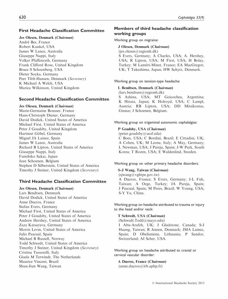
First Headache Classification Committee
Jes Olesen, Denmark (Chairman)Andre Bes, FranceRobert Kunkel, USAJames W Lance, AustraliaGiuseppe Nappi, ItalyVolker Pfaffenrath, GermanyFrank Clifford Rose, United KingdomBruce S Schoenberg, USADieter Soyka, GermanyPeer Tfelt-Hansen, Denmark (Secretary)K Michael A Welch, USAMarica Wilkinson, United Kingdom
Second Headache Classification Committee
Jes Olesen, Denmark (Chairman)Marie-Germaine Bousser, FranceHans-Christoph Diener, GermanyDavid Dodick, United States of AmericaMichael First, United States of AmericaPeter J Goadsby, United KingdomHartmut Gobel, GermanyMiguel JA Lainez, SpainJames W Lance, AustraliaRichard B Lipton, United States of AmericaGiuseppe Nappi, ItalyFumihiko Sakai, JapanJean Schoenen, BelgiumStephen D Silberstein, United States of AmericaTimothy J Steiner, United Kingdom (Secretary)
Third Headache Classification Committee
Jes Olesen, Denmark (Chairman)Lars Bendtsen, DenmarkDavid Dodick, United States of AmericaAnne Ducros, FranceStefan Evers, GermanyMichael First, United States of AmericaPeter J Goadsby, United States of AmericaAndrew Hershey, United States of AmericaZaza Katsarava, GermanyMorris Levin, United States of AmericaJulio Pascual, SpainMichael B Russell, NorwayTodd Schwedt, United States of AmericaTimothy J Steiner, United Kingdom (Secretary)Cristina Tassorelli, ItalyGisela M Terwindt, The NetherlandsMaurice Vincent, BrazilShuu-Jiun Wang, Taiwan
Members of third headache classification
working groups
Working group on migraine:
J Olesen, Denmark (Chairman)([email protected])S Evers, Germany; A Charles, USA; A. Hershey,USA; R Lipton, USA; M First, USA; H Bolay,Turkey; M Lanteri-Minet, France; EA MacGregor,UK; T Takeshima, Japan; HW Schytz, Denmark.
Working group on tension-type headache:
L Bendtsen, Denmark (Chairman)([email protected])S Ashina, USA; MT Goicochea, Argentina;K Hirata, Japan; K Holroyd, USA; C Lampl,Austria; RB Lipton, USA; DD Mitsikostas,Greece; J Schoenen, Belgium.
Working group on trigeminal autonomic cephalalgias:
P Goadsby, USA (Chairman)([email protected])C Boes, USA; C Bordini, Brazil; E Cittadini, UK;A Cohen, UK; M Leone, Italy; A May, Germany;L Newman, USA; J Pareja, Spain; J-W Park, SouthKorea; T Rozen, USA; E Waldenlind, Sweden.
Working group on other primary headache disorders:
S-J Wang, Taiwan (Chairman)([email protected])A Ducros, France; S Evers, Germany; J-L Fuh,Taiwan; A Ozge, Turkey; JA Pareja, Spain;J Pascual, Spain; M Peres, Brazil; W Young, USA;S-Y Yu, China.
Working group on headache attributed to trauma or injury
to the head and/or neck:
T Schwedt, USA (Chairman)([email protected])I Abu-Arafeh, UK; J Gladstone, Canada; S-JHuang, Taiwan; R Jensen, Denmark; JMA Lainez,Spain; D Obelieniene, Lithuania; P Sandor,Switzerland; AI Scher, USA.
Working group on headache attributed to cranial or
cervical vascular disorder:
A Ducros, France (Chairman)([email protected])
630 Cephalalgia 33(9)
� International Headache Society 2013

M Arnold, Switzerland; M Dichgans, Germany;E Houdart, France; J Ferro, Portugal; E Leroux,Canada; Y-S Li, China; A Singhal, USA;G Tietjen, USA.
Working group on headache attributed to non-vascular
intracranial disorder:
DW Dodick, USA (Chairman)([email protected])S Evers, Germany; D Friedman, USA; S Kirby,Canada; B Mokri, USA; J Pascual (Spain); MPeres, Brazil; A Purdy, Canada; K Ravishankar,India; P Sandor, Switzerland; W Schievink, USA;R Stark, Australia; F Taylor, USA.
Working group on headache attributed to a substance or
its withdrawal:
MB Russell, Norway (Chairman)([email protected])L Bendtsen, Denmark; J-L Fuh, Taiwan;Z Katsarava, Germany; AV Krymchantowski,Brazil; M Leone, Italy; K Ravishankar, India;A Tugrul, Turkey; NJ Wiendels, The Netherlands.
Working group on headache attributed to infection:
C Tassorelli, Italy (Chairman)([email protected])E Marchioni, Italy; V Osipova, Russia;K Ravishankar, India; L Savi, Italy; F Sakai,Japan; JR Berger, (USA).
Working group on headache attributed to disorder of
homoeostasis:
J Pascual, Spain (Chairman)([email protected])M Bigal, Brazil; C Bordini, Brazil; J GonzalezMenacho, Spain; F Mainardi, Italy; A Ozge,Turkey; J Pereira-Monteiro, Portugal; M Serrano-Duenas, Ecuador.
Working group on headache or facial pain attributed to
disorder of the cranium, neck, eyes, ears, nose, sinuses,
teeth, mouth or other facial or cervical structure:
M Levin, USA (Chairman)([email protected])
R Cady, USA; C Fernandez de las Penas, Spain;D Friedman, USA; V Guidetti, Italy; J Lance,Australia; P Svensson, Denmark.
Working group on headache attributed to psychiatric
disorder:
M Vincent, Brazil (Chairman)([email protected])M First, USA; E Loder, USA; AE Lake III, USA;F Radat, France; JI Escobar, USA.
Working group on painful cranial neuropathies and other
facial pains:
Z Katsarava, Germany (Chairman)([email protected])R Benoliel, Israel; C Sommer, Germany; A Woda,France; J Zakrzewska UK; V Aggarwal, UK;L Bonamico, Argentina; D Ettlin, USA; S Graff-Radford, USA; J-P Goulet, Canada;S Jaaskelainen, Finland; V Limmroth, Germany;A Michelotti, Italy; D Nixdorf, USA;M Obermann, Germany; R Ohrbach, USA;J Pereira-Monteiro, Portugal; P Pionchon, France;T Renton, UK; S De Siqueira, Brazil; C Wober-Bingol, Austria.
Working group for appendix disorders and criteria:
GM Terwindt, The Netherlands (Chairman)([email protected])
Acknowledgements
The work of the Headache Classification Committee of theInternational Headache Society is financially supported
exclusively by the International Headache Society. Therehas been no commercial sponsorship of the InternationalClassification of Headache Disorders, 3rd edition.
We gratefully acknowledge the support of Timothy Steiner,first for his efforts as honorary secretary of the Classification
Committee and second for his work on copy-editing and pre-paration of this manuscript.
ICHD-3 beta 631
� International Headache Society 2013
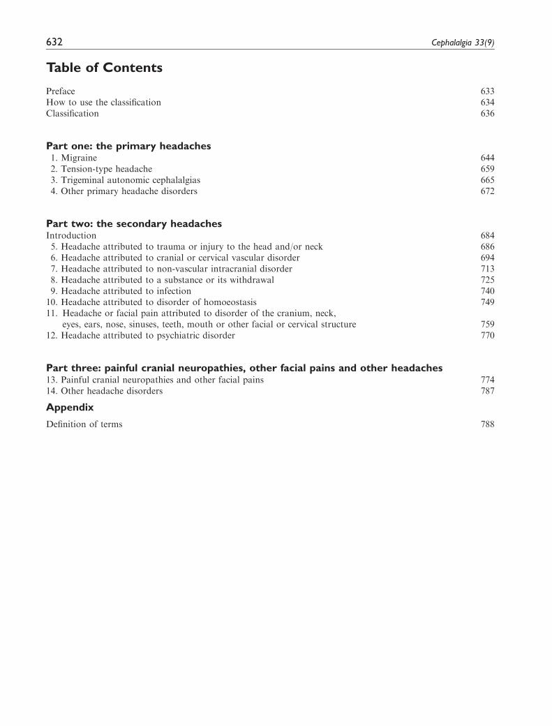
Table of Contents
Preface 633How to use the classification 634Classification 636
Part one: the primary headaches1. Migraine 6442. Tension-type headache 6593. Trigeminal autonomic cephalalgias 6654. Other primary headache disorders 672
Part two: the secondary headachesIntroduction 6845. Headache attributed to trauma or injury to the head and/or neck 6866. Headache attributed to cranial or cervical vascular disorder 6947. Headache attributed to non-vascular intracranial disorder 7138. Headache attributed to a substance or its withdrawal 7259. Headache attributed to infection 740
10. Headache attributed to disorder of homoeostasis 74911. Headache or facial pain attributed to disorder of the cranium, neck,
eyes, ears, nose, sinuses, teeth, mouth or other facial or cervical structure 75912. Headache attributed to psychiatric disorder 770
Part three: painful cranial neuropathies, other facial pains and other headaches13. Painful cranial neuropathies and other facial pains 77414. Other headache disorders 787
Appendix
Definition of terms 788
632 Cephalalgia 33(9)

Preface
After two very successful editions of the InternationalClassification of Headache Disorders (ICHD), a thirdis now close to being final. The members of theClassification Committee have all worked hard forthree years in order to accomplish this beta version.Most members have chaired the work on a specificchapter of the classification, assisted by a number ofother experts. For this edition, there has been a sub-stantial body of evidence available for the classifica-tion work, in contrast to our previous editions, whichwere mostly based on the opinions of experts. Wehave tried to be conservative, making changes onlywhere there was good published evidence to supportchange or where the need for change was intuitivelyobvious.
This is the first time that we have published a betaversion ahead of the final version. The main reason is tosynchronize ICHD-3 with the World HealthOrganization’s next revision (11th edition) of theInternational Classification of Diseases (ICD-11). Thisclassification is already well advanced, and we have notonly secured a very good representation of headachewithin ICD-11 but also ensured congruence betweenICD-11 and ICHD-3 beta. However, ICD-11 nowenters a phase of field trials, and ICHD-3 should dothe same. Such a test period will allow identificationand correction of mistakes and enable a broad inputfrom the members of the International HeadacheSociety.
ICD-11 diagnostic codes will not be finalized untiltwo or three years from now, but it would be a majoradvantage for ICHD-3 to be able to include thesecodes along with our own. WHO’s ICD-11 codeswill be used by health authorities for official diagnos-tic coding, and in many cases they will be employed
for reimbursement purposes; we must have themright.
We publish ICHD-3 beta immediately on theInternational Headache Society’s website, and shortlyafter as an issue of Cephalalgia. Field-testing will con-tinue for 2 or maybe 3 years. Small amendments arelikely both to ICHD-3 and to the diagnostic codes ofICD-11, and these will be incorporated. At that time,we shall publish ICHD-3 in final form in Cephalalgia.
ICHD-3 beta is published only in English, but thosethroughout the world who wish to make their own care-ful translations of parts or in toto are welcome to do sosubject to the conditions stated above. The final versionof ICHD-3 should be translated into as many languagesas possible, and these translations published, as hap-pened to the second and first editions. As we expectICHD-3 beta to be very similar to the final version,translation work begun now is likely to remain useful.Any changes necessitated later by the outcomes of field-testing can be made easily.
Clinicians and researchers should start using the cri-teria of ICHD-3 beta. There are many improvementsover ICHD-II, and it would be unhelpful to continue touse ICHD-II for scientific work. We encourage readersto study ICHD-3 beta very closely, and document andcomment on any inconsistencies they may find.Comments should be sent not to me but to the chairmenof the relevant working groups. Their names and emailaddresses are found in this publication and on the IHSwebsite.
Jes Olesen
ChairmanHeadache Classification Committee
International Headache Society
ICHD-3 beta 633

How to use this classification
This extensive document is not intended to be learnt byheart. Even members of the Classification Committeeare unable to remember all of it. It is a document thatshould be consulted time and time again. In this wayyou will soon get to know the diagnostic criteria for 1.1Migraine without aura, 1.2 Migraine with aura, themajor subtypes of 2. Tension-type headache, 3.1Cluster headache and a few others. The rest willremain something to look up. In clinical practice youdo not need the classification for the obvious case ofmigraine or tension-type headache, but it is useful whenthe diagnosis is uncertain. For research, the classifica-tion is indispensable and every patient entered into aresearch project, be it a drug trial or a study of patho-physiology or biochemistry, must fulfil a set of diagnos-tic criteria.
1. This classification is hierarchical, and you mustdecide how detailed you want to make your diag-nosis. This can range from the first-digit level to thefifth. First, one gets a rough idea about whichgroup the patient belongs to. Is it, for example,1. Migraine or 2. Tension-type headache or 3.Trigeminal autonomic cephalalgias? Then oneobtains information allowing a more detailed diag-nosis. The desired detail depends on the purpose. Ingeneral practice only the first- or second-digit diag-noses are usually applied, whereas in specialistpractice and headache centres a diagnosis at thefourth- or fifth-digit level is appropriate.
2. For most purposes, patients receive a diagnosisaccording to the headache phenotypes that theycurrently present, or that they have presentedwithin the last year. For genetic and some otheruses, occurrence during the whole lifetime is used.
3. Each distinct type, subtype or subform of headachethat the patient has must be separately diagnosedand coded. Thus, a severely affected patient in aheadache centre may receive three diagnoses andcodes: 1.1 Migraine without aura, 1.2 Migrainewith aura and 8.2 Medication-overuse headache.
4. When a patient receives more than one diagnosis,these should be listed in the order of importance tothe patient.
5. When one type of headache in a particular patientfulfils two different sets of diagnostic criteria, thenall other available information should be used todecide which of the alternatives is the correct ormore likely diagnosis. This could include the
longitudinal headache history (how did the head-ache start?), the family history, the effect of drugs,menstrual relationship, age, gender and a range ofother features. Fulfilment of the diagnostic criteriafor 1. Migraine, 2. Tension-type headache or 3.Trigeminal autonomic cephalalgias, or any of theirsubtypes, always trumps fulfilment of criteria forthe probable diagnostic categories of each, whichare last-described in the respective groups. Inother words, a patient whose headache fulfils cri-teria for both 1.5 Probable migraine and 2.1Infrequent episodic tension-type headache shouldbe coded to the latter. Nevertheless, considerationshould always be given to the possibility that someheadache attacks meet one set of criteria, whereasother attacks meet another set. In such cases, twodiagnoses exist and both should be coded.
6. To receive a particular headache diagnosis thepatient must, in many cases, experience a minimumnumber of attacks of (or days with) that headache.This number is specified in the diagnostic criteriafor the headache type, subtype or subform.Further, the headache must fulfil a number ofother requirements described within the criteriaunder separate letter headings: A, B, C etc. Someletter headings are monothetic: that is, they expressa single requirement. Other letter headings arepolythetic, requiring for example any two out offour listed characteristics.
7. The full set of diagnostic criteria is provided forsome headache disorders only at the first- andsecond-digit levels. Diagnostic criteria at thethird- and fourth-digit levels then demand, as cri-terion A, fulfilment of the criteria for levels oneand/or two and, in criterion B and onwards, specifythe further specific criteria to be fulfilled.
8. The frequency of primary headache disorders variesfrom attacks every one to two years to attacksdaily. The severity of attacks also varies. ICHD-3beta does not generally provide a possibility to codefor frequency or severity, but recommends that fre-quency and severity be specified in free text.
9. Primary or secondary headache or both: When anew headache occurs for the first time in close tem-poral relation to another disorder that is known tocause headache, or fulfils other criteria for causa-tion by that disorder, the new headache is coded asa secondary headache attributed to the causativedisorder. This remains true even when the
634 Cephalalgia 33(9)
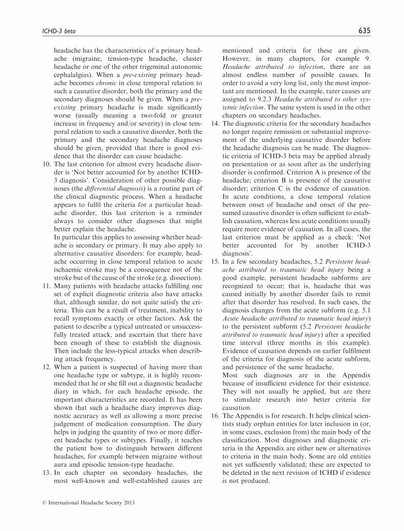
headache has the characteristics of a primary head-ache (migraine, tension-type headache, clusterheadache or one of the other trigeminal autonomiccephalalgias). When a pre-existing primary head-ache becomes chronic in close temporal relation tosuch a causative disorder, both the primary and thesecondary diagnoses should be given. When a pre-existing primary headache is made significantlyworse (usually meaning a two-fold or greaterincrease in frequency and/or severity) in close tem-poral relation to such a causative disorder, both theprimary and the secondary headache diagnosesshould be given, provided that there is good evi-dence that the disorder can cause headache.
10. The last criterion for almost every headache disor-der is ‘Not better accounted for by another ICHD-3 diagnosis’. Consideration of other possible diag-noses (the differential diagnosis) is a routine part ofthe clinical diagnostic process. When a headacheappears to fulfil the criteria for a particular head-ache disorder, this last criterion is a reminderalways to consider other diagnoses that mightbetter explain the headache.In particular this applies to assessing whether head-ache is secondary or primary. It may also apply toalternative causative disorders: for example, head-ache occurring in close temporal relation to acuteischaemic stroke may be a consequence not of thestroke but of the cause of the stroke (e.g. dissection).
11. Many patients with headache attacks fulfilling oneset of explicit diagnostic criteria also have attacksthat, although similar, do not quite satisfy the cri-teria. This can be a result of treatment, inability torecall symptoms exactly or other factors. Ask thepatient to describe a typical untreated or unsuccess-fully treated attack, and ascertain that there havebeen enough of these to establish the diagnosis.Then include the less-typical attacks when describ-ing attack frequency.
12. When a patient is suspected of having more thanone headache type or subtype, it is highly recom-mended that he or she fill out a diagnostic headachediary in which, for each headache episode, theimportant characteristics are recorded. It has beenshown that such a headache diary improves diag-nostic accuracy as well as allowing a more precisejudgement of medication consumption. The diaryhelps in judging the quantity of two or more differ-ent headache types or subtypes. Finally, it teachesthe patient how to distinguish between differentheadaches, for example between migraine withoutaura and episodic tension-type headache.
13. In each chapter on secondary headaches, themost well-known and well-established causes are
mentioned and criteria for these are given.However, in many chapters, for example 9.Headache attributed to infection, there are analmost endless number of possible causes. Inorder to avoid a very long list, only the most impor-tant are mentioned. In the example, rarer causes areassigned to 9.2.3 Headache attributed to other sys-temic infection. The same system is used in the otherchapters on secondary headaches.
14. The diagnostic criteria for the secondary headachesno longer require remission or substantial improve-ment of the underlying causative disorder beforethe headache diagnosis can be made. The diagnos-tic criteria of ICHD-3 beta may be applied alreadyon presentation or as soon after as the underlyingdisorder is confirmed. Criterion A is presence of theheadache; criterion B is presence of the causativedisorder; criterion C is the evidence of causation.In acute conditions, a close temporal relationbetween onset of headache and onset of the pre-sumed causative disorder is often sufficient to estab-lish causation, whereas less acute conditions usuallyrequire more evidence of causation. In all cases, thelast criterion must be applied as a check: ‘Notbetter accounted for by another ICHD-3diagnosis’.
15. In a few secondary headaches, 5.2 Persistent head-ache attributed to traumatic head injury being agood example, persistent headache subforms arerecognized to occur; that is, headache that wascaused initially by another disorder fails to remitafter that disorder has resolved. In such cases, thediagnosis changes from the acute subform (e.g. 5.1Acute headache attributed to traumatic head injury)to the persistent subform (5.2 Persistent headacheattributed to traumatic head injury) after a specifiedtime interval (three months in this example).Evidence of causation depends on earlier fulfilmentof the criteria for diagnosis of the acute subform,and persistence of the same headache.Most such diagnoses are in the Appendixbecause of insufficient evidence for their existence.They will not usually be applied, but are thereto stimulate research into better criteria forcausation.
16. The Appendix is for research. It helps clinical scien-tists study orphan entities for later inclusion in (or,in some cases, exclusion from) the main body of theclassification. Most diagnoses and diagnostic cri-teria in the Appendix are either new or alternativesto criteria in the main body. Some are old entitiesnot yet sufficiently validated; these are expected tobe deleted in the next revision of ICHD if evidenceis not produced.
� International Headache Society 2013
ICHD-3 beta 635

Classification
ICHD-3 code Diagnosis
1. Migraine
1.1 Migraine without aura1.2 Migraine with aura1.2.1 Migraine with typical aura1.2.1.1 Typical aura with headache1.2.1.2 Typical aura without headache
1.2.2 Migraine with brainstem aura1.2.3 Hemiplegic migraine1.2.3.1 Familial hemiplegic migraine (FHM)1.2.3.1.1 Familial hemiplegic migraine type 1 (FHM1)1.2.3.1.2 Familial hemiplegic migraine type 2 (FHM2)1.2.3.1.3 Familial hemiplegic migraine type 3 (FHM3)1.2.3.1.4 Familial hemiplegic migraine, other loci
1.2.3.2 Sporadic hemiplegic migraine1.2.4 Retinal migraine
1.3 Chronic migraine1.4 Complications of migraine1.4.1 Status migrainosus1.4.2 Persistent aura without infarction1.4.3 Migrainous infarction1.4.4 Migraine aura-triggered seizure
1.5 Probable migraine1.5.1 Probable migraine without aura1.5.2 Probable migraine with aura
1.6 Episodic syndromes that may be associated with migraine1.6.1 Recurrent gastrointestinal disturbance1.6.1.1 Cyclical vomiting syndrome1.6.1.2 Abdominal migraine
1.6.2 Benign paroxysmal vertigo1.6.3 Benign paroxysmal torticollis
2. Tension-type headache (TTH)
2.1 Infrequent episodic tension-type headache2.1.1 Infrequent episodic tension-type headache associated with pericranial tenderness2.1.2 Infrequent episodic tension-type headache not associated with pericranial tenderness
2.2 Frequent episodic tension-type headache2.2.1 Frequent episodic tension-type headache associated with pericranial tenderness2.2.2 Frequent episodic tension-type headache not associated with pericranial tenderness
2.3 Chronic tension-type headache2.3.1 Chronic tension-type headache associated with pericranial tenderness2.3.2 Chronic tension-type headache not associated with pericranial tenderness
2.4 Probable tension-type headache2.4.1 Probable infrequent episodic tension-type headache2.4.2 Probable frequent episodic tension-type headache2.4.3 Probable chronic tension-type headache
3. Trigeminal autonomic cephalalgias (TACs)
3.1 Cluster headache3.1.1 Episodic cluster headache3.1.2 Chronic cluster headache
3.2 Paroxysmal hemicrania3.2.1 Episodic paroxysmal hemicrania3.2.2 Chronic paroxysmal hemicrania
636 Cephalalgia 33(9)
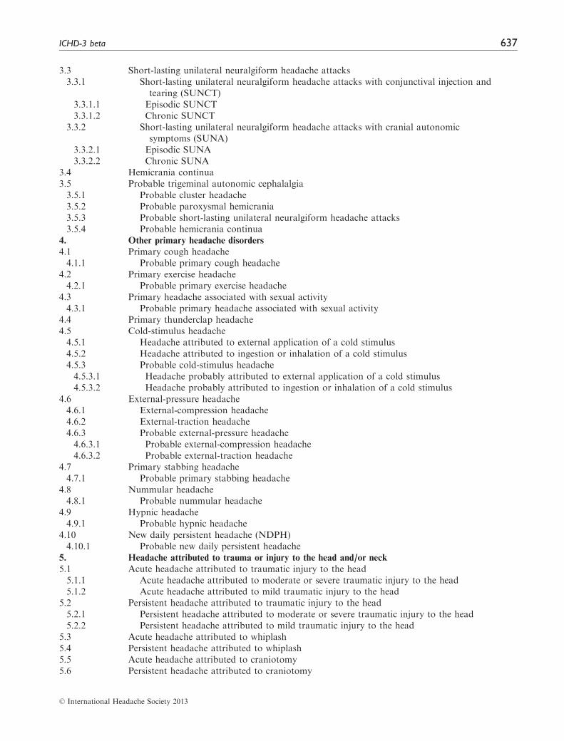
3.3 Short-lasting unilateral neuralgiform headache attacks3.3.1 Short-lasting unilateral neuralgiform headache attacks with conjunctival injection and
tearing (SUNCT)3.3.1.1 Episodic SUNCT3.3.1.2 Chronic SUNCT
3.3.2 Short-lasting unilateral neuralgiform headache attacks with cranial autonomicsymptoms (SUNA)
3.3.2.1 Episodic SUNA3.3.2.2 Chronic SUNA
3.4 Hemicrania continua3.5 Probable trigeminal autonomic cephalalgia3.5.1 Probable cluster headache3.5.2 Probable paroxysmal hemicrania3.5.3 Probable short-lasting unilateral neuralgiform headache attacks3.5.4 Probable hemicrania continua
4. Other primary headache disorders
4.1 Primary cough headache4.1.1 Probable primary cough headache
4.2 Primary exercise headache4.2.1 Probable primary exercise headache
4.3 Primary headache associated with sexual activity4.3.1 Probable primary headache associated with sexual activity
4.4 Primary thunderclap headache4.5 Cold-stimulus headache4.5.1 Headache attributed to external application of a cold stimulus4.5.2 Headache attributed to ingestion or inhalation of a cold stimulus4.5.3 Probable cold-stimulus headache4.5.3.1 Headache probably attributed to external application of a cold stimulus4.5.3.2 Headache probably attributed to ingestion or inhalation of a cold stimulus
4.6 External-pressure headache4.6.1 External-compression headache4.6.2 External-traction headache4.6.3 Probable external-pressure headache4.6.3.1 Probable external-compression headache4.6.3.2 Probable external-traction headache
4.7 Primary stabbing headache4.7.1 Probable primary stabbing headache
4.8 Nummular headache4.8.1 Probable nummular headache
4.9 Hypnic headache4.9.1 Probable hypnic headache
4.10 New daily persistent headache (NDPH)4.10.1 Probable new daily persistent headache
5. Headache attributed to trauma or injury to the head and/or neck
5.1 Acute headache attributed to traumatic injury to the head5.1.1 Acute headache attributed to moderate or severe traumatic injury to the head5.1.2 Acute headache attributed to mild traumatic injury to the head
5.2 Persistent headache attributed to traumatic injury to the head5.2.1 Persistent headache attributed to moderate or severe traumatic injury to the head5.2.2 Persistent headache attributed to mild traumatic injury to the head
5.3 Acute headache attributed to whiplash5.4 Persistent headache attributed to whiplash5.5 Acute headache attributed to craniotomy5.6 Persistent headache attributed to craniotomy
� International Headache Society 2013
ICHD-3 beta 637
6. Headache attributed to cranial or cervical vascular disorder
6.1 Headache attributed to ischaemic stroke or transient ischaemic attack6.1.1 Headache attributed to ischaemic stroke (cerebral infarction)6.1.2 Headache attributed to transient ischaemic attack (TIA)
6.2 Headache attributed to non-traumatic intracranial haemorrhage6.2.1 Headache attributed to non-traumatic intracerebral haemorrhage6.2.2 Headache attributed to non-traumatic subarachnoid haemorrhage (SAH)6.2.3 Headache attributed to non-traumatic acute subdural haemorrhage (ASDH)
6.3 Headache attributed to unruptured vascular malformation6.3.1 Headache attributed to unruptured saccular aneurysm6.3.2 Headache attributed to arteriovenous malformation (AVM)6.3.3 Headache attributed to dural arteriovenous fistula (DAVF)6.3.4 Headache attributed to cavernous angioma6.3.5 Headache attributed to encephalotrigeminal or leptomeningeal angiomatosis
(Sturge Weber syndrome)6.4 Headache attributed to arteritis6.4.1 Headache attributed to giant cell arteritis (GCA)6.4.2 Headache attributed to primary angiitis of the central nervous system (PACNS)6.4.3 Headache attributed to secondary angiitis of the central nervous system (SACNS)
6.5 Headache attributed to cervical carotid or vertebral artery disorder6.5.1 Headache or facial or neck pain attributed to cervical carotid or vertebral artery
dissection6.5.2 Post-endarterectomy headache6.5.3 Headache attributed to carotid or vertebral angioplasty
6.6 Headache attributed to cerebral venous thrombosis (CVT)6.7 Headache attributed to other acute intracranial arterial disorder6.7.1 Headache attributed to an intracranial endovascular procedure6.7.2 Angiography headache6.7.3 Headache attributed to reversible cerebral vasoconstriction syndrome (RCVS)6.7.3.1 Headache probably attributed to reversible cerebral vasoconstriction
syndrome (RCVS)6.7.4 Headache attributed to intracranial arterial dissection
6.8 Headache attributed to genetic vasculopathy6.8.1 Cerebral Autosomal Dominant Arteriopathy with Subcortical Infarcts and
Leukoencephalopathy (CADASIL)6.8.2 Mitochondrial Encephalopathy, Lactic Acidosis and Stroke-like episodes (MELAS)6.8.3 Headache attributed to another genetic vasculopathy
6.9 Headache attributed to pituitary apoplexy7. Headache attributed to non-vascular intracranial disorder
7.1 Headache attributed to increased cerebrospinal fluid pressure7.1.1 Headache attributed to idiopathic intracranial hypertension (IIH)7.1.2 Headache attributed to intracranial hypertension secondary to metabolic, toxic or
hormonal causes7.1.3 Headache attributed to intracranial hypertension secondary to hydrocephalus
7.2 Headache attributed to low cerebrospinal fluid pressure7.2.1 Post-dural puncture headache7.2.2 CSF fistula headache7.2.3 Headache attributed to spontaneous intracranial hypotension
7.3 Headache attributed to non-infectious inflammatory disease7.3.1 Headache attributed to neurosarcoidosis7.3.2 Headache attributed to aseptic (non-infectious) meningitis7.3.3 Headache attributed to other non-infectious inflammatory disease7.3.4 Headache attributed to lymphocytic hypophysitis7.3.5 Syndrome of transient Headache and Neurological Deficits with cerebrospinal fluid
Lymphocytosis (HaNDL)
� International Headache Society 2013
638 Cephalalgia 33(9)

7.4 Headache attributed to intracranial neoplasia7.4.1 Headache attributed to intracranial neoplasm7.4.1.1 Headache attributed to colloid cyst of the third ventricle
7.4.2 Headache attributed to carcinomatous meningitis7.4.3 Headache attributed to hypothalamic or pituitary hyper- or hyposecretion
7.5 Headache attributed to intrathecal injection7.6 Headache attributed to epileptic seizure7.6.1 Hemicrania epileptica7.6.2 Post-ictal headache
7.7 Headache attributed to Chiari malformation type I (CM1)7.8 Headache attributed to other non-vascular intracranial disorder8. Headache attributed to a substance or its withdrawal
8.1 Headache attributed to use of or exposure to a substance8.1.1 Nitric oxide (NO) donor-induced headache8.1.1.1 Immediate NO donor-induced headache8.1.1.2 Delayed NO donor-induced headache
8.1.2 Phosphodiesterase (PDE) inhibitor-induced headache8.1.3 Carbon monoxide (CO)-induced headache8.1.4 Alcohol-induced headache8.1.4.1 Immediate alcohol-induced headache8.1.4.2 Delayed alcohol-induced headache
8.1.5 Headache induced by food and/or additive8.1.5.1 Monosodium glutamate (MSG)-induced headache
8.1.6 Cocaine-induced headache8.1.7 Histamine-induced headache8.1.7.1 Immediate histamine-induced headache8.1.7.2 Delayed histamine-induced headache
8.1.8 Calcitonin gene-related peptide (CGRP)-induced headache8.1.8.1 Immediate CGRP-induced headache8.1.8.2 Delayed CGRP-induced headache
8.1.9 Headache attributed to exogenous acute pressor agent8.1.10 Headache attributed to occasional use of non-headache medication8.1.11 Headache attributed to long-term use of non-headache medication8.1.12 Headache attributed to exogenous hormone8.1.13 Headache attributed to use of or exposure to other substance
8.2 Medication-overuse headache (MOH)8.2.1 Ergotamine-overuse headache8.2.2 Triptan-overuse headache8.2.3 Simple analgesic-overuse headache8.2.3.1 Paracetamol (acetaminophen)-overuse headache8.2.3.2 Acetylsalicylic acid-overuse headache8.2.3.3 Other non-steroidal anti-inflammatory drug (NSAID)-overuse headache
8.2.4 Opioid-overuse headache8.2.5 Combination-analgesic-overuse headache8.2.6 Medication-overuse headache attributed to multiple drug classes not individually
overused8.2.7 Medication-overuse headache attributed to unverified overuse of multiple drug classes8.2.8 Medication-overuse headache attributed to other medication
8.3 Headache attributed to substance withdrawal8.3.1 Caffeine-withdrawal headache8.3.2 Opioid-withdrawal headache8.3.3 Oestrogen-withdrawal headache8.3.4 Headache attributed to withdrawal from chronic use of other substance
� International Headache Society 2013
ICHD-3 beta 639

9. Headache attributed to infection
9.1 Headache attributed to intracranial infection9.1.1 Headache attributed to bacterial meningitis or meningoencephalitis9.1.1.1 Acute headache attributed to bacterial meningitis or meningoencephalitis9.1.1.2 Chronic headache attributed to bacterial meningitis or meningoencephalitis9.1.1.3 Persistent headache attributed to past bacterial meningitis or meningoencephalitis
9.1.2 Headache attributed to viral meningitis or encephalitis9.1.2.1 Headache attributed to viral meningitis9.1.2.2 Headache attributed to viral encephalitis
9.1.3 Headache attributed to intracranial fungal or other parasitic infection9.1.3.1 Acute headache attributed to intracranial fungal or other parasitic infection9.1.3.2 Chronic headache attributed to intracranial fungal or other parasitic infection
9.1.4 Headache attributed to brain abscess9.1.5 Headache attributed to subdural empyema
9.2 Headache attributed to systemic infection9.2.1 Headache attributed to systemic bacterial infection9.2.1.1 Acute headache attributed to systemic bacterial infection9.2.1.2 Chronic headache attributed to systemic bacterial infection
9.2.2 Headache attributed to systemic viral infection9.2.2.1 Acute headache attributed to systemic viral infection9.2.2.2 Chronic headache attributed to systemic viral infection
9.2.3 Headache attributed to other systemic infection9.2.3.1 Acute headache attributed to other systemic infection9.2.3.2 Chronic headache attributed to other systemic infection
10. Headache attributed to disorder of homoeostasis
10.1 Headache attributed to hypoxia and/or hypercapnia10.1.1 High-altitude headache10.1.2 Headache attributed to aeroplane travel10.1.3 Diving headache10.1.4 Sleep apnoea headache
10.2 Dialysis headache10.3 Headache attributed to arterial hypertension10.3.1 Headache attributed to phaeochromocytoma10.3.2 Headache attributed to hypertensive crisis without hypertensive encephalopathy10.3.3 Headache attributed to hypertensive encephalopathy10.3.4 Headache attributed to pre-eclampsia or eclampsia10.3.5 Headache attributed to autonomic dysreflexia
10.4 Headache attributed to hypothyroidism10.5 Headache attributed to fasting10.6 Cardiac cephalalgia10.7 Headache attributed to other disorder of homoeostasis11. Headache or facial pain attributed to disorder of the cranium, neck, eyes, ears, nose, sinuses,
teeth, mouth or other facial or cervical structure
11.1 Headache attributed to disorder of cranial bone11.2 Headache attributed to disorder of the neck11.2.1 Cervicogenic headache11.2.2 Headache attributed to retropharyngeal tendonitis11.2.3 Headache attributed to craniocervical dystonia
11.3 Headache attributed to disorder of the eyes11.3.1 Headache attributed to acute glaucoma11.3.2 Headache attributed to refractive error11.3.3 Headache attributed to heterophoria or heterotropia (latent or persistent squint)11.3.4 Headache attributed to ocular inflammatory disorder11.3.5 Headache attributed to trochleitis
11.4 Headache attributed to disorder of the ears
� International Headache Society 2013
640 Cephalalgia 33(9)

11.5 Headache attributed to disorder of the nose or paranasal sinuses11.5.1 Headache attributed to acute rhinosinusitis11.5.2 Headache attributed to chronic or recurring rhinosinusitis
11.6 Headache attributed to disorder of the teeth or jaw11.7 Headache attributed to temporomandibular disorder (TMD)11.8 Head or facial pain attributed to inflammation of the stylohyoid ligament11.9 Headache or facial pain attributed to other disorder of cranium, neck, eyes, ears, nose,
sinuses, teeth, mouth or other facial or cervical structure12. Headache attributed to psychiatric disorder
12.1 Headache attributed to somatization disorder12.2 Headache attributed to psychotic disorder13. Painful cranial neuropathies and other facial pains
13.1 Trigeminal neuralgia13.1.1 Classical trigeminal neuralgia13.1.1.1 Classical trigeminal neuralgia, purely paroxysmal13.1.1.2 Classical trigeminal neuralgia with concomitant persistent facial pain
13.1.2 Painful trigeminal neuropathy13.1.2.1 Painful trigeminal neuropathy attributed to acute Herpes zoster13.1.2.2 Post-herpetic trigeminal neuropathy13.1.2.3 Painful post-traumatic trigeminal neuropathy13.1.2.4 Painful trigeminal neuropathy attributed to multiple sclerosis (MS) plaque13.1.2.5 Painful trigeminal neuropathy attributed to space-occupying lesion13.1.2.6 Painful trigeminal neuropathy attributed to other disorder
13.2 Glossopharyngeal neuralgia13.3 Nervus intermedius (facial nerve) neuralgia13.3.1 Classical nervus intermedius neuralgia13.3.2 Nervus intermedius neuropathy attributed to Herpes zoster
13.4 Occipital neuralgia13.5 Optic neuritis13.6 Headache attributed to ischaemic ocular motor nerve palsy13.7 Tolosa-Hunt syndrome13.8 Paratrigeminal oculosympathetic (Raeder’s) syndrome13.9 Recurrent painful ophthalmoplegic neuropathy13.10 Burning mouth syndrome (BMS)13.11 Persistent idiopathic facial pain (PIFP)13.12 Central neuropathic pain13.12.1 Central neuropathic pain attributed to multiple sclerosis (MS)13.12.2 Central post-stroke pain (CPSP)
14. Other headache disorders
14.1 Headache not elsewhere classified14.2 Headache unspecified
A. Appendix
A1. Migraine
A1.1 Migraine without auraA1.1.1 Pure menstrual migraine without auraA1.1.2 Menstrually related migraine without auraA1.1.3 Non-menstrual migraine without aura
A1.2 Migraine with aura (alternative criteria)A1.2.1 Migraine with typical aura (alternative criteria)
A1.3 Chronic migraine (alternative criteria)A1.3.1 Chronic migraine with pain-free periodsA1.3.2 Chronic migraine with continuous pain
A1.4 Complications of migraineA1.4.5 Migraine aura status
� International Headache Society 2013
ICHD-3 beta 641
A1.6 Episodic syndromes that may be associated with migraineA1.6.4 Infantile colicA1.6.5 Alternating hemiplegia of childhoodA1.6.6 Vestibular migraine
A2. Tension-type headache (alternative criteria)A3. Trigeminal-autonomic cephalalgias (TACs)
A3.6 Undifferentiated trigeminal autonomic cephalalgiaA4. Other primary headache disorders
A4.11 Epicrania fugaxA5. Headache attributed to trauma or injury to the head and/or neck
A5.1 Acute headache attributed to traumatic injury to the headA5.1.1.1 Delayed-onset acute headache attributed to moderate or severe traumatic injury to the headA5.1.2.1 Delayed-onset acute headache attributed to mild traumatic injury to the head
A5.2 Persistent headache attributed to traumatic injury to the headA5.2.1.1 Delayed-onset persistent headache attributed to moderate or severe traumatic injury to
the headA5.2.2.1 Delayed-onset persistent headache attributed to mild traumatic injury to the head
A5.7 Headache attributed to radiosurgery of the brainA5.8 Acute headache attributed to other trauma or injury to the head and/or neckA5.9 Persistent headache attributed to other trauma or injury to the head and/or neckA6. Headache attributed to cranial or cervical vascular disorder
A6.10 Persistent headache attributed to past cranial or cervical vascular disorderA7. Headache attributed to non-vascular intracranial disorder
A7.6 Headache attributed to epileptic seizureA7.6.3 Post-electroconvulsive therapy (ECT) headache
A7.9 Persistent headache attributed to past non-vascular intracranial disorderA8. Headache attributed to a substance or its withdrawal
A8.4 Persistent headache attributed to past use of or exposure to a substanceA9. Headache attributed to infection
A9.1 Headache attributed to intracranial infectionA9.1.3.3 Persistent headache attributed to past intracranial fungal or other parasitic infection
A9.1.6 Headache attributed to other infective space-occupying lesionA9.3 Headache attributed to human immunodeficiency virus (HIV) infectionA10. Headache attributed to disorder of homoeostasis
A10.7 Head and/or neck pain attributed to orthostatic (postural) hypotensionA10.8 Headache attributed to other disorder of homeostasisA10.8.1 Headache attributed to travel in spaceA10.8.2 Headache attributed to other metabolic or systemic disorder
A10.9 Persistent headache attributed to past disorder of homoeostasisA11. Headache or facial pain attributed to disorder of the cranium, neck, eyes, ears, nose, sinuses,
teeth, mouth or other facial or cervical structure
A11.2 Headache attributed to disorder of the neckA11.2.4 Headache attributed to upper cervical radiculopathyA11.2.5 Headache attributed to cervical myofascial pain
A11.5 Headache attributed to disorder of the nose or paranasal sinusesA11.5.3 Headache attributed to disorder of the nasal mucosa, turbinates or septum
A12. Headache attributed to psychiatric disorder
A12.3 Headache attributed to depressive disorderA12.4 Headache attributed to separation anxiety disorderA12.5 Headache attributed to panic disorderA12.6 Headache attributed to specific phobiaA12.7 Headache attributed to social anxiety disorder (social phobia)A12.8 Headache attributed to generalized anxiety disorderA12.9 Headache attributed to post-traumatic stress disorderA12.10 Headache attributed to acute stress disorder
� International Headache Society 2013
642 Cephalalgia 33(9)

Part one
The primary headaches
1. Migraine2. Tension-type headache3. Trigeminal autonomic cephalalgias4. Other primary headache disorders
ICHD-3 beta 643

1. Migraine
1.1 Migraine without aura1.2 Migraine with aura
1.2.1 Migraine with typical aura1.2.1.1 Typical aura with headache1.2.1.2 Typical aura without headache
1.2.2 Migraine with brainstem aura1.2.3 Hemiplegic migraine
1.2.3.1 Familial hemiplegic migraine (FHM)1.2.3.1.1 Familial hemiplegic migraine type 11.2.3.1.2 Familial hemiplegic migraine type 21.2.3.1.3 Familial hemiplegic migraine type 31.2.3.1.4 Familial hemiplegic migraine, other
loci1.2.3.2 Sporadic hemiplegic migraine
1.2.4 Retinal migraine1.3 Chronic migraine1.4 Complications of migraine
1.4.1 Status migrainosus1.4.2 Persistent aura without infarction1.4.3 Migrainous infarction1.4.4 Migraine aura-triggered seizure
1.5 Probable migraine1.5.1 Probable migraine without aura1.5.2 Probable migraine with aura
1.6 Episodic syndromes that may be associated withmigraine1.6.1 Recurrent gastrointestinal disturbance
1.6.1.1 Cyclical vomiting syndrome1.6.1.2 Abdominal migraine
1.6.2 Benign paroxysmal vertigo1.6.3 Benign paroxysmal torticollis
Coded elsewhere:Migraine-like headache secondary to another disorder(symptomatic migraine) is coded as a secondary head-ache attributed to that disorder.
General comment
Primary or secondary headache or both?
When a new headache with the characteristics ofmigraine occurs for the first time in close temporalrelation to another disorder known to cause headache,or fulfils other criteria for causation by that disorder,the new headache is coded as a secondary headacheattributed to the causative disorder. When pre-existingmigraine becomes chronic in close temporal relation tosuch a causative disorder, both the initial migrainediagnosis and the secondary diagnosis should begiven. 8.2 Medication-overuse headache is a particu-larly important example of this: both the episodic or
chronic migraine diagnosis and the diagnosis 8.2Medication-overuse headache should be given whenmedication overuse is present. When pre-existingmigraine is made significantly worse (usually meaninga two-fold or greater increase in frequency and/orseverity) in close temporal relation to such a causativedisorder, both the initial migraine diagnosis and thesecondary headache diagnosis should be given, pro-vided that there is good evidence that the disordercan cause headache.
Introduction
Migraine is a common disabling primary headachedisorder. Epidemiological studies have documentedits high prevalence and high socio-economic and perso-nal impacts. In the Global Burden of Disease Survey2010, it was ranked as the third most prevalent disorderand seventh-highest specific cause of disabilityworldwide.
Migraine has two major subtypes. 1.1 Migrainewithout aura is a clinical syndrome characterizedby headache with specific features and associatedsymptoms. 1.2 Migraine with aura is primarily charac-terized by the transient focal neurological symptomsthat usually precede or sometimes accompany theheadache. Some patients also experience a premoni-tory phase, occurring hours or days before the head-ache, and a headache resolution phase. Premonitoryand resolution symptoms include hyperactivity,hypoactivity, depression, cravings for particularfoods, repetitive yawning, fatigue and neck stiffnessand/or pain.
When a patient fulfils criteria for more than onesubtype of migraine, all subtypes should be diagnosedand coded. For example, a patient who has frequentattacks with aura but also some attacks without aurashould be coded as 1.2 Migraine with aura and 1.1Migraine without aura. Attacks of either type areincluded in the diagnostic criteria for 1.3 Chronicmigraine.
1.1 Migraine without aura
Previously used terms:Common migraine; hemicrania simplex.
Description:Recurrent headache disorder manifesting in attackslasting 4-72 hours. Typical characteristics of the head-ache are unilateral location, pulsating quality, moder-ate or severe intensity, aggravation by routine physicalactivity and association with nausea and/or photopho-bia and phonophobia.
� International Headache Society 2013
644 Cephalalgia 33(9)
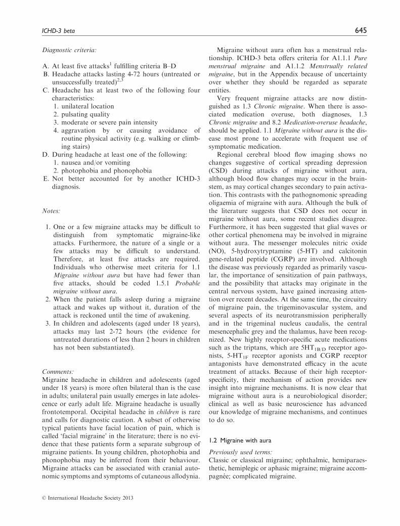
Diagnostic criteria:
A. At least five attacks1 fulfilling criteria B–DB. Headache attacks lasting 4-72 hours (untreated or
unsuccessfully treated)2,3
C. Headache has at least two of the following fourcharacteristics:1. unilateral location2. pulsating quality3. moderate or severe pain intensity4. aggravation by or causing avoidance of
routine physical activity (e.g. walking or climb-ing stairs)
D. During headache at least one of the following:1. nausea and/or vomiting2. photophobia and phonophobia
E. Not better accounted for by another ICHD-3diagnosis.
Notes:
1. One or a few migraine attacks may be difficult todistinguish from symptomatic migraine-likeattacks. Furthermore, the nature of a single or afew attacks may be difficult to understand.Therefore, at least five attacks are required.Individuals who otherwise meet criteria for 1.1Migraine without aura but have had fewer thanfive attacks, should be coded 1.5.1 Probablemigraine without aura.
2. When the patient falls asleep during a migraineattack and wakes up without it, duration of theattack is reckoned until the time of awakening.
3. In children and adolescents (aged under 18 years),attacks may last 2-72 hours (the evidence foruntreated durations of less than 2 hours in childrenhas not been substantiated).
Comments:Migraine headache in children and adolescents (agedunder 18 years) is more often bilateral than is the casein adults; unilateral pain usually emerges in late adoles-cence or early adult life. Migraine headache is usuallyfrontotemporal. Occipital headache in children is rareand calls for diagnostic caution. A subset of otherwisetypical patients have facial location of pain, which iscalled ‘facial migraine’ in the literature; there is no evi-dence that these patients form a separate subgroup ofmigraine patients. In young children, photophobia andphonophobia may be inferred from their behaviour.Migraine attacks can be associated with cranial auto-nomic symptoms and symptoms of cutaneous allodynia.
Migraine without aura often has a menstrual rela-tionship. ICHD-3 beta offers criteria for A1.1.1 Puremenstrual migraine and A1.1.2 Menstrually relatedmigraine, but in the Appendix because of uncertaintyover whether they should be regarded as separateentities.
Very frequent migraine attacks are now distin-guished as 1.3 Chronic migraine. When there is asso-ciated medication overuse, both diagnoses, 1.3Chronic migraine and 8.2 Medication-overuse headache,should be applied. 1.1 Migraine without aura is the dis-ease most prone to accelerate with frequent use ofsymptomatic medication.
Regional cerebral blood flow imaging shows nochanges suggestive of cortical spreading depression(CSD) during attacks of migraine without aura,although blood flow changes may occur in the brain-stem, as may cortical changes secondary to pain activa-tion. This contrasts with the pathognomonic spreadingoligaemia of migraine with aura. Although the bulk ofthe literature suggests that CSD does not occur inmigraine without aura, some recent studies disagree.Furthermore, it has been suggested that glial waves orother cortical phenomena may be involved in migrainewithout aura. The messenger molecules nitric oxide(NO), 5-hydroxytryptamine (5-HT) and calcitoningene-related peptide (CGRP) are involved. Althoughthe disease was previously regarded as primarily vascu-lar, the importance of sensitization of pain pathways,and the possibility that attacks may originate in thecentral nervous system, have gained increasing atten-tion over recent decades. At the same time, the circuitryof migraine pain, the trigeminovascular system, andseveral aspects of its neurotransmission peripherallyand in the trigeminal nucleus caudalis, the centralmesencephalic grey and the thalamus, have been recog-nized. New highly receptor-specific acute medicationssuch as the triptans, which are 5HT1B/D receptor ago-nists, 5-HT1F receptor agonists and CGRP receptorantagonists have demonstrated efficacy in the acutetreatment of attacks. Because of their high receptor-specificity, their mechanism of action provides newinsight into migraine mechanisms. It is now clear thatmigraine without aura is a neurobiological disorder;clinical as well as basic neuroscience has advancedour knowledge of migraine mechanisms, and continuesto do so.
1.2 Migraine with aura
Previously used terms:Classic or classical migraine; ophthalmic, hemiparaes-thetic, hemiplegic or aphasic migraine; migraine accom-pagnee; complicated migraine.
� International Headache Society 2013
ICHD-3 beta 645
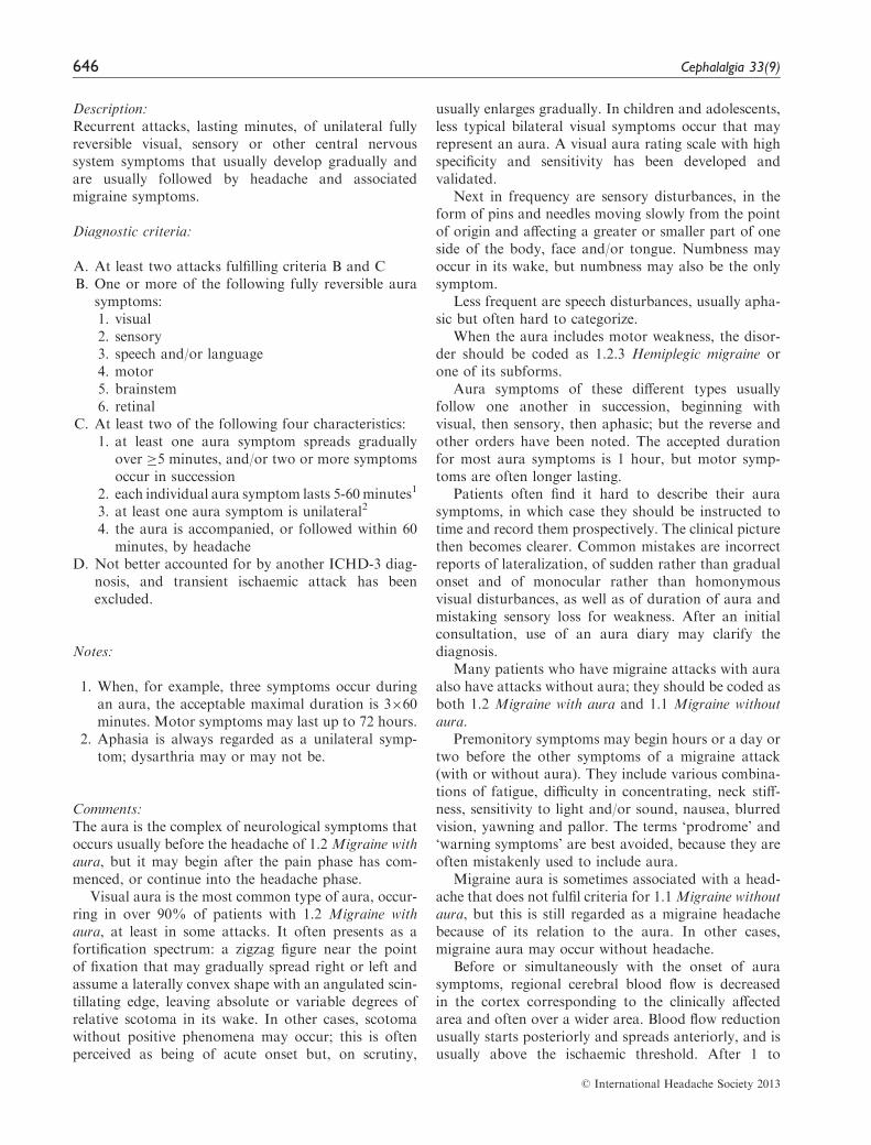
Description:Recurrent attacks, lasting minutes, of unilateral fullyreversible visual, sensory or other central nervoussystem symptoms that usually develop gradually andare usually followed by headache and associatedmigraine symptoms.
Diagnostic criteria:
A. At least two attacks fulfilling criteria B and CB. One or more of the following fully reversible aura
symptoms:1. visual2. sensory3. speech and/or language4. motor5. brainstem6. retinal
C. At least two of the following four characteristics:1. at least one aura symptom spreads gradually
over �5 minutes, and/or two or more symptomsoccur in succession
2. each individual aura symptom lasts 5-60minutes1
3. at least one aura symptom is unilateral2
4. the aura is accompanied, or followed within 60minutes, by headache
D. Not better accounted for by another ICHD-3 diag-nosis, and transient ischaemic attack has beenexcluded.
Notes:
1. When, for example, three symptoms occur duringan aura, the acceptable maximal duration is 3�60minutes. Motor symptoms may last up to 72 hours.
2. Aphasia is always regarded as a unilateral symp-tom; dysarthria may or may not be.
Comments:The aura is the complex of neurological symptoms thatoccurs usually before the headache of 1.2 Migraine withaura, but it may begin after the pain phase has com-menced, or continue into the headache phase.
Visual aura is the most common type of aura, occur-ring in over 90% of patients with 1.2 Migraine withaura, at least in some attacks. It often presents as afortification spectrum: a zigzag figure near the pointof fixation that may gradually spread right or left andassume a laterally convex shape with an angulated scin-tillating edge, leaving absolute or variable degrees ofrelative scotoma in its wake. In other cases, scotomawithout positive phenomena may occur; this is oftenperceived as being of acute onset but, on scrutiny,
usually enlarges gradually. In children and adolescents,less typical bilateral visual symptoms occur that mayrepresent an aura. A visual aura rating scale with highspecificity and sensitivity has been developed andvalidated.
Next in frequency are sensory disturbances, in theform of pins and needles moving slowly from the pointof origin and affecting a greater or smaller part of oneside of the body, face and/or tongue. Numbness mayoccur in its wake, but numbness may also be the onlysymptom.
Less frequent are speech disturbances, usually apha-sic but often hard to categorize.
When the aura includes motor weakness, the disor-der should be coded as 1.2.3 Hemiplegic migraine orone of its subforms.
Aura symptoms of these different types usuallyfollow one another in succession, beginning withvisual, then sensory, then aphasic; but the reverse andother orders have been noted. The accepted durationfor most aura symptoms is 1 hour, but motor symp-toms are often longer lasting.
Patients often find it hard to describe their aurasymptoms, in which case they should be instructed totime and record them prospectively. The clinical picturethen becomes clearer. Common mistakes are incorrectreports of lateralization, of sudden rather than gradualonset and of monocular rather than homonymousvisual disturbances, as well as of duration of aura andmistaking sensory loss for weakness. After an initialconsultation, use of an aura diary may clarify thediagnosis.
Many patients who have migraine attacks with auraalso have attacks without aura; they should be coded asboth 1.2 Migraine with aura and 1.1 Migraine withoutaura.
Premonitory symptoms may begin hours or a day ortwo before the other symptoms of a migraine attack(with or without aura). They include various combina-tions of fatigue, difficulty in concentrating, neck stiff-ness, sensitivity to light and/or sound, nausea, blurredvision, yawning and pallor. The terms ‘prodrome’ and‘warning symptoms’ are best avoided, because they areoften mistakenly used to include aura.
Migraine aura is sometimes associated with a head-ache that does not fulfil criteria for 1.1Migraine withoutaura, but this is still regarded as a migraine headachebecause of its relation to the aura. In other cases,migraine aura may occur without headache.
Before or simultaneously with the onset of aurasymptoms, regional cerebral blood flow is decreasedin the cortex corresponding to the clinically affectedarea and often over a wider area. Blood flow reductionusually starts posteriorly and spreads anteriorly, and isusually above the ischaemic threshold. After 1 to
� International Headache Society 2013
646 Cephalalgia 33(9)

several hours, gradual transition into hyperaemiaoccurs in the same region. Cortical spreading depres-sion of Leao is the likely underlying mechanism.
Systematic studies have demonstrated that manypatients with visual aura occasionally have symptomsin the extremities and/or speech symptoms. Conversely,patients with symptoms in the extremities and/or speechor language symptoms almost always also experiencevisual aura symptoms at least during some attacks. Adistinction between migraine with visual aura, migrainewith hemiparaesthetic aura and migraine with speechand/or language aura is probably artificial, and there-fore is not recognized in this classification. They are allcoded as 1.2.1 Migraine with typical aura. Patients withaura symptoms arising from the brainstem are coded as1.2.2 Migraine with brainstem aura, but they almostalways have additional typical aura symptoms.Patients with 1.2.3 Hemiplegic migraine have motorweakness, and this is classified as a separate subformbecause of genetic and pathophysiological differencesfrom migraine with typical aura. Such patients oftenhave brainstem symptoms in addition.
The previously defined syndromes, migraine withprolonged aura and migraine with acute-onset aura,have been abandoned. The great majority of patientswith such attacks have other attacks that fulfil criteriafor one of the recognized subforms of 1.2 Migraine withaura, and should be coded to that diagnosis. The restshould be coded to 1.5.2 Probable migraine with aura,specifying the atypical feature (prolonged aura or acuteonset aura) in parenthesis. The diagnosis is usually evi-dent after a careful history alone, although there arerare secondary mimics including carotid dissection,arteriovenous malformation and seizure.
1.2.1 Migraine with typical aura
Description:Migraine with aura in which aura consists of visualand/or sensory and/or speech/language symptoms,but no motor weakness, and is characterized by gradualdevelopment, duration of each symptom no longer than1 hour, a mix of positive and negative features andcomplete reversibility.
Diagnostic criteria:
A. At least two attacks fulfilling criteria B and CB. Aura consisting of visual, sensory and/or speech/
language symptoms, each fully reversible, but nomotor, brainstem or retinal symptoms
C. At least two of the following four characteristics:1. at least one aura symptom spreads gradually
over �5 minutes, and/or two or more symptomsoccur in succession
2. each individual aura symptom lasts 5-60minutes1
3. at least one aura symptom is unilateral2
4. the aura is accompanied, or followed within 60minutes, by headache
D. Not better accounted for by another ICHD-3 diag-nosis, and transient ischaemic attack has beenexcluded.
Notes:
1. When for example three symptoms occur during anaura, the acceptable maximal duration is 3�60minutes.
2. Aphasia is always regarded as a unilateral symp-tom; dysarthria may or may not be.
1.2.1.1 Typical aura with headache
Description:Migraine with typical aura in which aura is accompa-nied or followed within 60 minutes by headache with orwithout migraine characteristics.
Diagnostic criteria:
A. Fulfils criteria for 1.2.1 Migraine with typical auraB. Headache, with or without migraine characteristics,
accompanies or follows the aura within 60 minutes.
1.2.1.2 Typical aura without headache
Description:Migraine with typical aura in which aura is neitheraccompanied nor followed by headache of any sort.
Diagnostic criteria:
A. Fulfils criteria for 1.2.1 Migraine with typical auraB. No headache accompanies or follows the aura
within 60 minutes.
Comments:In some patients, a typical aura is always followed bymigraine headache, but many patients have, in addi-tion, attacks with aura followed by a less distinct head-ache or even without headache. A number of patientshave, exclusively, 1.2.1.2 Typical aura without headache.
In the absence of headache fulfilling criteria for 1.1Migraine without aura, the precise diagnosis of aura andits distinction from mimics that may signal serious
� International Headache Society 2013
ICHD-3 beta 647
disease (e.g. transient ischaemic attack) becomesmore difficult and often requires investigation.When aura occurs for the first time after age 40,when symptoms are exclusively negative (e.g. hemiano-pia) or when aura is prolonged or very short, othercauses, particularly transient ischaemic attacks,should be ruled out.
1.2.2 Migraine with brainstem aura
Previously used terms:Basilar artery migraine; basilar migraine; basilar-typemigraine.
Description:Migraine with aura symptoms clearly originating fromthe brainstem, but no motor weakness.
Diagnostic criteria:
A. At least two attacks fulfilling criteria B-DB. Aura consisting of visual, sensory and/or speech/
language symptoms, each fully reversible, but nomotor1 or retinal symptoms
C. At least two of the following brainstem symptoms:1. dysarthria2. vertigo3. tinnitus4. hypacusis5. diplopia6. ataxia7. decreased level of consciousness
D. At least two of the following four characteristics:1. at least one aura symptom spreads gradually
over �5 minutes, and/or two or more symptomsoccur in succession
2. each individual aura symptom lasts 5-60minutes2
3. at least one aura symptom is unilateral3
4. the aura is accompanied, or followed within 60minutes, by headache
E. Not better accounted for by another ICHD-3 diag-nosis, and transient ischaemic attack has beenexcluded.
Notes:
1. When motor symptoms are present, code as 1.2.3Hemiplegic migraine.
2. When for example three symptoms occur during anaura, the acceptable maximal duration is 3�60minutes.
3. Aphasia is always regarded as a unilateral symp-tom; dysarthria may or may not be.
Comments:Originally the terms basilar artery migraine or basilarmigraine were used but, as involvement of the basilarartery is unlikely, the term migraine with brainstem aurais preferred.
There are typical aura symptoms in addition to thebrainstem symptoms during most attacks. Manypatients who have attacks with brainstem aura alsoreport other attacks with typical aura and should becoded for both 1.2.1 Migraine with typical aura and1.2.2 Migraine with brainstem aura.
Many of the symptoms listed under criterion C mayoccur with anxiety and hyperventilation, and thereforeare subject to misinterpretation.
1.2.3 Hemiplegic1 migraine
Description:Migraine with aura including motor weakness.
Diagnostic criteria:
A. At least two attacks fulfilling criteria B and CB. Aura consisting of both of the following:
1. fully reversible motor weakness2. fully reversible visual, sensory and/or speech/
language symptomsC. At least two of the following four characteristics:
1. at least one aura symptom spreads graduallyover �5 minutes, and/or two or more symptomsoccur in succession
2. each individual non-motor aura symptom lasts5–60 minutes, and motor symptoms last <72hours2
3. at least one aura symptom is unilateral3
4. the aura is accompanied, or followed within 60minutes, by headache
D. Not better accounted for by another ICHD-3 diag-nosis, and transient ischaemic attack and strokehave been excluded.
Notes:
1. The term plegic means paralysis in most languages,but most attacks are characterized by motorweakness.
2. In some patients, motor weakness may last weeks.3. Aphasia is always regarded as a unilateral symp-
tom; dysarthria may or may not be.
Comment:It may be difficult to distinguish weakness from sensoryloss.
� International Headache Society 2013
648 Cephalalgia 33(9)
1.2.3.1 Familial hemiplegic migraine (FHM)
Description:Migraine with aura including motor weakness, and atleast one first- or second-degree relative has migraineaura including motor weakness.
Diagnostic criteria:
A. Fulfils criteria for 1.2.3 Hemiplegic migraineB. At least one first- or second-degree relative has had
attacks fulfilling criteria for 1.2.3 Hemiplegicmigraine.
Comments:New genetic data have allowed a more precise defini-tion of 1.2.3.1 Familial hemiplegic migraine (FHM)than was possible previously. Specific genetic subtypeshave been identified: in FHM1 there are mutations inthe CACNA1A gene (coding for a calcium channel)on chromosome 19; in FHM2 there are mutations inthe ATP1A2 gene (coding for a K/Na-ATPase) onchromosome 1; and in FHM3 there are mutationsin the SCN1A gene (coding for a sodium channel)on chromosome 2. There may be other loci not yetidentified. When genetic testing is done, the geneticsubtype (if discovered) should be specified at thefifth digit.
It has been shown that 1.2.3.1 Familial hemiplegicmigraine (FHM) very often presents with brainstemsymptoms in addition to the typical aura symptoms,and that headache almost always occurs. Rarely,during FHM attacks, disturbances of consciousness(sometimes including coma), confusion, fever andCSF pleocytosis can occur.
1.2.3.1 Familial hemiplegic migraine (FHM) may bemistaken for epilepsy and (unsuccessfully) treated assuch. FHM attacks can be triggered by (mild) headtrauma. In approximately 50% of FHM families,chronic progressive cerebellar ataxia occurs indepen-dently of the migraine attacks.
1.2.3.1.1 Familial hemiplegic migraine type 1 (FHM1)
Diagnostic criteria:
A. Fulfils criteria for 1.2.3.1 Familial hemiplegicmigraine
B. A causative mutation on the CACNA1A gene hasbeen demonstrated.
1.2.3.1.2 Familial hemiplegic migraine type 2 (FHM2)
Diagnostic criteria:
A. Fulfils criteria for 1.2.3.1 Familial hemiplegicmigraine
B. A causative mutation on the ATP1A2 gene has beendemonstrated.
1.2.3.1.3 Familial hemiplegic migraine type 3 (FHM3)
Diagnostic criteria:
A. Fulfils criteria for 1.2.3.1 Familial hemiplegicmigraine
B. A causative mutation on the SCN1A gene has beendemonstrated.
1.2.3.1.4 Familial hemiplegic migraine, other loci
Diagnostic criteria:
A. Fulfils criteria for 1.2.3.1 Familial hemiplegic migraineB. Genetic testing has demonstrated no mutation on
the CACNA1A, ATP1A2 or SCN1A genes.
1.2.3.2 Sporadic hemiplegic migraine
Description:Migraine with aura including motor weakness, and nofirst- or second-degree relative has migraine auraincluding motor weakness.
Diagnostic criteria:
A. Fulfils criteria for 1.2.3 Hemiplegic migraineB. No first- or second-degree relative fulfils criteria for
1.2.3 Hemiplegic migraine.
Comments:Epidemiological studies have shown that sporadic casesoccur with approximately the same prevalence as famil-ial cases.
The attacks in 1.2.3.2 Sporadic hemiplegic migrainehave the same clinical characteristics as those in 1.2.3.1Familial hemiplegic migraine. Some apparently sporadiccases have known FHM mutations, and in some a first-or second-degree relative later develops hemiplegicmigraine, thus completing fulfilment of the criteria for1.2.3.1 Familial hemiplegic migraine and requiring achange of diagnosis.
� International Headache Society 2013
ICHD-3 beta 649
Sporadic cases usually require neuroimaging andother tests to rule out other causes. A lumbar puncturemay be necessary to rule out 7.3.5 Syndrome of transi-ent Headache and Neurological Deficits with cerebrosp-inal fluid Lymphocytosis (HaNDL).
1.2.4 Retinal migraine
Description:Repeated attacks of monocular visual disturbance,including scintillations, scotomata or blindness, asso-ciated with migraine headache.
Diagnostic criteria:
A. At least two attacks fulfilling criteria B and CB. Aura consisting of fully reversible monocular posi-
tive and/or negative visual phenomena (e.g. scintil-lations, scotomata or blindness) confirmed duringan attack by either or both of the following:1. clinical visual field examination2. the patient’s drawing (made after clear instruc-
tion) of a monocular field defectC. At least two of the following three characteristics
1. the aura spreads gradually over �5 minutes2. aura symptoms last 5-60 minutes3. the aura is accompanied, or followed within 60
minutes, by headacheD. Not better accounted for by another ICHD-3 diag-
nosis, and other causes of amaurosis fugax havebeen excluded.
Comments:Some patients who complain of monocular visual dis-turbance in fact have hemianopia. Some cases withoutheadache have been reported, but migraine cannot beascertained as the underlying aetiology.
1.2.4 Retinal migraine is an extremely rare cause oftransient monocular visual loss. Cases of permanentmonocular visual loss associated with migraine havebeen described. Appropriate investigations are requiredto exclude other causes of transient monocularblindness.
1.3 Chronic migraine1,2
Description:Headache occurring on 15 or more days per month formore than 3 months, which has the features of migraineheadache on at least 8 days per month.
Diagnostic criteria:
A. Headache (tension-type-like and/or migraine-like)on �15 days per month for >3 months2 and ful-filling criteria B and C
B. Occurring in a patient who has had at least fiveattacks fulfilling criteria B-D for 1.1 Migraine with-out aura and/or criteria B and C for 1.2 Migrainewith aura
C. On �8 days per month for >3 months, fulfilling anyof the following3:1. criteria C and D for 1.1 Migraine without aura2. criteria B and C for 1.2 Migraine with aura3. believed by the patient to be migraine at onset
and relieved by a triptan or ergot derivativeD. Not better accounted for by another ICHD-3
diagnosis.
Notes:
1. The diagnosis of 1.3 Chronic migraine excludes thediagnosis of 2. Tension-type headache or its sub-types because tension-type-like headache is withinthe diagnostic criteria for 1.3 Chronic migraine.
2. The reason for singling out chronic from episodicmigraine is that it is impossible to distinguish theindividual episodes of headache in patients withsuch frequent or continuous headaches. In fact,the characteristics of the headache may changenot only from day to day but even within thesame day. It is extremely difficult to keep suchpatients medication-free in order to observe thenatural history of the headache. In this situation,attacks with or without aura are both counted, aswell as tension-type-like headaches. The mostcommon cause of symptoms suggestive of chronicmigraine is medication overuse, as defined under8.2 Medication-overuse headache. Around 50% ofpatients apparently with 1.3 Chronic migrainerevert to an episodic migraine subtype after drugwithdrawal; such patients are in a sense wronglydiagnosed as 1.3 Chronic migraine. Equally, manypatients apparently overusing medication do notimprove after drug withdrawal, and the diagnosisof 8.2 Medication-overuse headache may in a sensebe inappropriate (assuming that chronicity inducedby drug overuse is always reversible). For thesereasons, and because of the general rule, patientsmeeting criteria for 1.3 Chronic migraine and for 8.2Medication-overuse headache should be given bothdiagnoses. After drug withdrawal, migraine willeither revert to the episodic subtype or remainchronic, and be re-diagnosed accordingly; in thelatter case, the diagnosis of 8.2 Medication-overuse
� International Headache Society 2013
650 Cephalalgia 33(9)

headache may be rescinded. In some countries, it isusual practice to diagnose 8.2 Medication-overuseheadache only on discharge.
3. Characterization of frequently recurring headachegenerally requires a headache diary to record infor-mation on pain and associated symptoms day-by-day for at least 1 month. Sample diaries are avail-able at http://www.i-h-s.org.
1.4 Complications of migraine
Comment:Code separately for both the migraine subtype and forthe complication.
1.4.1 Status migrainosus
Description:A debilitating migraine attack lasting for more than 72hours.
Diagnostic criteria:
A. A headache attack fulfilling criteria B and CB. Occurring in a patient with 1.1 Migraine
without aura and/or 1.2 Migraine with aura, andtypical of previous attacks except for its durationand severity
C. Both of the following characteristics:1. unremitting for >72 hours1
2. pain and/or associated symptoms aredebilitating2
D. Not better accounted for by another ICHD-3diagnosis.
Notes:
1. Remissions of up to 12 hours because of medica-tion or sleep are accepted.
2. Milder cases, not meeting criterion C2, are coded1.5.1 Probable migraine without aura.
Comments:Headache with the features of 1.4.1 Status migraino-sus may often be caused by medication overuse.When headache in these circumstances meets the cri-teria for 8.2 Medication-overuse headache, code for1.3 Chronic migraine and 8.2 Medication-overuseheadache but not for 1.4.1 Status migrainosus. Whenoveruse of medication is of shorter duration than3 months, code for the appropriate migraine sub-type(s) only.
1.4.2 Persistent aura without infarction
Description:Aura symptoms persisting for 1 week or more withoutevidence of infarction on neuroimaging.
Diagnostic criteria:
A. Aura fulfilling criterion BB. Occurring in a patient with 1.2 Migraine
with aura and typical of previous auras exceptthat one or more aura symptoms persists for �1week
C. Neuroimaging shows no evidence of infarctionD. Not better accounted for by another ICHD-3
diagnosis.
Comments:Persistent aura symptoms are rare but well documen-ted. They are often bilateral and may last for monthsor years. The 1-week minimum in criterion B is basedon the opinion of experts and should be formallystudied.
Diagnostic work-up must distinguish 1.4.2 Persistentaura without infarction from 1.4.3 Migrainous infarc-tion, and exclude symptomatic aura as a resultof cerebral infarction of other causes. Attackslasting more than 1 hour and less than 1 weekand not fulfilling criteria for 1.2.1 Migraine withtypical aura are coded 1.5.2 Probable migraine withaura.
1.4.3 Migrainous infarction
Description:One or more migraine aura symptoms associated withan ischaemic brain lesion in the appropriate territorydemonstrated by neuroimaging.
Diagnostic criteria:
A. A migraine attack fulfilling criteria B and CB. Occurring in a patient with 1.2 Migraine with aura
and typical of previous attacks except that one ormore aura symptoms persists for >60 minutes
C. Neuroimaging demonstrates ischaemic infarction ina relevant area
D. Not better accounted for by another diagnosis.
Comments:Ischaemic stroke in a migraine sufferer may be categor-ized as cerebral infarction of other cause coexisting withmigraine, cerebral infarction of other cause presenting
� International Headache Society 2013
ICHD-3 beta 651
with symptoms resembling migraine with aura, or cere-bral infarction occurring during the course of a typicalmigraine with aura attack. Only the last fulfils criteriafor 1.4.3 Migrainous infarction.
1.4.3 Migrainous infarction mostly occurs in the pos-terior circulation and in younger women
A two-fold increased risk of ischaemic stroke inpatients with migraine with aura patients has beendemonstrated in several population-based studies.However, it should be noted that these infarctionsare not migrainous infarctions. The mechanisms ofthe increased risk of ischaemic stroke in migrainesufferers remain unclear; likewise, the relationshipbetween frequency of aura and the nature ofaura symptoms denoting the increase in risk isunknown. Most studies have shown a lack of asso-ciation between migraine without aura and ischaemicstroke.
1.4.4 Migraine aura-triggered seizure
Description:A seizure triggered by an attack of migraine with aura.
Diagnostic criteria:
A. A seizure fulfilling diagnostic criteria for one type ofepileptic attack, and criterion B below
B. Occurring in a patient with 1.2 Migraine with aura,and during, or within 1 hour after, an attack ofmigraine with aura
C. Not better accounted for by another diagnosis.
Comment:Migraine and epilepsy are prototypical examples ofparoxysmal brain disorders. Although migraine-likeheadaches are quite frequently seen in the epilepticpostictal period, sometimes a seizure occurs during orfollowing a migraine attack. This phenomenon, some-times referred to as migralepsy, is a rare event, origin-ally described in patients with 1.2 Migraine with aura.Evidence for association with 1.1Migraine without aurais still lacking.
1.5 Probable migraine
Previously used term:Migrainous disorder.
Coded elsewhere:Migraine-like headache secondary to another disorder(symptomatic migraine) is coded according to thatdisorder.
Description:Migraine-like attacks missing one of the featuresrequired to fulfil all criteria for a subtype of migrainecoded above, and not fulfilling criteria for anotherheadache disorder.
Diagnostic criteria:
A. Attacks fulfilling all but one of criteria A-D for 1.1Migraine without aura, or all but one of criteria A-Cfor 1.2 Migraine with aura
B. Not fulfilling ICHD-3 criteria for any other head-ache disorder
C. Not better accounted for by another ICHD-3diagnosis.
Comment:In making a headache diagnosis, attacks that fulfil cri-teria for both 2. Tension-type headache and 1.5Probable migraine are coded as the former in accor-dance with the general rule that a definite diagnosisalways trumps a probable diagnosis. However, inpatients who already have a migraine diagnosis, andwhere the issue is to count the number of attacks theyare having (e.g. as an outcome measure in a drug trial),attacks fulfilling criteria for 1.5 Probable migraineshould be counted as migraine. The reason for this isthat mild migraine attacks, or attacks treated early,often do not achieve all characteristics necessary for amigraine attack diagnosis but nevertheless respond tospecific migraine treatments.
1.5.1 Probable migraine without aura
Diagnostic criteria:
A. Attacks fulfilling all but one of criteria A–D for 1.1Migraine without aura
B. Not fulfilling ICHD-3 criteria for any other head-ache disorder
C. Not better accounted for by another ICHD-3diagnosis.
1.5.2 Probable migraine with aura
Diagnostic criteria:
A. Attacks fulfilling all but one of criteria A–C for 1.2Migraine with aura or any of its subforms
B. Not fulfilling ICHD-3 criteria for any other head-ache disorder
C. Not better accounted for by another ICHD-3diagnosis.
� International Headache Society 2013
652 Cephalalgia 33(9)

1.6 Episodic syndromes that may be associated with
migraine
Previously used terms:Childhood periodic syndromes; periodic syndromes ofchildhood.
Comments:This group of disorders occurs in patients who alsohave 1.1 Migraine without aura or 1.2 Migraine withaura, or who have an increased likelihood todevelop either of these disorders. Although historicallynoted to occur in childhood, they may also occur inadults.
Additional conditions that may also occur in thesepatients include episodes of motion sickness and peri-odic sleep disorders including sleep walking, sleep talk-ing, night terrors and bruxism.
1.6.1 Recurrent gastrointestinal disturbance
Previously used terms:Chronic abdominal pain; functional abdominal pain;functional dyspepsia; irritable bowel syndrome; func-tional abdominal pain syndrome.
Description:Recurrent episodic attacks of abdominal pain and/ordiscomfort, nausea and/or vomiting, occurring infre-quently, chronically or at predictable intervals, thatmay be associated with migraine.
Diagnostic criteria:
A. At least five attacks with distinct episodes ofabdominal pain and/or discomfort and/or nauseaand/or vomiting
B. Normal gastrointestinal examination andevaluation
C. Not attributed to another disorder.
1.6.1.1 Cyclic vomiting syndrome
Description:Recurrent episodic attacks of intense nausea andvomiting, usually stereotypical in the individual andwith predictable timing of episodes. Attacks may beassociated with pallor and lethargy. There is completeresolution of symptoms between attacks.
Diagnostic criteria:
A. At least five attacks of intense nausea and vomiting,fulfilling criteria B and C
B. Stereotypical in the individual patient and recurringwith predictable periodicity
C. All of the following:1. nausea and vomiting occur at least four times
per hour2. attacks last �1 hour and up to 10 days3. attacks occur �1 week apart
D. Complete freedom from symptoms between attacksE. Not attributed to another disorder.1
Note:
1. In particular, history and physical examination donot show signs of gastrointestinal disease.
Comments:1.6.1.1 Cyclic vomiting syndrome is typically a self-limit-ing episodic condition occurring in childhood, with per-iods of complete normality between episodes. Thecyclic nature is the hallmark, and is predictable.
This disorder was not included as a childhood peri-odic syndrome in ICHD-I, but it was in ICHD-II. Theclinical features of this syndrome resemble those foundin association with migraine headaches, and multiplethreads of research over the last years have suggestedthat cyclic vomiting syndrome is a condition related tomigraine.
1.6.1.2 Abdominal migraine
Description:An idiopathic disorder seen mainly in children as recur-rent attacks of moderate to severe midline abdominalpain, associated with vasomotor symptoms, nausea andvomiting, lasting 2–72 hours and with normalitybetween episodes. Headache does not occur duringthese episodes.
Diagnostic criteria:
A. At least five attacks of abdominal pain, fulfillingcriteria B–D
B. Pain has at least two of the following threecharacteristics:1. midline location, periumbilical or poorly
localized2. dull or ‘just sore’ quality3. moderate or severe intensity
C. During attacks, at least two of the following:1. anorexia2. nausea3. vomiting4. pallor
� International Headache Society 2013
ICHD-3 beta 653
D. Attacks last 2-72 hours when untreated or unsuc-cessfully treated
E. Complete freedom from symptoms between attacksF. Not attributed to another disorder.1
Note:
1. In particular, history and physical examination donot show signs of gastrointestinal or renal disease,or such disease has been ruled out by appropriateinvestigations.
Comments:Pain of 1.6.1.2 Abdominal migraine is severe enough tointerfere with normal daily activities.
In young children the presence of headache isoften overlooked. A careful history of presence orabsence of headache must be taken and, ifheadache or head pain during attacks is identified,a diagnosis of 1.1 Migraine without aura should beconsidered.
Children may find it difficult to distinguish anor-exia from nausea. Pallor is often accompaniedby dark shadows under the eyes. In a fewpatients, flushing is the predominant vasomotorphenomenon.
Most children with abdominal migraine will developmigraine headache later in life.
1.6.2 Benign paroxysmal vertigo
Description:A disorder characterized by recurrent brief attacks ofvertigo, occurring without warning and resolving spon-taneously, in otherwise healthy children.
Diagnostic criteria:
A. At least five attacks fulfilling criteria B and CB. Vertigo1 occurring without warning, maximal at
onset and resolving spontaneously after minutes tohours without loss of consciousness
C. At least one of the following associated symptomsor signs:1. nystagmus2. ataxia3. vomiting4. pallor5. fearfulness
D. Normal neurological examination and audiometricand vestibular functions between attacks
E. Not attributed to another disorder.
Note:
1. Young children with vertigo may not be able todescribe vertiginous symptoms. Parental observa-tion of episodic periods of unsteadiness may beinterpreted as vertigo in young children.
Comments:Posterior fossa tumours, seizures and vestibular disor-ders must be excluded.
The relationship between 1.6.2 Benign paroxysmalvertigo and A1.6.6 Vestibular migraine (see Appendix)needs to be further examined.
1.6.3 Benign paroxysmal torticollis
Description:Recurrent episodes of head tilt to one side, perhapswith slight rotation, which remit spontaneously. Thecondition occurs in infants and small children, withonset in the first year.
Diagnostic criteria:
A. Recurrent attacks1 in a young child, fulfilling cri-teria B and C
B. Tilt of the head to either side, with or without slightrotation, remitting spontaneously after minutes todays
C. At least one of the following associated symptomsor signs:1. pallor2. irritability3. malaise4. vomiting5. ataxia2
D. Normal neurological examination between attacksE. Not attributed to another disorder.
Notes:
1. Attacks tend to recur monthly.2. Ataxia is more likely in older children within the
affected age group.
Comments:The child’s head can be returned to the neutral positionduring attacks: some resistance may be encountered,but can be overcome.
The differential diagnosis includes gastro-oesopha-geal reflux, idiopathic torsional dystonia and complexpartial seizure, but particular attention must be paid to
� International Headache Society 2013
654 Cephalalgia 33(9)
the posterior fossa and craniocervical junction wherecongenital or acquired lesions may produce torticollis.These observations need further validation by patientdiaries, structured interviews and longitudinal datacollection.
1.6.3 Benign paroxysmal torticollis may evolve into1.6.2 Benign paroxysmal vertigo or 1.2 Migraine withaura (particularly 1.2.2 Migraine with brainstem aura),or cease without further symptoms.
Bibliography
1.1 Migraine in general
Arruda MA, Guidetti V, Galli F, et al. Primary headaches inchildhood–A population-based study. Cephalalgia 2010;30:1056–1064.
Diener HC and Silberstein SD. Medication overuse headache. In:Olesen J, Goadsby PJ, Ramadan NM, et al. (eds). TheHeadaches, 3rd ed. Philadelphia: Lippincott Williams &Wilkins 2006;971–979.
Gelfand AA, Fullerton HJ and Goadsby PJ. Child neurology:Migraine with aura in children. Neurology 2010; 75(5): e16–e19.
Goadsby PJ. Recent advances in the diagnosis and managementof migraine. BMJ 2006; 332:25–29.
Goadsby PJ. Migraine pathophysiology. Headache 2005; 45Suppl 1: S14–S24.
Katsarava Z, Manack A, Yoon MS, et al. Chronic migraine:classification and comparisons. Cephalalgia 2011; 31:520–529.
Lipton RB, Bigal ME, Steiner TJ, et al. Classification of primaryheadaches. Neurology 2004; 63:427–435.
Martelletti P, Haimanot RT, Lainez MJ, et al. The GlobalCampaign (GC) to Reduce the Burden of HeadacheWorldwide. The International Team for Specialist Education(ITSE). J Headache Pain 2005; 6:261–263.
Silberstein SD. Migraine. Lancet 2004; 363:381–391.Vos T, Flaxman AD, Naghavi M, Lozano R, et al. Years livedwith disability (YLD) for 1160 sequelae of 289 diseases andinjuries 1990–2010: A systematic analysis for the global burdenof disease study 2010. Lancet 2012; 380: 2163–2196.
1.2 Migraine with aura
Cao Y, Welch KM, Aurora S, et al. Functional MRI-BOLD ofvisually triggered headache in patients with migraine. ArchNeurol 1999; 56:548–554.
Charles A and Brennan K. Cortical spreading depression – newinsights and persistent questions. Cephalalgia 2009; 29:1115–1124.
Cologno D, Torelli P and Manzoni GC. Migraine with aura: Areview of 81 patients at 10–20 years’ follow-up. Cephalalgia1998; 18:690–696.
Cutrer FM, Sorensen AG, Weisskoff RM, et al. Perfusion-weighted imaging defects during spontaneous migrainousaura. Ann Neurol 1998; 43:25–31.
Eriksen MK, Thomsen LL, Andersen I, et al. Clinical character-istics of 362 patients with familial migraine with aura.Cephalalgia 2004; 24:564–575.
Eriksen MK, Thomsen LL and Olesen J. The Visual Aura RatingScale (VARS) for migraine aura diagnosis. Cephalalgia 2005;25:801–810.
Hadjikhani N, Sanchez del Rio M, Wu O, et al. Mechanisms ofmigraine aura revealed by functional MRI in human visualcortex. PNAS 2001; 98:4687–4692.
Hansen JM, Lipton R, Dodick D, et al. Migraine headache ispresent in the aura phase – a prospective study. Neurology2012; 79:2044–2049.
Jensen K, Tfelt-Hansen P, Lauritzen M, et al. Classic migraine. Aprospective recording of symptoms. Acta Neurol Scand 1986;73:359–362.
Kallela M, Wessman M, Farkkila M, et al. Clinical characteris-tics of migraine in a population-based twin sample: Similaritiesand differences between migraine with and without aura.Cephalalgia 1999; 19:151–158.
Kelman L. The premonitory symptoms (prodrome): A tertiarycare study of 893 migraineurs. Headache 2004; 44:865–872.
Lauritzen M. Pathophysiology of the migraine aura. The spread-ing depression theory. Brain 1994; 117 (Pt 1):199–210.
Leao AAP. Spreading depression of activity in the cerebralcortex. J Neurophysiol 1944; 7:359–390.
Olesen J, Friberg L, Olsen TS, et al. Timing and topography ofcerebral blood flow, aura, and headache during migraineattacks. Ann Neurol 1990; 28:791–798.
Queiroz LP, Friedman DI, Rapoport AM, et al. Characteristicsof migraine visual aura in Southern Brazil and Northern USA.Cephalalgia 2011; 31:1652–1658.
Rasmussen BK and Olesen J. Migraine with aura and migrainewithout aura: An epidemiological study. Cephalalgia 1992;12:221–228 (discussion 186).
Salhofer-Polanyi S, Frantal S, Brannath W, et al. Prospectiveanalysis of factors related to migraine aura – The PAMINAStudy. Headache 2012; 52:1236–1245.
Schoonman GG, Evers DJ, Terwindt GM, et al. The prevalenceof premonitory symptoms in migraine: A questionnaire studyin 461 patients. Cephalalgia 2006; 26:1209–1213.
Vibeke U, Gervil M, Kyvik KO, et al. Evidence of a geneticfactor in migraine with aura: A population-based Danishtwin study. Ann Neurol 1999; 45:242–246.
1.2.1 Migraine with typical aura
Eriksen MK, Thomsen LL and Olesen J. Implications of clinicalsubtypes of migraine with aura. Headache 2006; 46:286–297.
Matharu MJ and Goadsby PJ. Post-traumatic chronic paroxys-mal hemicrania (CPH) with aura. Neurology 2001; 56:273–275.
Morrison DP. Abnormal perceptual experiences in migraine.Cephalalgia 1990; 10:273–277.
Silberstein SD, Niknam R, Rozen TD, et al. Cluster headachewith aura. Neurology 2000; 54:219–221.
Wijman CA, Wolf PA, Kase CS, et al. Migrainous visual accom-paniments are not rare in late life: The Framingham Study.Stroke 1998; 29:1539–1543.
1.2.2 Migraine with brainstem aura
Ambrosini A, D’Onofrio M, Grieco GS, et al. Familial basilarmigraine associated with a new mutation in the ATP1A2 gene.Neurology 2005; 65:1826–1828.
Bickerstaff ER. Basilar artery migraine. Lancet 1961; i: 15.Caplan LR. Migraine and vertebrobasilar ischemia. Neurology1991; 41:55–61.
Eriksen MK, Thomsen LL and Olesen J. Implications of clinicalsubtypes of migraine with aura. Headache 2006; 46:286–297.
Kirchmann M, Thomsen LL and Olesen J. Basilar-type migraine:Clinical, epidemiologic, and genetic features. Neurology 2006;66:880–886.
Lempert T, Neuhauser H and Daroff RB. Vertigo as a symptomof migraine. Ann NY Acad Sci 2009; 1164:242–251.
Sturzenegger MH and Meienberg O. Basilar artery migraine: Afollow-up study of 82 cases. Headache 1985; 25:408–415.
� International Headache Society 2013
ICHD-3 beta 655
Swanson JW and Vick NA. Basilar artery migraine 12 patients,with an attack recorded electroencephalographically.Neurology 1978; 28:782–786.
Thomsen LL, Eriksen MK, Roemer SF, et al. A population-based study of familial hemiplegic migraine suggests reviseddiagnostic criteria. Brain 2002; 125:1379–1391.
Thomsen LL, Ostergaard E, Olesen J, et al. Evidence for a sepa-rate type of migraine with aura: Sporadic hemiplegic migraine.Neurology 2003; 60:595–601.
1.2.3 Hemiplegic migraine
Ambrosini A, D’Onofrio M, Grieco GS, et al. Familial basilarmigraine associated with a new mutation in the ATP1A2 gene.Neurology 2005; 65:1826–1828.
De Fusco M, Marconi R, Silvestri L, et al. Haploinsufficiency ofATP1A2 encoding the Naþ/Kþ pump alpha2 subunit asso-ciated with familial hemiplegic migraine type 2. Nat Genet2003; 33:192–196.
de Vries B, Frants RR, Ferrari M, et al. Molecular genetics ofmigraine. Human Genet 2009; 126:115–132.
Dichgans M, Freilinger T, Eckstein G, et al. Mutation in theneuronal voltage-gated sodium channel SCN1A in familialhemiplegic migraine. Lancet 2005; 366:371–377.
Dreier JP, Jurkat-Rott K, Petzold GC, et al. Opening of theblood-brain barrier preceding cortical edema in a severeattack of FHM type II. Neurology 2005; 64:2145–2147.
Eriksen MK, Thomsen LL and Olesen J. Implications of clinicalsubtypes of migraine with aura. Headache 2006; 46:286–297.
Hansen JM, Schytz HW, Larsen VA, et al. Hemiplegic migraineaura begins with cerebral hypoperfusion: Imaging in the acutephase. Headache 2011; 51:1289–1296.
Hansen JM, Thomsen LL, Olesen J, et al. Coexisting typicalmigraine in familial hemiplegic migraine. Neurology 2010;74:594–600.
Iizuka T, Takahashi Y, Sato M, et al. Neurovascular changes inprolonged migraine aura in FHM with a novel ATP1A2 genemutation. J Neurol Neurosurg Psychiat 2012; 83:205–212.
Jurkat-Rott K, Freilinger T, Dreier JP, et al. Variability of famil-ial hemiplegic migraine with novel A1A2 Naþ/Kþ-ATPasevariants. Neurology 2004; 62:1857–1861.
Kirchmann M, Thomsen LL and Olesen J. Basilar-type migraine:clinical, epidemiologic, and genetic features. Neurology 2006;66:880–886.
Leo L, Gherardini L, Barone V, et al. Increased susceptibility tocortical spreading depression in the mouse model of familialhemiplegic migraine type 2. PLoS Genet 2011; 7: e1002129.
Thomsen LL, Eriksen MK, Roemer SF, et al. A population-based study of familial hemiplegic migraine suggests reviseddiagnostic criteria. Brain 2002; 125:1379–1391.
Thomsen LL, Kirchmann M, Bjornsson A, et al. The geneticspectrum of a population-based sample of familial hemiplegicmigraine. Brain 2007; 130:346–356.
Thomsen LL, Ostergaard E, Olesen J, et al. Evidence for a sepa-rate type of migraine with aura: Sporadic hemiplegic migraine.Neurology 2003; 60:595–601.
Vanmolkot KRJ, Kors EE, Turk U, et al. Two de novo mutationsin the Na,K-ATPase gene ATP1A2 associated with pure famil-ial hemiplegic migraine. Eur J Human Genet 2006; 14:555–560.
1.2.4 Retinal migraine
Carroll D. Retinal migraine. Headache 1970; 10: 9–13.Chronicle EP and Mulleners WM. Visual system dysfunction inmigraine: A review of clinical and psychophysical findings.Cephalalgia 1996; 16: 525–535.
Hedges TR. Isolated ophthalmic migraine in the differential diag-nosis of cerebro-ocular ischemia. Stroke 1976; 7: 379–381.
Grosberg BM, Solomon S, Friedman DI and Lipton RB. Retinalmigraine reappraised. Cephalalgia 2006; 26:1275–1286.
Hill DL, Daroff RB, Ducros A, et al. Most cases labeled as‘retinal migraine’ are not migraine. J Neuroophthalmol 2007;27:3–8.
Martin TJ and Corbett JJ. Disorders of the eye. In SilbersteinSD, Lipton RB and Dalessio DJ, editors.Wolff’s Headache andOther Head Pain. New York: Oxford University Press 2001;459–474.
Troost T and Zagami AS. Ophthalmoplegic migraine and retinalmigraine. In Olesen J, Tfelt-Hansen P and Welch KMA, edi-tors. The Headaches. Philadelphia: Lippincott Williams &Wilkins 2000; 511–516.
1.3 Chronic migraine
Aurora SK. Is chronic migraine one end of a spectrum ofmigraine or a separate entity? Cephalalgia 2009; 29: 597–605.
Bigal ME and Lipton RB. Concepts and mechanisms of migrainechronification. Headache 2008; 48:7–15.
Bigal M, Rapoport A, Sheftell F, et al. The InternationalClassification of Headache Disorders revised criteria forchronic migraine – field testing in a headache specialty clinic.Cephalalgia 2007; 27: 230–234.
Bigal ME, Serrano D, Reed M and Lipton RB. Chronic migrainein the population: burden, diagnosis, and satisfaction withtreatment. Neurology 2008; 71: 559–566.
Bigal ME, Sheftell FD, Rapoprt AM, et al. Chronic daily head-ache in a tertiary care population: Correlation between theInternational Headache Society diagnostic criteria and pro-posed revisions of criteria for chronic daily headache.Cephalalgia 2002; 22:432–438.
Bloudek LM, Stokes M, Buse DC, et al. Cost of healthcare forpatients with migraine in five European countries: results fromthe International Burden of Migraine Study (IBMS).J Headache Pain 2012; 13:361–378.
Buse DC, Manack AN, Fanning KM, et al. Chronic migraineprevalence, disability, and sociodemographic factors. Resultsfrom the American Migraine Prevalence and Prevention Study.Headache 2012; 52:1456–1470.
Buse D, Manack A, Serrano D, et al. Headache impact ofchronic and episodic migraine: results from the AmericanMigraine Prevalence and Prevention study. Headache 2012;52:3–17.
Diamond S. A view of chronic daily headache. Headache Quart2000; 11: 177.
Mathew NT, Stubits E and Nigam MR. Transformation ofmigraine into daily headache: Analysis of factors. Headache1982; 22:66–68.
Diener HC, Dodick DW, Goadsby PJ, et al. Chronic migraine –classification, characteristics and treatment. Nat Rev Neurol2012; 8:162–171.
Goadsby PJ and Hargreaves R. Refractory migraine and chronicmigraine: pathophysiological mechanisms. Headache 2008; 48:799–804.
Katsarava Z, Manack A, Yoon MS, et al. Chronicmigraine: classification and comparisons. Cephalalgia 2011;31:520–529.
Manzoni GC, Bonavita V, Bussone G, et al; ANIRCEF(Associazione Neurologica Italiana Ricerca Cefalee). Chronicmigraine classification: current knowledge and future perspec-tives. J Headache Pain 2001; 12:585–592.
Mathew NT, Stubits E and Nigam MP. Transformed or evolu-tive migraine. Headache 1987; 27:102–106.
� International Headache Society 2013
656 Cephalalgia 33(9)
Natoli JL, Manack A, Dean B, et al. Global prevalence ofchronic migraine: a systematic review. Cephalalgia 2010; 30:599–609.
Scher AI, Stewart WF, Liberman J and Lipton RB. Prevalence offrequent headache in a population sample. Headache 1998; 38:497–506.
Scher AI, Stewart WF, Ricci JA and Lipton RB. Factors asso-ciated with the onset and remission of chronic daily headachein a population-based study. Pain 2003; 106:81–89.
Silberstein SD, Lipton RB and Sliwinski M. Classification ofdaily and near-daily headaches: Field trial of revised IHS cri-teria. Neurology 1996; 47:871–875.
Silberstein SD, Lipton RB, Solomon S and Mathew NT.Classification of daily and near-daily headaches: Proposedrevisions to the IHS criteria. Headache 1994; 34: 1–7.
Stewart WF, Scher AI and Lipton RB. Stressful life events andrisk of chronic daily headache: Results from the frequent head-ache epidemiology study. Cephalalgia 2001; 21: 279.
Zeeberg P, Olesen J and Jensen R. Probable medication-overuseheadache: The effect of a 2-month drug-free period. Neurology2006; 66:1894–1898.
Zeeberg P, Olesen J and Jensen R. Medication overuse headacheand chronic migraine in a specialized headache centre: Field-testing proposed new appendix criteria. Cephalalgia 2009;29:214–220.
1.4.1 Status migrainosus
Akhtar ND, Murray MA and Rothner AD. Statusmigrainosus in children and adolescents. Semin PediatrNeurol 2001; 8:27–33.
Couch JR and Diamond S. Status migrainosus. Causative andtherapeutic aspects. Headache 1983; 23:94–101.
Cure J and Rothrock J. Prolonged status migrainosus compli-cated by cerebellar infarction. Headache 2007; 47:1091–1092.
Gentile S, Rainero I, Daniele D, et al. Reversible MRI abnorm-alities in a patient with recurrent status migrainosus.Cephalalgia 2009; 29:687–690.
Lanfranconi S, Corti S, Bersano A, et al. Aphasic and visual aurawith increased vasogenic leakage: An atypical migrainosusstatus. J Neurol Sci 2009; 285:227–229.
Perucca P, Terzaghi M and Manni R. Status epilepticus migrai-nosus: Clinical, electrophysiologic, and imaging characteristics.Neurology 2010; 75:373–374.
Raskin NH. Treatment of status migrainosus: The Americanexperience. Headache 1990; 30 Suppl 2: 550–553.
1.4.2 Persistent aura without infarction
Ambrosini A, de Noordhout AM and Schoenen J.Neuromuscular transmission in migraine patients with pro-longed aura. Acta Neurol Belg 2001; 101:166–170.
Bento MS and Esperanca P. Migraine with prolonged aura.Headache 2000; 40:52–53.
Chen WT, Fuh JL, Lu SR and Wang SJ. Persistent migrainousvisual phenomena might be responsive to lamotrigine.Headache 2001; 41:823–825.
Chen WT, Lin YY, Fuh JL, et al. Sustained visual cortex hyper-excitability in migraine with persistent visual aura. Brain 2011;134 (Pt 8):2387–2395.
Evans RW and Lay CL. A persistent migraine aura. Headache2000; 40:696–698.
Haan J, Sluis P, Sluis IH and Ferrari MD. Acetazolamide treat-ment for migraine aura status. Neurology 2000; 55:1588–1589.
Haas DC. Prolonged migraine aura status. Ann Neurol 1982;11:197–199.
Liu GT, Schatz NJ, Galetta SK, et al. Persistent positive visualphenomena in migraine. Neurology 1995; 45:664–668.
Luda E, Bo E, Sicuro L, et al. Sustained visual aura: A totallynew variation of migraine. Headache 1991; 31:582–583.
Relja G, Granato A, Ukmar M, et al. Persistent aura withoutinfarction: Description of the first case studied with both brainSPECT and perfusion MRI. Cephalalgia 2005; 25:56–59.
Rothrock JF. Successful treatment of persistent migraine aurawith divalproex sodium. Neurology 1997; 48:261–262.
San-Juan OD and Zermeno PF. Migraine with persistent aura ina Mexican patient: Case report and review of the literature.Cephalalgia 2007; 27:456–460.
Smith M, Cros D and Sheen V. Hyperperfusion with vasogenicleakage by fMRI in migraine with prolonged aura. Neurology2002; 58:1308–1310.
Wang YF, Fuh JL, Chen WT and Wang SJ. The visual aurarating scale as an outcome predictor for persistent visualaura without infarction. Cephalalgia 2008; 28:1298–1304.
1.4.3 Migrainous infarction
Bono G, Minonzio G, Mauri M and Clerici AM. Complicationsof migraine: Migrainous infarction. Clin Exp Hypertens 2006;28:233–242.
Bousser MG, Conard J, Kittner S, et al. Recommendations onthe risk of ischemic stroke associated with use of combined oralcontraceptives and hormone replacement therapy in womenwith migraine. The International Headache Society TaskForce on Combined Oral Contraceptives & HormoneReplacement Therapy. Cephalalgia 2000; 20:155–156.
Chang CL, Donaghy M and Poulter N. Migraine and stroke inyoung women: case-control study. The WorldHealth Organization Collaborative Study of CardiovascularDisease and Steroid Hormone Contraception. BMJ 1999;318:13–18.
Connor CCR. Complicated migraine. A study of permanent neu-rological and visual defects. Lancet 1962; ii:1072–1075.
Laurell K, Artto V, Bendtsen L, et al. Migrainous infarction: ANordic multicenter study. Eur J Neurol 2011; 18:1220–1226.
MacGregor EA and Guillebaud J. Combined oral contraceptives,migraine and ischemic stroke. Clinical and ScientificCommittee of the Faculty of Family Planning andReproductive Health Care and the Family PlanningAssociation. Br J Fam Plann 1998; 24: 55–60.
Olesen J, Friberg L, Olsen TS, et al. Ischaemia-induced (sympto-matic) migraine attacks may be more frequent than migraineinduced ischemic insults. Brain 1993; 116: 187–202.
Rothrock JF, Walicke P, Swenson MR, et al. Migrainous stroke.Arch Neurol 1988; 45:63–67.
Schulz UG, Blamire AM, Davies P, et al. Normal cortical energymetabolism in migrainous stroke: A 31P-MR spectroscopystudy. Stroke 2009; 40:3740–3744.
Tietjen GE. The relationship of migraine and stroke.Neuroepidemiology 2000; 19: 13–19.
Tzourio C, Kittner SJ, Bousser MG, et al. Migraine and stroke inyoung women. Cephalalgia 2000; 20:190–199.
Wolf ME, Szabo K, Griebe M, et al. Clinical and MRI charac-teristics of acute migrainous infarction. Neurology 2011;76:1911–1917.
1.4.4 Migraine aura–triggered seizure
Belcastro V, Striano P, Kasteleijn-Nolst Trenite DG, et al.Migralepsy, hemicrania epileptica, post-ictal headache and‘ictal epileptic headache’: A proposal for terminology and clas-sification revision. J Headache Pain 2011; 12:289–294.
� International Headache Society 2013
ICHD-3 beta 657
Davies PT and Panayiotopoulos CP. Migraine triggered seizuresand epilepsy triggered headache and migraine attacks: A needfor re-assessment. J Headache Pain 2011; 12:287–288.
Friedenberg S and Dodick DW. Migraine-associated seizure: Acase of reversible MRI abnormalities and persistent nondomi-nant hemisphere syndrome. Headache 2000; 40:487–490.
Maggioni F, Mampreso E, Ruffatti S, et al. Migralepsy: Is thecurrent definition too narrow? Headache 2008; 48:1129–1132.
Marks DA and Ehrenberg BL. Migraine-related seizures inadults with epilepsy, with EEG correlation. Neurology 1993;43:2476–2483.
Merlino G, Valente MR, D’Anna S and Gigli GL. Seizures withprolonged EEG abnormalities during an attack of migrainewithout aura. Headache 2007; 47:919–922.
Parisi P and Kasteleijn-Nolst Trenite DGA. ‘Migralepsy’: A callfor revision of the definition. Epilepsia 2010; 51:932–933.
Rogawski MA. Common pathophysiologic mechanisms inmigraine and epilepsy. Arch Neurol 2008; 65:709–714.
Sances G, Guaschino E, Perucca P, et al. Migralepsy: A call forrevision of the definition. Epilepsia 2009; 50:2487–2496.
Ter Berg HW. Migraine-associated seizure: A case of reversibleMRI abnormalities and persistent nondominant hemispheresyndrome. Headache 2001; 41:326–328.
Velioglu SK and Ozmenoglu M. Migraine-related seizures in anepileptic population. Cephalalgia 1999; 19:797–801.
Verrotti A, Coppola G, Di Fonzo A, et al. Should ‘migralepsy’ beconsidered an obsolete concept? A multicenter retrospectiveclinical/EEG study and review of the literature. EpilepsyBehav 2011; 21:52–59.
1.5 Probable migraine
Granella F, Alessandro RD, Manzoni GC, et al. InternationalHeadache Society classification: Interobserver reliability in thediagnosis of primary headaches. Cephalalgia 1994; 14: 16–20.
Rains JC, Penzien DB, Lipchik GL, et al. Diagnosis of migraine:Empirical analysis of a large clinical sample of atypicalmigraine (IHS 1.7) patients and proposed revision of the IHScriteria. Cephalalgia 2001; 21:584–595.
Rasmussen BK, Jensen R and Olesen J. A population-basedanalysis of the diagnostic criteria of the InternationalHeadache Society. Cephalalgia 1991; 11:129–134.
Russell MB and Olesen J. Migrainous disorder and its relation tomigraine without aura and migraine with aura. A genetic epi-demiological study. Cephalalgia 1996; 16:431–435.
1.6.1 Recurrent gastrointestinal disturbance
Abu-Arafeh I and Russel G. Prevalence and clinical features ofabdominal migraine compared with those of migraine head-ache. Arch Dis Child 1995; 72:413–417.
Al-Twaijri WA and Shevell MI. Pediatric migraine equivalents:Occurrence and clinical features in practice. Pediatr Neurol2002; 26:365–368.
Dignan F, Abu-Arafeh I and Russell G. The prognosis of child-hood abdominal migraine. Arch Dis Child 2001; 84:415–418.
Drossman DA and Dumitrascu DL. Rome III: New standard forfunctional gastrointestinal disorders. J Gastrointestin Liver Dis2006; 15:237–241.
Farquar HA. Abdominal migraine in children. BMJ 1956;i:1082–1085.
Fleisher DR. Cyclic vomiting syndrome and migraine. J Pediatr1999; 134:533–535.
Haan J, Kors EE and Ferrari MD. Familial cyclic vomiting syn-drome. Cephalalgia 2002; 22:552–554.
Li BU. Cyclic vomiting syndrome: Age-old syndrome and newinsights. Semin Pediatr Neurol 2001; 8: 13–21.
Li BUK, Lefevre F, Chelimsky GG, et al. NASPGHANConsensus Statement on the Diagnosis and Management ofCVS. J Pediatr Gastroenterol Nutr 2008; 47:379–393.
Rashed H, Abell TL, Familoni BO, et al. Autonomic function incyclic vomiting syndrome and classic migraine. Dig Dis Sci1999; 44 Suppl 8: 74S-78S.
Russell G, Abu-Arafeh I and Symon DN. Abdominal migraine:Evidence for existence and treatment options. Paediatr Drugs2002; 4:1–8.
Thiessen PN. Recurrent abdominal pain. Pediatr Rev 2002;23:39–46.
Welch KM. Scientific basis of migraine: speculation on therelationship to cyclic vomiting. Dig Dis Sci 1999; 44 Suppl 8:26S–30S.
1.6.2 Benign paroxysmal vertigo
Drigo P, Carli G and Laverda AM. Benign paroxysmal vertigo ofchildhood. Brain Dev (Netherlands) 2001; 23: 38–41.
Dunn DW and Snyder CH. Benign paroxysmal vertigo of child-hood. Am J Dis Child 1976; 130:1099–1100.
Fenichel GM. Migraine as a cause of benign paroxysmal vertigoof childhood. J Pediatr 1967; 71:114–115.
1.6.3 Benign paroxysmal torticollis
Drigo, P, Carli G and Laverda AM. Benign paroxysmal torticol-lis of infancy. Brain Dev 2000; 22:169–172.
Giffin NJ, Benton S and Goadsby PJ. Benign paroxysmal torti-collis of infancy: Four new cases and linkage to CACNA1Amutation. Dev Med Child Neurol 2002; 44:490–493.
Rosman NP, Douglass LM, Sharif UM, Paolini J. The neurologyof benign paroxysmal torticollis of infancy: Report of 10new cases and review of the literature. J Child Neurol 2009;24:155–160.
� International Headache Society 2013
658 Cephalalgia 33(9)
2. Tension-type headache (TTH)
2.1 Infrequent episodic tension-type headache2.1.1 Infrequent episodic tension-type headache
associated with pericranial tenderness2.1.2 Infrequent episodic tension-type headache
not associated with pericranial tenderness2.2 Frequent episodic tension-type headache
2.2.1 Frequent episodic tension-type headacheassociated with pericranial tenderness
2.2.2 Frequent episodic tension-type headache notassociated with pericranial tenderness
2.3 Chronic tension-type headache2.3.1 Chronic tension-type headache associated
with pericranial tenderness2.3.2 Chronic tension-type headache not asso-
ciated with pericranial tenderness2.4 Probable tension-type headache
2.4.1 Probable infrequent episodic tension-typeheadache
2.4.2 Probable frequent episodic tension-typeheadache
2.4.3 Probable chronic tension-type headache
Previously used terms:Tension headache; muscle contraction headache; psy-chomyogenic headache; stress headache; ordinaryheadache; essential headache; idiopathic headache; psy-chogenic headache.
Coded elsewhere:Tension-type-like headache attributed to another disor-der is coded to that disorder.
General comment
Primary or secondary headache or both?
When a headache with the characteristics of tension-type characteristics occurs for the first time in closetemporal relation to another disorder that is knownto cause headache, or fulfils other criteria for causationby that disorder, the new headache is coded as asecondary headache attributed to the causativedisorder. When pre-existing tension-type headachebecomes chronic in close temporal relation to such acausative disorder, both the initial tension-type head-ache diagnosis and the secondary diagnosis should begiven. When pre-existing tension-type headache ismade significantly worse (usually meaning a two-foldor greater increase in frequency and/or severity) inclose temporal relation to such a causative disorder,both the initial tension-type headache diagnosis andthe secondary diagnosis should be given, providedthat there is good evidence that the disorder can
cause headache. In the case of chronic tension-typeheadache with medication overuse, a close temporalrelation is often difficult to establish. Therefore, bothdiagnoses, 2.3 Chronic tension-type headache and 8.2Medication-overuse headache, should be given in allsuch cases.
Introduction
2. Tension-type headache is very common, with a life-time prevalence in the general population rangingbetween 30% and 78% in different studies, and it hasa very high socio-economic impact.
Although this type of headache was previouslyconsidered to be primarily psychogenic, a numberof studies have appeared after publication of ICHD-I that strongly suggest a neurobiological basis, atleast for the more severe subtypes of tension-typeheadache.
The division of 2. Tension-type headache into episo-dic and chronic subtypes, which was introduced inICHD-I, has proved extremely useful. In ICHD-II,the episodic form was further subdivided into an infre-quent subform with headache episodes less than onceper month and a frequent subform. 2.3 Chronic tension-type headache is a serious disease, causing greatlydecreased quality of life and high disability. 2.2Frequent episodic tension-type headache can be asso-ciated with considerable disability, and sometimes war-rants treatment with expensive drugs. In contrast, 2.1Infrequent episodic tension-type headache, which occursin almost the entire population, usually has very littleimpact on the individual and, in most instances,requires no attention from the medical profession.The distinction of 2.1 Infrequent episodic tension-typeheadache from 2.2 Frequent episodic tension-type head-ache thus separates individuals who typically do notrequire medical management, and avoids categorizingalmost the entire population as having a significantheadache disorder, yet allows their headaches to beclassified.
The exact mechanisms of 2. Tension-type headacheare not known. Peripheral pain mechanisms are mostlikely to play a role in 2.1 Infrequent episodic tension-type headache and 2.2 Frequent episodic tension-typeheadache, whereas central pain mechanisms play amore important role in 2.3 Chronic tension-type head-ache. Increased pericranial tenderness recorded bymanual palpation is the most significant abnormal find-ing in patients with tension-type headache. The tender-ness is typically present interictally, is further increasedduring actual headache and increases with the intensityand frequency of headaches. Pericranial tenderness iseasily recorded by manual palpation by small rotatingmovements and a firm pressure (preferably aided by use
� International Headache Society 2013
ICHD-3 beta 659
of a palpometer) with the second and third fingers onthe frontal, temporal, masseter, pterygoid, sternoclei-domastoid, splenius and trapezius muscles. Local ten-derness scores of 0-3 for each muscle can be summed toyield a total tenderness score for each individual.Palpation is a useful guide for treatment strategy. Italso adds value and credibility to the explanationsgiven to the patient.
Increased tenderness is most likely of pathophysio-logical importance. The Classification Committeeencourages further research into the pathophysiologicalmechanisms and treatment of 2. Tension-type headache.With this aim, ICHD-II distinguished patients with andwithout such disorder of the pericranial muscles, andthis subdivision is maintained in ICHD-3 beta to sti-mulate further research in this area.
The diagnostic difficulty most often encounteredamong the primary headache disorders is to discrimi-nate between tension-type headache and mild migrainewithout aura. This is more so because patients withfrequent headaches often suffer from both disorders.It has been suggested to tighten the diagnostic criteriafor 2. Tension-type headache in the hope of excludingmigraine that phenotypically resembles tension-typeheadache. Such an increase in specificity would, atthe same time, reduce the sensitivity of the criteria,resulting in a larger proportion of patients whose head-aches could be classified only as 2.4 Probable tension-type headache or 1.5 Probable migraine. Stricter diag-nostic criteria for 2. Tension-type headache were pro-posed in the Appendix of ICHD-II, as A2. Tension-type headache, but so far with no evidence that such achange would be beneficial. These stricter diagnosticcriteria remain in the Appendix, for research purposesonly. The Classification Committee recommends com-parisons between patients diagnosed according to eachset of criteria, not only for characterization of clinicalfeatures but also for enquiry into pathophysiologicalmechanisms and response to treatments.
2.1 Infrequent episodic tension-type headache
Description:Infrequent episodes of headache, typically bilateral,pressing or tightening in quality and of mild to moder-ate intensity, lasting minutes to days. The pain does notworsen with routine physical activity and is not asso-ciated with nausea, but photophobia or phonophobiamay be present.
Diagnostic criteria:
A. At least 10 episodes of headache occurring on <1day per month on average (<12 days per year) andfulfilling criteria B-D
B. Lasting from 30 minutes to 7 daysC. At least two of the following four characteristics:
1. bilateral location2. pressing or tightening (non-pulsating) quality3. mild or moderate intensity4. not aggravated by routine physical activity such
as walking or climbing stairsD. Both of the following:
1. no nausea or vomiting2. no more than one of photophobia or
phonophobiaE. Not better accounted for by another ICHD-3
diagnosis.
2.1.1 Infrequent episodic tension-type headacheassociated with pericranial tenderness
Diagnostic criteria:
A. Episodes fulfilling criteria for 2.1 Infrequent episodictension-type headache
B. Increased pericranial tenderness on manualpalpation.
2.1.2 Infrequent episodic tension-type headache not asso-ciated with pericranial tenderness
Diagnostic criteria:
A. Episodes fulfilling criteria for 2.1 Infrequent episodictension-type headache
B. No increase in pericranial tenderness.
2.2 Frequent episodic tension-type headache
Description:Frequent episodes of headache, typically bilateral,pressing or tightening in quality and of mild to moder-ate intensity, lasting minutes to days. The pain does notworsen with routine physical activity and is not asso-ciated with nausea, but photophobia or phonophobiamay be present.
Diagnostic criteria:
A. At least 10 episodes of headache occurring on 1-14 days per month on average for >3 months(�12 and <180 days per year) and fulfilling criteriaB-D
B. Lasting from 30 minutes to 7 daysC. At least two of the following four characteristics:
1. bilateral location2. pressing or tightening (non-pulsating) quality
� International Headache Society 2013
660 Cephalalgia 33(9)
3. mild or moderate intensity4. not aggravated by routine physical activity such
as walking or climbing stairsD. Both of the following:
1. no nausea or vomiting2. no more than one of photophobia or
phonophobiaE. Not better accounted for by another ICHD-3
diagnosis.
Comments:2.2 Frequent episodic tension-type headache often coex-ists with 1.1 Migraine without aura. Coexisting ten-sion-type headache in migraineurs should preferablybe identified through use of a diagnostic headachediary. The treatment of migraine differs considerablyfrom that of tension-type headache, and it is impor-tant to educate patients to distinguish between theseheadache types if they are to select the right treatmentfor each while avoiding medication overuse and theadverse consequence of 8.2 Medication-overuseheadache.
When headache fulfils criteria for both 1.5Probable migraine and 2. Tension-type headache,code as 2. Tension-type headache (or as any subtypeof it for which the criteria are fulfilled) under thegeneral rule that definite diagnoses always trumpprobable diagnoses. When headache fulfils criteriafor both 1.5 Probable migraine and 2.4 Probable ten-sion-type headache, code as the former under the gen-eral rule of hierarchy, which puts 1. Migraine and itssubtypes before 2. Tension-type headache and itssubtypes.
2.2.1 Frequent episodic tension-type headache associatedwith pericranial tenderness
Diagnostic criteria:
A. Episodes fulfilling criteria for 2.2 Frequent episodictension-type headache
B. Increased pericranial tenderness on manualpalpation.
2.2.2 Frequent episodic tension-type headache notassociated with pericranial tenderness
Diagnostic criteria:
A. Episodes fulfilling criteria for 2.2 Frequent episodictension-type headache
B. No increase in pericranial tenderness.
2.3 Chronic tension-type headache
Coded elsewhere:4.10 New daily persistent headache.
Description:A disorder evolving from frequent episodic tension-type headache, with daily or very frequent episodes ofheadache, typically bilateral, pressing or tightening inquality and of mild to moderate intensity, lasting hoursto days, or unremitting. The pain does not worsen withroutine physical activity, but may be associated withmild nausea, photophobia or phonophobia.
Diagnostic criteria:
A. Headache occurring on �15 days per month onaverage for >3 months (�180 days per year), ful-filling criteria B-D
B. Lasting hours to days, or unremittingC. At least two of the following four characteristics:
1. bilateral location2. pressing or tightening (non-pulsating) quality3. mild or moderate intensity4. not aggravated by routine physical activity such
as walking or climbing stairsD. Both of the following:
1. no more than one of photophobia, phonophobiaor mild nausea
2. neither moderate or severe nausea nor vomitingE. Not better accounted for by another ICHD-3
diagnosis.
Comments:2.3 Chronic tension-type headache evolves over timefrom 2.2 Frequent episodic tension-type headache;when these criteria A-E are fulfilled by headache that,unambiguously, is daily and unremitting from less than24 hours after its first onset, code as 4.10 New dailypersistent headache. When the manner of onset is notremembered or is otherwise uncertain, code as 2.3Chronic tension-type headache.
Both 2.3 Chronic tension-type headache and 1.3Chronic migraine require headache on at least 15 daysper month. For 2.3 Chronic tension-type headache,headache on at least 15 days must meet the criteriafor 2. Tension-type headache and for 1.3 Chronicmigraine headache on at least 8 days must meet thecriteria for 1. Migraine. Therefore, a patient can fulfilcriteria for both these diagnoses, for example by havingheadache on 25 days per month meeting migraine cri-teria on 8 days and tension-type headache criteria on 17days. In these cases, only the diagnosis 1.3 Chronicmigraine should be given.
� International Headache Society 2013
ICHD-3 beta 661
In many uncertain cases there is overuse of medica-tion. When this fulfils criterion B for any of the sub-forms of 8.2 Medication-overuse headache and thecriteria for 2.3 Chronic tension-type headache are alsofulfilled, the rule is to code for both 2.3 Chronic tension-type headache and 8.2 Medication-overuse headache.After drug withdrawal, the diagnosis should be re-eval-uated: not uncommonly the criteria for 2.3 Chronic ten-sion-type headache will no longer be fulfilled, withreversion to one or other episodic subtype.
2.3.1 Chronic tension-type headache associated withpericranial tenderness
Diagnostic criteria:
A. Headache fulfilling criteria for 2.3 Chronic tension-type headache
B. Increased pericranial tenderness on manualpalpation.
2.3.2 Chronic tension-type headache not associated withpericranial tenderness
Diagnostic criteria:
A. Headache fulfilling criteria for 2.3 Chronic tension-type headache
B. No increase in pericranial tenderness.
2.4 Probable tension-type headache
Description:Tension-type-like headache missing one of the featuresrequired to fulfil all criteria for a subtype of tension-type headache coded above, and not fulfilling criteriafor another headache disorder.
Comment:Patients meeting one of the sets of criteria below mayalso meet the criteria for one of the subforms of 1.6Probable migraine. In such cases, all other availableinformation should be used to decide which of the alter-natives is the more likely.
2.4.1 Probable infrequent episodic tension-type headache
Diagnostic criteria:
A. One or more episodes of headache fulfilling all butone of criteria A-D for 2.1 Infrequent episodic ten-sion-type headache
B. Not fulfilling ICHD-3 criteria for any other head-ache disorder
C. Not better accounted for by another ICHD-3diagnosis.
2.4.2 Probable frequent episodic tension-type headache
Diagnostic criteria:
A. Episodes of headache fulfilling all but one of criteriaA-D for 2.2 Frequent episodic tension-type headache
B. Not fulfilling ICHD-3 criteria for any other head-ache disorder
C. Not better accounted for by another ICHD-3diagnosis.
2.4.3 Probable chronic tension-type headache
Diagnostic criteria:
A. Headache fulfilling all but one of criteria A-D for2.3 Chronic episodic tension-type headache
B. Not fulfilling ICHD-3 criteria for any other head-ache disorder
C. Not better accounted for by another ICHD-3diagnosis.
Bibliography
Ashina M. Nitric oxide synthase inhibitors for the treatment ofchronic tension-type headache. Expert Opin Pharmacother2002; 3: 395–399.
Ashina M. Neurobiology of chronic tension-type headache.Cephalalgia 2004; 24: 161–172.
Ashina M, Bendtsen L, Jensen R, et al. Muscle hardness inpatients with chronic tension-type headache: Relation toactual headache state. Pain 1999; 79: 201–205.
Ashina M, Bendtsen L, Jensen R, et al. Possible mechanismsof glyceryl-trinitrate-induced immediate headache in patientswith chronic tension-type headache. Cephalalgia 2000; 20:919–924.
Ashina M, Lassen LH, Bendtsen L, et al. Effect of inhibition ofnitric oxide synthase on chronic tension-type headache: A ran-domised crossover trial. Lancet 1999; 353: 287–289.
Ashina M, Stallknecht B, Bendtsen L, et al. Tender points arenot sites of ongoing inflammation – In vivo evidence in patientswith chronic tension-type headache. Cephalalgia 2003; 23: 109–116.
Ashina M, Stallknecht B, Bendtsen L, et al. In vivo evidence ofaltered skeletal muscle blood flow in chronic tension-typeheadache. Brain 2002; 125: 320–326.
Ashina S, Babenko L, Jensen R, et al. Increased muscular andcutaneous pain sensitivity in cephalic region in patients withchronic tension-type headache. Eur J Neurol 2005; 12: 543–549.
Ashina S, Bendtsen L, Ashina M, et al. Generalized hyperalgesiain patients with chronic tension-type headache. Cephalalgia2006; 26: 940–948.
Ashina S, Bendtsen L and Jensen R. Analgesic effect of amitrip-tyline in chronic tension-type headache is not directly related toserotonin reuptake inhibition. Pain 2004; 108: 108–114.
� International Headache Society 2013
662 Cephalalgia 33(9)
Bendtsen L. Central sensitization in tension-type headache -Possible pathophysiological mechanisms. Cephalalgia 2000;20: 486–508.
Bendtsen L, Bigal ME, Cerbo R, et al. Guidelines for controlledtrials of drugs in tension-type headache: Second edition.Cephalalgia 2010; 30: 1–16.
Bendtsen L, Evers S, Linde M, et al. EFNS guideline on thetreatment of tension-type headache - Report of an EFNStask force. Eur J Neurol 2010; 17: 1318–1325.
Bendtsen L, Jensen R. Amitriptyline reduces myofascial tender-ness in patients with chronic tension-type headache.Cephalalgia 2000; 20: 603–610.
Bendtsen L and Jensen R. Mirtazapine is effective in the prophy-lactic treatment of chronic tension-type headache. Neurology2004; 62: 1706–1711.
Bendtsen L and Jensen R. Tension-type headache: The mostcommon, but also the most neglected, headache disorder.Curr Opin Neurol 2006; 19: 305–309.
Bendtsen L, Jensen R, Jensen NK and Olesen J. Pressure-con-trolled palpation: A new technique which increases the relia-bility of manual palpation. Cephalalgia 1995; 15: 205–210.
Bendtsen L, Jensen R and Olesen J. A non-selective (amitripty-line), but not a selective (citalopram), serotonin reuptake inhi-bitor is effective in the prophylactic treatment of chronictension-type headache. J Neurol Neurosurg Psychiatry 1996;61: 285–290.
Bendtsen L, Jensen R and Olesen J. Decreased pain detection andtolerance thresholds in chronic tension-type headache. ArchNeurol 1996; 53: 373–376.
Bendtsen L, Jensen R and Olesen J. Qualitatively altered nocicep-tion in chronic myofascial pain. Pain 1996; 65: 259–264.
Buchgreitz L, Egsgaard LL, Jensen R, et al. Abnormal pain pro-cessing in chronic tension-type headache: A high-density EEGbrain mapping study. Brain 2008; 131: 3232–3238.
Buchgreitz L, Lyngberg AC, Bendtsen L and Jensen R.Frequency of headache is related to sensitization: A populationstudy. Pain 2006; 123: 19–27.
Buchgreitz L, Lyngberg AC, Bendtsen L and Jensen R. Increasedpain sensitivity is not a risk factor but a consequence of fre-quent headache: A population-based follow-up study. Pain2008; 137: 623–630.
Cathcart S, Petkov J and Pritchard D. Effects of induced stresson experimental pain sensitivity in chronic tension-type head-ache sufferers. Eur J Neurol 2008; 15: 552–558.
Cathcart S, Petkov J, Winefield AH, et al. Central mechanisms ofstress-induced headache. Cephalalgia 2010; 30: 285–295.
Cathcart S, Winefield AH, Lushington K and Rolan P. Noxiousinhibition of temporal summation is impaired in chronic ten-sion-type headache. Headache 2010; 50: 403–412.
Christensen M, Bendtsen L, Ashina M and Jensen R.Experimental induction of muscle tenderness and headache intension-type headache patients. Cephalalgia 2005; 25: 1061–1067.
Clark GT, Sakai S, Merrill R, et al. Cross-correlation betweenstress, pain, physical activity, and temporalis muscle EMG intension-type headache. Cephalalgia 1995; 15: 511–518.
Fernandez-de-Las-Penas C, Alonso-Blanco C, Cuadrado ML,et al. Myofascial trigger points and their relationship to head-ache clinical parameters in chronic tension-type headache.Headache 2006; 46: 1264–1272.
Fernandez-de-Las-Penas C, Alonso-Blanco C, Cuadrado MLand Pareja JA. Myofascial trigger points in the suboccipitalmuscles in episodic tension-type headache. Man Ther 2006;11: 225–230.
Fernandez-de-Las-Penas C, Cuadrado ML, Arendt-Nielsen L,et al. Increased pericranial tenderness, decreased pressure
pain threshold, and headache clinical parameters in chronictension-type headache patients. Clin J Pain 2007; 23: 346–352.
Fernandez-de-Las-Penas C, Cuadrado ML, Arendt-Nielsen L,et al. Myofascial trigger points and sensitization: An updatedpain model for tension-type headache. Cephalalgia 2007; 27:383–393.
Fernandez-de-Las-Penas C, Cuadrado ML and Pareja JA.Myofascial trigger points, neck mobility, and forward headposture in episodic tension-type headache. Headache 2007;47: 662–672.
Fernandez-de-Las-Penas C and Schoenen J. Chronic tension-typeheadache: What is new? Curr Opin Neurol 2009; 22: 254–261.
Fumal A and Schoenen J. Tension-type headache: Currentresearch and clinical management. Lancet Neurol 2008; 7:70–83.
Heckman BD and Holroyd KA. Tension-type headache and psy-chiatric comorbidity. Curr Pain Headache Rep 2006; 10: 439–447.
Holroyd KA, O’Donnell FJ, Stensland M, et al. Management ofchronic tension-type headache with tricyclic antidepressantmedication, stress management therapy, and their combina-tion: A randomized controlled trial. JAMA 2001; 285: 2208–2215.
Holroyd KA, Stensland M, Lipchik GL, et al. Psychosocial cor-relates and impact of chronic tension-type headaches.Headache 2000; 40: 3–16.
Hubbard DR and Berkoff GM. Myofascial trigger points showspontaneous needle EMG activity. Spine 1993; 18: 1803–1807.
Janke EA, Holroyd KA and Romanek K. Depression increasesonset of tension-type headache following laboratory stress.Pain 2004; 111: 230–238.
Jensen R. Mechanisms of spontaneous tension-type headaches:An analysis of tenderness, pain thresholds and EMG. Pain1996; 64: 251–256.
Jensen R. Pathophysiological mechanisms of tension-type head-ache: A review of epidemiological and experimental studies.Cephalalgia 1999; 19: 602–621.
Jensen R, Bendtsen L and Olesen J. Muscular factors are ofimportance in tension-type headache. Headache 1998; 38: 10–17.
Jensen R, Rasmussen BK, Pedersen B, et al. Cephalic muscletenderness and pressure pain threshold in a general population.Pain 1992; 48: 197–203.
Jensen R, Rasmussen BK, Pedersen B and Olesen J. Muscle ten-derness and pressure pain thresholds in headache. A popula-tion study. Pain 1993; 52: 193–199.
Jensen R and Stovner LJ. Epidemiology and comorbidity ofheadache. Lancet Neurol 2008; 7: 354–361.
Jensen R, Zeeberg P, Dehlendorff C and Olesen J. Predictors ofoutcome of the treatment programme in a multidisciplinaryheadache centre. Cephalalgia 2010; 30: 1214–1224.
Langemark M, Bach FW, Jensen TS and Olesen J. Decreasednociceptive flexion reflex threshold in chronic tension-typeheadache. Arch Neurol 1993; 50: 1061–1064.
Langemark M, Jensen K, Jensen TS and Olesen J. Pressure painthresholds and thermal nociceptive thresholds in chronic ten-sion-type headache. Pain 1989; 38: 203–210.
Langemark M and Olesen J. Pericranial tenderness in tensionheadache. A blind, controlled study. Cephalalgia 1987; 7:249–255.
Leistad R, Sand T, Westgaard R, et al. Stress-induced pain andmuscle activity in patients with migraine and tension-typeheadache. Cephalalgia 2006; 26: 64–73.
Lindelof K, Ellrich J, Jensen R and Bendtsen L. Central painprocessing in chronic tension-type headache. ClinNeurophysiol 2009; 120: 1364–1370.
� International Headache Society 2013
ICHD-3 beta 663

Lindelof K, Jung K, Ellrich J, et al. Low-frequencyelectrical stimulation induces long-term depression inpatients with chronic tension-type headache. Cephalalgia2010; 30: 860–867.
Lyngberg AC, Rasmussen BK, Jorgensen T and Jensen R. Hasthe prevalence of migraine and tension-type headache changedover a 12-year period? A Danish population survey. Eur JEpidemiol 2005; 20: 243–249.
Mathew NT. Tension-type headache. Curr Neurol Neurosci Rep2006; 6: 100–105.
Mork H, Ashina M, Bendtsen L, et al. Induction of prolongedtenderness in patients with tension-type headache by means ofa new experimental model of myofascial pain. Eur J Neurol2003; 10: 249–256.
Mork H, Ashina M, Bendtsen L, et al. Possible mechanisms ofpain perception in patients with episodic tension-type head-ache. A new experimental model of myofascial pain.Cephalalgia 2004; 24: 466–475.
Nestoriuc Y, Rief W and Martin A. Meta-analysis ofbiofeedback for tension-type headache: Efficacy, specificity,and treatment moderators. J Consult Clin Psychol 2008; 76:379–396.
Olesen J. Clinical and pathophysiological observations inmigraine and tension-type headache explained by integrationof vascular, supraspinal and myofascial inputs. Pain 1991; 46:125–132.
Pielsticker A, Haag G, Zaudig M and Lautenbacher S.Impairment of pain inhibition in chronic tension-type head-ache. Pain 2005;118 (1–2): 215–223.
Rasmussen BK, Jensen R, Schroll M and Olesen J. Epidemiologyof headache in a general population – A prevalence study.J Clin Epidemiol 1991; 44: 1147–1157.
Sandrini G, Rossi P, Milanov I, et al. Abnormal modulatoryinfluence of diffuse noxious inhibitory controls in migraineand chronic tension-type headache patients. Cephalalgia2006; 26: 782–789.
Schmidt-Hansen PT, Svensson P, Bendtsen L, et al. Increasedmuscle pain sensitivity in patients with tension-type headache.Pain 2007; 129: 113–121.
Schmidt-Wilcke T, Leinisch E, Straube A, et al. Gray matterdecrease in patients with chronic tension type headache.Neurology 2005; 65: 1483–1486.
Schoenen J, Bottin D, Hardy F and Gerard P. Cephalic andextracephalic pressure pain thresholds in chronic tension-typeheadache. Pain 1991; 47: 145–149.
Schoenen J, Gerard P, De P, et al. Multiple clinical and para-clinical analyses of chronic tension-type headache associated orunassociated with disorder of pericranial muscles. Cephalalgia1991; 11: 135–139.
Schwartz BS, Stewart WF, Simon D and Lipton RB.Epidemiology of tension-type headache. JAMA 1998; 4:381–383.
� International Headache Society 2013
664 Cephalalgia 33(9)
3. Trigeminal autonomic cephalalgias
(TACs)
3.1 Cluster headache3.1.1 Episodic cluster headache3.1.2 Chronic cluster headache
3.2 Paroxysmal hemicrania3.2.1 Episodic paroxysmal hemicrania3.2.2 Chronic paroxysmal hemicrania
3.3 Short-lasting unilateral neuralgiform headacheattacks3.3.1 Short-lasting unilateral neuralgiform head-
ache attacks with conjunctival injection andtearing (SUNCT)
3.3.1.1 Episodic SUNCT3.3.1.2 Chronic SUNCT
3.3.2 Short-lasting unilateral neuralgiform head-ache attacks with cranial autonomic symp-toms (SUNA)
3.3.2.1 Episodic SUNA3.3.2.2 Chronic SUNA
3.4 Hemicrania continua3.5 Probable trigeminal autonomic cephalalgia
3.5.1 Probable cluster headache3.5.2 Probable paroxysmal hemicrania3.5.3 Probable short-lasting unilateral neuralgi-
form headache attacks3.5.4 Probable hemicrania continua
General comment
Primary or secondary headache or both?
When a new headache with the characteristics of a tri-geminal autonomic cephalalgia (TAC) occurs for thefirst time in close temporal relation to another disorderknown to cause headache, or fulfils other criteria forcausation by that disorder, the new headache is codedas a secondary headache attributed to the causativedisorder. When a pre-existing TAC becomes chronicin close temporal relation to such a causative disorder,both the initial TAC diagnosis and the secondary diag-nosis should be given. When a pre-existing TAC ismade significantly worse (usually meaning a two-foldor greater increase in frequency and/or severity) in closetemporal relation to such a causative disorder, both theinitial TAC diagnosis and the secondary headache diag-nosis should be given, provided that there is good evi-dence that the disorder can cause headache.
Introduction
The trigeminal autonomic cephalalgias (TACs) sharethe clinical features of headache, which is usually later-alized, and often prominent cranial parasympathetic
autonomic features, which are again lateralized andipsilateral to the headache. Experimental and humanfunctional imaging suggests that these syndromes acti-vate a normal human trigeminal parasympatheticreflex, with clinical signs of cranial sympathetic dys-function being secondary.
Typical migraine aura can be seen, rarely, in associa-tion with TACs.
3.1 Cluster headache
Previously used terms:Ciliary neuralgia; erythro-melalgia of the head; erythro-prosopalgia of Bing; hemicrania angioparalytica; hemi-crania neuralgiformis chronica; histaminic cephalalgia;Horton’s headache; Harris-Horton’s disease; migrai-nous neuralgia (of Harris); petrosal neuralgia (ofGardner); Sluder’s neuralgia; spheno-palatine neural-gia; vidian neuralgia.
Coded elsewhere:Symptomatic cluster headache, secondary to anotherdisorder, is coded as a secondary headache attributedto that disorder.
Description:Attacks of severe, strictly unilateral pain which is orbi-tal, supraorbital, temporal or in any combination ofthese sites, lasting 15–180 minutes and occurring fromonce every other day to eight times a day. The pain isassociated with ipsilateral conjunctival injection, lacri-mation, nasal congestion, rhinorrhoea, forehead andfacial sweating, miosis, ptosis and/or eyelid oedema,and/or with restlessness or agitation.
Diagnostic criteria:
A. At least five attacks fulfilling criteria B–DB. Severe or very severe unilateral orbital, supraorbital
and/or temporal pain lasting 15–180 minutes (whenuntreated)1
C. Either or both of the following:1. at least one of the following symptoms or signs,
ipsilateral to the headache:a) conjunctival injection and/or lacrimationb) nasal congestion and/or rhinorrhoeac) eyelid oedemad) forehead and facial sweatinge) forehead and facial flushingf) sensation of fullness in the earg) miosis and/or ptosis
2. a sense of restlessness or agitationD. Attacks have a frequency between one every other
day and eight per day for more than half of the timewhen the disorder is active
� International Headache Society 2013
ICHD-3 beta 665
E. Not better accounted for by another ICHD-3diagnosis.
Note:
1. During part (but less than half) of the time-courseof 3.1 Cluster headache, attacks may be less severeand/or of shorter or longer duration.
Comments:Attacks occur in series lasting for weeks or months (so-called cluster periods) separated by remission periodsusually lasting months or years. About 10–15% ofpatients have 3.1.2 Chronic cluster headache, withoutsuch remission periods. In a large series with goodfollow-up, one-quarter of patients had only a singlecluster period. Such patients meet the criteria for andshould be coded as 3.1 Cluster headache.
The pain of 3.1 Cluster headache is maximal orbi-tally, supraorbitally, temporally or in any combinationof these sites, but may spread to other regions. Duringthe worst attacks, the intensity of pain is excruciating.Patients are usually unable to lie down, and character-istically pace the floor. Pain usually recurs on the sameside of the head during an individual cluster period.During a cluster period in 3.1.1 Episodic cluster head-ache, and at any time in 3.1.2 Chronic cluster headache,attacks occur regularly and may be provoked by alco-hol, histamine or nitroglycerin.
Age at onset is usually 20–40 years. For unknownreasons, men are afflicted three times more often thanwomen.
Acute attacks involve activation in the region of theposterior hypothalamic grey matter. 3.1 Cluster head-ache may be autosomal dominant in about 5% of cases.
Some patients have been described who have both3.1 Cluster headache and 13.1 Trigeminal neuralgia(sometimes referred to as cluster-tic syndrome). Theyshould receive both diagnoses. The importance of thisobservation is that both conditions must be treated forthe patient to become headache-free.
3.1.1 Episodic cluster headache
Description:Cluster headache attacks occurring in periods lastingfrom 7 days to 1 year, separated by pain-free periodslasting at least 1 month.
Diagnostic criteria:
A. Attacks fulfilling criteria for 3.1 Cluster headacheand occurring in bouts (cluster periods)
B. At least two cluster periods lasting from 7 days to 1year (when untreated) and separated by pain-freeremission periods of �1 month.
Comment:Cluster periods usually last between 2 weeks and 3months.
3.1.2 Chronic cluster headache
Description:Cluster headache attacks occurring for more than 1year without remission, or with remission periods last-ing less than 1 month.
Diagnostic criteria:
A. Attacks fulfilling criteria for 3.1 Cluster headache,and criterion B below
B. Occurring without a remission period, or withremissions lasting <1 month, for at least 1 year.
Comment:3.1.2 Chronic cluster headache may arise de novo (pre-viously referred to as primary chronic cluster headache),or evolve from 3.1.1 Episodic cluster headache (pre-viously secondary chronic cluster headache). In somepatients change occurs from 3.1.2 Chronic cluster head-ache to 3.1.1 Episodic cluster headache.
3.2 Paroxysmal hemicrania
Description:Attacks of severe, strictly unilateral pain which is orbi-tal, supraorbital, temporal or in any combination ofthese sites, lasting 2–30 minutes and occurring severalor many times a day. The attacks are associated withipsilateral conjunctival injection, lacrimation, nasalcongestion, rhinorrhoea, forehead and facial sweating,miosis, ptosis and/or eyelid oedema. They respondabsolutely to indomethacin.
Diagnostic criteria:
A. At least 20 attacks fulfilling criteria B-EB. Severe unilateral orbital, supraorbital and/or tem-
poral pain lasting 2–30 minutesC. At least one of the following symptoms or signs,
ipsilateral to the pain:1. conjunctival injection and/or lacrimation2. nasal congestion and/or rhinorrhoea3. eyelid oedema4. forehead and facial sweating
� International Headache Society 2013
666 Cephalalgia 33(9)
5. forehead and facial flushing6. sensation of fullness in the ear7. miosis and/or ptosis
D. Attacks have a frequency above five per day formore than half of the time
E. Attacks are prevented absolutely by therapeuticdoses of indomethacin1
F. Not better accounted for by another ICHD-3diagnosis.
Note:
1. In an adult, oral indomethacin should be used initi-ally in a dose of at least 150 mg daily and increasedif necessary up to 225 mg daily. The dose by injec-tion is 100–200 mg. Smaller maintenance doses areoften employed.
Comment:In contrast to cluster headache, there is no male pre-dominance. Onset is usually in adulthood, althoughchildhood cases are reported.
3.2.1 Episodic paroxysmal hemicrania
Description:Attacks of paroxysmal hemicrania occurring in periodslasting from 7 days to 1 year, separated by pain-freeperiods lasting at least 1 month.
Diagnostic criteria:
A. Attacks fulfilling criteria for 3.2 Paroxysmal hemi-crania and occurring in bouts
B. At least two bouts lasting from 7 days to 1 year(when untreated) and separated by pain-free remis-sion periods of �1 month.
3.2.2 Chronic paroxysmal hemicrania
Description:Attacks of paroxysmal hemicrania occurring for morethan 1 year without remission, or with remission peri-ods lasting less than 1 month.
Diagnostic criteria:
A. Attacks fulfilling criteria for 3.2 Paroxysmal hemi-crania, and criterion B below
B. Occurring without a remission period, or withremissions lasting <1 month, for at least 1 year.
Comment:Patients who fulfil criteria for both 3.2.2 Chronic par-oxysmal hemicrania and 13.1 Trigeminal neuralgia(sometimes referred to as CPH-tic syndrome) shouldreceive both diagnoses. Their recognition is important,as both disorders require treatment. The pathophysio-logical significance of the association is not yet clear.
3.3 Short-lasting unilateral neuralgiform headache attacks
Description:Attacks of moderate or severe, strictly unilateral headpain lasting seconds to minutes, occurring at least oncea day and usually associated with prominent lacrima-tion and redness of the ipsilateral eye.
Diagnostic criteria:
A. At least 20 attacks fulfilling criteria B–DB. Moderate or severe unilateral head pain, with orbi-
tal, supraorbital, temporal and/or other trigeminaldistribution, lasting for 1–600 seconds and occur-ring as single stabs, series of stabs or in a saw-tooth pattern
C. At least one of the following cranial autonomicsymptoms or signs, ipsilateral to the pain:1. conjunctival injection and/or lacrimation2. nasal congestion and/or rhinorrhoea3. eyelid oedema4. forehead and facial sweating5. forehead and facial flushing6. sensation of fullness in the ear7. miosis and/or ptosis
D. Attacks have a frequency of at least one a day formore than half of the time when the disorder isactive
E. Not better accounted for by another ICHD-3diagnosis.
Comments:Longer-duration attacks are characterized by multiplestabs or a saw-tooth pain pattern.
Two subtypes of 3.3 Short-lasting unilateral neuralgi-form headache attacks are recognized in ICHD-3 beta:3.3.1 Short-lasting unilateral neuralgiform headacheattacks with conjunctival injection and tearing(SUNCT) and 3.3.2 Short lasting unilateral neuralgi-form headache attacks with cranial autonomic symptoms(SUNA). 3.3.1 SUNCT may be a subform of 3.3.2SUNA, although this requires further study.Meanwhile, each is classified as a separate subtype,described below.
3.3.1 SUNCT and 3.3.2 SUNA are usually trigger-able without a refractory period. This is in contrast to
� International Headache Society 2013
ICHD-3 beta 667

13.1 Trigeminal neuralgia, which usually has a refrac-tory period after each attack.
3.3.1 Short-lasting unilateral neuralgiform headacheattacks with conjunctival injection and tearing(SUNCT)
Diagnostic criteria:
A. Attacks fulfilling criteria for 3.3 Short-lasting unilat-eral neuralgiform headache attacks
B. Both of conjunctival injection and lacrimation(tearing).
Comments:The literature suggests that the most common mimic of3.3.1 SUNCT is a lesion in the posterior fossa.
Patients have been described in whom there is over-lap between 3.3.1 SUNCT and 13.1 Trigeminal neural-gia. Differentiation is clinically complex. Such patientsshould receive both diagnoses.
Patients with both 3.3.1 SUNCT and 3.1 Clusterheadache have been reported; the pathophysiologicalsignificance of this overlap is yet to be determined.
3.3.1.1 Episodic SUNCT
Description:Attacks of SUNCT occurring in periods lasting from 7days to 1 year, separated by pain-free periods lasting 1month or more.
Diagnostic criteria:
A. Attacks fulfilling criteria for 3.3.1 Short-lasting uni-lateral neuralgiform headache attacks with conjuncti-val injection and tearing and occurring in bouts
B. At least two bouts lasting from 7 days to 1 year andseparated by pain-free remission periods of �1month.
3.3.1.2 Chronic SUNCT
Description:Attacks of SUNCT occurring for more than 1 yearwithout remission, or with remission periods lastingless than 1 month.
Diagnostic criteria:
A. Attacks fulfilling criteria for 3.3.1 Short-lasting uni-lateral neuralgiform headache attacks with conjuncti-val injection and tearing, and criterion B below
B. Occurring without a remission period, or withremissions lasting <1 month, for at least 1 year.
3.3.2 Short lasting unilateral neuralgiform headacheattacks with cranial autonomic symptoms (SUNA)
A. Attacks fulfilling criteria for 3.3 Short-lasting unilat-eral neuralgiform headache attacks, and criterion Bbelow
B. Only one or neither of conjunctival injection andlacrimation (tearing).
3.3.2.1 Episodic SUNA
Description:Attacks of SUNA occurring in periods lasting from 7days to 1 year, separated by pain-free periods lasting atleast 1 month.
Diagnostic criteria:
A. Attacks fulfilling criteria for 3.3.2 Short-lasting uni-lateral neuralgiform headache attacks with cranialautonomic symptoms and occurring in bouts
B. At least two bouts lasting from 7 days to 1 year andseparated by pain-free remission periods of �1month.
3.3.2.2 Chronic SUNA
Description:Attacks of SUNA occurring for more than 1 year with-out remission, or with remission periods lasting lessthan 1 month.
Diagnostic criteria:
A. Attacks fulfilling criteria for 3.3.2 Short-lasting uni-lateral neuralgiform headache attacks with cranialautonomic symptoms, and criterion B below
B. Occurring without a remission period, or withremissions lasting <1 month, for at least 1 year.
3.4 Hemicrania continua
Description:Persistent, strictly unilateral headache, associated withipsilateral conjunctival injection, lacrimation, nasalcongestion, rhinorrhoea, forehead and facial sweating,miosis, ptosis and/or eyelid oedema, and/or with rest-lessness or agitation. The headache is absolutely sensi-tive to indomethacin.
� International Headache Society 2013
668 Cephalalgia 33(9)

Diagnostic criteria:
A. Unilateral headache fulfilling criteria B-DB. Present for >3 months, with exacerbations of mod-
erate or greater intensityC. Either or both of the following:
1. at least one of the following symptoms or signs,ipsilateral to the headache:a) conjunctival injection and/or lacrimationb) nasal congestion and/or rhinorrhoeac) eyelid oedemad) forehead and facial sweatinge) forehead and facial flushingf) sensation of fullness in the earg) miosis and/or ptosis
2. a sense of restlessness or agitation, or aggrava-tion of the pain by movement
D. Responds absolutely to therapeutic doses ofindomethacin1
E. Not better accounted for by another ICHD-3diagnosis.
Note:
1. In an adult, oral indomethacin should be used initi-ally in a dose of at least 150 mg daily and increasedif necessary up to 225 mg daily. The dose by injec-tion is 100–200 mg. Smaller maintenance doses areoften employed.
Comments:Migrainous symptoms such as photophobiaand phonophobia are often seen in 3.4 Hemicraniacontinua.
3.4 Hemicrania continua has been included under 3.Trigeminal autonomic cephalalgias in ICHD-3 beta(previously it was under 4. Other primary headache dis-orders) on the basis that the pain is typically unilateral,as are the cranial autonomic symptoms when present.Brain imaging studies show important overlapsbetween all disorders included here, notably activationin the region of the posterior hypothalamic grey. Inaddition, the absolute response to indomethacin of3.4 Hemicrania continua is shared with 3.2Paroxysmal hemicrania.
3.4.1 Hemicrania continua, remitting subtype
Description:Hemicrania continua characterized by pain that is notcontinuous but is interrupted by remission periods of atleast 1 day.
Diagnostic criteria:
A. Headache fulfilling criteria for 3.4 Hemicrania con-tinua, and criterion B below
B. Headache is not daily or continuous, but inter-rupted by remission periods of �1 day withouttreatment.
Comment:3.4.1 Hemicrania continua, remitting subtype can arisede novo or from 3.4.2 Hemicrania continua, unremittingsubtype.
3.4.2 Hemicrania continua, unremitting subtype
Description:Hemicrania continua characterized by continuous pain,without remission periods of at least 1 day, for at least 1year.
Diagnostic criteria:
A. Headache fulfilling criteria for 3.4 Hemicrania con-tinua, and criterion B below
B. Headache is daily and continuous for at least 1 year,without remission periods of �1 day.
Comment:3.4.2Hemicrania continua, unremitting subtype can arisede novo or evolve from 3.4.1 Hemicrania continua,remitting subtype. The majority of patients have theunremitting subtype from onset.
3.5 Probable trigeminal autonomic cephalalgia
Description:Headache attacks which are believed to be a type of 3.Trigeminal autonomic cephalalgia, but which are miss-ing one of the features required to fulfil all criteria forany of the subtypes coded above, and do not fulfil allcriteria for another headache disorder.
Diagnostic criteria:
A. Headache attacks fulfilling all but one of criteriaA-D for 3.1 Cluster headache, criteria A-E for 3.2Paroxysmal hemicrania, criteria A-D for 3.3 Short-lasting unilateral neuralgiform headache attacks orcriteria A-D for 3.4 Hemicrania continua
B. Not fulfilling ICHD-3 criteria for any other head-ache disorder
C. Not better accounted for by another ICHD-3diagnosis.
� International Headache Society 2013
ICHD-3 beta 669
Comment:Patients may be coded 3.5.1 Probable cluster headache,3.5.2 Probable paroxysmal hemicrania, 3.5.3 Probableshort-lasting unilateral neuralgiform headache attacksor 3.5.4 Probable hemicrania continua. Such patientseither have not had a sufficient number of typicalattacks (e.g. a first bout of cluster headache), or havehad, but fail to fulfil one of the other criteria.
Bibliography
Bahra A, May A and Goadsby PJ. Cluster headache: A prospec-tive clinical study in 230 patients with diagnostic implications.Neurology 2002; 58: 354–361.
Benoliel R and Sharav Y. Trigeminal neuralgia with lacrimationor SUNCT syndrome? Cephalalgia 1998; 18: 85–90.
Bing R. Uber traumatische Erythromelalgie undErthroprosopalgie. Nervenarzt 1930; 3: 506–512.
Boes CJ, Matharu MS and Goadsby PJ. The paroxysmal hemi-crania-tic syndrome. Cephalalgia 2003; 23: 24–28.
Bordini C, Antonaci F, Stovner LJ, et al. ‘Hemicrania continua’:A clinical review. Headache 1991; 31: 20–26.
Bouhassira D, Attal N, Esteve M and Chauvin M. SUNCT syn-drome. A case of transformation from trigeminal neuralgia.Cephalalgia 1994; 14: 168–170.
Broeske D, Lenn NJ and Cantos E. Chronic paroxysmal hemi-crania in a young child: Possible relation to ipsilateral occipitalinfarction. J Child Neurol 1993; 8: 235–236.
Bussone G, Leone M, Volta GD, et al. Short-lasting unilateralneuralgiform headache attacks with tearing and conjunctivalinjection: The first symptomatic case. Cephalalgia 1991; 11:123–127.
Caminero AB, Pareja JA and Dobato JL. Chronic paroxysmalhemicrania-tic syndrome. Cephalalgia 1998; 18: 159–161.
Cittadini E and Goadsby PJ. Hemicrania Continua: A clinicalstudy of 39 patients with diagnostic implications. Brain 2010;133: 1973–1986.
Cittadini E, Matharu MS and Goadsby PJ. Paroxysmal hemicra-nia: A prospective clinical study of thirty-one cases. Brain2008; 131: 1142–1155.
Cohen AS, Matharu MS and Goadsby PJ. Short-lastingUnilateral Neuralgiform Headache Attacks with Conjunctivalinjection and Tearing (SUNCT) or cranial Autonomic features(SUNA). A prospective clinical study of SUNCT and SUNA.Brain 2006; 129: 2746–2760.
De Benedittis G. SUNCT syndrome associated with cavernousangioma of the brain stem. Cephalalgia 1996; 16: 503–506.
Ekbom K. Ergotamine tartrate orally in Horton’s ’histaminiccephalalgia’ (also called Harris’s ciliary neuralgia). ActaPsychiat Scand 1947; 46: 106–113.
Ekbom K. Nitroglycerin as a provocative agent in cluster head-ache. Arch Neurol 1968; 19: 487–493.
Empl M, Goadsby PJ and Kaube H. Migraine with aura,episodic cluster headache, and SUNCT syndrome consecu-tively in a patient: Trigemino-vascular trinity. Cephalalgia2003; 23: 584.
Eulenberg A. Lehrbuch der Nervenkrankheiten. 2nd ed. Berlin:Hirschwald 1878.
Goadsby PJ. Pathophysiology of cluster headache: A trigeminalautonomic cephalgia. Lancet Neurol 2002; 1: 37–43.
Goadsby PJ and Lipton RB. A review of paroxysmal hemicra-nias, SUNCT syndrome and other short-lasting headacheswith autonomic features, including new cases. Brain 1997;120: 193–209.
Goadsby PJ and Lipton RB. Paroxysmal hemicrania-tic syn-drome. Headache 2001; 41: 608–609.
Goadsby PJ, Matharu MS and Boes CJ. SUNCT syndrome ortrigeminal neuralgia with lacrimation. Cephalalgia 2001; 21:82–83.
Hannerz J. Trigeminal neuralgia with chronic paroxysmal hemi-crania: The CPH-tic syndrome. Cephalalgia 1993; 13: 361–364.
Hannerz J. The second case of chronic paroxysmal hemicrania-ticsyndrome [Editorial comment]. Cephalalgia 1998; 18: 124.
Harris W. Ciliary (migrainous) neuralgia and its treatment. BMJ1936; 1: 457–460.
Horton BT. Histaminic cephalgia. Lancet 1952; 72: 92–98.Irimia P, Cittadini E, Paemeleire K, et al. Unilateral photophobiaor phonophobia in migraine compared with trigeminal auto-nomic cephalalgias. Cephalalgia 2008; 28: 626–630.
Klimek A. Cluster-tic syndrome. Cephalalgia 1987; 7: 161–162.Kreiner M. Use of streptomycin-lidocaine injections in the treat-ment of the cluster-tic syndrome. Clinical perspectives and acase report. J Craniomaxillofac Surg 1996; 24: 289–292.
Kudrow L. Cluster headache: Mechanisms and management.Oxford: Oxford University Press 1980.
Kudrow DB and Kudrow L. Successful aspirin prophylaxis in achild with chronic paroxysmal hemicrania. Headache 1989; 29:280–281.
Manzoni GC. Gender ratio of cluster headache over the years: Apossible role of changes in lifestyle. Cephalalgia 1998; 18: 138–142.
Manzoni GC, Micieli G, Granella F, et al. Cluster headache -Course over ten years in 189 patients. Cephalalgia 1991; 11:169–174.
Manzoni GC, Terzano MG, Bono G, et al. Cluster headache –Clinical findings in 180 patients. Cephalalgia 1983; 3: 21–30.
Martinez-Salio A, Porta-Etessam J, Perez-Martinez D, et al.Chronic paroxysmal hemicrania-tic syndrome. Headache2000; 40: 682–685.
May A, Bahra A, Buchel C, et al. Hypothalamic activation incluster headache attacks. Lancet 1998; 352: 275–278.
May A and Goadsby PJ. The trigeminovascular system inhumans: Pathophysiological implications for primary head-ache syndromes of the neural influences on the cerebral circu-lation. J Cereb Blood Flow Metab 1999; 19: 115–127.
Monzillo PH, Sanvito WL and Da Costa AR. Cluster-tic syn-drome: Report of five new cases. Arq Neuropsiquiatr 2000; 58:518–521.
Morales F, Mostacero E, Marta J and Sanchez S. Vascular mal-formation of the cerebellopontine angle associated withSUNCT syndrome. Cephalalgia 1994; 14: 301–302.
Mulleners WM and Verhagen WIM. Cluster-tic syndrome.Neurology 1996; 47: 302.
Newman LC, Lipton RB and Solomon S. Hemicrania continua:Ten new cases and a review of the literature. Neurology 1994;44: 2111–2114.
Obermann M, Yoon M-S, Dommes P, et al. Prevalence of tri-geminal autonomic symptoms in migraine: A population-basedstudy. Cephalalgia 2007; 27: 504–509.
Olivarius de Fine B. Hemicrania neuralgiformis chronica(Chronic migrainous neuralgia). Quoted by Sjaastad O,editor. Proceedings of the Scandinavian Migraine SocietyAnnual Meeting 1971; p. 8.
Pascual J and Berciano J. Relief of cluster-tic syndrome by thecombination of lithium and carbamazepine. Cephalalgia 1993;13: 205–206.
Romberg MH. Lehrbuch der Nervenkrankheiten des Menschen.Berlin: Dunker 1840.
Russell MB, Andersson PG, Thomsen LL and Iselius L. Clusterheadache is an autosomal dominantly inherited disorder in
� International Headache Society 2013
670 Cephalalgia 33(9)

some families: A complex segregation analysis. J Med Genet1995; 32: 954–956.
Sanahuja J, Vazquez P and Falguera M. Paroxysmal hemicrania-tic syndrome responsive to acetazolamide. Cephalalgia 2005;25: 547–549.
Silberstein SD, Niknam R, Rozen TD and Young WB. Clusterheadache with aura. Neurology 2000; 54: 219–221.
Sjaastad O and Dale I. Evidence for a new (?) treatable headacheentity. Headache 1974; 14: 105–108.
Sjaastad O, Saunte C, Salvesen R, et al. Shortlasting unilateralneuralgiform headache attacks with conjunctival injection, tear-ing, sweating, and rhinorrhea. Cephalalgia 1989; 9: 147–156.
Sjostrand C, Waldenlind E and Ekbom K. A follow up study of60 patients after an assumed first period of cluster headache.Cephalalgia 2000; 20: 653–657.
Sluder G. The syndrome of sphenopalatine ganglion neurosis.Am J Med 1910; 140: 868–878.
Solomon S, Apfelbaum RI and Guglielmo KM. The cluster-tic syndrome and its surgical therapy. Cephalalgia 1985; 5:83–89.
Sprenger T and Goadsby PJ. What has functional neuroimagingdone for primary headache...and for the clinical neurologist?J Clin Neurosci 2010; 17: 547–553.
Vail HH. Vidian neuralgia. Ann Otol Rhinol Laryngol 1932; 41:837–856.
Watson P and Evans R. Cluster-tic syndrome. Headache 1985;25: 123–126.
Zukerman E, Peres MFP, Kaup AO, et al. Chronicparoxysmal hemicrania-tic syndrome. Neurology 2000; 54:1524–1526.
� International Headache Society 2013
ICHD-3 beta 671
4. Other primary headache disorders
4.1 Primary cough headache4.1.1 Probable primary cough headache
4.2 Primary exercise headache4.2.1 Probable primary exercise headache
4.3 Primary headache associated with sexual activity4.3.1 Probable primary headache associated with
sexual activity4.4 Primary thunderclap headache4.5 Cold-stimulus headache
4.5.1 Headache attributed to external applicationof a cold stimulus
4.5.2 Headache attributed to ingestion or inhala-tion of a cold stimulus
4.5.3 Probable cold-stimulus headache4.5.3.1 Headache probably attributed to
external application of a coldstimulus
4.5.3.2 Headache probably attributed toingestion or inhalation of a coldstimulus
4.6 External-pressure headache4.6.1 External-compression headache4.6.2 External-traction headache4.6.3 Probable external-pressure headache
4.6.3.1 Probable external-compressionheadache
4.6.3.2 Probable external-traction headache4.7 Primary stabbing headache
4.7.1 Probable primary stabbing headache4.8 Nummular headache
4.8.1 Probable nummular headache4.9 Hypnic headache
4.9.1 Probable hypnic headache4.10 New daily persistent headache (NDPH)
4.10.1 Probable new daily persistentheadache
General comment
Primary or secondary headache or both?
When a headache with the characteristics of any of thedisorders classified here occurs for the first time in closetemporal relation to another disorder that is known tocause headache, or fulfils other criteria for causation bythat disorder, the new headache is coded as a secondaryheadache attributed to the causative disorder. When apre-existing headache with the characteristics of any ofthe disorders classified here becomes chronic, or is madesignificantly worse (usually meaning a two-fold orgreater increase in frequency and/or severity), in closetemporal relation to such a causative disorder, both theinitial headache diagnosis and the secondary headache
diagnosis should be given, provided that there is goodevidence that the disorder can cause headache.
Introduction
This chapter includes a number of primary headachedisorders that are clinically heterogeneous. Theirpathogenesis is still poorly understood, and their treat-ments are suggested on the basis of anecdotal reports oruncontrolled trials. Headaches with similar characteris-tics to several of these disorders can be symptomatic ofanother disorder (i.e. secondary headaches); when theyfirst present, they demand careful evaluation by ima-ging and/or other appropriate tests.
The onset of some of these headaches, for example,4.2 Primary exercise headache, 4.3 Primary headacheassociated with sexual activity and 4.4 Primary thunder-clap headache, can be acute, and affected patients aresometimes assessed in emergency departments.Appropriate and full investigation (neuroimaging, inparticular) is mandatory in these cases.
This chapter also includes some clinical entities, suchas 4.7 Primary stabbing headache and 4.9 Hypnic head-ache, that are primary in most cases. In addition, moreand more evidence indicates that 4.8 Nummular head-ache is a primary headache disorder, and therefore ithas been moved from the Appendix of ICHD-II to thischapter of ICHD-3 beta. Two headache disorders ori-ginally in chapter 13 of ICHD-II have also been movedto this chapter: 4.5 Cold-stimulus headache and 4.6External-pressure headache. The latter includes 4.6.1External-compression headache and the newly addedentity, 4.6.2 External-traction headache, because theseseem more likely to be primary headache disorders inthat they are brought on by physiological (non-dama-ging) stimuli. In contrast, 3.4 Hemicrania continua, ori-ginally in this chapter in ICHD-II, is now moved tochapter 3 because evidence indicates that it rightlybelongs to 3. Trigeminal autonomic cephalalgias.
The headache disorders in this chapter can begrouped into four categories: (1) headaches associatedwith physical exertion, including 4.1 Primary coughheadache, 4.2 Primary exercise headache, 4.3 Primaryheadache attributed to sexual activity and 4.4 Primarythunderclap headache; (2) headaches attributed to directphysical stimuli, including 4.5 Cold-stimulus headacheand 4.6 External-pressure headache; (3) epicranial head-aches (i.e. head pain over the scalp), including 4.7Primary stabbing headache and 4.8 Nummular headache(as well as A4.11 Epicrania fugax in the Appendix);and (4) other miscellaneous primary headachedisorders including 4.9 Hypnic headache and 4.10 Newdaily persistent headache. Therefore, the codingsequence is rearranged in ICHD-3 beta according tothese groupings.
� International Headache Society 2013
672 Cephalalgia 33(9)
4.1 Primary cough headache
Previously used terms:Benign cough headache; Valsalva-manœuvre headache.
Description:Headache precipitated by coughing or other Valsalva(straining) manœuvre, but not by prolonged physicalexercise, in the absence of any intracranial disorder.
Diagnostic criteria:
A. At least two headache episodes fulfilling criteria B-D
B. Brought on by and occurring only in associationwith coughing, straining and/or other Valsalvamanœuvre
C. Sudden onsetD. Lasting between 1 second and 2 hoursE. Not better accounted for by another ICHD-3
diagnosis.
Comments:4.1 Primary cough headache is a rare condition,accounting for 1% or fewer of all headache patientsconsulting neurological clinics. However, one reportfound one-fifth of patients with cough seen in a chestmedicine clinic had cough headache.
4.1 Primary cough headache arises moments after thecough, reaches its peak almost immediately, and thensubsides over several seconds to a few minutes(although some patients experience mild to moderateheadache for 2 hours). It is usually bilateral and poster-ior, and predominantly affects patients older than40 years of age. There is a significant correlationbetween the frequency of cough and the severity ofthe headache. Associated symptoms such as vertigo,nausea and sleep abnormality have been reported byup to two-thirds of patients with 4.1 Primary coughheadache.
Although indomethacin (50–200 mg/day) is usuallyeffective in treatment of 4.1 Primary cough headache, afew symptomatic cases have been reported to respondto this treatment. The syndrome of cough headache issymptomatic in about 40% of cases, and the majorityof patients in whom this is so have Arnold-Chiari mal-formation type I. Other reported causes include CSFhypotension, carotid or vertebrobasilar diseases,middle cranial fossa or posterior fossa tumours, mid-brain cyst, basilar impression, platybasia, subdural hae-matoma, cerebral aneurysms and reversible cerebralvasoconstriction syndrome. Diagnostic neuroimagingplays an important role in the search for possible intra-cranial lesions or abnormalities. As subtentorial
tumours accounted for more than 50% of intracranialspace-occupying lesions in children, cough headache inpaediatric patients should be considered symptomaticuntil proved otherwise.
4.1.1 Probable primary cough headache
Diagnostic criteria:
A. Either of the following:1. a single headache episode fulfilling criteria B-D2. at least two headache episodes fulfilling criterion
B and either of criteria C and DB. Brought on by and occurring only in association
with coughing, straining and/or other Valsalvamanœuvre
C. Sudden onsetD. Lasting between 1 second and 2 hoursE. Not fulfilling ICHD-3 criteria for any other head-
ache disorderF. Not better accounted for by another ICHD-3
diagnosis.
4.2 Primary exercise headache
Previously used terms:Primary exertional headache; benign exertionalheadache.
Coded elsewhere:Exercise-induced migraine is coded under 1. Migraineaccording to its subtype.
Description:Headache precipitated by any form of exercise in theabsence of any intracranial disorder.
Diagnostic criteria:
A. At least two headache episodes fulfilling criteria Band C
B. Brought on by and occurring only during or afterstrenuous physical exercise
C. Lasting <48 hoursD. Not better accounted for by another ICHD-3
diagnosis.
Comments:4.2 Primary exercise headache occurs particularly in hotweather or at high altitude. Subforms such as ‘weight-lifters’ headache are recognized but not individuallyclassified. Unlike 4.1 Primary cough headache, whichcan be triggered by short-lasting trains of efforts (i.e.Valsalva-like manœuvres), 4.2 Primary exercise
� International Headache Society 2013
ICHD-3 beta 673

headache is usually precipitated by sustained physicallystrenuous exercise.
Headache had a pulsating character in most respon-dents with exercise headache in the Vaga study (less soamong adolescent sufferers, of whom almost half hadheadache durations of less than 5 minutes).
There are reports of prevention in some patients byingestion of ergotamine tartrate. Indomethacin hasbeen found effective in the majority of the cases.
The pathophysiological mechanisms underlying4.2 Primary exercise headache are unknown. Mostinvestigators believe it is vascular in origin, hypothesiz-ing that venous or arterial distension, secondary to phy-sical exercise, is the pain-inducing mechanism. Therecent finding that patients with primary exercise head-ache have significantly higher prevalence of internaljugular venous valve incompetence (70% comparedwith 20% of controls), suggests that intracranialvenous congestion caused by retrograde jugularvenous flow may play a role in the pathophysiologyof this disorder.
Symptomatic cases occur. On first occurrence ofheadache with these characteristics, it is mandatory toexclude subarachnoid haemorrhage, arterial dissectionand reversible cerebral vasoconstriction syndrome(RCVS).
4.2.1 Probable primary exercise headache
Diagnostic criteria:
A. Either of the following:1. a single headache episode fulfilling criteria B
and C2. at least two headache episodes fulfilling criterion
B but not criterion CB. Brought on by and occurring only during or after
strenuous physical exerciseC. Lasting <48 hoursD. Not fulfilling ICHD-3 criteria for any other head-
ache disorderE. Not better accounted for by another ICHD-3
diagnosis.
4.3 Primary headache associated with sexual activity
Previously used terms:Benign sex headache; benign vascular sexual headache;coital cephalalgia; coital headache; intercourse head-ache; orgasmic cephalalgia; orgasmic headache; sexualheadache.
Coded elsewhere:Postural headache occurring after coitus should becoded as 7.2.3 Headache attributed to spontaneous
intracranial hypotension because it is most probably aresult of CSF leakage.
Description:Headache precipitated by sexual activity, usually start-ing as a dull bilateral ache as sexual excitementincreases and suddenly becoming intense at orgasm,in the absence of any intracranial disorder.
Diagnostic criteria:
A. At least two episodes of pain in the head and/orneck fulfilling criteria B-D
B. Brought on by and occurring only during sexualactivity
C. Either or both of the following:1. increasing in intensity with increasing sexual
excitement2. abrupt explosive intensity just before or with
orgasmD. Lasting from 1 minute to 24 hours with severe
intensity and/or up to 72 hours with mild intensityE. Not better accounted for by another ICHD-3
diagnosis.
Comments:Two subforms (preorgasmic headache and orgasmicheadache) were included in ICHD-I and ICHD-II, butclinical studies since have been unable to distinguishthese; therefore, 4.3 Headache associated with sexualactivity is now regarded as a single entity with variablepresentation.
Recent studies have shown that up to 40% of allcases run a chronic course over more than a year.
Some patients experience only one attack of 4.3Primary headache attributed to sexual activity duringtheir lives; they should be diagnosed as 4.3.1 Probableprimary headache associated with sexual activity. Forfurther research on this headache type, it is recom-mended to include only patients with at least twoattacks.
Epidemiological research has further shown that 4.3Primary headache associated with sexual activity canoccur at any sexually active age, is more prevalent inmales than in females (ratios range from 1.2:1 to 3:1),occurs independently of the type of sexual activity, isnot accompanied by autonomic or vegetative symp-toms in most cases, is bilateral in two-thirds and uni-lateral in one-third of cases and is diffuse or occipitallylocalized in 80% of cases. Attack frequency of 4.3Primary headache attributed to sexual activity shouldalways be related to the frequency of sexual activity.
4.3 Primary headache associated with sexual activityis not associated with disturbance of consciousness,
� International Headache Society 2013
674 Cephalalgia 33(9)
vomiting or visual, sensory or motor symptoms(whereas symptomatic sexual headache may be). Onthe first onset of headache with sexual activity, it ismandatory to exclude subarachnoid haemorrhage,arterial dissection and reversible cerebral vasoconstric-tion syndrome (RCVS). Multiple explosive headachesduring sexual activities should be considered as 6.7.3Headache attributed to reversible cerebral vasoconstric-tion syndrome (RCVS) (qv) until proven otherwise byangiographic studies (including conventional, magneticresonance or computed tomography angiography) ortranscranial Doppler ultrasonography. Of note, vaso-constrictions may not be observed at the early stage ofRCVS; therefore, follow-up studies may be needed.
4.3.1 Probable primary headache associated with sexualactivity
Diagnostic criteria:
A. Either of the following:1. a single headache episode fulfilling criteria B-D2. at least two headache episodes fulfilling criterion
B and either but not both of criteria C and DB. Brought on by and occurring only during sexual
activityC. Either or both of the following:
1. increasing in intensity with increasing sexualexcitement
2. abrupt explosive intensity just before or withorgasm
D. Lasting from 1 minute to 24 hours with severeintensity and/or up to 72 hours with mild intensity
E. Not fulfilling ICHD-3 criteria for any other head-ache disorder
F. Not better accounted for by another ICHD-3diagnosis.
4.4 Primary thunderclap headache
Previously used term:Benign thunderclap headache.
Coded elsewhere:4.1 Primary cough headache, 4.2 Primary exercise head-ache and 4.3 Primary headache associated with sexualactivity can all present as thunderclap headache. Whensuch headache is attributed uniquely to one of thesetriggers, it should be coded accordingly as one ofthese headache types.
Description:High-intensity headache of abrupt onset, mimickingthat of ruptured cerebral aneurysm, in the absence ofany intracranial pathology.
Diagnostic criteria:
A. Severe head pain fulfilling criteria B and CB. Abrupt onset, reaching maximum intensity in <1
minuteC. Lasting for �5 minutesD. Not better accounted for by another ICHD-3
diagnosis.
Comment:Evidence that thunderclap headache exists as a primarydisorder is poor: the search for an underlying causeshould be expedited and exhaustive. Thunderclap head-ache is frequently associated with serious vascularintracranial disorders, particularly subarachnoid hae-morrhage: it is mandatory to exclude this and a rangeof other such conditions including intracerebral hae-morrhage, cerebral venous thrombosis, unruptured vas-cular malformation (mostly aneurysm), arterialdissection (intra- and extracranial), reversible cerebralvasoconstriction syndrome (RCVS) and pituitary apo-plexy. Other organic causes of thunderclap headacheare meningitis, colloid cyst of the third ventricle, CSFhypotension and acute sinusitis (particularly with baro-trauma). 4.4 Primary thunderclap headache should be adiagnosis of last resort, reached only when all organiccauses have been demonstrably excluded. This impliesnormal brain imaging, including the brain vessels, and/or normal CSF. Of note, vasoconstrictions may not beobserved in the early stage of RCVS. For this reason,probable primary thunderclap headache is not a diagno-sis that should be made, even temporarily.
4.5 Cold-stimulus headache
Description:Headache brought on by a cold stimulus applied exter-nally to the head or ingested or inhaled.
4.5.1 Headache attributed to external application of acold stimulus
Description:Generalized headache following exposure of the unpro-tected head to a very low environmental temperature.
Diagnostic criteria:
A. At least two acute headache episodes fulfilling cri-teria B and C
B. Brought on by and occurring only during applica-tion of an external cold stimulus to the head
C. Resolving within 30 minutes after removal of thecold stimulus
� International Headache Society 2013
ICHD-3 beta 675

D. Not better accounted for by another ICHD-3diagnosis.
Comment:This headache is a result of external cooling of thehead, such as occurs during exposure in very coldweather, diving into cold water or receiving cryother-apy. Some patients develop intense, short-lasting, stab-bing headache midfrontally, although the pain can beunilateral and temporal, frontal or retro-orbital.
4.5.2 Headache attributed to ingestion or inhalation of acold stimulus
Previously used terms:Ice-cream headache; brain-freeze headache.
Description:Short-lasting frontal or temporal pain, which may beintense, induced in susceptible people by passage ofcold material (solid, liquid or gaseous) over the palateand/or posterior pharyngeal wall.
Diagnostic criteria:
A. At least two episodes of acute frontal or temporalheadache fulfilling criteria B and C
B. Brought on by and occurring immediately after acold stimulus to the palate and/or posterior pharyn-geal wall from ingestion of cold food or drink orinhalation of cold air
C. Resolving within 10 minutes after removal of thecold stimulus
D. Not better accounted for by another ICHD-3diagnosis.
Comments:4.5.2 Headache attributed to ingestion or inhalation of acold stimulus is common in the general population,especially among those with 1. Migraine. Rapid inges-tion of crushed ice slurry is particularly likely to pro-voke this headache, but eating ice-cream, even slowly,can do so.
Headache is frontal or temporal, and most com-monly bilateral (but may be lateralized to the side ofusual migraine headache in those who have unilateralheadache as part of 1. Migraine).
4.5.3 Probable cold-stimulus headache
Diagnostic criteria:
A. A single headache episode fulfilling criteria B and C
B. Brought on by and occurring only during or imme-diately after a cold stimulus applied externally tothe head or ingested or inhaled
C. Resolving within 10 minutes after removal of thecold stimulus
D. Not fulfilling ICHD-3 criteria for any other head-ache disorder
E. Not better accounted for by another ICHD-3diagnosis.
Comment:Codable subforms are 4.5.3.1Headache probably attrib-uted to external application of a cold stimulus and 4.5.3.2Headache probably attributed to ingestion or inhalationof a cold stimulus.
4.6 External-pressure headache
Description:Headache resulting from sustained compression of ortraction on pericranial soft tissues.
Comment:4.6 External-pressure headache is a primary headachedisorder because compression and traction are toosubtle to cause damage to the scalp; in other words,they are physiological stimuli.
4.6.1 External-compression headache
Description:Headache resulting from sustained compression of peri-cranial soft tissues, for example by a tight band aroundthe head, hat or helmet, or goggles worn during swim-ming or diving, without damage to the scalp.
Diagnostic criteria:
A. At least two episodes of headache fulfilling criteriaB–D
B. Brought on by and occurring within 1 hour duringsustained external compression of the forehead orscalp
C. Maximal at the site of external compressionD. Resolving within 1 hour after external compression
is relievedE. Not better accounted for by another ICHD-3
diagnosis.
4.6.2 External-traction headache
Previously used term:Ponytail headache.
� International Headache Society 2013
676 Cephalalgia 33(9)

Description:Headache resulting from sustained traction on pericra-nial soft tissues, without damage to the scalp.
Diagnostic criteria:
A. At least two episodes of headache fulfilling criteriaB–D
B. Brought on by and occurring only during sustainedexternal traction on the scalp
C. Maximal at the traction siteD. Resolving within 1 hour after traction is relievedE. Not better accounted for by another ICHD-3
diagnosis.
Comment:The duration of headache varies with the severity andduration of the external traction. Although headache ismaximal at the site of traction, it often extends to otherareas of the head.
4.6.3 Probable external-pressure headache
Diagnostic criteria:
A. Either of the following:1. a single episode of headache fulfilling criteria
B–D2. at least two episodes of headache fulfilling criter-
ion B and either but not both of criteria C and DB. Brought on by and occurring only during sustained
external compression of or traction on the foreheadand/or scalp
C. Maximal at the compression or traction siteD. Resolving within 1 hour after compression or trac-
tion is relievedE. Not fulfilling ICHD-3 criteria for any other head-
ache disorderF. Not better accounted for by another ICHD-3
diagnosis.
Comment:Codable subforms are 4.6.3.1 Probable external-com-pression headache and 4.6.3.2 Probable external-tractionheadache.
4.7 Primary stabbing headache
Previously used terms:Ice-pick pains; jabs and jolts; needle-in-the-eye syn-drome; ophthalmodynia periodica; sharp short-livedhead pain.
Description:Transient and localized stabs of pain in the head thatoccur spontaneously in the absence of organic diseaseof underlying structures or of the cranial nerves.
Diagnostic criteria:
A. Head pain occurring spontaneously as a single stabor series of stabs and fulfilling criteria B–D
B. Each stab lasts for up to a few secondsC. Stabs recur with irregular frequency, from one to
many per dayD. No cranial autonomic symptomsE. Not better accounted for by another ICHD-3
diagnosis.
Comments:Studies show 80% of stabs last 3 seconds or less; rarely,stabs last for 10–120 seconds. Attack frequency is gen-erally low, with one or a few per day. In rare cases,stabs occur repetitively over days, and there has beenone description of status lasting 1 week.
4.7 Primary stabbing headache involves extratrigem-inal regions in 70% of cases. It may move from onearea to another, in either the same or the oppositehemicranium: in only one-third of patients it has afixed location. When stabs are strictly localized to onearea, structural changes at this site and in the distribu-tion of the affected cranial nerve must be excluded.
A few patients have accompanying symptoms, butnot including cranial autonomic symptoms. The latterhelp to differentiate 4.7 Primary stabbing headache from3.3 Short-lasting unilateral neuralgiform headacheattacks.
4.7 Primary stabbing headache is more commonlyexperienced by people with 1. Migraine, in whichcases stabs tend to be localized to the site habituallyaffected by migraine headaches.
4.7.1 Probable primary stabbing headache
Diagnostic criteria:
A. Head pain occurring spontaneously as a single stabor series of stabs and fulfilling two only of criteriaB–D
B. Each stab lasts for up to a few secondsC. Stabs recur with irregular frequency, from one to
many per dayD. No cranial autonomic symptomsE. Not fulfilling ICHD-3 criteria for any other head-
ache disorderF. Not better accounted for by another ICHD-3
diagnosis.
� International Headache Society 2013
ICHD-3 beta 677
4.8 Nummular headache
Previously used term:Coin-shaped headache.
Description:Pain of highly variable duration, but often chronic, in asmall circumscribed area of the scalp in the absence ofany underlying structural lesion.
Diagnostic criteria:
A. Continuous or intermittent head pain fulfilling cri-terion B
B. Felt exclusively in an area of the scalp, with all ofthe following four characteristics:1. sharply contoured2. fixed in size and shape3. round or elliptical4. 1–6 cm in diameter
C. Not better accounted for by another ICHD-3diagnosis.
Comments:The painful area may be localized in any part of thescalp, but is usually in the parietal region. Rarely, 4.8Nummular headache is bi- or multifocal, each sympto-matic area retaining all the characteristics of nummularheadache. Pain intensity is generally mild to moderate,but occasionally severe. Superimposed on the back-ground pain, spontaneous or triggered exacerbationsmay occur. Duration is highly variable: in up to 75%of published cases, the disorder has been chronic (pre-sent for longer than 3 months), but cases have also beendescribed with durations of seconds, minutes, hoursor days.
The affected area commonly shows variable combi-nations of hypaesthesia, dysaesthesia, paraesthesia,allodynia and/or tenderness.
Other causes, in particular structural and dermato-logical lesions, must be excluded by history, physicalexamination and appropriate investigations.
4.8.1 Probable nummular headache
Diagnostic criteria:
A. Continuous or intermittent head pain fulfilling cri-terion B
B. Felt exclusively in an area of the scalp, with threeonly of the following four characteristics:1. sharply contoured2. fixed in size and shape3. round or elliptical
4. 1–6 cm in diameterC. Not fulfilling ICHD-3 criteria for any other head-
ache disorderD. Not better accounted for by another ICHD-3
diagnosis.
4.9 Hypnic headache
Previously used terms:Hypnic headache syndrome; ‘alarm clock’ headache.
Description:Frequently recurring headache attacks developing onlyduring sleep, causing wakening and lasting for up to 4hours, without characteristic associated symptoms andnot attributed to other pathology.
Diagnostic criteria:
A. Recurrent headache attacks fulfilling criteria B-EB. Developing only during sleep, and causing
wakeningC. Occurring on �10 days per month for >3 monthsD. Lasting �15 minutes and for up to 4 hours after
wakingE. No cranial autonomic symptoms or restlessnessF. Not better accounted for by another ICHD-3
diagnosis.
Comments:4.9 Hypnic headache usually begins after age 50 years,but may occur in younger people. The pain is usuallymild to moderate, but severe pain is reported by one-fifth of patients. Pain is bilateral in about two-thirds ofcases. Attacks usually last from 15 to 180 minutes, butlonger durations have been described. Most cases arepersistent, with daily or near daily headaches, but anepisodic subform (on less than 15 days per month) mayoccur. Although it was thought that the features of 4.9Hypnic headache were generally tension-type-like,recent studies found that patients could present withmigraine-like features and some patients had nauseaduring attacks.
Onset of 4.9 Hypnic headache is probably not relatedto sleep stage. A recent MRI study showed grey mattervolume reduction in the hypothalamus in patients with4.9 Hypnic headache.
Lithium, caffeine, melatonin and indomethacin havebeen effective treatments in several reported cases.Distinction from one of the subtypes of 3. Trigeminalautonomic cephalalgias, especially 3.1 Cluster headache,is necessary for effective management.
Other possible causes of headache developing duringand causing wakening from sleep should be ruled out,
� International Headache Society 2013
678 Cephalalgia 33(9)

with particular attention given to sleep apnoea, noctur-nal hypertension, hypoglycaemia and medication over-use; intracranial disorders must also be excluded.However, the presence of sleep apnoea syndrome doesnot necessarily exclude the diagnosis of 4.9 Hypnicheadache.
4.9.1 Probable hypnic headache
Diagnostic criteria:
A. Recurrent headache attacks fulfilling criterion Band two only of criteria C-E
B. Developing only during sleep, and causingwakening
C. Occurring on �10 days per month for >3 monthsD. Lasting �15 minutes and for up to 4 hours after
wakingE. No cranial autonomic symptoms or restlessnessF. Not fulfilling ICHD-3 criteria for any other head-
ache disorderG. Not better accounted for by another ICHD-3
diagnosis.
4.10 New daily persistent headache (NDPH)
Previously used terms:Chronic headache with acute onset; de novo chronicheadache.
Description:Persistent headache, daily from its onset, which isclearly remembered. The pain lacks characteristic fea-tures, and may be migraine-like or tension-type-like, orhave elements of both.
Diagnostic criteria:
A. Persistent headache fulfilling criteria B and CB. Distinct and clearly remembered onset, with pain
becoming continuous and unremitting within 24hours
C. Present for >3 monthsD. Not better accounted for by another ICHD-3
diagnosis.
Comments:4.10 New daily persistent headache (NDPH) is uniquein that headache is daily from onset, and very soonunremitting, typically occurring in individuals withouta prior headache history. Patients with this disorderinvariably recall and can accurately describe such an
onset; if they cannot do so, another diagnosis shouldbe made. Nevertheless, patients with prior headache(1. Migraine or 2. Tension-type headache) are notexcluded from this diagnosis, but they should notdescribe increasing headache frequency prior to itsonset. Similarly, patients with prior headache shouldnot describe exacerbation followed by medicationoveruse.
4.10 New daily persistent headache (NDPH) mayhave features suggestive of either 1. Migraine or2. Tension-type headache. Even though criteria for 1.3Chronic migraine and/or 2.3 Chronic tension-type head-ache may also be fulfilled, the default diagnosis is 4.10New daily persistent headache (NDPH) whenever thecriteria for this disorder are met. In contrast, when thecriteria for both 4.10 New daily persistent headache(NDPH) and 3.4 Hemicrania continua are met, thenthe latter is the default diagnosis.
Abortive drug use may exceed the limits defined ascausative of 8.2 Medication-overuse headache (qv). Insuch cases, the diagnosis of 4.10 New daily persistentheadache cannot be made unless the onset of dailyheadache clearly predates the medication overuse.When this is so, both diagnoses, 4.10 New daily persis-tent headache (NDPH) and 8.2 Medication-overuseheadache, should be given.
In all cases, other secondary headaches such as 7.1Headache attributed to increased cerebrospinal fluidpressure, 7.2 Headache attributed to low cerebrospinalfluid pressure and 5.1 Acute headache attributed to trau-matic injury to the head should be ruled out by appro-priate investigations.
4.10 New daily persistent headache (NDPH)has two subforms: a self-limiting subform thattypically resolves within several months withouttherapy, and a refractory form that is resistant toaggressive treatment regimens. These are not separatelycoded.
4.10.1 Probable new daily persistent headache
Diagnostic criteria:
A. Persistent headache fulfilling criteria B and CB. Distinct and clearly remembered onset, with pain
becoming continuous and unremitting within 24hours
C. Present for <3 monthsD. Not fulfilling ICHD-3 criteria for any other head-
ache disorderE. Not better accounted for by another ICHD-3
diagnosis.
� International Headache Society 2013
ICHD-3 beta 679

Bibliography
4.1 Primary cough headache
Chen PK, Fuh JL and Wang SJ. Cough headache: A study of 83consecutive patients. Cephalalgia 2009; 29: 1079–1085.
Chen YY, Lirng JF, Fuh JL, et al. Primary cough headache isassociated with posterior fossa crowdedness: A morphometricMRI study. Cephalalgia 2004; 24: 694–699.
Cohen ME and Duffner PK, eds. Brain tumors in children.Principles of diagnosis and treatment. New York: Raven Press1994.
Cutrer FM and Boes CJ. Cough, exertional, and sex headaches.Neurol Clin 2004; 22: 133–149.
Ozge C, Atis� S, Ozge A, et al. Cough headache: frequency, char-acteristics and the relationship with the characteristics ofcough. Eur J Pain 2005; 9: 383–388.
Pascual J. Primary cough headache. Curr Pain Headache Rep2005; 9: 272–276.
Pascual J, Gonzalez-Mandly A, Martın R and Oterino A.Headaches precipitated by cough, prolonged exercise orsexual activity: A prospective etiological and clinical study.J Headache Pain 2008; 9: 259–266.
Pascual J. Iglesias F, Oterino A, et al. Cough, exertional, andsexual headaches: An analysis of 72 benign and symptomaticcases. Neurology 1996; 46: 1520–1524.
Perini F, Toso V. Benign cough ‘cluster’ headache. Cephalalgia1998; 18: 493–494.
Raskin NH. The cough headache syndrome treatment. Neurology1995; 45: 1784.
4.2 Primary exercise headache
Buzzi MG, Formisano R, Colonnese C, et al. Chiari-associatedexertional, cough and sneeze headache responsive to medicaltherapy. Headache 2003; 43: 404–406.
Chen SP, Fuh JL, Lu SR and Wang SJ. Exertionalheadache: A survey of 1963 adolescents. Cephalalgia 2009;29: 401–407.
Doepp F, Valdueza JM and Schreiber SJ. Incompetence of inter-nal jugular valve in patients with primary exertional headache:A risk factor? Cephalalgia 2008; 28: 182–185.
Edis RH and Silbert PL. Sequential benign sexual headache andexertional headache (letter). Lancet 1988; 30: 993.
Green MW. A spectrum of exertional headaches. Headache 2001;4: 1085–1092.
Heckmann JG, Hilz MJ, Muck-Weymann M and Neundorfer B.Benign exertional headache/benign sexual headache: A disor-der of myogenic cerebrovascular autoregulation? Headache1997; 37: 597–598.
Lance JW and Lambros J. Unilateral exertional headache asasymptom of cardiac ischemia. Headache 1998; 38: 315–316.
Lipton RB, Lowenkopf T, Bajwa ZH, et al. Cardiac cephalgia: Atreatable form of exertional headache. Neurology 1997; 49:813–816.
McCrory P. Recognizing exercise-induced headache. Phys SportsMed 1997; 25: 33–43.
Pascual J, Iglesias F, Oterino A, et al. Cough, exertional, andsexual headaches: An analysis of 72 benign and symptomaticcases. Neurology 1996; 46: 1520–1524.
Silbert PL, Edis RH, Stewart-Wynne EG and Gubbay SS. Benignvascular sexual headache and exertional headache:Interrelationships and long term prognosis. J NeurolNeurosurg Psychiat 1991; 54: 417–421.
Sjaastad O and Bakketeig LS. Exertional headache. I.Vaga study of headache epidemiology. Cephalalgia 2002; 22:784–790.
Sjaastad O and Bakketeig LS. Exertional headache-II. Clinicalfeatures Vaga study of headache epidemiology. Cephalalgia2003; 23: 803–807.
Wang SJ and Fuh JL. The ‘Other’ headaches: Primary cough,exertion, sex, and primary stabbing headaches. Curr PainHeadache Rep 2010; 14: 41–46.
4.3 Primary headache associated with sexual
activity
Biehl K, Evers S and Frese A. Comorbidity of migraine andheadache associated with sexual activity. Cephalalgia 2007;27: 1271–1273.
Chakravarty A. Primary headaches associated with sexual activ-ity – Some observations in Indian patients. Cephalalgia 2006;26: 202–207.
D’Andrea G, Granella F and Verdelli F. Migraine with auratriggered by orgasm. Cephalalgia 2002; 22: 485–486.
Evers S, Peikert A and Frese A. Sexual headache in young ado-lescence: A case report. Headache 2009; 49: 1234–1235.
Frese A, Eikermann A, Frese K, et al. Headache associated withsexual activity: Demography, clinical features, and comorbid-ity. Neurology 2003; 61: 796–800.
Frese A, Rahmann A, Gregor N, et al. Headache associated withsexual activity: Prognosis and treatment options. Cephalalgia2007; 27: 1265–1270.
Kumar KL and Reuler JB. Uncommon headaches: Diagnosisand treatment. J Gen Int Med 1993; 8: 333–341.
Lance JW. Headaches related to sexual activity. J NeurolNeurosurg Psychiat 1976; 39: 1226–1230.
Landtblom AM, Fridriksson S, Boivie J, et al. Sudden onsetheadache: A prospective study of features, incidence andcauses. Cephalalgia 2002; 22: 354–360.
Lundberg PO and Osterman PO. Intercourse and headache. In:Genazzani AR, Nappi G, Facchinetti F and Martignoni E(eds). Pain and reproduction. Parthenon Publishing 1988:149–153.
Pascual J, Iglesias F, Oterino A, et al. Cough, exertional, andsexual headaches: An analysis of 72 benign and symptomaticcases. Neurology 1996; 46: 1520–1524.
Pascual J, Gonzalez-Mandly A, Martın R and Oterino A.Headaches precipitated by cough, prolonged exercise orsexual activity: A prospective etiological and clinical study.J Headache Pain 2008; 9: 259–266.
Yeh YC, Fuh JL, Chen SP and Wang SJ. Clinical features, ima-ging findings and outcomes of headache associated with sexualactivity. Cephalalgia 2010; 30: 1329–1335.
4.4 Primary thunderclap headache
Bassi P, Bandera R, Loiero M, et al. Warning signs in subarach-noid hemorrhage: A cooperative study. Acta Neurol Scand1991; 84: 277–281.
Chen SP, Fuh JL, Liang JF, et al. Recurrent primary thunderclapheadache and benign CNS angiopathy: Spectra of the samedisorder? Neurology 2006; 67: 2164–2169.
Dodick DW, Brown RD, Britton JW and Huston J.Nonaneurysmal thunderclap headache with diffuse, multifocal,segmental and reversible vasospasm. Cephalalgia 1999; 19:118–123.
Garg RK. Recurrent thunderclap headache associated withreversible vasospasm causing stroke. Cephalalgia 2001; 21:78–79.
Landtblom AM, Fridriksson S, Boivie J, et al. Sudden onsetheadache: A prospective study of features incidence andcauses. Cephalalgia 2002; 22: 354–360.
� International Headache Society 2013
680 Cephalalgia 33(9)
Linn FHH, Rinkel GJE, Algra A and van Gijn J. Headachecharacteristics in subarachnoid haemorrhage and benignthunderclap headache. J Neurol Neurosurg Psychiat 1998; 65:791–793.
Linn FHH, Rinkel GJE, Algra A and van Gijn J. Follow-up ofidiopathic thunderclap headache in general practice. J Neurol1999; 246: 946–948.
Linn FHH and Wijdicks EFM. Causes and management of thun-derclap headache: A comprehensive review. Neurologist 2002;8: 279–289.
Markus HS. A prospective follow-up of thunderclap headachemimicking subarachnoid haemorrhage. J Neurol NeurosurgPsychiatry 1991; 54: 1117–1125.
Maurino J, Saposnik G, Lepera S, et al. Multiple simultaneousintracerebral haemorrhages. Arch Neurol 2001; 58: 629–632.
Nowak DA, Rodiek SO, Henneken S, et al. Reversible segmentalcerebral vasoconstriction (Call-Fleming syndrome): Are cal-cium channel inhibitors a potential treatment option?Cephalalgia 2003; 23: 218–222.
Schwedt TJ, Matharu MS and Dodick DW. Thunderclap head-ache. Lancet Neurol 2006; 5: 621–631.
Slivka A and Philbrook B. Clinical and angiographic features ofthunderclap headache. Headache 1995; 35: 1–6.
Sturm JW and Macdonell RAL. Recurrent thunderclap headacheassociated with reversible intracerebral vasospasm causingstroke. Cephalalgia 2000; 20: 132–135.
Wijdicks EFM, Kerkhoff H and van Gjin J. Cerebral vasospasmand unruptured aneurysm in thunderclap headache. Lancet1988; ii: 1020.
Witham TF and Kaufmann AM. Unruptured cerebral aneurysmproducing a thunderclap headache. Am J Emergency Med2000; 1: 88–90.
4.5 Cold-stimulus headache
Bird N, MacGregor A andWilkinson MIP. Ice-cream headache –Site, duration, and relationship to migraine. Headache 1992;32: 35–38.
Burkhart CG and Burkhart CN. Ice cream headaches withcryotherapy of actinic keratoses. Int J Dermatol 2006; 45:1116–1117.
Drummond PD and Lance JW. Neurovascular disturbances inheadache patients. Clin Exp Neurol 1984; 20: 93–99.
Fasano VA, Broggi G, Lo Russo G and Zeme S. Headacheinduced by freezing external carotid artery branches. AdvNeurol 1982; 33: 399.
Fuh JL, Wang SJ, Lu SR and Juang KD. Ice-cream headache–Alarge survey of 8359 adolescents. Cephalalgia 2003; 23: 977–981.
Kaczorowski M and Kaczorowski J. Ice-cream evoked headaches(ICE-H) study: randomised trial of accelerated versus cautiousice-cream eating regimen. BMJ 2002; 21: 1445–1446.
Mattsson P. Headache caused by drinking cold water is commonand related to active migraine. Cephalalgia 2001; 21: 230–235.
Raskin NH and Knittle SC. Ice cream headache andorthostatic symptoms in patients with migraine. Headache1976; 16: 222–225.
Selekler HM, Erdogan MS and Budak F. Prevalence and clinicalcharacteristics of an experimental model of ’ice-cream head-ache’ in migraine and episodic tension-type headache patients.Cephalalgia 2004; 24: 293–297.
4.6 External-pressure headache
Blau JN. Ponytail headache: A pure extracranial headache.Headache 2004; 44: 411–413.
Krymchantowski AV. Headaches due to external compression.Curr Pain Headache Rep 2010; 14: 321–324.
Pestronk A and Pestronk S. Goggle migraine. N Engl J Med1983; 308: 226–227.
4.7 Primary stabbing headache
Dangond F and Spierings EL. Idiopathic stabbing headacheslasting a few seconds. Headache 1993; 33: 257–258.
Fuh JL, Kuo KH and Wang SJ: Primary stabbing headache in aheadache clinic. Cephalalgia 2007; 27: 1005–1009.
Fusco C, Pisani F and Faienza C. Idiopathic stabbing headache:Clinical characteristics of children and adolescents. BrainDevelop 2003; 25: 237–240.
Martins IP, Parreira E and Costa I. Extratrigeminal ice-pickstatus. Headache 1995; 35: 107–110.
Pareja JA, Ruiz J, de Isla C, et al. Idiopathic stabbing headache(jabs and jolts syndrome). Cephalalgia 1996; 16: 93–96.
Raskin NH and Schwartz RK. Icepick-like pain. Neurology 1980;30: 203–205.
Selekler HM and Budak F. Idiopathic stabbing headache andexperimental ice cream headache (short-lived headaches). EurNeurol 2004; 51: 6–9.
Shin JH, Song HK, Lee JH, et al. Paroxysmal stabbing headachein the multiple dermatomes of the head and neck: a variant ofprimary stabbing headache or occipital neuralgia? Cephalalgia2007; 27: 1101–1108.
Sjaastad O, Pettersen H and Bakketeig LS. The Vaga study;epidemiology of headache I: The prevalence of ultrashort par-oxysms. Cephalalgia 2001; 21: 207–215.
Sjaastad, O, Pettersen, H and Bakketeig, LS. Long-lasting cepha-lic jabs (?) The Vaga study of headache epidemiology.Cephalalgia 2005; 25: 581–592.
Soriani S, Battistella PA, Arnaldi C, et al. Juvenile idiopathicstabbing headache. Headache 1996; 36: 565–567.
4.8 Nummular headache
Cuadrado ML, Valle B, Fernandez de las Penas C, et al. Bifocalnummular headache: The first three cases. Cephalalgia 2009;29: 583–586.
Cuadrado ML, Valle B, Fernandez-de-las-Penas C, et al.Pressure pain sensitivity of the head in patients withnummular headache: A cartographic study. Cephalalgia 2010;30: 200–206.
Fernandez-de-las Penas C, Cuadrado ML, Barriga FJ and ParejaJA. Local decrease of pressure pain threshold in nummularheadache. Headache 2006; 46: 1195–1198.
Fernandez-de-las-Penas C, Cuadrado ML, Barriga FJ and ParejaJA. Pericranial tenderness is not related to nummular head-ache. Cephalalgia 2007; 27: 182–186.
Grosberg BM, Solomon S and Lipton RB. Nummular headache.Curr Pain Headache Rep 2007; 11: 310–312.
Guerrero AL, Cortijo E, Herrero-Velazquez S, et al.Nummular headache with and without exacerbations:Comparative characteristics in a series of 72 patients.Cephalalgia 2012; 32: 649–653.
Moon J, Ahmed K and Garza I. Case series of sixteen patientswith nummular headache. Cephalalgia 2010; 12: 1527–1530.
Pareja JA, Caminero AB, Serra J, et al. Numular headache: Acoin-shaped cephalgia. Neurology 2002; 58: 1678–1679.
Pareja JA, Cuadrado ML, Fernandez de las Penas C, et al.Nummular headache with trophic changes inside the painfularea. Cephalalgia 2008; 28: 186–190.
Pareja JA, Montojo T and Alvarez M. Nummular HeadacheUpdate. Curr Neurol Neurosci Rep 2012; 12: 118–124.
� International Headache Society 2013
ICHD-3 beta 681

Pareja JA, Pareja J, Barriga FJ, et al. Nummular headache. Aprospective series of 14 new cases. Headache 2004; 44: 611–614.
Ruscheweyh R, Buchheister A, Gregor N, et al. Nummular head-ache: Six new cases and lancinating pain attacks as possiblemanifestation. Cephalalgia 2010; 30: 249–253.
4.9 Hypnic headache
Centonze V, D’Amico D, Usai S, et al. First Italian case ofhypnic headache, with literature review and discussion ofnosology. Cephalalgia 2001; 21: 71–74.
Dodick DW. Polysomnography in hypnic headache syndrome.Headache 2000; 40: 748–752.
Dodick DW, Jones JM and Capobianco DJ. Hypnic headache:Another indomethacin-responsive headache syndrome?Headache 2000; 40: 830–835.
Donnet A and Lanteri-Minet M. A consecutive series of 22cases of hypnic headache in France. Cephalalgia 2009; 29:928–934.
Evers S and Goadsby PJ. Hypnic headache: Clinical features,pathophysiology, and treatment. Neurology 2003; 60: 905–909.
Gil-Gouveia R and Goadsby PJ. Secondary ‘hypnic headache’.J Neurol 2007; 254: 646–654.
Holle D, Naegel S, Krebs S, et al. Hypothalamic gray mattervolume loss in hypnic headache. Ann Neurol 2011; 69: 533–539.
Holle D, Naegel S, Krebs S, et al. Clinical characteristics andtherapeutic options in hypnic headache. Cephalalgia 2010; 30:1435–1442.
Holle D, Wessendorf TE, Zaremba S, et al. Serial polysomno-graphy in hypnic headache. Cephalalgia 2011; 31: 286–290.
Liang JF, Fuh JL, Yu HY, et al. Clinical features, polysomno-graphy and outcome in patients with hypnic headache.Cephalalgia 2008; 28: 209–215.
Newman LC, Lipton RB and Solomon S. The hypnic headachesyndrome: A benign headache disorder of the elderly.Neurology 1990; 40: 1904–1905.
Peres MF, Masruha MR, Zukerman E, et al. Potential therapeu-tic use of melatonin in migraine and other headache disorders.Expert Opin Investig Drugs 2006; 15: 367–375.
Raskin NH. The hypnic headache syndrome. Headache 1988; 28:534–536.
4.10 New daily persistent headache
Bigal ME, Lipton RB, Tepper SJ, et al. Primary chronic dailyheadache and its subtypes in adolescents and adults. Neurology2004; 14: 843–847.
Bigal ME, Rapoport AM, Tepper SJ, et al. The classification ofchronic daily headache in adolescents – A comparison betweenthe second edition of the international classification of head-ache disorders and alternative diagnostic criteria. Headache2005; 45:582–589.
Castillo J, Munoz P, Guitera V and Pascual J. Epidemiology ofchronic daily headache in the general population. Headache1999; 39: 190–196.
Chakravarty A. Chronic daily headache in children and adoles-cents: A clinic based study from India. Cephalalgia 2005; 25:795–800.
Donnet A and Levrier O. A consecutive series of ten cases of newdaily persistent headache: Clinical presentation and morphol-ogy of the venous system. Neurology 2009; 72: A419.
Evans RW. New daily persistent headache. Curr Pain HeadacheRep 2003; 7: 303–307.
Evans RW and Rozen TD. Etiology and treatment of new dailypersistent headache. Headache 2001; 41: 830–832.
Goadsby PJ and Boes C. New daily persistent headache. J NeurolNeurosurg Psychiat 2002; 72 Suppl 2: ii6-ii9.
Grande RB, Aaseth K, Lundqvist C, et al. Prevalence of newdaily persistent headache in the general population. TheAkershus study of chronic headache. Cephalalgia 2009; 29:1149–1155.
Kung E, Tepper SJ, Rapoport AM, et al. New dailypersistent headache in the pediatric population. Cephalalgia2009; 29: 17–22.
Li D and Rozen TD. The clinical characteristics of new dailypersistent headache. Cephalalgia 2002; 22: 66–69.
Mack KJ. New daily persistent headache in children and adults.Curr Pain Headache Rep 2009; 13: 47–51.
Mack KJ. What incites new daily persistent headache in children?Pediatr Neurol 2004; 31: 122–125.
Meineri P, Torre E, Rota E and Grasso E. New daily persistentheadache: Clinical and serological characteristics in a retro-spective study. Neurol Sci 2004; 25 Suppl 3: S281-S282.
Peng KP, Fuh JL, Yuan HK, et al. New daily persistent head-ache: Should migrainous features be incorporated? Cephalalgia2011; 31: 1561–1569.
Peres MF, Lucchetti G, Mercante JP and Young WB. New dailypersistent headache and panic disorder. Cephalalgia 2011; 31:250–253.
Prakash S and Shah ND. Postinfectious new daily persistentheadache may respond to intravenous methylprednisolone.J Headache Pain 2010; 11: 59–66.
Robbins MS, Grosberg BM, Napchan U, et al. Clinical andprognostic subforms of new daily-persistent headache.Neurology 2010; 74: 1358–1364.
Rozen TD, Roth JM and Denenberg N. Cervical spine jointhypermobility: A possible predisposing factor for new dailypersistent headache. Cephalalgia 2006; 26: 1182–1185.
Rozen T and Swidan SZ. Elevation of CSF tumor necrosis factoralpha levels in new daily persistent headache and treatmentrefractory chronic migraine. Headache 2007; 47: 1050–1055.
Santoni JR and Santoni-Williams CJ. Headache and painful lym-phadenopathy in extracranial or systemic infection: Etiology ofnew daily persistent headaches. Intern Med 1993; 32: 530–532.
Silberstein SD, Lipton RB, Solomon S and Mathew NT.Classification of daily and near daily headaches: Proposed revi-sions to the IHS classification. Headache 1994; 34: 1–7.
Takase Y, Nakano M, Tatsumi C and Matsuyama T. Clinicalfeatures, effectiveness of drug-based treatment, and prognosisof new daily persistent headache (NDPH): 30 cases in Japan.Cephalalgia 2004; 24: 955–959.
Vanast WJ. New daily persistent headaches: Definition of abenign syndrome. Headache 1986; 26: 317.
Young WB and Swanson JW. New daily-persistent headache:The switched-on headache. Neurology 2010; 74: 1338–1339.
� International Headache Society 2013
682 Cephalalgia 33(9)

Part two
The secondary headaches
5. Headache attributed to trauma or injury to the head and/or neck6. Headache attributed to cranial or cervical vascular disorder7. Headache attributed to non-vascular intracranial disorder8. Headache attributed to a substance or its withdrawal9. Headache attributed to infection
10. Headache attributed to disorder of homoeostasis11. Headache or facial pain attributed to disorder of cranium, neck, eyes, ears, nose, sinuses, teeth, mouth or other
facial or cranial structure12. Headache attributed to psychiatric disorder
ICHD-3 beta 683
Introduction to the secondary headaches
When a patient has headache for the first time, or a newheadache type, and at the same time develops a braintumour, it is straightforward to conclude that headacheis secondary to the tumour. Such patients shall be givenonly one headache diagnosis – 7.4 Headache attributedto intracranial neoplasia (or one of its subforms) – evenwhen the headache phenomenologically is migraine,tension-type headache or cluster headache. In otherwords, a de novo headache occurring with another dis-order recognized to be capable of causing it is alwaysdiagnosed as secondary.The situation is different when the patient has pre-viously had a type of primary headache that becomesworse in close temporal relation to the occurrence ofanother disorder. Three possible explanations for thisworsening exist: that it is coincidental; that it is anaggravation of the primary headache, causally relatedto the other disorder; that it represents a new headache,again causally related to the other disorder. The rulesfor attribution developed in ICHD-II allowed one ortwo diagnoses in such circumstances, but relied on jud-gement. They have been modified in ICHD-3 beta inorder to be less open to interpretation.
When a new headache occurs for the first time in close
temporal relation to another disorder that is known to
cause headache, or fulfils other criteria for causation by
that disorder, the new headache is coded as a secondary
headache attributed to the causative disorder. This
remains true even when the headache has the charac-
teristics of a primary headache (migraine, tension-type
headache, cluster headache or one of the other trigem-
inal autonomic cephalalgias).
When a pre-existing primary headache becomes chronic
in close temporal relation to such a causative disorder,
both the primary and the secondary diagnoses should
be given. When a pre-existing primary headache is
made significantly worse (usually meaning a two-fold
or greater increase in frequency and/or severity) in close
temporal relation to such a causative disorder, both the
primary and the secondary headache diagnoses should
be given, provided that there is good evidence that the
disorder can cause headache.
ICHD-II standardized the format of the diagnostic cri-teria for secondary headaches, but this was not withoutproblems. A revision was necessary, and this revision isadopted in ICHD-3 beta:
General diagnostic criteria for secondary headaches:
A. Any headache fulfilling criterion C
B. Another disorder scientifically documented to beable to cause headache has been diagnosed1
C. Evidence of causation demonstrated by at least twoof the following2:1. headache has developed in temporal relation to
the onset of the presumed causative disorder2. one or both of the following:
a) headache has significantly worsened in paral-lel with worsening of the presumed causativedisorder
b) headache has significantly improved in paral-lel with improvement of the presumed causa-tive disorder
3. headache has characteristics typical for the cau-sative disorder3
4. other evidence exists of causation4
D. Not better accounted for by another ICHD-3diagnosis.
Notes:
1. As headache is extremely prevalent, it can occursimultaneously with another disorder by chanceand without a causal relation. Therefore, a second-ary headache can be definitely diagnosed only whensolid evidence exists from published scientific stu-dies that the disorder specified in criterion B is cap-able of causing headache. Scientific evidence cancome from large clinical studies observing close tem-poral relationships between the disorder and head-ache outcomes after treatment of the disorder, orfrom smaller studies using advanced scanning meth-ods, blood tests or other paraclinical tests, even ifthese are not readily available to the diagnosingphysician who will use these criteria. In otherwords, study methods that are not useful in routineuse of the diagnostic criteria may nonetheless beuseful for establishing general causal relationshipsas the basis of criterion B. Throughout ICHD-3beta, however, diagnostic criteria restrict themselvesto information reasonably available to the diagnos-ing physician in a typical clinical situation.
2. The general criteria require two separate evidentialfeatures to be present, and allow up to four types ofevidence, as set out. Not all of these four types areappropriate for all disorders, and not all four needform part of the specific criteria for a particularsecondary headache when this is so. There are afew secondary headaches for which evidence of cau-sation depends very heavily on onset in temporalrelation to the presumed cause. Examples are thesubtypes of 7.2 Headache attributed to low cere-brospinal fluid pressure, which are usually ortho-static but not invariably, so that this characteristic
� International Headache Society 2013
684 Cephalalgia 33(9)
cannot be relied on as a diagnostic criterion. Insuch cases, criterion D is of particular importance.
3. An example is very sudden (thunderclap) onset ofheadache in 6.2.2 Headache attributed to non-traumatic subarachnoid haemorrhage (SAH). Thecharacteristics (if any) must be specified for eachsecondary headache.
4. This is to be specified (if appropriate) for each sec-ondary headache. One example of this kind of
evidence is accordance between the site of the head-ache and the location of a presumed causative dis-order. Another is variations in parallel betweenheadache features (such as intensity) and markersof activity of the presumed causative disorder (e.g.changes on neuroimaging, or in other laboratorymeasures [such as erythrocyte sedimentation ratein 6.4.1 Headache attributed to giant cell arteritis(GCA)]).
� International Headache Society 2013
ICHD-3 beta 685
5. Headache attributed to trauma or injury
to the head and/or neck
5.1 Acute headache attributed to traumatic injury tothe head5.1.1 Acute headache attributed to moderate or
severe traumatic injury to the head5.1.2 Acute headache attributed to mild traumatic
injury to the head5.2 Persistent headache attributed to traumatic injury
to the head5.2.1 Persistent headache attributed to moderate
or severe traumatic injury to the head5.2.2 Persistent headache attributed to mild trau-
matic injury to the head5.3 Acute headache attributed to whiplash5.4 Persistent headache attributed to whiplash5.5 Acute headache attributed to craniotomy5.6 Persistent headache attributed to craniotomy
General comment
Primary or secondary headache or both?
When a headache occurs for the first time in close tem-poral relation to trauma or injury to the head and/orneck, it is coded as a secondary headache attributed tothe trauma or injury. This remains true when the newheadache has the characteristics of any of the primaryheadache disorders classified in Part one of ICHD-3beta. When a pre-existing headache with the character-istics of a primary headache disorder becomes chronic,or is made significantly worse (usually meaning a two-fold or greater increase in frequency and/or severity), inclose temporal relation to such trauma or injury, boththe initial headache diagnosis and a diagnosis of 5.Headache attributed to trauma or injury to the headand/or neck (or one of its subtypes) should be given.
Introduction
The subtypes of 5. Headache attributed to trauma orinjury to the head and/or neck are among the mostcommon secondary headache disorders. During thefirst 3 months from onset they are considered acute; ifthey continue beyond that period they are designatedpersistent. This time period is consistent with ICHD-IIdiagnostic criteria, although the term persistent hasbeen adopted in place of chronic.There are no specific headache features known to distin-guish the subtypes of 5.Headache attributed to trauma orinjury to the head and/or neck from other headache types;most often these resemble tension-type headache ormigraine. Consequently their diagnosis is largely depen-dent on the close temporal relation between the trauma
or injury and headache onset. Consistently with those ofICHD-II, the diagnostic criteria of ICHD-3 beta for allsubtypes require that headache must be reported to havedeveloped within 7 days of trauma or injury, or within 7days after regaining consciousness and/or the ability tosense and report pain when these have been lost follow-ing trauma or injury. Although this 7-day interval issomewhat arbitrary, and although some experts arguethat headache may develop after a longer interval in aminority of patients, there is not enough evidence at thistime to change this requirement.Headache may occur as an isolated symptom followingtrauma or injury or as one of a constellation of symp-toms, commonly including dizziness, fatigue, reducedability to concentrate, psychomotor slowing, mildmemory problems, insomnia, anxiety, personalitychanges and irritability. When several of these symp-toms follow head injury, the patient may be consideredto have a post-concussion syndrome.The pathogenesis of 5.Headache attributed to trauma orinjury to the head and/or neck is often unclear.Numerous factors that may contribute to its develop-ment include, but are not limited to, axonal injury,alterations in cerebral metabolism, alterations in cere-bral haemodynamics, underlying genetic predisposi-tion, psychopathology and a patient’s expectations ofdeveloping headache after head injury. Recent research,using advanced neuroimaging modalities, suggests apotential for detecting brain structural abnormalitiesfollowing minor trauma that are not detectable throughconventional diagnostic tests. Post-traumatic sleep dis-turbances, mood disturbances and psychosocial stres-sors can plausibly influence the development andperpetuation of headache. The overuse of abortiveheadache medications may contribute to the persistenceof headache after head injury through the developmentof 8.2 Medication-overuse headache. Clinicians mustconsider this possibility whenever such headache per-sists beyond the initial post-trauma phase.Risk factors for the development of 5. Headache attrib-uted to trauma or injury to the head and/or neck mayinclude a previous history of headache, less severeinjury, female gender and the presence of comorbidpsychiatric disorders. The association between repeti-tive head trauma and the development of headacheshould be investigated further. The degree to which apatient’s expectation of headache following head injuryand litigation regarding such headache promote itsdevelopment and persistence is still widely debated.The majority of evidence suggests that malingering isa factor in only a small minority of patients. Those withpending litigation and those without are similar regard-ing headache characteristics, cognitive test results,treatment responses and improvement in symptomsover time. Furthermore, symptom resolution does not
� International Headache Society 2013
686 Cephalalgia 33(9)
typically occur following legal settlements. InLithuania, for example, a country in which there islittle expectation of developing headache after headinjury, and a lack of insurance against personalinjury, rates of 5.2 Persistent headache attributed totraumatic injury to the head are low.5. Headache attributed to trauma or injury to the headand/or neck is also reported in children, although lessoften than in adults. The clinical presentations of thesubtypes are similar in children and adults, and thediagnostic criteria in children are the same.
5.1 Acute headache attributed to traumatic injury to
the head
Coded elsewhere:Trauma as a result of acceleration/deceleration move-ments of the head, with flexion/extension of the neck, isclassified as whiplash. Acute headache attributed tosuch trauma is coded as 5.3 Acute headache attributedto whiplash. Acute headache attributed to surgical cra-niotomy performed for reasons other than traumatichead injury is coded as 5.5 Acute headache attributedto craniotomy.
Description:Headache of less than 3 months’ duration caused bytraumatic injury to the head.
Diagnostic criteria:
A. Any headache fulfilling criteria C and DB. Traumatic injury to the head1 has occurredC. Headache is reported to have developed within 7
days after one of the following:1. the injury to the head2. regaining of consciousness following the injury
to the head3. discontinuation of medication(s) that impair
ability to sense or report headache followingthe injury to the head
D. Either of the following:1. headache has resolved within 3 months after the
injury to the head2. headache has not yet resolved but 3 months have
not yet passed since the injury to the headE. Not better accounted for by another ICHD-3
diagnosis.
Note:
1. Traumatic injury to the head is defined as a struc-tural or functional injury resulting from the actionof external forces on the head. These include
striking the head with or the head striking anobject, penetration of the head by a foreign body,forces generated from blasts or explosions, andother forces yet to be defined.
Comment:The stipulation that headache must be reported to havedeveloped within 7 days is somewhat arbitrary (seeIntroduction). Compared with longer intervals, a 7-day interval yields diagnostic criteria with higher speci-ficity for 5.1 Acute headache attributed to traumaticinjury to the head (i.e. stronger evidence of causation)but a correlative loss of sensitivity. Further research isneeded into whether or not a different interval might bemore appropriate. In the meantime, Appendix criteriafor A5.1.1.1 Delayed-onset acute headache attributed tomoderate or severe traumatic injury to the head andA5.1.2.1 Delayed-onset acute headache attributed tomild traumatic injury to the head (qv) may be usedwhen the interval between injury and headache onsetis greater than 7 days.
5.1.1 Acute headache attributed to moderate or severetraumatic injury to the head
Diagnostic criteria:
A. Headache fulfilling criteria for 5.1 Acute headacheattributed to traumatic injury to the head
B. Injury to the head associated with at least one of thefollowing:1. loss of consciousness for >30 minutes2. Glasgow Coma Scale (GCS) score <133. post-traumatic amnesia1 lasting >24 hours4. alteration in level of awareness for >24 hours5. imaging evidence of a traumatic head injury such
as intracranial haemorrhage and/or braincontusion.
Note:
1. The duration of post-traumatic amnesia is definedas the time between head injury and recovery ofmemory of current events and those occurring inthe last 24 hours.
5.1.2 Acute post-traumatic headache attributed to mildtraumatic injury to the head
Diagnostic criteria:
A. Headache fulfilling criteria for 5.1 Acute headacheattributed to traumatic injury to the head
� International Headache Society 2013
ICHD-3 beta 687

B. Injury to the head fulfilling both of the following:1. associated with none of the following:
a) loss of consciousness for >30 minutesb) Glasgow Coma Scale (GCS) score <13c) post-traumatic amnesia lasting >24 hoursd) altered level of awareness for >24 hourse) imaging evidence of a traumatic head injury
such as intracranial haemorrhage and/orbrain contusion
2. associated, immediately following the headinjury, with one or more of the following symp-toms and/or signs:a) transient confusion, disorientation or
impaired consciousnessb) loss of memory for events immediately before
or after the head injuryc) two or more other symptoms suggestive of
mild traumatic brain injury: nausea, vomiting,visual disturbances, dizziness and/or vertigo,impaired memory and/or concentration.
Comment:The diagnostic criteria for mild traumatic injury to thehead and for moderate or severe traumatic injury to thehead allow for substantial variability in the severity ofhead injury classified in each category. This has ledsome experts to suggest inclusion of additional cate-gories: headache attributed to very mild traumaticinjury to the head and headache attributed to verysevere traumatic injury to the head. Although there isinsufficient evidence for adding these categories at pre-sent, future studies should investigate the utility ofdoing so.
5.2 Persistent headache attributed to traumatic injury to
the head
Coded elsewhere:Trauma as a result of acceleration/deceleration move-ments of the head, with flexion/extension of the neck, isclassified as whiplash. Persistent headache attributed tosuch trauma is coded as 5.4 Persistent headache attrib-uted to whiplash. Persistent headache attributed to sur-gical craniotomy performed for reasons other thantraumatic head injury is coded as 5.6 Persistent head-ache attributed to craniotomy.
Description:Headache of greater than 3 months’ duration caused bytraumatic injury to the head.
Diagnostic criteria:
A. Any headache fulfilling criteria C and DB. Traumatic injury to the head1 has occurredC. Headache is reported to have developed within
7 days after one of the following:1. the injury to the head2. regaining of consciousness following the injury
to the head3. discontinuation of medication(s) that impair
ability to sense or report headache followingthe injury to the head
D. Headache persists for >3 months after the injury tothe head
E. Not better accounted for by another ICHD-3diagnosis.
Note:
1. Traumatic injury to the head is defined as a struc-tural or functional injury resulting from the actionof external forces on the head. These include strik-ing the head with or the head striking an object,penetration of the head by a foreign body, forcesgenerated from blasts or explosions, and otherforces yet to be defined.
Comments:The stipulation that headache must be reported to havedeveloped within 7 days is somewhat arbitrary (seeIntroduction). Compared with longer intervals, a 7-day interval yields diagnostic criteria with higher speci-ficity for 5.2 Persistent headache attributed to traumaticinjury to the head (i.e. stronger evidence of causation)but a correlative loss of sensitivity. Further research isneeded into whether or not a different interval might bemore appropriate. In the meantime, Appendix criteriafor A5.2.1.1 Delayed-onset persistent headache attribu-ted to moderate or severe traumatic injury to the headand A5.2.2.1 Delayed-onset persistent headache attribu-ted to mild traumatic injury to the head (qv) may be usedwhen the interval between injury and headache onset isgreater than 7 days.To be consistent with ICHD-II diagnostic criteria forchronic post-traumatic headache and with the time inter-val used in the diagnoses of other secondary headachedisorders, 3 months is the time interval after whichheadache attributed to head injury is considered persis-tent. Further research is needed to investigate whethershorter or longer intervals may be more appropriatelyadopted.
� International Headache Society 2013
688 Cephalalgia 33(9)
5.2.1 Persistent headache attributed to moderate orsevere traumatic injury to the head
Diagnostic criteria:
A. Headache fulfilling criteria for 5.2 Persistent head-ache attributed to traumatic injury to the head
B. Injury to the head associated with at least one of thefollowing:1. loss of consciousness for >30 minutes2. Glasgow Coma Scale (GCS) score <133. post-traumatic amnesia1 lasting >24 hours4. alteration in level of awareness for >24 hours5. imaging evidence of a traumatic head injury such
as intracranial haemorrhage and/or braincontusion.
Note:
1. The duration of post-traumatic amnesia is definedas the time between head injury and recovery ofmemory of current events and of those occurringin the last 24 hours.
Comment:When headache following head injury becomes persis-tent, the possibility of 8.2 Medication-overuse headacheneeds to be considered.
5.2.2 Persistent headache attributed to mild traumaticinjury to the head
Diagnostic criteria:
A. Headache fulfilling criteria for 5.2 Persistent head-ache attributed to traumatic injury to the head
B. Head injury fulfilling both of the following:1. associated with none of the following:
a) loss of consciousness for >30 minutesb) Glasgow Coma Scale (GCS) score <13c) post-traumatic amnesia lasting >24 hoursd) altered level of awareness for >24 hourse) imaging evidence of a traumatic head injury
such as intracranial haemorrhage and/orbrain contusion
2. associated, immediately following the headinjury, with one or more of the following symp-toms and/or signs:a) transient confusion, disorientation or
impaired consciousnessb) loss of memory for events immediately before
or after the head injury
c) two or more other symptoms suggestive ofmild traumatic brain injury: nausea, vomiting,visual disturbances, dizziness and/or vertigo,impaired memory and/or concentration.
Comment:When headache following head injury becomes persis-tent, the possibility of 8.2 Medication-overuse headacheneeds to be considered.
5.3 Acute headache attributed to whiplash1
Description:Headache of less than 3 months’ duration caused bywhiplash.
Diagnostic criteria:
A. Any headache fulfilling criteria C and DB. Whiplash1, associated at the time with neck pain
and/or headache, has occurredC. Headache has developed within 7 days after the
whiplashD. Either of the following:
1. headache has resolved within 3 months after thewhiplash
2. headache has not yet resolved but 3 months havenot yet passed since the whiplash
E. Not better accounted for by another ICHD-3diagnosis.
Note:
1. Whiplash is defined as sudden and inadequatelyrestrained acceleration/deceleration movements ofthe head with flexion/extension of the neck.Whiplash may occur after either high or lowimpact forces.
Comments:Whiplash most commonly occurs in the context of amotor vehicle accident.5.3 Acute headache attributed to whiplash may occur asan isolated symptom or with a constellation of othersymptoms that relate to the neck, as well as somaticextracervical, neurosensory, behavioural, cognitiveand/or mood symptoms. Whiplash itself may beclassified according to the severity of the clinical pre-sentation, using a scheme such as that presentedby the Quebec Task Force on Whiplash-AssociatedDisorders.
� International Headache Society 2013
ICHD-3 beta 689
5.4 Persistent headache attributed to whiplash
Description:Headache of greater than 3 months’ duration caused bywhiplash.
Diagnostic criteria:
A. Any headache fulfilling criteria C and DB. Whiplash1, associated at the time with neck pain
and/or headache, has occurredC. Headache has developed within 7 days after the
whiplashD. Headache persists for >3 months after the whiplashE. Not better accounted for by another ICHD-3
diagnosis.
Note:
1. Whiplash is defined as sudden and inadequatelyrestrained acceleration/deceleration movements ofthe head with flexion/extension of the neck.Whiplash may occur after either high or lowimpact forces.
Comment:When post-whiplash headache becomes persistent, thepossibility of 8.2 Medication-overuse headache needs tobe considered.
5.5 Acute headache attributed to craniotomy
Description:Headache of less than 3 months’ duration caused bysurgical craniotomy.
Diagnostic criteria:
A. Any headache fulfilling criteria C and DB. Surgical craniotomy1 has been performedC. Headache is reported to have developed within 7
days after one of the following:1. the craniotomy2. regaining of consciousness following the
craniotomy3. discontinuation of medication(s) that impair
ability to sense or report headache followingthe craniotomy
D. Either of the following:1. headache has resolved within 3 months after the
craniotomy
2. headache has not yet resolved but 3 months havenot yet passed since the craniotomy
E. Not better accounted for by another ICHD-3diagnosis.
Note:
1. When the craniotomy was performed follow-ing head injury, code as 5.1.1 Acute headache attrib-uted to moderate or severe traumatic injury to thehead.
Comments:5.5 Acute headache attributed to craniotomy may occurin more than two-thirds of patients following surgicalcraniotomy. In the majority of cases, it resolves withinthe acute post-operative period. It is more commonafter surgery of the skull base compared with otherlocations. Although the pain of 5.5 Acute headacheattributed to craniotomy is often felt maximally at thesite of craniotomy, it may be more diffuse and resembletension-type headache or migraine.Exclusion of other secondary headache disorders thatmay occur following craniotomy is necessary prior toassigning the diagnosis of 5.5 Acute headache attributedto craniotomy. Although there are numerous potentialaetiologies of headache following craniotomy, consid-erations should include cervicogenic headache (as aresult of positioning during surgery), headache fromcerebrospinal fluid leak, infections, hydrocephalus andintracranial haemorrhage.
5.6 Persistent headache attributed to craniotomy
Description:Headache of greater than 3 months’ duration caused bysurgical craniotomy.
Diagnostic criteria:
A. Any headache fulfilling criteria C and DB. Surgical craniotomy1 has been performedC. Headache is reported to have developed within 7
days after one of the following:1. the craniotomy2. regaining of consciousness following the
craniotomy3. discontinuation of medication(s) that impairs
ability to sense or report headache followingthe craniotomy
� International Headache Society 2013
690 Cephalalgia 33(9)
D. Headache persists for >3 months after thecraniotomy
E. Not better accounted for by another ICHD-3diagnosis.
Note:
1. When the craniotomy was performed followinghead injury, code as 5.2.1 Persistent headacheattributed to moderate or severe traumatic injury tothe head.
Comments:About a quarter of patients who develop 5.5Acute headache attributed to craniotomy go on toexperience 5.6 Persistent headache attributed tocraniotomy.When headache following craniotomy becomes persis-tent, the possibility of 8.2 Medication-overuse headacheneeds to be considered.
Bibliography
Traumatic Brain Injury (TBI) Task Force for the US Departmentof the Army. TBI Task Force Report to the Surgeon General.US Army 2008 @ http://www.armymedicine.army.mil/prr/tbitfr.html (accessed 13 February 2013).
Introduction
Aoki Y, Inokuchi R, Gunshin M, et al. Diffusion tensor imagingstudies of mild traumatic brain injury: a meta-analysis.J Neurol Neurosurg Psychiat 2012; 83: 870–876.
Faux S and Sheedy J. A prospective controlled study in the pre-valence of posttraumatic headache following mild traumaticbrain injury. Pain Med 2008; 9: 1001–1011.
Kirk C, Naquib G and Abu-Arafeh I. Chronic post-traumaticheadache after head injury in children and adolescents. DevMed Child Neurol 2008; 50: 422–425.
Lucas S, Hoffman JM, Bell KR, Walker W and Dikmen S.Characterization of headache after traumatic brain injury.Cephalalgia 2012; 32: 600–606.
Nampiaparampil DE. Prevalence of chronic pain aftertraumatic brain injury: A systematic review. JAMA 2008;300: 711–719.
Russell MB and Olesen J. Migraine associated with head trauma.Eur J Neurol 1996; 3: 424–428.
Theeler BJ, Flynn FG and Erickson JC. Headaches after concus-sion in US soldiers returning from Iraq or Afghanistan.Headache 2010; 50: 1262–1272.
5.1, 5.2 Acute or persistent headache attributed to
traumatic injury to the head
Afari N, Harder LH, Madra NJ, et al. PTSD, combat injury, andheadache in Veterans Returning from Iraq/Afghanistan.Headache 2009; 49: 1267–1276.
Alfano DP. Emotional and pain-related factors in neuropsycho-logical assessment following mild traumatic brain injury. BrainCogn 2006; 60:194–196.
Bazarian JJ, Wong T, Harris M, et al. Epidemiology and predic-tors of post-concussive syndrome after minor head injury in anemergency population. Brain Inj 1999; 13: 173–189.
Bazarian JJ, Zhong J, Blyth B, et al, Peterson D. Diffusion tensorimaging detects clinically important axonal damage after mildtraumatic brain injury: A pilot study. J Neurotrauma 2007; 24:1447–1459.
Borgaro SR, Prigatano GP, Kwasnica C and Rexer JL. Cognitiveand affective sequelae in complicated and uncomplicated mildtraumatic brain injury. Brain Inj 2003; 17: 189–198.
Buzzi MG, Bivona U, Matteis M, et al. Cognitive and psycholo-gical patterns in post-traumatic headache following severetraumatic brain injury. Cephalalgia 2003; 23: 672 (P4L22).
Couch JR and Bearss C. Chronic daily headache in the post-trauma syndrome: Relation to extent of head injury.Headache 2001; 41: 559–564.
Couch JR, Lipton RB, Stewart WF and Scher AI. Head or neckinjury increases the risk of chronic daily headache: A popula-tion-based study. Neurology 2007; 69: 1169–1177.
Couch JR, Lipton R and Stewart WF. Is post-traumatic head-ache classifiable and does it exist? Eur J Neurol 2009; 16: 12–13.
De Benedittis G and De Santis A. Chronic post-traumatic head-ache: Clinical, psychopathological features and outcome deter-minants. J Neurosurg Sci 1983; 27: 177–186.
De Kruijk JR, Leffers P, Menheere PP, et al. Prediction of post-traumatic complaints after mild traumatic brain injury: Earlysymptoms and biochemical markers. J Neurol NeurosurgPsychiat 2002; 73: 727–732.
Evans RW. Post-traumatic headaches. Neurol Clin N Am 2004;22: 237–249.
Formisano R, Bivona U, Catani S, et al. Post-traumatic head-ache: facts and doubts. J Headache Pain 2009; 10: 145–152.
Gladstone J. From psychoneurosis to ICHD-2: An overview ofthe state of the art in post-traumatic headache. Headache 2009;49: 1097–1111.
Jensen OK and Nielsen FF. The influence of sex and pre-trau-matic headache on the incidence and severity of headache afterhead injury. Cephalalgia 1990; 10: 285–293.
King NS. Emotional, neuropsychological, and organic factors:Their use in the prediction of persisting postconcussion symp-toms after moderate and mild head injuries. J NeurolNeurosurg Psychiat 1996; 61: 75–81.
King NS, Crawford S, Wenden FJ, et al. Early prediction ofpersisting post-concussion symptoms following mild and mod-erate head injuries. Br J Clin Psychol 1999; 38 (Pt 1): 15–25.
Lahz S and Bryant RA. Incidence of chronic pain following trau-matic brain injury. Arch Phys Med Rehabil 1996; 77: 889–891.
Leininger BE, Gramling SE, Farrell AD, et al.Neuropsychological deficits in symptomatic minor headinjury patients after concussion and mild concussion.J Neurol Neurosurg Psychiat 1990; 53: 293–296.
Lenaerts ME. Post-traumatic headache: From classification chal-lenges to biological underpinnings. Cephalalgia 2008; 28 Suppl1: 12–15.
Lew HL, Lin PH, Fuh JL, et al. Characteristics and treatment ofheadache after traumatic brain injury: A focused review. Am JPhys Med Rehabil 2006; 85: 619–627.
Martins HA, Ribas VR, Martins BB, et al. Post-traumatic head-ache. Arq Neuropsiquiatr 2009; 67: 43–45.
McAllister TW, Saykin AJ, Flashman LA, et al. Brain activationduring working memory 1 month after mild traumatic brain
� International Headache Society 2013
ICHD-3 beta 691

injury: A functional MRI study. Neurology 1999; 53: 1300–1308.
Metting Z, Rodiger LA, De Keyser J and van der Naalt J.Structural and functional neuroimaging in mild-to-moderatehead injury. Lancet Neurol 2007; 6: 699–710.
Mickeviciene D, Schrader H, Nestvold K, et al. A controlledhistorical cohort study on the post-concussion syndrome. EurJ Neurol 2002; 9: 581–587.
Mickeviciene D, Schrader H, Obelieniene D, et al. A controlledprospective inception cohort study on the post-concussion syn-drome outside the medicolegal context. Eur J Neurol 2004; 11:411–419.
Neely ET, Midgette LA and Scher AI. Clinical review and epi-demiology of headache disorders in US service members: Withemphasis on post-traumatic headache. Headache 2009; 49:1089–1096.
Nampiaparampil DE. Prevalence of chronic pain after traumaticbrain injury: A systematic review. JAMA 2008; 300: 711–719.
Obermann M, Nebel K, Schumann C, et al. Gray matter changesrelated to chronic posttraumatic headache. Neurology 2009; 73:978–983.
Obermann M, Holle D and Katsarava Z. Post-traumatic head-ache. Expert Rev Neurother 2009; 9: 1361–1370.
Packard RC. Current concepts in chronic post-traumatic head-ache. Curr Pain Headache Rep 2005; 9: 59–64.
Packard RC. Posttraumatic headache: Permanency and relation-ship to legal settlement. Headache 1992; 32: 496–500.
Packard RC. Epidemiology and pathogenesis of posttraumaticheadache. J Head Trauma Rehabil 1999; 14: 9–21.
Rimel RW, Giordani B, Barth JT, et al. Disability caused byminor head injury. Neurosurgery 1981; 9: 221–228.
Ruff RL, Ruff SS and Wang XF. Headaches among OperationIraqi Freedom/Operation Enduring Freedom veterans withmild traumatic brain injury associated with exposures to explo-sions. J Rehabil Res Dev 2008; 45: 941–952.
Sarmento E, Moreira P, Brito C, et al. Proton spectroscopy inpatients with post-traumatic headache attributed to mild headinjury. Headache 2009; 49: 1345–1352.
Schaumann-von Stosch R, Schmidt H and Sandor P.Posttraumatic headache – IHS chapter 5. Cephalalgia 2008;28: 908–909.
Sheedy J, Harvey E, Faux S, et al. Emergency department assess-ment of mild traumatic brain injury and the prediction of post-concussive symptoms: A 3-month prospective study. J HeadTrauma Rehabil 2009; 24: 333–343.
Sheftell FD, Tepper SJ, Lay CL and Bigal ME. Post-traumaticheadache: Emphasis on chronic types following mild closedhead injury. Neurol Sci 2007; 28: S203-S207.
Solomon S. Post-traumatic headache: Commentary: An over-view. Headache 2009; 49: 1112–1115.
Stovner LJ, Schrader H, Mickeviciene D, et al. Postconcussionheadache: Reply to editorial. Eur J Neurol 2009; 16: e14.
Stovner LJ, Schrader H, Mickeviciene D, et al. Headache afterconcussion. Eur J Neurol 2009; 16: 112–120.
Tatrow K, Blanchard EB, Hickling EJ and Silverman DJ.Posttraumatic headache: Biopsychosocial comparisons withmultiple control groups. Headache 2003; 43: 755–766.
Theeler BJ, Flynn FG and Erickson JC. Headaches after concus-sion in US soldiers returning from Iraq or Afghanistan.Headache 2010; 50: 1262–1272.
Theeler BJ and Erickson JC. Mild head trauma and chronic head-aches in returning US soldiers. Headache 2009; 49: 529–334.
Thornhill S, Teasdale GM, Murray GD, et al. Disability in youngpeople and adults one year after head injury: Prospectivecohort study. BMJ 2000; 320: 1631–1635.
Uomoto JM and Esselman PC. Traumatic brain injury andchronic pain: Differential types and rates by head injury sever-ity. Arch Phys Med Rehabil 1993; 74: 61–64.
Walker WC, Seel RT, Curtiss G and Warden DL. Headache aftermoderate and severe traumatic brain injury: A longitudinalanalysis. Arch Phys Med Rehabil 2005; 86: 1793–1800.
Yamaguchi M. Incidence of headache and severity of head injury.Headache 1992; 32: 427–431.
Yang CC, Hua MS, Tu YK and Huang SJ. Early clinical char-acteristics of patients with persistent post-concussion symp-toms: A prospective study. Brain Inj 2009; 23: 299–306.
Yang CC, Tu YK, Hua MS and Huang SJ. The associationbetween the postconcussion symptoms and clinical outcomesfor patients with mild traumatic brain injury. J Trauma 2007;62: 657–663.
Zasler ND. Posttraumatic headache: Caveats and controversies.J Head Trauma Rehabil 1999; 14: 1–8.
5.3, 5.4 Acute or persistent headache attributed to
whiplash
Obelieniene D, Schrader H, Bovim G, et al. Pain after whiplash:A prospective controlled inception cohort study. J NeurolNeurosurg Psychiat 1999; 66: 279–283.
Obermann M, Nebel K, Riegel A, et al. Incidence and predictorsof chronic headache attributed to whiplash injury. Cephalalgia2010; 30: 528–534.
O’Neill B, Haddon W, Jr, Kelley AB and Sorenson WW.Automobile head restraints–Frequency of neck injury claimsin relation to the presence of head restraints. Am J PublicHealth 1972; 62: 399–406.
Richter M, Otte D, Pohlemann T, et al. Whiplash-type neckdistortion in restrained car drivers: frequency, causes andlong-term results. Eur Spine J 2000; 9: 109–117.
Spitzer WO, Skovron ML, Salmi LR, et al. Scientific monographof the Quebec Task Force on Whiplash-Associated Disorders:Redefining ‘whiplash’ and its management. Spine 1995; 20Suppl 8: 1S-73S.
5.5, 5.6 Acute or persistent headache attributed to
craniotomy
De Benedittis G, Lorenzetti A, Spagnoli D, et al. Postoperativepain in neurosurgery: A pilot study in brain surgery.Neurosurgery 1996; 38: 466–470.
De Gray LC and Matta BF. Acute and chronic pain followingcraniotomy: A review. Anaesthesia 2005; 60: 693–704.
Gee JR, Ishaq Y and Vijayan N. Post craniotomy headache.Headache 2003; 43: 276–278.
Harner SG, Beatty CW and Ebersold MJ. Headache after acous-tic neuroma excision. Am J Otol 1993; 14: 552–555.
Kaur A, Selwa L, Fromes G andRoss D. Persistent headache aftersupratentorial craniotomy. Neurosurgery 2000; 47: 633–636.
Rocha Filho P. Post-craniotomy headache after acoustic neu-roma surgery. Cephalalgia 2010; 30: 509–510.
Rocha-Filho PAS, Gherpelli JLD, De Siqueira JTT and RabelloGD. Post-craniotomy headache: Characteristics, behavior andeffect on quality of life in patients operated for treatment ofsupratentorial intracranial aneurysms. Cephalalgia 2008; 28:41–48.
� International Headache Society 2013
692 Cephalalgia 33(9)
Rocha-Filho PAS, Gherpelli JLD, de Siqueira JTT and RabelloGD. Post-craniotomy headache: A proposed revision of IHSdiagnostic criteria. Cephalalgia 2010; 30: 560–566.
Schaller B and Baumann A. Headache after removal of vestibularschwannoma via the retrosigmoid approach: A long-termfollow-up study. Otolaryngol Head Neck Surgery 2003; 128:387–395.
Thibault M, Girard F, Moumdijian R, et al. Craniotomy siteinfluences postoperative pain following neurosurgical proce-dures: A retrospective study. Can J Anesth 2007; 54: 544–548.
Vijayan N. Postoperative headache in acoustic neuroma.Headache 1995; 2: 98–100.
� International Headache Society 2013
ICHD-3 beta 693
6. Headache attributed to cranial or cervical
vascular disorder
6.1 Headache attributed to ischaemic stroke or transi-ent ischaemic attack6.1.1 Headache attributed to ischaemic stroke (cer-
ebral infarction)6.1.2 Headache attributed to transient ischaemic
attack (TIA)6.2 Headache attributed to non-traumatic intracranial
haemorrhage6.2.1 Headache attributed to non-traumatic intra-
cerebral haemorrhage6.2.2 Headache attributed to non-traumatic subar-
achnoid haemorrhage (SAH)6.2.3 Headache attributed to non-traumatic acute
subdural haemorrhage (ASDH)6.3 Headache attributed to unruptured vascular
malformation6.3.1 Headache attributed to unruptured saccular
aneurysm6.3.2 Headache attributed to arteriovenous malfor-
mation (AVM)6.3.3 Headache attributed to dural arteriovenous
fistula (DAVF)6.3.4 Headache attributed to cavernous angioma6.3.5 Headache attributed to encephalotrigeminal
or leptomeningeal angiomatosis (SturgeWeber syndrome)
6.4 Headache attributed to arteritis6.4.1 Headache attributed to giant cell arteritis
(GCA)6.4.2 Headache attributed to primary angiitis of
the central nervous system (PACNS)6.4.3 Headache attributed to secondary angiitis of
the central nervous system (SACNS)6.5 Headache attributed to cervical carotid or vertebral
artery disorder6.5.1 Headache or facial or neck pain attributed to
cervical carotid or vertebral artery dissection6.5.2 Post-endarterectomy headache6.5.3 Headache attributed to carotid or vertebral
angioplasty6.6 Headache attributed to cerebral venous thrombosis
(CVT)6.7 Headache attributed to other acute intracranial
arterial disorder6.7.1 Headache attributed to an intracranial endo-
vascular procedure6.7.2 Angiography headache6.7.3 Headache attributed to reversible cerebral
vasoconstriction syndrome (RCVS)6.7.3.1 Headache probably attributed to rever-
sible cerebral vasoconstriction syn-drome (RCVS)
6.7.4 Headache attributed to intracranial arterialdissection
6.8 Headache attributed to genetic vasculopathy6.8.1 Cerebral Autosomal Dominant Arteriopathy
with Subcortical Infarcts andLeukoencephalopathy (CADASIL)
6.8.2 Mitochondrial Encephalopathy, LacticAcidosis and Stroke-like episodes (MELAS)
6.8.3 Headache attributed to another geneticvasculopathy
6.9 Headache attributed to pituitary apoplexy
General comment
Primary or secondary headache or both?
When a headache occurs for the first time in close tem-poral relation to a cranial or cervical vascular disorder,it is coded as a secondary headache attributed to thatdisorder. This remains true when the new headache hasthe characteristics of any of the primary headache dis-orders classified in Part one of ICHD-3 beta. When apre-existing headache with the characteristics of a pri-mary headache disorder becomes chronic, or is madesignificantly worse (usually meaning a two-fold orgreater increase in frequency and/or severity), in closetemporal relation to a cranial or cervical vascular dis-order, both the initial headache diagnosis and a diag-nosis of 6. Headache attributed to cranial or cervicalvascular disorder (or one of its subtypes) should begiven, provided that there is good evidence that thedisorder can cause headache.
Introduction
The diagnosis of headache and its causal link is easy inmost of the vascular conditions listed below because theheadache presents both acutely and with neurologicalsigns and because it often remits rapidly. The close tem-poral relationship between the headache and these neuro-logical signs is therefore crucial to establishing causation.
In many of these conditions, such as ischaemic orhaemorrhagic stroke, headache is overshadowed byfocal signs and/or disorders of consciousness. Inothers, such as subarachnoid haemorrhage, headacheis usually the prominent symptom. In a number ofother conditions that can induce both headache andstroke, such as dissections, cerebral venous thrombosis,giant cell arteritis and central nervous system angiitis,headache is often an initial warning symptom. It istherefore crucial to recognize the association of head-ache with these disorders in order to diagnose correctlythe underlying vascular disease and start appropriatetreatment as early as possible, thus preventing poten-tially devastating neurological consequences.
� International Headache Society 2013
694 Cephalalgia 33(9)

All of these conditions can occur in patients whohave previously suffered a primary headache of anytype. A clue that points to an underlying vascular con-dition is the onset, usually sudden, of a new headache,so far unknown to the patient. Whenever this occurs,vascular conditions should urgently be looked for.
For headache attributed to any of the vascular dis-orders listed here, the diagnostic criteria include when-ever possible:
A. Headache fulfilling criterion CB. A cranial or cervical vascular disorder known to be
able to cause headache has been demonstratedC. Evidence of causation demonstrated by at least two
of the following:1. headache has developed in temporal relation to
the onset of the cranial or cervical vasculardisorder
2. either or both of the following:a) headache has significantly worsened in paral-
lel with worsening of the cranial or cervicalvascular disorder
b) headache has significantly improved in paral-lel with improvement of the cranial or cervicalvascular disorder
3. headache has characteristics typical for the cra-nial or cervical vascular disorder
4. other evidence exists of causationD. Not better accounted for by another ICHD-3
diagnosis.
6.1 Headache attributed to ischaemic stroke or transient
ischaemic attack
6.1.1 Headache attributed to ischaemic stroke (cerebralinfarction)
Description:Headache caused by ischaemic stroke, usually withacute onset and associated with focal neurologicalsigns. It has a self-limited course, and is very rarelythe presenting or a prominent feature of ischaemicstroke.
Diagnostic criteria:
A. Any new headache fulfilling criterion CB. Acute ischaemic stroke has been diagnosedC. Evidence of causation demonstrated by at least one
of the following:1. headache has developed in very close temporal
relation to other symptoms and/or clinical signsof ischaemic stroke, or has led to the diagnosis ofischaemic stroke
2. headache has significantly improved in parallelwith stabilization or improvement of othersymptoms or clinical or radiological signs ofischaemic stroke
D. Not better accounted for by another ICHD-3diagnosis.
Comments:6.1.1 Headache attributed to ischaemic stroke (cerebralinfarction) is accompanied by focal neurological signsand/or alterations in consciousness, which in most casesallows easy differentiation from the primary headaches.It is usually of moderate intensity, and has no specificcharacteristics. It can be bilateral or unilateral ipsilat-eral to the stroke. Rarely, an acute ischaemic stroke,notably a cerebellar infarction, can present with an iso-lated sudden (even thunderclap) headache.
Headache accompanies ischaemic stroke in up toone-third of cases; it is more frequent in basilar- thanin carotid-territory strokes. It is of little practical valuein establishing stroke aetiology except that headache isvery rarely associated with lacunar infarcts but extre-mely common in acute arterial wall disorders such asdissection or reversible cerebral vasoconstriction syn-drome. In these latter conditions, headache may bedirectly caused by the arterial wall lesions and mayprecede ischaemic stroke.
6.1.2 Headache attributed to transient ischaemic attack(TIA)
Description:Headache caused by a transient ischaemic attack (TIA)and accompanied by the sudden-onset transient focalsigns of a TIA. It lasts less than 24 hours.
Diagnostic criteria:
A. Any new headache fulfilling criterion CB. Transient ischaemic attack (TIA) has been diagnosedC. Evidence of causation demonstrated by both of the
following:1. headache has developed simultaneously with
other symptoms and/or clinical signs of TIA2. headache resolves within 24 hours
D. Not better accounted for by another ICHD-3diagnosis.
Comments:A TIA is a transient episode of neurological dysfunc-tion caused by focal brain or retinal ischaemia withoutclinical, imaging or other evidence of acute cerebral orretinal infarction. Symptoms of a TIA typically, but not
� International Headache Society 2013
ICHD-3 beta 695

invariably, last less than 1 hour. Although morecommon with basilar- than carotid-territory TIA, head-ache is very rarely a prominent symptom of TIA.
The differential diagnosis between 6.1.2 Headacheattributed to transient ischaemic attack and an attackof 1.2 Migraine with aura may be particularly difficult.The mode of onset is crucial: the focal deficit is typicallysudden in a TIA and more frequently progressive in amigrainous aura. Furthermore, positive phenomena(e.g. scintillating scotoma) are far more common inmigrainous aura than in TIA, whereas negative phe-nomena are more usual in TIA.
The coincidence of an otherwise typical TIA and asevere headache should prompt the search for somearterial disorders that can directly induce severe head-ache (arterial dissection, among others).
6.2 Headache attributed to non-traumatic intracranial
haemorrhage
Coded elsewhere:Headache attributed to traumatic intracerebral and/orsubarachnoid haemorrhage or to traumatic intracereb-ral, subdural or epidural haematoma is coded as 5.1.1Acute headache attributed to moderate or severe trau-matic injury to the head or 5.2.1 Persistent headacheattributed to moderate or severe traumatic injury to thehead.
Description:Headache caused by non-traumatic intracranial hae-morrhage, with, generally, sudden (even thunderclap)onset. Depending on the type of haemorrhage, it maybe isolated or associated with focal neurological deficits.
6.2.1 Headache attributed to non-traumaticintracerebral haemorrhage
Description:Headache caused by non-traumatic intracerebral hae-morrhage, usually with acute onset and associated withfocal neurological signs. It can, rarely, be the presentingand prominent feature of non-traumatic intracerebralhaemorrhage.
Diagnostic criteria:
A. Any new headache fulfilling criterion CB. Intracerebral haemorrhage (ICH)1 in the absence of
head trauma has been diagnosedC. Evidence of causation demonstrated by at least two
of the following:1. headache has developed in close temporal rela-
tion to other symptoms and/or clinical signs ofICH, or has led to the diagnosis of ICH
2. headache has significantly improved in parallelwith stabilization or improvement of othersymptoms or clinical or radiological signs ofICH
3. headache has at least one of the following threecharacteristics:a) sudden or thunderclap onsetb) maximal on the day of its onsetc) localized in accordance with the site of the
haemorrhageD. Not better accounted for by another ICHD-3
diagnosis.
Note:
1. Through usage, the term intracerebral is taken inthis context to include intracerebellar.
Comments:6.2.1 Headache attributed to non-traumatic intracerebralhaemorrhage is more often a result of associated sub-arachnoid blood and local compression than intracra-nial hypertension. Headache is more usual and moresevere in haemorrhagic than in ischaemic stroke, and6.2.1 Headache attributed to non-traumatic intracerebralhaemorrhage can occasionally present as thunderclapheadache.
The headache is usually overshadowed by focal def-icits or coma, but it can be the prominent early featureof some intracerebral haemorrhages, notably cerebellarhaemorrhage, which may require emergency surgicaldecompression.
6.2.2 Headache attributed to non-traumaticsubarachnoid haemorrhage (SAH)
Description:Headache caused by non-traumatic subarachnoid hae-morrhage (SAH), typically severe and sudden, peakingin seconds (thunderclap headache) or minutes. It can bethe sole symptom of SAH.
Diagnostic criteria:
A. Any new headache fulfilling criterion CB. Subarachnoid haemorrhage (SAH) in the absence
of head trauma has been diagnosedC. Evidence of causation demonstrated by at least two
of the following:1. headache has developed in close temporal
relation to other symptoms and/or clinicalsigns of SAH, or has led to the diagnosis of SAH
� International Headache Society 2013
696 Cephalalgia 33(9)
2. headache has significantly improved in parallelwith stabilization or improvement of othersymptoms or clinical or radiological signs ofSAH
3. headache has sudden or thunderclap onsetD. Not better accounted for by another ICHD-3
diagnosis.
Comments:SAH is the most common cause of persistent,intense and incapacitating headache of abrupt onset(thunderclap headache), and is a serious condition(mortality rate is 40–50% and 10–20% of patients diebefore arriving at hospital; 50% of survivors are leftdisabled).
6.2.2 Headache attributed to non-traumatic subarach-noid haemorrhage (SAH) may nonetheless be moderateand without any associated signs. The abrupt onset isthe key feature. Any patient with headache of abruptonset or thunderclap headache should be evaluated forSAH. Diagnosis is confirmed by non-contrast-enhanced CT scan, which has a sensitivity of 98% inthe first 12 hours after onset (dropping to 93% at 24hours and 50% at 7 days). If CT results are non-diag-nostic, a lumbar puncture is essential. Xanthochromiais present in 100% of cases with aneurysmal SAH whencerebrospinal fluid (CSF) is collected between 12 hoursand 2 weeks after the onset of symptoms and analysedspectrophotometrically. MRI is not indicated as aninitial diagnostic test for SAH; however, FLAIR andgradient-echo T2-weighted images may be useful whenthe CT is normal and the CSF abnormal.
Initial misdiagnosis occurs in one-quarter to one-half of patients; the most common specific misdiagnosisis migraine, but often, in these cases, no cause is iden-tified. The most common reasons for misdiagnosis arefailure to obtain appropriate neuroimaging, or misin-terpretation, or failure to perform a lumbar puncture incases where this is required. Delayed diagnosis oftenhas a catastrophic outcome.
SAH is a neurointerventional emergency. After diag-nosis of SAH, the next urgent step is to identify a rup-tured aneurysm (80% of cases of spontaneous SAHresult from ruptured saccular aneurysms).
6.2.3 Headache attributed to non-traumatic acutesubdural haemorrhage (ASDH)
Description:Headache caused by non-traumatic acute subdural hae-morrhage (ASDH), typically severe and sudden, peak-ing in seconds (thunderclap headache) or minutes. It isusually accompanied or rapidly followed by focal signsand decrease in consciousness.
Diagnostic criteria:
A. Any new headache fulfilling criterion CB. Acute subdural haemorrhage (ASDH) in the
absence of head trauma has been diagnosedC. Evidence of causation demonstrated by at least two
of the following:1. headache has developed in very close temporal
relation to other symptoms and/or clinical signsof ASDH, or has led to the diagnosis of ASDH
2. either or both of the following:a) headache has significantly worsened in paral-
lel with worsening of ASDHb) headache has significantly improved in paral-
lel with improvement of other symptoms orclinical or radiological signs of ASDH
3. headache has either or both of the following twocharacteristics:a) sudden or thunderclap onsetb) localized in accordance with the site of the
haemorrhageD. Not better accounted for by another ICHD-3
diagnosis.
Comments:Most cases of ASDH occur after head trauma andshould be coded accordingly. Non-traumatic ASDHwithout other intracranial haemorrhage (‘pureASDH’) is rare and represents a life-threatening condi-tion. It is a neurosurgical emergency.
The bleeding may be from arterial or venous origin.Reported causes include ‘spontaneous’ cortical arteryrupture, aneurysm rupture, arteriovenous malforma-tions and dural arteriovenous fistulae, tumours ormetastasis, coagulopathies, moya-moya disease, cere-bral venous thrombosis and intracranial hypotension.Isolated cases or small series have mostly been reportedby neurosurgeons. Headache is described in 25–100%of cases depending on the series and the underlyingcause. Isolated headache can be the presenting sign;but usually it is associated or followed by a rapid neu-rological deterioration.
6.3 Headache attributed to unruptured vascular
malformation
Coded elsewhere:Headache attributed to ruptured vascular malforma-tion is coded as 6.2.1 Headache attributed to intracer-ebral haemorrhage or 6.2.2 Headache attributedto subarachnoid haemorrhage or, rarely, 6.2.3Headache attributed to non traumatic acute subduralhaemorrhage.
� International Headache Society 2013
ICHD-3 beta 697

Description:Headache secondary to an unruptured intracranial vas-cular malformation (occurring without haemorrhage).Depending on the type of malformation, the headachemay have a chronic course with recurrent attacksmimicking episodic primary headaches, or an acuteand self-limited course.
6.3.1 Headache attributed to unruptured saccularaneurysm
Diagnostic criteria:
A. Any new headache fulfilling criterion CB. An unruptured saccular aneurysm has been
diagnosedC. Evidence of causation demonstrated by at least two
of the following:1. headache has developed in close temporal rela-
tion to other symptoms and/or clinical signs ofunruptured saccular aneurysm, or has led to itsdiagnosis
2. either or both of the following:a) headache has significantly worsened in paral-
lel with other symptoms or clinical or radiolo-gical signs of growth of the saccular aneurysm
b) headache has resolved after treatment of thesaccular aneurysm
3. either or both of the following:a) headache has sudden or thunderclap onsetb) headache is associated with a painful IIIrd
nerve palsyD. Not better accounted for by another ICHD-3 diag-
nosis, and intracranial haemorrhage and reversiblecerebral vasoconstriction syndrome have beenexcluded by appropriate investigations.
Comments:Headache is reported by approximately one-fifth ofpatients with unruptured cerebral aneurysm, butwhether this association is incidental or causal is anunresolved issue.
6.3.1 Headache attributed to unruptured saccularaneurysm usually has no specific features. On theother hand, a new-onset headache can reveal a sympto-matic but unruptured saccular aneurysm. A classic vari-ety is acute IIIrd nerve palsy with retro-orbital pain anda dilated pupil, indicating an aneurysm of the posteriorcommunicating cerebral artery or termination of thecarotid artery. Such painful IIIrd nerve palsy is anemergency, signalling impending rupture or progressiveenlargement of the arterial malformation.
Several retrospective studies have shown that abouthalf of patients with an aneurysmal subarachnoid
haemorrhage reported the occurrence of a sudden andsevere headache within the 4 weeks prior to the diag-nosis of aneurysmal rupture. Setting aside the possibi-lity of memory biases, this suggests these headaches area result of sudden enlargement of the arterial malfor-mation (‘sentinel headache’) or to mild subarachnoidhaemorrhage that is not diagnosed as such (‘warningleak’). Evidence for the existence of sentinel headachesis poor. Moreover, the term warning leak should not beused, because a leak indicates a subarachnoid haemor-rhage. Given that at least one in three patients withaneurysmal subarachnoid haemorrhage is initially mis-diagnosed, and given the risks of re-bleeding, patientswith sudden severe headaches should undergo completeinvestigation, including cerebral imaging, CSF studyand cerebral angiography (MRA or CT angiography).
6.3.2 Headache attributed to arteriovenousmalformation (AVM)
Diagnostic criteria:
A. Any headache fulfilling criterion CB. An arteriovenous malformation (AVM) has been
diagnosedC. Evidence of causation demonstrated by at least two
of the following:1. headache has developed in close temporal rela-
tion to other symptoms and/or clinical signs ofAVM, or has led to the discovery of an AVM
2. either or both of the following:a) headache has significantly worsened in paral-
lel with worsening of the AVMb) headache has significantly improved in paral-
lel with improvement of the AVM3. headache is localized to the site of the AVM
D. Not better accounted for by another ICHD-3 diag-nosis, and intracranial haemorrhage has beenexcluded by appropriate investigations.
Comments:Cases have been reported highlighting the associationof AVM with a variety of headaches such as 3.1 Clusterheadache, 3.2.2 Chronic paroxysmal hemicrania and3.3.1 Short-lasting unilateral neuralgiform headachewith conjunctival injection and tearing (SUNCT), butthese cases had atypical features. There is no good evi-dence of a relationship between AVM and these pri-mary headache disorders.
1.2 Migraine with aura has been reported in up to58% of women with AVM. A strong argument infavour of a causal relationship is the overwhelmingcorrelation between the side of the headache, or ofthe aura, and the side of the AVM. There is thus a
� International Headache Society 2013
698 Cephalalgia 33(9)

strong suggestion that AVM can cause attacks ofmigraine with aura (symptomatic migraine). Yet in alarge AVM series, presenting features frequentlyincluded epilepsy or focal deficits with or without hae-morrhage and, much more rarely, migraine-likesymptoms.
6.3.3 Headache attributed to dural arteriovenous fistula(DAVF)
Diagnostic criteria:
A. Any new headache fulfilling criterion CB. A dural arteriovenous fistula (DAVF) has been
diagnosedC. Evidence of causation demonstrated by at least two
of the following:1. headache has developed in close temporal rela-
tion to other symptoms and/or clinical signs ofDAVF, or has led to the diagnosis of DAVF
2. either or both of the following:a) headache has significantly worsened in paral-
lel with other symptoms or clinical or radiolo-gical signs of growth of the DAVF
b) headache has significantly improved aftertreatment of the DAVF
3. at least one of the following:a) headache is accompanied by pulsatile tinnitusb) headache is accompanied by ophthalmoplegiac) headache is both progressive and worse in the
morning and/or during coughing and/orbending over
4. headache is localized to the site of the DAVFD. Not better accounted for by another ICHD-3 diag-
nosis, and intracerebral haemorrhage and cerebralvenous thrombosis have been excluded by appropri-ate investigations.
Comment:Studies devoted to 6.3.3 Headache attributed to duralarteriovenous fistula are lacking. A painful pulsatile tin-nitus can be a presenting symptom, as well as headachewith features of intracranial hypertension as a result ofdecrease in venous outflow and sometimes to sinusthrombosis. Carotidocavernous fistulae may presentas painful ophthalmoplegia.
6.3.4 Headache attributed to cavernous angioma
Coded elsewhere:Headache attributed to cerebral haemorrhage or sei-zure secondary to cavernous angioma is coded as6.2.1 Headache attributed to intracerebral haemorrhageor 7.6 Headache attributed to epileptic seizure.
Diagnostic criteria:
A. Any new headache fulfilling criterion CB. A cavernous angioma has been diagnosedC. Evidence of causation demonstrated by at least two
of the following:1. headache has developed in close temporal rela-
tion to other symptoms and/or clinical signs ofcavernous angioma
2. either or both of the following:a) headache has significantly worsened in parallel
with other symptoms or clinical or radiologicalsigns of growth of the cavernous angioma
b) headache has significantly improved orresolved after removal of the cavernousangioma
3. headache is localized to the site of the cavernousangioma
D. Not better accounted for by another ICHD-3 diag-nosis, and intracerebral haemorrhage has beenexcluded by appropriate investigations.
Comments:Cavernous angiomas are increasingly recognized onMRI. Isolated case reports suggest that some cavernousangiomas may trigger SUNCT-like or migraine-likeattacks. However, there is still no good study devotedto 6.3.4 Headache attributed to cavernous angioma.
In a series of 126 symptomatic patients with caver-nous angiomas and KRIT 1 mutations, only 4%reported headache as a presenting symptom. On thecontrary, headache is commonly reported as a conse-quence of cerebral haemorrhage or of seizures, whichare the two main manifestations of cavernous angio-mas; such headache should be coded to either of theseaccordingly.
6.3.5 Headache attributed to encephalotrigeminal orleptomeningeal angiomatosis (Sturge Weber syndrome)
Coded elsewhere:Headache attributed to seizure secondary to SturgeWeber syndrome is coded as 7.6 Headache attributedto epileptic seizure.
Diagnostic criteria:
A. Any new headache fulfilling criterion CB. Facial angioma is present, together with neuroima-
ging evidence of meningeal angioma ipsilateral to itC. Evidence of causation demonstrated by at least two
of the following:1. headache has developed in close temporal rela-
tion to other symptoms and/or clinical signs
� International Headache Society 2013
ICHD-3 beta 699

and/or imaging evidence of the meningealangioma
2. headache has significantly worsened inparallel with other symptoms or clinical or radi-ological signs of growth of the meningealangioma
3. headache is migraine-like, either bilateral orlocalized to the site of the angioma, and asso-ciated with aura contralateral to the site of theangioma
D. Not better accounted for by another ICHD-3diagnosis.
Comment:6.3.5 Headache attributed to encephalotrigeminal or lep-tomeningeal angiomatosis (Sturge Weber syndrome) ispoorly documented. More than 90% of cases of SturgeWeber syndrome have seizures, and half report post-seizure headaches, which should be coded accordingly.Isolated reports suggest that encephalotrigeminal or lep-tomeningeal angiomatosis may be a cause of sympto-matic migraine, particularly of attacks with prolongedauras (possibly related to chronic oligaemia).
6.4 Headache attributed to arteritis
Description:Headache caused by and symptomatic of an inflamma-tion of cervical, cranial and/or brain arteries. Headachemay be the sole symptom of arteritis.
Diagnostic criteria:
A. Any new headache fulfilling criterion CB. Arteritis has been diagnosedC. Evidence of causation demonstrated by either or
both of the following:1. headache has developed in close temporal relation
to other symptoms and/or clinical signs of onset ofarteritis, or has led to the diagnosis of arteritis
2. either or both of the following:a) headache has significantly worsened in paral-
lel with worsening of arteritisb) headache has significantly improved in paral-
lel with improvement of arteritisD. Not better accounted for by another ICHD-3
diagnosis.
6.4.1 Headache attributed to giant cell arteritis (GCA)
Previously used term:Headache attributed to temporal arteritis.
Description:Headache caused by and symptomatic of giant cellarteritis. Headache may be the sole symptom of giantcell arteritis, a disease most conspicuously associatedwith headache, which is a result of inflammation ofcranial arteries, especially branches of the external car-otid artery. The features of the headache are variable.
Diagnostic criteria:
A. Any new headache fulfilling criterion CB. Giant cell arteritis (GCA) has been diagnosedC. Evidence of causation demonstrated by at least two
of the following:1. headache has developed in close temporal rela-
tion to other symptoms and/or clinical or biolo-gical signs of onset of GCA, or has led to thediagnosis of GCA
2. either or both of the following:a) headache has significantly worsened in paral-
lel with worsening of GCAb) headache has significantly improved or
resolved within 3 days of high-dose steroidtreatment
3. headache is associated with scalp tendernessand/or jaw claudication
D. Not better accounted for by another ICHD-3diagnosis.
Comments:Of all arteritides and collagen vascular diseases, giantcell arteritis is the disease most conspicuously asso-ciated with headache, which is a result of inflamma-tion of cranial arteries, especially branches of theexternal carotid artery. The variability in the featuresof 6.4.1 Headache attributed to giant cell arteritis andin the other symptoms of GCA (polymyalgia rheuma-tica, jaw claudication) are such that any recent per-sisting headache in a patient over 60 years of ageshould suggest GCA and lead to appropriateinvestigations.
Recent repeated attacks of amaurosis fugax asso-ciated with headache are strongly suggestive of GCAand should prompt urgent investigations. The majorrisk is of blindness as a result of anterior ischaemicoptic neuropathy, which can be prevented by immedi-ate steroid treatment; the time interval between visualloss in one eye and in the other is usually less than 1week. Patients with GCA are also at risk of cerebralischaemic events and of dementia.
Histological diagnosis can be difficult, because thetemporal artery may appear uninvolved in some areas(skip lesions), pointing to the necessity of serialsectioning.
� International Headache Society 2013
700 Cephalalgia 33(9)
6.4.2 Headache attributed to primary angiitis of the cen-tral nervous system (PACNS)
Previously used term:Headache attributed to isolated CNS angiitis or gran-ulomatous CNS angiitis.
Description:Headache caused by and symptomatic of primaryangiitis of the central nervous system. Headache is thedominant symptom of this disorder, but lacks specificfeatures.
Diagnostic criteria:
A. Any new headache fulfilling criterion CB. Primary angiitis of the central nervous system
(PACNS) has been diagnosedC. Evidence of causation demonstrated by either or
both of the following:1. headache has developed in close temporal rela-
tion to other symptoms and/or clinical signs ofonset of PACNS, or has led to the diagnosis ofPACNS
2. either or both of the following:a) headache has significantly worsened in paral-
lel with worsening of PACNSb) headache has significantly improved in paral-
lel with improvement in PACNS resultingfrom steroid and/or immunosuppressivetreatment
D. Not better accounted for by another ICHD-3 diag-nosis, and CNS infection, CNS neoplasia and rever-sible cerebral vasoconstriction syndrome have beenexcluded by appropriate investigations.
Comments:Headache is the dominant symptom in CNS angiitis(either primary or secondary). It is present in 50–80%of cases according to the diagnostic methods used,angiography and histology, respectively. Neverthelessit has no specific features and is therefore of littlediagnostic value until other signs are present, suchas focal deficits, seizures, altered cognition or disor-ders of consciousness. However, the absence of bothheadache and CSF pleocytosis makes CNS angiitisunlikely.
The pathogenesis of 6.4.2 Headache attributed toprimary angiitis of the central nervous system ismultifactorial: inflammation, stroke (ischaemic orhaemorrhagic), raised intracranial pressure and/or sub-arachnoid haemorrhage.
The effect of treatment is far less dramatic than in6.4.1 Headache attributed to giant cell arteritis.
Histologically proven primary CNS angiitis remains aserious and not infrequently lethal condition.
6.4.3 Headache attributed to secondary angiitis of thecentral nervous system (SACNS)
Description:Headache caused by and symptomatic of secondaryangiitis of the central nervous system. Headache is thedominant symptom of this disorder, but lacks specificfeatures.
Diagnostic criteria:
A. Any new headache fulfilling criterion CB. Secondary angiitis of the central nervous system
(SACNS) (angiitis of the CNS in the presence ofsystemic angiitis) has been diagnosed
C. Evidence of causation demonstrated by either orboth of the following:1. headache has developed in close temporal rela-
tion to other symptoms and/or clinical signs ofonset of SACNS
2. either or both of the following:a) headache has significantly worsened in paral-
lel with worsening of the systemic angiitisb) headache has significantly improved in paral-
lel with improvement in the systemic angiitisresulting from steroid and/or immunosup-pressive treatment
D. Not better accounted for by another ICHD-3diagnosis.
Comments:Headache is the dominant symptom in CNS angiitis(either primary or secondary). It is present in 50–80%of cases according to the diagnostic methods used,angiography and histology, respectively. Neverthelessit has no specific features and is therefore of littlediagnostic value until other signs are present such asfocal deficits, seizures, altered cognition or disordersof consciousness. However, the absence of both head-ache and CSF pleocytosis makes CNS angiitisunlikely.
The difficulty here is two-fold: 1) diagnosing CNSangiitis in a patient known to have one of the manyconditions that can cause angiitis; 2) finding the under-lying condition (inflammatory, infectious, malignant,toxic) in a patient presenting with CNS angiitis.
The pathogenesis of 6.4.3 Headache attributed to sec-ondary angiitis of the central nervous system ismultifactorial: inflammation, stroke (ischaemic or hae-morrhagic), raised intracranial pressure and/or subar-achnoid haemorrhage.
� International Headache Society 2013
ICHD-3 beta 701

6.5 Headache attributed to cervical carotid or vertebral
artery disorder
Description:Headache and/or pain in the face and/or neck causedby non-inflammatory lesions affecting the cervical car-otid and/or vertebral arteries. The pain generally has asudden (even thunderclap) onset. It can remain isolatedor be a warning symptom preceding the focal deficits ofischaemic stroke.
Diagnostic criteria:
A. Any new headache and/or facial or neck pain ful-filling criterion C
B. A cervical artery lesion has been demonstrated, or asurgical or radiological intervention has been per-formed on a cervical artery
C. Evidence of causation demonstrated by at least twoof the following:1. pain has developed in close temporal relation to
other local signs of cervical artery disorder, or hasled to the diagnosis of cervical artery disorder
2. either or both of the following:a) pain has significantly worsened in parallel
with other signs of the cervical artery lesionb) pain has significantly improved or resolved
within 1 month of its onset3. pain is unilateral and ipsilateral to the affected
cervical arteryD. Not better accounted for by another ICHD-3
diagnosis.
6.5.1 Headache or facial or neck pain attributed tocervical arterial dissection
Description:Headache and/or pain in the face and/or neck causedby dissection of a cervical carotid or vertebral artery.The pain is usually ipsilateral to the dissected vessel andgenerally has a sudden (even thunderclap) onset. It canremain isolated or be a warning symptom precedingischaemic stroke.
Diagnostic criteria:
A. Any new headache and/or facial or neck pain ful-filling criterion C
B. Cervical carotid or vertebral dissection has beendiagnosed
C. Evidence of causation demonstrated by at least twoof the following:1. pain has developed in close temporal relation to
other local signs of cervical artery dissection, or
has led to the diagnosis of cervical arterydissection
2. either or both of the following:a) pain has significantly worsened in parallel
with other signs of the cervical artery lesionb) pain has significantly improved or resolved
within 1 month of its onset3. either or both of the following:
a) pain is severe and continuous for days orlonger
b) pain precedes signs of acute retinal and/orcerebral ischaemia
4. pain is unilateral and ipsilateral to the affectedcervical artery
D. Not better accounted for by another ICHD-3diagnosis.
Comments:Headache with or without neck pain can be the onlymanifestation of cervical artery dissection. It is by farthe most frequent symptom (55–100% of cases), andthe most frequent inaugural symptom (33–86% ofcases), of this disorder.
6.5.1 Headache or facial or neck pain attributed tocervical arterial dissection is usually unilateral (ipsilat-eral to the dissected artery), severe and persistent (fora mean of 4 days). However, it has no constant spe-cific pattern and it can sometimes be very misleading,mimicking other headaches such as 1. Migraine, 3.1Cluster headache or 4.4 Primary thunderclap headache.Associated signs (of cerebral or retinal ischaemia andlocal signs) are common: a painful Horner’s syn-drome, painful tinnitus of sudden onset or painfulXIIth nerve palsy are highly suggestive of carotidartery dissection.
Cervical artery dissection may be associated withintracranial artery dissection, which is a potentialcause of subarachnoid haemorrhage. 6.7.4Headache attributed to intracranial arterial dissectionmay be present in addition to 6.5.1 Headache orfacial or neck pain attributed to cervical arterialdissection.
6.5.1 Headache or facial or neck pain attributed tocervical arterial dissection usually precedes the onset ofischaemic signs, and therefore requires early diagnosisand treatment. Diagnosis is based on cervical MRIwith fat suppression, Duplex scanning, MRA and/orCTA and, in doubtful cases, conventional angiogra-phy. Several of these investigations are commonlyneeded as any of them can be normal. There havebeen no randomized trials of treatment, but there isa consensus in favour of heparin followed by warfarinfor 3–6 months according to the quality of the arterialrecovery.
� International Headache Society 2013
702 Cephalalgia 33(9)

6.5.2 Post-endarterectomy headache
Description:Headache caused by the surgical procedure of carotidendarterectomy. Pain can also involve the neck and face.It can remain isolated or be a warning symptom preced-ing the focal deficits of (mostly haemorrhagic) stroke.
Diagnostic criteria:
A. Any new headache fulfilling criterion CB. Carotid endarterectomy has been performedC. Evidence of causation demonstrated by at least two
of the following:1. headache develops within 1 week of carotid
endarterectomy2. headache resolves within 1 month after carotid
endarterectomy3. headache is unilateral, on the side of the carotid
endarterectomy, and has one of the followingthree characteristics:a) diffuse mild painb) cluster headache-like pain occurring once or
twice a day in attacks lasting 2–3 hoursc) pulsating severe pain
D. Not better accounted for by another ICHD-3 diag-nosis, and arterial dissection has been excluded byappropriate investigations.
Comment:Three subforms of 6.5.2 Post-endarterectomy headachehave been described (but are not separately coded). Themost frequent (up to 60% of cases) is a diffuse, mildisolated headache occurring in the first few days aftersurgery. It is a benign self-limiting condition. Thesecond subform (reported in up to 38% of cases) is aunilateral cluster headache-like pain with attacks, last-ing 2–3 hours, occurring once or twice a day. It resolvesin about 2 weeks. The third subform is part of the rarehyperperfusion syndrome, with a unilateral pulsatingand severe pain occurring 3 days after surgery. Itoften precedes a rise in blood pressure and the onsetof seizures or neurological deficits on or about theseventh day. Urgent treatment is required, as thesesymptoms can herald cerebral haemorrhage.
6.5.3 Headache attributed to carotid or vertebralangioplasty
Description:Headache caused by the surgical procedure of cervicalangioplasty. Pain can also involve the neck and face. Itcan remain isolated or be a warning symptom preced-ing the focal deficits of (mostly haemorrhagic) stroke.
Diagnostic criteria:
A. Any new headache, fulfilling criterion CB. Carotid or vertebral angioplasty has been
performedC. Evidence of causation demonstrated by all of the
following:1. headache has developed within 1 week of the
angioplasty2. headache has resolved within 1 month after the
angioplasty3. headache is on the same side as the angioplasty
D. Not better accounted for by another ICHD-3 diag-nosis, and arterial dissection has been excluded byappropriate investigations.
Comments:Percutaneous transluminal angioplasty (PTA) andstenting versus surgery are presently undergoing rando-mized trials. Data on headache are still scarce, andheadache is not mentioned in large series of carotidPTA. In a small series of 53 patients, cervical painoccurred during balloon inflation in one-half ofpatients and head pain in one-third, mostly disappear-ing within seconds of balloon deflation.
6.5.3 Headache attributed to carotid or vertebralangioplasty has been reported as part of the rare hyper-perfusion syndrome.
6.6 Headache attributed to cerebral venous thrombosis
(CVT)
Description:Headache caused by cerebral venous thrombosis. It hasno specific characteristics: it is most often diffuse, pro-gressive and severe, but can be unilateral andsudden (even thunderclap), or mild, and sometimes ismigraine-like.
Diagnostic criteria:
A. Any new headache, fulfilling criterion CB. Cerebral venous thrombosis (CVT) has been
diagnosedC. Evidence of causation demonstrated by both of the
following:1. headache has developed in close temporal rela-
tion to other symptoms and/or clinical signs ofCVT, or has led to the discovery of CVT
2. either or both of the following:a) headache has significantly worsened in paral-
lel with clinical or radiological signs of exten-sion of the CVT
� International Headache Society 2013
ICHD-3 beta 703
b) headache has significantly improved orresolved after improvement of the CVT
D. Not better accounted for by another ICHD-3diagnosis.
Comments:Headache is by far the most frequent symptom ofcerebral venous thrombosis (CVT), present in 80–90% of cases, and also the most frequent inauguralsymptom. 6.6 Headache attributed to cerebral venousthrombosis has no specific characteristics, but mostoften is diffuse, progressive and severe, and associatedwith other signs of intracranial hypertension. It canalso be unilateral and sudden, and sometimes verymisleading, mimicking 1. Migraine, 4.4 Primary thun-derclap headache, 7.2 Headache attributed to low cere-brospinal fluid pressure or 6.2.2 Headache attributed tonon-traumatic subarachnoid haemorrhage (CVT can bea cause of SAH).
Headache can be the only manifestation of CVT but,in over 90% of cases, it is associated with focal signs(neurological deficits or seizures) and/or signs of intra-cranial hypertension, subacute encephalopathy orcavernous sinus syndrome.
Given the absence of specific characteristics of 6.6Headache attributed to cerebral venous thrombosis, anyrecent persisting headache should raise suspicion, par-ticularly in the presence of an underlying prothrombo-tic condition. Diagnosis is based on neuroimaging(MRI with T2*-weighted images plus MRA, or CTscan plus CT angiography, and intra-arterial angiogra-phy in doubtful cases). Treatment should be started asearly as possible and includes symptomatic treatment,heparin followed by at least 6 months of oral anticoa-gulation and, whenever indicated, treatment of theunderlying cause.
6.7 Headache attributed to other acute intracranial arterial
disorder
6.7.1 Headache attributed to an intracranialendovascular procedure
Description:Unilateral headache caused directly by an intracranialendovascular procedure, ipsilateral to the procedureand lasting less than 24 hours.
Diagnostic criteria:
A. Any new headache fulfilling criterion CB. Intracranial angioplasty or embolization has been
performed
C. Evidence of causation demonstrated by all of thefollowing:1. headache has developed within seconds of the
procedure2. headache has resolved within 24 hours after the
end of the procedure3. headache is severe, unilateral and ipsilateral to
the procedureD. Not better accounted for by another ICHD-3 diag-
nosis, and arterial dissection has been excluded byappropriate investigations.
Comment:A very specific subform of 6.7.1 Headache attributed toan intracranial endovascular procedure has beenreported after balloon inflation or embolization of anAVM or aneurysm. It is a severe pain of abrupt onset,localized in a specific area according to the arteryinvolved, occurring within a few seconds of the proce-dure and disappearing rapidly.
6.7.2 Angiography headache
Description:Headache caused directly by cerebral angiography,either diffuse, burning and severe or, in people withmigraine, with the clinical features of a migraine attack.
Diagnostic criteria:
A. Any new headache fulfilling criterion CB. Intra-arterial carotid or vertebral angiography has
been performedC. Evidence of causation demonstrated by at least two
of the following:1. headache has developed during the angiography2. headache has resolved within 72 hours after the
end of the angiography3. headache is either of the following:
a) diffuse, burning and severeb) in a patient with migraine, having the features
of 1.1 Migraine without aura or 1.2 Migrainewith aura
D. Not better accounted for by another ICHD-3diagnosis.
Comments:Intracarotid or intravertebral injection of contrastinduces a diffuse severe headache with a burning sensa-tion, which resolves spontaneously. It can also trigger amigraine attack in a person who has 1. Migraine. In thelatter case, the patient should have both diagnoses: the
� International Headache Society 2013
704 Cephalalgia 33(9)

appropriate subtype of 1. Migraine and 6.7.2Angiography headache.
Contrast angiography is contraindicated in patientsaffected by any subform of 1.2.3 Hemiplegic migrainebecause it may trigger a life-threatening attack, withprolonged hemiplegia and coma.
6.7.3 Headache attributed to reversible cerebralvasoconstriction syndrome (RCVS)
Description:Headache caused by reversible cerebral vasoconstric-tion syndrome, typically thunderclap headache recur-ring over 1–2 weeks, often triggered by sexualactivity, exertion, Valsalva manœuvres and/or emotion.Headache can remain the sole symptom of RCVS.
Diagnostic criteria:
A. Any new headache fulfilling criterion CB. Reversible cerebral vasoconstriction syndrome
(RCVS) has been diagnosedC. Evidence of causation demonstrated by at least one
of the following:1. headache, with or without focal deficits and/or
seizures, has led to angiography (with ‘stringsand beads’ appearance) and diagnosis of RCVS
2. headache has either or both of the followingcharacteristics:a) recurrent during �1 month, and with thun-
derclap onsetb) triggered by sexual activity, exertion, Valsalva
manœuvres, emotion, bathing and/orshowering
3. no new significant headache occurs >1 monthafter onset
D. Not better accounted for by another ICHD-3 diag-nosis, and aneurysmal subarachnoid haemorrhagehas been excluded by appropriate investigations.
Comments:Reversible cerebral vasoconstriction syndrome (RCVS)is a poorly understood condition, characterized clini-cally by severe diffuse headaches that typically are ofthe thunderclap type, mimicking aneurysmal SAH.RCVS is the most frequent cause of thunderclap head-ache recurring over a few days or weeks. 6.7.3Headache attributed to reversible cerebral vasoconstric-tion syndrome may rarely have other modes of onset:progressing rapidly over hours or more slowly overdays. Headache is often the only symptom of RCVS,but the condition can be associated with fluctuatingfocal neurological deficits and sometimes seizures.
Angiography is, by definition, abnormal, with alter-nating segments of arterial constriction and dilatation(‘strings and beads’ appearance). However, MR-, CT-and even catheter-angiography can be normal duringthe first week after clinical onset. Patients with recur-ring thunderclap headache and a normal angiogram,but fulfilling all other criteria for RCVS, should beconsidered as having 6.7.3.1 Headache probably attrib-uted to reversible cerebral vasoconstriction syndrome.Brain MRI is abnormal in 30-80% of cases, showingvarious patterns of lesions including intracranial hae-morrhages (convexity subarachnoid, intracerebral and/or subdural), cerebral infarctions and/or cerebraloedema corresponding to ‘posterior reversible encepha-lopathy syndrome’.
At least half of cases of RCVS are secondary, mainlypostpartum and/or following exposure to vasoactivesubstances including illicit drugs, alpha-sympathomi-metics and serotoninergic drugs. The disease is self-lim-iting in 1–3 months, with resolution of the headacheand disappearance of the arterial abnormalities (hence‘reversible’). However, strokes as a result of RCVS canproduce permanent impairment.
6.7.3.1 Headache probably attributed to reversiblecerebral vasoconstriction syndrome (RCVS)
Description:Headache typical for reversible cerebral vasoconstric-tion syndrome (RCVS), namely thunderclap headacherecurring over 1–2 weeks and triggered by sexual activ-ity, exertion, Valsalva manœuvres and/or emotion, butthe intracranial arterial beading typical of RCVS hasnot been demonstrated by cerebral angiography.
Diagnostic criteria:
A. Any new headache fulfilling criterion CB. Reversible cerebral vasoconstriction syndrome
(RCVS) is suspected, but cerebral angiography isnormal
C. Probability of causation demonstrated by all of thefollowing:1. at least two headaches within 1 month, with all
three of the following characteristics:a) thunderclap onset, and peaking in <1 minuteb) severe intensityc) lasting �5 minutes
2. at least one thunderclap headache has been trig-gered by one of the following:a) sexual activity (just before or at orgasm)b) exertionc) Valsalva-like manœuvred) emotione) bathing and/or showering
� International Headache Society 2013
ICHD-3 beta 705
f) bending3. no new thunderclap or other significant head-
ache occurs >1 month after onsetD. Not fulfilling ICHD-3 criteria for any other head-
ache disorderE. Not better accounted for by another ICHD-3
diagnosis, and aneurysmal subarachnoid haemor-rhage has been excluded by appropriateinvestigations.
Comment:Large series of patients with confirmed RCVS haveshown that up to 75% of patients presented with iso-lated headaches. The arterial abnormalities of RCVSmay be difficult to demonstrate. Some RCVS casesneed repeated CT- or MR-angiography 2-3 weeksafter headache onset and others need invasive conven-tional angiography to be detected. In patients who haverecurrent, triggered thunderclap headaches typical forRCVS over a period of less than 1 month and normalinitial cerebral angiography, and in whom anothercause of the headaches has been excluded by appropri-ate investigations, a diagnosis of 6.7.3.1Headache prob-ably attributed to reversible cerebral vasoconstrictionsyndrome (RCVS) can be made.
6.7.4 Headache attributed to intracranial arterialdissection
Description:Headache caused by dissection of an intracranialartery. The pain is mostly unilateral, ipsilateral to thedissected vessel, and generally has a sudden (even thun-derclap) onset. It can remain isolated or be a warningsymptom preceding (mostly haemorrhagic) stroke.
Diagnostic criteria:
A. Any new headache fulfilling criterion CB. An intracranial arterial dissection has been
diagnosedC. Evidence of causation demonstrated by at least two
of the following:1. headache has developed in close temporal rela-
tion to other symptoms and/or clinical signs ofintracranial dissection, or has led to the diagno-sis of intracranial dissection
2. headache resolves within 1 month of its onset3. headache has either or both of the following
characteristics:a) sudden or thunderclap onsetb) severe intensity
4. headache is unilateral and ipsilateral to thedissection
D. Not better accounted for by another ICHD-3diagnosis.
Comment:Dissection can affect any intracranial artery and mayinduce ischaemic infarcts, compression of adjacent struc-tures or intracranial haemorrhages (subarachnoid or intra-cerebral). Acute headache is often the presenting symptomand can be the sole symptom of this disorder.
6.8 Headache attributed to genetic vasculopathy
Description:Headache occurring as part of the phenotypic spectrumof the genetic cerebral vasculopathies, mostly manifest-ing as recurrent attacks of headache, which may havethe features of migraine with or without aura. Attackscan recur for years, and are usually associated fromonset, or after a variable delay, with other manifesta-tions of the causative mutation.
Diagnostic criteria:
A. Recurrent attacks of headache fulfilling criterion CB. A genetic vasculopathy has been diagnosed by
appropriate genetic testingC. Headache is either:
1. migraine-like2. the presenting symptom of stroke-like episodes
D. Not better accounted for by another ICHD-3diagnosis.
6.8.1 Cerebral Autosomal Dominant Arteriopathy withSubcortical Infarcts and Leukoencephalopathy(CADASIL)
Description:An autosomal dominant (with some sporadic cases)small-artery disease of the brain characterized clinicallyby recurrent small deep infarcts, subcortical dementia,mood disturbances and, in one-third of cases, bymigraine with aura (which is usually the first symptomof the disease).
Diagnostic criteria:
A. Recurrent attacks of migraine with typical, hemiple-gic or prolonged aura, fulfilling criterion C
B. Cerebral Autosomal Dominant Arteriopathy withSubcortical Infarcts and Leukoencephalopathy(CADASIL) has been demonstrated by genetic testingfor NOTCH-3 mutations and/or skin biopsy evidence
� International Headache Society 2013
706 Cephalalgia 33(9)
C. Either or both of the following:1. migraine with aura was the earliest clinical mani-
festation of CADASIL2. attacks of migraine with aura improve or cease
when other manifestations of CADASIL (e.g.ischaemic stroke, mood disturbances and/orcognitive dysfunction) appear and worsen
D. Not better accounted for by another ICHD-3diagnosis.
Comments:CADASIL is an autosomal dominant disease, withsome sporadic cases, involving the smooth musclecells in the media of small arteries of the brain. It is aresult of mutations of the NOTCH-3 gene; the diagno-sis is made by screening for NOTCH-3 mutations or bya simple skin biopsy with immunostaining of NOTCH-3 antibodies.
CADASIL is characterized clinically by recurrentsmall deep infarcts, subcortical dementia, mood distur-bances and, in one-third of cases, by migraine withaura. In such cases, this is usually the first symptomof the disease, appearing at a mean age of 30 years,some 15 years before ischaemic strokes and 20–30years before death. Migraine attacks are typical of 1.2Migraine with aura except for an unusual frequency ofprolonged aura.
MRI is always abnormal, with striking white matterchanges on T2-weighted images.
6.8.2 Mitochondrial Encephalopathy, Lactic Acidosisand Stroke-like episodes (MELAS)
Description:A genetically heterogeneous mitochondrial disorderwith a variable clinical phenotype, including featuresof central nervous system involvement (seizures, hemi-paresis, hemianopia, cortical blindness, sensorineuraldeafness and/or episodic vomiting) and, frequently,headache, which is either recurrent in migraine-likeattacks or a presenting symptom of stroke-likeepisodes.
Diagnostic criteria:
A. Recurrent attacks of headache fulfilling criterion CB. A mitochondrial genetic abnormality associated
with MELAS has been demonstratedC. Either or both of the following:
1. recurrent migraine attacks with or without aura2. acute headache preceding focal neurological def-
icits and/or seizuresD. Not better accounted for by another ICHD-3
diagnosis.
Comment:MELAS syndrome, comprising mitochondrial myopa-thy, encephalopathy, lactic acidosis and stroke-likeepisodes, is a genetically heterogeneous mitochondrialdisorder with a variable clinical phenotype. The dis-order is accompanied by features of central nervoussystem involvement, including seizures, hemiparesis,hemianopia, cortical blindness, sensorineural deafnessand episodic vomiting. Headache is frequent inMELAS, either as recurrent migraine-like attacks oras the presenting symptom of stroke-like episodes.The high frequency of migraine-like attacks as partof MELAS has led to the hypothesis that mitochon-drial mutations may play a role in migraine withaura, but the 3243 mutation was not detected intwo groups of subjects with 1.2 Migraine with aura.Other yet-undetected mutations may play a role inboth migraine and ischaemic stroke, as migraineattacks, mostly with aura, also occur in other mito-chondrial disorders.
6.8.3 Headache attributed to other genetic vasculopathy
Description:Migraine with or without aura occurring as part of thephenotypic spectrum of a genetic vasculopathy otherthan those described above.
Diagnostic criteria:
A. Recurrent attacks of migraine with or without aura,fulfilling criterion C
B. A genetic vasculopathy has been demonstrated byappropriate genetic testing
C. Migraine attacks are understood to be part of thesyndrome associated with the genetic vasculopathy
D. Not better accounted for by another ICHD-3diagnosis.
Comment:Recurrent migraine attacks have been reported as partof the clinical spectrum of the autosomal dominantdisorder, retinal vasculopathy with cerebral leuko-dystrophy (RVCL), which is caused by TREX1mutations, and of hereditary infantile hemiparesis,retinal arterial tortuosity and leukoencephalopathy(HIHRATL), a condition occurring as a result ofCOL4A1 mutations. Only a few families with eitherdisorder have been reported. Because of the presenceof other severe manifestations, migraine has not beensystematically investigated in these pedigrees. Itappears that RVCL was mainly associated with attacksof 1.1 Migraine without aura and HIHRATL withattacks of 1.2 Migraine with aura.
� International Headache Society 2013
ICHD-3 beta 707
6.9 Headache attributed to pituitary apoplexy
Description:Headache caused by pituitary apoplexy, usually withsudden (even thunderclap) onset and severe intensity,and accompanied from onset or later by visual symp-toms and/or hypopituitarism.
Diagnostic criteria:.
A. Any new headache fulfilling criterion CB. Acute haemorrhagic pituitary infarction has been
diagnosedC. Evidence of causation demonstrated by at least two
of the following:1. headache has developed in close temporal rela-
tion to other symptoms and/or clinical signs ofpituitary apoplexy, or has led to the diagnosis ofpituitary apoplexy
2. either or both of the following:a) headache has significantly worsened in paral-
lel with other symptoms and/or clinical signsof pituitary apoplexy
b) headache has significantly improved in paral-lel with other symptoms and/or clinical signsof improvement of pituitary apoplexy
3. headache is severe and of sudden or thunderclaponset
D. Not better accounted for by another ICHD-3diagnosis.
Comments:The rare clinical syndrome of pituitary apoplexy is anacute, life-threatening condition. It is one of the causesof thunderclap headache. Most cases occur as the firstpresentation of rapid enlargement of non-functioningpituitary macroadenomas as a result of haemorrhageand/or infarction. It is one of the causes of non-aneurysmal subarachnoid haemorrhage.
MRI is more sensitive than CT scan for detectingintrasellar pathology.
Bibliography
6.1.1 Headache attributed to ischaemic stroke
(cerebral infarction)
Ferro JM, Melo TP, Oliveira V, et al. A multivariate study of head-ache associated with ischemic stroke.Headache 1995; 35: 315–319.
Fisher CM. Headache in acute cerebrovascular disease. In:Vinken PH and Bruyn GW eds. Headache and cranial neur-algias. Handbook of clinical neurology. Amsterdam: Elsevier1968; 5: 124–156.
Gorelick PB, Hier DB, Caplan LR and Langenberg D. Headache inacute cerebrovascular disease. Neurology 1986; 36: 1445–1450.
Portenoy RK, Abissi CJ, Lipton RB, et al. Headache in cerebro-vascular disease. Stroke 1984; 15: 1009–1012.
Schwedt TJ and Dodick DW. Thunderclap stroke: Embolic cer-ebellar infarcts presenting as thunderclap headache. Headache2006; 46; 520–522.
Verdelho A, Ferro JM, Melo T, et al. Headache in acute stroke.A prospective study in the first 8 days. Cephalalgia 2008; 28;346–354.
6.1.2 Headache attributed to transient ischaemic
attack (TIA)
Caplan LR. Migraine and vertebrobasilar ischemia. Neurology1991; 41: 55–61.
Ferro JM, Costa I, Melo TP, et al. Headache associated withtransient ischemic attacks. Headache 1995; 35: 544–548.
Fisher CM. Migraine accompaniments versus arterioscleroticischemia. Trans Am Neurol Assoc 1968; 93: 211–213.
Fisher CM. Cerebral ischemia: Less familiar types. ClinNeurosurg 1971; 18: 267–336.
Fisher CM. Late-life migraine accompaniments as a cause ofunexplained transient ischemic attacks. Can J Med Sci 1980;7: 9–17.
Martsen BH, Sorensen PS and Marquardsen J. Transientischemic attacks in young patients: a thromboembolic ormigrainous manifestation? A ten-year follow-up of 46 patients.J Neurol Neurosurg Psychiat 1990; 53: 1029–1033.
6.2.1 Headache attributed to non-traumatic intra-
cerebral haemorrhage
Ferro JM, Melo TP and Guerreiro M. Headaches in intracerebralhemorrhage survivors. Neurology 1998; 50: 203–207.
Gorelick PB, Hier DB, Caplan LR and Langenberg P. Headachein acute cerebrovascular disease. Neurology 1986; 36: 1445–1450.
Jensen TS and Gorrelick PB. Headache associated with strokeand intracranial hematoma. In: The Headaches. 2nd edition.J Olesen, P Telt-Hansen and KMA Welch eds. Philadelphia:Lippincott Williams and Wilkins 2000, 781–787.
Melo TP, Pinto AN and Ferro JM. Headache in intracerebralhematomas. Neurology 1996; 47: 494–500.
Schuaib A, Metz L and Hing T. Migraine and intra-cerebralhemorrhage. Cephalalgia 1989; 9: 59–61.
Verdelho A, Ferro JM, Melo T, et al. Headache in acute stroke.A prospective study in the first 8 days. Cephalalgia 2008; 28;346–354.
6.2.2 Headache attributed to non-traumatic
subarachnoid haemorrhage (SAH)
Bassi P, Bandera R, Loiero M, et al. Warning signs in subarach-noid hemorrhage: A cooperative study. Acta Neurol Scand1991; 84: 277–281.
Edlow JA and Caplan LR. Avoiding pitfalls in the diagnosis ofsubarachnoid hemorrhage. NEJM 2000; 342: 29–36.
Evans RW. Diagnostic testing for the evaluation of headaches.Neurology Clinics 1996; 14: 1–26.
Linn FHH, Rinkel GJE, Algra A and van Gijn J.Headache characteristics in subarachnoid haemorrhage andbenign thunderclap headache. J Neurol Neurosurg Psych1998; 65: 791–793.
Mayberg MR, Batjer HH, Dacey R, et al. Guidelines for themanagement of aneurysmal subarachnoid hemorrhage. Astatement for healthcare professionals from a special writinggroup of the Stroke Council of the American HeartAssociation. Stroke 1994; 25: 2315–2328.
� International Headache Society 2013
708 Cephalalgia 33(9)
Ramirez-Lassepas M, Espinosa CE, Cicero JJ, et al. Predictors ofintracranial pathologic findings in patients who seek emer-gency care because of headache. Arch Neurol 1997; 54: 1506–1509.
Seymour JJ, Moscati RM and Jehle DV. Response of headachesto non-narcotic analgesics resulting in missed intracranialhemorrhage. Am J Emerg Med 1995; 13: 43–45.
Sidman R, Vconnolly E and Lemke T. Subarachnoid hemorrhagediagnosis: Lumbar puncture is still needed when the computedtomography scan is normal. Acad EmergMed 1996; 3: 827–831.
Van der Wee N, Rinkel GJE, Hasan D and van Gijn J. Detectionof subarachnoid hemorrhage on early CT: is lumbar puncturestill needed after a negative scan? J Neurol Neurosurg Psychiat1995; 58: 357–359.
Verweij RD, Wijdicks EFM and van Gijn J. Warning headachein aneurysmal subarachnoid hemorrhage. A case control study.Arch Neurol 1988; 45: 1019–1020.
Weir B. Diagnostic aspects of SAH. In: Weir B. Subarachnoidhemorrhage: causes and cures. New York: Oxford UniversityPress 1998, 144–176.
6.2.3 Headache attributed to non-traumatic acute
subdural haemorrhage (ASDH)
Chhiber SS and Singh JP. Acute spontaneous subdural hema-toma of arterial origin: A report of four cases and review ofliterature. Neurol India 2010; 58: 654–658.
Depreitere B, Van Calenbergh F and van Loon J. A clinicalcomparison of non-traumatic acute subdural haematomaseither related to coagulopathy or of arterial origin withoutcoagulopathy. Acta Neurochir (Wien) 2003; 145: 541–546.
Koerbel A, Ernemann U and Freudenstein D. Acute subduralhematoma without subarachnoid hemorrhage caused by rup-ture of internal carotid artery bifurcation aneurysm: Casereport and review of literature. Br J Radiol 2005; 78: 646–650.
Missori P, Fenga L, Maraglino C, et al. Spontaneous acute sub-dural hematomas. A clinical comparison with traumatic acutesubdural hematomas. Acta Neurochir (Wien) 2000; 142: 697–701.
de Noronha RJ, Sharrack B, Hadjivassiliou M and RomanowskiCA. Subdural haematoma: a potentially serious consequenceof spontaneous intracranial hypotension. J Neurol NeurosurgPsychiatry 2003; 74: 752–755.
Ogawa K, Oishi M, Mizutani T, et al. Dural arteriovenous fistulaon the convexity presenting with pure acute subdural hema-toma. Acta Neurol Belg 2010; 110: 190–192.
Takahashi S, Shinoda J and Hayashi T. Cerebral Venous SinusThrombosis in an Adult Patient Presenting as Headache andAcute Subdural Hematoma. J Stroke Cerebrovasc Dis 2010; 21:338–340.
6.3.1 Headache attributed to unruptured saccular
aneurysm
Byruma EP, McGregor JM and Christoforidisa GA.Thunderclap headache without subarachnoid hemorrhageassociated with regrowth of previously coil-occluded aneur-ysms. Am J Neuroradiol 2009; 30: 1059–1061.
Day JW and Raskin NH. Thunderclap headache: Symptom ofunruptured cerebral aneurysm. Lancet 1986; 2: 1247–1248.
Linn FHH, Wijdicks EFM, van der Graaf Y, et al. Prospectivestudy of sentinel headache in aneurysmal subarachnoid hae-morrhage. Lancet 1994; 344: 590–593.
Markus HS. A prospective follow-up of thunderclap headachemimicking subarachnoid haemorrhage. J Neurol NeurosurgPsychiat 1991; 54: 1117–1125.
Mas JL, Baron JC, Bousser MG and Chiras J. Stroke, migraineand inracranial aneurysm: A case report. Stroke 1986; 17:1019–1021.
Ostergard JR and Ramadan N. Unruptured vascular malforma-tions and subarachnoid hemorrhage. In J Olesen, P Tfelt-Hansen and KMA Welch eds. The Headaches 2nd edition.Philadelphia: Lippincott Williams and Wilkins 2000: 789–796.
Raps EC, Rogers JD, Galetta DL, et al. The clinical spectrumof unruptured intracranial aneurysms. Arch Neurol 1993; 50:265–268.
Schievink WI. Intracranial aneurysms. NEJM 1997; 336: 28–40.Wijdicks EFM, Kerkhoff H and van Gijn J. Long-term follow-upof 71 patients with thunderclap headache mimicking subarach-noid haemorrhage. Lancet 1988; 2: 68–70.
6.3.2 Headache attributed to arteriovenous mal-
formation (AVM)
Bruyn GW. Intracranial arteriovenous malformation andmigraine. Cephalalgia 1984; 4: 191–207.
Haas DC. Arteriovenous malformations and migraine: Casereports and an analysis of the relationship. Headache 1991;31: 509–513.
Troost BT, Mark LE and Maroon JC. Resolution of classicmigraine after removal of an occipital lobe AVM. AnnNeurol 1979; 5: 199–201.
6.3.3 Headache attributed to dural arteriovenous
fistula (DAVF)
Garza I. Images from headache: A ‘noisy’ headache: Dural arter-iovenous fistula resembling new daily persistent headache.Headache 2008; 48: 1120–1121.
Malek AM, Halbach VV, Dowd CF and Higashida RT.Diagnosis and treatment of dural arteriovenous fistulas.Neuroimaging Clin N Am 1998; 8: 445–468.
6.3.4 Headache attributed to cavernous angioma
Afridi S and Goadsby PJ. New onset migraine with a brain stemcavernous angioma. J Neurol Neurosurg Psychiat 2003; 74:680–682.
De Benedittis G. SUNCT syndrome associated with cavernousangioma of the brain stem. Cephalalgia 1996; 16: 503–506.
Denier C, Labauge P, Brunereau L, et al. Clinical features ofcerebral cavernous malformations patients with krit1 muta-tions. Ann Neurol 2004; 55: 213–220.
Epstein MA, Beerman PH and Schut L. Cavernous angiomapresenting as atypical facial and head pain. J Child Neurol1990; 5: 27–30.
Kivelev J, Niemela M, Kivisaari R and Hernesniemi J.Intraventricular cerebral cavernomas: a series of 12 patientsand review of the literature. J Neurosurg 2010; 112: 140–149.
Robinson JR, Awad IA and Little JR. Natural history of thecavernous angioma. J Neurosurg 1991; 75: 709–714.
6.3.5 Headache attributed to encephalotrigeminal
or leptomeningeal angiomatosis (Sturge Weber
syndrome)
Chabriat H, Pappata S, Traykov L, et al. Angiomatosede Sturge Weber responsable d’une hemiplegie sansinfarctus cerebral en fin de grossesse. Rev Neurol (Paris)1996; 152: 536–541.
Klapper J. Headache in Sturge-Weber syndrome. Headache 1994;34: 521–522.
� International Headache Society 2013
ICHD-3 beta 709
Lisotto C, Mainardi F, Maggioni F and Zanchin G. Headache inSturge–Weber syndrome: a case report and review of theLiterature. Cephalalgia 2004; 24: 1001–1004.
Pascual-Castroviejo I, Pascual-Pascual SI, Velazquez-Fragua Rand Viano J. Sturge-Weber syndrome: Study of 55 patients.Can J Neurol Sci 2008; 35: 301–307.
6.4.1 Headache attributed to giant cell arteritis
(GCA)
Caselli RJ and Hunder GG. Neurologic aspects of giant cell (tem-poral) arteritis. Rheum Dis Clin North Am 1993; 19: 941–953.
Gonzalez-Gay MA, Barros S, Lopez-Diaz MJ, et al. Giant cellarteritis: Disease patterns of clinical presentation in a series of240 patients. Medicine (Baltimore) 2005; 84: 269–276.
Hunder GG. Giant cell (temporal) arteritis. Rheum Dis ClinNorth Am 1990; 16: 399–409.
Lee AG and Brazis PW. Temporal arteritis: A clinical approach.J Am Geriatr Soc 1999; 47: 1364–1370.
Mukhtyar C, Guillevin L, Cid MC, et al. EULAR recommenda-tions for the management of large vessel vasculitis. Ann RheumDis 2009; 68: 318–323.
Solomon S and Cappa KG. The headache of temporal arteritis. JAm Geriatr Soc 1987; 35: 163–165.
Thielen KR, Wydicks EFM and Nichols DA. Giant cell (tem-poral) arteritis: Involvement of the vertebral and internal car-otid arteries. Mayo Clin Proc 1998; 73: 444–446.
6.4.2, 6.4.3 Headache attributed to primary or
secondary angiitis of the central nervous system
Calabrese LH, Furlan AH, Gragg LA and Ropos TH. Primaryangiitis of the central nervous system: Diagnostic criteria andclinical approach. Cleve J Med 1992: 59: 293–306.
Calabrese LH, Duna GF and Lie JT. Vasculitis in the centralnervous system. Arthritis Rheum 1997; 40: 1189–1201.
Hajj-Ali RA, Singhal AB, Benseler S, et al. Primary angiitis of theCNS. Lancet Neurol 2011; 10: 561–572.
Harris KG, Tran DD, Sickels WJ, et al. Diagnosing intracranialvasculitis: The roles or MR and angiography. Am JNeuroradiol 1994; 15: 317–330.
Kumar R, Wijdicks EFM, Brown RD, et al. Isolated angiitis ofthe CNS presenting as subarachnoid haemorrhage. J NeurolNeurosurg Psych 1997; 62: 649–651.
Lie JT. Primary (granulomatous) angiitis of the central nervoussystem: A clinicopathologic analysis of 15 new cases and areview of the literature. Hum Pathol 1992; 23: 164–171.
Moore PM. Vasculitis of the central nervous system. SeminNeurol 1994; 14: 313–319.
Salvarani C, Brown RD, Jr, Calamia KT, et al. Primary centralnervous system vasculitis: Analysis of 101 patients. Ann Neurol2007; 62; 442–451.
Savage COS, Harper L, Cockwell P, et al. ABC of arterial andvascular disease: Vasculitis. BMJ 2000; 320: 1325–1328.
6.5.1 Headache or facial or neck pain attributed to
cervical carotid or vertebral artery dissection
Arnold M, Cumurciuc R, Stapf C, et al. Pain as the only symp-tom of cervical artery dissection. J Neurol Neurosurg Psychiat2006; 77: 1021–1024.
Biousse V, D’Anglejan-Chatillon J, Touboul PJ, et al. Timecourse of symptoms in extracranial carotid artery dissections.A series of 80 patients. Stroke 1995; 26: 235–239.
Debette S and Leys D. Cervical-artery dissections: Predisposingfactors, diagnosis, andoutcome.LancetNeurol 2009; 8: 668–678.
Fisher CM. The headache and pain of spontaneous carotid dis-section. Headache 1982; 22: 60–65.
Guillon B, Levy C and Bousser MG. Internal carotid artery dis-section: An update. J Neurol Sci 1998; 153: 146–158.
Nakatomi H, Nagata K, Kawamoto S and Shiokawa Y.Ruptured dissecting aneurysm as a cause of subarachnoidhemorrhage of unverified etiology. Stroke 1997; 28: 1278–1282.
Ramadan NM, Tietjen GE, Levine SR and Welch KMA.Scintillating scotomata associated with internal carotidartery dissection: Report of three cases. Neurology 1991; 41:1084–1087.
Silbert PL, Mokri B and Schievink WI. Headache and neck painin spontaneous internal carotid and vertebral artery dissec-tions. Neurology 1995; 45: 1517–1522.
SturzeneggerM.Headache and neck pain. The warning symptomsof vertebral artery dissection. Headache 1994; 34: 187–193.
Tzourio C, Benslamia L, Guillon B, et al. Migraine and the riskof cervical artery dissection: A case-control study. Neurology2002; 59: 435–437.
6.5.2 Post-endarterectomy headache
Breen JC, Caplan LR, DeWitt LD, et al. Brain edema after car-otid surgery. Neurology 1996; 46: 175–181.
De Marinis M, Zaccaria A, Faraglia V, et al. Post endarterect-omy headache and the role of the oculo-sympathetic system.J Neurol Neurosurg Psychiat 1991; 54: 314–317.
Ille O, Woimant F, Pruna A, et al. Hypertensiveencephalopathy after bilateral carotid endarterectomy. Stroke1995; 26: 488–491.
Leviton A, Caplan L and Salzman E. Severe headache after car-otid endarterectomy. Headache 1975; 15: 207–209.
Tehindrazanarivelo A, Lutz G, Petitjean C and Bousser MG.Headache following carotid endarterectomy: A prospectivestudy. Cephalalgia 1991; 11 suppl 11: 353.
6.5.3 Headache attributed to carotid or vertebral
angioplasty
Dietrich EB, Ndiaye M and Reid DB. Stenting in the carotidartery. Experience in 110 patients. J Endovasc Surg 1996; 3:42–62.
Gil-Peralta A, Mayol A, Gonzalez Marcos JR, et al.Percutaneous transluminal angioplasty of the symptomaticatherosclerotic carotid arteries; Results, complications andfollow-up. Stroke 1996; 27: 2271–2273.
McCabe DJH, Brown MM and Clifton A. Fatal cerebral reper-fusion hemorrhage after carotid stenting. Stroke 1999; 30:2483–2486.
Munari LM, Belloni G, Moschini L, et al. Carotid pain duringpercutaneous angioplasty. Pathophysiology and clinical fea-tures. Cephalalgia 1994; 14: 127–131.
Schoser BG, Heesen C, Eckert B and Thie A. Cerebral hyperper-fusion injury after percutaneous transluminal angioplasty ofextracranial arteries. J Neurol 1997; 244: 101–104.
6.6 Headache attributed to cerebral venous
thrombosis (CVT)
Aidi S, Chaunu MP, Biousse V and Bousser MG. Changingpattern of headache pointing to cerebral venous thrombosisafter lumbar puncture and intra venous high dose cortico-ster-oids. Headache 1999; 39: 559–564.
Biousse V, Ameri A and Bousser MG. Isolated intracranialhypertension as the only sign of cerebral venous thrombosis.Neurology 1999; 53: 1537–1542.
� International Headache Society 2013
710 Cephalalgia 33(9)
Bousser MG and Ferro JM. Cerebral venous thrombosis: Anupdate. Lancet Neurol 2007; 6; 162–170.
Cumurciuc R, Crassard I, Sarov M, et al. Headache as the onlyneurological sign of cerebral venous thrombosis: A series of 17cases. J Neurol Neurosurg Psychiat 2005; 76; 1084–1087.
De Bruijn SFTM, Stam J, Kappelle LJ for CVST study group.Thunderclap headache as first symptom of cerebral venoussinus thrombosis. Lancet 1996; 348: 1623–1625.
Leker RR and Steiner I. Features of dural sinus thrombosis simu-lating pseudotumor cerebri. Eur J Neurol 1999; 6: 601–604.
Newman DS, Levine SR, Curtis VL and Welch KMA. Migrainelike visual phenomena associated with cerebral venous throm-bosis. Headache 1989; 29: 82–85.
Wasay M, Kojan S, Dai AI, et al. Headache in Cerebral VenousThrombosis: Incidence, pattern and location in 200 consecutivepatients. J Headache Pain 2011; 11: 137–139.
6.7.1 Headache attributed to an intracranial
endovascular procedure
Gil-Gouveia R, Fernandes Sousa R, Lopes L, et al. Headachesduring angiography and endovascular procedures. J Neurol2007; 254; 591–596.
Martins IP, Baeta E, Paiva T, et al. Headaches during intracra-nial endovascular procedures: A possible model for vascularheadache. Headache 1993; 23: 227–233.
Nichols FT, Mawad M, Mohr JP, et al. Focal headache duringballoon inflation in the vertebral and basilar arteries. Headache1993; 33: 87–89.
Nichols FT, Mawad M, Mohr JP, et al. Focal headache duringballoon inflation in the internal carotid and middle cerebralarteries. Stroke 1990; 21: 555–559.
6.7.2 Angiography headache
Gil-Gouveia RS, Sousa RF, Lopes L, et al. Post-angiographyheadaches. J Headache Pain 2008; 9: 327–330.
Ramadan NM, Gilkey SJ, Mitchell M, et al. Postangiographyheadache. Headache 1995; 35: 21–24.
Shuaib A and Hachinski VC. Migraine and the risks from angio-graphy. Arch Neurol 1988; 45: 911–912.
6.7.3 Headache attributed to reversible cerebral
vasoconstriction syndrome (RCVS) and 6.7.3.1
Headache probably attributed to reversible cere-
bral vasoconstriction syndrome
Call GK, Fleming MC, Sealfon S, et al. Reversible cerebral seg-mental vasoconstriction. Stroke 1988; 19: 1159–1170.
Calabrese LH, Dodick DW, Schwedt TJ and Singhal AB.Narrative review: Reversible cerebral vasoconstriction syn-dromes. Ann Intern Med 2007; 146: 34–44.
Chen SP, Fuh JL, Lirng JF, et al. Recurrent primary thunderclapheadache and benign CNS angiopathy: Spectra of the samedisorder? Neurology 2006; 67: 2164–2169.
Chen SP, Fuh JL, Wang SJ, et al. Magnetic resonance angiogra-phy in reversible cerebral vasoconstriction syndromes. AnnNeurol 2010; 67: 648–656.
Ducros A, Boukobza M, Porcher R, et al. The clinical and radi-ological spectrum of reversible cerebral vasoconstriction syn-drome. A prospective series of 67 patients. Brain 2007; 130:3091–3101.
Ducros A and Bousser MG. Thunderclap headache. BMJ 2012;345: e8557.
Ducros A, Fiedler U, Porcher R, et al. Hemorrhagic manifesta-tions of reversible cerebral vasoconstriction syndrome.
Frequency, features, and risk factors. Stroke 2010; 41:2505–2511.
Dodick DW, Brown RD, Britton JW and Huston J. Non aneur-ysmal thunderclap headache with diffuse, multifocal segmentaland reversible vasospasm. Cephalalgia 1999; 19: 118–123.
Singhal AB, Hajj-Ali RA, Topcuoglu MA, et al. Reversible cere-bral vasoconstriction syndromes: Analysis of 139 cases. ArchNeurol 2011; 68: 1005–1012.
6.7.4 Headache attributed to intracranial arterial
dissection
Dlamini N, Freeman JL, Mackay MT, et al. Intracranial dissec-tion mimicking transient cerebral arteriopathy in childhoodarterial ischemic stroke. J Child Neurol 2011; 26: 1203–1206.
Sharif AA, Remley KB and Clark HB. Middle cerebral arterydissection. A clinicopathologic study. Neurology 1995; 45:1929–1231.
Szatmary Z, Boukobza M, Vahedi K, et al. Orgasmic headacheand middle cerebral artery dissection. J Neurol NeurosurgPsychiat 2006; 77: 693–694.
6.8.1 Cerebral Autosomal Dominant Arteriopathy
with Subcortical Infarcts and
Leukoencephalopathy (CADASIL)
Chabriat H, Tournier-Lasserve E, Vahedi K, et al. Autosomaldominant migraine with MRI white matter abnormalities map-ping to the CADASIL locus. Neurology 1995; 45: 1086–1091.
Chabriat H, Vahedi K, Iba-Zizen MT, et al. Clinical spectrum ofCADASIL: A study of 7 families. Lancet 1995; 346: 934–939.
Joutel A, Corpechot C, Ducros A, et al. ‘Notch 3’ mutations inCADASIL, a hereditary adult-onset condition causing strokeand dementia. Nature 1996; 383: 707–710.
Vahedi K, Chabriat H, Levy C, et al. Migraine with aura andbrain magnetic resonance imaging abnormalities in patientswith CADASIL. Arch Neurol 2004; 61: 1237–1240.
6.8.2 Mitochondrial Encephalopathy, Lactic
Acidosis and Stroke-like episodes (MELAS)
Klopstock A, May P, Siebel E, et al. Mitochondrial DNA inmigraine with aura. Neurology 1996; 46: 1735–1738.
Koo B, Becker L, Chuang S, et al. Mitochondrial encephalomyo-pathy, lactic acidosis, stroke-like episodes (MELAS): Clinical,radiological, pathological and genetic observations. AnnNeurol 1993; 34: 25–32.
Ojaimi J, Katsabanis S, Bower S, et al. Mitochondrial DNAin stroke and migraine with aura. Cerebrovasc Dis 1998; 8:102–106.
Pavlakis SG, Phillips PC, Di Mauro S, et al. Mitochondrial myo-pathy, encephalopathy, lactic acidosis and stroke-like episodes:A distinct clinical syndrome. Ann Neurol 1984; 16: 481–488.
6.8.3 Headache attributed to another genetic
vasculopathy
Gould DB, Phalan FC, van Mil SE, et al. Role of COL4A1 insmall-vessel disease and hemorrhagic stroke. NEJM 2006; 354:1489–1496..
Hottenga JJ, Vanmolkot KR, Kors EE, et al. The 3p21.1-p21.3hereditary vascular retinopathy locus increases the risk forRaynaud’s phenomenon and migraine. Cephalalgia 2005; 25:1168–1172.
Richards A, van den Maagdenberg AM, Jen JC, et al. C-terminaltruncations in human 3’-5’ DNA exonuclease TREX1 cause
� International Headache Society 2013
ICHD-3 beta 711
autosomal dominant retinal vasculopathy with cerebral leuko-dystrophy. Nat Genet 2007; 39: 1068–1070.
Terwindt GM, Haan J, Ophoff RA, et al. Clinical and geneticanalysis of a large Dutch family with autosomal dominantvascular retinopathy, migraine and Raynaud’s phenomenon.Brain 1998; 121: 303–316.
Vahedi K, Boukobza M, Massin P, et al. Clinical and brain MRIfollow-up study of a family with COL4A1 mutation. Neurology2007; 69: 1564–1568..
Vahedi K, Massin P, Guichard JP, et al. Hereditary infantilehemiparesis, retinal arteriolar tortuosity, and leukoencephalo-pathy. Neurology 2003; 60: 57–63.
6.9 Headache attributed to pituitary apoplexy
Carral F. Pituitary apoplexy. Arch Neurol 2001; 58: 1143–1144.
Chakeres DW, Curtin A and Ford G. Magnetic resonance ima-ging of pituitary and parasellar abnormalities. Radiol ClinNorth Am 1989; 27: 265–281.
Da Motta LA, de Mello PA, de Lacerda CM, et al. Pituitaryapoplexy. Clinical course, endocrine evaluations and treatmentanalysis. J Neurosurg Sci 1991; 43: 25–36.
Dodick DW and Wijdicks EFM.. Pituitary apoplexy presentingas thunderclap headache. Neurology 1998; 50: 1510–1511.
Hernandez A, Angeles Del Real M, Aguirre M, et al. Pituitaryapoplexy: A transient benign presentation mimicking with sub-arachnoid hemorrhage with negative angiography. Eur JNeurol 1998; 5: 499–501.
Lee CC, Cho AS and Carter WA. Emergency department pre-sentation of pituitary apoplexy. Am J Emerg Med 2000; 18:328–331.
McFadzean RM, Doyle D, Rampling R, et al. Pituitary apoplexyand its effect on vision. Neurosurgery 1991; 29: 669–675.
� International Headache Society 2013
712 Cephalalgia 33(9)

7. Headache attributed to non-vascular
intracranial disorder
7.1 Headache attributed to increased cerebrospinalfluid pressure7.1.1 Headache attributed to idiopathic intracra-
nial hypertension (IIH)7.1.2 Headache attributed to intracranial hyperten-
sion secondary to metabolic, toxic or hormo-nal causes
7.1.3 Headache attributed to intracranial hyperten-sion secondary to hydrocephalus
7.2 Headache attributed to low cerebrospinal fluidpressure7.2.1 Post-dural puncture headache7.2.2 CSF fistula headache7.2.3 Headache attributed to spontaneous intracra-
nial hypotension7.3 Headache attributed to non-infectious inflamma-
tory intracranial disease7.3.1 Headache attributed to neurosarcoidosis7.3.2 Headache attributed to aseptic (non-infec-
tious) meningitis7.3.3 Headache attributed to other non-infectious
inflammatory intracranial disease7.3.4 Headache attributed to lymphocytic
hypophysitis7.3.5 Syndrome of transient Headache and
Neurological Deficits with cerebrospinalfluid Lymphocytosis (HaNDL)
7.4 Headache attributed to intracranial neoplasia7.4.1 Headache attributed to intracranial neoplasm
7.4.1.1 Headache attributed to colloid cyst ofthe third ventricle
7.4.2 Headache attributed to carcinomatousmeningitis
7.4.3 Headache attributed to hypothalamic orpituitary hyper- or hyposecretion
7.5 Headache attributed to intrathecal injection7.6 Headache attributed to epileptic seizure
7.6.1 Hemicrania epileptica7.6.2 Post-ictal headache
7.7 Headache attributed to Chiari malformation type I(CM1)
7.8 Headache attributed to other non-vascular intra-cranial disorder
General comment
Primary or secondary headache or both?
When a headache occurs for the first time in close tem-poral relation to a non-vascular intracranial disorder, itis coded as a secondary headache attributed to thatdisorder. This remains true when the new headache
has the characteristics of any of the primary headachedisorders classified in Part one of ICHD-3 beta. When apre-existing headache with the characteristics of a pri-mary headache disorder becomes chronic, or is madesignificantly worse (usually meaning a two-fold orgreater increase in frequency and/or severity), in closetemporal relation to a non-vascular intracranial disor-der, both the initial headache diagnosis and a diagnosisof 7. Headache attributed to non-vascular intracranialdisorder (or one of its subtypes) should be given, pro-vided that there is good evidence that the disorder cancause headache.
Introduction
In this chapter, the headaches are attributed to changesin intracranial pressure. Both increased and decreasedcerebrospinal fluid (CSF) pressure can lead to head-ache. Other causes of headache here are non-infectiousinflammatory diseases, intracranial neoplasia, seizures,rare conditions such as intrathecal injections and Chiarimalformation type I, and other non-vascular intracra-nial disorders.
Compared with those on primary headaches, thereare few epidemiological studies of these headache types.Controlled trials of therapy are almost non-existent.
For headache attributed to any of the non-vascularintracranial disorders listed here, the diagnostic criteriainclude whenever possible:
A. Headache fulfilling criterion CB. A non-vascular intracranial disorder known to be
able to cause headache has been diagnosedC. Evidence of causation demonstrated by at least two
of the following:1. headache has developed in temporal relation to
the onset of the non-vascular intracranial disorder2. either or both of the following:
a) headache has significantly worsened in paral-lel with worsening of the non-vascular intra-cranial disorder
b) headache has significantly improved in paral-lel with improvement in the non-vascularintracranial disorder
3. headache has characteristics typical for the non-vascular intracranial disorder
4. other evidence exists of causationD. Not better accounted for by another ICHD-3
diagnosis.
Headache persisting for more than 1 month aftersuccessful treatment or spontaneous resolution of theintracranial disorder usually has other mechanisms.Headache persisting for more than 3 months after treat-ment or remission of intracranial disorders is defined in
� International Headache Society 2013
ICHD-3 beta 713

the Appendix for research purposes. Such headacheexists but has been poorly studied; Appendix entriesare intended to stimulate further research into suchheadaches and their mechanisms.
7.1 Headache attributed to increased cerebrospinal fluid
pressure
Coded elsewhere:Headache attributed to intracranial pressure or hydro-cephalus secondary to an intracranial neoplasm iscoded as 7.4.1 Headache attributed to intracranialneoplasm.
Description:Headache caused by increased cerebrospinal fluid(CSF) pressure, usually accompanied by other symp-toms and/or clinical signs of intracranial hypertension.It remits after normalization of CSF pressure.
Diagnostic criteria:
A. Any headache fulfilling criterion CB. Increased CSF pressure (>250 mm CSF) measured
by lumbar puncture (performed in the lateral decu-bitus position, without sedative medications), epi-dural or intraventricular monitoring, with normalCSF chemistry and cellularity
C. Evidence of causation demonstrated by either orboth of the following:1. headache has developed in temporal relation to
intracranial hypertension2. headache is relieved by reducing intracranial
pressureD. Not better accounted for by another ICHD-3
diagnosis.
7.1.1 Headache attributed to idiopathic intracranialhypertension (IIH)
Previously used terms:Headache attributed to benign intracranial hyperten-sion (BIH); pseudotumour cerebri; meningeal hydrops;serous meningitis.
Description:Headache caused by idiopathic intracranial hyperten-sion (IIH), usually accompanied by other symptomsand/or clinical signs of IIH. It remits after normaliza-tion of cerebrospinal fluid pressure.
Diagnostic criteria:
A. Any headache fulfilling criterion C
B. Idiopathic intracranial hypertension (IIH) has beendiagnosed, with CSF pressure >250mm CSF(measured by lumbar puncture performed in thelateral decubitus position, without sedative med-ications, or by epidural or intraventricularmonitoring)
C. Evidence of causation demonstrated by at least twoof the following:1. headache has developed in temporal relation to
IIH, or led to its discovery2. headache is relieved by reducing intracranial
hypertension3. headache is aggravated in temporal relation to
increase in intracranial pressureD. Not better accounted for by another ICHD-3
diagnosis.
Comments:Idiopathic intracranial hypertension (IIH) most com-monly occurs in young obese women.
IIH should be diagnosed with caution in thosewith altered mental status and in patients withCSF pressure below 250 mm CSF. In some patients,especially children, an opening pressure of up to 280mm CSF is normal, but, for most, an opening pres-sure above 280 mm CSF should be consideredelevated.
Body mass index is only weakly related to CSF pres-sure, and a mildly elevated CSF pressure should not bedismissed in obese patients.
CSF pressure varies when lumbar epidural pres-sure monitoring is done for 1 hour or more, so asingle measurement performed within minutes maynot be indicative of the average CSF pressure over24 hours. Diagnostic CSF pressure measurementshould be made when the patient is not receivingtreatment to lower the intracranial pressure.Neuroimaging findings consistent with the diagnosisof IIH include empty sella turcica, distension of theperioptic subarachnoid space, flattening of the poster-ior sclerae, protrusion of the optic nerve papillae intothe vitreous and transverse cerebral venous sinusstenosis.
Although the majority of patients with IIH havepapilloedema, IIH without papilloedema has beenobserved. Other symptoms or signs of IIH includepulse-synchronous tinnitus, transient visual obscura-tions, neck or back pain and diplopia.7.1.1 Headache attributed to idiopathic intracranialhypertension (IIH) lacks specific features. It is fre-quently described as frontal, retro-orbital, ‘pressurelike’ or explosive; migraine-like headache may alsooccur.
� International Headache Society 2013
714 Cephalalgia 33(9)

7.1.2 Headache attributed to intracranial hypertensionsecondary to metabolic, toxic or hormonal causes
Coded elsewhere:Headache attributed to increased intracranial pressureas a result of head trauma, vascular disorder or intra-cranial infection is coded to whichever of these is thecause. Headache attributed to raised intracranial pres-sure occurring as a side effect of medication is coded as8.1.11 Headache attributed to long-term use of non-head-ache medication.
Description:Headache caused by intracranial hypertension second-ary to a variety of systemic disorders and accompaniedby other symptoms and/or clinical signs of intracranialhypertension. It remits with resolution of the systemicdisorder.
Diagnostic criteria:
A. Any headache fulfilling criterion CB. A metabolic, toxic or hormonal disorder has
been diagnosed, with CSF pressure >250 mm CSF(measured by lumbar puncture performedin the lateral decubitus position, without sedativemedications, or by epidural or intraventricularmonitoring) and with normal CSF chemistry andcellularity
C. Evidence of causation demonstrated by either orboth of the following:1. headache has developed in temporal
relation to the metabolic, toxic or hormonaldisorder
2. either or both of the following:a) headache has significantly worsened in paral-
lel with worsening of the metabolic, toxic orhormonal disorder
b) headache has significantly improved in paral-lel with improvement in the metabolic, toxicor hormonal disorder
D. Not better accounted for by another ICHD-3diagnosis.
Comments:Potential causes of intracranial hypertension includeacute hepatic failure, hypercarbia, acute hypertensivecrisis, Reye’s hepatocerebral syndrome and heartfailure.
Removal of the inciting agent or treatment of thesecondary cause may not be sufficient to normalizethe high intracranial pressure; additional treatment isoften required to prevent visual loss, and to relieveheadache and other symptoms.
7.1.3 Headache attributed to intracranial hypertensionsecondary to hydrocephalus
Description:Headache caused by hydrocephalus, accompanied byother symptoms and/or clinical signs of increased cere-brospinal fluid pressure or hydrocephalus. It remitsafter resolution of the hydrocephalus.
Diagnostic criteria:
A. Any headache fulfilling criterion CB. Hydrocephalus has been diagnosed, with CSF pres-
sure >250 mm CSF (measured by lumbar punctureperformed in the lateral decubitus position, withoutsedative medications, or by epidural or intraventri-cular monitoring)
C. Evidence of causation demonstrated by either orboth of the following:1. headache has developed in temporal relation to
the hydrocephalus2. either or both of the following:
a) headache has significantly worsened in paral-lel with worsening of the hydrocephalus
b) headache has significantly improved in paral-lel with improvement in the hydrocephalus
D. Not better accounted for by another ICHD-3diagnosis.
Comment:Normal-pressure hydrocephalus usually does not causeheadache; occasionally, mild dull headache is reported.
7.2 Headache attributed to low cerebrospinal fluid
pressure
Description:Orthostatic headache in the presence of low cerebrosp-inal fluid (CSF) pressure (either spontaneous or second-ary), or CSF leakage, usually accompanied by neckpain, tinnitus, changes in hearing, photophobia and/or nausea. It remits after normalization of CSF pres-sure or successful sealing of the CSF leak.
Diagnostic criteria:
A. Any headache fulfilling criterion CB. Low CSF pressure (<60 mm CSF) and/or evidence
of CSF leakage on imagingC. Headache has developed in temporal relation to the
low CSF pressure or CSF leakage, or led to itsdiscovery
D. Not better accounted for by another ICHD-3diagnosis.
� International Headache Society 2013
ICHD-3 beta 715
Comment:7.2 Headache attributed to low cerebrospinal fluid pres-sure is usually but not invariably orthostatic. Headachethat significantly worsens soon after sitting upright orstanding and/or improves after lying horizontally islikely to be caused by low CSF pressure, but thiscannot be relied on as a diagnostic criterion. Evidenceof causation may depend on onset in temporal relationto the presumed cause together with exclusion of otherdiagnoses.
7.2.1 Post-dural puncture headache
Previously used term:Post-lumbar puncture headache.
Description:Headache occurring within 5 days of a lumbar punc-ture, caused by cerebrospinal fluid (CSF) leakagethrough the dural puncture. It is usually accompaniedby neck stiffness and/or subjective hearing symptoms. Itremits spontaneously within 2 weeks, or after sealing ofthe leak with autologous epidural lumbar patch.
Diagnostic criteria:
A. Any headache fulfilling criterion CB. Dural puncture has been performedC. Headache has developed within 5 days of the dural
punctureD. Not better accounted for by another ICHD-3
diagnosis.
Comment:Independent risk factors for 7.2.1 Post-dural punctureheadache have recently been demonstrated:female gender, age between 31 and 50 years, aprevious history of 7.2.1 Post-dural puncture headacheand orientation of the needle bevel perpendicular to thelong axis of the spinal column at the time of the duralpuncture.
7.2.2 CSF fistula headache
Description:Orthostatic headache occurring after a procedure ortrauma causing a persistent cerebrospinal fluid (CSF)leakage resulting in low intracranial pressure. It remitsafter successful sealing of the CSF leak.
Diagnostic criteria:
A. Any headache fulfilling criterion CB. Both of the following:
1. a procedure has been performed, or trauma hasoccurred, known sometimes to cause persistentCSF leakage (CSF fistula)
2. low CSF pressure (<60 mm CSF) and/or evi-dence of low CSF pressure and/or of CSF leak-age on MRI, myelography, CT myelography orradionuclide cisternography
C. Headache has developed in temporal relation to theprocedure or trauma
D. Not better accounted for by another ICHD-3diagnosis.
7.2.3 Headache attributed to spontaneous intracranialhypotension
Previously used terms:Headache attributed to spontaneous low CSF pressureor primary intracranial hypotension; low CSF-volumeheadache; hypoliquorrhoeic headache.
Description:Orthostatic headache caused by low cerebrospinal fluid(CSF) pressure of spontaneous origin. It is usuallyaccompanied by neck stiffness and subjective hearingsymptoms. It remits after normalization of CSFpressure.
Diagnostic criteria:
A. Any headache fulfilling criterion CB. Low CSF pressure (<60 mm CSF) and/or evidence
of CSF leakage on imagingC. Headache has developed in temporal relation to the
low CSF pressure or CSF leakage, or has led to itsdiscovery
D. Not better accounted for by another ICHD-3diagnosis.
Comments:7.2.3 Headache attributed to spontaneous intracranialhypotension cannot be diagnosed in a patient who hashad a dural puncture within the prior month.
The headache in patients with spontaneous CSFleaks or spontaneously low CSF pressure may resemble7.2.1 Post-dural puncture headache, occurring immedi-ately or within seconds of assuming an upright positionand resolving quickly (within 1 minute) after lying hor-izontally. Alternatively it may show delayed response topostural change, worsening after minutes or hours ofbeing upright and improving, but not necessarily resol-ving, after minutes or hours of being horizontal.Although there is a clear postural component inmost cases of 7.2.3 Headache attributed to spontaneous
� International Headache Society 2013
716 Cephalalgia 33(9)
intracranial hypotension, it may not be as dramatic orimmediate as in 7.2.1 Post-dural puncture headache. Theorthostatic nature of the headache at its onset should besought when eliciting a history, as this feature maybecome much less obvious over time.
Although autologous epidural blood patches (EBPs)are frequently effective in sealing CSF leaks, theresponse to a single EBP may not be permanent, andcomplete relief of symptoms may not be achieved untiltwo or more EBPs have been performed. However,some degree of sustained improvement, beyond a fewdays, is generally expected. In some cases, sustainedimprovement cannot be achieved with EBPs and surgi-cal intervention may be required.
In patients with typical orthostatic headache and noapparent cause, after exclusion of postural orthostatictachycardia syndrome (POTS) it is reasonable in clin-ical practice to provide autologous lumbar EBP.
It is not clear that all patients have an active CSFleak, despite a compelling history or brain imagingsigns compatible with CSF leakage. Cisternography isan outdated test, now infrequently used; it is signifi-cantly less sensitive than other imaging modalities(MRI, CT or digital subtraction myelography). Duralpuncture to measure CSF pressure directly is not neces-sary in patients with positive MRI signs such as duralenhancement with contrast.
The underlying disorder in 7.2.3 Headache attributedto spontaneous intracranial hypotensionmay be low CSFvolume. A history of a trivial increase in intracranialpressure (e.g. on vigorous coughing) is sometimes eli-cited. Postural headache has been reported after coitus:such headache should be coded as 7.2.3 Headacheattributed to spontaneous intracranial hypotensionbecause it is most probably a result of CSF leakage.
7.3 Headache attributed to non-infectious inflammatory
intracranial disease
Description:Headache in the presence of a non-infectious inflamma-tory intracranial disease, usually with lymphocyticpleocytosis in the cerebrospinal fluid. It remits afterresolution of the inflammatory disorder.
Diagnostic criteria:
A. Any headache fulfilling criterion CB. A non-infectious inflammatory disease known to be
able to cause headache has been diagnosedC. Evidence of causation demonstrated by either or
both of the following:1. headache has developed in temporal relation to
the onset of the non-infectious inflammatorydisease
2. either or both of the following:a) headache has significantly worsened in paral-
lel with worsening of the non-infectiousinflammatory disease
b) headache has significantly improved in paral-lel with improvement of the non-infectiousinflammatory disease
D. Not better accounted for by another ICHD-3diagnosis.
7.3.1 Headache attributed to neurosarcoidosis
Description:Headache caused by neurosarcoidosis, associated withaseptic meningitis, cranial nerve lesions, intracranialspace-occupying lesion(s) on brain MRI, periventricu-lar inflammatory focal lesions and/or homogeneouslyenhancing mass lesions on brain or spinal MRI thatare confirmed on biopsy as non-caseating granulomas.
Diagnostic criteria:
A. Any headache fulfilling criterion CB. Neurosarcoidosis has been diagnosedC. Evidence of causation demonstrated by at least two
of the following:1. headache has developed in temporal relation to
the onset of neurosarcoidosis2. either or both of the following:
a) headache has significantly worsened in paral-lel with worsening of neurosarcoidosis
b) headache has significantly improved in paral-lel with improvement in neurosarcoidosis
3. headache is accompanied by one or more cranialnerve palsies
D. Not better accounted for by another ICHD-3diagnosis.
Comment:Other manifestations of neurosarcoidosis include asep-tic meningitis, cranial nerve lesions, intracranial space-occupying lesion(s) on brain MRI, periventricularinflammatory focal lesions and/or homogeneouslyenhancing mass lesions on brain or spinal MRI thatare confirmed on biopsy as non-caseating granulomas.
7.3.2 Headache attributed to aseptic (non-infectious)meningitis
Description:Headache caused by aseptic meningitis, associated withother symptoms and/or clinical signs of meningeal irri-tation. It resolves after resolution of the meningitis.
� International Headache Society 2013
ICHD-3 beta 717

Diagnostic criteria:
A. Any headache fulfilling criterion CB. Aseptic meningitis has been diagnosed by CSF
examinationC. Evidence of causation demonstrated by at least two
of the following:1. headache has developed in temporal relation to
the onset of aseptic meningitis, or led to itsdiscovery
2. either or both of the following:a) headache has significantly worsened in paral-
lel with worsening of aseptic meningitisb) headache has significantly improved in paral-
lel with improvement in aseptic meningitis3. headache is accompanied by other symptoms
and/or clinical signs of meningeal inflammationincluding neck stiffness (meningismus) and/orphotophobia
D. Not better accounted for by another ICHD-3diagnosis.
Comments:The CSF in patients with aseptic meningitis showslymphocytic pleocytosis, mildly elevated protein andnormal glucose in the absence of infectious organisms.
Aseptic meningitis may occur after exposure to cer-tain drugs, including ibuprofen or other NSAIDS,immunoglobulins, penicillin or trimethoprim, intrathe-cal injections and/or insufflations.
7.3.3 Headache attributed to other non-infectiousinflammatory intracranial disease
Description:Headache caused by any of a variety of autoimmunedisorders, associated with other symptoms and/or clin-ical signs of the causative disorder. It remits after suc-cessful treatment of the autoimmune disorder.
Diagnostic criteria:
A. Any headache fulfilling criterion CB. A non-infectious inflammatory disease known to be
able to cause headache, other than those describedabove, has been diagnosed
C. Evidence of causation demonstrated by either orboth of the following:1. headache has developed in temporal relation to the
onset of the non-infectious inflammatory disease2. either or both of the following:
a) headache has significantly worsened in paral-lel with worsening of the non-infectiousinflammatory disease
b) headache has significantly improved in paral-lel with improvement in the non-infectiousinflammatory disease
D. Not better accounted for by another ICHD-3diagnosis.
Comment:Headache can be causally associated with, but is notusually a presenting or prominent symptom of, acutedemyelinating encephalomyelitis (ADEM), systemiclupus erythematosus (SLE), Behcet’s syndrome andother systemic or focal (e.g. limbic encephalitis) auto-immune syndromes.
7.3.4 Headache attributed to lymphocytic hypophysitis
Description:Headache caused by lymphocytic hypophysitis, asso-ciated with pituitary enlargement and, in half ofcases, with hyperprolactinaemia. It remits after success-ful treatment of the lymphocytic hypophysitis.
Diagnostic criteria:
A. Any headache fulfilling criterion CB. Lymphocytic hypophysitis has been diagnosedC. Evidence of causation demonstrated by either or
both of the following:1. headache has developed in temporal relation to
the onset of the lymphocytic hypophysitis2. either or both of the following:
a) headache has significantly worsened in paral-lel with worsening of the lymphocytichypophysitis
b) headache has significantly improved in paral-lel with improvement in the lymphocytichypophysitis
D. Not better accounted for by another ICHD-3diagnosis.
Comments:Lymphocytic hypophysitis is associated with pituitaryenlargement and homogeneous contrast enhancementon brain MRI. It is accompanied by hyperprolactinae-mia in 50% of cases or autoantibodies against hypo-physeal cytosol protein in 20% of cases.
The disorder typically develops at the end of preg-nancy or during the post-partum period, but it can alsooccur in men.
� International Headache Society 2013
718 Cephalalgia 33(9)
7.3.5 Syndrome of transient Headache and NeurologicalDeficits with cerebrospinal fluid Lymphocytosis(HaNDL)
Previously used terms:Migraine with cerebrospinal pleocytosis; pseudomi-graine with lymphocytic pleocytosis.
Description:Migraine-like headache episodes (typically one totwelve) accompanied by neurological deficits includinghemiparaesthesia, hemiparesis and/or dysphasia, butpositive visual symptoms only uncommonly, lastingseveral hours. There is lymphocytic pleocytosis. Thedisorder resolves spontaneously within 3 months.
Diagnostic criteria:
A. Episodes of migraine-like headache fulfilling criteriaB and C
B. Both of the following:1. accompanied or shortly preceded by the onset of
at least one of the following transient neurologi-cal deficits lasting >4 hoursa) hemiparaesthesiab) dysphasiac) hemiparesis
2. associated with CSF lymphocytic pleocytosis(>15 white cells per ml), with negative aetiologi-cal studies
C. Evidence of causation demonstrated by either orboth of the following:1. headache and transient neurological deficits
have developed or significantly worsened in tem-poral relation to the CSF lymphocytic pleocyto-sis, or led to its discovery
2. headache and transient neurological deficitshave significantly improved in parallel withimprovement in the CSF lymphocyticpleocytosis
D. Not better accounted for by another ICHD-3diagnosis.
Comments:The clinical picture of 7.3.5 Syndrome of transientHeadache and Neurological Deficits with cerebrospinalfluid Lymphocytosis (HaNDL) is of 1–12 discrete epi-sodes of transient neurological deficits accompanied orfollowed by moderate to severe headache. Most of theepisodes last hours, but some may last for more than 24hours. The neurological manifestations include sensorysymptoms in about three-quarters of cases, aphasia intwo-thirds and motor deficits in a little over half.Migraine-aura-like visual symptoms are relatively
uncommon (fewer than 20% of cases). The syndromeresolves within 3 months.
In addition to CSF lymphocytosis (up to 760cells/ml), there are elevations of CSF total protein (upto 250 mg/dl) in >90% of cases and of CSF pressure(up to 400 mm CSF) in more than 50% of cases. Thepresence of a viral prodrome in at least one-quarter ofcases has raised the possibility of an autoimmunepathogenesis of 7.3.5 Syndrome of transient Headacheand Neurological Deficits with cerebrospinal fluidLymphocytosis (HaNDL). A recent description of anti-bodies to a subunit of the T-type voltage-gated calciumchannel CACNA1H in the sera of two patients withthis disorder supports this view.
Papilloedema is occasionally present. Routine CTand MRI scans (with or without intravenous con-trast) and angiography are invariably normal whenperformed outside of an episode. Ictal brain imagingmay show delayed brain perfusion without increaseddiffusion-weighted imaging changes, and narrowingof cerebral arteries. Also, grey matter oedema andsulcal enhancement have been described in a singlepatient. Microbiological studies have been uniformlynormal. EEG and SPECT scans may show focallyabnormal areas consistent with the focal neurologicaldeficits.
Most patients with this syndrome have no prior his-tory of migraine. The clinician must consider otherdiagnoses that may share some of its clinical features,including 1.2.3 Hemiplegic migraine (although muta-tions of the CACNA1A gene, the cause of 1.2.3.1.1Familial hemiplegic migraine type 1 (FHM1), havebeen excluded in several patients with 7.3.5 Syndromeof transient Headache and Neurological Deficits withcerebrospinal fluid Lymphocytosis (HaNDL)), neuro-borreliosis, neurosyphilis, neurobrucellosis, myco-plasma, granulomatous and neoplastic arachnoiditis,encephalitis and CNS vasculitis.
7.4 Headache attributed to intracranial neoplasia
Description:Headache caused by intracranial neoplasia.
Diagnostic criteria:
A. Any headache fulfilling criterion CB. Intracranial neoplasia has been diagnosedC. Evidence of causation demonstrated by at least one
of the following:1. headache has developed in temporal relation to
the intracranial neoplasia, or led to its discovery2. headache has significantly worsened in parallel
with worsening of the intracranial neoplasia
� International Headache Society 2013
ICHD-3 beta 719
3. headache has significantly improved in temporalrelation to successful treatment of the intracra-nial neoplasia
D. Not better accounted for by another ICHD-3diagnosis.
7.4.1 Headache attributed to intracranial neoplasm
Description:Headache, usually progressive, worse in the morningand aggravated by Valsalva-like manœuvres, causedby one or more space-occupying intracranial tumours.
Diagnostic criteria:
A. Headache fulfilling criterion CB. A space-occupying intracranial neoplasm has been
demonstratedC. Evidence of causation demonstrated by at least two
of the following:1. headache has developed in temporal relation to
development of the neoplasm, or led to itsdiscovery
2. either or both of the following:a) headache has significantly worsened in paral-
lel with worsening of the neoplasmb) headache has significantly improved in tem-
poral relation to successful treatment of theneoplasm
3. headache has at least one of the following threecharacteristics:a) progressiveb) worse in the morning or after daytime
nappingc) aggravated by Valsalva-like manœuvres
D. Not better accounted for by another ICHD-3diagnosis.
7.4.1.1 Headache attributed to colloid cyst of the thirdventricle
Description:Headache caused by colloid cyst of the third ventricle,presenting very characteristically as recurrent attackswith thunderclap onset and reduced level or loss ofconsciousness.
Diagnostic criteria:
A. Headache fulfilling criterion CB. A colloid cyst of the third ventricle has been
demonstrated
C. Evidence of causation demonstrated by both of thefollowing:1. headache has developed in temporal relation to
development of the colloid cyst, or led to itsdiscovery
2. either or both of the following:a) headache is recurrent, with thunderclap onset
and accompanied by reduced level or loss ofconsciousness
b) headache has significantly improved orresolved in temporal relation to successfultreatment of the colloid cyst
D. Not better accounted for by another ICHD-3diagnosis.
Comment:The vast majority of colloid cysts of the third ventricleare discovered incidentally, having been asymptomatic.Nevertheless, their position immediately adjacent to theforamen of Monro can, on occasion, result in suddenobstructive hydrocephalus, causing headache withthunderclap onset and reduced level or loss of con-sciousness. This highly characteristic presentationshould lead to rapid diagnosis. 7.4.1.1 Headache attrib-uted to colloid cyst of the third ventricle signals a life-threatening emergency.
7.4.2 Headache attributed to carcinomatous meningitis
Description:Headache caused by carcinomatous meningitis, usuallyaccompanied by signs of encephalopathy and/or cranialnerve palsies.
Diagnostic criteria:
A. Any headache fulfilling criterion CB. Carcinomatous meningitis (in the presence of sys-
temic neoplasia known to be associated with carci-nomatous meningitis) has been demonstrated
C. Evidence of causation demonstrated by at least twoof the following:1. headache has developed in temporal relation to
development of the carcinomatous meningitis2. either or both of the following:
a) headache has significantly worsened in paral-lel with worsening of the carcinomatousmeningitis
b) headache has significantly improved in paral-lel with improvement in the carcinomatousmeningitis
3. headache is associated with cranial nerve palsiesand/or encephalopathy
� International Headache Society 2013
720 Cephalalgia 33(9)
D. Not better accounted for by another ICHD-3diagnosis.
7.4.3 Headache attributed to hypothalamic or pituitaryhyper- or hyposecretion
Description:Headache caused by a pituitary adenoma and hypotha-lamic or pituitary hyper- or hyposecretion, usuallyaccompanied by disorder of temperature regulation,abnormal emotional state and/or altered thirst or appe-tite. It remits after successful treatment of the under-lying disorder.
Diagnostic criteria:
A. Any headache fulfilling criterion CB. Hypothalamic or pituitary hyper- or hyposecretion
(including prolactin, growth hormone (GH) and/oradrenocorticotropic hormone (ACTH) hypersecre-tion), associated with pituitary adenoma, has beendemonstrated
C. Evidence of causation demonstrated by at least twoof the following:1. headache has developed in temporal relation to
onset of hypothalamic or pituitary hyper- orhyposecretion
2. either or both of the following:a) headache has significantly worsened in paral-
lel with worsening of the hypothalamic orpituitary hyper- or hyposecretion
b) headache has significantly improved in paral-lel with improvement in the hypothalamic orpituitary hyper- or hyposecretion
3. headache is associated with at least one of thefollowing:a) disorder of temperature regulationb) abnormal emotional statec) altered thirst and/or appetite
D. Not better accounted for by another ICHD-3diagnosis.
7.5 Headache attributed to intrathecal injection
Description:Headache experienced in both upright and recumbentpostures, caused by and occurring within 4 days of anintrathecal injection and remitting within 14 days.
Diagnostic criteria:
A. Any headache fulfilling criterion CB. An intrathecal injection has been given
C. Evidence of causation demonstrated by at least twoof the following:1. headache has developed within 4 days of the
intrathecal injection2. headache has significantly improved within 14
days after the intrathecal injection3. signs of meningeal irritation
D. Not better accounted for by another ICHD-3diagnosis.
Comments:Headache usually develops within 4 days after intrathe-cal injection, and is present in both upright and recum-bent postures.
When headache persists beyond 14 days, alternativediagnoses should be considered, such as 7.2.2 CSF fis-tula headache, meningitis or leptomeningeal disease.
7.6 Headache attributed to epileptic seizure
Coded elsewhere:There is a complex and bidirectional associationbetween migraine and epilepsy. Where the two coexist,without either being a risk factor for the other,migraine is coded under 1. Migraine according to itssubtype. Where migraine is comorbid with certainforms of epilepsy, such as benign occipital epilepsy,benign rolandic epilepsy and corticoreticular epilepsywith absence seizures, again it is coded under 1.Migraine according to its subtype. Where migraine-like or other headache and epilepsy are both part of aspecific brain disorder (e.g. MELAS), the headache iscoded to that disorder. Where a seizure occurs duringor immediately following a migraine aura (‘migra-lepsy’), it is coded as 1.4.4 Migraine aura-triggeredseizure.
Description:Headache caused by an epileptic seizure, occurringduring and/or after the seizure and remitting sponta-neously within hours or up to 3 days.
Diagnostic criteria:
A. Any headache fulfilling criterion CB. The patient is having or has recently had an epilep-
tic seizureC. Evidence of causation demonstrated by both of the
following:1. headache has developed simultaneously with
onset of the seizure2. headache has resolved spontaneously after the
seizure has terminated
� International Headache Society 2013
ICHD-3 beta 721
D. Not better accounted for by another ICHD-3diagnosis.
Comment:Pre-ictal headache has also been evaluated in a smallstudy of 11 patients with intractable focal epilepsy.Headache was frontotemporal, ipsilateral to the focusin nine patients with temporal lobe epilepsy (TLE) andcontralateral in one with TLE and one with frontal lobeepilepsy. More studies are needed to establish the exis-tence of pre-ictal headache, and determine its preva-lence and clinical features, in patients with partial andgeneralized epilepsy.
7.6.1 Hemicrania epileptica
Description:Headache occurring during a partial epilepticseizure, ipsilateral to the epileptic discharge, and remit-ting immediately or soon after the seizure hasterminated.
Diagnostic criteria:
A. Any headache fulfilling criterion CB. The patient is having a partial epileptic seizureC. Evidence of causation demonstrated by both of the
following:1. headache has developed simultaneously with
onset of the partial seizure2. either or both of the following:
a) headache has significantly improved immedi-ately after the partial seizure has terminated
b) headache is ipsilateral to the ictal dischargeD. Not better accounted for by another ICHD-3
diagnosis.
7.6.2 Post-ictal headache
Description:Headache caused by and occurring within 3 hours afteran epileptic seizure, and remitting spontaneously within72 hours after seizure termination.
Diagnostic criteria:
A. Any headache fulfilling criterion CB. The patient has recently had a partial or generalized
epileptic seizureC. Evidence of causation demonstrated by both of the
following:1. headache has developed within 3 hours after the
epileptic seizure has terminated
2. headache has resolved within 72 hours after theepileptic seizure has terminated
D. Not better accounted for by another ICHD-3diagnosis.
Comment:7.6.2 Post-ictal headache occurs in over 40% of patientswith either temporal lobe epilepsy or frontal lobe epi-lepsy and in up to 60% of patients with occipital lobeepilepsy. It occurs more frequently after generalizedtonic-clonic seizures than other seizure types.
7.7 Headache attributed to Chiari malformation type I
(CM1)
Description:Headache caused by Chiari type I malformation,usually occipital or suboccipital, of short duration(less than 5 minutes) and provoked by cough or otherValsalva-like manœuvres. It remits after the successfultreatment of the Chiari malformation.
Diagnostic criteria:
A. Headache fulfilling criterion CB. Chiari malformation type 1 (CM1) has been
demonstrated1
C. Evidence of causation demonstrated by at least twoof the following:1. either or both of the following:
a) headache has developed in temporal relationto the CM1
b) headache has resolved within 3 months aftersuccessful treatment of the CM1
2. headache has at least one of the following threecharacteristics:a) precipitated by cough or other Valsalva-like
manœuvreb) occipital or suboccipital locationc) lasting <5 minutes
3. headache is associated with other symptomsand/or clinical signs of brainstem, cerebellar,lower cranial nerve and/or cervical spinal corddysfunction
D. Not better accounted for by another ICHD-3diagnosis.2
Notes:
1. Diagnosis of Chiari malformation by MRI requiresa 5-mm caudal descent of the cerebellar tonsils or3-mm caudal descent of the cerebellar tonsils pluscrowding of the subarachnoid space at the
� International Headache Society 2013
722 Cephalalgia 33(9)
craniocervical junction as evidenced by compres-sion of the CSF spaces posterior and lateral tothe cerebellum, or reduced height of the supraocci-put, or increased slope of the tentorium, or kinkingof the medulla oblongata.
2. Patients with spontaneous intracranial hypotensionsecondary to CSF leak may demonstrate MRI evi-dence of secondary tonsillar descent and CM1.These patients may also present with headacherelated to cough or other Valsalva-like manœuvre(and are correctly coded as 7.2.3 Headache attrib-uted to spontaneous intracranial hypotension).Therefore, in all patients presenting with headacheand CM1, CSF leak must be excluded.
Comments:7.7 Headache attributed to Chiari malformation type I(CM1) is often descriptively similar to 4.1 Primarycough headache with the exception, sometimes, oflonger duration (minutes rather than seconds).
Almost all (95%) patients with CM1 report a con-stellation of five or more distinct symptoms.
An MRI database showed tonsillar herniation of atleast 5 mm in 0.7% of the population. The clinical con-text of CMI is important as many of these subjects can beasymptomatic. Some patients exhibit ‘Chiari-like’ symp-toms with minimal cerebellar tonsillar herniation,whereas others may be asymptomatic with large hernia-tions. No correlation exists between the amount of her-niation and the severity of headache or level ofdisability in presenting patients. Rigid adherence tothe clinical and radiological criteria described above isrecommended prior to surgical intervention, to avoidan unnecessary surgical procedure that has significantpotential for surgical morbidity.
These criteria for 7.7 Headache attributed to Chiarimalformation type I (CM1) require validation.Prospective studies with long-term surgical outcomeare needed.
7.8 Headache attributed to other non-vascular
intracranial disorder
Description:Headache caused by a non-vascular intracranial disor-der other than those described above.
Diagnostic criteria:
A. Any headache fulfilling criterion CB. A non-vascular intracranial disorder known to be
able to cause headache, other than those describedabove, has been demonstrated
C. Evidence of causation demonstrated by at least twoof the following:1. headache has developed in temporal relation to
the onset of the non-vascular intracranialdisorder
2. either or both of the following:a) headache has developed or significantly wor-
sened in parallel with worsening of the non-vascular intracranial disorder
b) headache has significantly improved in paral-lel with improvement of the non-vascularintracranial disorder
3. headache has characteristics typical for the non-vascular intracranial disorder
4. other evidence exists of causationD. Not better accounted for by another ICHD-3
diagnosis.
Bibliography
7.1.1 Headache attributed to idiopathic
intracranial hypertension (IIH)
Avery RA, Shah SS, Licht DJ, et al. Reference range for cere-brospinal fluid opening pressure in children. NEJM 2010; 363:891–893.
Friedman DI and Jacobson DM. Idiopathic intracranial hyper-tension. J Neuroophthalmol 2004; 24: 138–145.
Corbett JJ and Mehta MP. Cerebrospinal fluid pressure innormal obese subjects and patients with pseudotumor cerebri.Neurology 1983; 33: 1386–1388.
Whiteley W, Al-Shahi R, Warlow CP, et al. CSF opening pres-sure: Reference interval and the effect of body mass index.Neurology 2006; 67: 1690–1691.
7.2.1 Post-dural puncture headache
Amorim JA, Gomes de Barros MV and Valenca MM. Post-dural(post-lumbar) puncture headache: Risk factors and clinical fea-tures. Cephalalgia 2012; 32: 916–923.
Bezov D, Lipton RB and Ashina S. Post-dural puncture head-ache: Part I diagnosis, epidemiology, etiology and pathophy-siology. Headache 2010; 50: 1144–1152.
7.2.3 Headache attributed to spontaneous intra-
cranial hypotension
Mea E, Chiapparini L, Savoiardo M, et al. Application of IHScriteria to headache attributed to spontaneous intracranialhypotension in a large population. Cephalalgia 2009; 29:418–422.
Schievink WI. Spontaneous spinal cerebrospinal fluid leaks andintracranial hypotension. JAMA 2006; 295: 2286–2296.
Schievink WI, Dodick DW, Mokri B, et al. Diagnostic criteriafor headache due to spontaneous intracranial hypotension: Aperspective. Headache 2011; 51: 1442–1444.
Schievink WI, Maya MM, Louy C, et al. Diagnostic criteria forspontaneous spinal CSF leaks and intracranial hypotension.Am J Neuroradiol 2008; 29: 853–856.
Schwedt TJ and Dodick DW. Spontaneous intracranial hypoten-sion. Curr Pain Headache Rep 2007; 11: 56–61.
� International Headache Society 2013
ICHD-3 beta 723
7.3.5 Syndrome of transient Headache and
Neurological Deficits with cerebrospinal fluid
Lymphocytosis (HaNDL)
Bartleson JD, Swanson JW and Whisnant JP. A Migrainoussyndrome with cerebrospinal fluid pleocytosis. Neurology1981; 31: 1257–1262.
Berg MJ and Williams LS. The transient syndrome of headachewith neurologic deficits and CSF lymphocytosis (review).Neurology 1995; 45: 1648–1654.
Chapman KM, Szczygielski BI, Toth C, et al. Pseudomigrainewith lymphocytic pleocytosis: A calcium channelopathy?Clinical description of 10 cases and genetic analysis of thefamilial hemiplegic migraine gene CACNA1A. Headache2003; 43: 892–895.
Fuentes B, Diez Tejedor E, Pascual J, et al. Cerebral blood flowchanges in pseudomigraine with pleocytosis analyzed by singlephoton emission computed tomography. A spreading depres-sion mechanism? Cephalalgia 1998; 18: 570–573.
Fumal A, Vandenheede M, Coppola G, et al. The syndrome oftransient headache with neurological deficits and CSF lympho-cytosis (HaNDL): Electrophysiological findings suggesting amigrainous pathophysiology. Cephalalgia 2005; 25: 754–758.
Gomez-Aranda F, Canadillas F, Marti-Masso JF, et al.Pseudomigraine with temporary neurological symptoms andlymphocytic pleocytosis: A report of fifty cases. Brain 1997;120: 1105–1113.
Kurtuncu M, Kaya D, Zuliani L, et al. CACNA1A antibodiesassociated with headache with neurological deficits and cere-brospinal fluid lymphocytosis (HaNDL). Cephalalgia 2013; 33:123–129.
Morrison DG, Phuah HK, Reddy AT, et al. Ophthalmologicinvolvement in the syndrome of headache, neurologic deficits,and cerebrospinal fluid lymphocytosis. Ophthalmology 2003;110: 115–118.
Parissis D, Ioannidis P, Balamoutsos G and Karacostas D.Confusional state in the syndrome of HaNDL. Headache2011; 51: 1285–1288.
7.6 Headache attributed to epileptic seizure
Forderreuther S, Henkel A, Noachtar S and Straube A.Headache associated with epileptic seizures: Epidemiologyand clinical characteristics. Headache 2002; 42: 649–655.
Ito M, Adachi N, Nakamura F, et al. Multi-centre study on post-ictal headache in patients with localization-related epilepsy.Psychiatry Clin Neurosci 2002; 53: 385–389.
Ito M, Adachi N, Nakamura F, et al. Characteristics of postictalheadache in patients with partial epilepsy. Cephalalgia 2004;24:23–28.
Karaali-Savrun F, Goksan B, Yeni SN, et al. Seizure-relatedheadache in patients with epilepsy. Seizure 2002; 11: 67–69.
Kwan P, Man CBL, Leung H, et al. Headache in patients withepilepsy: A prospective incidence study. Epilepsia 2008; 49:1099–1102.
Leniger T, Isbruch K, Von den Driesch S, et al. Seizure-associated headache in epilepsy. Epilepsia 2001; 42: 1176–1179.
Schachter SC, Richman K, Loder E and Beluk S. Self-reported characteristics of postictal headaches. J Epilepsy1995; 8: 41–43.
Schmidt Botha S, Schutte C-M, Olorunju S and Kakaza M.Postictal headache in South African adult patients with gener-alized epilepsy in a tertiary care setting: A cross-sectionalstudy. Cephalalgia 2012; 30: 1495–1501.
Schon F and Blau JN. Post-epileptic headache and migraine.J Neurol Neurosurg Psychiat 1987; 50: 1148–1152.
Syversten M, Helde G, Stovner LJ and Brodtkorb E.Headache add to the burden of epilepsy. J Headache Pain2007; 8: 224–230.
Yankovsky AE, Andermann F and Bernasconi A. Characteristicsof headache associated with intractable partial epilepsy.Epilepsia 2005; 46: 1241–1245.
7.7 Headache attributed to Chiari malformation
type I (CM1)
Chen P-K, Fuh J-L and Wang S-J. Cough headache:A study of 83 consecutive patients. Cephalalgia 2009; 29:1079–1085.
Pascual J, Iglesias F, Oterino A, et al. Cough, exertional, andsexual headaches: An analysis of 72 benign and symptomaticcases. Neurology 1996; 46: 1520–1524.
Pascual J, Gonzalez-Mandly A, Martın R and Oterino A.Headaches precipitated by cough, prolonged exercise orsexual activity: A prospective etiological and clinical study.J Headache Pain 2008; 9: 259–266.
Grazzi L and Usai S. Headache and Chiari malformation inyoung age: Clinical aspects and differential diagnosis. NeurolSci 2011; 32 Suppl 3: S299–S301.
Mea E, Chiapparini L, Leone M, et al. Chronic daily headache inthe adults: Differential diagnosis between symptomatic ChiariI malformation and spontaneous intracranial hypotension.Neurol Sci 2011; 32 Suppl 3: S291–S294.
� International Headache Society 2013
724 Cephalalgia 33(9)
8. Headache attributed to a substance or its
withdrawal
8.1 Headache attributed to use of or exposure to asubstance8.1.1 Nitric oxide (NO) donor-induced headache
8.1.1.1 Immediate NO donor-induced headache8.1.1.2 Delayed NO donor-induced headache
8.1.2 Phosphodiesterase (PDE) inhibitor-inducedheadache
8.1.3 Carbon monoxide (CO)-induced headache8.1.4 Alcohol-induced headache
8.1.4.1 Immediate alcohol-induced headache8.1.4.2 Delayed alcohol-induced headache
8.1.5 Headache induced by food and/or additive8.1.5.1 Monosodium glutamate (MSG)-
induced headache8.1.6 Cocaine-induced headache8.1.7 Histamine-induced headache
8.1.7.1 Immediate histamine-induced headache8.1.7.2 Delayed histamine-induced headache
8.1.8 Calcitonin gene-related peptide (CGRP)-induced headache
8.1.8.1 Immediate CGRP-induced headache8.1.8.2 Delayed CGRP-induced headache
8.1.9 Headache attributed to exogenous acutepressor agent
8.1.10 Headache attributed to occasional use ofnon-headache medication
8.1.11 Headache attributed to long-term use ofnon-headache medication
8.1.12 Headache attributed to exogenous hormone8.1.13 Headache attributed to use of or exposure
to other substance8.2 Medication-overuse headache (MOH)
8.2.1 Ergotamine-overuse headache8.2.2 Triptan-overuse headache8.2.3 Simple analgesic-overuse headache
8.2.3.1 Paracetamol (acetaminophen)-overuseheadache
8.2.3.2 Acetylsalicylic acid-overuse headache8.2.3.3 Other non-steroidal anti-inflammatory
drug (NSAID)-overuse headache8.2.4 Opioid-overuse headache8.2.5 Combination-analgesic-overuse headache8.2.6 Medication-overuse headache attributed to
multiple drug classes not individuallyoverused
8.2.7 Medication-overuse headache attributed tounverified overuse of multiple drug classes
8.2.8 Medication-overuse headache attributed toother medication
8.3 Headache attributed to substance withdrawal8.3.1 Caffeine-withdrawal headache8.3.2 Opioid-withdrawal headache
8.3.3 Oestrogen-withdrawal headache8.3.4 Headache attributed to withdrawal from
chronic use of other substance
Coded elsewhere:7.1.2 Headache attributed to intracranial hypertensionsecondary to metabolic, toxic or hormonal causes; 7.3.2Headache attributed to aseptic (non-infectious)meningitis.
General comment
Primary or secondary headache or both?
When a headache occurs for the first time in closetemporal relation to exposure to or withdrawal froma substance, it is coded as a secondary headacheattributed to exposure to or withdrawal from thatsubstance. This remains true when the new headachehas the characteristics of any of the primary headachedisorders classified in Part one of ICHD-3 beta. Whena pre-existing headache with the characteristics of aprimary headache disorder becomes chronic, or ismade significantly worse (usually meaning a two-foldor greater increase in frequency and/or severity), inclose temporal relation to exposure to or withdrawalfrom a substance, both the initial headache diagnosisand a diagnosis of 8. Headache attributed to a sub-stance or its withdrawal (or one of its subtypes)should be given, provided that there is good evidencethat exposure to or withdrawal from that substancecan cause headache.
Introduction
People with 1. Migraine are physiologically and per-haps psychologically hyperresponsive to a variety ofinternal and external stimuli. Alcohol, food and foodadditives, and chemical and drug ingestion and with-drawal, have all been reported to provoke or activatemigraine in susceptible individuals. The association isoften based on anecdotal data and reports of adversedrug reactions. The fact that these stimuli are asso-ciated with headache does not prove causation oreliminate the need to consider other aetiologies.Because common events happen commonly, the asso-ciation between a headache and an exposure to asubstance may be mere coincidence. Headache canoccur on the basis of chance. Headache can be asymptom of a systemic disease, and drugs given totreat such a condition will be associated with head-ache. In acute migraine drug trials, headache, as wellas associated symptoms, is listed as an adverse drugreaction despite that it is a symptom of the treateddisorder and not the result of treatment. Some
� International Headache Society 2013
ICHD-3 beta 725
disorders may predispose to drug-related headache.Alone, neither the drug nor the condition would pro-duce headache.
The general criteria for the headache disorders listedhere are:
A. Headache fulfilling criterion CB. Use of or exposure to a substance known to be able
to cause headache has occurredC. Evidence of causation demonstrated by two of the
following:1. headache has developed in temporal relation to
use of or exposure to the substance2. headache has significantly improved or resolved
after removal of the substance3. headache has characteristics typical for use of or
exposure to the substance4. other evidence exists of causation
D. Not better accounted for by another ICHD-3diagnosis.
8.1 Headache attributed to use of or exposure to a
substance
Description:Headache caused by use of or exposure to a substance,with onset immediately or within hours.
Comments:8.1 Headache attributed to use of or exposure to a sub-stance can be caused by a toxic substance, as anunwanted effect of a substance in normal therapeuticuse or in experimental studies.
Headache as a side effect has been recorded withmany drugs, often merely reflecting the high prevalenceof headache. Only when it occurs more often after anactive drug than after placebo in double-blind con-trolled trials can headache be regarded as a true sideeffect. The double-blind design can also be used experi-mentally to study the relationship between drug effectsand headache. In some cases, for example nitric oxide(NO) donors, such studies have led to a deeper under-standing of the involvement of neurotransmittermechanisms in primary headaches.
In general, people with 1. Migraine are much moresusceptible to such headaches than other individuals,and the same may be true for people with 2. Tension-type headache or 3.1 Cluster headache. A number ofsubstances, such as NO donors and histamine, inducean immediate headache in both normal volunteers andin migraineurs. However, it is now clear that peoplewho have primary headache disorders also develop adelayed headache, one to several hours after the sub-stance has been cleared from the blood. Knowledge of
the potential headache-inducing effects of substances inclinical use is important in order to label these sub-stances appropriately. Combinations such as alcoholand disulfiram may cause headache when individualagents might not.
Paradoxically, the headache encountered bymost people after heavy alcohol use may be a positivefeature because it encourages avoidance of excessivedrinking.
Substances that cause headache through theirtoxic effects, such as carbon monoxide, cannot bestudied experimentally and the causal relationshipbetween exposure and headache has therefore tobe demonstrated in clinical cases where the sub-stance has been used accidentally or for suicideattempt.
8.1.1 Nitric oxide (NO) donor-induced headache
Description:Headache caused immediately, or after a delay, byacute exposure to a nitric oxide donor.
Comments:8.1.1 Nitric oxide (NO) donor-induced headache istypically frontotemporal and pulsating. All NOdonors (e.g. amyl nitrate, erythrityl tetranitrate,pentaerythrityl tetranitrate, glyceryl trinitrate [GTN],isosorbide mono- or dinitrate, sodium nitroprusside,mannitol hexanitrate) can cause headache of thissubform.
GTN induces immediate headache in most normalpeople, but can also cause a delayed headache inmigraineurs which fulfils the diagnostic criteria for1.1 Migraine without aura. In people with 2.3Chronic tension-type headache, GTN has been shownto induce a delayed headache which has the character-istics of 2. Tension-type headache (the effect isunknown in those with 2.1 Infrequent episodic ten-sion-type headache or 2.2 Frequent episodic tension-type headache). These delayed headaches occur, onaverage, 5–6 hours after exposure. People with 3.Cluster headache develop delayed headache onlyduring cluster periods: GTN usually induces a clusterheadache attack 1–2 hours after intake.
Headache is a side effect of therapeutic use of nitro-glycerine. With chronic use, tolerance develops within aweek, and GTN-induced headache disappears in mostpatients within that time. Other NO donors used ther-apeutically may also produce headache. Isosorbidemononitrate has been the subject of one formaldouble-blind placebo-controlled study, and causes amuch longer-lasting headache than GTN owing to itsslow release of NO.
� International Headache Society 2013
726 Cephalalgia 33(9)
8.1.1.1 Immediate NO donor-induced headache
Previously used terms:Nitroglycerine headache; dynamite headache; hot dogheadache.
Diagnostic criteria:
A. Any headache fulfilling criterion CB. Absorption of a nitric oxide (NO) donor has
occurredC. Evidence of causation demonstrated by all of the
following:1. headache has developed within 1 hour after
absorption of the NO donor2. headache has resolved within 1 hour after release
of NO has ended3. headache has at least one of the following four
characteristics:a) bilateralb) mild to moderate intensityc) pulsating qualityd) aggravated by physical activity
D. Not better accounted for by another ICHD-3diagnosis.
8.1.1.2 Delayed NO donor-induced headache
Diagnostic criteria:
A. Headache, in a person affected by a primary head-ache disorder and with the characteristics of thatheadache type, fulfilling criterion C
B. Absorption of a nitric oxide (NO) donor hasoccurred
C. Evidence of causation demonstrated by both of thefollowing:1. headache has developed within 2–12 hours after
exposure to the NO donor, and after NO iscleared from the blood
2. headache has resolved within 72 hours afterexposure
D. Not better accounted for by another ICHD-3diagnosis.
8.1.2 Phosphodiesterase (PDE) inhibitor-inducedheadache
Description:Headache caused by intake of a phosphodiesteraseinhibitor, resolving spontaneously within 72 hours.
Diagnostic criteria:
A. Any headache fulfilling criterion CB. A single dose of a phosphodiesterase (PDE) inhibi-
tor has been takenC. Evidence of causation demonstrated by all of the
following:1. headache has developed within 5 hours of intake
of the PDE inhibitor2. headache has resolved within 72 hours of onset3. headache has at least one of the following four
characteristics:a) bilateralb) mild to moderate intensityc) pulsating qualityd) aggravated by physical activity
D. Not better accounted for by another ICHD-3diagnosis.
Comment:Phosphodiesterases (PDEs) are enzymes that breakdown cGMP and cAMP. The PDE-5 inhibitors, silde-nafil and dipyridamole, increase levels of cGMP and/orcAMP. The resultant headache usually has the charac-teristics of tension-type headache, but in people with 1.Migraine (who should be warned of this side effect) ithas the characteristics of 1.1 Migraine without aura.
8.1.3 Carbon monoxide (CO)-induced headache
Previously used term:Warehouse workers’ headache.
Description:Headache caused by exposure to carbon monoxide,resolving spontaneously within 72 hours after itselimination.
Diagnostic criteria:
A. Bilateral headache fulfilling criterion CB. Exposure to carbon monoxide (CO) has occurredC. Evidence of causation demonstrated by all of the
following:1. headache has developed within 12 hours of expo-
sure to CO2. headache intensity varies with the severity of CO
intoxication3. headache has resolved within 72 hours of elim-
ination of COD. Not better accounted for by another ICHD-3
diagnosis.
� International Headache Society 2013
ICHD-3 beta 727

Comments:Typically, carboxyhaemoglobin levels of 10–20% causeamild headache without gastrointestinal or neurologicalsymptoms, levels of 20–30% cause a moderate pulsatingheadache and irritability, and levels of 30–40% cause asevere headache with nausea, vomiting and blurredvision. At levels above 40%, headache is usually not acomplaint because of the change in consciousness.
There are no good studies of the long-term effects ofCO intoxication on headache, but there is some evi-dence of chronic post-CO intoxication headache.
8.1.4 Alcohol-induced headache
Description:Headache caused immediately, or after a delay, byingestion of alcohol (usually in the form of alcoholicbeverages).
8.1.4.1 Immediate alcohol-induced headache
Previously used term:Cocktail headache.
Diagnostic criteria:
A. Any headache fulfilling criterion CB. Alcohol has been ingestedC. Evidence of causation demonstrated by all of the
following:1. headache has developed within 3 hours of alco-
hol ingestion2. headache has resolved within 72 hours after
alcohol ingestion has ceased3. headache has at least one of the following three
characteristics:a) bilateralb) pulsating qualityc) aggravated by physical activity
D. Not better accounted for by another ICHD-3diagnosis.
Comment:The effective dose of alcohol to cause 8.1.4.1 Immediatealcohol-induced headache is variable, and can be verysmall in people with 1. Migraine (who, at other times,may tolerate alcohol at the same level as non-migrai-neurs). 8.1.4.1 Immediate alcohol-induced headache ismuch rarer than 8.1.4.2Delayed alcohol-induced headache.
8.1.4.2 Delayed alcohol-induced headache
Previously used term:Hangover headache.
Description:Headache caused, after a delay of hours, by ingestion ofalcohol (usually in the form of alcoholic beverages). Itresolves spontaneously within 72 hours.
Diagnostic criteria:
A. Any headache fulfilling criterion CB. Alcohol has been ingestedC. Evidence of causation demonstrated by all of the
following:1. headache has developed within 5–12 hours after
ingestion of alcohol2. headache has resolved within 72 hours of onset3. headache has at least one of the following three
characteristics:a) bilateralb) pulsating qualityc) aggravated by physical activity
D. Not better accounted for by another ICHD-3diagnosis.
Comment:8.1.4.2 Delayed alcohol-induced headache is one of thecommonest types of secondary headache. It remainsunclear whether additional substances play a role,such as nicotine in cigarette smoke, which is ofteninhaled at the same time as alcohol ingestion.Whether the delayed headache is a toxic effect or amanifestation of mechanisms similar to those in8.1.1.2 Delayed NO donor-induced headache is an unre-solved question.
8.1.5 Headache induced by food and/or additive
Previously used term:Dietary headache.
Coded elsewhere:An episode of migraine triggered by a specific food oradditive is coded as the appropriate subtype of 1.Migraine.
Description:Headache caused by ingestion of a food or anadditive containing one or more specific substances,which may not be identified, to which the patient issensitive.
Diagnostic criteria:
A. Any headache fulfilling criterion CB. A food or an additive containing one or more spe-
cific substances, not necessarily identified but
� International Headache Society 2013
728 Cephalalgia 33(9)

capable of causing headache in sensitive subjects,has been ingested
C. Evidence of causation demonstrated by all of thefollowing:1. headache has developed within 12 hours of
ingestion of the food or additive2. headache has resolved within 72 hours after
ingestion of the food or additive3. headache has at least one of the following four
characteristics:a) bilateralb) mild to moderate intensityc) pulsating qualityd) aggravated by physical activity
D. Not better accounted for by another ICHD-3diagnosis.
Comment:Monosodium glutamate, which is a well-establishedcause of headache, has a separate subcoding below.Phenylethylamine, tyramine and aspartame have beenincriminated, but without conclusive evidence.
8.1.5.1 Monosodium glutamate (MSG)-inducedheadache
Previously used term:Chinese restaurant syndrome.
Coded elsewhere:An episode of migraine triggered by monosodium glu-tamate ingestion is coded as the appropriate subtype of1. Migraine.
Diagnostic criteria:
A. Any headache fulfilling criterion CB. Monosodium glutamate (MSG) has been ingestedC. Evidence of causation demonstrated by all of the
following:1. headache has developed within 1 hour of MSG
ingestion2. headache has resolved within 72 hours after
MSG ingestion3. headache has at least one of the following five
characteristics:a) bilateralb) mild to moderate intensityc) pulsating qualityd) associated with flushing of the face, pressure
in the face and chest, burning sensations in theneck, shoulders and/or chest, dizziness andabdominal discomfort
e) aggravated by physical activity
D. Not better accounted for by another ICHD-3diagnosis.
Comment:8.1.5.1 Monosodium glutamate (MSG)-inducedheadache is typically pressing/tightening or burning inquality, but may be pulsating in people with 1.Migraine. It is commonly associated with flushing of theface, pressure in the face and chest, burning sensations inthe neck, shoulders and/or chest, dizziness and abdominaldiscomfort.
8.1.6 Cocaine-induced headache
Description:Headache caused by administration of cocaine by anyroute.
Diagnostic criteria:
A. Any headache fulfilling criterion CB. Cocaine has been administered by any routeC. Evidence of causation demonstrated by all of the
following:1. headache has developed within 1 hour of cocaine
administration2. headache has resolved within 72 hours after
cocaine administration3. headache has at least one of the following four
characteristics:a) bilateralb) mild to moderate intensityc) pulsating qualityd) aggravated by physical activity
D. Not better accounted for by another ICHD-3diagnosis.
Comment:The principal routes of cocaine administration are oral(‘chewing’), intranasal (‘snorting’), intravenous (‘main-lining’) and inhalation (smoking).
8.1.7 Histamine-induced headache
Description:Headache caused immediately, or after a delay, byacute exposure to histamine.
Comments:Histamine has similar effect whether administered sub-cutaneously, by inhalation or intravenously. Themechanism is primarily mediated via the H1 receptor,and is almost completely blocked by mepyramine.
� International Headache Society 2013
ICHD-3 beta 729
Histamine causes an immediate headache in mostpeople, but can also cause a delayed headache inmigraineurs, which fulfils the diagnostic criteria for1.1 Migraine without aura. In people with 2. Tension-type headache, histamine may induce a delayed head-ache which has the characteristics of that disorder.These delayed headaches occur, on average, 5–6 hoursafter exposure. People with 3. Cluster headache developdelayed headache with the characteristics of that disor-der only during cluster periods, usually 1–2 hours afterexposure.
8.1.7.1 Immediate histamine-induced headache
Diagnostic criteria:
A. Any headache fulfilling criterion CB. Histamine has been administeredC. Evidence of causation demonstrated by all of the
following:1. headache has developed within 1 hour of hista-
mine absorption2. headache has resolved within 1 hour after
absorption of histamine has ceased3. headache has at least one of the following four
characteristics:a) bilateralb) mild to moderate intensityc) pulsating qualityd) aggravated by physical activity
D. Not better accounted for by another ICHD-3diagnosis.
8.1.7.2 Delayed histamine-induced headache
Diagnostic criteria:
A. Headache, in a person affected by a primaryheadache disorder and with the characteristicsof that headache type, fulfilling criterion C
B. Histamine has been administeredC. Evidence of causation demonstrated by both of the
following:1. headache has developed within 2–12 hours after
administration of histamine2. headache has resolved within 72 hours after
administration of histamineD. Not better accounted for by another ICHD-3
diagnosis.
8.1.8 Calcitonin gene-related peptide (CGRP)-inducedheadache
Description:Headache caused immediately, or after a delay, by acuteexposure to calcitonin gene-related peptide (CGRP).
Comments:Calcitonin gene-related peptide (CGRP), administeredby infusion, causes an immediate headache. It can alsocause a delayed headache in migraineurs, on average5–6 hours after exposure, which fulfils the diagnosticcriteria for 1.1 Migraine without aura.
The CGRP antagonist, telcagepant, is effective in theacute treatment of migraine.
8.1.8.1 Immediate CGRP-induced headache
Diagnostic criteria:
A. Any headache fulfilling criterion CB. Calcitonin gene-related peptide (CGRP) has been
administeredC. Evidence of causation demonstrated by all of the
following:1. headache has developed within 1 hour of CGRP
absorption2. headache has resolved within 1 hour after
absorption of CGRP has ceased3. headache has at least one of the following four
characteristics:a) bilateralb) mild to moderate intensityc) pulsating qualityd) aggravated by physical activity
D. Not better accounted for by another ICHD-3diagnosis.
8.1.8.2 Delayed CGRP-induced headache
Diagnostic criteria:
A. Headache, in a person affected by a primary head-ache disorder and with the characteristics of thatheadache type, fulfilling criterion C
B. Calcitonin gene-related peptide (CGRP) has beenadministered
C. Evidence of causation demonstrated by both of thefollowing:1. headache has developed within 2–12 hours after
administration of CGRP2. headache has resolved within 72 hours after
administration of CGRP has ceased
� International Headache Society 2013
730 Cephalalgia 33(9)
D. Not better accounted for by another ICHD-3diagnosis.
8.1.9 Headache attributed to exogenous acute pressoragent
Description:Headache occurring during, and caused by, an acuterise in blood pressure induced by an exogenous pressoragent.
Diagnostic criteria:
A. Any headache fulfilling criterion CB. An acute rise in blood pressure has followed admin-
istration of an exogenous pressor agentC. Evidence of causation demonstrated by both of the
following:1. headache has occurred within 1 hour of admin-
istration of the pressor agent2. headache has resolved within 72 hours after
administration of the pressor agent has ceasedD. Not better accounted for by another ICHD-3
diagnosis.
8.1.10 Headache attributed to occasional use of non-headache medication
Description:Headache occurring as an acute adverse event afteroccasional use of a medication taken for purposesother than the treatment of headache.
Diagnostic criteria:
A. Any headache fulfilling criterion CB. One or more doses of medication have been taken
for purposes other than the treatment of headacheC. Evidence of causation demonstrated by both of the
following:1. headache has developed within minutes to hours
of intake2. headache has resolved within 72 hours after
intake has ceasedD. Not better accounted for by another ICHD-3
diagnosis.
Comments:8.1.10 Headache attributed to occasional use of non-headache medication has been reported as an adverseevent after use of many drugs. The following are themost commonly incriminated: atropine, digitalis,
disulfiram, hydralazine, imipramine, nicotine, nifedi-pine, nimodipine.
The headache characteristics are not very welldefined in the literature, and probably depend on thedrug, but in most cases headache is dull, continuous,diffuse and of moderate to severe intensity.
8.1.11 Headache attributed to long-term use of non-headache medication
Coded elsewhere:Headache developing as an adverse event during hor-mone therapy is coded as 8.1.12 Headache attributed toexogenous hormone. Headache developing as a compli-cation of long-term overuse of acute headache medica-tion by a person with a headache disorder is coded as8.2 Medication-overuse headache or one of its subtypes.
Description:Headache developing as an adverse event during long-term use of a medication taken for purposes other thanthe treatment of headache, and not necessarilyreversible.
Diagnostic criteria:
A. Headache present on �15 days per month and ful-filling criterion C
B. Long-term use of a medication taken for purposesother than the treatment of headache
C. Evidence of causation demonstrated by at least twoof the following:1. headache has developed in temporal relation to
the commencement of medication intake2. one or more of the following:
a) headache has significantly worsened after anincrease in dosage of the medication
b) headache has significantly improved orresolved after a reduction in dosage of themedication
c) headache has resolved after cessation of themedication
3. the medication is recognized to cause headache,in at least some people, during long-term use
D. Not better accounted for by another ICHD-3diagnosis.
Comments:The dosage and duration of exposure that may result inheadache during long-term use varies from medicationto medication. Similarly, the time required for resolu-tion varies – if the effect is reversible.
8.1.11 Headache attributed to long-term use of non-headache medication can be a result of direct
� International Headache Society 2013
ICHD-3 beta 731

pharmacological effect of the medication, such as vaso-constriction producing malignant hypertension, or to asecondary effect such as drug-induced intracranialhypertension. The latter is a recognized complicationof long-term use of anabolic steroids, amiodarone,lithium carbonate, nalidixic acid, thyroid hormonereplacement therapy, tetracycline and minocycline.
8.1.12 Headache attributed to exogenous hormone
Description:Headache developing as an adverse event during regu-lar intake of exogenous hormones, usually for contra-ception or as hormone replacement therapy.
Diagnostic criteria:
A. Any headache fulfilling criterion CB. Regular intake of one or more exogenous hormonesC. Evidence of causation demonstrated by both of the
following:1. headache has developed in temporal relation to
the commencement of hormone intake2. one or more of the following:
a) headache has significantly worsened after anincrease in dosage of the hormone
b) headache has significantly improved orresolved after a reduction in dosage of thehormone
c) headache has resolved after cessation of hor-mone intake
D. Not better accounted for by another ICHD-3diagnosis.
Comments:Regular use of exogenous hormones, typically for con-traception or hormone replacement therapy, can beassociated with an increase in frequency or new devel-opment of migraine or other headache. The general ruleis applied that when a headache occurs for the first timein close temporal relation to regular use of exogenoushormones, it is coded as 8.1.12 Headache attributed toexogenous hormone. When a pre-existing headache withthe characteristics of a primary headache disorderbecomes chronic, or is made significantly worse (usuallymeaning a two-fold or greater increase in frequencyand/or severity), in close temporal relation to regularuse of exogenous hormones, both the initial headachediagnosis and a diagnosis of 8.1.12 Headache attributedto exogenous hormone should be given.
When a woman with 8.1.12 Headache attributedto exogenous hormone also experiences 8.3.3Oestrogen withdrawal headache, both diagnoses shouldbe given.
8.1.13 Headache attributed to use of or exposure toother substance
Description:Headache occurring during or soon after, and causedby, use of or exposure to a substance other than thosedescribed above, including herbal, animal or otherorganic or inorganic substances given by physiciansor non-physicians with medicinal intent although notlicensed as medicinal products.
Diagnostic criteria:
A. Any headache fulfilling criterion CB. Exposure to a substance other than those described
aboveC. Evidence of causation demonstrated by both of the
following:1. headache has developed within 12 hours of
exposure2. headache has resolved within 72 hours after
exposureD. Not better accounted for by another ICHD-3
diagnosis.
Comments:8.1.13 Headache attributed to use of or exposure to othersubstance includes headache caused by herbal, animalor other organic or inorganic substances given by phy-sicians or non-physicians with medicinal intentalthough not licensed as medicinal products.
8.1.13 Headache attributed to use of orexposure to other substance has been reported afterexposure to a number of other organic and inorganicsubstances. The following are most commonlyincriminated:
Inorganic compounds:
arsenic, borate, bromate, chlorate, copper, iodine, lead,
lithium, mercury, tolazoline hydrochloride.
Organic compounds:
aniline, balsam, camphor, carbon disulfide, carbon tet-
rachloride, clordecone, EDTA, heptachlor, hydrogen
sulfide, kerosene, long-chain alcohols, methyl alcohol,
methyl bromide, methyl chloride, methyl iodine,
naphthalene, organophosphorous compounds (para-
thion, pyrethrum).
The characteristics of 8.1.13 Headache attributed to useof or exposure to other substance are not well defined inthe literature, and almost certainly vary with the agent.In most cases it is dull, diffuse, continuous and of mod-erate to severe intensity.
� International Headache Society 2013
732 Cephalalgia 33(9)
8.2 Medication-overuse headache (MOH)
Previously used terms:Rebound headache; drug-induced headache; medica-tion-misuse headache.
Coded elsewhere:Patients with a pre-existing primary headache who, inassociation with medication overuse, develop a newtype of headache or a marked worsening of their pre-existing headache that, in either case, meets the criteriafor 8.2 Medication-overuse headache (or one of its sub-types), should be given both this diagnosis and thediagnosis of the pre-existing headache. Patients whomeet criteria for both 1.3 Chronic migraine and 8.2Medication-overuse headache should be given bothdiagnoses.
Description:Headache occurring on 15 or more days per monthdeveloping as a consequence of regular overuse ofacute or symptomatic headache medication (on 10 ormore, or 15 or more days per month, depending on themedication) for more than 3 months. It usually, but notinvariably, resolves after the overuse is stopped.
General comment:In the criteria set out below for the various subtypes,the specified numbers of days of medication use con-sidered to constitute overuse are based on expert opi-nion rather than on formal evidence.
Diagnostic criteria:
A. Headache occurring on �15 days per month in apatient with a pre-existing headache disorder
B. Regular overuse for >3 months of one or moredrugs that can be taken for acute and/or sympto-matic treatment of headache1
C. Not better accounted for by another ICHD-3diagnosis.
Note:
1. Patients should be coded for one or more subtypesof 8.2 Medication-overuse headache according tothe specific medication(s) overused and the criteriafor each below. For example, a patient who fulfilsthe criteria for 8.2.2 Triptan-overuse headache andthe criteria for one of the subforms of 8.2.3 Simpleanalgesic-overuse headache should receive boththese codes. The exception occurs when patientsoveruse combination-analgesic medications, whoare coded 8.2.5 Combination-analgesic-overuse
headache and not according to each constituent ofthe combination-analgesic medication.Patients who use multiple drugs for acute or symp-tomatic treatment of headache may do so in amanner that constitutes overuse even though noindividual drug or class of drug is overused; suchpatients should be coded 8.2.6 Medication-overuseheadache attributed to multiple drug classes not indi-vidually overused.Patients who are clearly overusing multiple drugsfor acute or symptomatic treatment of headachebut cannot give an adequate account of theirnames and/or quantities are coded 8.2.7Medication-overuse headache attributed to unverifiedoveruse of multiple drug classes until better informa-tion is available. In almost all cases, this necessi-tates diary follow-up.
Comments:8.2 Medication-overuse headache is an interactionbetween a therapeutic agent used excessively and a sus-ceptible patient. Among those with a previous primaryheadache diagnosis, most have 1. Migraine or 2.Tension-type headache (or both); only a small minorityhave other primary headache diagnoses such as 3.3Chronic cluster headache or 4.10 New daily persistentheadache.
The diagnosis of 8.2 Medication-overuse headache isextremely important clinically. Approximately half ofpeople with headache on 15 or more days per month formore than 3 months have 8.2 Medication-overuse head-ache. Evidence shows that the majority of patients withthis disorder improve after discontinuation of the over-used medication, as does their responsiveness to pre-ventative treatment. Simple advice on the causes andconsequences of 8.2 Medication-overuse headache is anessential part of its management. An explanatory bro-chure is often all that is necessary to prevent or discon-tinue medication overuse. Prevention is especiallyimportant in patients prone to frequent headache.
However, the behaviour of some patients with 8.2Medication-overuse headache is similar to that seenwith other drug addictions, and the Severity ofDependence Scale (SDS) score is a significant predictorof medication overuse among headache patients.
8.2.1 Ergotamine-overuse headache
Diagnostic criteria:
A. Headache fulfilling criteria for 8.2 Medication-over-use headache
B. Regular intake of ergotamine on �10 days permonth for >3 months.
� International Headache Society 2013
ICHD-3 beta 733
Comments:Bioavailability of ergots is so variable that a minimumdose cannot be defined.
A patient who fulfils the criteria for 8.2.1Ergotamine-overuse headache and has regularly usedor overused other drug(s) for the acute or symptomatictreatment of headache for more than 3 months shouldbe given all other applicable codes.
8.2.2 Triptan-overuse headache
Diagnostic criteria:
A. Headache fulfilling criteria for 8.2 Medication-over-use headache
B. Regular intake of one or more triptans,1 in anyformulation, on �10 days per month for >3months.
Note:
1. The triptan(s) will usually be specified inparenthesis.
Comments:Triptan overuse may increase migraine frequency tothat of 1.3 Chronic migraine. Evidence suggests thatthis occurs sooner with triptan overuse than with ergo-tamine overuse.
A patient who fulfils the criteria for 8.2.2 Triptan-overuse headache and has regularly used or overusedother drug(s) for the acute or symptomatic treatmentof headache for more than 3 months should be given allother applicable codes.
8.2.3 Simple analgesic-overuse headache
Comment:A patient who fulfils the criteria for 8.2.3 Simple analge-sic-overuse headache (or one of its subtypes) and hasregularly used or overused other drug(s) for the acuteor symptomatic treatment of headache for more than 3months should be given all other applicable codes.
8.2.3.1 Paracetamol (acetaminophen)-overuse headache
Diagnostic criteria:
A. Headache fulfilling criteria for 8.2 Medication-over-use headache
B. Regular intake of paracetamol on �15 days permonth for >3 months.
8.2.3.2 Acetylsalicylic acid-overuse headache
Diagnostic criteria:
A. Headache fulfilling criteria for 8.2 Medication-over-use headache
B. Regular intake of acetylsalicylic acid on �15 daysper month for >3 months.
8.2.3.3 Other non-steroidal anti-inflammatory drug(NSAID)-overuse headache
Diagnostic criteria:
A. Headache fulfilling criteria for 8.2 Medication-over-use headache
B. Regular intake of one or more NSAIDs1 other thanacetylsalicylic acid on �15 days per month for >3months.
Note:
1. The NSAID(s) will usually be specified inparenthesis.
8.2.4 Opioid-overuse headache
Diagnostic criteria:
1. Headache fulfilling criteria for 8.2 Medication-over-use headache
2. Regular intake of one or more opioids1 on �10days per month for >3 months.
Note:
1. The opioid(s) will usually be specified inparenthesis.
Comments:Prospective studies indicate that patients overusingopioids have the highest relapse rate after withdrawaltreatment.
A patient who fulfils the criteria for 8.2.4Opioid-overuse headache (or one of its subtypes) andhas regularly used or overused other drug(s) for theacute or symptomatic treatment of headache for morethan 3 months should be given all other applicablecodes.
� International Headache Society 2013
734 Cephalalgia 33(9)
8.2.5 Combination-analgesic-overuse1 headache
Diagnostic criteria:
A. Headache fulfilling criteria for 8.2 Medication-overuse headache
B. Regular intake of one or more combination-analgesic medications1,2 on �10 days/month for>3 months.
Notes:
1. The term combination-analgesic is used specificallyfor formulations combining drugs of two or moreclasses, each with analgesic effect or acting asadjuvants.
2. The combination-analgesic(s) will usually be speci-fied in parenthesis.
Comments:Many combination-analgesics are marketed. Theytend to be widely used by people with headache,and are very commonly implicated in medication-overuse headache. For this reason, 8.2.5Combination-analgesic-overuse headache has a separatecoding.
The most commonly overused combination-analgesics are tablets combining simple analgesicswith opioids, butalbital and/or caffeine.
8.2.6 Medication-overuse headache attributed to multi-ple drug classes not individually overused
Diagnostic criteria:
A. Headache fulfilling criteria for 8.2 Medication-over-use headache
B. Regular intake of any combination of ergotamine,triptans, simple analgesics, NSAIDs and/oropioids1 on a total of �10 days per month for >3months without overuse of any single drug or drugclass alone.2
Notes:
1. The drugs or drug classes will usually be specified inparenthesis.
2. ‘Without overuse of any single drug ordrug class alone’ means criterion B has notbeen fulfilled for any of the specific subforms8.2.1–8.2.5.
8.2.7 Medication-overuse headache attributed to unveri-fied overuse of multiple drug classes
Diagnostic criteria:
A. Headache fulfilling criteria for 8.2 Medication-over-use headache
B. Both of the following:1. regular intake of any combination of ergota-
mine, triptans, simple analgesics, NSAIDs and/or opioids on �10 days per month for >3months
2. the identity, quantity and/or pattern of use oroveruse of these classes of drug cannot be reli-ably established.
Comment:Patients who are clearly overusing multiple medicationsfor acute or symptomatic treatment of headache, butcannot give an accurate account of what, when or howmuch, are encountered not uncommonly. Although aprospective diary record over several weeks might pro-vide the information, it would also delay withdrawal,which is clearly required.
8.2.8 Medication-overuse headache attributed to othermedication
Diagnostic criteria:
A. Headache fulfilling criteria for 8.2 Medication-over-use headache
B. Regular overuse, on �10 days per month for >3months, of one or more medications other thanthose described above,1 taken for acute or sympto-matic treatment of headache.
Note:
1. The medication(s) will usually be specified inparenthesis.
8.3 Headache attributed to substance withdrawal
Description:Headache following, and caused by, withdrawal fromexposure to a medication or other substance.
8.3.1 Caffeine-withdrawal headache
Description:Headache developing within 24 hours after regular con-sumption of caffeine in excess of 200 mg/day for more
� International Headache Society 2013
ICHD-3 beta 735
than 2 weeks, which has been interrupted. It resolvesspontaneously within 7 days in the absence of furtherconsumption.
Diagnostic criteria:
A. Headache fulfilling criterion CB. Caffeine consumption of >200 mg/day for >2
weeks, which has been interrupted or delayedC. Evidence of causation demonstrated by both of the
following:1. headache has developed within 24 hours after
last caffeine intake2. either or both of the following:
a) headache is relieved within 1 hour by intake ofcaffeine 100 mg
b) headache has resolved within 7 days aftertotal caffeine withdrawal
D. Not better accounted for by another ICHD-3diagnosis.
8.3.2 Opioid-withdrawal headache
Description:Headache developing within 24 hours after daily con-sumption of opioid(s) for more than 3 months, whichhas been interrupted. It resolves spontaneously within 7days in the absence of further consumption.
Diagnostic criteria:
A. Headache fulfilling criterion CB. Opioid intake daily for >3 months, which has been
interruptedC. Evidence of causation demonstrated by both of the
following:1. headache has developed within 24 hours after
last opioid intake2. headache has resolved within 7 days after total
opioid withdrawalD. Not better accounted for by another ICHD-3
diagnosis.
8.3.3 Oestrogen-withdrawal headache
Description:Headache or migraine developing within 5 daysafter daily consumption of exogenous oestrogenfor 3 weeks or longer, which has been interrupted(usually during the pill-free interval of combinedoral contraception or following a course of replace-ment or supplementary oestrogen). It resolves
spontaneously within 3 days in the absence offurther consumption.
Diagnostic criteria:
A. Headache or migraine fulfilling criterion CB. Daily use of exogenous oestrogen for �3 weeks,
which has been interruptedC. Evidence of causation demonstrated by both of the
following:1. headache or migraine has developed within 5
days after the last use of oestrogen2. headache or migraine has resolved within 3 days
of its onsetD. Not better accounted for by another ICHD-3
diagnosis.
Comment:Oestrogen-withdrawal following cessation of a courseof exogenous estrogens (such as during the pill-freeinterval of combined oral contraceptives or followinga course of replacement or supplementary oestrogen)can induce headache and/or migraine.
8.3.4 Headache attributed to withdrawal from chronicuse of other substance
Description:Headache following, and caused by, withdrawal fromchronic use of or exposure to a medication or substanceother than those described above.
Diagnostic criteria:
A. Headache fulfilling criterion CB. Daily intake of a substance other than those
described above for >3 months, which has beeninterrupted
C. Evidence of causation demonstrated by both of thefollowing:1. headache has developed in close temporal rela-
tion to withdrawal from use of the substance2. headache has resolved within 3 months after
total withdrawal from use of the substanceD. Not better accounted for by another ICHD-3
diagnosis.
Comment:It has been suggested, but without sufficient evidence,that withdrawal from chronic use of the following sub-stances may cause headache: corticosteroids, tricyclicantidepressants, selective serotonin reuptake inhibitors
� International Headache Society 2013
736 Cephalalgia 33(9)

(SSRIs), non-steroidal anti-inflammatory drugs(NSAIDs).
Bibliography
8.1 Headache attributed to use of or exposure to a
substance
Altura BM, Altura BT and Gebrewold A. Alcoholinduced spasm of cerebral blood vessels. J Mental Sci 2000;104: 972–999.
Ashina M, Bendtsen L, Jensen R and Olesen J. Nitric oxide-induced headache in patients with chronic tension-type head-ache. Brain 2000; 123: 1830–1837.
Askmark H, Lundberg PO and Olsson S. Drug related headache.Headache 1989; 29: 441–444.
Beck HG, Schulze WH and Suter GM. Carbon monoxide-adomestic hazard. JAMA 1940; 115: 1.
Birk S, Kruuse C, Petersen KA, et al. The headache-inducingeffect of cilostazol in human volunteers. Cephalalgia 2006;26: 1304–1309.
Bonnet GF and Nepveux P. Migraine due to tyramine. Sem Hop1971; 47: 2441–2445.
Brewerton TD, Murphy DL, Lesem MD, et al. Headacheresponses following m-chlorophenylpiperazine in bulimicsand controls. Headache 1992; 32: 217–222.
Cleophas TJ, Niemeyer MG, van der Wall EE and van derMeulen J. Nitrate-induced headache in patients with stableangina pectoris: Beneficial effect of starting on a low dose.Angiology 1996; 47: 679–685.
Council of Scientific Affairs. Aspartame: Review of safety issues.JAMA 1985; 254: 400–402.
Cregler LL and Mark H. Medical complications of cocaineabuse. NEJM 1986; 315: 1495–1501.
Dhopesh V, Maany I and Herring C. The relationship ofcocaine to headache in polysubstance abusers. Headache1991; 31: 17–19.
Dhuna A, Pascual-Leone A and Belgrade M. Cocaine-relatedvascular headaches. J Neurol Neurosurg Psychiat 1991; 54:803–806.
De Marinis M, Janiri L and Agnoli A. Headache in the use andwithdrawal of opiates and other associated substances ofabuse. Headache 1991; 31: 159–163.
Ekbom K. Nitroglycerin as a provocative agent in cluster head-ache. Arch Neurol 1968; 19: 487–493.
el-Mallakh RS. Marijuana and migraine. Headache 1987; 27:442–443.
el-Mallakh RS, Kranzler HR and Kamanitz JR. Headaches andpsychoactive substance use. Headache 1991; 31: 584–587.
Fanciullacci M, Alessandri M, Figini M, et al. Increase in plasmacalcitonin gene-related peptide from the extracerebral circula-tion during nitroglycerin-induced cluster headache attack. Pain1995; 60: 119–123.
Forbes HS, Cobb S and Fremont-Smith F. Cerebral edema andheadache following carbon monoxide asphyxia. Arch NeurolPsychiatr 1924; 11: 164.
Ghose K and Carrol JD. Mechanisms of tyramine-inducedmigraine: Similarities with dopamine and interactions with dis-ulfiram and propranolol. Neuropsychiobiol 1984; 12: 122–126.
Gore ME and Salmon PR. Chinese restaurant syndrome: Fact orfiction. Lancet 1980; 318: 251–252.
Hanington E and Harper AM. The role of tyramine in the etiol-ogy of migraine and related studies on the cerebral and intra-cerebral circulations. Headache 1968; 8: 84–97.
Iversen HK, Nielsen TM, Olesen J and Tfelt-Hansen P.Intravenous nitroglycerin as an experimental model of vascularheadache. Basic characteristics. Pain 1989; 38: 17–24.
Kenney RA and Tidball CS. Human susceptibility to oral mono-sodium 1-glutamate. Am J Clin Nutr 1972; 25: 140–146.
Krabbe AA and Olesen J. Headache provocation by continuousintravenous infusion of histamine, clinical results and receptormechanisms. Pain 1980; 8:253–259.
Kruuse C, Thomsen LL, Jacobsen TB and Olesen J. The phos-phodiesterase 5 inhibitor sildenafil has no effect on cerebralblood flow or blood velocity, but nevertheless induces head-ache in healthy subjects. J Cereb Blood Flow Metab 2002; 22:1124–1131.
Kruuse C, Jacobsen TB, Lassen LH, et al. Dipyridamole dilateslarge cerebral arteries concomitant to headache induction inhealthy subjects. J Cereb Blood Flow Metab 2000; 20: 1372–1379.
Lassen LH, Thomsen LL and Olesen J. Histamine inducesmigraine via the H receptor. Support for the NO-hypothesisof migraine. Neuroreport 1995; 6: 1475–1479.
Leon AS, Hunninghake DB, Bell C, et al. Safety of long-termdoses of aspartame. Arch Int Med 1989; 149: 2318–2324.
Leone M, Attanasio A, Croci D, et al. The serotonergic agent m-chlorophenylpiperazine induced migraine attacks: A controlledstudy. Neurology 2000; 55: 136–139.
Lichten E, Lichten J, Whitty A and Pieper D. The confirmationof a biochemical marker for women’s hormonal migraine: Thedepo-oestradiol challenge test. Headache 1996; 36: 367–371.
Lipton RB, Kwong CM and Solomon S. Headaches in hospita-lized cocaine users. Headache 1989; 29: 225–228.
Magos AL, Brewster E, Singh R, et al. The effects of norethister-one in postmenopausal women on oestrogen replacement ther-apy: A model for the premenstrual syndrome. Br J ObstetGynaecol 1986; 93: 1290–1296.
Merrit JE and Williams PB. Vasospasm contributes tomonosodium glutamate-induced headache. Headache 1990;30: 575–580.
Murphree AB, Greenberg LA and Carrol RB.Neuropharmacologic effects of substances other than ethanolin alcoholic beverages. Fed Proc 1967; 26: 1468–1473.
Nappi RE, Cagnacci A, Granella F, et al. Course of primaryheadaches during hormone replacement therapy. Maturitas2001; 38: 157–163.
Schiffmann SS, Buckley CE, Sampson HA et al. Aspartame andsusceptibility to headache. NEJM 1987; 317: 1181–1185.
Seltzer S. Foods and drug combinations, responsible for headand neck pain. Cephalalgia 1982; 2: 111–124.
Sicuteri F, Bene ED, Poggioni M and Bonazzi A. Unmaskinglatent dysnociception in healthy subjects. Headache 1987; 27:180–185.
Tarasoff L and Kelly MF. Monosodium L-glutamate: A double-blind study and review. FoodChemToxicol 1993; 31: 1019–1035.
Thomsen LL, Kruse C, Iversen HK and Olesen J. A Nitric oxidedonor triggers genuine migraine attacks. Eur J Neurol 1994; 1:71–80.
Yang WH, Drouin MA, Herbert M, et al. The monosodiumglutamate symptom complex: Assessment in a double blind,placebo controlled, randomized study. J Allergy ClinImmunol 1997; 99: 757–762.
8.2 Medication-overuse headache
Aaseth K, Grande RB, Saltyt_e Bent J, et al. 3-years-follow-up ofsecondary chronic headaches. The Akershus study of chronicheadache. Eur J Pain 2011; 15: 186–92.
� International Headache Society 2013
ICHD-3 beta 737

Ala-Hurula V, Myllyla V and Hokkanen E. Ergotamine abuse:Results of ergotamine discontinuation with special reference tothe plasma concentrations. Cephalalgia 1982; 2: 189–195.
Ala-Hurula V, Myllyla V, Hokkanen E and Tokola O.Tolfenamic acid and ergotamine abuse. Headache 1981; 21:240–242.
Allgulander C. History and current status of sedative-hypnoticdrug use and abuse. Acta Psychiatr Scand 1986; 73: 465–478.
Andersson PG. Ergotamine headache. Headache 1975; 15:118–121.
Bigal ME, Rapoport AM, Sheftell FD, et al. Transformedmigraine and Medication overuse in a tertiary headachecentre - Clinical characteristics and treatment outcomes.Cephalalgia 2004; 24: 483–490.
deMarinis M, Janiri L and Agnoli A. Headache in the use andwithdrawal of opiates and other associated substances ofabuse. Headache 1991; 31: 159–163.
Diener HC, Dichgans J, Scholz E, et al. Analgesic-inducedchronic headache: Long-term results of withdrawal therapy.J Neurol 1989; 236: 9–14.
Evers S, Gralow I, Bauer B, et al. Sumatriptan and ergotamineoveruse and drug-induced headache: A clinicoepidemiologicstudy. Clin Neuropharmacol 1999; 22: 201–206.
Fanciullaci M, Alessandri M, Pietrini U, et al. Long-term ergo-tamine abuse: Effect on adrenergically induced mydriasis. ClinPharm Ther 1992; 51: 302–307.
Fritsche G, Frettloh J, Huppe M, et al. Prevention of medicationoveruse in patients with migraine. Pain 2010; 151: 404–413.
Fuh JL, Wang SJ, Lu SR and Juang KD. Does medication over-use headache represent a behavior of dependence? Pain 2005;119: 49–55.
Gaist D, Hallas J, Sindrup SH and Gram LF. Is overuse ofsumatriptan a problem? A population-based study. Eur JClin Pharmacol 1996; 50: 161–165.
Gaist D, Tsiropoulus I, Sindrup SH, et al. Inappropriate use ofsumatriptan: Population based register and interview study.BMJ 1998; 316: 1352–1353.
Gossop M, Darke S, Griffiths P, et al. The Severity ofDependence Scale (SDS): psychometric properties of the SDSin English and Australian samples of heroin, cocaine andamphetamine users. Addiction 1995; 90: 607–614.
Granella F, Farina S, Malferrari G and Manzoni GC. Drugabuse in chronic headache: A clinicoepidemiologic study.Cephalalgia 1987; 7: 15–19.
Grande RB, Aaseth K, Saltyte Benth J, et al. The Severity ofDependence Scale detects people with medication overuse:The Akershus study of chronic headache. J Neurol NeurosurgPsychiat 2009; 80: 784–789.
Grande RB, Aaseth K, Saltyt_e Bent J, et al. Reduction of med-ication-overuse headache after modified brief intervention. TheAkershus study on chronic headache. Eur J Neurol 2011; 18:129–37.
Hering R and Steiner TJ. Abrupt outpatient withdrawal frommedication in analgesic-abusing migraineurs. Lancet 1991;337: 1442–1443.
Horton BT and Peters GA. Clinical manifestations of excessiveuse of ergotamine preparations and management of withdra-wal effect: Report of 52 cases. Headache 1963; 3: 214–226.
Katsarava Z, Fritsche G, Muessig M, et al. Clinical features ofwithdrawal headache following overuse of triptans and otherheadache drugs. Neurology 2001; 57: 1694–1698.
Kaube H, May A, Diener HC and Pfaffenrath V. Sumatriptanmisuse in daily chronic headache. BMJ 1994; 308: 1573.
Limmroth V, Katsarava Z, Fritsche G and Diener HC. Headacheafter frequent use of new 5-HT agonists zolmitriptan and nar-atriptan. Lancet 1999; 353: 378.
Limmroth V, Katsarava Z, Fritsche G, et al. Features of medica-tion overuse headache following overuse of different acuteheadache drugs. Neurology 2002; 59: 1011–1014.
Lucas RN and Falkowski W. Ergotamine and methysergideabuse in patients with migraine. Br J Psychiatry 1973; 122:199–203.
Lundqvist C, Grande RB, Aaseth K and Russell MB. The sever-ity of dependence scale predicts prognosis of medication over-use headache. The Akershus study of chronic headache. Pain2012; 153: 682–686.
Mathew NT, Kurman R and Perez F. Drug induced refractoryheadache - Clinical features and management. Headache 1990;30: 634–638.
Michultka DM, Blanchard EB, Appelbaum KA, et al.The refractory headache patient–2. High medication consump-tion (analgesic rebound) headache. Behav Res Ther 1989; 27:411–420.
Nicolodi M, DelBianco PL and Sicuteri F. The way to seroto-nergic use and abuse in migraine. Int J Clin Pharmacol Res1997l17: 79–84.
Page H. Rebound headache from ergotamine withdrawal. JAMA1981; 246: 719.
Rapoport A, Stang P, Gutterman DL, et al. Analgesic reboundheadache in clinical practice: Data from a physician survey.Headache 1996; 36: 14–19.
Roswell AR, Neylan C and Wilkinson M. Ergotamine inducedheadache in migrainous patients. Headache 1973; 13: 65–67.
Saper JR. Drug overuse among patients with headache. NeurolClin 1983; 1: 465–477.
Schnider P, Aull S, Baumgartner C, et al. Long-term outcome ofpatients with headache and drug abuse after inpatient with-drawal: Five-year followup. Cephalalgia 1996; 16: 481–485.
Schnider P, Aull S and Feucht M. Use and abuse of analgesics intension-type headache. Cephalalgia 1994; 14: 162–167.
Seller EM, Busto UE, Kaplan HL, et al. Comparative abuseliability of codeine and naratriptan. Clin Pharmacol Ther1998; 63: 121.
Tfelt-Hansen P and Krabbe AA. Ergotamine. Do patients benefitfrom withdrawal? Cephalalgia 1981; 1: 29–32.
Von Korff M, Galer BS and Stang P. Chronic use of sympto-matic headache medications. Pain 1995; 62: 179–186.
Walker J, Parisi S and Olive D. Analgesic rebound headache:Experience in a community hospital. Southern Med J 1993;86: 1202–1205.
8.3 Headache attributed to substance withdrawal
Abbott PJ. Caffeine: A toxicological overview. Med J Aust 1986;145: 518–521.
Epstein MT, Hockaday JM and Hockaday TDR. Migraine andreproductive hormones through the menstrual cycle. Lancet1975; i: 543–548.
Laska EM, Sunshine A, Mueller F, et al. Caffeine as an analgesicadjuvant. JAMA 1984; 251: 1711–1718.
Lichten E, Lichten J, Whitty A and Pieper D. The confirmationof a biochemical marker for women’s hormonal migraine: Thedepo-oestradiol challenge test. Headache 1996; 36: 367–371.
Silverman K, Evans SM, Strain EC and Griffiths RR.Withdrawal syndrome after the double-blind cessation of caf-feine consumption. NEJM 1992; 327: 1109–1114.
� International Headache Society 2013
738 Cephalalgia 33(9)
Somerville BW. Estrogen-withdrawal migraine. I. Duration ofexposure required and attempted prophylaxis by premenstrualestrogen administration. Neurology 1975; 25: 239–244.
Somerville BW. Estrogen-withdrawal migraine. II. Attemptedprophylaxis by continuous estradiol administration.Neurology 1975; 25: 245–250.
Somerville BW. The role of estradiol withdrawal in the etiologyof menstrual migraine. Neurology 1972; 22: 355–365.
Somerville BW. The role of progesterone in menstrual migraine.Neurology 1971; 21: 853–859.
van Dusseldorp M and Katan MB. Headache caused by caffeinewithdrawal among moderate coffee drinkers switched fromordinary to decaffeinated coffee: A 12 week double blindtrial. BMJ 1990; 300: 1558–1559.
� International Headache Society 2013
ICHD-3 beta 739
9. Headache attributed to infection
9.1 Headache attributed to intracranial infection9.1.1 Headache attributed to bacterial meningitis
or meningoencephalitis9.1.1.1 Acute headache attributed to bacterial
meningitis or meningoencephalitis9.1.1.2 Chronic headache attributed to bacterial
meningitis or meningoencephalitis9.1.1.3 Persistent headache attributed to past
bacterial meningitis ormeningoencephalitis
9.1.2 Headache attributed to viral meningitis orencephalitis
9.1.2.1 Headache attributed to viral meningitis9.1.2.2 Headache attributed to viral encephalitis
9.1.3 Headache attributed to intracranial fungalor other parasitic infection
9.1.3.1 Acute headache attributed to intracra-nial fungal or other parasitic infection
9.1.3.2 Chronic headache attributed to intra-cranial fungal or other parasiticinfection
9.1.4 Headache attributed to brain abscess9.1.5 Headache attributed to subdural empyema
9.2 Headache attributed to systemic infection9.2.1 Headache attributed to systemic bacterial
infection9.2.1.1 Acute headache attributed to systemic
bacterial infection9.2.1.2 Chronic headache attributed to systemic
bacterial infection9.2.2 Headache attributed to systemic viral
infection9.2.2.1 Acute headache attributed to systemic
viral infection9.2.2.2 Chronic headache attributed to systemic
viral infection9.2.3 Headache attributed to other systemic
infection9.2.3.1 Acute headache attributed to other sys-
temic infection9.2.3.2 Chronic headache attributed to other
systemic infection
Coded elsewhere:Headache disorders attributed to extracranial infections ofthe head (such as ear, eye and sinus infections) are codedas subtypes of 11. Headache or facial pain attributed todisorder of the cranium, neck, eyes, ears, nose, sinuses,teeth, mouth or other facial or cervical structure.
General comment
Primary or secondary headache or both?
When a headache occurs for the first time in close tem-poral relation to an infection, it is coded as a secondaryheadache attributed to that infection. This remains truewhen the new headache has the characteristics of any ofthe primary headache disorders classified in Part one ofICHD-3 beta. When a pre-existing headache with thecharacteristics of a primary headache disorder becomeschronic, or is made significantly worse (usually meaninga two-fold or greater increase in frequency and/orseverity), in close temporal relation to an infection,both the initial headache diagnosis and a diagnosis of9. Headache attributed to infection (or one of its sub-types) should be given, provided that there is good evi-dence that that infection can cause headache.
Acute, chronic or persistent?
9. Headache attributed to infection is usually the conse-quence of active infection, resolving within 3 months oferadication of the infection. In some cases, depending onthe pathogenic agent, the infection cannot be treatedeffectively and remains active. The headache in thesecases may not abate, because the cause remains present,and after 3 months is referred to as chronic. In other, rarercases, the infection resolves or is eradicated but the head-ache does not remit; after 3 months, such headache istermed persistent (in keeping with other secondary head-aches). Accordingly, acute and chronic subforms of head-ache attributed to active or recent infection have beendefined, in some cases in contrast to persistent subformsof post-infectious headache (see for example 9.1.1.1 Acuteheadache attributed to bacterial meningitis or meningoen-cephalitis, 9.1.1.2 Chronic headache attributed to bacterialmeningitis or meningoencephalitis and 9.1.1.3 Persistentheadache attributed to past bacterial meningitis or menin-goencephalitis). The purpose is to distinguish and keepseparate two probably different causative mechanismsand two different management approaches.
Introduction
Headache is a common accompaniment of systemicviral infections such as influenza. It is also commonwith sepsis; more rarely it may accompany other sys-temic infections.
In intracranial infections, headache is usually the firstand the most frequently encountered symptom.Occurrence of a new type of headache which is diffuseand associated with focal neurological signs and/oraltered mental state, a general feeling of illness and/orfever should direct attention towards an intracranial
� International Headache Society 2013
740 Cephalalgia 33(9)
infection even in the absence of neck stiffness.Unfortunately, there are no good prospective studiesof the headaches associated with intracranial infectionand the diagnostic criteria for some of the subtypes of 9.1Headache attributed to intracranial infection are at leastpartly reliant on expert consensus (including the views ofexperts in neuroinfection) when evidence is lacking.
The general criteria for this chapter, adhered to asfar as possible, are as follows:
A. Headache fulfilling criterion CB. An infection, or sequela of an infection, known to
be able to cause headache has been diagnosedC. Evidence of causation demonstrated by at least two
of the following:1. headache has developed in temporal relation to
the onset of the infection2. either or both of the following:
a) headache has significantly worsened in paral-lel with worsening of the infection
b) headache has significantly improved orresolved in parallel with improvement in orresolution of the infection
3. headache has characteristics typical for theinfection
D. Not better accounted for by another ICHD-3diagnosis.
9.1 Headache attributed to intracranial infection
Description:Headache of variable duration, and in rare cases per-sistent, caused by intracranial bacterial, viral, fungal orother parasitic infection or by a sequela of any of these.
9.1.1 Headache attributed to bacterial meningitis ormeningoencephalitis
Description:Headache of variable duration caused by bacterialmeningitis or meningoencephalitis. It may develop ina context of mild flu-like symptoms. It is typicallyacute and associated with neck stiffness, nausea, feverand changes in mental state and/or other neurologicalsymptoms and/or signs. In most cases it resolves oncethe infection has been eradicated, but rarely it becomespersistent.
Diagnostic criteria:
A. Headache of any duration fulfilling criterion CB. Bacterial meningitis or meningoencephalitis has
been diagnosedC. Evidence of causation demonstrated by at least two
of the following:
1. headache has developed in temporal relation tothe onset of the bacterial meningitis ormeningoencephalitis
2. headache has significantly worsened in parallelwith worsening of the bacterial meningitis ormeningoencephalitis
3. headache has significantly improved in parallelwith improvement in the bacterial meningitis ormeningoencephalitis
4. headache is either or both of the following:a) holocranialb) located in the nuchal area and associated with
neck stiffnessD. Not better accounted for by another ICHD-3
diagnosis.
Comments:Headache is the commonest and may be the first symp-tom of these infections. 9.1.1 Headache attributed tobacterial meningitis or meningoencephalitis should besuspected whenever headache is associated with fever,altered mental state (including reduced vigilance), focalneurological deficits or generalized seizures. In the caseof encephalitis, associated deficits include disturbancesof speech or hearing, double vision, loss of sensation insome parts of the body, muscle weakness, partialparalysis in the arms and legs, hallucinations, person-ality changes, impaired judgement, loss of conscious-ness, sudden severe dementia and/or memory loss.
Nevertheless, in most cases of intracranial bacterialinfection it is extremely difficult to distinguish involve-ment purely of the meninges from involvement purelyof the encephalon. Furthermore, this distinction doesnot lead to different approaches to evaluation or choiceof treatment. Therefore, headache attributed to bacter-ial meningitis and headache attributed to bacterialencephalitis have been included in the same subgroupof 9.1.1 Headache attributed to bacterial meningitis ormeningoencephalitis.
A variety of microorganisms may cause meningitisand/or encephalitis, including Streptococcus pneumo-niae, Neisseria meningitides and Listeriamonocytogenes.
Direct stimulation of the sensory terminals locatedin the meninges by the bacterial infection causes theonset of headache. Bacterial products (toxins), media-tors of inflammation such as bradykinin, prostaglan-dins and cytokines and other agents released byinflammation not only directly cause pain but alsoinduce pain sensitization and neuropeptide release.In the case of encephalitis, increased intracranial pres-sure may also play a role in causing headache.
In most cases, headache remits with resolution of theinfection. However, the infection may remain active for
� International Headache Society 2013
ICHD-3 beta 741
months, leading to chronic headache. In a minority ofcases, headache persists for more than 3 months afterresolution of the causative infection. Three separatesubforms of 9.1.1 Headache attributed to bacterialmeningitis or meningoencephalitis are thereforedescribed because pathophysiology and treatment aredifferent depending on whether the infection has beencompletely eradicated or remains active.
9.1.1.1 Acute headache attributed to bacterial meningitisor meningoencephalitis
Diagnostic criteria:
A. Headache fulfilling criteria for 9.1.1 Headacheattributed to bacterial meningitis or meningoencepha-litis, and criterion C below
B. Bacterial meningitis or meningoencephalitis remainsactive or has recently resolved
C. Headache has been present for <3 months.
9.1.1.2 Chronic headache attributed to bacterial menin-gitis or meningoencephalitis
Diagnostic criteria:
A. Headache fulfilling criteria for 9.1.1 Headacheattributed to bacterial meningitis or meningoencepha-litis, and criterion C below
B. Bacterial meningitis or meningoencephalitisremains active or has resolved within the last3 months
C. Headache has been present for >3 months.
9.1.1.3 Persistent headache attributed to past bacterialmeningitis or meningoencephalitis
Diagnostic criteria:
A. Headache previously fulfilling criteria for 9.1.1Headache attributed to bacterial meningitis or menin-goencephalitis, and fulfilling criterion C below
B. Bacterial meningitis or meningoencephalitis hasresolved
C. Headache has persisted for >3 months afterresolution of the bacterial meningitis ormeningoencephalitis
D. Not better accounted for by another ICHD-3diagnosis.
9.1.2 Headache attributed to viral meningitis orencephalitis
Description:Headache caused by viral meningitis or encephalitis,typically with neck stiffness and fever and variablyassociated, according to the extent of the infection,with neurological symptoms and/or signs includingchanges in mental state.
Diagnostic criteria:
A. Any headache fulfilling criterion CB. Viral meningitis or encephalitis has been diagnosedC. Evidence of causation demonstrated by at least two
of the following:1. headache has developed in temporal relation to
the onset of the viral meningitis or encephalitis2. headache has significantly worsened in parallel with
worsening of the viral meningitis or encephalitis3. headache has significantly improved in parallel
with improvement in the viral meningitis orencephalitis
4. headache is either or both of the following:a) holocranialb) located in the nuchal area and associated with
neck stiffnessD. Not better accounted for by another ICHD-3
diagnosis.
Comments:9.1.2 Headache attributed to viral meningitis or encepha-litis should be suspected whenever headache is asso-ciated with fever, stiff neck, light sensitivity, nauseaand/or vomiting.
Enteroviruses account for most cases of 9.1.2Headacheattributed to viral meningitis or encephalitis; Herpes sim-plex, adenovirus, mumps and others may also be respon-sible. CSF polymerase chain reaction (PCR) gives thespecific diagnosis in the majority of cases. Positive CSFPCR for Herpes simplex virus (HSV) types 1 or 2 andserology for HSV-1&2 DNA presume the diagnosis ofHerpes simplex encephalitis. In some cases, CSF PCR ispositive forHumanHerpes virus (HHV) types 6 or 7. It hasbeen documented that PCR sensitivity is reduced by morethan half when the test is performed 1 week after the onsetof the symptoms, causing false negatives. When PCR per-formed after 1 week is negative, the diagnosis can be madeon the basis of an altered CSF/blood antibody ratio.
As with 9.1.1 Headache attributed to bacterialmeningitis or meningoencephalitis, it may be difficultto distinguish involvement purely of the meningesfrom involvement purely of the encephalon. Thedistinction is nonetheless important to make and
� International Headache Society 2013
742 Cephalalgia 33(9)

maintain, because the two conditions differ prognos-tically, the expectation being worse with encephaliticinvolvement. For this reason, separate criteria aregiven for 9.1.2.1 Headache attributed to viral menin-gitis and 9.1.2.2 Headache attributed to viralencephalitis.
Also at variance from 9.1.1 Headache attributed tobacterial meningitis or meningoencephalitis, a persis-tent post-infectious subform of 9.1.2 Headache attrib-uted to viral meningitis or encephalitis is not supportedby evidence and has not, therefore, beencontemplated.
9.1.2.1 Headache attributed to viral meningitis
Diagnostic criteria:
A. Headache fulfilling criteria for 9.1.2 Headacheattributed to viral meningitis or encephalitis
B. Neuroimaging shows enhancement of theleptomeninges.
9.1.2.2 Headache attributed to viral encephalitis
Diagnostic criteria:
A. Headache fulfilling criteria for 9.1.2 Headacheattributed to viral meningitis or encephalitis
B. Either or both of the following:1. neuroimaging shows diffuse brain oedema2. at least one of the following:
a) altered mental stateb) focal neurological deficitsc) seizures.
Comment:9.1.2.2 Headache attributed to viral encephalitis shouldbe suspected whenever headache is associated withaltered mental state (including impaired vigilance),focal neurological deficits and/or seizures. Pain isusually diffuse, with the focus in frontal and/orretro-orbital areas, severe or extremely severe, throb-bing or pressing type. Other commonly associatedneurological deficits are disturbances of speech orhearing, double vision, loss of sensation in someparts of the body, muscle weakness, partial paralysisin the arms and legs, ataxia, hallucinations,personality changes, loss of consciousness and/ormemory loss.
9.1.3 Headache attributed to intracranial fungal or otherparasitic infection
Description:Headache of variable duration caused by intracranialfungal or other parasitic infection. It is usually observedin a context of congenital or acquired immunosuppres-sion. In most cases it resolves once the infection hasbeen eradicated, but rarely it becomes persistent.
Diagnostic criteria:
A. Any headache fulfilling criterion CB. Intracranial fungal or other parasitic infection has
been diagnosedC. Evidence of causation demonstrated by at least two
of the following:1. headache has developed in temporal relation to
the onset of the intracranial fungal or otherparasitic infection
2. headache has significantly worsened in parallelwith worsening of the intracranial fungal orother parasitic infection
3. headache has significantly improved in parallelwith improvement in the intracranial fungal orother parasitic infection
4. headache develops progressively,1 and is eitheror both of the following:a) holocranialb) located in the nuchal area and associated with
neck stiffnessD. Not better accounted for by another ICHD-3
diagnosis.
Note:
1. The clinical symptoms tend to evolve over weeks, inparallel with the level of immunosuppression.
Comments:9.1.3 Headache attributed to intracranial fungal or otherparasitic infection should be suspected whenever head-ache is associated with fever, progressively alteredmental state (including impaired vigilance) and/or mul-tiple focal neurological deficits of increasing severity,and neuroimaging shows enhancement of the leptome-ninges and/or diffuse brain oedema.
Early diagnosis is best made by CT or MRI. BesidesCSF culture and CSF PCR investigations, other testson CSF and blood are available. These include directdetection of the pathogen (cytological detection, micro-scopic visualization, culture and identification of fungalelements in the biological materials under observation)
� International Headache Society 2013
ICHD-3 beta 743
and tests for indirect detection of the pathogen (identi-fication of an antigen or another element of the cap-sule). In the case of aspergillosis, the galattomannanantigen can be detected in biological fluids (serum,bronchoalveolar washing liquid or CSF). In other sys-temic fungal infections, serum 1,3-�-D-glucan may bediagnostically helpful. The India ink test enables stain-ing of the capsule of cryptococcus.
It is noteworthy that fungal and parasitic infectionsof the meninges or encephalon are almost exclusivelyobserved in immunodepressed patients or old people.More specifically, the following groups are to be con-sidered at risk:
1. people with significant neutropaenia (<500 neutro-phils/mm3) detected in close temporal relation tothe infection
2. people who have undergone allogenic graft of stemcells
3. people undergoing chronic steroid therapy (prednisone0.3mg/kg/day or equivalent for more than 3 weeks)
4. people with ongoing or recent (within the previous90 days) treatment with immunosuppressor drugs(cyclosporine, TNF blockers, monoclonal antibo-dies, analogues of nucleosides)
5. people with severe hereditary immunodeficiency.
A persistent post-infectious subform of 9.1.3 Headacheattributed to intracranial fungal or other parasitic infec-tion is not well supported by evidence; it appears only inthe Appendix as A9.1.3.3 Persistent headache attributedto past intracranial fungal or other parasitic infection.
9.1.3.1 Acute headache attributed to intracranial fungalor other parasitic infection
Diagnostic criteria:
A. Headache fulfilling criteria for 9.1.3 Headacheattributed to intracranial fungal or other parasiticinfection, and criterion C below
B. Intracranial fungal or other parasitic infectionremains active or has recently resolved
C. Headache has been present for <3 months.
9.1.3.2 Chronic headache attributed to intracranialfungal or other parasitic infection
Diagnostic criteria:
A. Headache fulfilling criteria for 9.1.3 Headacheattributed to intracranial fungal or other parasiticinfection, and criterion C below
B. Intracranial fungal or other parasitic infection remainsactive or has resolved within the last 3 months
C. Headache has been present for >3 months.
9.1.4 Headache attributed to brain abscess
Description:Headache caused by brain abscess, usually associatedwithfever, focal neurological deficit(s) and/or altered mentalstate (including impaired vigilance).
Diagnostic criteria:
A. Any headache fulfilling criterion CB. A brain abscess has been demonstratedC. Evidence of causation demonstrated by at least two
of the following:1. headache has developed in temporal relation to
development of the abscess, or led to its discovery2. headache has significantly worsened in parallel
with deterioration of the abscess shown by anyof the following:a) worsening of other symptoms and/or clinical
signs arising from the abscessb) evidence of enlargement of the abscessc) evidence of rupture of the abscess
3. headache has significantly improved in parallelwith improvement in the abscess
4. headache has at least one of the following threecharacteristics:a) intensity increasing gradually, over several
hours or days, to moderate or severeb) aggravated by straining or other Valsalva
manœuvrec) accompanied by nausea
D. Not better accounted for by another ICHD-3diagnosis.
Comments:The most common organisms causing brain abscessinclude streptococcus, staphylococcus aureus, bacteroidesspecies and enterobacter. Recently, brain abscesses havealso been reported with aspergillosis and blastomycosis.
Predisposing factors include infections of the para-nasal sinuses, ears, jaws, teeth or lungs.
Direct compression and irritation of the meningealand/or arterial structures and increased intracranialpressure are the mechanisms causing 9.1.4 Headacheattributed to brain abscess.
9.1.5 Headache attributed to subdural empyema
Description:Headache caused by a subdural empyema, usuallyassociated with fever and symptoms and/or clinical
� International Headache Society 2013
744 Cephalalgia 33(9)
signs of meningeal irritation and increased intracranialpressure.
Diagnostic criteria:
A. Any headache fulfilling criterion CB. Subdural empyema has been demonstratedC. Evidence of causation demonstrated by at least two
of the following:1. headache has developed in temporal relation to
development of the empyema, or led to itsdiscovery
2. headache has significantly worsened in parallelwith deterioration of the empyema shown by anyof the following:a) worsening of other symptoms and/or clinical
signs arising from the empyemab) evidence of enlargement of the empyemac) evidence of rupture of the empyema
3. headache has significantly improved in parallelwith improvement in the empyema
4. headache has either or both of the followingcharacteristics:a) unilateral, or more intense on one sideb) associated with tenderness of the skull
D. Not better accounted for by another ICHD-3diagnosis.
Comments:Subdural empyema is often secondary to sinusitis orotitis media. It may also be a complication ofmeningitis.
9.1.5 Headache attributed to subdural empyema iscaused by meningeal irritation, increased intracranialpressure and/or fever.
9.2 Headache attributed to systemic infection
Coded elsewhere:Headache attributed to meningitis or encephalitisaccompanying systemic infection should be codedaccordingly under 9.1 Headache attributed to intracra-nial infection.
Description:Headache of variable duration caused by systemicinfection, usually accompanied by other symptomsand/or clinical signs of the infection.
Comments:Headache in systemic infections is usually a relativelyinconspicuous symptom, and diagnostically unhelp-ful. These conditions are mostly dominated by fever,general malaise and other systemic symptoms.
Nevertheless, some systemic infections, particularlyinfluenza, have headache as a prominent symptomalong with fever and others. When systemic infectionis accompanied by meningitis or encephalitis, any head-ache attributed to the infection should be coded tothese disorders as a subtype of 9.1 Headache attributedto intracranial infection.
In infectious disease, headache commonly coexistswith fever and may be dependent on it, but headachecan also occur in the absence of fever. The exactnature of these mechanisms remains to be investigated.Meanwhile, the great variability in their propensity forcausing headache indicates that systemic infections donot have this effect simply through fever and exogenousor endogenous pyrogens. The mechanisms causingheadache include direct effects of the microorganismsthemselves. Several cells are likely to be involved (acti-vated microglia and monocytic macrophages, activatedastrocytes and blood-brain barrier and endothelialcells), along with several immunoinflammatory media-tors (cytokines, glutamate, COX-2/PGE2 system,NO–iNOS system and reactive oxygen species system).
9.2.1 Headache attributed to systemic bacterial infection
Description:Headache caused by and occurring in association withother symptoms and/or clinical signs of a systemic bac-terial infection, in the absence of meningitis ormeningoencephalitis.
Diagnostic criteria:
A. Headache of any duration fulfilling criterion CB. Both of the following:
1. systemic bacterial infection has been diagnosed2. there is no evidence of meningitic or meningoen-
cephalitic involvementC. Evidence of causation demonstrated by at least two
of the following:1. headache has developed in temporal relation to
onset of the systemic bacterial infection2. headache has significantly worsened in parallel
with worsening of the systemic bacterialinfection
3. headache has significantly improved or resolvedin parallel with improvement in or resolution ofthe systemic bacterial infection
4. headache has either or both of the followingcharacteristics:a) diffuse painb) moderate or severe intensity
D. Not better accounted for by another ICHD-3diagnosis.
� International Headache Society 2013
ICHD-3 beta 745
9.2.1.1 Acute headache attributed to systemic bacterialinfection
Diagnostic criteria:
A. Headache fulfilling criteria for 9.2.1 Headacheattributed to systemic bacterial infection, and criter-ion C below
B. The systemic bacterial infection remains active orhas recently resolved
C. Headache has been present for <3 months.
9.2.1.2 Chronic headache attributed to systemic bacter-ial infection
Diagnostic criteria:
A. Headache fulfilling criteria for 9.2.1 Headacheattributed to systemic bacterial infection, and criter-ion C below
B. The systemic bacterial infection remains active orhas resolved within the last 3 months
C. Headache has been present for >3 months.
9.2.2 Headache attributed to systemic viral infection
Description:Headache caused by and occurring in associationwith other symptoms and/or clinical signs of a systemicviral infection, in the absence of meningitis orencephalitis.
Diagnostic criteria:
A. Headache of any duration fulfilling criterion CB. Both of the following:
1. systemic viral infection has been diagnosed2. there is no evidence of meningitic or encephalitic
involvementC. Evidence of causation demonstrated by at least two
of the following:1. headache has developed in temporal relation to
onset of the systemic viral infection2. headache has significantly worsened in parallel
with worsening of the systemic viral infection3. headache has significantly improved or resolved
in parallel with improvement in or resolution ofthe systemic viral infection
4. headache has either or both of the followingcharacteristics:a) diffuse pain
b) moderate or severe intensityD. Not better accounted for by another ICHD-3
diagnosis.
9.2.2.1 Acute headache attributed to systemic viralinfection
Diagnostic criteria:
A. Headache fulfilling criteria for 9.2.2 Headacheattributed to systemic viral infection, and criterionC below
B. The systemic viral infection remains active or hasrecently resolved
C. Headache has been present for <3 months.
9.2.2.2 Chronic headache attributed to systemic viralinfection
Diagnostic criteria:
A. Headache fulfilling criteria for 9.2.2 Headacheattributed to systemic viral infection, and criterionC below
B. The systemic viral infection remains active or hasresolved within the last 3 months
C. Headache has been present for >3 months.
9.2.3 Headache attributed to other systemic infection
Description:Headache caused by and occurring in association withother symptoms and/or clinical signs of a systemic fungalinfection or infestation by protozoal or other parasites,in the absence of meningitis or meningoencephalitis.
Diagnostic criteria:
A. Any headache fulfilling criterion CB. Both of the following:
1. systemic fungal infection, or infestation by pro-tozoal or other parasites, has been diagnosed
2. there is no evidence of meningitic or meningoen-cephalitic involvement
C. Evidence of causation demonstrated by at least twoof the following:1. headache has developed in temporal relation to
onset of the systemic infection or infestation
� International Headache Society 2013
746 Cephalalgia 33(9)
2. headache has significantly worsened in parallelwith worsening of the systemic infection orinfestation
3. headache has significantly improved in parallelwith improvement in the systemic infection orinfestation
4. headache has either or both of the followingcharacteristics:a) diffuse painb) moderate or severe intensity
D. Not better accounted for by another ICHD-3diagnosis.
Comment:This is a heterogenous and ill-defined group of sys-temic infections, most frequently seen in immuno-suppressed patients or in specific geographicalareas. The fungi most commonly involved are thepathogenic fungi (Cryptococcus neoformans,Histoplasma capsulatum and Coccidioides immitis)and the opportunistic fungi (Candida species,Aspergillus species and others). Among protozoa,Pneumocystis carinii and Toxoplasma gondii infesta-tions may be associated with headache. Headachehas also been reported with the nematodeStrongyloides stercoralis.
9.2.3.1 Acute headache attributed to other systemicinfection
Diagnostic criteria:
A. Headache fulfilling criteria for 9.2.3 Headacheattributed to other systemic infection, and criterionC below
B. The systemic infection remains active or hasrecently resolved
C. Headache has been present for <3 months.
9.2.3.2 Chronic headache attributed to other systemicinfection
Diagnostic criteria:
A. Headache fulfilling criteria for 9.2.3 Headacheattributed to other systemic infection, and criterionC below
B. The systemic infection remains active or hasresolved within the last 3 months
C. Headache has been present for >3 months.
Bibliography
9.1.1 Headache attributed to bacterial meningitis
or meningoencephalitis
Bohr V, Hansen B, Kjersen H, et al. Sequelae from bacterialmeningitis and their relation to the clinical condition duringacute illness, based on 667 questionnaire returns. Part II of athree part series. J Infect 1983; 7(2): 102–110.
Brooks RG, Licitra CM and Peacock MG. Encephalitis causedby Coxiella burnetii. Ann Neurol 1986; 20: 91–93.
Drexler ED. Severe headache: When to worry, what to do.Postgrad Med 1990; 87: 164–170, 173–180.
Francke E. The many causes of meningitis. Postgrad Med 1987;82: 175–178, 181–183, 187–188.
Gedde-Dahl TW, Lettenstrom GS and Bovre K. Coverage formeningococcal disease in the Norwegian morbidity and mor-tality statistics. NIPH Ann 1980; 3: 31–35.
Helbok R, Broessner G, Pfausler B and Schmutzhard E. Chronicmeningitis. J Neurol 2009; 256: 168–175.
Jones HR and Siekert RG. Neurological manifestation of infec-tive endocarditis. Brain 1989; 112: 1295–1315.
Pachner AR and Steere AC. Neurological findings of Lyme dis-ease. Yale Biol Med 1984; 57: 481–483.
Pachner AR and Steere AC. The triad of neurologic manifesta-tions of Lyme disease: Meningitis, cranial neuritis, and radicu-loneuritis. Neurology 1985; 35: 47–53.
Tonjum T. Nilsson F, Bruun JH and Hanebeg B. The early phaseof meningococcal disease. NIPH Ann 1983; 6: 175–181.
van de Beek D, de Gans J, Spanjaard L, et al. Clinical featuresand prognostic factors in adults with bacterial meningitis.NEJM 2004; 351(18): 1849–1859.
Zhang SR, Zhang YS and Zhao XD. Tuberculous meningitiswith hydrocephalus: A clinical and CT study. Chung Hua NeiKo Tsa Chih 1989; 28: 202–204.
9.1.2 Headache attributed to viral meningitis or
encephalitis
Ambrose HE, Granerod J, Clewley JP, et al. UK Aetiology ofEncephalitis Study Group. Diagnostic strategy used to estab-lish etiologies of encephalitis in a prospective cohort of patientsin England. J Clin Microbiol 2011; 49: 3576–3583.
Davis LE and McLaren LC. Relapsing herpes simplexencephalitis following antiviral therapy. Ann Neurol 1983; 13:192–195.
Denes E, Labach C, Durox H, et al. Intrathecal synthesis ofspecific antibodies as a marker of herpes simplex encephalitisin patients with negative PCR. Swiss Med Wkly 2010; 140:w13107.
Desmond RA, Accortt NA, Talley L, et al. Enteroviral meningi-tis: Natural history and outcome of pleconaril therapy.Antimicrob Agents Chemother 2006; 50: 2409–2414.
Domachowske JB, Cunningham CK, Cummings DL, et al.Acute manifestations and neurologic sequelae of Epstein-Barr virus encephalitis in children. Pediatr Infect Dis J 1996;15: 871–875.
Domingues RB, Kuster GW, Onuki de Castro FL, et al.Headache features in patients with dengue virus infection.Cephalalgia 2006; 26: 879–882.
Farazmand P, Woolley PD and Kinghorn GR. Mollaret’s menin-gitis and herpes simplex virus type 2 infections. Int J STDAIDS 2011; 22: 306–307.
� International Headache Society 2013
ICHD-3 beta 747
Kennedy PG. Retrospective analysis of 46 cases of simplex ence-phalitis seen in Glasgow between 1962 and 1985. OJM 1988;86: 533–540.
Kennedy PG, Adams IH, Graham DI and Clements GB. A clin-ico-pathological study of herpes simplex encephalitis.Neuropathol Appl Neurobiol 1998; 14: 395–415.
Mutton K and Guiver M. Laboratory techniques for human viralencephalitis diagnosis. Infect Disord Drug Targets 2011; 11(3):206–234.
Poneprasert B. Japanese encephalitis in children in northernThailand. Southeast Asian J Trop Med Public Health 1989;20:599–603.
Poulikakos PJ, Sergi EE, Margaritis AS, et al. A case of recurrentbenign lymphocytic (Mollaret’s) meningitis and review of theliterature. J Infect Public Health 2010; 3: 192–195.
Saged JI, Weinstein Mo and Miller DC. Chronic encephalitispossibly due to herpes simplex virus: Two cases. Neurology1985; 35: 1470–1472.
Sauerbrei A and Wutzler P. Laboratory diagnosis of central ner-vous system infections caused by herpesviruses. J Clin Virol2002; 25 Suppl 1: S45–S51.
Singer JI, Maur PR, Riley JP and Smith PB. Management ofcentral nervous system infections during an epidemic of enter-oviral aseptic meningitis. J Pediatr 1980; 96: 559–563.
Takeuchi S, Takasato Y, Masaoka H, et al. Hemorrhagic ence-phalitis associated with Epstein-Barr virus infection. J ClinNeurosci 2010; 17: 153–154.
9.1.3 Headache attributed to intracranial fungal or
other parasitic infection
Cochius JI, Burns RJ and Willoughby JO. CNS cryptococcosis:Unusual aspects. Clin Exp Neurol 1989; 26: 183–191.
Contini C. Clinical and diagnostic management of toxoplasmosisin the immunocompromised patient. Parassitologia 2008; 50:45–50.
Drake KW and Adam RD. Coccidioidal meningitis and brainabscesses: Analysis of 71 cases at a referral center. Neurology2009; 73: 1780–1786.
Onishi A, Sugiyama D, Kogata Y, et al. Diagnosticaccuracy of serum 1,3-�-D-glucan for pneumocystis jirovecipneumonia, invasive candidiasis, and invasive aspergillosis:Systematic review and meta-analysis. J Clin Microbiol 2012;50: 7–15.
Patil SA, Katyayani S and Arvind N. Significance of antibodydetection in the diagnosis of cryptococcal meningitis.J Immunoassay Immunochem 2012; 33: 140–148.
Prandota J. Recurrent headache as the main symptom ofacquired cerebral toxoplasmosis in nonhuman immunodefi-ciency virus-infected subjects with no lymphadenopathy: Theparasite may be responsible for the neurogenic inflammationpostulated as a cause of different types of headaches. Am JTher 2007; 14: 63–105.
Singh RR, Chaudhary SK, Bhatta NK, et al. Clinical and etio-logical profile of acute febrile encephalopathy in eastern Nepal.Indian J Pediatr 2009; 76: 1109–1111.
9.1.4 Headache attributed to brain abscess
Chalstrey S, Pfleiderer AG and Moffat DA. Persisting incidenceand mortality of sinogenic cerebral abscess: A continuingreflection of late clinical diagnosis. J R Soc Med 1991; 84:193–195.
Chun CH, Johnson JD, Hofstetter M and Raff MJ. Brainabscess: A study of 45 consecutive cases. Medicine 1986; 65:415–431.
Harris LF, Maccubbin DA, Triplett JN and Haws FB. Brainabscess: Recent experience at a community hospital. SouthMed J 1985; 78: 704–707.
Kulay A, Ozatik N and Topucu I. Otogenic intracranialabscesses. Acta Neurochir (Wien) 1990; 107: 140–146.
Seven H, Coskun BU, Calis AB, et al. Intracranial abscessesassociated with chronic suppurative otitis media. Eur ArchOtorhinolaryngol 2005; 262: 847–851.
Yen PT, Chan ST and Huang TS. Brain abscess: With specialreference to otolaryngologic sources of infection. OtolaryngolHead Neck Surg 1995; 113: 15–22.
9.1.5 Headache attributed to subdural empyema
Hodges J, Anslow P and Gillet G. Subdural empyema:Continuing diagnostic problems in the CT scan era. QJM1986; 59: 387–393.
Leotta N, Chaseling R, Duncan G and Isaacs D. Intracranialsuppuration. J Paediatr Child Health 2005; 41: 508–512.
McIntyre PB, Lavercombe PS, Kemp RJ and McCormack JG.Subdural and epidural empyema: Diagnostic and therapeuticproblems. Med J Aust 1991; 154: 653–657.
Sellik JA. Epidural abscess and subdural empyema. J AmOsteopath Assoc 1989; 89: 806–810.
9.2 Headache attributed to systemic infection
Arredondo M, Hackett J, de Bethencourt FR, et al. Prevalence ofXMRV infection in different risk populations in Spain. AIDSRes Hum Retroviruses 2012; 28: 1089–1094.
Capelli E, Zola R, Lorusso L, et al. Chronic fatigue syndrome/myalgic encephalomyelitis: An update. Int J ImmunopatholPharmacol 2010; 23(4): 981–989.
De Marinis M and Welch KM, Headache associated with non-cephalic infections: Classification and mechanisms.Cephalalgia 1992; 12: 197–201.
Hou CC, Lin H, Chang CP, et al. Oxidative stress and pyrogenicfever pathogenesis. Eur J Pharmacol 2011; 667(1–3): 6–12.
� International Headache Society 2013
748 Cephalalgia 33(9)
10. Headache attributed to disorder of
homoeostasis
10.1 Headache attributed to hypoxia and/or hypercapnia10.1.1 High-altitude headache10.1.2 Headache attributed to aeroplane travel10.1.3 Diving headache10.1.4 Sleep apnoea headache
10.2 Dialysis headache10.3 Headache attributed to arterial hypertension
10.3.1 Headache attributed tophaeochromocytoma
10.3.2 Headache attributed to hypertensive crisiswithout hypertensive encephalopathy
10.3.3 Headache attributed to hypertensiveencephalopathy
10.3.4 Headache attributed to pre-eclampsia oreclampsia
10.3.5 Headache attributed to autonomicdysreflexia
10.4 Headache attributed to hypothyroidism10.5 Headache attributed to fasting10.6 Cardiac cephalalgia10.7 Headache attributed to other disorder of
homoeostasis
Coded elsewhere:7.1.2 Headache attributed to intracranial hypertensionsecondary to metabolic, toxic or hormonal causes.
General comment
Primary or secondary headache or both?
When a headache occurs for the first time in close tem-poral relation to a disorder of homoeostasis, it is codedas a secondary headache attributed to that disorder.This remains true when the new headache has the char-acteristics of any of the primary headache disordersclassified in Part one of ICHD-3 beta. When a pre-existing headache with the characteristics of a primaryheadache disorder becomes chronic, or is made signifi-cantly worse (usually meaning a two-fold or greaterincrease in frequency and/or severity), in close temporalrelation to a disorder of homoeostasis, both the initialheadache diagnosis and a diagnosis of 10. Headacheattributed to a disorder of homoeostasis (or one of itssubtypes) should be given, provided that there is goodevidence that that disorder can cause headache.
Introduction
The mechanisms behind causation of the various sub-types of 10. Headache attributed to disorder of
homoeostasis are various. Nevertheless, it is possibleto set out general diagnostic criteria, applicable inmost cases, as follows:
A. Headache fulfilling criterion CB. A disorder of homoeostasis known to be able to
cause headache has been diagnosedC. Evidence of causation demonstrated by at least two
of the following:1. headache has developed in temporal relation to
the onset of the disorder of homoeostasis2. either or both of the following:
a) headache has significantly worsened in paral-lel with worsening of the disorder ofhomoeostasis
b) headache has significantly improved afterresolution of the disorder of homoeostasis
3. headache has characteristics typical for the dis-order of homoeostasis
D. Not better accounted for by another ICHD-3diagnosis.
10.1 Headache attributed to hypoxia and/or hypercapnia
Description:Headache caused by hypoxia and/or hypercapnia andoccurring in conditions of exposure to one or both.
Diagnostic criteria:
A. Any headache fulfilling criterion CB. Exposure to conditions of hypoxia and/or
hypercapniaC. Evidence of causation demonstrated by at least one
of the following:1. headache has developed in temporal relation to
the exposure2. either or both of the following:
a) headache has significantly worsened in paral-lel with increasing exposure to hypoxia and/orhypercapnia
b) headache has significantly improved in paral-lel with improvement in hypoxia and/orhypercapnia
D. Not better accounted for by another ICHD-3diagnosis.
10.1.1 High-altitude headache
Description:Headache, usually bilateral and aggravated by exertion,caused by ascent above 2500 metres. It resolves spon-taneously within 24 hours after descent.
� International Headache Society 2013
ICHD-3 beta 749

Diagnostic criteria:
A. Headache fulfilling criterion CB. Ascent to altitude above 2500 m has taken placeC. Evidence of causation demonstrated by at least two
of the following:1. headache has developed in temporal relation to
the ascent2. either or both of the following:
a) headache has significantly worsened in paral-lel with continuing ascent
b) headache has resolved within 24 hours afterdescent to below 2500 m
3. headache has at least two of the following threecharacteristics:a) bilateral locationb) mild or moderate intensityc) aggravated by exertion, movement, straining,
coughing and/or bendingD. Not better accounted for by another ICHD-3
diagnosis.
Comments:10.1.1 High-altitude headache is a frequent complica-tion of ascent to altitude, occurring in more than30% of mountaineers. Risk factors include a historyof 1. Migraine, low arterial oxygen saturation, highperceived degree of exertion and fluid intake below 2litres in 24 hours.
Most cases of 10.1.1 High-altitude headache respondto simple analgesics such as paracetamol (acetamino-phen) or ibuprofen. However, acute mountain sickness(AMS) consists of at least moderate headache com-bined with one or more of nausea, anorexia, fatigue,photophobia, dizziness and sleep disturbances.Acetazolamide (125 mg, two to three times daily) andsteroids may reduce susceptibility to AMS. Other pre-ventative strategies include 2 days of acclimatizationprior to engaging in strenuous exercise at high altitudes,liberal fluid intake and avoidance of alcohol.
10.1.2 Headache attributed to aeroplane travel
Description:Headache, often severe, usually unilateral and periocu-lar and without autonomic symptoms, occurringduring and caused by aeroplane travel. It remits afterlanding.
Diagnostic criteria:
A. At least two episodes of headache fulfillingcriterion C
B. The patient is travelling by aeroplane
C. Evidence of causation demonstrated by at least twoof the following:1. headache has developed exclusively during aero-
plane travel2. either or both of the following:
a) headache has worsened in temporal relationto ascent after take-off and/or descent priorto landing of the aeroplane
b) headache has spontaneously improved within30 minutes after the ascent or descent of theaeroplane is completed
3. headache is severe, with at least two of the fol-lowing three characteristics:a) unilateral locationb) orbitofrontal location (parietal spread may
occur)c) jabbing or stabbing quality (pulsation may
also occur)D. Not better accounted for by another ICHD-3
diagnosis.
Comments:10.1.2 Headache attributed to aeroplane travel occursduring landing in more than 85% of patients. Side-shift between different flights occurs in around 10%of cases. Nasal congestion, a stuffy feeling of the faceor tearing may occur ipsilaterally, but these have beendescribed in fewer than 5% of cases.
The presence of a sinus disorder should be excluded.
10.1.3 Diving headache
Coded elsewhere:1. Migraine, 2. Tension-type headache, 4.2 Primaryexercise headache, 4.5 Cold-stimulus headache, 4.6.1External compression headache and 11.2.1 Cervicogenicheadache can occur during a dive. In these instances,diving should be considered a precipitating factorrather than the cause, and the headache should becoded as these disorders accordingly.
Description:Headache caused by diving below 10 metres, occurringduring the dive and often intensified on resurfacing, inthe absence of decompression illness. It is usuallyaccompanied by symptoms of carbon dioxide intoxica-tion. It remits quickly with oxygen or, if this is notgiven, spontaneously within 3 days after the dive hasended.
Diagnostic criteria:
A. Any headache fulfilling criterion CB. Both of the following:
� International Headache Society 2013
750 Cephalalgia 33(9)
1. the patient is diving at a depth greater than10m
2. there is no evidence of decompression illnessC. Evidence of causation demonstrated by at least one
of the following:1. headache has developed during the dive2. either or both of the following:
a) headache has worsened as the dive iscontinued
b) either of the following:(i) headache has spontaneously resolved
within 3 days of completion of the dive(ii) headache has remitted within 1 hour after
treatment with 100% oxygen3. at least one of the following symptoms of CO2
intoxication:a) mental confusionb) light-headednessc) motor incoordinationd) dyspnoeae) facial flushing
D. Not better accounted for by another ICHD-3diagnosis.
Comment:There is some evidence that hypercapnia in theabsence of hypoxia is associated with headache. Thebest clinical example of headache attributed to hyper-capnia is 10.1.3 Diving headache. Hypercapnia(arterial pCO2 >50 mmHg) is known to cause relaxa-tion of cerebrovascular smooth muscle, leading tointracranial vasodilatation and increased intracranialpressure. Carbon dioxide may accumulate in a diverwho intentionally holds his or her breath intermit-tently (skip breathing) in a mistaken attempt to con-serve air, or takes shallow breaths to minimizebuoyancy variations in the narrow passages of awreck or cave. Divers may also hypoventilate unin-tentionally when a tight wetsuit or buoyancy compen-sator jacket restricts chest wall expansion, or whenventilation is inadequate in response to physical exer-tion. Strenuous exercise increases the rate of CO2
production more than 10-fold, resulting in a transientelevation of pCO2 to more than 60 mmHg. 10.1.3Diving headache usually intensifies during the decom-pression phase of the dive or on resurfacing.
10.1.4 Sleep apnoea headache
Description:Morning headache, usually bilateral and with a dura-tion of less than 4 hours, caused by sleep apnoea. Thedisorder resolves with successful treatment of the sleepapnoea.
Diagnostic criteria:
A. Headache present on awakening after sleep and ful-filling criterion C
B. Sleep apnoea (apnoea-hypopnoea index �5) hasbeen diagnosed1
C. Evidence of causation demonstrated by at least twoof the following:1. headache has developed in temporal relation to
the onset of sleep apnoea2. either or both of the following:
a) headache has worsened in parallel with wor-sening of sleep apnoea
b) headache has significantly improved orremitted in parallel with improvement in orresolution of sleep apnoea
3. headache has at least one of the following threecharacteristics:a) recurs on >15 days per monthb) all of the following:
(i) bilateral location(ii) pressing quality(iii) not accompanied by nausea, photophobia
or phonophobiac) resolves within 4 hours
D. Not better accounted for by another ICHD-3diagnosis.
Note:
1. The apnoea-hypopnoea index is calculated bydividing the number of apnoeic events bythe number of hours of sleep (5–15/hours¼mild;15–30/hours¼moderate; >30/hours¼ severe).
Comments:10.1.4 Sleep apnoea headache seems to be less fre-quent and of longer duration than previouslyassumed. A definitive diagnosis requires overnightpolysomnography. Although morning headache is sig-nificantly more common in patients with sleep apnoeathan in the general population, headache present onawakening is a non-specific symptom which occurs ina variety of primary and secondary headache disor-ders, in sleep-related respiratory disorders other thansleep apnoea (e.g. Pickwickian syndrome, chronicobstructive pulmonary disorder), and in other pri-mary sleep disorders such as periodic leg movementsof sleep.
It is unclear whether the mechanism of 10.1.4 Sleepapnoea headache is related to hypoxia, hypercapnia ordisturbance in sleep.
� International Headache Society 2013
ICHD-3 beta 751
10.2 Dialysis headache
Description:Headache with no specific characteristics occurringduring and caused by haemodialysis. It resolves spon-taneously within 72 hours after the haemodialysis ses-sion has ended.
Diagnostic criteria:
A. At least three episodes of acute headache fulfillingcriterion C
B. The patient is on haemodialysisC. Evidence of causation demonstrated by at least two
of the following:1. each headache has developed during a session of
haemodialysis2. either or both of the following:
a) each headache has worsened during the dialy-sis session
b) each headache has resolved within 72 hoursafter the end of the dialysis session
3. headache episodes cease altogether after success-ful kidney transplantation and termination ofhaemodialysis
D. Not better accounted for by another ICHD-3diagnosis.
Comments:10.2 Dialysis headache commonly occurs in associationwith hypotension and dialysis disequilibrium syn-drome. This syndrome may begin as headache andthen progress to obtundation and finally coma, withor without seizures. It is relatively rare, and may beprevented by changing dialysis parameters.
Low magnesium and high sodium levels may be riskfactors for developing 10.2 Dialysis headache.
As caffeine is rapidly removed by dialysis,8.3.1 Caffeine-withdrawal headache should be consid-ered in patients who consume large quantities ofcaffeine.
10.3 Headache attributed to arterial hypertension
Description:Headache, often bilateral and pulsating, caused byarterial hypertension, usually during an acute rise insystolic (to �180 mmHg) and/or diastolic (to �120mmHg) blood pressure. It remits after normalizationof blood pressure.
Diagnostic criteria:
A. Any headache fulfilling criterion C
B. Hypertension defined as systolic pressure �180mmHg and/or diastolic pressure �120 mmHg hasbeen demonstrated
C. Evidence of causation demonstrated by either orboth of the following:1. headache has developed in temporal relation to
the onset of hypertension2. either or both of the following:
a) headache has significantly worsened in paral-lel with worsening hypertension
b) headache has significantly improved in paral-lel with improvement in hypertension
D. Not better accounted for by another ICHD-3diagnosis.
Comments:Mild (140–159/90–99 mmHg) or moderate (160–179/100–109 mmHg) chronic arterial hypertension doesnot appear to cause headache. Whether moderatehypertension predisposes to headache at all remainscontroversial, but there is some evidence that it does.
Ambulatory blood pressure monitoring in patientswith mild and moderate hypertension has shown noconvincing relationship between blood pressure fluctua-tions over a 24-hour period and presence or absence ofheadache.
10.3.1 Headache attributed to phaeochromocytoma
Coded elsewhere:When hypertensive encephalopathy is present, headacheis coded as 10.3.3 Headache attributed to hypertensiveencephalopathy. When the diagnosis of phaeochromocy-toma has not yet been made, and hypertensive encepha-lopathy is not present, patients may meet the diagnosticcriteria for 10.3.2 Headache attributed to hypertensivecrisis without hypertensive encephalopathy.
Description:Headache attacks, usually severe and of short duration(less than 1 hour) and accompanied by sweating,palpitations, pallor and/or anxiety, caused byphaeochromocytoma.
Diagnostic criteria:
A. Recurrent discrete short-lasting headache episodesfulfilling criterion C
B. Phaeochromocytoma has been demonstratedC. Evidence of causation demonstrated by at least two
of the following:1. headache episodes have commenced in temporal
relation to development of the phaeochromocy-toma, or led to its discovery
� International Headache Society 2013
752 Cephalalgia 33(9)

2. either or both of the following:a) individual headache episodes develop in tem-
poral relation to abrupt rises in bloodpressure
b) individual headache episodes remit in tem-poral relation to normalization of bloodpressure
3. headache is accompanied by at least one of thefollowing:a) sweatingb) palpitationsc) anxietyd) pallor
4. headache episodes remit entirely after removalof the phaeochromocytoma
D. Not better accounted for by another ICHD-3diagnosis.
Comments:10.3.1 Headache attributed to phaeochromocytomaoccurs as a paroxysmal headache in 51–80% of patientswith phaeochromocytoma. It is often severe, frontal oroccipital and usually described as either pulsating orconstant in quality. An important feature of the head-ache is its short duration: less than 15 minutes in 50%and less than 1 hour in 70% of patients. Associatedfeatures include apprehension and/or anxiety, oftenwith a sense of impending death, tremor, visual distur-bances, abdominal or chest pain, nausea, vomiting andoccasionally paraesthesia. The face can blanch or flushduring the attack.
The diagnosis of phaeochromocytoma is establishedby the demonstration of increased excretion of catecho-lamines or catecholamine metabolites, and can usuallybe secured by analysis of a single 24-hour urine samplecollected when the patient is hypertensive orsymptomatic.
10.3.2 Headache attributed to hypertensive crisiswithout hypertensive encephalopathy
Coded elsewhere:10.3.1 Headache attributed to phaeochromocytoma.
Description:Headache, usually bilateral and pulsating, caused by aparoxysmal rise of arterial hypertension (systolic �180mmHg and/or diastolic �120 mmHg). It remits afternormalization of blood pressure.
Diagnostic criteria:
A. Headache fulfilling criterion CB. Both of the following:
1. a hypertensive crisis1 is occurring2. there are no clinical features or other evidence of
hypertensive encephalopathyC. Evidence of causation demonstrated by at least two
of the following:1. headache has developed during the hypertensive
crisis2. either or both of the following:
a) headache has significantly worsened in paral-lel with increasing hypertension
b) headache has significantly improved orresolved in parallel with improvement in orresolution of the hypertensive crisis
3. headache has at least one of the following threecharacteristics:a) bilateral locationb) pulsating qualityc) precipitated by physical activity
D. Not better accounted for by another ICHD-3diagnosis.
Note:
1. A hypertensive crisis is defined as a paroxysmal risein systolic (to �180 mmHg) and/or diastolic (to�120 mmHg) blood pressure.
Comment:Paroxysmal hypertension may occur in association withfailure of baroreceptor reflexes (after carotid endarter-ectomy or subsequent to irradiation of the neck) or inpatients with enterochromaffin cell tumours.
10.3.3 Headache attributed to hypertensiveencephalopathy
Description:Headache, usually bilateral and pulsating, caused bypersistent blood pressure elevation to 180/120 mmHgor above and accompanied by symptoms of encephalo-pathy such as confusion, lethargy, visual disturbancesor seizures. It improves after normalization of bloodpressure.
Diagnostic criteria:
A. Headache fulfilling criterion CB. Hypertensive encephalopathy has been diagnosedC. Evidence of causation demonstrated by at least two
of the following:1. headache has developed in temporal relation to
the onset of the hypertensive encephalopathy2. either or both of the following:
� International Headache Society 2013
ICHD-3 beta 753
a) headache has significantly worsened in paral-lel with worsening of the hypertensiveencephalopathy
b) headache has significantly improved orresolved in parallel with improvement in orresolution of the hypertensive encephalopathy
3. headache has at least two of the following threecharacteristics:a) diffuse painb) pulsating qualityc) aggravated by physical activity
D. Not better accounted for by another ICHD-3diagnosis.
Comments:Hypertensive encephalopathy presents with persistentelevation of blood pressure to �180/120 mmHg andat least two of confusion, reduced level of conscious-ness, visual disturbances including blindness, and sei-zures. It is thought to occur when compensatorycerebrovascular vasoconstriction can no longer preventcerebral hyperperfusion as blood pressure rises. Asnormal cerebral autoregulation of blood flow is over-whelmed, endothelial permeability increases and cere-bral oedema occurs. On MRI, this is often mostprominent in the parieto-occipital white matter.
Although hypertensive encephalopathy in patientswith chronic arterial hypertension is usually accompa-nied by a diastolic blood pressure of >120 mmHg, andby grade III or IV hypertensive retinopathy (Keith-Wagener-Barker classification), previously normoten-sive individuals may develop signs of encephalopathywith blood pressures as low as 160/100 mmHg.Hypertensive retinopathy may not be present at thetime of clinical presentation.
Any cause of hypertension can lead to hypertensiveencephalopathy. Headache attributed to hypertensiveencephalopathy should be coded as 10.3.3 Headacheattributed to hypertensive encephalopathy, regardless ofthe underlying cause.
10.3.4 Headache attributed to pre-eclampsia or eclampsia
Description:Headache, usually bilateral and pulsating, occurring inwomen during pregnancy or the immediate puerperiumwith pre-eclampsia or eclampsia. It remits after resolu-tion of the pre-eclampsia or eclampsia.
Diagnostic criteria:
A. Headache, in a woman who is pregnant or in thepuerperium (up to 4 weeks postpartum), fulfillingcriterion C
B. Pre-eclampsia or eclampsia has been diagnosedC. Evidence of causation demonstrated by at least two
of the following:1. headache has developed in temporal relation to
the onset of the pre-eclampsia or eclampsia2. either or both of the following:
a) headache has significantly worsened in paral-lel with worsening of the pre-eclampsia oreclampsia
b) headache has significantly improved orresolved in parallel with improvement in orresolution of the pre-eclampsia or eclampsia
3. headache has at least two of the following threecharacteristics:a) bilateral locationb) pulsating qualityc) aggravated by physical activity
D. Not better accounted for by another ICHD-3diagnosis.
Comments:Pre-eclampsia and eclampsia appear to involve a strongmaternal inflammatory response, with broad immuno-logical systemic activity. A placenta appears essentialfor their development, although case reports indicatethat eclampsia can occur in the puerperium as well asduring pregnancy.
Pre-eclampsia and eclampsia are multi-system disor-ders with various forms. Their diagnosis requires hyper-tension (>140/90 mmHg) documented on two bloodpressure readings at least 4 hours apart, or a rise indiastolic pressure of �15 mmHg or in systolic pressureof �30 mmHg, coupled with urinary protein excretion>0.3 g/24 hours. In addition, tissue oedema, thrombo-cytopaenia and abnormalities in liver function can occur.
10.3.5 Headache attributed to autonomic dysreflexia
Description:Throbbing severe headache, with sudden onset, inpatients with spinal cord injury and autonomic dysre-flexia. The latter, which can be life-threatening, mani-fests as a paroxysmal rise in blood pressure amongother symptoms and clinical signs, and is often trig-gered by bladder or bowel irritation (by infection, dis-tension or impaction).
Diagnostic criteria:
A. Headache of sudden onset, fulfilling criterion CB. Presence of spinal cord injury and autonomic dysre-
flexia documented by a paroxysmal rise above base-line in systolic pressure of �30 mmHg and/ordiastolic pressure �20 mmHg
� International Headache Society 2013
754 Cephalalgia 33(9)
C. Evidence of causation demonstrated by at least twoof the following:1. headache has developed in temporal relation to
the rise in blood pressure2. either or both of the following:
a) headache has significantly worsened in paral-lel with increase in blood pressure
b) headache has significantly improved in paral-lel with decrease in blood pressure
3. headache has at least two of the following fourcharacteristics:a) severe intensityb) pounding or throbbing (pulsating) qualityc) accompanied by diaphoresis cranial to the
level of the spinal cord injuryd) triggered by bladder or bowel reflexes
D. Not better accounted for by another ICHD-3diagnosis.
Comments:The time to onset of autonomic dysreflexia after spinalcord injury is variable and has been reported from 4days to 15 years.
Given that autonomic dysreflexia can be a life-threatening condition, its prompt recognition and ade-quate management are critical. Typically, 10.3.5Headache attributed to autonomic dysreflexia is asudden-onset, severe headache accompanied by severalother symptoms and clinical signs including increasedblood pressure, altered heart rate and diaphoresis cra-nial to the level of spinal cord injury. These are trig-gered by noxious or non-noxious stimuli, usually ofvisceral origin (bladder distension, urinary tract infec-tion, bowel distension or impaction, urological proce-dures, gastric ulcer and others) but also of somaticorigin (pressure ulcers, ingrown toenail, burns,trauma or surgical or invasive diagnostic procedures).
10.4 Headache attributed to hypothyroidism
Description:Headache, usually bilateral and non-pulsatile, inpatients with hypothyroidism and remitting after nor-malization of thyroid hormone levels.
Diagnostic criteria:
A. Headache fulfilling criterion CB. Hypothyroidism has been demonstratedC. Evidence of causation demonstrated by at least two
of the following:1. headache has developed in temporal relation to
the onset of hypothyroidism, or led to itsdiscovery
2. either or both of the following:a) headache has significantly worsened in paral-
lel with worsening of the hypothyroidismb) headache has significantly improved or
resolved in parallel with improvement in orresolution of the hypothyroidism
3. headache has at least one of the following threecharacteristics:a) bilateral locationb) non-pulsatile qualityc) constant over time
D. Not better accounted for by another ICHD-3diagnosis.
Comments:It has been estimated that approximately 30% ofpatients with hypothyroidism suffer from 10.4Headache attributed to hypothyroidism. Its mechanismis unclear. There is a female preponderance and often ahistory of migraine in childhood. The headache is notassociated with nausea or vomiting.
In the presence of hypothyroidism, headache canalso be a manifestation of pituitary adenoma (coded7.4.3 Headache attributed to hypothalamic or pituitaryhyper- or hyposecretion).
10.5 Headache attributed to fasting
Coded elsewhere:An episode of migraine triggered by fasting is coded as1. Migraine or one of its subtypes.
Description:Diffuse non-pulsating headache, usually mild to mod-erate, occurring during and caused by fasting for atleast 8 hours. It is relieved after eating.
Diagnostic criteria:
A. Diffuse headache not fulfilling the criteria for 1.Migraine or any of its subtypes but fulfilling criter-ion C below
B. The patient has fasted for �8 hoursC. Evidence of causation demonstrated by both of the
following:1. headache has developed during fasting2. headache has significantly improved after eating
D. Not better accounted for by another ICHD-3diagnosis.
Comments:10.5 Headache attributed to fasting is significantly morecommon in people who have a prior history of
� International Headache Society 2013
ICHD-3 beta 755

headache. Even though the typical headache attributedto fasting is diffuse, non-pulsating and mild to moder-ate in intensity, in those with a prior history of migrainethe headache may resemble 1.1 Migraine without aura.If the criteria for this disorder are met, the headacheshould be coded accordingly (fasting being a precipitat-ing factor).
The likelihood of headache developing as a resultof a fast increases with the duration of the fast.Nevertheless, 10.5 Headache attributed to fastingdoes not appear to be related to duration of sleep,to caffeine withdrawal or to hypoglycaemia.Although headache may occur under conditions ofhypoglycaemia-induced brain dysfunction, there is noconclusive evidence to support a causal association.10.5 Headache attributed to fasting can occur in theabsence of hypoglycaemia, insulin-induced hypogly-caemia does not precipitate headache in migraine suf-ferers, and headache is not a complaint of patientspresenting to the emergency department with sympto-matic hypoglycaemia.
10.6 Cardiac cephalalgia
Description:Migraine-like headache, usually but not always aggra-vated by exercise, occurring during an episode of myo-cardial ischaemia. It is relieved by nitroglycerine.
Diagnostic criteria:
A. Any headache fulfilling criterion CB. Acute myocardial ischaemia has been demonstratedC. Evidence of causation demonstrated by at least two
of the following:1. headache has developed in temporal relation to
onset of acute myocardial ischaemia2. either or both of the following:
a) headache has significantly worsened in paral-lel with worsening of the myocardialischaemia
b) headache has significantly improved orresolved in parallel with improvement in orresolution of the myocardial ischaemia
3. headache has at least two of the following fourcharacteristics:a) moderate to severe intensityb) accompanied by nauseac) not accompanied by phototophia or
phonophobiad) aggravated by exertion
4. headache is relieved by nitroglycerine or deriva-tives of it
D. Not better accounted for by another ICHD-3diagnosis.
Comments:Diagnosis must include careful documentation of head-ache and simultaneous cardiac ischaemia during tread-mill or nuclear cardiac stress testing. However, 10.6Cardiac cephalalgia occurring at rest has beendescribed.
Failure to recognize and correctly diagnose 10.6Cardiac cephalalgia can have serious consequences.Therefore, distinguishing this disorder from 1.1Migraine without aura is of crucial importance, particu-larly as vasoconstrictor medications (e.g. triptans,ergots) are indicated in the treatment of migraine butcontraindicated in patients with ischaemic heart dis-ease. Both disorders can produce severe head painaccompanied by nausea, and both can be triggered byexertion. Migraine-like headache may be triggered byangina treatment such as nitroglycerine.
10.7 Headache attributed to other disorder of
homoeostasis
Description:Headache caused by any disorder of homoeostasis notdescribed above.
Diagnostic criteria:
A. Any headache fulfilling criterion CB. A disorder of homoeostasis other than those
described above, and known to be able to causeheadache, has been diagnosed
C. Evidence of causation demonstrated by either orboth of the following:1. headache has developed in temporal relation to
the onset of the disorder of homoeostasis2. either or both of the following:
a) headache has significantly worsened in paral-lel with worsening of the disorder ofhomoeostasis
b) headache has significantly improved orresolved in parallel with improvement in orresolution of the disorder of homoeostasis
D. Not better accounted for by another ICHD-3diagnosis.
Comment:Although relationships between headache and a varietyof systemic and metabolic diseases have been proposed,
� International Headache Society 2013
756 Cephalalgia 33(9)
systematic evaluation of these relationships has notbeen performed and there is insufficient evidence onwhich to build operational diagnostic criteria.
Bibliography
Bigal ME and Gladstone J. The metabolic headaches. Curr PainHeadache Rep 2008; 12: 292–295.
10.1.1 High-altitude headache
Appenzeller O. Altitude headache. Headache 1972; 12: 126–129.Burtscher M, Mairer K, Wille M and Broessner G. Risk factorsfor high-altitude headache in mountaineers. Cephalalgia 2011;31: 706–711.
Clarke C. Acute mountain sickness: Medical problems associatedwith acute and subacute exposure to hypobaric hypoxia.Postgrad Med J 2006; 82: 748–753.
Ginsberg MD. Carbon monoxide intoxication: Clinical features,neuropathology and mechanisms of injury. J Toxicol ClinToxicol 1985; 23: 281–288.
Heckerling PS, Leikiin JB, Maturen A and Perkins JT. Predictorsof occult carbon monoxide poisoning in patients with headacheand dizziness. Ann Intern Med 1987; 107: 174–176.
Jafarian S, Gorouhi F and Lotfi J. Reverse association betweenhigh-altitude headache and nasal congestion. Cephalalgia 2007;27: 899–903.
Lipton RB, Mazer C, Newman LC and Solomon S. Sumatriptanrelieves migraine-like headaches associated with carbon mon-oxide exposure. Headache 1997; 37: 392–395.
Porcelli J and Gugelchuk G. A trek to the top: A review of acutemountain sickness. J Am Osteopath Assoc 1995; 95: 718–720.
Serrano-Duenas M. High-altitude headache. Expert RevNeurother 2007; 7: 245–248.
Schoonman GG, Sandor PS, Agosti RM, et al. Normobarichypoxia and nitroglycerin as trigger factors for migraine.Cephalalgia 2006; 26: 816–819.
Silber E, Sonnenberg P, Collier DJ, et al. Clinical features ofheadache at altitude: A prospective study. Neurology 2003;60: 1167–1171.
Wilson MH, Newman S and Imray CH. The cerebral effects ofascent to high altitudes. Lancet Neurol 2009; 8: 175–191.
10.1.2 Headache attributed to aeroplane travel
Berilgen MS and Mungen B. Headache associated with airplanetravel: Report of six cases. Cephalalgia 2006; 26: 707–711.
Berilgen MS and Mungen B. A new type of headache. Headacheassociated with airplane travel: Preliminary diagnostic criteriaand possible mechanisms of aetiopathognesis. Cephalalgia2011; 31: 1266–1273.
Mainardi F, Lissotto C, Maggioni F and Zanchin G. Headacheattributed to airplane travel (‘Airplane headache’). Clinical pro-file based on a large case series. Cephalalgia 2012; 32: 592–599.
10.1.3 Diving headache
Cheshire WP and Ott MC Jr. Headache in divers.Headache 2001;41: 235–247.
Di Fabio R, Vanacore N, Davassi C, et al. Scuba diving is notassociated with high prevalence of headache: A cross-sectionalstudy in men. Headache 2012; 52: 385–392.
Edmonds RC, Greene ER, Schoene RB, et al. Diving and suba-quative medicine. 3rd Ed. Oxford: Butterworth-Heinemann1992: 404–406.
Englund M and Risberg J. Self-reported headache during satura-tion diving. Aviat Space Environ Med 2003; 74: 236–241.
Sliwka U, Kransney JA, Simon SG, et al. Effects of sustainedlow-level elevations of carbon dioxide on cerebral blood flowand autoregulation of the intracerebral arteries in humans.Aviat Space Environ Med 1998; 69: 299–306.
10.1.4 Sleep apnoea headache
Aldrich MS and Chauncey JB. Are morning headaches part ofobstructive sleep apnea syndrome? Arch Intern Med 1990; 150:1265–1267.
Alberti A, Mazzotta G, Gallinella E, et al. Headache character-istics in obstructive sleep apnea syndrome and insomnia. ActaNeurol Scand 2005; 111: 309–316.
Chen PK, Fuh JL, Lane HY, et al. Morning headache in habitualsnorers: Frequency, characteristics, predictors and impacts.Cephalalgia 2011; 31: 829–836.
Greenough GP, Nowell PD and Sateia MJ. Headache complaintsin relation to nocturnal oxygen saturation among patients withsleep apnea syndrome. Sleep Med 2002; 3: 361–364.
Goksan B, Gunduz A, Karadeniz D, et al. Morning headache insleep apnoea: Clinical and polysomnographic evaluation andresponse to nasal continuous positive airway pressure.Cephalalgia 2009; 29: 635–641.
Kristiansen HA, Kvaerner KJ, Akre H, et al. Tension-type head-ache and sleep apnea in the general population. J HeadachePain 2011; 12: 63–69.
Kristiansen HA, Kvaerner KJ, Akre H, et al. Sleep apnea head-ache in the general population. Cephalalgia 2012; 32: 451–458.
Loh NK, Dinner DS, Foldvary DO, et al. Do patients withobstructive sleep apnea wake up with headaches? Arch InternMed 1999; 159: 1765–1768.
Luchesi LM, Speciali JG, Santos-Silva R, et al. Nocturnal awa-kening with headache and its relationship with sleep disordersin a population sample of adult inhabitants of Sao Paulo City,Brazil. Cephalalgia 2010; 30: 1477–1485.
Mitsikostas DD, Vikelis M and Viskos A. Refractory chronicheadache associated with obstructive sleep apnoea syndrome.Cephalalgia 2008; 28: 139–143.
Ozge A, Ozge C, Kaleagasi H, et al. Headache in patients withchronic obstructive pulmonary disease: Effects of chronichypoxemia. J Headache Pain 2006; 7: 37–43.
Poceta JS and Dalessio DJ. Identification and treatment of sleepapnea in patients with chronic headache. Headache1995;35:586–589.
10.2 Dialysis headache
Antoniazzi AL, Bigal ME, Bordini CA and Speciali JG.Headache associated with dialysis. The IHS criteria revisited.Cephalalgia 2003; 23: 146–149.
Gksel BK, Torun D, Karaca S, et al. Is low blood magnesiumlevel associated with hemodialysis headache? Headache 2006;46: 40–45.
Antonazzzi AL and Corrado AP. Dialysis headache. Curr PainHeadache Rep 2007; 11: 297–303.
Jameson MD and Wiegmann TB. Principles, uses, and complica-tions of hemodialysis.Medical ClinNorth Am 1990; 74: 945–960.
10.3 Headache attributed to arterial hypertension
Dodick DW. Recurrent short-lasting headache associated withparoxysmal hypertension: A clonidine-responsive syndrome.Cephalalgia 2000; 20: 509–514.
� International Headache Society 2013
ICHD-3 beta 757
Gipponi S, Venturelli E, Rao R, et al. Hypertension is a factorassociated with chronic daily headache. Neurol Sci 2010; 31Suppl 1: 171–173.
Furlan JC. Headache attributed to autonomic dysreflexia.Neurology 2011; 77: 792–798.
Gus M, Fuchs FD, Pimentel M, et al. Behavior of ambulatoryblood pressure surrounding episodes of headache in mildlyhypertensive patients. Arch Intern Med 2001; 161: 252–255.
Kruszewski P, Bieniaszewski L, Neubauer J and Krupa-Wojciechowska B. Headache in patients with mild to moderatehypertension is generally not associated with simultaneousblood pressure elevation. J Hypertension 2000; 18: 437–444.
Lance JW and Hinterberger H. Symptom of pheochromocytomawith particular reference to headache, correlated with catecho-lamine production. Arch Neurol 1976; 33: 281–288.
Land SH and Donovan T. Pre-eclampsia and eclampsia head-ache: Classification recommendation [letter]. Cephalalgia1999; 19: 67–69.
Loh KC, Shlossberg AH, Abbott EC, et al.Phaeochromocytoma: A ten-year survey. Quart J Med 1997;90: 51–60.
Mannelli M, Ianni L, Cilotti A and Conti A. Pheochromocytomain Italy: A multicentric retrospective study. Eur J Endocrinol1999; 141: 619–624.
Thomas JE, Rooke ED and Kvale WF. The neurologists experi-ence with pheochromocytoma. JAMA 1966; 197: 754–758.
Vaughan CJ and Delanty N. Hypertensive emergencies. Lancet2000; 356: 411–417.
Walker JJ. Pre-eclampsia. Lancet 2000; 56: 1260–1265.Weiss NS. Relation of high blood pressure to headache,epistaxis, and selected other symptoms. The United StatesHealth Examination Survey of Adults. NEJM 1972; 287:631–633.
Zampaglione B, Pascale C, Marchisio M and Cavallo-Perin P.Hypertensive urgencies and emergencies. Prevalence and clin-ical presentation. Hypertension 1996; 27: 144–147.
10.4 Headache attributed to hypothyroidism
Airaghi L and Catania A. Endocrine headache. In: Seminars inheadache management. Neuroendocrinological aspects of head-ache, vol 4, number 4. BC Decker Inc 1999: 1–15.
Amy JR. Tests of thyroid function in chronic headache patients.Headache 1987; 27: 351–353.
Arafah BM, Prunty D, Ybarra J, et al. The dominant role ofincreased intrasellar pressure in the pathogenesis hypopituitar-ism, hyperprolactinemia, and headache in patients with pitui-tary adenomas. J Clin Endocrinol Metab 2000; 85: 1789–1793.
Fenichel NM. Chronic headache due to masked hypothyroidism.Ann Intern Med 1948; 29: 456–460.
Levy MJ, Matharu MS, Meeran K, et al. The clinical character-istics of headache in patients with pituitary tumours. Brain2005; 128: 1921–1930.
Moreau T. Headache in hypothyroidism. Prevalence and out-come under thyroid hormone therapy. Cephalalgia 1988; 18:687–689.
10.5 Headache attributed to fasting
Dalton K. Food intake prior to migraine attacks. Study of 2,313spontaneous attacks. Headache 1975; 15: 188–193.
Dexter JD, Roberts J and Byer JA. The five hour glucose toler-ance test and effect of low sucrose diet in migraine. Headache1978; 18: 91–94.
Malouf R and Brust JCM. Hypoglycemia: Causes, neurologicalmanifestations, and outcome. Ann Neurol 1985; 17: 421–430.
Mosek AC and Korczyn AD. Yom Kippur Headache. Neurology1995; 45: 1953–1955.
Pearce J. Insulin induced hypoglycaemia in migraine. J NeurolNeurosurg Psychiat 1971; 34: 154–156.
Service FJ. Hypoglycemic disorders. In: Wyngaarden JB, SmithLH and Bennett JC, eds. Cecil’s textbook of medicine, 18th ed.Philadelphia: WB Saunders 1992: 1310–1317.
10.6 Cardiac cephalalgia
Blacky RA, Rittlemeyer JT and Wallace MR. Headache angina.Am J Cardiol 1987; 60: 730.
Chen SP, Fuh JL, Yu WC, et al. Cardiac cephalalgia: Case seriesand review of the literature with new ICDH-II criteria revis-ited. Eur Neurol 2004; 24: 231–234.
Bowen J and Oppenheimer G. Headache as a presentation ofangina: Reproduction of symptoms during angioplasty.Headache 1993; 33: 238–239.
Fleetcroft R and Maddocks JL. Headache due to ischaemic heartdisease. J Roy Soc Med 1985; 78: 676.
Grace A, Horgan J, Breathnach K and Staunton H. Anginalheadache and its basis. Cephalalgia 1997; 17: 195–196.
Gutierrez-Morlote J and Pascual J. Cardiac cephalalgia is notnecessarily and exertional headache: Case report. Cephalalgia2002; 22: 765–766.
Lefkowitz D and Biller J. Bregmatic headache as a manifestationof myocardial ischemia. Arch Neurol 1982; 39: 130.
Lipton RB, Lowenkopf T, Bajwa ZH, et al. Cardiac cephalgia: Atreatable form of exertional headache. Neurology 1997; 49:813–816.
Vernay D, Deffond D, Fraysse P and Dordain G. Walk head-ache: An unusual manifestation of ischemic heart disease.Headache 1989; 29: 350–351.
Wei JH and Wang HF. Cardiac cephalalgia: Case reports andreview. Cephalalgia 2008: 28: 892–896.
� International Headache Society 2013
758 Cephalalgia 33(9)
11. Headache or facial pain attributed to
disorder of the cranium, neck, eyes, ears,
nose, sinuses, teeth, mouth or other facial
or cervical structure
11.1 Headache attributed to disorder of cranial bone11.2 Headache attributed to disorder of the neck
11.2.1 Cervicogenic headache11.2.2 Headache attributed to retropharyngeal
tendonitis11.2.3 Headache attributed to craniocervical
dystonia11.3 Headache attributed to disorder of the eyes
11.3.1 Headache attributed to acute glaucoma11.3.2 Headache attributed to refractive error11.3.3 Headache attributed to heterophoria or
heterotropia (latent or persistent squint)11.3.4 Headache attributed to ocular inflamma-
tory disorder11.3.5 Headache attributed to trochleitis
11.4 Headache attributed to disorder of the ears11.5 Headache attributed to disorder of the nose or
paranasal sinuses11.5.1 Headache attributed to acute rhinosinusitis11.5.2 Headache attributed to chronic or recur-
ring rhinosinusitis11.6 Headache attributed to disorder of the teeth or
jaw11.7 Headache attributed to temporomandibular dis-
order (TMD)11.8 Head or facial pain attributed to inflammation of
the stylohyoid ligament11.9 Headache or facial pain attributed to other disor-
der of cranium, neck, eyes, ears, nose, sinuses,teeth, mouth or other facial or cervical structure
Coded elsewhere:Headaches that are caused by head or neck trauma areclassified under 5. Headache attributed to trauma orinjury to the head and/or neck. This is true in particularfor post-whiplash headache, despite the likely possibi-lity that these headaches are attributable to pathologyin the neck. Neuralgiform headaches manifesting withfacial, neck and/or head pain are classified under 13.Painful cranial neuropathies and other facial pains.
General comment
Primary or secondary headache or both?
When a headache occurs for the first time in close tem-poral relation to a cranial, cervical, facial, neck, eye, ear,nose, sinus, dental or mouth disorder known to causeheadache, it is coded as a secondary headache attributedto that disorder. This remains true when the new
headache has the characteristics of any of the primaryheadache disorders classified in Part one of ICHD-3beta. When a pre-existing headache with the character-istics of a primary headache disorder becomes chronic,or is made significantly worse (usually meaning a two-fold or greater increase in frequency and/or severity), inclose temporal relation to a cranial, cervical, facial, neck,eye, ear, nose, sinus, dental or mouth disorder, both theinitial headache diagnosis and a diagnosis of 11.Headache or facial pain attributed to disorder of the cra-nium, neck, eyes, ears, nose, sinuses, teeth, mouth or otherfacial or cervical structure (or one of its subtypes) shouldbe given, provided that there is good evidence that thatdisorder can cause headache.
Introduction
Disorders of the cervical spine and of other structuresof the neck and head have not infrequently beenregarded as common causes of headache, as manyheadaches seem to originate from the cervical, nuchalor occipital regions or are localized there. Degenerativechanges in the cervical spine can be found in virtuallyall people over 40 years of age. However, large-scalecontrolled studies have shown that such changes areequally widespread among people with and peoplewithout headache. Spondylosis or osteochondrosis aretherefore not conclusive as the explanation of head-ache. A similar situation applies to other widespreaddisorders: chronic sinusitis, temporomandibular disor-ders and refractive errors of the eyes.
Without specific criteria it would be possible for vir-tually any type of headache to be classified as 11.Headache or facial pain attributed to disorder of thecranium, neck, eyes, ears, nose, sinuses, teeth, mouth orother facial or cervical structure. It is not sufficientmerely to list manifestations of headaches in order todefine them, as these manifestations are not unique.The purpose of the criteria in this chapter is not todescribe headaches in all their possible subforms, butrather to establish specific causal relationships betweenheadaches and facial pain and the disorders of the cra-nium, neck, eyes, ears, nose, sinuses, teeth, mouth andother facial or cranial structures where these exist. Forthis reason it has been necessary to identify strict spe-cific operational criteria for cervicogenic headache andother causes of headache described in this chapter. It isnot possible here to take account of diagnostic teststhat are unconfirmed or for which quality criteriahave not been investigated. Instead, the aim of therevised criteria is to motivate the development of reli-able and valid operational tests to establish specificcausal relationships between headaches and craniocer-vical disorders.
� International Headache Society 2013
ICHD-3 beta 759

For these reasons, and because of the variety of cau-sative disorders dealt with in this chapter, it is difficultto describe a general set of criteria for headache and/orfacial pain attributed to them. However, in most casesthere is conformity with the following:
A. Headache or facial pain fulfilling criterion CB. Clinical, laboratory and/or imaging evidence of a
disorder or lesion of the cranium, neck, eyes, ears,nose, sinuses, teeth, mouth or other facial or cervi-cal structure known to be able to cause headache
C. Evidence that the pain can be attributed to the dis-order or lesion
D. Not better accounted for by another ICHD-3diagnosis.
11.1 Headache attributed to disorder of cranial bone
Description:Headache caused by a disorder or lesion of the cranialbones.
Diagnostic criteria:
A. Any headache fulfilling criterion CB. Clinical, laboratory and/or imaging evidence of a
disorder or lesion of the cranial bones known tobe able to cause headache
C. Evidence of causation demonstrated by at least twoof the following:1. headache has developed in temporal relation to
the onset of the cranial bone disorder or appear-ance of the lesion
2. either or both of the following:a) headache has significantly worsened in paral-
lel with worsening of the cranial bone disorderor lesion
b) headache has significantly improved in paral-lel with improvement in the cranial bone dis-order or lesion
3. headache is exacerbated by pressure applied tothe cranial bone lesion
4. headache is localized to the site of the cranialbone lesion
D. Not better accounted for by another ICHD-3diagnosis.
Comment:Most disorders of the skull (e.g. congenital abnormal-ities, fractures, tumours, metastases) are usually notaccompanied by headache. Exceptions of importanceare osteomyelitis, multiple myeloma and Paget’s dis-ease. Headache may also be caused by lesions of themastoid, and by petrositis.
11.2 Headache attributed to a disorder of the neck
Coded elsewhere:Headache caused by neck trauma is classified under 5.Headache attributed to trauma or injury to the head and/or neck or one of its subtypes.
Description:Headache caused by a disorder involving any structurein the neck, including bony, muscular and other softtissue elements.
11.2.1 Cervicogenic headache
Coded elsewhere:Headache causally associated with cervical myofascialpain sources (myofascial trigger points) may, if itmeets other criteria, be coded as 2.1.1 Infrequent epi-sodic tension-type headache associated with pericranialtenderness, 2.2.1 Frequent episodic tension-type head-ache associated with pericranial tenderness or 2.3.1Chronic tension-type headache associated with pericra-nial tenderness. It seems appropriate to add anAppendix diagnosis A11.2.5 Headache attributed tocervical myofascial pain, and await evidence thatthis type of headache is more closely related toother cervicogenic headaches than to 2. Tension-typeheadache. Clearly, there are many cases that overlapthese two categories, for which diagnosis can bechallenging.
Description:Headache caused by a disorder of the cervical spineand its component bony, disc and/or soft tissue ele-ments, usually but not invariably accompanied byneck pain.
Diagnostic criteria:
A. Any headache fulfilling criterion CB. Clinical, laboratory and/or imaging evidence of a
disorder or lesion within the cervical spine or softtissues of the neck, known to be able to causeheadache
C. Evidence of causation demonstrated by at least twoof the following:1. headache has developed in temporal relation to
the onset of the cervical disorder or appearanceof the lesion
2. headache has significantly improved or resolvedin parallel with improvement in or resolution ofthe cervical disorder or lesion
3. cervical range of motion is reduced and head-ache is made significantly worse by provocativemanœuvres
� International Headache Society 2013
760 Cephalalgia 33(9)
4. headache is abolished following diagnostic block-ade of a cervical structure or its nerve supply
D. Not better accounted for by another ICHD-3diagnosis.
Comments:Features that tend to distinguish 11.2.1 Cervicogenicheadache from 1. Migraine and 2. Tension-type head-ache include side-locked pain, provocation of typicalheadache by digital pressure on neck muscles and byhead movement, and posterior-to-anterior radiation ofpain. However, although these may be features of11.2.1 Cervicogenic headache, they are not unique toit, and they do not necessarily define causal relation-ships. Migrainous features such as nausea, vomitingand photo/phonophobia may be present with 11.2.1Cervicogenic headache, although to a generally lesserdegree than in 1. Migraine, and may differentiatesome cases from 2. Tension-type headache.
Tumours, fractures, infections and rheumatoidarthritis of the upper cervical spine have not been vali-dated formally as causes of headache, but are never-theless accepted as such when demonstrated to be soin individual cases. Cervical spondylosis and osteo-chondritis may or may not be valid causes fulfillingcriterion B, depending on the individual case. Whencervical myofascial pain is the cause, the headacheshould probably be coded under 2. Tension-type head-ache. However, awaiting further evidence, an alterna-tive diagnosis of A11.2.5Headache attributed to cervicalmyofascial pain is included in the Appendix.
Headache caused by upper cervical radiculopathyhas been postulated and, considering the now well-understood convergence between upper cervical andtrigeminal nociception, this is a logical cause of head-ache. Pending further evidence, this diagnosis is foundin the Appendix as A11.2.4 Headache attributed toupper cervical radiculopathy.
11.2.2 Headache attributed to retropharyngealtendonitis
Description:Headache caused by inflammation or calcification inthe retropharyngeal soft tissues, and usually broughton by stretching or compression of upper cervical pre-vertebral muscles.
Diagnostic criteria:
A. Any headache fulfilling criterion CB. Retropharyngeal tendonitis has been demonstrated
by imaging evidence of abnormal swelling of pre-vertebral soft tissues at upper cervical spine levels
C. Evidence of causation demonstrated by at least twoof the following:1. headache has developed in temporal relation to
the onset of the retropharyngeal tendonitis2. either or both of the following:
a) headache has significantly worsened in paral-lel with progression of the retropharyngealtendonitis
b) headache has significantly improved orresolved in parallel with improvement in orresolution of the retropharyngeal tendonitis
3. headache is made significantly worse by exten-sion of the neck, rotation of the head and/orswallowing
4. there is tenderness over the spinous processes ofthe upper three cervical vertebrae
D. Not better accounted for by another ICHD-3diagnosis.
Comments:Body temperature and erythrocyte sedimentation rate(ESR) are usually elevated in retropharyngeal tendoni-tis. Although retroflexion of the neck most consistentlyaggravates pain, the same usually occurs also with rota-tion of the head and swallowing. Tissues over the trans-verse processes of the upper three vertebrae are usuallytender to palpation.
Calcification in prevertebral tissues is best seen onCT or MRI, but plain films of the neck can alsoreveal this. In several cases, amorphous calcific materialhas been aspirated from the swollen prevertebraltissues.
Upper carotid dissection (or another lesion in oraround the carotid artery) should be ruled out beforethe diagnosis of 11.2.2 Headache attributed to retro-pharyngeal tendonitis is confirmed.
11.2.3 Headache attributed to craniocervical dystonia
Description:Headache caused by dystonia involving neck muscles,with abnormal movements or defective posturing of theneck or head as a result of muscular hyperactivity.
Diagnostic criteria:
A. Neck and posterior head pain fulfilling criterion CB. Craniocervical dystonia is demonstrated by abnor-
mal movements or defective posturing of the neckor head as a result of muscular hyperactivity
C. Evidence of causation demonstrated by at least twoof the following:1. headache has developed in temporal relation to
the onset of craniocervical dystonia
� International Headache Society 2013
ICHD-3 beta 761
2. headache has significantly worsened in parallelwith progression of the craniocervical dystonia
3. headache has significantly improved or resolvedin parallel with improvement in or resolution ofthe craniocervical dystonia
4. headache location corresponds to the location ofthe dystonic muscle(s)
D. Not better accounted for by another ICHD-3diagnosis.
Comments:Focal dystonias of the head and neck accompanied by11.2.3Headache attributed to craniocervical dystonia arepharyngeal dystonia, spasmodic torticollis, mandibulardystonia, lingual dystonia and a combination of thecranial and cervical dystonias (segmental craniocervicaldystonia).
Pain is presumably caused by local muscle contrac-tion and secondary changes in sensitization.
11.3 Headache attributed to disorder of the eyes
Description:Headache caused by a disorder involving one or botheyes.
11.3.1 Headache attributed to acute glaucoma
Description:Headache, usually unilateral, caused by acute narrow-angle glaucoma and associated with other symptomsand clinical signs of this disorder.
Diagnostic criteria:
A. Any headache fulfilling criterion CB. Acute narrow-angle glaucoma has been diagnosedC. Evidence of causation demonstrated by at least two
of the following:1. headache has developed in temporal relation to
the onset of glaucoma2. headache has significantly worsened in parallel
with progression of glaucoma3. headache has significantly improved or resolved
in parallel with improvement in or resolution ofglaucoma
4. pain location includes the affected eyeD. Not better accounted for by another ICHD-3
diagnosis.
Comment:Acute glaucoma generally causes eye and/or periorbitalpain, visual acuity loss (blurring), nausea and vomiting.
When intraocular pressure rises above 30 mmHg, therisk of permanent visual loss rises dramatically, whichmakes early diagnosis essential.
11.3.2 Headache attributed to refractive error
Description:Headache caused by ocular refractive error(s), generallysymptomatic after prolonged visual tasks.
Diagnostic criteria:
A. Any headache fulfilling criterion CB. Uncorrected or miscorrected refractive error(s) in
one or both eyesC. Evidence of causation demonstrated by at least two
of the following:1. headache has developed and/or significantly
worsened in temporal relation to the onset orworsening of the refractive error(s)
2. headache has significantly improved after cor-rection of the refractive error(s)
3. headache is aggravated by prolonged visualtasks at an angle or distance at which vision isimpaired
4. headache significantly improves when the visualtask is discontinued
D. Not better accounted for by another ICHD-3diagnosis.
Comment:Most patients with 11.3.2Headache attributed to refrac-tive error will seek advice from an ophthalmologist.Although refractive error is much less commonly acause of headache than is generally believed, there issome evidence for it in children, as well as a numberof supportive cases in adults.
11.3.3 Headache attributed to heterophoria orheterotropia (latent or persistent squint)
Description:Headache caused by latent or persistent strabismus,usually occurring after prolonged visual tasks.
Diagnostic criteria:
A. Frontal headache fulfilling criterion CB. Strabismus has been identified, with at least one of
the following symptoms:1. blurred vision2. diplopia3. difficulty switching from near to far focus and/or
vice versa
� International Headache Society 2013
762 Cephalalgia 33(9)
C. Evidence of causation demonstrated by at least twoof the following:1. headache has developed in temporal relation to
the onset of the strabismus, or led to itsdiscovery
2. headache has significantly improved after cor-rection of the strabismus
3. headache is aggravated by sustained visual tasks4. headache is alleviated by closing one eye and/or
discontinuation of the visual taskD. Not better accounted for by another ICHD-3
diagnosis.
Comment:Most patients with 11.3.3 Headache attributed to het-erophoria or heterotropia will seek advice from anophthalmologist. There is little evidence for this causeof headache other than a number of supportive cases.
11.3.4 Headache attributed to ocular inflammatorydisorder
Description:Headache caused by ocular inflammatory conditionssuch as iritis, uveitis, scleritis or conjunctivitis and asso-ciated with other symptoms and clinical signs of thedisorder.
Diagnostic criteria:
A. Periorbital headache and eye pain fulfillingcriterion C
B. Clinical, laboratory and/or imaging evidence ofocular inflammatory disease such as iritis, uveitis,cyclitis, scleritis, choroiditis, conjunctivitis or cor-neal inflammation
C. Evidence of causation demonstrated by at least twoof the following:1. headache has developed in temporal relation to
the onset of the ocular disorder2. either or both of the following:
a) headache has significantly worsened in paral-lel with worsening of the ocular disorder
b) headache has significantly improved orresolved in parallel with improvement in orresolution of the ocular disorder
3. either or both of the following:a) headache significantly improves with topical
application of local anaesthetic agent to theeye
b) headache is aggravated by pressure applied tothe eye
4. in the case of a unilateral eye disorder, headacheis localized ipsilateral to it
D. Not better accounted for by another ICHD-3diagnosis.
Comments:Ocular inflammation takes many forms, and may becategorized variously by anatomical site (e.g. iritis,cyclitis, choroiditis), by course (i.e. acute, subacute,chronic), by presumed cause (e.g. endogenous orexogenous infectious agents, lens-related, traumatic)or by type of inflammation (granulomatous, non-granulomatous).
Because of nociceptive field overlap and convergence(leading to complex pain referral), any ocular source ofpain may lead to headache in any region. Nevertheless,if the eye disorder is unilateral, headache is likely to belocalized ipsilateral to it.
11.3.5 Headache attributed to trochleitis
Coded elsewhere:An episode of migraine triggered by trochleitis is codedas 1. Migraine or one of its subtypes.
Description:Headache, usually frontal and/or periorbital in loca-tion, with or without eye pain, caused by peritrochlearinflammation. It is often exacerbated by downwardmovements of the eye.
Diagnostic criteria:
A. Periorbital and/or frontal headache fulfilling criter-ion C
B. Clinical and/or imaging evidence of trochlearinflammation
C. Evidence of causation demonstrated by at least twoof the following:1. unilateral ocular pain2. headache is exacerbated by movement of the eye,
particularly downward in adduction3. headache is significantly improved by injection
of local anaesthetic or steroid agent into the peri-trochlear region
4. in the case of a unilateral trochleitis, headache islocalized ipsilateral to it
D. Not better accounted for by another ICHD-3diagnosis.
Comments:Trochleitis, defined as inflammation of the trochleaand/or sheath of the superior oblique muscle, canlead to eye pain and frontal headache that are aggra-vated by movements of the eye involving the superior
� International Headache Society 2013
ICHD-3 beta 763

oblique muscle. Although not common, it is not rare,and must be considered when evaluating unilateral peri-orbital head pain.
Trochleitis can also trigger an episode of 1.Migraine, which is coded accordingly.
11.4 Headache attributed to disorder of the ears
Description:Headache caused by an inflammatory, neoplastic orother disorder of one or both ears and associatedwith other symptoms and/or clinical signs of thedisorder.
Diagnostic criteria:
A. Any headache fulfilling criterion CB. Clinical, laboratory and/or imaging evidence of an
infectious, neoplastic or other irritative disorder orlesion of one or both ears, known to be able tocause headache
C. Evidence of causation demonstrated by at least twoof the following:1. headache has developed in temporal relation to
the onset of the ear disorder or appearance ofthe ear lesion
2. either or both of the following:a) headache has significantly worsened in paral-
lel with worsening or progression of the eardisorder or lesion
b) headache has significantly improved orresolved in parallel with improvement in orresolution of the ear disorder or lesion
3. headache is exacerbated by pressure applied tothe affected ear(s) or periauricular structures
4. in the case of a unilateral ear disorder or lesion,headache is localized ipsilateral to it
D. Not better accounted for by another ICHD-3diagnosis.
Comment:Because of nociceptive field overlap and convergence inthe nociceptive pathways of the head and neck, it seemsclear that a painful disorder or lesion of the ear maylead to headache. It is highly unlikely that headache insuch conditions can occur in the absence of ear pain,the typical manifestation of otological pathology.
11.5 Headache attributed to disorder of the nose or
paranasal sinuses
Previously used term:The term ‘sinus headache’ is outmoded because it hasbeen applied both to primary headaches and headache
supposedly attributed to various conditions involvingnasal or sinus structures.
Description:Headache caused by a disorder of the nose and/or para-nasal sinuses and associated with other symptoms and/or clinical signs of the disorder.
11.5.1 Headache attributed to acute rhinosinusitis
Description:Headache caused by acute rhinosinusitis and associatedwith other symptoms and/or clinical signs of thisdisorder.
Diagnostic criteria:
A. Any headache fulfilling criterion CB. Clinical, nasal endoscopic and/or imaging evidence
of acute rhinosinusitisC. Evidence of causation demonstrated by at least two
of the following:1. headache has developed in temporal relation to
the onset of the rhinosinusitis2. either or both of the following:
a) headache has significantly worsened in paral-lel with worsening of the rhinosinusitis
b) headache has significantly improved orresolved in parallel with improvement in orresolution of the rhinosinusitis
3. headache is exacerbated by pressure applied overthe paranasal sinuses
4. in the case of a unilateral rhinosinusitis, head-ache is localized ipsilateral to it
D. Not better accounted for by another ICHD-3diagnosis.
Comments:1. Migraine and 2. Tension-type headache can be mis-taken for 11.5.1 Headache attributed to acute rhinosinu-sitis because of similarity in location of the headacheand, in the case of migraine, because of the commonlyaccompanying nasal autonomic symptoms. The pre-sence or absence of purulent nasal discharge and/orother features diagnostic of acute rhinosinusitis helpto differentiate these conditions. However, an episodeof 1.Migrainemay be triggered or exacerbated by nasalor sinus pathology.
Pain as a result of pathology in the nasal mucosa orrelated structures is usually perceived as frontal orfacial, but may be referred more posteriorly. Simplyfinding pathological changes on imaging of acute rhi-nosinusitis, correlating with the patient’s pain descrip-tion, is not enough to secure the diagnosis of 11.5.1
� International Headache Society 2013
764 Cephalalgia 33(9)

Headache attributed to acute rhinosinusitis. Treatmentresponse to local anaesthesia is compelling evidence,but may also not be pathognomonic.
11.5.2 Headache attributed to chronic or recurringrhinosinusitis
Description:Headache caused by a chronic infectious or inflamma-tory disorder of the paranasal sinuses and associatedwith other symptoms and/or clinical signs of thedisorder.
Diagnostic criteria:
A. Any headache fulfilling criterion CB. Clinical, nasal endoscopic and/or imaging evidence
of current or past infection or other inflammatoryprocess within the paranasal sinuses
C. Evidence of causation demonstrated by at least twoof the following:1. headache has developed in temporal relation to
the onset of chronic rhinosinusitis2. headache waxes and wanes in parallel with the
degree of sinus congestion, drainage and othersymptoms of chronic rhinosinusitis
3. headache is exacerbated by pressure applied overthe paranasal sinuses
4. in the case of a unilateral rhinosinusitis, head-ache is localized ipsilateral to it
D. Not better accounted for by another ICHD-3diagnosis.
Comment:It has been controversial whether or not chronic sinuspathology can produce persistent headache. Recent stu-dies seem to support such causation.
11.6 Headache attributed to disorder of the teeth or jaw
Description:Headache caused by a disorder involving the teeth and/or jaw.
Diagnostic criteria:
A. Any headache fulfilling criterion CB. Clinical and/or imaging evidence of a disorder or
lesion of one or more teeth and/or the jaw, knownto be able to cause headache
C. Evidence of causation demonstrated by at least twoof the following:1. headache has developed in temporal relation to the
onset of the disorder or appearance of the lesion
2. either or both of the following:a) headache has significantly worsened in paral-
lel with worsening or progression of the dis-order or lesion
b) headache has significantly improved orresolved in parallel with improvement in orresolution of the disorder or lesion
3. headache is exacerbated by pressure applied tothe lesion
4. in the case of a unilateral disorder or lesion,headache is localized ipsilateral to it
D. Not better accounted for by another ICHD-3diagnosis.
Comment:Disorders of the teeth usually cause toothache and/orfacial pain, and those causing headache are rare. Painfrom the teeth may be referred, however, and causediffuse headache. The most common cause of 11.6Headache attributed to disorder of the teeth or jaw isperiodontitis or pericoronitis as the result of infectionor traumatic irritation around a partially erupted lowerwisdom tooth.
11.7 Headache attributed to temporomandibular disorder
(TMD)
Description:Headache caused by a disorder involving structures inthe temporomandibular region.
Diagnostic criteria:
A. Any headache fulfilling criterion CB. Clinical and/or imaging evidence of a pathological
process affecting the temporomandibular joint(TMJ), muscles of mastication and/or or associatedstructures
C. Evidence of causation demonstrated by at least twoof the following:1. headache has developed in temporal relation to
the onset of the temporomandibular disorder2. either or both of the following:
a) headache has significantly worsened in paral-lel with progression of the temporomandibu-lar disorder
b) headache has significantly improved orresolved in parallel with improvement inor resolution of the temporomandibulardisorder
3. the headache is produced or exacerbated byactive jaw movements, passive movementsthrough the range of motion of the jaw and/or provocative manœuvres applied to
� International Headache Society 2013
ICHD-3 beta 765
temporomandibular structures such as pressureon the TMJ and surrounding muscles ofmastication
4. headache, when unilateral, is ipsilateral to theside of the temporomandibular disorder
D. Not better accounted for by another ICHD-3diagnosis.
Comments:11.7 Headache attributed to temporomandibular disorder(TMD) is usually most prominent in the preauricularareas of the face, masseter muscles and/or temporalregions. Pain generators include disk displacements,joint osteoarthritis, joint hypermobility and regionalmyofascial pain. 11.7 Headache attributed to temporo-mandibular disorder (TMD) tends to be unilateral whenthe temporomandibular complex is the generator ofpain, but may be bilateral when muscular involvementis present. Pain referral to the face is common.
Diagnosis of TMD can be difficult, with some con-troversy regarding the relative importance of clinicaland radiographic evidence. The use of diagnostic cri-teria evolved by the International RDC/TMDConsortium Network and Orofacial Pain SpecialInterest Group is recommended.
There is some overlap between 11.7 Headache attrib-uted to temporomandibular disorder (TMD) as a result ofmuscular tension and 2. Tension-type headache. Whenthe diagnosis of TMD is uncertain, the headache shouldbe coded as 2. Tension-type headache or one of its sub-types (presumably with pericranial muscle tenderness).
11.8 Headache or facial pain attributed to inflammation of
the stylohyoid ligament
Previously used term:Eagle’s syndrome.
Description:Unilateral headache, with neck, pharyngeal and/orfacial pain, caused by inflammation of the stylohyoidligament and usually provoked or exacerbated by headturning.
Diagnostic criteria:
A. Any head, neck, pharyngeal and/or facial pain ful-filling criterion C
B. Radiological evidence of calcified or elongated sty-lohyoid ligament
C. Evidence of causation demonstrated by at least twoof the following:1. pain is provoked or exacerbated by digital pal-
pation of the stylohyoid ligament
2. pain is provoked or exacerbated by headturning
3. pain is significantly improved by local injectionof local anaesthetic agent to the stylohyoid liga-ment, or by styloidectomy
4. pain is ipsilateral to the inflamed stylohyoidligament
D. Not better accounted for by another ICHD-3diagnosis.
Comment:11.8 Headache or facial pain attributed to inflammationof the stylohyoid ligament is generally perceived in theoropharynx, neck and/or face, but some patientsexperience more diffuse headache.
11.9 Headache or facial pain attributed to other
disorder of cranium, neck, eyes, ears, nose,
sinuses, teeth, mouth or other facial or cervical
structure
Description:Headache and/or facial pain caused by a disorder of thecranium, neck, eyes, ears, nose, sinuses, teeth, mouth orother facial or cervical structure not described above.
Diagnostic criteria:
A. Any headache and/or facial pain fulfillingcriterion C
B. A disorder or lesion of cranium, neck, eyes, ears,nose, sinuses, teeth, mouth or other facial orcervical structure not described above butknown to be able to cause headache has beendiagnosed
C. Evidence of causation demonstrated by at least twoof the following:1. headache and/or facial pain has developed in
temporal relation to the onset of the disorderor appearance of the lesion
2. either or both of the following:a) headache and/or facial pain has significantly
worsened in parallel with progression of thedisorder or lesion
b) headache and/or facial pain has significantlyimproved or resolved in parallel withimprovement in or resolution of the disorderor lesion
3. headache and/or facial pain is exacerbated bypressure applied to the lesion
4. headache and/or facial pain is localized in accor-dance with the site of the lesion
D. Not better accounted for by another ICHD-3diagnosis.
� International Headache Society 2013
766 Cephalalgia 33(9)

Bibliography
11.1 Headache attributed to disorder of cranial
bone
Bhatoe HS and Deshpande GU. Primary cranial Ewing’s sar-coma. Br J Neurosurg 1998; 12: 165–169.
Hayashi T, Kuroshima Y, Yoshida K, et al. Primary osteosar-coma of the sphenoid bone with extensive periosteal extension –Case report. Neurol Med Chir (Tokyo) 2000; 40: 419–422.
Scherer A, Engelbrecht V, Nawatny J, et al. MRI of the cerebel-lopontine angle in patients with cleidocranial dysostosis.Rofo Fortschr Geb Rontgenstr Neuen Bildgeb Verfahr 2001;173: 315–318.
11.2.1 Cervicogenic headache
Antonaci F, Fredriksen TA and Sjaastad O. Cervicogenicheadache: Clinical presentation, diagnostic criteria, anddifferential diagnosis. Curr Pain Headache Rep 2001; 5:387–392.
Antonaci F, Ghirmai S, Bono G, et al. Cervicogenic headache:Evaluation of the original diagnostic criteria. Cephalalgia 2001;21: 573–583.
Bogduk N, Corrigan B, Kelly P, et al. Cervical Headache. Med JAust 1985; 143: 202–207.
Bogduk N. Cervicogenic headache: Anatomic basis and patho-physiologic mechanisms. Curr Pain Headache Rep 2001; 5:382–386.
Bogduk N: Headache and the neck. In: Goadsby PJ andSilberstein SD (eds): Headache. Boston: Butterworth-Heinemann 1997: 369–381.
Fredriksen TA and Sjaastad O. Cervicogenic headache: Currentconcepts of pathogenesis related to anatomical structure. ClinExp Rheumatol 2000; 18 (2 Suppl 19): S16–18.
Gobel H and Edmeads JG. Disorders of the skull and cervicalspine. In: Olesen J, Tfelt-Hansen P and Welch KMA (eds). TheHeadaches. 2nd edition. Philadelphia: Lippincott Williams &Wilkins 2000: 891–898.
Knackstedt H, Bansevicius D, Kjersti A, et al. Cervicogenicheadache in the general population: The Akershus study ofchronic headache. Cephalalgia 2010; 30: 1468–1476.
Lance JW and Anthony M. Neck-tongue syndrome onsudden turning of the head. J Neurol Neurosurg Psychiat1980; 43: 97–101.
Leone M, D’Amico D, Grazzi L, et al. Cervicogenic headache: Acritical review of the current diagnostic criteria. Pain 1998; 78:1–5.
Leone M, D’Amico D, Moschiano F, et al. Possible identificationof cervicogenic headache among patients with migraine: Ananalysis of 374 headaches. Headache 1995; 35: 461–464.
Lord S, Barnsley L, Wallis B and Bogduk N. Third occipitalheadache: A prevalence study. J Neurol Neurosurg Psychiat1994; 57: 1187–1190.
Lord SM and Bogduk N. The cervical synovial joints as sourcesof post-traumatic headache. J Musculoskel Pain 1996; 4: 81–94.
Poughias L, Kruszewski P and Inan L. Cervicogenic headache: Aclinical review with special emphasis on therapy. Funct Neurol1997; 12: 305–317.
Sjaastad O, Fredriksen TA, Stolt-Nielsen A, et al. Cervicogenicheadache: The importance of sticking to the criteria. FunctNeurol 2002; 17: 35–36.
Sjaastad O and Bakketeig LS. Prevalence of cervicogenic head-ache: Vaga study of headache epidemiology. Acta NeurolScand 2008; 117: 173–180.
11.2.2 Headache attributed to retropharyngeal
tendonitis
Eastwood JD, Hudgins PA and Malone D. Retropharyngealeffusion in acute calcific prevertebral tendonitis: Diagnosiswith CT and MR imaging. Am J Neuroradiol 1998; 19: 1789–1792.
Ekbom K, Torhall J, Annell K and Traff J. Magnetic resonanceimaging in retropharyngeal tendonitis. Cephalalgia 1994; 14:266–269.
Pearce JM. Longus cervicis colli ‘myositis’ (syn: retropharyngealtendonitis). J Neurol Neurosurg Psychiat 1996; 61: 324–329.
Sarkozi J and Fam AG. Acute calcific retropharyngeal tendoni-tis: An unusual cause of neck pain. Arthritis Rheum 1984; 27:708–710.
11.2.3 Headache attributed to craniocervical
dystonia
Csala B and Deuschl G. Craniocervical dystonia. Pragmatic gen-eral concept or nosologic entity? Nervenarzt 1994; 65: 75–94.
Friedman J and Standaert DG. Dystonia and its disorders.Neurol Clin 2001; 19: 681–705.
Gobel H and Deuschl G. Dauerkontraktionen kranialer oderzervikaler Muskeln. Munchener Medizinische Wochenschrift1997; 139: 456–458.
Gobel H, Heinze A, Heinze-Kuhn K and Austermann K.Botulinum toxin A in the treatment of headache syndromesand pericranial pain syndromes. Pain 2001; 91: 195–199.
Markham CH. The dystonias. Curr Opin Neurol Neurosurg 1992;5: 301–307.
11.3 Headache attributed to disorder of the eyes
Akinci A, Guven A, Degerliyurt A, et al. The correlation betweenheadache and refractive errors. J AAPOS 2008; 12: 290–293.
Daroff RB. Ocular causes of headache. Headache 1998; 38:661–667.
Daum KM, Good G and Tijerina L. Symptoms in video displayterminal operators and the presence of small refractive errors.J Am Optom Assoc 1988; 59: 691–697.
Gerling J, Janknecht P and Kommerell G. Orbital pain in opticneuritis and anterior ischemic optic neuropathy. Neuro-Ophthalmology 1998; 19: 93–99.
Gobel H and Martin TJ. Ocular disorders. In: Olesen J, Tfelt-Hansen P and Welch KMA. The Headaches. 2nd edition.Philadelphia: Lippincott Williams & Wilkins 2000: 899–904.
Gordon GE, Chronicle EP and Rolan P. Why do we still notknow whether refractive error causes headaches? Towards aframework for evidence based practice. Ophthalmic PhysiolOpt 2001;21: 45–50.
Lewis J and Fourman S. Subacute angle-closure glaucoma as acause of headache in the presence of a white eye. Headache1998; 38: 684–686.
McCluskey PJ, Lightman S, Watson PG, et al. Posterior scleritis.Clinical features, systemic associations, and outcome in a largeseries of patients. Ophthalmology 1999; 106: 2380–2386.
Tychsen L, Tse DT, Ossoinig K and Anderson RL. Trochleitiswith superior oblique myositis. Ophthalmology 1984; 91:1075–1079.
Yanguela J, Pareja JA, Lopez N and Sanchez del Rio M.Trochleitis and migraine headache. Neurology 2002; 58:802–805.
Yanguela J, Sanchez del Rio M, Bueno A, et al. Primary tro-chlear headache. A new cephalgia generated and modulated onthe trochlear region. Neurology 2004; 62: 1134–1140.
� International Headache Society 2013
ICHD-3 beta 767

Zaragoza-Casares P, Gomez-Fernandez T, Gomez de Liano MAand Zaragoza-Garcia P. Bilateral idiopathic trochleitis as acause of frontal cephalgia. Headache 2009; 49: 476–477.
11.5 Headache attributed to disorder of the nose
or paranasal sinuses
Aaseth K, Grande RB, Benth JS, et al. 3-year follow-up of sec-ondary chronic headaches: The Akershus study of chronicheadache. Eur J Pain 2011; 15: 186–192.
Aaseth K, Grande RB, Kvaerner K, et al. Chronic rhinosinusitisgives a ninefold increased risk of chronic headache. TheAkershus study of chronic headache. Cephalalgia 2010; 30:152–160.
Abu-Bakra M and Jones NS. Prevalence of nasal mucosal con-tact points in patients with facial pain compared with patientswithout facial pain. J Laryngol Otol 2001; 115: 629–632.
Blumenthal HJ. Headache and sinus disease. Headache 2001; 41:883–888.
Boes CJ, Swanson JW and Dodick DW. Chronic paroxysmalhemicrania presenting as otalgia with a sensation of externalacoustic meatus obstruction: Two cases and a pathophysiolo-gic hypothesis. Headache 1998; 38: 787–791.
Cady RK, Dodick DW, Levine HL, et al. Sinus headache:A neurology, otolaryngology, allergy and primary careconsensus on diagnosis and treatment. Mayo Clin Proc 2005;80: 908–916.
Close LG and Aviv J. Headaches and disease of the nose andparanasal sinuses. Semin Neurol 1997; 17: 351–354.
De Vuyst D, De Schepper AM and Parizel PM. Chronic cocaineabuse. JBR-BTR 2001; 84: 60.
Gobel H and Baloh RW. Disorders of ear, nose, and sinus. In:Olesen J, Tfelt-Hansen P and Welch KMA. The Headaches.2nd edition. Philadelphia: Lippincott Williams & Wilkins 2000:905–912.
Kenny TJ, Duncavage J, Bracikowski J, et al. Prospective analy-sis of sinus symptoms and correlation with paranasal com-puted tomography scan. Otolaryngol Head Neck Surg 2001;125: 40–43.
Lam DK, Lawrence HP and Tenenbaum HC. Aural symptoms intemporomandibular disorder patients attending a craniofacialpain unit. J Orofac Pain 2001; 15: 146–157.
Lanza DC and Kennedy DW. Adult rhinosinusitisdefined. Report of the Rhinosinusitis Task ForceCommittee of the American Academy of OtolaryngologyHead and Neck Surgery. Otolaryngol Head Neck Surg 1997;117: S1-S7.
Levine HL. Patients with headache and visual disturbance: adifferentiation between migraine and sinus headache. ArchOtolaryngol Head Neck Surg 2000: 126: 234–235.
Murphy E and Merrill RL. Non-odontogenic toothache. J IrDent Assoc 2001; 47: 46–58.
Pinto A, De Rossi SS, McQuone S and Sollecito TP. Nasal muco-sal headache presenting as orofacial pain: A review of the lit-erature and a case report. Oral Surg Oral Med Oral Pathol OralRadiol Endod 2001; 92: 180–183.
Sandstrom M, Wilen J, Oftedal G and Hansson Mild K. Mobilephone use and subjective symptoms. Comparison of symptomsexperienced by users of analogue and digital mobile phones.Occup Med (Lond) 2001; 51: 25–35.
Seiden AM and Martin VT. Headache and the frontal sinus.Otolaryngol Clin North Am 2001; 34: 227–241.
Sydbom A, Blomberg A, Parnia S, et al. Health effects of dieselexhaust emissions. Eur Respir J 2001; 17: 733–746.
Tosun F, Gerek M and Ozkaptan Y. Nasal surgery for contactpoint headaches. Headache 2000; 40: 237–240.
West B and Jones NS. Endoscopy-negative, computed tomogra-phy-negative facial pain in a nasal clinic. Laryngoscope 2001;111 (4 Pt 1): 581–586.
11.6 Headache attributed to disorder of the teeth
or jaw
Allen DT, Voytovich MC and Allen JC. Painful chewing andblindness: Signs and symptoms of temporal arteritis. J AmDent Assoc 2000; 131: 1738–1741.
Ciancaglini R and Radaelli G. The relationship between head-ache and symptoms of temporomandibular disorder in thegeneral population. J Dent 2001; 29: 93–98.
Egermark I, Carlsson GE and Magnusson T. A 20-year long-itudinal study of subjective symptoms of temporomandibulardisorders from childhood to adulthood. Acta Odontol Scand2001; 59: 40–48.
Epstein JB, Caldwell J and Black G. The utility of panoramicimaging of the temporomandibular joint in patients with tem-poromandibular disorders. Oral Surg Oral Med Oral PatholOral Radiol Endod 2001; 92: 236–239.
Henrikson T, Ekberg EC and Nilner M. Symptoms and signs oftemporomandibular disorders in girls with normal occlusionand Class II malocclusion. Acta Odontol Scand 1997; 55:229–235.
Ivanhoe CB, Lai JM and Francisco GE. Bruxism after braininjury: successful treatment with botulinum toxin-A. ArchPhys Med Rehabil 1997; 78: 1272–1273.
Kirveskari P. Prediction of demand for treatment of temporo-mandibular disorders. J Oral Rehabil 2001; 28: 572–575.
Magnusson T, Egermark I and Carlsson GE. A longitudinalepidemiologic study of signs and symptoms of temporoman-dibular disorders from 15 to 35 years of age. J Orofac Pain2000; 14: 310–319.
Marcusson A, List T, Paulin G and Dworkin S.Temporomandibular disorders in adults with repaired cleftlip and palate: A comparison with controls. EOS 2001; 23:193–204.
Sonnesen L, Bakke M and Solow B. Malocclusion traits andsymptoms and signs of temporomandibular disorders in chil-dren with severe malocclusion. Eur J Orthod 1998; 20: 543–559.
11.7 Headache attributed to temporomandibular
disorder (TMD)
Ciancaglini R and Radaelli G. The relationship between head-ache and symptoms of temporomandibular disorder in thegeneral population. J Dent 2001; 29: 93–98.
Dworkin SF. Research diagnostic criteria for temporomandibu-lar disorders: Current status & future relevance. J Oral Rehabil2010; 37: 734–743.
Jacome D. Primary yawning headache. Cephalalgia 2001; 21:697–699.
List T, Wahlund K and Larsson B. Psychosocial functioning anddental factors in adolescents with temporomandibular disor-ders: A case-control study. J Orofac Pain 2001; 15: 218–227.
Molina OF, dos Santos Junior J, Nelson SJ and Nowlin T.Profile of TMD and bruxer compared to TMD and nonbruxerpatients regarding chief complaint, previous consultations,modes of therapy, and chronicity. Cranio 2000; 18: 205–219.
Ogus H. Degenerative disease of the temporomandibular jointand pain-dysfunction syndrome. J Roy Soc Med 1978; 71:748–754.
Schiffman E, Ohrbach R, Truelove E, et al. Diagnostic criteriafor temporomandibular disorders (DC/TMD) for clinical andresearch applications: Recommendations of the International
� International Headache Society 2013
768 Cephalalgia 33(9)
RDC/TMD Consortium Network and Orofacial Pain SpecialInterest Group. J Orofacial Pain 2013 (in press).
Schiffman ES, Ohrbach R, List T, et al. Diagnostic criteria forheadache attributed to temporomandibular disorders (TMD).Cephalalgia 2012; 32: 683–692.
11.8 Head or facial pain attributed to inflammation
of the stylohyoid ligament
Colby CC and Del Gaudio JM. Stylohyoid complex syndrome: Anew diagnostic classification. Arch Otolaryngol Head NeckSurg 2011; 137: 248–252.
� International Headache Society 2013
ICHD-3 beta 769
12. Headache attributed to psychiatric
disorder
12.1 Headache attributed to somatization disorder12.2 Headache attributed to psychotic disorder
Coded elsewhere:Headache attributed to a substance use disorder (e.g.dependence), headache attributed to substance withdra-wal, headache attributed to acute intoxication andheadache attributed to medication overuse are allcoded under 8. Headache attributed to a substance orits withdrawal.
General comment
Primary or secondary headache or both?
Headaches are common, and so are psychiatric disor-ders. Therefore, frequent coexistence by chance alone isexpected.
When a headache occurs for the first time in closetemporal relation to a psychiatric disorder, however, acausal relationship may be present. If causation is con-firmed, the headache must be coded as a secondaryheadache attributed to that disorder. This remainstrue when the new headache has the characteristics ofany of the primary headache disorders classified in Partone of ICHD-3 beta. When a pre-existing headachewith the characteristics of a primary headache disorderbecomes chronic, or is made significantly worse (usuallymeaning a two-fold or greater increase in frequencyand/or severity), in close temporal relation to a psychia-tric disorder, both the initial headache diagnosis and adiagnosis of 12. Headache attributed to psychiatric dis-order (or one of its subtypes) should be given, providedthat there is good evidence that that disorder can causeheadache. When a causal relationship cannot be con-firmed, the pre-existing primary headache and the psy-chiatric disorder are diagnosed separately.
Chronic headache attributed to and persisting afterresolution of a psychiatric disorder has not yet beendescribed.
Introduction
Evidence supporting psychiatric causes of headacheremains scarce. Thus, the diagnostic categories in thissection of the classification are limited to those fewcases in which a headache occurs in the context andas a direct consequence of a psychiatric conditionknown to be symptomatically manifested by headache.
Diagnostic criteria must be restrictive enough not toinclude false positive cases, but must set the thresholdlow enough to admit the majority of affected patients.
In the vast majority of cases of 12. Headache attributedto psychiatric disorder, the diagnosis is based on perso-nal evaluation of case histories and physical examina-tions rather than objective diagnostic biomarkers.
Headache disorders may, of course, occur in associa-tion with psychiatric disorders without any causal con-nection. Headache disorders occur coincidentally witha number of psychiatric disorders, including depressivedisorders (major depressive disorders, single episode orrecurrent; persistent depressive disorder), anxiety disor-ders (separation anxiety disorder, panic disorder, socialanxiety disorder and generalized anxiety disorder) andtrauma- and stress-related disorders (reactive attach-ment disorder, acute stress disorder, post-traumaticstress disorder, adjustment disorders). In such cases,when there is no evidence of a causal relationship,both a primary headache diagnosis and a separate psy-chiatric diagnosis should be made.
Epidemiological data nonetheless show that head-ache and psychiatric disorders occur together at fre-quencies higher than would be expected by chance.Confounding factors may in part explain these appar-ent comorbidities. For example, patients who have onediagnosis are more likely to be diagnosed with otherconditions simply because they receive more medicalscrutiny. Genuine comorbidities also are possible,such as between migraine and depression, indicatingthe likelihood of an underlying association. Putativecasual associations include the headache causing thepsychiatric condition, the psychiatric condition causingthe headache, reciprocal influence between the head-ache and the psychiatric condition and a commonunderlying factor causing both.
Although it is suggested that headache occurringexclusively in association with some common psychia-tric disorders such as depressive disorders, anxiety dis-orders and trauma/stress-related disorders might beconsidered as attributed to these disorders, because ofuncertainties concerning the causal relationships andrelative lack of evidence in this context, criteria forheadaches attributed to these psychiatric disordershave been included only in the Appendix. Further clar-ification of the mechanisms underlying these causalassociations is necessary for sturdy conclusions.
Evidence suggests that the presence of a comorbidpsychiatric disorder tends to worsen the course of 1.Migraine and/or 2. Tension-type headache by increasingthe frequency and severity of the headache and/ormaking it less responsive to treatment. Thus, identifica-tion and treatment of any comorbid psychiatric condi-tion is important for the proper management of theseheadaches. In children and adolescents, primary head-ache disorders (migraine, episodic tension-type head-ache and especially chronic tension-type headache)are often comorbid with psychiatric disorder.
� International Headache Society 2013
770 Cephalalgia 33(9)

Sleep disorders, post-traumatic stress disorder, socialanxiety disorder (school phobia) attention-deficit/hyperactivity disorder (ADHD), conduct disorder,learning disorder, enuresis, encopresis and tic disordershould be carefully looked for and treated when found,considering their negative burden in disability andprognosis of paediatric headache.
To ascertain whether a headache should be attribu-ted to a psychiatric disorder, it is necessary to deter-mine whether or not there is a concomitant psychiatricdisorder. It is recommended to inquire about com-monly comorbid psychiatric symptoms such as depres-sive and anxiety disorders in all headache patients.When a psychiatric disorder is suspected to be a possi-ble cause of the headache condition, then an evaluationby an experienced psychiatrist or psychologist isrecommended.
12.1 Headache attributed to somatization disorder
Description:Headache occurring as part of the symptomatic presen-tation of a somatization disorder.
Diagnostic criteria:
A. Any headache fulfilling criterion CB. A diagnosis has been made of somatization disorder
characterized by both of the following:1. a history of multiple physical symptoms begin-
ning before age 30 years, which either have notbeen fully explained by a known medical condi-tion or, when there is a related medical condi-tion, are in excess of what would be expectedfrom the history, physical examination, orlaboratory findings
2. during the course of the disorder, all of thefollowing:a) at least four pain symptoms from or during
four different sites or functions (e.g. fromhead, chest, back, abdomen, joints, extremi-ties and/or rectum, and/or during menstrua-tion, sexual intercourse and/or urination)
b) at least two gastrointestinal symptoms otherthan pain (e.g. nausea, bloating, vomitingother than during pregnancy, diarrhoea and/or intolerance of several different foods)
c) at least one sexual symptom other than pain(e.g. sexual indifference, erectile or ejaculatorydysfunction, irregular menses, excessive men-strual bleeding and/or vomiting throughoutpregnancy)
d) at least one pseudoneurological symptom notlimited to pain (e.g. conversion symptomssuch as impaired coordination or balance,
paralysis or localized weakness, difficultyswallowing or lump in the throat, aphonia,urinary retention, hallucinations, loss oftouch or pain sensation, double vision, blind-ness, deafness, seizures, dissociation symp-toms such as amnesia and/or loss ofconsciousness other than fainting)
C. Evidence of causation demonstrated by at least oneof the following:1. headache has evolved or significantly worsened
in intensity in parallel with the development ofother somatic symptoms attributed to somatiza-tion disorder
2. constant or remitting headache parallels in timethe fluctuation of other somatic symptomsattributed to somatization disorder
3. headache has remitted in parallel with remissionof the other somatic symptoms attributed tosomatization disordero
D. Not better accounted for by another ICHD-3diagnosis.
Comments:Somatization disorder is characterized by a combina-tion of multiple distressing symptoms and an excessiveor maladaptive response to these symptoms or asso-ciated health concerns. Symptoms include gastric and/or other intestinal problems or dysfunctions, back pain,pain in the arms, legs or joints, headaches, chest painand/or dyspnoea, dizziness, feeling tired and/or havinglow energy, and sleep troubles. The patient’s suffering isauthentic, whether or not it is medically explained.Patients typically experience distress and a high levelof functional impairment. The symptoms may or maynot accompany diagnosed general medical disorders orpsychiatric disorders. There may be a high level of med-ical care utilization, which rarely alleviates the patient’sconcerns. From the clinician’s point of view, many ofthese patients seem unresponsive to therapies, and newinterventions or therapies may only exacerbate the pre-senting symptoms or lead to new side effects and com-plications. Some patients feel that their medicalassessment and treatment have been inadequate.
It should be noted that somatization disorder per seis not included in the fifth edition of the Diagnostic andStatistical Manual of Mental Disorders (DSM-5), thelatest revision of the American PsychiatricAssociation’s diagnostic manual, scheduled for releasein May 2013; it has been replaced by the categorySomatic Symptom Disorder, characterized by one ormore somatic symptoms associated with disproportion-ate and persistent thoughts about the seriousness ofone’s symptoms, persistently high level of anxietyabout health or symptoms or excessive time and
� International Headache Society 2013
ICHD-3 beta 771
energy devoted to these symptoms or health concerns.Given the enormous heterogeneity of this category (i.e.it includes both individuals with headaches who havedisproportionate concerns about the seriousness of theheadache as well as classic cases of somatization disor-der with a lifelong pattern of multiple somatic symp-toms including headache), it was decided that it wouldbe possible to assert attribution only when headachewas part of a larger pattern of multiple somatic com-plaints. Thus, ICHD-3 beta continues to refer to theDSM-IV definition of somatization disorder.
12.2 Headache attributed to psychotic disorder
Description:Headache as a manifestation of a delusion whose con-tent involves a mechanism that the patient believesexplains the headache (e.g. headache is the result of adevice implanted in the head by aliens).
Diagnostic criteria:
A. Any headache fulfilling criterion CB. Presence of a delusion whose content involves a
mechanism that would explain the headache (e.g.the patient believes that a device has been implantedinto his or her head, which is causing a headache, orthat he or she has a brain tumour causing headachedespite irrefutable proof to the contrary)
C. Evidence of causation demonstrated by either orboth of the following:1. headache has developed with or after the onset
of the delusion2. headache has remitted after remission of the
delusionD. Not better accounted for by another ICHD-3
diagnosis.
Comments:Delusions are false fixed beliefs, based on incorrectinferences about reality, that are firmly held despiteobvious proof to the contrary. They may involve afalse belief that a serious medical condition (e.g. braintumour or aneurysm) is present and causes the head-ache, despite repeated proofs and appropriate author-itative reassurances that no such medical condition ispresent. The content of the delusion may be morebizarre, such as the idea of a transmitter being surgi-cally implanted into one’s head and causing theheadache.
When the patient first develops a headache (e.g. oneof the primary headache disorders classified in Part oneof ICHD-3 beta) and then develops a delusionalexplanation for the headache, such as its being a
result of a brain tumour despite no medical evidencein support of that belief, the headache may not beattributed to the psychiatric disorder; instead, the head-ache should be coded as a primary headache disorderand the patient given the additional psychiatric diagno-sis of delusional disorder, somatic type.
Bibliography
Allet JL and Allet RE. Somatoform disorders in neurologicalpractice. Curr Opin Psychiatry 2006; 19: 413–420.
Borkum JM. Chronic headaches and the neurobiology of soma-tization. Curr Pain Headache Rep 2010; 14: 55–61.
Canestri P, Galli F, Guidetti V and Tomaciello A. Chronic dailyheadache in children and adolescents: A two years follow-up.Cephalalgia 2001; 21: 288.
Curioso EP, Young WB, Shecter AL and Kaiser R. Psychiatriccomorbidity predicts outcome in chronic daily headachepatients. Neurology 1999; 52 (Suppl 2): A471.
Gambini O, Islam L, Demartini B and Scarone S. Psychiatricissues in patients with headaches. Neurol Sci 2010; 31 Suppl1: S111-S113.
Guidetti V, Galli F, Fabrizi P, et al. Headache and psychiatriccomorbidity: Clinical aspects and outcome in an 8-year follow-up study. Cephalalgia 1998; 18: 455–462.
Hung CI, Liu CY, Cheng YT and Wang SJ. Migraine: A missinglink between somatic symptoms and major depressive disorder.J Affect Disord 2009; 117(1–2): 108–115.
Lake A. Behavioral and nonpharmacologic treatments of head-ache. Med Clin North Am 2001; 85: 1055–1075.
Lake AE 3rd, Rains JC, Penzien DB and Lipchik, GL. Headacheand psychiatric comorbidity: historical context, research rele-vance, and clinical implications. Headache 2005; 45: 493–506.
Maizels M and Burchette R. Somatic symptoms in headachepatients: The influence of headache diagnosis, frequency, andcomorbidity. Headache 2004; 44: 983–993.
Marazzitti D, Toni C, Pedri S, et al. Prevalence of headachesyndromes in panic disorder. Int Clin Psychopharmacol 1999;14: 247–251.
Mitsikostas DD and Thomas AM. Comorbidity of headache anddepressive disorders. Cephalalgia 1999; 19:211–217.
Nicholson RA. Chronic headache: The role of the psychologist.Curr Pain Headache Rep 2010; 14: 47–54.
Pakalnis A, Greenberg G, Drake ME and Paolich J. Pediatricmigraine prophylaxis with divalproex. J Child Neurol 2001; 16:731–734.
Radat F, Psychopathology and headache. Rev Neurol 2000; 156Suppl 4: 4S62–67.
Radat F, Milowska D and Valade D. Headaches secondary topsychiatric disorders (HSPD): a retrospective study of 87patients. Headache 2011; 51: 789–795.
Radat F, Sakh D, Lutz G, et al. Psychiatric comorbidity is relatedto headache induced by chronic substance use in migraineurs.Headache 1999; 39: 477–480.
Radat F and Swendsen J. Psychiatric comorbidity in migraine: Areview. Cephalalgia 2005; 25: 165–178.
Smitherman TA and Baskin SM. Headache secondary to psy-chiatric disorders. Curr Pain Headache Rep 2008; 12: 305–310.
Voigt K, Nagel A, Meyer B, et al. Towards positive diagnosticcriteria: A systematic review of somatoform disorder diagnosesand suggestions for future classification. J Psychosom Res2010; 68: 403–414.
Yutzy S. Somatoform disorders. In: Tasman A, Kay J andLieberman JA (eds). Psychiatry, 2nd ed. Chichester: JohnWiley and Sons 2003: 1419–20.
� International Headache Society 2013
772 Cephalalgia 33(9)

Part three
Painful cranial neuropathies, other facial pains and other headaches
13. Painful cranial neuropathies and other facial pains14. Other headache disorders
ICHD-3 beta 773

13. Painful cranial neuropathies and other
facial pains
13.1 Trigeminal neuralgia13.1.1 Classical trigeminal neuralgia
13.1.1.1 Classical trigeminal neuralgia,purely paroxysmal
13.1.1.2 Classical trigeminal neuralgiawith concomitant persistentfacial pain
13.1.2 Painful trigeminal neuropathy13.1.2.1 Painful trigeminal neuropathy
attributed to acute Herpes zoster13.1.2.2 Post-herpetic trigeminal
neuropathy13.1.2.3 Painful post-traumatic trigeminal
neuropathy13.1.2.4 Painful trigeminal neuropathy
attributed to multiple sclerosis(MS) plaque
13.1.2.5 Painful trigeminal neuropathyattributed to space-occupyinglesion
13.1.2.6 Painful trigeminal neuropathyattributed to other disorder
13.2 Glossopharyngeal neuralgia13.3 Nervus intermedius (facial nerve) neuralgia
13.3.1 Classical nervus intermedius neuralgia13.3.2 Nervus intermedius neuropathy attributed
to Herpes zoster13.4 Occipital neuralgia13.5 Optic neuritis13.6 Headache attributed to ischaemic ocular motor
nerve palsy13.7 Tolosa-Hunt syndrome13.8 Paratrigeminal oculosympathetic (Raeder’s)
syndrome13.9 Recurrent painful ophthalmoplegic neuropathy13.10 Burning mouth syndrome (BMS)13.11 Persistent idiopathic facial pain (PIFP)13.12 Central neuropathic pain
13.12.1 Central neuropathic pain attributed tomultiple sclerosis (MS)
13.12.2 Central post-stroke pain (CPSP)
Introduction
Pain in the head and neck is mediated by afferent fibresin the trigeminal nerve, nervus intermedius, glossophar-yngeal and vagus nerves and the upper cervical rootsvia the occipital nerves. Stimulation of these nerves bycompression, distortion, exposure to cold or otherforms of irritation or by a lesion in central pathwaysmay give rise to stabbing or constant pain felt in thearea innervated.
The cause may be clear, such as infection by Herpeszoster or a structural abnormality demonstrated byimaging, but in some cases there may be no causeapparent for neuralgic pain.
Trigeminal and glossopharyngeal neuralgias presenta problem of terminology. When pain is found to resultfrom compression of the nerve by a vascular loop atoperation, the neuralgia should strictly be regarded assecondary. As many patients do not come to operation,it remains uncertain as to whether they have primary orsecondary neuralgias. For this reason the term classicalrather than primary has been applied to those patientswith a typical history even though a vascular source ofcompression may be discovered during its course. Theterm secondary can then be reserved for those patientsin whom a neuroma or similar lesion is demonstrated.
Definitions of terms used in this chapter1:
Pain: An unpleasant sensory and emotional experienceassociated with actual or potential tissue damage, ordescribed in terms of such damage.
Neuropathic pain: Pain (qv) caused by a lesion ordisease of the somatosensory nervous system.
Central neuropathic pain: Pain (qv) caused by alesion or disease of the central somatosensory nervoussystem.
Peripheral neuropathic pain: Pain (qv) caused by alesion or disease of the peripheral somatosensory ner-vous system.
Neuropathy: A disturbance of function or pathologicalchange in a nerve or nerves (in one nerve: mononeuro-pathy; in several nerves: mononeuropathy multiplex;when diffuse and bilateral: polyneuropathy). The termneuropathy is not intended to cover neurapraxia, neu-rotmesis, section of a nerve, disturbances of a nerve as aresult of transient impact such as a blow, stretching orepileptic discharge (the term neurogenic applies to painattributed to such temporary perturbations).
Neuralgia: Pain in the distribution of a nerve ornerves. (Common usage, especially in Europe, oftenimplies a paroxysmal quality, but the term neuralgiashould not be reserved for paroxysmal pains.)
Note:
1. International Association for the Study of Pain:Taxonomy. URL: http://www.iasp-pain.org.
13.1 Trigeminal neuralgia
Description:A disorder characterized by recurrent unilateral briefelectric shock-like pains, abrupt in onset and termina-tion, limited to the distribution of one or more divisions
� International Headache Society 2013
774 Cephalalgia 33(9)
of the trigeminal nerve and triggered by innocuous sti-muli. It may develop without apparent cause or be aresult of another diagnosed disorder. There may or maynot be, additionally, persistent background facial painof moderate intensity.
13.1.1 Classical trigeminal neuralgia
Previously used term:Tic douloureux.
Description:Trigeminal neuralgia developing without apparentcause other than neurovascular compression.
Diagnostic criteria:
A. At least three attacks of unilateral facial pain ful-filling criteria B and C
B. Occurring in one or more divisions of the trigeminalnerve, with no radiation beyond the trigeminaldistribution
C. Pain has at least three of the following fourcharacteristics:1. recurring in paroxysmal attacks lasting from a
fraction of a second to 2 minutes2. severe intensity3. electric shock-like, shooting, stabbing or sharp
in quality4. precipitated by innocuous stimuli to the affected
side of the face1
D. No clinically evident neurological deficit2
E. Not better accounted for by another ICHD-3diagnosis.
Notes:
1. Some attacks may be, or appear to be, sponta-neous, but there must be at least three that areprecipitated in this way to meet this criterion.
2. Hypoaesthesia or hypoalgesia in the affected tri-geminal region always indicates axonal damage.When either is present, there is trigeminal neuropa-thy and extensive diagnostic work-up is necessaryto exclude symptomatic cases. There are somepatients with hyperalgesia in the painful region,which should not necessarily lead to a diagnosisof trigeminal neuropathy because it may reflectthe patient’s increased attention to the painful side.
Comments:The term classical (rather than primary) trigeminalneuralgia is used because, according to current
evidence, 13.1.1 Classical trigeminal neuralgia iscaused by neurovascular compression, most frequentlyby the superior cerebellar artery. Imaging (preferablyMRI) should be done to exclude secondary causes and,in the majority of patients, to demonstrate neurovascu-lar compression of the trigeminal nerve.
Many patients with 13.1.1 Classical trigeminal neur-algia have a memorable onset of pain.
13.1.1 Classical trigeminal neuralgia usually appearsin the second or third divisions. The pain never crossesto the opposite side but it may rarely occur bilaterally.Following a painful paroxysm there is usually a refrac-tory period during which pain cannot be triggered.When very severe, the pain often evokes contractionof the muscle of the face on the affected side (tic dou-loureux). Mild autonomic symptoms such as lacrima-tion and/or redness of the eye may be present.
The duration of pain attacks can change over timeand become more prolonged as well as severe. They canresult in psychosocial dysfunction, significantly impair-ing quality of life and often leading to weight loss.
Between paroxysms. most patients are asympto-matic. In the subform 13.1.1.2 Classical trigeminal neur-algia with concomitant persistent facial pain, there isprolonged background pain in the affected area.
13.1.1 Classical trigeminal neuralgiamay be precededby a period of atypical continuous pain termed pre-trigeminal neuralgia in the literature.
13.1.1.1 Classical trigeminal neuralgia, purely paroxysmal
Description:Trigeminal neuralgia without persistent backgroundfacial pain.
Diagnostic criteria:
A. Recurrent attacks of unilateral facial pain fulfillingcriteria for 13.1.1 Classical trigeminal neuralgia
B. No persistent facial pain between attacksC. Not better accounted for by another ICHD-3
diagnosis.
Comment:13.1.1.1 Classical trigeminal neuralgia, purely paroxysmalis usually responsive, at least initially, to pharmacother-apy (especially carbamazepine or oxcarbazepine).
13.1.1.2 Classical trigeminal neuralgia with concomitantpersistent facial pain
Previously used terms:Atypical trigeminal neuralgia; trigeminal neuralgiatype 2.
� International Headache Society 2013
ICHD-3 beta 775
Description:Trigeminal neuralgia with persistent background facialpain.
Diagnostic criteria:
A. Recurrent attacks of unilateral facial pain fulfillingcriteria for 13.1.1 Classical trigeminal neuralgia
B. Persistent facial pain of moderate intensity in theaffected area
C. Not better accounted for by another ICHD-3diagnosis.
Comments:13.1.1.2 Classical trigeminal neuralgia with concomitantpersistent facial pain has been referred to as atypicaltrigeminal neuralgia or, recently, as trigeminal neural-gia type 2.
Central sensitization may account for the persistentfacial pain. Neurovascular compression on MRI is lesslikely to be demonstrated. 13.1.1.2 Classical trigeminalneuralgia with concomitant persistent facial painresponds poorly to conservative treatment and to neu-rosurgical interventions. It is less likely to be triggeredby innocuous stimuli.
13.1.2 Painful trigeminal neuropathy
Description:Head and/or facial pain in the distribution of one ormore branches of the trigeminal nerve caused byanother disorder and indicative of neural damage.The pain is highly variable in quality and intensityaccording to the cause.
13.1.2.1 Painful trigeminal neuropathy attributed toacute Herpes zoster
Description:Unilateral head and/or facial pain of less than 3months’ duration in the distribution of one or morebranches of the trigeminal nerve, caused by and asso-ciated with other symptoms and/or clinical signs ofacute Herpes zoster.
Diagnostic criteria:
A. Unilateral head and/or facial pain lasting <3months and fulfilling criterion C
B. Either or both of the following:1. herpetic eruption has occurred in the territory of
a trigeminal nerve branch or branches2. varicella zoster virus DNA has been detected in
the CSF by polymerase chain reaction
C. Evidence of causation demonstrated by both of thefollowing:1. pain preceded the herpetic eruption by<7 days
2. pain is located in the distribution of the sametrigeminal nerve branch or branches
D. Not better accounted for by another ICHD-3diagnosis.
Comments:Herpes zoster affects the trigeminal ganglion in 10–15%of cases, with the ophthalmic division being singled outin some 80% of patients. Rarely, pain is not followedby an eruption or rash (zoster sine herpete). The diag-nosis in such cases is confirmed by polymerase chainreaction detection of varicella zoster virus DNA in thecerebrospinal fluid.
13.1.2.1 Painful trigeminal neuropathy attributed toacute Herpes zoster is usually burning, stabbing/shoot-ing, tingling or aching, and accompanied by cutaneousallodynia.
Ophthalmic herpes may be associated with IIIrd,IVth and VIth cranial nerve palsies. Herpes zoster iscommon in immunocompromised patients, occurringin about 10% of those with lymphoma and 25% ofpatients with Hodgkin’s disease.
13.1.2.2 Post-herpetic trigeminal neuropathy
Previously used term:Post-herpetic trigeminal neuralgia.
Description:Unilateral head and/or facial pain persisting orrecurring for at least 3 months in the distributionof one or more branches of the trigeminal nerve,with variable sensory changes, caused by Herpeszoster.
Diagnostic criteria:
A. Unilateral head and/or facial pain persisting orrecurring for �3 months and fulfillingcriterion C
B. History of acute Herpes zoster affecting a trigeminalnerve branch or branches
C. Evidence of causation demonstrated by both of thefollowing:1. pain developed in temporal relation to the acute
Herpes zoster2. pain is located in the distribution of the same
trigeminal nerve branch or branchesD. Not better accounted for by another ICHD-3
diagnosis.
� International Headache Society 2013
776 Cephalalgia 33(9)
Comments:Following acute Herpes zoster, post-herpetic neuralgiais more prevalent in the elderly.
The first division of the trigeminal nerve is mostcommonly affected in 13.1.2.2 Post-herpetic trigeminalneuropathy, but the second and third divisions can beinvolved also. Typically the pain is burning and itching.Itching of affected areas may be very prominent andextremely bothersome. Sensory abnormalities and allo-dynia are usually present in the territory involved. Paleor light purple scars may be present as sequelae of theherpetic eruption.
13.1.2.3 Painful post-traumatic trigeminal neuropathy
Previously used term:Anaesthesia dolorosa.
Coded elsewhere:Here are described painful post-traumatic neuropathies;most trigeminal nerve injuries do not result in pain andtherefore have no place in ICHD-3 beta.
Description:Unilateral facial or oral pain following trauma to thetrigeminal nerve, with other symptoms and/or clinicalsigns of trigeminal nerve dysfunction.
Diagnostic criteria:
A. Unilateral facial and/or oral pain fulfillingcriterion C
B. History of an identifiable traumatic event1 to the tri-geminal nerve, with clinically evident positive (hyper-algesia, allodynia) and/or negative (hypoaesthesia,hypoalgesia) signs of trigeminal nerve dysfunction
C. Evidence of causation demonstrated by both of thefollowing:1. pain is located in the distribution of the same
trigeminal nerve2. pain has developed within 3–6 months of the
traumatic eventD. Not better accounted for by another ICHD-3
diagnosis.
Note:
1. The traumatic event may be mechanical, chemical,thermal or caused by radiation.
Comment:Pain duration ranges widely from paroxysmal to con-stant, and may be mixed. Specifically following
radiation-induced postganglionic injury, neuropathymay appear after more than 3 months.
13.1.2.4 Painful trigeminal neuropathy attributed tomultiple sclerosis (MS) plaque
Description:Unilateral head and/or facial pain in the distribution ofa trigeminal nerve and with the characteristics of clas-sical trigeminal neuralgia, induced by a multiple sclero-sis plaque affecting the trigeminal nerve root andassociated with other symptoms and/or clinical signsof multiple sclerosis.
Diagnostic criteria:
A. Head and/or facial pain with the characteristics of13.1.1 Classical trigeminal neuralgia with or withoutconcomitant persistent facial pain, but not necessa-rily unilateral
B. Multiple sclerosis (MS) has been diagnosedC. An MS plaque affecting the trigeminal nerve root
has been demonstrated by MRI or by routine elec-trophysiological studies (blink reflex or trigeminalevoked potentials) indicating impairment of theaffected trigeminal nerve(s)
D. Not better accounted for by another ICHD-3diagnosis.
Comments:Current studies indicate that about 7% of MSpatients have a syndrome that is similar to 13.1.1Classical trigeminal neuralgia. However, symptoms oftrigeminal neuralgia are very rarely a presenting featureof MS.
Symptoms of 13.1.2.4 Painful trigeminal neuropathyattributed to multiple sclerosis (MS) plaque are morelikely to be bilateral than those of 13.1.1 Classical tri-geminal neuralgia.
Patients with 13.1.2.4 Painful trigeminal neuropa-thy attributed to multiple sclerosis (MS) plaquebenefit less from pharmacological interventionsthan those with 13.1.1 Classical trigeminal neuralgia.
13.1.2.5 Painful trigeminal neuropathy attributed tospace-occupying lesion
Description:Unilateral head and/or facial pain in the distribution ofa trigeminal nerve and with the characteristics of clas-sical trigeminal neuralgia, induced by contact betweenthe affected trigeminal nerve and a space-occupyinglesion.
� International Headache Society 2013
ICHD-3 beta 777

Diagnostic criteria:
A. Unilateral head and/or facial pain with the charac-teristics of 13.1.1 Classical trigeminal neuralgia withor without concomitant persistent facial pain andfulfilling criterion C
B. A space-occupying lesion, and contact between thelesion and the affected trigeminal nerve, have beendemonstrated by imaging
C. Pain has developed after contact occurred betweenthe lesion and the trigeminal nerve, or led to itsdiscovery
D. Not better accounted for by another ICHD-3diagnosis.
Comment:Patients with 13.1.2.5 Painful trigeminal neuropathyattributed to space-occupying lesion have clinicallydetectable sensory signs or electrophysiologicalabnormalities.
13.1.2.6 Painful trigeminal neuropathy attributed toother disorder
Diagnostic criteria:
A. Head and/or facial pain with the characteristics of13.1.1 Classical trigeminal neuralgia with or withoutconcomitant persistent facial pain, but not necessa-rily unilateral
B. A disorder, other than those described above butknown to be capable of causing painful trigeminalneuropathy, has been diagnosed
C. Pain has developed after onset of the disorder, orled to its discovery
D. Not better accounted for by another ICHD-3diagnosis.
13.2 Glossopharyngeal neuralgia
Previously used term:Vagoglossopharyngeal neuralgia.
Description:A severe, transient, stabbing, unilateral pain experiencedin the ear, base of the tongue, tonsillar fossa and/orbeneath the angle of the jaw. It is commonly provokedby swallowing, talking and/or coughing, and may remitand relapse in the fashion of classical trigeminal neuralgia.
Diagnostic criteria:
A. At least three attacks of unilateral pain fulfillingcriteria B and C
B. Pain is located in the posterior part of the tongue,tonsillar fossa, pharynx, beneath the angle of thelower jaw and/or in the ear
C. Pain has at least three of the following fourcharacteristics:1. recurring in paroxysmal attacks lasting from a
few seconds to 2 minutes2. severe intensity3. shooting, stabbing or sharp in quality4. precipitated by swallowing, coughing, talking or
yawningD. No clinically evident neurological deficitE. Not better accounted for by another ICHD-3
diagnosis.
Comments:13.2 Glossopharyngeal neuralgia is felt in the distribu-tions of the auricular and pharyngeal branches of thevagus nerve as well as branches of the glossopharyngealnerve. Prior to its development, unpleasant sensationscan be experienced in affected areas for weeks to severalmonths.
13.2 Glossopharyngeal neuralgia is less severe than13.1.1 Classical trigeminal neuralgia but can be badenough for patients to lose weight. These two disorderscan occur together.
In rare cases, attacks of pain are associated withvagal symptoms such as cough, hoarseness, syncopeand/or bradycardia. Some authors have proposeddistinguishing between pharyngeal, otalgic andvagal subtypes of neuralgia, and suggested usingthe term vagoglossopharyngeal neuralgia when painis accompanied by asystole, convulsions andsyncope.
Imaging may show neurovascular compression ofthe glossopharyngeal nerve. There are single reportsof secondary glossopharyngeal neuropathy caused byneck trauma, multiple sclerosis, tonsillar or regionaltumours, cerebello-pontine angle tumours andArnold-Chiari malformation.
13.2 Glossopharyngeal neuralgia is usually respon-sive, at least initially, to pharmacotherapy, especiallyantiepileptics. It has been suggested that applicationof local anaesthetic to the tonsil and pharyngeal wallcan prevent attacks for a few hours.
13.3 Nervus intermedius (facial nerve) neuralgia
Description:A rare disorder characterized by brief paroxysms ofpain felt deeply in the auditory canal, sometimes radiat-ing to the parieto-occipital region. It may develop with-out apparent cause or as a complication of Herpeszoster.
� International Headache Society 2013
778 Cephalalgia 33(9)

13.3.1 Classical nervus intermedius neuralgia
Description:Nervus intermedius neuralgia developing withoutapparent cause.
Diagnostic criteria:
A. At least three attacks of unilateral pain fulfillingcriteria B and C
B. Pain is located in the auditory canal, sometimesradiating to the parieto-occipital region
C. Pain has at least three of the following fourcharacteristics:1. recurring in paroxysmal attacks lasting from a
few seconds to minutes2. severe intensity3. shooting, stabbing or sharp in quality4. precipitated by stimulation of a trigger area in
the posterior wall of the auditory canal and/orperiauricular region
D. No clinically evident neurological deficitE. Not better accounted for by another ICHD-3
diagnosis.
Comments:Disorders of lacrimation, salivation and/or taste some-times accompany the pain of 13.3.1 Classical nervusintermedius neuralgia. In view of the complex and over-lapping innervation of the external ear, deriving fromthe trigeminal (auriculotemporal nerve), facial (nervusintermedius), glossopharyngeal, vagus and second cra-nial nerves, attribution of neuralgias to a single nervemay not be easy in this body region if a specific neuro-vascular contact cannot be visualized.
The pain of 13.3.1 Classical nervus intermedius neur-algia can result in psychological effects and significantlyimpair quality of life.
13.3.2 Secondary nervus intermedius neuropathy attrib-uted to acute Herpes zoster
Previously used term:Ramsay Hunt syndrome.
Description:Unilateral pain felt deeply in the auditory canal, some-times radiating to the parieto-occipital region, asso-ciated with facial paresis and caused by Herpes zosterof the nervus intermedius.
Diagnostic criteria:
A. Unilateral facial pain fulfilling criterion C
B. Herpetic eruption has occurred in the ear and/ororal mucosa, in the territory of the nervusintermedius
C. Evidence of causation demonstrated by both of thefollowing:1. pain has preceded the herpetic eruption by <7
days2. pain is localized to the distribution of the nervus
intermediusD. Clinical features of peripheral facial paresisE. Not better accounted for by another ICHD-3
diagnosis.
Comments:The most frequent cause of secondary nervus interme-dius neuropathy is Herpes zoster. A very few cases aredescribed resulting from other disorders such as neuro-vascular compression, and there are rare familial casesassociated with occipital neuralgia.
In Ramsay Hunt syndrome, zoster lesions in the earor oral mucosa accompanied by facial paresis arepathognomonic, but the original description pointedto additional symptoms such as vertigo, tinnitus, acous-tic disturbances and nausea.
13.3.2 Secondary nervus intermedius neuropathyattributed to Herpes zoster should be treated with cor-tisone and acyclovir as early as possible.
13.4 Occipital neuralgia
Description:Unilateral or bilateral paroxysmal, shooting or stab-bing pain in the posterior part of the scalp, in thedistribution of the greater, lesser or third occipitalnerves, sometimes accompanied by diminished sensa-tion or dysaesthesia in the affected area and com-monly associated with tenderness over the involvednerve(s).
Diagnostic criteria:
A. Unilateral or bilateral pain fulfilling criteria B-EB. Pain is located in the distribution of the greater,
lesser and/or third occipital nervesC. Pain has two of the following three characteristics:
1. recurring in paroxysmal attacks lasting from afew seconds to minutes
2. severe intensity3. shooting, stabbing or sharp in quality
D. Pain is associated with both of the following:1. dysaesthesia and/or allodynia apparent during
innocuous stimulation of the scalp and/or hair2. either or both of the following:
a) tenderness over the affected nerve branches
� International Headache Society 2013
ICHD-3 beta 779

b) trigger points at the emergence of the greateroccipital nerve or in the area of distributionof C2
E. Pain is eased temporarily by local anaesthetic blockof the affected nerve
F. Not better accounted for by another ICHD-3diagnosis.
Comments:The pain of 13.4 Occipital neuralgia may reach thefronto-orbital area through trigeminocervical inter-neuronal connections in the trigeminal spinal nuclei.
13.4 Occipital neuralgia must be distinguished fromoccipital referral of pain arising from the atlantoaxialor upper zygapophyseal joints or from tender triggerpoints in neck muscles or their insertions.
13.5 Optic neuritis
Previously used term:Retrobulbar neuritis.
Description:Pain behind one or both eyes caused by demyelinationof the optic nerve(s) and accompanied by impairmentof central vision.
Diagnostic criteria:
A. Unilateral or bilateral headache fulfilling criterion CB. Clinical, electrophysiological, imaging and/or
laboratory evidence confirming the presence ofoptic neuritis
C. Evidence of causation demonstrated by both of thefollowing:1. headache has developed in temporal relationship
to optic neuritis2. headache has either or both of the following
features:a) localized in retro-orbital, orbital, frontal and/
or temporal regionsb) aggravated by eye movement
D. Not better accounted for by another ICHD-3diagnosis.
Comments:13.5 Optic neuritis is often a manifestation of multiplesclerosis. Pain may precede impairment of vision.Clinical series report the prevalence of head pain inoptic neuritis to be about 90%.
There is a high incidence (90%) of pain with eyemovement when there is an orbital segment enhance-ment in cranial MRI, and a high
probability (70%) of no such pain when there is noenhancement.
13.6 Headache attributed to ischaemic ocular motor
nerve palsy
Description:Unilateral frontal and/or periorbital pain caused byand associated with other symptoms and/or clinicalsigns of ischaemic paresis of the ipsilateral IIIrd, IVthor VIth cranial nerve.
Diagnostic criteria:
A. Unilateral headache fulfilling criterion CB. Clinical and imaging findings confirming an ischae-
mic ocular motor nerve palsyC. Evidence of causation demonstrated by both of the
following:1. headache has developed in temporal relation to
the motor nerve palsy2. headache is localized around the ipsilateral brow
and eyeD. Not better accounted for by another ICHD-3
diagnosis.
Comments:The majority of ocular motor nerve palsies are painful,regardless of the presence or absence of diabetes. 13.6Headache attributed to ischaemic ocular motor nervepalsy can occur prior to or concurrently with theonset of diplopia.
Pain is most frequent in patients with IIIrd nervepalsies, less so in VIth nerve paresis and least frequentin cases of IVth nerve paresis.
13.7 Tolosa-Hunt syndrome
Description:Unilateral orbital pain associated with paresis of one ormore of the IIIrd, IVth and/or VIth cranial nervescaused by a granulomatous inflammation in the caver-nous sinus, superior orbital fissure or orbit.
Diagnostic criteria:
A. Unilateral headache fulfilling criterion CB. Both of the following:
1. granulomatous inflammation of the cavernoussinus, superior orbital fissure or orbit, demon-strated by MRI or biopsy
2. paresis of one or more of the ipsilateral IIIrd,IVth and/or VIth cranial nerves
� International Headache Society 2013
780 Cephalalgia 33(9)
C. Evidence of causation demonstrated by both of thefollowing:1. headache has preceded paresis of the IIIrd, IVth
and/or VIth nerves by �2 weeks, or developedwith it
2. headache is localized around the ipsilateral browand eye
D. Not better accounted for by another ICHD-3diagnosis.
Comments:Some reported cases of 13.7 Tolosa-Hunt syndrome hadadditional involvement of the Vth nerve (commonly thefirst division) or optic, VIIth or VIIIth nerves.Sympathetic innervation of the pupil is occasionallyaffected. The syndrome has been caused by granuloma-tous material in the cavernous sinus, superior orbitalfissure or orbit in some biopsied cases.
Careful follow-up is required to exclude other causesof painful ophthalmoplegia such as tumours, vasculitis,basal meningitis, sarcoid or diabetes mellitus.
Pain and paresis of 13.7 Tolosa-Hunt syndromeresolve when it is treated adequately withcorticosteroids.
13.8 Paratrigeminal oculosympathetic (Raeder’s)
syndrome
Description:Constant, unilateral pain in the distribution of theophthalmic division of the trigeminal nerve, sometimesextending to the maxillary division, accompanied byHorner’s syndrome and caused by a disorder in themiddle cranial fossa or of the carotid artery.
Diagnostic criteria:
A. Constant, unilateral headache fulfilling criterion CB. Imaging evidence of underlying disease of either the
middle cranial fossa or of the ipsilateral carotidartery
C. Evidence of causation demonstrated by both of thefollowing:1. headache has developed in temporal relation to
the onset of the underlying disorder2. headache has either or both of the following
features:a) localized to the distribution of the ophthalmic
division of the trigeminal nerve, with or with-out spread to the maxillary division
b) aggravated by eye movementD. Ipsilateral Horner’s syndromeE. Not better accounted for by another ICHD-3
diagnosis.
Comment:The original description of 13.8 Paratrigeminal oculo-sympathetic (Raeder’s) syndrome was useful becausethe involvement of oculopupillary sympathetic fibresindicated a lesion of the middle cranial fossa. It isregarded as a classical example of clinico-anatomicalmethodology in the early 20th century. Whether theterm Raeder’s syndrome should be used today is heavilydebated, but painful Horner’s syndrome is still consid-ered by some authors to be a diagnostically useful indi-cation of a middle cranial fossa lesion or of carotidartery dissection.
13.9 Recurrent painful ophthalmoplegic neuropathy
Previously used term:Ophthalmoplegic migraine.
Description:Repeated attacks of paresis of one or more ocular cra-nial nerves (commonly the IIIrd), with ipsilateralheadache.
Diagnostic criteria:
A. At least two attacks fulfilling criterion BB. Unilateral headache accompanied by ipsilateral par-
esis of one, two or all three ocular motor nervesC. Orbital, parasellar or posterior fossa lesion has been
excluded by appropriate investigationD. Not better accounted for by another ICHD-3
diagnosis.
Comments:The old and inappropriate term ophthalmoplegicmigraine was rejected because this syndrome is notmigrainous but rather a recurrent painful neuropathy.
Recent data suggest that headache can develop up to14 days prior to ocular motor paresis. Gadoliniumenhancement or nerve thickening can be demonstratedusing MRI. Treatment with corticosteroids is beneficialin some patients.
13.10 Burning mouth syndrome (BMS)
Previously used terms:Stomatodynia, or glossodynia when confined to thetongue.
Description:An intraoral burning or dysaesthetic sensation, recur-ring daily for more than 2 hours per day over morethan 3 months, without clinically evident causativelesions.
� International Headache Society 2013
ICHD-3 beta 781
Diagnostic criteria:
A. Oral pain fulfilling criteria B and CB. Recurring daily for >2 hours per day for >3 monthsC. Pain has both of the following characteristics:
1. burning quality2. felt superficially in the oral mucosa
D. Oral mucosa is of normal appearance and clinicalexamination including sensory testing is normal
E. Not better accounted for by another ICHD-3diagnosis.
Comments:The pain of 13.10 Burning mouth syndrome (BMS) isusually bilateral and its intensity fluctuates. The mostcommon site is the tip of the tongue. Subjective drynessof the mouth, dysaesthesia and altered taste may bepresent.
There is a high menopausal female prevalence, andsome studies show comorbid psychosocial and psychia-tric disorders. Recent laboratory and brain imaginginvestigations have indicated changes in central andperipheral nervous systems.
Whether secondary burning mouth syndrome attribu-ted to a local (candidiasis, lichen planus, hyposaliva-tion) or systemic disorder (medication induced,anaemia, deficiencies of vitamin B12 or folic acid,Sjogren’s syndrome, diabetes) should be considered asan entity is a matter for debate. Current evidence doesnot justify inclusion even in the Appendix.
13.11 Persistent idiopathic facial pain (PIFP)
Previously used term:Atypical facial pain.
Description:Persistent facial and/or oral pain, with varying presen-tations but recurring daily for more than 2 hours perday over more than 3 months, in the absence of clinicalneurological deficit.
Diagnostic criteria:
A. Facial and/or oral pain fulfilling criteria B and CB. Recurring daily for >2 hours per day for >3
monthsC. Pain has both of the following characteristics:
1. poorly localized, and not following the distribu-tion of a peripheral nerve
2. dull, aching or nagging qualityD. Clinical neurological examination is normalE. A dental cause has been excluded by appropriate
investigations
F. Not better accounted for by another ICHD-3diagnosis.
Comments:A wide variety of words are used to describe the char-acter of 13.11 Persistent idiopathic facial pain (PIFP)but it is most often depicted as dull, nagging or aching.It can have sharp exacerbations, and is aggravated bystress. Pain may be described as either deep or super-ficial. With time, it may spread to a wider area of thecraniocervical region.
13.11 Persistent idiopathic facial pain (PIFP) may becomorbid with other pain conditions such as chronicwidespread pain and irritable bowel syndrome. In addi-tion, it presents with high levels of psychiatric comor-bidity and psychosocial disability.
A continuum seems to exist from 13.11 Persistentidiopathic facial pain (PIFP) induced by insignificanttrauma to 13.1.2.3 Painful post-traumatic trigeminalneuropathy caused obviously by significant insult tothe peripheral nerves. 13.11 Persistent idiopathic facialpain (PIFP) may originate from a minor operation orinjury to the face, maxillae, teeth or gums but persistafter healing of the initial noxious event and withoutany demonstrable local cause. However, psychophysi-cal or neurophysiological tests may demonstrate sen-sory abnormalities.
The term atypical odontalgia has been applied to acontinuous pain in one or more teeth or in a toothsocket after extraction, in the absence of any usualdental cause. This is thought to be a subform of 13.11Persistent idiopathic facial pain (PIFP), although it ismore localized, the mean age at onset is younger andgenders are more balanced. Based on the history oftrauma, atypical odontalgia may also be a subform of13.1.2.3 Painful post-traumatic trigeminal neuropathy.These subforms, if they exist, have not been sufficientlystudied to propose diagnostic criteria.
13.12 Central neuropathic pain
Description:Unilateral or bilateral craniocervical pain with variablepresentation, with or without sensory changes, of cen-tral origin. Depending on the cause, it may be constantor remitting and relapsing.
13.12.1 Central neuropathic pain attributed to multiplesclerosis (MS)
Description:Unilateral or bilateral craniocervical pain with variablepresentation, with or without sensory changes, attribu-ted to a demyelinating lesion of the central ascending
� International Headache Society 2013
782 Cephalalgia 33(9)

connections of the trigeminal nerve in a person withmultiple sclerosis. It commonly remits and relapses.
Diagnostic criteria:
A. Facial and/or head pain fulfilling criterion CB. Multiple sclerosis (MS) has been diagnosed, with
MRI demonstration of a demyelinating lesion inthe brain stem or ascending projections of the tri-geminal nuclei
C. Pain has developed in temporal relation to thedemyelinating lesion, or led to its discovery
D. Not better accounted for by another ICHD-3diagnosis.
Comment:Non-painful sensory abnormalities (usually dysaesthe-sia but also hypoaesthesia, anaesthesia, hypoalgesia,paraesthesia, etc.) may coexist with pain in 13.12.1Central neuropathic pain attributed to multiple sclerosis(MS). Pain may be paroxysmal, as in 13.1.2.4 Painfultrigeminal neuropathy attributed to multiple sclerosis(MS) plaque, or continuous.
13.12.2 Central post-stroke pain (CPSP)
Description:Usually unilateral facial and/or head pain, with vary-ing presentations involving parts or all of the cranio-cervical region and associated with impairedsensation, occurring within 6 months of and causedby stroke. It is not explicable by a lesion of theperipheral trigeminal or other cranial or cervicalnerves.
Diagnostic criteria:
A. Facial and/or head pain fulfilling criterion CB. Ischaemic or haemorrhagic stroke has occurredC. Evidence of causation demonstrated by both of the
following:1. pain has developed within 6 months after the
stroke2. imaging (usually MRI) has demonstrated a vas-
cular lesion in an appropriate siteD. Not better accounted for by another ICHD-3
diagnosis.
Comments:13.12.2 Central post-stroke pain (CPSP) is attributedto a lesion of the ascending projections of the trigem-inal nuclei. Cervical spinothalamic pathways and cor-tical processing may also play a significant role.
Therefore, symptoms may also involve the trunk andlimbs of the affected side.
Craniocervical pain following a thalamic lesion ispart of a hemisyndrome. With lateral medullary lesions,hemifacial pain may occur in isolation but is more oftenaccompanied by crossed hemidysaesthesia.
Bibliography
13.1.1 Classical trigeminal neuralgia
Benoliel R and Sharav Y. Trigeminal neuralgia with lacrimationor SUNCT syndrome? Cephalalgia 1998; 18: 85–90.
Benoliel R, Eliav E and Sharav Y. Self-reports of pain-relatedawakenings in persistent orofacial pain patients. J Orofac Pain2009; 23: 330–338.
Bowsher D. Trigeminal neuralgia: A symptomatic study on 126successive patients with and without previous intervention.Pain Clinic 2000; 12: 93–101.
Cruccu G, Gronseth G, Alksne J, et al. AAN-EFNS guidelineson trigeminal neuralgia management. Eur J Neurol 2008: 15;1013–1028.
Drangsholt M and Truelove E. Trigeminal neuralgia mistaken astemporomandibular disorder. J Evid Base Dent Pract 2001; 1:41–50.
Fromm GH, Graff-Radford SB, Terrence CF and Sweet WH.Pre-trigeminal neuralgia. Neurology 1990; 40: 1493–1495.
Katusic S, Beard CM, Bergstralh E and Kurland LT. Incidenceand clinical features of trigeminal neuralgia, Rochester,Minnesota, 1945–1984. Ann Neurol 1990; 27: 89–95.
Obermann M, Yoon MS, Ese D, et al. Impaired trigeminal noci-ceptive processing in patients with trigeminal neuralgia.Neurology 2007; 69: 835–841.
Pareja JA, Cuadrado ML, Caminero AB, et al. Duration ofattacks of first division trigeminal neuralgia. Cephalalgia2005; 25: 305–308.
Rasmussen P. Facial pain. II. A prospective survey of 1052patients with a view of: character of the attacks, onset,course, and character of pain. Acta Neurochir (Wien) 1990;107: 121–128.
Rasmussen P. Facial pain. III. A prospective study of the locali-zation of facial pain in 1052 patients. Acta Neurochir (Wien)1991; 108: 53–63.
Rasmussen P. Facial pain. IV. A prospective study of 1052patients with a view of: precipitating factors, associated symp-toms, objective psychiatric and neurological symptoms. ActaNeurochir (Wien) 1991; 108: 100–109.
Tolle T, Dukes E and Sadosky A. Patient burden of trigeminalneuralgia: Results from a cross-sectional survey of health stateimpairment and treatment patterns in six European countries.Pain Practice 2006; 6: 153–160.
Zakrzewska JM. Diagnosis and differential diagnosis of trigem-inal neuralgia. Clin J Pain 2002; 18: 14–21.
13.1.2.1 Painful trigeminal neuropathy attributed
to acute Herpes zoster
Bowsher D. The management of postherpetic neuralgia. PostgradMed J 1997; 73: 623–629.
Dworkin RH and Portenoy RK. Pain and its persistence inherpes zoster. Pain 1996; 67: 241–252.
Ragozzino MW, Melton LJ, Kerland LT, et al. Population-basedstudy of Herpes Zoster and its sequelae. Medicine 1982; 61:310–316.
� International Headache Society 2013
ICHD-3 beta 783

13.1.2.2 Post-herpetic trigeminal neuropathy
Alvarez FK, de Siqueira SR, Okada M, et al. Evaluation of thesensation in patients with trigeminal post-herpetic neuralgia.J Oral Pathol Med 2007; 36: 347–350.
Lazaro C, Caseras X and Banos MD. Postherpetic neuralgia: Adescriptive analysis of patients seen in pain clinics. Reg AnesthPain Med 2003; 18: 315–320.
13.1.2.3 Painful post-traumatic trigeminal
neuropathy
Ardekian L and Dodson TB. Complications associated with theplacement of dental implants. Oral Maxillofacial Surg Clin NAmer 2003; 15: 243–249.
Benoliel R, Zadik Y, Eliav E and Sharav Y. Peripheral painfultraumatic trigeminal neuropathy: Clinical features in 91 casesand proposal of novel diagnostic criteria. J Orofac Pain 2012;26: 49–58.
Jaaskelainen S K, Teerijoki-Oksa T and Forssell H.Neurophysiologic and quantitative sensory testing in the diag-nosis of trigeminal neuropathy and neuropathic pain. Pain2005; 117: 349–357.
Polycarpou N, Ng YL, Canavan D, et al. Prevalence of persistentpain after endodontic treatment and factors affecting its occur-rence in cases with complete radiographic healing. Int Endod J2005; 38: 169–178.
Queral-Godoy E, Figueiredo R, Valmaseda-Castellon E, et al.Frequency and evolution of lingual nerve lesions followinglower third molar extraction. J Oral Maxillofac Surg 2006;64: 402–407.
Renton T, Adey-Viscuso D, Meechan JG and Yilmaz Z.Trigeminal nerve injuries in relation to the local anaesthesiain mandibular injections. Br Dent J 2010; 209: E15.
Renton T and Yilmaz Z. Profiling of patients presenting withposttraumatic neuropathy of the trigeminal nerve. J OrofacPain 2011; 25: 333–344.
13.1.2.4 Painful trigeminal neuropathy attributed
to multiple sclerosis (MS) plaque
Cruccu G, Biasiotta A, Di RS, et al. Trigeminal neuralgia andpain related to multiple sclerosis. Pain 2009; 143: 186–191.
Mueller D, Obermann M, Yoon MS, et al. Prevalence of trigem-inal neuralgia and persistent idiopathic facial pain: A popula-tion-based study. Cephalalgia 2011; 31: 1542–1548.
O’Connor AB, Schwid SR, Herrmann DN, et al. Pain associatedwith multiple sclerosis: systematic review and proposed classi-fication. Pain 2008; 137: 96–111.
13.1.2.5 Painful trigeminal neuropathy attributed
to space-occupying lesion
Cheng TM, Cascino TL and Onofrio BM. Comprehensive studyof diagnosis and treatment of trigeminal neuralgia secondaryto tumors. Neurology 1993; 43: 2298–2302.
13.2 Glossopharyngeal neuralgia
Kandan SR, Khan S, Jeyaretna DS, et al. Neuralgia of the glos-sopharyngeal and vagal nerves: Long-term outcome followingsurgical treatment and literature review. Br J Neurosurg 2010;24: 441–446.
Katusic S, Williams DB, Beard CM, et al. Incidence and clinicalfeatures of glossopharyngeal neuralgia, Rochester, Minnesota,1945–1984. Neuroepidemiology 1991; 10: 266–275.
Patel A, Kassam A, Horowitz M and Chang YF.Microvascular decompression in the management of glosso-pharyngeal neuralgia: Analysis of 217 cases. Neurosurgery2002; 50: 705–710.
Rushton JG, Stevens JC and Miller RH. Glossopharyngeal(vagoglossopharyngeal) neuralgia: A study of 217 cases. ArchNeurol 1981; 38: 201–205.
13.3 Nervus intermedius (facial nerve) neuralgia
Alfieri A and Strauss C. Microvascular decompression may be aneffective treatment for nervus intermedius neuralgia.J Laryngol Otol 2011; 125: 765.
Bruyn GW. Nervus intermedius neuralgia (Hunt). In: Rose FC(ed) Headache. Handbook of Clinical Neurology. Amsterdam:Elsevier 1986; 4: 487–494.
Pulec JL. Geniculate neuralgia: Long-term results of surgicaltreatment. Ear Nose Throat J 2002; 81: 30–33.
Riederer F, Sandor PS, Linnebank M and Ettlin DA. Familialoccipital and nervus intermedius neuralgia in a Swiss family.J Headache Pain 2010; 11: 335–338.
13.4 Occipital neuralgia
Bartsch T and Goadsby P. Anatomy and physiology of painreferral in primary and cervicogenic headache disorders.Headache Curr 2005; 2: 42–48.
Boes Ch. C2 myelitis presenting with neuralgiform occipital pain.Neurology 2005; 64: 1093–1094.
Bogduk N, et al. The anatomy and pathophysiology of neck pain.Phys Med Rehabil Clin North Amer 2005; 14: 455–472.
Ehni G and Benner B. Occipital neuralgia and the C1-C2 arthro-sis syndrome. NEJM 1984; 310: 127.
13.5 Optic neuritis
Du Y, Yang J, Li JJ, et al. Unilateral optic neuritis in a Chinesepopulation in three centers. J Clin Neurosci 2011; 18: 902–904.
Fazzone HE, Lefton DR and Kupersmith MJ. Optic neuritis:Correlation of pain and magnetic resonance imaging.Ophthalmology 2003; 110: 1646–1649.
Optic Neuritis Study Group. The clinical profile of optic neuritis.Experience of the Optic Neuritis Treatment Trial. ArchOphthalmol 1991; 109: 1673–1678.
13.6 Headache attributed to ischaemic ocular
motor nerve palsy
Kennard C. Disorders of eye movements I. In Swash M andOxbury J (eds). Clinical neurology. Edinburgh: ChurchillLivingstone 1991: 446–447.
Waind APB. Ocular nerve palsy associated with severe headache.BMJ 1956; 2: 901–902.
Wilker S, Rucker J, Newman N, et al. Pain in ischemic ocularmotor nerve palsies. Br J Ophthalmol 2009; 93: 1657–1659.
13.7 Tolosa-Hunt syndrome
Cakirer S. MRI findings in Tolosa-Hunt syndrome before andafter systemic corticosteroid therapy. Eur J Radiol 2003; 45:83–90.
Cohn DF, Carasso R and Streifler M. Painful ophthalmoplegia:The Tolosa-Hunt syndrome. Eur Neurol 1979; 18: 373–381.
de Arcaya AA, Cerezal L, Canga A, et al. Neuroimaging diag-nosis of Tolosa-Hunt syndrome: MRI contribution. Headache1999; 39: 321–325.
� International Headache Society 2013
784 Cephalalgia 33(9)

Goto Y, Goto I and Hosokawa S. Neurological and radiologicalstudies in painful ophthalmoplegia: Tolosa-Hunt syndromeand orbital pseudotumour. J Neurol 1989; 236: 448–451.
La Mantia L, Curone M, Rapoport AM and Bussone G. Tolosa-Hunt syndrome: Critical literature review based on IHS 2004criteria. Cephalalgia 2006; 26: 772–781.
Odabasi Z, Gokcil Z, Atilla S, et al. The value of MRI in a case ofTolosa-Hunt syndrome. Clin Neurol Neurosurg 1997; 99: 151–154.
Straube A, Bandmann O, Buttner U and Schmidt H. A contrastenhanced lesion of the III nerve on MR of a patient withophthalmoplegic migraine as evidence for a Tolosa-Hunt syn-drome. Headache 1993; 33: 446–448.
13.8 Paratrigeminal oculosympathetic (Raeder’s)
syndrome
Shoja MM, Tubbs RS, Ghabili K, et al. Johan Georg Raeder andparatrigeminal sympathetic paresis. Childs Nerv Syst 2010; 26:373–376.
Solomon S. Raeder syndrome. Arch Neurol 2001; 58: 661–662.Goadsby PJ. Raeder’s syndrome: Paratrigeminal paralysis of theoculopupillary sympathetic system. J Neurol NeurosurgPsychiat 2002; 72: 297–299.
13.9 Recurrent painful ophthalmoplegic
neuropathy
Bharucha DX, Campbell TB, Valencia I, et al. MRI findings inpediatric ophthalmoplegic migraine: A case report and litera-ture review. Pediatric Neurol 2007; 37: 59–63.
Doran M and Larner AJ. MRI findings in ophthalmoplegicmigraine: Nosological implications. J Neurol 2004; 251: 100–101.
Gelfand AA, Gelfand JM, Prabakhar P and Goadsby PJ.Ophthalmoplegic ’’migraine’’ or recurrent ophthalmoplegiccranial neuropathy: New cases and a systematic review. JChild Neurol 2012; 27: 759–766.
Lance JW and Zagami AS. Ophthalmoplegic migraine: A recur-rent demyelinating neuropathy? Cephalalgia 2001; 21: 84–89.
Weiss AH and Phillips JO. Ophthalmoplegic migraine. PediatricNeurol 2004; 30: 64–66.
13.10 Burning mouth syndrome (BMS)
Bergdahl M and Bergdahl J. Burning mouth syndrome:Prevalence and associated factors. J Oral Pathol Med 1999;28: 350–354.
Eliav E, Kamran B, Schaham R, et al. Evidence of chorda tym-pani dysfunction in patients with burning mouth syndrome. JAm Dent Assoc 2007; 138: 628–633.
Forssell H, Jaaskelainen S, Tenovuo O and Hinkka S. Sensorydysfunction in burning mouth syndrome. Pain 2002; 99: 41–47.
Jaaskelainen SK, Forssell H and Tenovuo O. Abnormalities ofthe blink reflex in burning mouth syndrome. Pain 1997; 73:455–460.
Lauria G, Majorana A, Borgna M, et al. Trigeminal small-fibersensory neuropathy causes burning mouth syndrome. Pain2005; 115: 332–337.
Patton LL, Siegel MA, Benoliel R and De Laat A. Managementof burning mouth syndrome: Systematic review and manage-ment recommendations. Oral Surg Oral Med Oral Pathol OralRadiol Endod 2007; 103 Suppl 39: 1–13.
Sardella A, Gualerzi A, Lodi G, et al. Morphological evaluationof tongue mucosa in burning mouth syndrome. Arch Oral Biol2012; 57: 94–101.
Scala A, Checchi L, Montevecchi M, et al. Update on burningmouth syndrome: Overview and patient management. Crit RevOral Biol Med 2003; 14: 275–291.
Woda A and Pionchon P. A unified concept of idiopathic orofa-cial pain: Clinical features. J Orofac Pain 1999; 13: 172–184.
13.11 Persistent idiopathic facial pain (PIFP)
Aggarwal VR, McBeth J, Lunt M, et al. Development and vali-dation of classification criteria for idiopathic orofacial pain foruse in population-based studies. J Orofac Pain 2007; 21: 203–215.
Aggarwal VR, McBeth J, Lunt M, et al. Epidemiology of chronicsymptoms that are frequently unexplained: Do they sharecommon associated factors? Int J Epidemiol 2006; 35; 468–476.
Forssell H, Tenovuo O, Silvoniemi P and Jaaskelainen SK.Differences and similarities between atypical facial pain andtrigeminal neuropathic pain. Neurology 2007; 69: 1451–1459.
List T, Leijon G and Svensson P. Somatosensory abnormalitiesin atypical odontalgia: A case-control study. Pain 2008; 139:333–341.
Pfaffenrath V, Rath M, Pollmann W and Keeser W. Atypicalfacial pain – Application of the IHS criteria in a clinicalsample. Cephalalgia 1993; 13 Suppl 12: 84–88.
Sardella A, Demarosi F, Barbieri C and Lodi G. An up-to-dateview on persistent idiopathic facial pain. Minerva Stomatol2009; 58: 289–299.
13.12 Central neuropathic pain
Abhinav K, Love S, Kalantzis G, et al. Clinicopathologicalreview of patients with and without multiple sclerosis treatedby partial sensory rhizotomy for medically refractory trigem-inal neuralgia: A 12-year retrospective study. Clin NeurolNeurosurg 2012; 114: 361–365.
Cruccu G, Biasiotta A, Di Rezze S, et al. Trigeminalneuralgia and pain related to multiple sclerosis. Pain 2009;143: 186–91.
Jensen TS, Rasmussen P and Reske-Nielsen E. Association oftrigeminal neuralgia with multiple sclerosis: Clinical patholo-gical features. Acta Neurol Scand 1982; 65: 182–189.
Putzki N, Pfriem A, Limmroth V, et al. Prevalence of migraine,tension-type headache and trigeminal neuralgia in multiplesclerosis. Eur J Neurol 2009; 16: 262–267.
13.12.1 Central neuropathic pain attributed to
multiple sclerosis (MS)
Mills RJ, Young CA and Smith ET. Central trigeminal involve-ment in multiple sclerosis using high-resolution MRI at 3 T. BrJ Radiol 2010; 83: 493–498.
Osterberg A, Boivie J and Thuomas KA. Central pain in multiplesclerosis – Prevalence and clinical characteristics. Eur J Pain2005; 9: 531–542.
Osterberg A and Boivie J. Central pain in multiple sclerosis –Sensory abnormalities. Eur J Pain 2010; 14: 104–110.
13.12.2 Central post-stroke pain
Bowsher D, Leijon G and Thuomas KA. Central poststroke pain.Correlation of MRI with clinical pain characteristics and sen-sory abnormalities. Neurology 1998; 51: 1352–1358.
Fitzek S, Baumgartner U, Fitzek C, et al. Mechanisms andPredictors of chronic facial pain in lateral medullary infarction.Ann Neurol 2001; 49: 493–500.
� International Headache Society 2013
ICHD-3 beta 785

Hong JH, Bai DS, Jeong JY, et al. Injury of the spino-thalamo-cortical pathway is necessary for central post-stroke pain. EurNeurol 2010; 64: 163–168.
Kalita J, Kumar B, Misra UK and Pradhan PK. Central poststroke pain: Clinical, MRI, and SPECT correlation. Pain Med2011; 12: 282–288.
Klit H, Finnerup NB and Jensen TS. Central post-stroke pain:Clinical characteristics, pathophysiology, and management.Lancet Neurol 2009; 8: 857–868.
MacGowan DJ, Janal MN, Clark WC, et al. Central poststrokepain and Wallenberg’s lateral medullary infarction: frequency,character, and determinants in 63 patients. Neurology 1997; 49:120–125.
Tuveson B, Leffler AS and Hansson P. Influence of heterotopicnoxious conditioning stimulation on spontaneous pain anddynamic mechanical allodynia in central post-stroke painpatients. Pain 2009; 143: 84–91.
� International Headache Society 2013
786 Cephalalgia 33(9)
14. Other headache disorders
14.1 Headache not elsewhere classified14.2 Headache unspecified
Introduction
In order to make this classification exhaustive thereare, in appropriate cases, subcategories for condi-tions that fulfil all but one criterion for specific dis-orders. Still there may be headaches that cannot fitinto any of the existing chapters because they arebeing described for the first time, or because theresimply is not enough information available. Thischapter is intended for these types or subtypes ofheadaches.
14.1 Headache not elsewhere classified
Previously used term:Headache not classifiable.
Diagnostic criteria:
A. Headache with characteristic features suggestingthat it is a unique diagnostic entity
B. Does not fulfil criteria for any of the headache dis-orders described above.
Comment:Several new headache entities have been described inthe time between the first edition of The InternationalClassification of Headache Disorders and this third edi-tion. It is anticipated that there are more entities still tobe described. Such headaches, until classified, can becoded as 14.1 Headache not elsewhere classified.
14.2 Headache unspecified
Previously used term:Headache not classifiable.
Diagnostic criteria:
A. Headache is or has been presentB. Not enough information is available to classify the
headache at any level of this classification.
Comment:It is also apparent that a diagnosis must be made in alarge number of patients where very little information isavailable, allowing only to state that they have head-ache but not which type of headache. Such patients arecoded as 14.2 Headache unspecified. This code, how-ever, must never be used as an excuse for not gatheringdetailed information about a headache when suchinformation is available. It should be used only in situa-tions where information cannot be obtained becausethe patient is dead, unable to communicate orunavailable.
� International Headache Society 2013
ICHD-3 beta 787
Appendix
A1. MigraineA2. Tension-type headache (alternative criteria)A3. Trigeminal-autonomic cephalalgias (TACs)A4. Other primary headache disordersA5. Headache attributed to trauma or injury to the head and/or neckA6. Headache attributed to cranial or cervical vascular disorderA7. Headache attributed to non-vascular intracranial disorderA8. Headache attributed to a substance or its withdrawalA9. Headache attributed to infectionA10. Headache attributed to disorder of homoeostasisA11. Headache or facial pain attributed to disorder of the cranium, neck, eyes, ears, nose, sinuses, teeth, mouth or
other facial or cervical structureA12. Headache attributed to psychiatric disorder
788 Cephalalgia 33(9)
A1. Migraine
A1.1 Migraine without auraA1.1.1 Pure menstrual migraine without auraA1.1.2 Menstrually related migraine without auraA1.1.3 Non-menstrual migraine without aura
A1.2 Migraine with aura (alternative criteria)A1.2.1 Migraine with typical aura (alternative criteria)
A1.3 Chronic migraine (alternative criteria)A1.3.1 Chronic migraine with pain-free periodsA1.3.2 Chronic migraine with continuous pain
A1.4 Complications of migraineA1.4.5 Migraine aura status
A1.6 Episodic syndromes that may be associated with migraineA1.6.4 Infantile colicA1.6.5 Alternating hemiplegia of childhoodA1.6.6 Vestibular migraine
A2. Tension-type headache (alternative criteria)A3. Trigeminal-autonomic cephalalgias (TACs)
A3.6 Undifferentiated trigeminal autonomic cephalalgiaA4. Other primary headache disorders
A4.11 Epicrania fugaxA5. Headache attributed to trauma or injury to the head and/or neck
A5.1 Acute headache attributed to traumatic injury to the headA5.1.1.1 Delayed-onset acute headache attributed to moderate or severe traumatic injury to the headA5.1.2.1 Delayed-onset acute headache attributed to mild traumatic injury to the head
A5.2 Persistent headache attributed to traumatic injury to the headA5.2.1.1 Delayed-onset persistent headache attributed to moderate or severe traumatic injury to the
headA5.2.2.1 Delayed-onset persistent headache attributed to mild traumatic injury to the head
A5.7 Headache attributed to radiosurgery of the brainA5.8 Acute headache attributed to other trauma or injury to the head and/or neckA5.9 Persistent headache attributed to other trauma or injury to the head and/or neckA6. Headache attributed to cranial or cervical vascular disorder
A6.10 Persistent headache attributed to past cranial or cervical vascular disorderA7. Headache attributed to non-vascular intracranial disorder
A7.6 Headache attributed to epileptic seizureA7.6.3 Post-electroconvulsive therapy (ECT) headacheA7.9 Persistent headache attributed to past non-vascular intracranial disorderA8. Headache attributed to a substance or its withdrawal
A8.4 Persistent headache attributed to past use of or exposure to a substanceA9. Headache attributed to infection
A9.1 Headache attributed to intracranial infectionA9.1.3.3 Persistent headache attributed to past intracranial fungal or other parasitic infection
A9.1.6 Headache attributed to other infective space-occupying lesionA9.3 Headache attributed to human immunodeficiency virus (HIV) infectionA10. Headache attributed to disorder of homoeostasis
A10.7 Head and/or neck pain attributed to orthostatic (postural) hypotensionA10.8 Headache attributed to other disorder of homeostasisA10.8.1 Headache attributed to travel in spaceA10.8.2 Headache attributed to other metabolic or systemic disorder
A10.9 Persistent headache attributed to past disorder of homoeostasis
� International Headache Society 2013
ICHD-3 beta 789
A11. Headache or facial pain attributed to disorder of the cranium, neck, eyes, ears, nose, sinuses, teeth,
mouth or other facial or cervical structure
A11.2 Headache attributed to disorder of the neckA11.2.4 Headache attributed to upper cervical radiculopathyA11.2.5 Headache attributed to cervical myofascial pain
A11.5 Headache attributed to disorder of the nose or paranasal sinusesA11.5.3 Headache attributed to disorder of the nasal mucosa, turbinates or septum
A12. Headache attributed to psychiatric disorder
A12.3 Headache attributed to depressive disorderA12.4 Headache attributed to separation anxiety disorderA12.5 Headache attributed to panic disorderA12.6 Headache attributed to specific phobiaA12.7 Headache attributed to social anxiety disorder (social phobia)A12.8 Headache attributed to generalized anxiety disorderA12.9 Headache attributed to post-traumatic stress disorderA12.10 Headache attributed to acute stress disorder
� International Headache Society 2013
790 Cephalalgia 33(9)
Introduction
An Appendix was first added to the second edition ofThe International Classification of Headache Disorders(ICHD-II). It had several purposes, which are retainedin ICHD-3 beta.
The primary purpose of the Appendix is to presentresearch criteria for a number of novel entities thathave not been sufficiently validated by research con-ducted so far. The experience of the experts in theClassification Committee, and publications of variablequality, suggest that there are still a number of diag-nostic entities that are believed to be real but for whichbetter scientific evidence must be presented before theycan be formally accepted. Therefore, as happenedbetween ICHD-II and ICHD-3 beta, it is anticipated
that some disorders now in the Appendix will moveinto the main body of the classification at the nextrevision.
In a few places the Appendix presents alternativesets of diagnostic criteria to those in the main body ofthe classification. This is again because clinical experi-ence and a certain amount of published evidence sug-gest that the alternative criteria may be preferable, butthe committee does not yet feel that the evidence issufficient to change the main classification.
Finally, the Appendix is used as a first step in elim-inating disorders historically included as diagnosticentities in previous editions of ICHD, but for whichsufficient evidence has still not been published.
� International Headache Society 2013
ICHD-3 beta 791

A1. Migraine
A1.1 Migraine without aura
A1.1.1 Pure menstrual migraine without aura
Diagnostic criteria:
A. Attacks, in a menstruating woman,1 fulfilling cri-teria for 1.1 Migraine without aura and criterion Bbelow
B. Documented and prospectively recorded evidenceover at least three consecutive cycles has confirmedthat attacks occur exclusively on day 1 � 2 (i.e. days�2 to þ3)2 of menstruation1 in at least two out ofthree menstrual cycles and at no other times of thecycle.
Notes:
1. For the purposes of ICHD-3 beta, menstruation isconsidered to be endometrial bleeding resultingfrom either the normal menstrual cycle or fromthe withdrawal of exogenous progestogens, as inthe use of combined oral contraceptives or cyclicalhormone replacement therapy.
2. The first day of menstruation is day 1 and the pre-ceding day is day �1; there is no day 0.
A1.1.2 Menstrually related migraine without aura
Diagnostic criteria:
A. Attacks, in a menstruating woman,1 fulfilling cri-teria for 1.1 Migraine without aura and criterion Bbelow
B. Documented and prospectively recorded evidenceover at least three consecutive cycles has confirmedthat attacks occur on day 1 � 2 (i.e. days �2 to þ3)2
of menstruation1 in at least two out of three men-strual cycles, and additionally at other times of thecycle.
Notes:
1. For the purposes of ICHD-3 beta, menstruation isconsidered to be endometrial bleeding resultingfrom either the normal menstrual cycle or fromthe withdrawal of exogenous progestogens, as inthe use of combined oral contraceptives or cyclicalhormone replacement therapy.
2. The first day of menstruation is day 1 and the pre-ceding day is day �1; there is no day 0.
A1.1.3 Non-menstrual migraine without aura
Diagnostic criteria:
A. Attacks, in a menstruating woman,1 fulfilling cri-teria for 1.1 Migraine without aura and criterion Bbelow
B. Attacks do not fulfil criterion B for A1.1.1 Puremenstrual migraine without aura or A1.1.2Menstrually related migraine without aura.
Note:
1. For the purposes of ICHD-3 beta, menstruation isconsidered to be endometrial bleeding resultingfrom either the normal menstrual cycle or fromthe withdrawal of exogenous progestogens, as inthe use of combined oral contraceptives or cyclicalhormone replacement therapy.
Comments:This subclassification of 1.1 Migraine without aura isclearly applicable only to menstruating women asdefined above.
The importance of distinguishing between A1.1.1Pure menstrual migraine without aura and A1.1.2Menstrually related migraine without aura is that hor-mone prophylaxis is more likely to be effective for theformer. Documented prospectively recorded evidence,kept for a minimum of three cycles, is necessary toconfirm the diagnosis because many women over-report an association between attacks andmenstruation.
Menstrual attacks are mostly migraine without aura.In women who have both 1.1 Migraine without auraand 1.2 Migraine with aura, the latter does not appearto be associated with menstruation.
The mechanism(s) of migraine may be differentwith endometrial bleeding resulting from the normalmenstrual cycle and bleeding as a result of the with-drawal of exogenous progestogens (as occurs withcombined oral contraception and cyclical hormonereplacement therapy). For example, the endogenousmenstrual cycle results from complex hormonalchanges in the hypothalamic-pituitary-ovarian axisresulting in ovulation, which is suppressed by use ofcombined oral contraceptives. Therefore researchshould separate these subpopulations. Managementstrategies may also differ for these distinctsubpopulations.
There is some evidence that menstrual migraineattacks, at least in some women, result from oestrogenwithdrawal, although other hormonal and biochemical
� International Headache Society 2013
792 Cephalalgia 33(9)
changes at this time of the cycle may also be relevant.When pure menstrual migraine or menstrually relatedmigraine is considered to be associated with exogenousoestrogen withdrawal, both codes A1.1.1 Pure men-strual migraine without aura or A1.1.2 Menstruallyrelated migraine without aura and 8.3.3 Oestrogen-withdrawal headache should be used.
The menstrual relation may change over a woman’sreproductive lifetime.
A1.2 Migraine with aura (alternative criteria)
Alternative diagnostic criteria:
A. At least two attacks fulfilling criteria B and CB. One or more of the following fully reversible aura
symptoms:1. visual2. sensory3. speech and/or language4. motor5. brainstem6. retinal
C. At least three of the following six characteristics:1. at least one aura symptom spreads gradually
over �5 minutes2. two or more aura symptoms occur in succession3. each individual aura symptom lasts 5–60 min1
4. at least one aura symptom is unilateral2
5. at least one aura symptom is positive3
6. the aura is accompanied, or followed within 60minutes, by headache
D. Not better accounted for by another ICHD-3diagnosis.
Notes:
1. When, for example, three symptoms occur duringan aura, the acceptable maximal duration is 3�60minutes. Motor symptoms may last up to 72 hours.
2. Aphasia is always regarded as a unilateral symp-tom; dysarthria may or may not be.
3. Scintillations and pins and needles are positivesymptoms of aura.
A1.2.1 Migraine with typical aura (alternative criteria)
Alternative diagnostic criteria:
A. At least two attacks fulfilling criteria B and CB. Aura consisting of visual, sensory and or speech/
language symptoms, each fully reversible, but nomotor, brainstem or retinal symptoms
C. At least three of the following six characteristics:1. at least one aura symptom spreads gradually
over �5 minutes2. two or more aura symptoms occur in succession3. each individual aura symptom lasts 5–60
minutes1
4. at least one aura symptom is unilateral2
5. at least one aura symptom is positive3
6. the aura is accompanied, or followed within 60minutes, by headache
D. Not better accounted for by another ICHD-3diagnosis.
Notes:
1. When for example three symptoms occur during anaura, the acceptable maximal duration is 3�60minutes.
2. Aphasia is always regarded as a unilateral symp-tom; dysarthria may or may not be.
3. Scintillations and pins and needles are positivesymptoms of aura.
A1.3 Chronic migraine (alternative criteria)
Alternative diagnostic criteria:
A. Headache (tension-type-like and/or migraine-like)on �15 days per month for >3 months and fulfillingcriteria B and C
B. Occurring in a patient who has had at least fiveattacks fulfilling criteria B–D for 1.1 Migraine with-out aura and/or criteria B and C for 1.2 Migrainewith aura
C. On �8 days per month for >3 months fulfilling anyof the following:1. criteria C and D for 1.1 Migraine without aura2. criteria B and C for 1.2 Migraine with aura3. criteria A and B for 1.5 Probable migraine
D. Not better accounted for by another ICHD-3diagnosis.
A1.3.1 Chronic migraine with pain-free periods
Diagnostic criteria:
A. Headache fulfilling criteria for 1.3 Chronic migraineand criterion B below
B. Interrupted by pain-free periods of >3 hours on �5days per month which are not attributed to drugtreatment.
� International Headache Society 2013
ICHD-3 beta 793
A1.3.2 Chronic migraine with continuous pain
Diagnostic criteria:
A. Headache fulfilling criteria for 1.3 Chronic migraineand criterion B below
B. Not interrupted by pain-free periods of >3 hours on�5 days per month unless these are attributed todrug treatment.
A1.4 Complications of migraine
A1.4.5 Migraine aura status
Diagnostic criteria:
A. Migraine fulfilling criteria for 1.2 Migraine withaura or one of its subtypes
B. At least two auras occur per day for �3 days.
Comment:Other neurological disorders including reversible cere-bral vasoconstriction syndrome, posterior reversibleencephalopathy syndrome and arterial dissectionshould be excluded by appropriate investigation.
A1.6 Episodic syndromes that may be associated with
migraine
A1.6.4 Infantile colic
Description:Excessive, frequent crying in a baby who appears to beotherwise healthy and well fed.
Diagnostic criteria:
A. Recurrent episodes of irritability, fussing or cryingfrom birth to 4 months of age, fulfilling criterion B
B. Both of the following:1. episodes last for �3 hours per day2. episodes occur on �3 days per week for �3
weeksC. Not attributed to another disorder.
Comments:Infantile colic affects one baby in five, but failure tothrive needs to be excluded.
Infants with colic have a higher likelihood of devel-oping 1.1 Migraine without aura or 1.2 Migraine withaura later in life. Mothers with 1. Migraine have beenfound to be 2.5 times more likely to have infants withcolic than mothers without. For fathers with 1.
Migraine, the likelihood of an infant with colic wasincreased two-fold.
A1.6.5 Alternating hemiplegia of childhood
Description:Infantile attacks of hemiplegia involving each side alter-nately, associated with a progressive encephalopathy,other paroxysmal phenomena and mental impairment.
Diagnostic criteria:
A. Recurrent attacks of hemiplegia alternating betweenthe two sides of the body and fulfilling criteria Band C
B. Onset before the age of 18 monthsC. At least one other paroxysmal phenomenon is asso-
ciated with the bouts of hemiplegia or occurs inde-pendently, such as tonic spells, dystonic posturing,choreoathetoid movements, nystagmus or otherocular motor abnormalities and/or autonomicdisturbances
D. Evidence of mental and/or neurological deficit(s)E. Not attributed to another disorder.
Comment:This is a heterogeneous neurodegenerative disorder. Arelationship with migraine is suggested on clinicalgrounds. The possibility that it is an unusual form ofepilepsy cannot be ruled out. Mutations in the ATP1A3gene (encoding the sodium-potassium [Naþ/Kþ]ATPase a3 subunit) are likely to be responsible for atleast 70% of cases.
A1.6.5 Vestibular migraine
Previously used terms:Migraine-associated vertigo/dizziness; migraine-relatedvestibulopathy; migrainous vertigo.
Diagnostic criteria:
A. At least five episodes fulfilling criteria C and DB. A current or past history of 1.1 Migraine without
aura or 1.2 Migraine with aura1
C. Vestibular symptoms2 of moderate or severe inten-sity,3 lasting between 5 minutes and 72 hours4
D. At least 50% of episodes are associated with at leastone of the following three migrainous features5:1. headache with at least two of the following four
characteristics:a) unilateral locationb) pulsating qualityc) moderate or severe intensity
� International Headache Society 2013
794 Cephalalgia 33(9)

d) aggravation by routine physical activity2. photophobia and phonophobia6
3. visual aura7
E. Not better accounted for by another ICHD-3 diag-nosis or by another vestibular disorder8.
Notes:
1. Code also for the underlying migraine diagnosis.2. Vestibular symptoms, as defined by the Barany
Society’s Classification of Vestibular Symptomsand qualifying for a diagnosis of A1.6.5Vestibular migraine, include:
a) spontaneous vertigo:(i) internal vertigo (a false sensation of self-
motion);(ii) external vertigo (a false sensation that the
visual surround is spinning or flowing);b) positional vertigo, occurring after a change of
head position;c) visually induced vertigo, triggered by a com-
plex or large moving visual stimulus;d) head motion-induced vertigo, occurring
during head motion;e) head motion-induced dizziness with nausea
(dizziness is characterized by a sensation ofdisturbed spatial orientation; other forms ofdizziness are currently not included in theclassification of vestibular migraine).
3. Vestibular symptoms are rated moderate when theyinterfere with but do not prevent daily activitiesand severe when daily activities cannot becontinued.
4. Duration of episodes is highly variable. About30% of patients have episodes lasting minutes,30% have attacks for hours and another 30%have attacks over several days. The remaining10% have attacks lasting seconds only, whichtend to occur repeatedly during head motion,visual stimulation or after changes of head posi-tion. In these patients, episode duration is definedas the total period during which short attacksrecur. At the other end of the spectrum, thereare patients who may take 4 weeks to recoverfully from an episode. However, the core episoderarely exceeds 72 hours.
5. One symptom is sufficient during a single episode.Different symptoms may occur during different epi-sodes. Associated symptoms may occur before,during or after the vestibular symptoms.
6. Phonophobia is defined as sound-induced discom-fort. It is a transient and bilateral phenomenon thatmust be differentiated from recruitment, which isoften unilateral and persistent. Recruitment leads
to an enhanced perception and often distortion ofloud sounds in an ear with decreased hearing.
7. Visual auras are characterized by bright scintillat-ing lights or zigzag lines, often with a scotoma thatinterferes with reading. Visual auras typicallyexpand over 5–20 minutes and last for less than60 minutes. They are often, but not alwaysrestricted to one hemifield. Other types of migraineaura, for example somatosensory or dysphasicaura, are not included as diagnostic criteria becausetheir phenomenology is less specific and mostpatients also have visual auras.
8. History and physical examinations do not suggestanother vestibular disorder or such a disorder hasbeen considered but ruled out by appropriate inves-tigations or such a disorder is present as a comorbidor independent condition, but episodes can beclearly differentiated. Migraine attacks may beinduced by vestibular stimulation. Therefore, thedifferential diagnosis should include other vestibu-lar disorders complicated by superimposedmigraine attacks.
Comments:
Other symptomsTransient auditory symptoms, nausea, vomiting, pros-tration and susceptibility to motion sickness may beassociated with A1.6.5 Vestibular migraine. However,as they also occur with various other vestibular disor-ders, they are not included as diagnostic criteria.
Relation to migraine aura and migraine withbrainstem auraBoth migraine aura and migraine with brainstem aura(formerly: basilar-type migraine) are terms defined byICHD-3 beta. Only a minority of patients with A1.6.5Vestibular migraine experience their vertigo in thetime frame of 5–60 minutes as defined for an aurasymptom. Even fewer have their vertigo immediatelybefore headache starts, as required for 1.2.1.1 Typicalaura with headache. Therefore, episodes of A1.6.5Vestibular migraine cannot be regarded as migraineauras.
Although vertigo is reported by more than 60% ofpatients with 1.2.2 Migraine with brainstem aura,ICHD-3 beta requires at least two brainstem symptomsin addition to visual, sensory or dysphasic aura symp-toms for this diagnosis. Fewer than 10% of patientswith A1.6.5 Vestibular migraine fulfil these criteria.Therefore, A1.6.5 Vestibular migraine and 1.2.2Migraine with brainstem aura are not synonymous,although individual patients may meet the diagnosticcriteria for both disorders.
� International Headache Society 2013
ICHD-3 beta 795
Relation to benign paroxysmal vertigoAlthough A1.6.5 Vestibular migraine may start at anyage, ICHD-3 beta specifically recognizes a childhood dis-order, 1.6.2 Benign paroxysmal vertigo. The diagnosisrequires five episodes of vertigo, occurring without warn-ing and resolving spontaneously after minutes to hours.Between episodes, neurological examination, audiometry,vestibular functions and EEG must be normal. A unilat-eral throbbing headache may occur during attacks but isnot a mandatory criterion. 1.6.2 Benign paroxysmal ver-tigo is regarded as one of the precursor syndromes ofmigraine. Therefore, previous migraine headaches arenot required for diagnosis. As the classification ofA1.6.5 Vestibular migraine does not involve any agelimit, the diagnosis can be applied in children when therespective criteria are met. Only children with differenttypes of vertigo attacks, for example short-duration epi-sodes of less than 5 minutes and longer-lasting ones ofmore than 5 minutes, should receive both these diagnoses.
Overlap with Meniere’s disease1. Migraine is more common in patients with Meniere’sdisease than in healthy controls. Many patients with fea-tures of both Meniere’s disease and A1.6.5 Vestibularmigraine have been reported. In fact, migraine andMeniere’s disease can be inherited as a symptom cluster.Fluctuating hearing loss, tinnitus and aural pressure mayoccur in A1.6.5 Vestibular migraine, but hearing lossdoes not progress to profound levels. Similarly, migraineheadaches, photophobia and even migraine auras arecommon duringMeniere attacks. The pathophysiologicalrelationship between A1.6.5 Vestibular migraine andMeniere’s disease remains uncertain. In the first yearafter onset of symptoms, differentiation between themmay be challenging, as Meniere’s disease can be mono-symptomatic with only vestibular symptoms in the earlystages of the disease.
When the criteria for Meniere’s disease are met,particularly hearing loss as documented by audiometry,Meniere’s disease shouldbediagnosed, evenwhenmigrainesymptoms occur during the vestibular attacks. Onlypatients who have two different types of attacks, onefulfilling the criteria for A1.6.5 Vestibular migraineand the other for Meniere’s disease, should be diagnosedwith both disorders. A future revision of ICHD mayinclude a vestibular migraine/Meniere’s disease overlapsyndrome.
Bibliography
Bisdorff A, von Brevern M, Lempert T and Newman-Toker DE(on behalf of the Committee for the Classification ofVestibular Disorders of the Barany Society). Classification ofvestibular symptoms: Towards an international classificationof vestibular disorders. J Vest Res 2009; 19: 1–13.
Brantberg K and Baloh RW. Similarity of vertigo attacks due toMeniere’s disease and benign recurrent vertigo both with andwithout migraine. Acta Otolaryngol 2011; 131: 722–727.
Cass SP, Ankerstjerne JKP, Yetiser S, et al. Migraine-relatedvestibulopathy. Ann Otol Rhinol Laryngol 1997; 106: 182–189.
Cutrer FM and Baloh RW. Migraine-associated dizziness.Headache 1992; 32: 300–304.
Dieterich M and Brandt T. Episodic vertigo related to migraine(90 cases): Vestibular migraine? J Neurol 1999; 246: 883–892.
LempertT,Olesen J, Furman J, et al. Vestibularmigraine:Diagnosticcriteria. Consensus document of the Barany Society and theInternational Headache Society. J Vest Res 2012; 22: 167–172.
Heinzen EL, Swoboda KJ, Hitomi Y, et al. De novo mutations inATP1A3 cause alternating hemiplegia of childhood. Nat Genet2012; 44: 1030–1034.
Neff BA, Staab JP, Eggers SD, et al. Auditory and vestibular symp-toms and chronic subjective dizziness in patients with Meniere’sdisease, vestibular migraine andMeniere’s disease with concomi-tant vestibular migraine. Otol Neurotol 2012; 33: 1235–1244.
Neuhauser H, Leopold M, von Brevern M, et al. The interrela-tions of migraine, vertigo, and migrainous vertigo. Neurology2001; 56: 436–441.
Neuhauser H, Radtke A, von Brevern M, et al. Migrainous ver-tigo: Prevalence and impact on quality of life. Neurology 2006;67: 1028–1033.
Oh AK, Lee H, Jen JC, et al. Familial benign recurrent vertigo.Am J Med Genet 2001; 100: 287–291.
Radtke A, Neuhauser H, von Brevern M, et al. Vestibularmigraine – Validity of clinical diagnostic criteria. Cephalalgia2011; 31: 906–913.
Versino M and Sances G. Dizziness and migraine: A causal rela-tionship? Funct Neurol 2003; 18: 97–101.
A2. Tension-type headache (alternative
criteria)
The following alternative criteria may be applied toA2.1 Infrequent episodic tension-type headache, A2.2Frequent episodic tension-type headache, A2.3 Chronictension-type headache. They define a core syndrome oftension-type headache. In other words these criteria arevery specific but have low sensitivity.
Alternative diagnostic criteria:
A. Episodes, or headache, fulfilling criterion A for[whichever of 2.1 Infrequent episodic tension-typeheadache, 2.2 Frequent episodic tension-type head-ache or 2.3 Chronic tension-type headache] and cri-teria B–D below
B. Episodes, or headache, fulfil criterion B for [which-ever of 2.1 Infrequent episodic tension-type headache,2.2 Frequent episodic tension-type headache or 2.3Chronic tension-type headache]
C. Headache has at least three of the following fourcharacteristics:1. bilateral location2. pressing/tightening (non-pulsating) quality
� International Headache Society 2013
796 Cephalalgia 33(9)
3. mild or moderate intensity4. not aggravated by routine physical activity such
as walking or climbing stairsD. No nausea, vomiting, photophobia or phonophobiaE. Not better accounted for by another ICHD-3
diagnosis.
A3. Trigeminal-autonomic cephalalgias
(TACs)
A3.6 Undifferentiated trigeminal autonomic cephalalgia
Description:A trigeminal autonomic cephalalgia-like disorderoccurring in children and adolescents with characteris-tics of the disorder not fully developed.
Comments:Incomplete brain development may alter the presenta-tion of trigeminal autonomic cephalalgias (TACs).Patients coded A3.6 Undifferentiated trigeminal auto-nomic cephalalgia would, typically, be children or ado-lescents whose headaches have characteristics stronglysuggestive of a TAC, but mixed and incomplete; forexample, they may have lateralized headache attackslasting 30 minutes with autonomic features, but with-out the expected responses to indomethacin, oxygen ortriptans.
Longitudinal studies are required to understandthese presentations better and in order to propose cri-teria for their diagnosis.
A4. Other primary headache disorders
A4.11 Epicrania fugax
Description:Brief paroxysmal head pain, with stabbing quality,describing a linear or zig-zag trajectory across the sur-face of one hemicranium.
Diagnostic criteria:
A. Recurrent stabbing head pain attacks lasting 1–10seconds, fulfilling criterion B
B. The pain is felt to move across the surface of onehemicranium in a linear or zig-zag trajectory, com-mencing and terminating in the territories of differ-ent nerves
C. Not better accounted for by another ICHD-3diagnosis.
Comments:A structural lesion must be excluded by history, physi-cal examination and, when appropriate, investigation.
Patients with A4.11 Epicrania fugax describe theirpainful experience in terms of the trajectory of thepain between two distant points on the head surface,with motion from onset to termination taking just a fewseconds. Such dynamic topography is a distinctive attri-bute that differentiates A4.11 Epicrania fugax fromother epicranial headaches and neuralgias. The onsetand termination points remain constant in each patient,with the pain strictly unilateral, although some patientshave shifting sides. The pain usually moves forward,but backward radiation is also possible. Forward-moving pain starts in a posterior hemicranial areaand tends to reach the ipsilateral eye or nose.Backward-moving pain starts in a frontal or periorbitalarea and tends to reach the occipital region. At the endof the attacks, ipsilateral autonomic signs such as lacri-mation, conjunctival injection and/or rhinorrhoea mayoccur.
Although the attacks are mostly spontaneous,they may occasionally be triggered by touch on thepoint of onset, which may remain tender in betweenattacks.
Bibliography
Cuadrado ML, Gomez-Vicente L, Porta-Etessam J, et al.Paroxysmal head pain with backward radiation. Will epicraniafugax go in the opposite direction? J Headache Pain 2010; 11:75–78.
Fontalba-Navas M and Arjona-Padillo A. Atypical migraineprogressing from nummular headache to epicrania fugax.Neurologia 2011; 26: 60–61.
Guerrero AL, Cuadrado ML, Porta-Etessam J, et al. Epicraniafugax: Ten new cases and therapeutic results. Headache 2010;50: 451–458.
Herrero-Velazquez S, Guerrero-Peral AL, Mulero P, et al.Epicrania fugax: The clinical characteristics of a series of 18patients. Rev Neurol 2011; 53: 531–537.
Mulero P, Guerrero AL, Herrero-Velazquez S, et al. EpicraniaFugax with backward radiation. Clinical characteristics of 9new cases. J Headache Pain 2011; 12: 535–539.
Pareja JA, Alvarez M and Montojo T. Epicrania fugax withbackward radiation. J Headache Pain 2012; 13: 175.
Pareja JA, Cuadrado ML, Fernandez de las Penas C, et al.Epicrania fugax: An ultrabrief paroxysmal epicranial pain.Cephalalgia 2008; 28: 257–263.
A5. Headache attributed to trauma or
injury to the head and/or neck
A5.1 Acute headache attributed to traumatic injury to
the head
Comment:The current stipulation that headache must begin (or bereported to have begun) within 7 days of head injury(or awareness of the injury) is somewhat arbitrary.Some data suggest that headache may begin after alonger interval. Future studies should continue to
� International Headache Society 2013
ICHD-3 beta 797
investigate the utility of diagnostic criteria for A5.1Acute headache attributed to traumatic injury to thehead that allow for headache to begin up to 30 daysafter the injury.
A5.1.1.1 Delayed-onset acute headache attributed tomoderate or severe traumatic injury to the head
Diagnostic criteria:
A. Any headache fulfilling criteria C and DB. Traumatic injury to the head has occurred, asso-
ciated with at least one of the following:1. loss of consciousness for >30 minutes2. Glasgow Coma Scale (GCS) <133. post-traumatic amnesia lasting >24 hours4. alteration in level of awareness for >24 hours5. imaging evidence of a traumatic head injury such
as intracranial haemorrhage and/or braincontusion
C. Time of onset of headache is uncertain, and/orheadache is reported to have developed >7 daysafter all of the following:1. the head injury2. regaining of consciousness following the head
injury (when applicable)3. discontinuation of medication(s) that impair
ability to sense or report headache followingthe head injury (when applicable)o
D. Either of the following:1. headache has resolved within 3 months after the
head injury2. headache has not yet resolved but 3 months have
not yet passed since the head injuryE. Not better accounted for by another ICHD-3
diagnosis.
A5.1.2.1 Delayed-onset acute headache attributed to mildtraumatic injury to the head
Diagnostic criteria:
A. Any headache fulfilling criteria C and DB. Traumatic injury to the head has occurred, fulfilling
both of the following:1. associated with none of the following:
a) loss of consciousness for >30 minutesb) Glasgow Coma Scale (GCS) <13c) post-traumatic amnesia lasting >24 hoursd) altered level of awareness for >24 hourse) imaging evidence of a traumatic head injury
such as intracranial haemorrhage and/orbrain contusion
2. associated, immediately following the headinjury, with one or more of the following symp-toms and/or signs:a) transient confusion, disorientation or
impaired consciousnessb) loss of memory for events immediately before
or after the injuryc) two or more other symptoms suggestive of
mild traumatic brain injury: nausea, vomiting,visual disturbances, dizziness and/or vertigo,impaired memory and/or concentration
C. Time of onset of headache is uncertain, and/orheadache is reported to have developed >7 daysafter all of the following:1. the head injury2. regaining of consciousness following the head
injury (when applicable)3. discontinuation of medication(s) that impair
ability to sense or report headache followingthe head injury (when applicable)o
D. Either of the following:1. headache has resolved within 3 months after the
head injury2. headache has not yet resolved but 3 months have
not yet passed since the head injuryE. Not better accounted for by another ICHD-3
diagnosis.
A5.2 Persistent headache attributed to traumatic injury to
the head
Comment:The current stipulation that headache must begin (or bereported to have begun) within 7 days of head injury(or awareness of the injury) is somewhat arbitrary.Some data suggest that headache may begin after alonger interval. Future studies should continue to inves-tigate the utility of diagnostic criteria for A5.2Persistent headache attributed to traumatic injury tothe head that allow for headache to begin up to 30days after the injury.
A5.2.1.1 Delayed-onset persistent headache attributed tomoderate or severe traumatic injury to the head
Diagnostic criteria:
A. Any headache fulfilling criteria C and DB. Traumatic injury to the head has occurred, asso-
ciated with at least one of the following:1. loss of consciousness for >30 minutes2. Glasgow Coma Scale (GCS) <133. post-traumatic amnesia lasting >24 hours4. alteration in level of awareness for >24 hours
� International Headache Society 2013
798 Cephalalgia 33(9)

5. imaging evidence of a traumatic head injury suchas intracranial haemorrhage and/or braincontusion.
C. Time of onset of headache is uncertain, and/orheadache is reported to have developed >7 daysafter all of the following:1. the head injury2. regaining of consciousness following the head
injury (when applicable)3. discontinuation of medication(s) that impair
ability to sense or report headache followingthe head injury (when applicable)
D. Headache persists for >3 months after the headinjury
E. Not better accounted for by another ICHD-3diagnosis.
A5.2.2.1 Delayed-onset persistent headache attributed tomild traumatic injury to the head
Diagnostic criteria:
A. Any headache fulfilling criteria C and DB. Traumatic injury to the head has occurred, fulfilling
both of the following:1. associated with none of the following:
a) loss of consciousness for >30 minutesb) Glasgow Coma Scale (GCS) <13c) post-traumatic amnesia lasting >24 hoursd) altered level of awareness for >24 hourse) imaging evidence of a traumatic head injury
such as intracranial haemorrhage and/orbrain contusion
2. associated, immediately following the headinjury, with one or more of the following symp-toms and/or signs:a) transient confusion, disorientation or
impaired consciousnessb) loss of memory for events immediately before
or after the injuryc) two or more other symptoms suggestive of
mild traumatic brain injury: nausea, vomiting,visual disturbances, dizziness and/or vertigo,impaired memory and/or concentration
C. Time of onset of headache is uncertain, and/orheadache is reported to have developed >7 daysafter all of the following:1. the head injury2. regaining of consciousness following the head
injury (when applicable)3. discontinuation of medication(s) that impair
ability to sense or report headache followingthe head injury (when applicable)
D. Headache persists for >3 months after the headinjury
E. Not better accounted for by another ICHD-3diagnosis.
A5.7 Headache attributed to radiosurgery of the brain
Diagnostic criteria:
A. Any new headache fulfilling criterion CB. Radiosurgery of the brain has been performedC. Evidence of causation demonstrated by both of the
following:1. headache has developed within 7 days after
radiosurgery2. headache has resolved within 3 months after
radiosurgeryD. Not better accounted for by another ICHD-3
diagnosis.
Comment:Although de novo headache has been described afterradiosurgery, most studies do not provide detaileddescriptions of its clinical characteristics, neither isit usually clear whether headache occurring afterradiosurgery represents an exacerbation of an under-lying headache disorder or a new headache. In caseswhere a previous history of headache was not pre-sent, the headache syndrome was short-lived,occurred more than a year after the procedure,and resembled migraine or thunderclap headache.Therefore, causal relationships between these head-aches and the radiosurgical procedures precedingthem were highly doubtful. Carefully controlledprospective studies are necessary to determinewhether A5.7 Headache attributed to radiosurgery ofthe brain exists as an entity and, if so, how it isrelated to the type and location of the lesion beingirradiated and/or the dosage and radiation fieldemployed.
A5.8 Acute headache attributed to other trauma or injury
to the head and/or neck
Diagnostic criteria:
A. Any headache fulfilling criteria C and DB. Trauma or injury to the head and/or neck of a type
not described above has occurredC. Evidence of causation demonstrated by either or
both of the following:1. headache has developed in close temporal rela-
tion to the trauma or injury
� International Headache Society 2013
ICHD-3 beta 799
2. other evidence exists of causation by the traumaor injury
D. Either of the following:1. headache has resolved within 3 months after the
trauma or injury2. headache persists but 3 months have not yet
passed since the trauma or injuryE. Not better accounted for by another ICHD-3
diagnosis.
A5.9 Persistent headache attributed to other trauma
or injury to the head and/or neck
Diagnostic criteria:
A. Any headache fulfilling criteria C and DB. Trauma or injury to the head and/or neck of a type
not described above has occurredC. Evidence of causation demonstrated by either or
both of the following:1. headache has developed in close temporal rela-
tion to the trauma or injury2. other evidence exists of causation by the trauma
or injuryD. Headache persists for >3 months after the trauma
or injuryE. Not better accounted for by another ICHD-3
diagnosis.
Bibliography
Lucas S, Hoffman JM, Bell KR, et al. Characterization ofheadache after traumatic brain injury. Cephalalgia 2012; 32:600–606.
Theeler BJ and Erickson JC. Post-traumatic headaches:Time for a revised classification? Cephalalgia 2012; 32:589–591.
Theeler BJ, Flynn FG and Erickson JC. Headaches after concus-sion in US soldiers returning from Iraq or Afghanistan.Headache 2010; 50: 1262–1272.
A6. Headache attributed to cranial or
cervical vascular disorder
A6.10 Persistent headache attributed to past cranial or
cervical vascular disorder
A. Headache previously diagnosed as 6. Headacheattributed to cranial or cervical vascular disorder orone of its subtypes or subforms, and fulfilling criter-ion C
B. The cranial or cervical vascular disorder causing theheadache has been effectively treated or has sponta-neously remitted
C. Headache has persisted for >3 months after effec-tive treatment or spontaneous remission of the vas-cular disorder
D. Not better accounted for by another ICHD-3diagnosis.
Comment:Headaches meeting the criteria for A6.10 Persistentheadache attributed to past cranial or cervical vasculardisorder, if they exist, have been poorly documented;research is needed to establish better criteria forcausation.
A7. Headache attributed to non-vascular
intracranial disorder
A7.6 Headache attributed to epileptic seizure
A7.6.3 Post-electroconvulsive therapy (ECT) headache
Diagnostic criteria:
A. Recurrent headache fulfilling criterion CB. A course of electroconvulsive therapy (ECT) has
been givenC. Evidence of causation demonstrated by all of the
following:1. headache has developed after �50% of ECT
sessions2. each headache has developed within 4 hours
after ECT3. each headache has resolved within 72 hours after
ECTD. Not better accounted for by another ICHD-3
diagnosis.
Comment:Clear descriptions of headache associated with electro-convulsive therapy are sparse. Published data are notadequate to define A7.6.3 Post-electroconvulsive therapy(ECT) headache operationally.
A7.9 Persistent headache attributed to past non-vascular
intracranial disorder
Diagnostic criteria:
A. Headache previously diagnosed as 7. Headacheattributed to non-vascular intracranial disorder orone of its subtypes or subforms, and fulfilling criter-ion C
� International Headache Society 2013
800 Cephalalgia 33(9)

B. The non-vascular intracranial disorder causing theheadache has been effectively treated or has sponta-neously remitted
C. Headache has persisted for >3 months after effec-tive treatment or spontaneous remission of the vas-cular disorder
D. Not better accounted for by another ICHD-3diagnosis.
Comment:Headaches meeting the criteria for A7.9 Persistentheadache attributed to past non-vascular intracranial dis-order, if they exist, have been poorly documented;research is needed to establish better criteria forcausation.
Bibliography
Belcastro V, Striano P, Kasteleijn-Nolst Trenite DGA, et al.Migralepsy, hemicrania epileptica, post-ictal headache and‘‘ictal epileptic headache’’: A proposal for terminology andclassification revision. J Headache Pain 2011; 12: 289–294.
Canuet L, Ishii R, Iwase M, et al. Cephalic auras of supplemen-tary motor area origin: An ictal MEG and SAM(g2) study.Epilepsy Behav 2008; 13: 570–574.
Dinwiddie SH, Huo D and Gottlieb O. The course of myalgiaand headache after electroconvulsive therapy. J ECT 2010; 26:116–120.
Mendez MF, Doss RC, Taylor JL and Arguello R. Relationshipof seizure variables to personality disorders in epilepsy.J Neuropsychiatry Clin Neurosci 1993; 5: 283–286.
Parisi P, Striano P, Kasteleijn-Nolst Trenite DGA, et al. ‘Ictalepileptic headache’: Recent concepts for new classification cri-teria. Cephalalgia 2012; 32: 723–724.
Schweder LJ, Wahlund B, Bergsholm P and Linaker OM.Questionnaire study about the practice of electroconvulsivetherapy in Norway. J ECT 2011; 27: 296–299.
Siegel AM, Williamson PD, Roberts DW, et al. Localized painassociated with seizures originating in the parietal lobe.Epilepsia 1999; 40: 845–855.
Young GB and Blume WT. Painful epileptic seizures. Brain 1983;106: 537–554.
A8. Headache attributed to a substance or
its withdrawal
A8.4 Persistent headache attributed to past use of or
exposure to a substance
Coded elsewhere:8.2 Medication-overuse headache.
Diagnostic criteria:
A. Headache previously diagnosed as 8.1 Headacheattributed to use of or exposure to a substance orone of its subtypes, and fulfilling criterion C
B. Use of or exposure to the substance has ceased
C. Headache has persisted for >3 months after expo-sure has ceased
D. Not better accounted for by another ICHD-3diagnosis.
A9. Headache attributed to infection
A9.1 Headache attributed to intracranial infection
A9.1.3.3 Persistent headache attributed to past intracra-nial fungal or other parasitic infection
Diagnostic criteria:
A. Headache previously fulfilling criteria for 9.1.3Headache attributed to intracranial fungal or otherparasitic infection, and fulfilling criterion C
B. Intracranial fungal or other parasitic infection hasresolved
C. Headache has persisted for >3 months after resolu-tion of the intracranial fungal or other parasiticinfection
D. Not better accounted for by another ICHD-3 diag-nosis, and hydrocephalus has been excluded byneuroimaging.
A9.1.6 Headache attributed to other infectivespace-occupying lesion
Diagnostic criteria:
A. Any headache fulfilling criterion CB. A space-occupying lesion of infective nature, other
than brain abscess or subdural empyema, has beendemonstrated
C. Evidence of causation demonstrated by at least twoof the following:1. headache has developed in temporal relation to
development of the infective space-occupyinglesion, or led to its discovery
2. headache has significantly worsened inparallel with deterioration of the infectivespace-occupying lesion, shown by any of thefollowing:a) worsening of other symptoms and/or clinical
signs arising from the infective space-occupy-ing lesion
b) evidence of enlargement of the infective space-occupying lesion
c) evidence of rupture of the infective space-occupying lesion
3. headache has significantly improved in parallelwith improvement in the infective space-occupy-ing lesion
� International Headache Society 2013
ICHD-3 beta 801

4. headache has at least one of the following threecharacteristics:a) intensity increasing gradually to moderate or
severeb) aggravated by straining or other Valsalva
manœuvrec) accompanied by nausea
D. Not better accounted for by another ICHD-3diagnosis.
A9.3 Headache attributed to human immunodeficiency
virus (HIV) infection
Coded elsewhere:Headache occurring in patients with HIV infection butcaused by a specific opportunistic infection should becoded to that infection. Headache caused by use ofantiretroviral drugs should be coded to 8.1.11Headache attributed to long-term use of non-headachemedication.
Diagnostic criteria:
A. Any headache fulfilling criterion CB. Both of the following:
1. systemic HIV infection has been demonstrated2. other ongoing systemic and/or intracranial infec-
tion has been excludedC. Evidence of causation demonstrated by at least two
of the following:1. headache has developed in temporal relation to
the onset of HIV infection2. headache has developed or significantly wor-
sened in temporal relation to worsening ofHIV infection as indicated by CD4 cell countand/or viral load
3. headache has significantly improved in parallelwith improvement in HIV infection as indicatedby CD4 cell count and/or viral load
D. Not better accounted for by another ICHD-3diagnosis.
Comments:Headache is reported by more than half of peopleinfected by human immunodeficiency virus (HIV)/acquired immune deficiency syndrome (AIDS), andmay be a part of the symptomatology of both acuteand chronic HIV infection (through aseptic meningitisand similar mechanisms). In most cases, headache isdull and bilateral, or has the features of a primary head-ache disorder (1.Migraine or 2. Tension-type headache).Headache severity, frequency and disability seem asso-ciated with severity of HIV infection as indicated byCD4 cell count and/or viral load, but not with the
duration of HIV infection or the number of prescribedantiretroviral medications. Only a minority of HIVpatients have headache attributable to opportunisticinfections, probably as a consequence of the availabilityof highly active antiretroviral therapy.
The rationale for separating A9.3 Headache attribu-ted to human immunodeficiency virus (HIV) infectionfrom headaches attributed to other infections is three-fold:
a) HIV infection is always both systemic and withinthe central nervous system;
b) the central nervous system infection may progressindependently of the systemic infection;
c) HIV infection is still not curable.
The positioning of A9.3 Headache attributed to humanimmunodeficiency virus (HIV) infection within theAppendix has been deemed necessary because it isextremely difficult to distinguish headache attributedpurely to HIV infection from the primary-like head-aches reported by most HIV patients. Application ofthese criteria in prospective studies may provide moreconclusive evidence.
During HIV infection, secondary meningitis and/orencephalitis associated with opportunistic infections orneoplasms can develop. The most common intracranialinfections associated with HIV infection and causingheadache are toxoplasmosis and cryptococcal meningi-tis. Headache occurring in patients with HIV infectionbut attributed to a specific opportunistic infectionshould be coded to that infection.
Antiretroviral drugs can also cause headache. Inthese cases, the headache should be coded as 8.1.11Headache attributed to long-term use of non-headachemedication.
Bibliography
Berger JR. Pearls: neurologic complications of HIV/AIDS. SeminNeurol 2010; 30: 66–70.
Brew BJ and Miller J. Human immunodeficiency virus-relatedheadache. Neurology 1993; 43: 1098–1100.
Denning DW. The neurological features of HIV infection.Biomed Pharmacother 1988; 42: 11–14.
Evers S, Wibbeke B, Reichelt D, et al. The impact of HIV infec-tion on primary headache. Unexpected findings from retro-spective, cross-sectional, and prospective analyses. Pain 2000;85: 191–200.
Hollander H and Strimgari S. Human immunodeficiency virus-associated meningitis. Clinical course and correlations. Am JMed 1987; 83: 813–816.
Kirkland KE, Kirkland K, Many Jr WJ and SmithermanTA. Headache among patients with HIV disease:prevalence, characteristics, and associations. Headache 2011;52: 455–466.
Mirsattari SM, Power C and Nath A. Primary headaches in HIV-infected patients. Headache 1999; 39: 3–10.
� International Headache Society 2013
802 Cephalalgia 33(9)
Norval DA. Symptoms and sites of pain experienced by AIDSpatients. S Afr Med J 2004; 94: 450–454.
Rinaldi R, Manfredi R, Azzimondi G, et al. Recurrent ‘migraine-like’ episodes in patients with HIV disease. Headache 2007; 37:443–448.
Valcour V, Chalermchai T, Sailasuta N, et al; on behalf of theRV254/SEARCH 010 Study Group. Central nervous systemviral invasion and inflammation during acute HIV infection.J Infect Dis 2012; 206: 275–282.
Weinke T, Rogler G, Sixt C, et al. Cryptococcosis in AIDSpatients: Observations concerning CNS involvement.J Neurol 1989; 236: 38–42.
A10. Headache attributed to disorder of
homoeostasis
A10.7 Head and/or neck pain attributed to orthostatic
(postural) hypotension
Description:Pain, mostly in the back of the neck but sometimesspreading upwards to the occipital region (‘coathanger’distribution), attributed to postural hypotension anddeveloping only in upright posture.
Diagnostic criteria:
A. Headache fulfilling criterion CB. Orthostatic (postural) hypotension has been
demonstratedC. Evidence of causation demonstrated by two of the
following:1. headache develops exclusively during upright
posture2. headache spontaneously improves in horizontal
posture3. headache is mostly in the back of the neck,
sometimes spreading upwards to the occipitalregion (‘coathanger’ distribution)
D. Not better accounted for by another ICHD-3diagnosis.
Comment:When specifically asked, 75% of patients with ortho-static hypotension reported neck pain.
A10.8 Headache attributed to other disorder of
homoeostasis
A10.8.1 Headache attributed to travel in space
Description:Non-specific headache caused by travel in space. Themajority of headache episodes are not associated withsymptoms of space motion sickness.
Diagnostic criteria:
A. Any new headache fulfilling criterion CB. The subject is travelling through spaceC. Evidence of causation demonstrated by both of the
following:1. headache has occurred exclusively during space
travel2. headache has spontaneously improved on return
to earthD. Not better accounted for by another ICHD-3
diagnosis.
Comment:Of the 16 male and one female astronauts who partici-pated in a survey, 12 (71%) reported at least one head-ache episode experienced while in space, whereas theyhad not suffered from headache when on earth.
A10.8.2 Headache attributed to other metabolic or sys-temic disorder
Headaches attributed to the following disorders mayoccur, but are not sufficiently validated:
anaemia, adrenocortical insufficiency, mineralocorticoid
deficiency, hyperaldosteronism, polycythaemia, hyper-
viscosity syndrome, thrombotic thrombocytopaenic pur-
pura, plasmapheresis, anticardiolipin antibody
syndrome, Cushing’s disease, hyponatraemia, hyperthyr-
oidism, hyperglycaemia, hypercalcaemia, systemic lupus
erythematosus, chronic fatigue syndrome, fibromyalgia.
Well-controlled, prospective studies are needed todefine more clearly the incidence and characteristicsof headaches that occur in association with these dis-orders. In each case, only those patients who meetwell-established diagnostic criteria for the disordersthemselves should be evaluated.
A10.9 Persistent headache attributed to past disorder of
homoeostasis
Diagnostic criteria:
A. Headache previously diagnosed as 10.Headache attrib-uted todisorderofhomoeostasis, and fulfillingcriterionC
B. The disorder of homoeostasis causing the headache hasbeen effectively treated or has spontaneously remitted
C. Headache has persisted for >3 months after effec-tive treatment or spontaneous remission of the dis-order of homoeostasis
D. Not better accounted for by another ICHD-3diagnosis.
� International Headache Society 2013
ICHD-3 beta 803
Bibliography
Cariga P, Ahmed S, Mathias CJ and Gardner BP. The prevalenceand association of neck (coat-hanger) pain and orthostatic(postural) hypotension in human spinal cord injury. SpinalCord 2002; 40: 77–82.
Mathias CJ, Mallipeddi R and Bleasdale-Barr K. Symptomsassociated with orthostatic hypotension in pure autonomic fail-ure and multiple system atrophy. J Neurol 1999; 246: 893–898.
Vein AA, Koppen H, Haan J, et al. Space headache: A newsecondary headache. Cephalalgia 2009; 29: 683–686.
A11. Headache or facial pain attributed to
disorder of the cranium, neck, eyes, ears,
nose, sinuses, teeth, mouth or other facial
or cervical structure
A11.2 Headache attributed to disorder of the neck
A11.2.4 Headache attributed to upper cervicalradiculopathy
Diagnostic criteria:
A. Head and/or neck pain fulfilling criterion CB. Clinical or radiological evidence of a C2 or C3
radiculopathyC. Evidence of causation demonstrated by both of the
following:1. at least two of the following:
a) pain has developed in temporal relation to onsetof the radiculopathy, or led to its discovery
b) pain has significantly improved or signifi-cantly worsened in parallel with improvementin or worsening of the radiculopathy
c) pain is temporarily abolished by local anaes-thesia of the relevant nerve root
2. headache is ipsilateral to the radiculopathyD. Not better accounted for by another ICHD-3
diagnosis.
Comment:Pain is usually posterior but may radiate to more ante-rior regions. Often there are lancinations of pain in oneof the areas subserved by the upper cervical roots onone or both sides, generally in the occipital, retroauri-cular or upper posterior cervical regions.
A11.2.5 Headache attributed to cervical myofascial pain
Diagnostic criteria:
A. Head and/or neck pain fulfilling criterion CB. A source of myofascial pain in the muscles of the
neck, including reproduceable trigger points, hasbeen demonstrated
C. Evidence of causation demonstrated by at least twoof the following:1. either or both of the following:
a) pain has developed in temporal relation toonset of the cervical myofascial pain disorder
b) pain has significantly improved in parallelwith improvement in the cervical myofascialpain disorder
2. significant pressure-tenderness is elicited in cer-vical muscles corresponding to the pain per-ceived by the patient
3. pain is temporarily abolished by local anaesthe-sic injections into trigger points, or by triggerpoint massage
D. Not better accounted for by another ICHD-3diagnosis.
Comment:Myofascial pain and its relation to so-called ‘triggerpoints’ is controversial. It has been difficult consistentlyto demonstrate supposed trigger points, and responseto treatment varies.
A11.5 Headache attributed to disorder of the nose or
paranasal sinuses
A11.5.3 Headache attributed to disorder of the nasalmucosa, turbinates or septum
Diagnostic criteria:
A. Any headache fulfilling criterion CB. Clinical, nasal endoscopic and/or imaging evidence
of a hypertrophic or inflammatory process withinthe nasal cavity1
C. Evidence of causation demonstrated by at least twoof the following:1. headache has developed in temporal relation to
the onset of the intranasal lesion2. headache has significantly improved or signifi-
cantly worsened in parallel with improvementin (with or without treatment) or worsening ofthe nasal lesion
3. headache has significantly improved followinglocal anaesthesia of the mucosa in the regionof the lesion
4. headache is ipsilateral to the site of the lesionD. Not better accounted for by another ICHD-3
diagnosis.
Note:Examples are concha bullosa and nasal septal spur.
� International Headache Society 2013
804 Cephalalgia 33(9)
A12. Headache attributed to psychiatric
disorder
Introduction
Headaches are commonly associated with various psy-chiatric disorders, but evidence of a causal relation-ship is lacking for most. The following are offered ascandidate criterion sets to facilitate research into thepossible causal relationships between certain psychia-tric disorders and headache. It is not recommendedthat they be used routinely in clinical practice todescribe the association between comorbid headacheand psychiatric disorders. In the vast majority ofcases, headache associated with these disorders mostprobably reflects common underlying risk factors oraetiologies.
In order to make any of the diagnoses listed below, itis crucial to establish a causal relationship between theheadache and the psychiatric disorder in question.Thus, either the headache onset occurs simultaneouslywith the psychiatric disorder or the headache clearlyworsens after the psychiatric disorder becomes evident.Definite biomarkers and clinical proof of headache cau-sation are difficult to obtain, and the diagnosis shouldbe based on high levels of clinical suspicion. Thus, forexample, in a child with separation anxiety disorder,headache should be attributed to this disorder only inthose cases where it occurs solely in the context ofactual or threatened separation, without any betterexplanation. Similarly, in an adult with panic disorder,headache should be attributed to the disorder only inthose cases where it occurs solely as one of the symp-toms of a panic attack.
A12.3 Headache attributed to depressive disorder
Diagnostic criteria:
A. Any headache fulfilling criterion CB. Major depressive disorder (single episode or recur-
rent) or persistent depressive disorder has beendiagnosed according to DSM-5 criteria
C. Headache occurs exclusively during depressiveepisodes
D. Not better accounted for by another ICHD-3diagnosis.
Comment:Many antidepressants, especially tricyclic antidepres-sants, are effective against headache disorders evenwhen depression is not present. This makes it difficultto determine whether remission of or improvement in aheadache disorder associated with depression and trea-ted with a tricyclic antidepressant is, in fact, evidence of
causation. Remission of headache is more suggestive ofa psychiatric cause when a major depressive disorderimproves under treatment with other type of antide-pressants shown to be less effective in headachetreatment.
A12.4 Headache attributed to separation anxiety disorder
Diagnostic criteria:
A. Any headache fulfilling criterion CB. Separation anxiety disorder has been diagnosed
according to DSM-5 criteriaC. Headache occurs exclusively in the context of actual
or threatened separation from home or from majorattachment figures
D. Not better accounted for by another ICHD-3diagnosis.
Comment:Separation anxiety disorder is persistent, typically last-ing at least 6 months, although a shorter durationmay meet diagnostic criteria in cases of acute onsetor exacerbation of severe symptoms (e.g. school refu-sal, or complete inability to separate from home orattachment figures). The disorder causes clinically sig-nificant distress and/or impairment in social, aca-demic, occupational and/or other important areas offunctioning.
A12.5 Headache attributed to panic disorder
Diagnostic criteria:
A. Any headache fulfilling criterion CB. Recurrent unexpected panic attacks fulfilling DSM-
5 criteria for panic disorderC. Headache occurs exclusively during panic attacksD. Not better accounted for by another ICHD-3
diagnosis.
A12.6 Headache attributed to specific phobia
Diagnostic criteria:
A. Any headache fulfilling criterion CB. Specific phobia has been diagnosed according to
DSM-5 criteriaC. Headache occurs exclusively when the patient is
exposed or anticipating exposure to the phobicstimulus
D. Not better accounted for by another ICHD-3diagnosis.
� International Headache Society 2013
ICHD-3 beta 805
Comment:Specific phobias typically last 6 or more months, caus-ing clinically significant distress and/or impairment insocial, occupational and/or other important areas offunctioning.
A12.7 Headache attributed to social anxiety disorder
(social phobia)
Diagnostic criteria:
A. Any headache fulfilling criterion CB. Social anxiety disorder (social phobia) has been
diagnosed according to DSM-5 criteriaC. Headache occurs solely when the patient is exposed
or anticipating exposure to social situationsD. Not better accounted for by another ICHD-3
diagnosis.
Comment:In social anxiety disorder (social phobia), there ismarked fear or anxiety about one or more social situa-tions in which the individual is exposed to possiblescrutiny by others. Examples include social interactions(e.g. having a conversation), being observed (e.g. eatingor drinking) or performing in front of others (e.g.giving a speech). The person fears that he or she willact in a way or show anxiety symptoms that will causehim or her to be negatively evaluated (e.g. be humi-liated, embarrassed or rejected) or that will offendothers. In children, the fear or anxiety may be expressedby crying, tantrums, freezing, clinging, shrinking orfailure to speak in social situations. The fear or anxietyis out of proportion to the actual threat posed by thesocial situation. The disorder is persistent, typicallylasting 6 or more months.
A12.8 Headache attributed to generalized anxiety disorder
Diagnostic criteria:
A. Any headache fulfilling criterion CB. Generalized anxiety disorder has been diagnosed
according to DSM-5 criteriaC. Headache occurs solely during periods of anxietyD. Not better accounted for by another ICHD-3
diagnosis.
Comment:Patients with generalized anxiety disorder presentexcessive anxiety and worry (apprehensive expectation)
about two (or more) domains of activities or events(e.g. family, health, finances, school/work difficulties),on more days than not, for 3 months or more.Symptoms may include restlessness or feeling keyedup or on edge, and muscle tension. Behaviours asso-ciated with this disorder include avoidance of activitiesor events with possible negative outcomes, markedinvestment of time and effort in preparing for activitiesor events with possible negative outcomes, marked pro-crastination in behaviour or decision-making becauseof worries, and repeatedly seeking reassurance becauseof worries.
A12.9 Headache attributed to post-traumatic stress
disorder
Diagnostic criteria:
A. Any headache fulfilling criterion CB. Post-traumatic stress disorder (PTSD) has been
diagnosed according to DSM-5 criteriaC. The headache first developed after exposure to the
trauma stressor and occurs solely in the context ofother symptoms of PTSD1
D. Not better accounted for by another ICHD-3diagnosis.
Note:
1. For example, headache occurs on exposure toreminders of the trauma.
Comments:Exposure to actual or threatened death, serious injuryor sexual violation may occur directly by experiencingthe event, or it may occur indirectly: by witnessing theevent; by learning that the event occurred to a closefamily member or friend; by experiencing repeated orextreme exposure to aversive details of the event (e.g.first responders collecting human remains; police offi-cers repeatedly exposed to details of child abuse). Thisis not true of exposure through electronic media, tele-vision, movies or pictures, unless this exposure is work-related.
Given the high rate of comorbid depression withpost-traumatic stress disorder (PTSD), the diagnosisof A12.9 Headache attributed to post-traumatic stressdisorder should be reserved for those patients whoseheadache is not explained by comorbid depression(i.e. cases of headache attributed to PTSD in patientswithout comorbid depression).
� International Headache Society 2013
806 Cephalalgia 33(9)
Definition of terms
Accompanying symptoms: Symptoms that typicallyaccompany rather than precede or follow headache.In migraine, for example, the most frequent arenausea, vomiting, photophobia and phonophobia;osmophobia, diarrhoea and other symptoms occurmore rarely.
Anorexia: Lack of appetite and dislike for food to amild degree.
Attack of headache (or pain): Headache (or pain) thatbuilds up, remains at a certain level for minutes to 72hours, then wanes until it is gone completely.
Aura: Early symptoms of an attack of migraine withaura, being the manifestations of focal cerebral dys-function. The aura typically lasts 20–30 minutes andprecedes the headache. See also: Focal symptoms,Prodrome, Premonitory symptoms, Warning symp-toms and Neurological symptoms.
Central neuropathic pain: Pain (qv) caused by a lesionor disease of the central somatosensory nervoussystem (see also Neuropathic pain).
Chronic: In pain terminology, chronic denotes persis-tence over a period longer than three months. Inheadache terminology, it retains this meaning for sec-ondary headache disorders. For primary headachedisorders that are more usually episodic (qv), chronicis used whenever attacks of headache (qv) occur onmore days than not over a period longer than 3months. The trigeminal autonomic cephalalgias arethe exception: in these disorders, chronic is not useduntil the disorder has been unremitting for more thanone year.
Close temporal relation: This term is used to describethe relation between an organic disorder and head-ache. Specific temporal relations may be known fordisorders of acute onset where causation is likely, buthave often not been studied sufficiently. For chronicdisorders the temporal relation as well as causationare often very difficult to ascertain.
Cluster headache attack: One episode of continuouspain lasting 15–180 minutes.
Cluster period: The time during which cluster headacheattacks occur regularly and at least once every otherday.
Cluster remission period: The time during which attackscease to occur spontaneously and cannot be inducedwith alcohol or nitroglycerine. To be considered aremission, the attack-free period must exceed onemonth.
Duration of attack: Time from onset until terminationof an attack of headache (or pain) (qv) meeting
criteria for a particular headache type or subtype.After migraine or cluster headache, a low-gradenon-pulsating headache without accompanyingsymptoms may persist, but this is not part of theattack and is not included in duration. If the patientfalls asleep during an attack and wakes up relieved,duration is until time of awakening. If an attack ofmigraine is successfully relieved by medication butsymptoms recur within 48 hours, these may representa relapse of the same attack or a new attack.Judgement is required to make the distinction (seealso Frequency of attacks).
Episodic: Recurring and remitting in a regular or irre-gular pattern of attacks of headache (or pain) (qv) ofconstant or variable duration. Through long usagethe term has acquired special meaning in the contextof episodic cluster headache, referring to the occur-rence of cluster periods separated by cluster remissionperiods (qv) rather than to attacks. Similar usage hasbeen adopted in paroxysmal hemicrania.
Facial pain: Pain below the orbitomeatal line, above theneck and anterior to the pinnae.
Focal symptoms: Symptoms of focal brain (usually cer-ebral) disturbance such as occur in migraine aura.
Fortification spectrum: Angulated, arcuate and gradu-ally enlarging visual hallucination typical of migrai-nous visual aura.
Frequency of attacks: The rate of occurrence of attacksof headache (or pain) (qv) per time period (commonlyone month). Successful relief of a migraine attackwith medication may be followed by relapse within48 hours. The IHS Guidelines for Controlled Trials ofDrugs in Migraine, 2nd edition, recommended as apractical solution, especially in differentiating attacksrecorded as diary entries over the previous month, tocount as distinct attacks only those that are separatedby an entire day headache-free.
Headache: Pain (qv) located above the orbitomeatalline.
Headache days: Number of days during an observedperiod of time (commonly 1 month) affected by head-ache for any part or the whole of the day.
Heterophoria: Latent strabismus.Heterotropia: Manifest strabismus.Intensity of pain: Degree of pain (qv) usually expressedin terms of its functional consequence and scored ona verbal four-point scale: 0, no pain; 1, mild pain,does not interfere with usual activities; 2, moderatepain, inhibits but does not wholly prevent usual
� International Headache Society 2013
ICHD-3 beta 807

activities; 3, severe pain, prevents all activities. It mayalso be expressed on a visual analogue scale.
Lancinating: Brief, electric, shock-like along a root ornerve.
Neuralgia: Pain (qv) in the distribution of a nerve ornerves. (Common usage, especially in Europe, oftenimplies a paroxysmal or lancinating (qv) quality, butthe term neuralgia should not be reserved for parox-ysmal pains.)
Neuritis: A special case of neuropathy (qv); the term isnow reserved for inflammatory processes affectingnerves.
Neuroimaging: CT, MRI, PET, SPECT or scintigraphyof the brain.
Neuropathic pain: Pain (qv) caused by a lesion or dis-ease of the somatosensory nervous system.
Neuropathy: A disturbance of function or pathologicalchange in a nerve or nerves (in one nerve: mono-neuropathy; in several nerves: mononeuropathy mul-tiplex; when diffuse and bilateral: polyneuropathy).The term neuropathy is not intended to cover neura-praxia, neurotmesis, section of a nerve, disturbancesof a nerve due to transient impact such as a blow,stretching or epileptic discharge (the term neurogenicapplies to pain attributed to such temporaryperturbations).
New headache: Any type, subtype or subform of head-ache (qv) from which the patient was not previouslysuffering.
Not sufficiently validated: Of doubtful validity as a diag-nostic entity judged from the experience of the sub-committee and/or controversy in the literature.
Nuchal region: Dorsal (posterior) aspect of upper neckincluding the region of insertion of neck muscles onthe cranium.
Pain: An unpleasant sensory and emotional experienceassociated with actual or potential tissue damage, ordescribed in terms of such damage (see also:Neuropathic pain, Central neuropathic pain andPeripheral neuropathic pain).
Pericranial muscles: Neck muscles, muscles of mastica-tion, facial muscles of expression and speech andmuscles of the inner ear (tensor tympani, stapedius).
Peripheral neuropathic pain: Pain (qv) caused by alesion or disease of the peripheral somatosensory ner-vous system (see also Neuropathic pain).
Phonophobia: Hypersensitivity to sound, usually caus-ing avoidance.
Photophobia: Hypersensitivity to light, usually causingavoidance.
Premonitory symptoms: Symptoms preceding and fore-warning of a migraine attack by 2–48 hours,
occurring before the aura in migraine with aura andbefore the onset of pain in migraine without aura.Among the common premonitory symptoms are: fati-gue, elation, depression, unusual hunger, craving forcertain foods.
Pressing/tightening: Pain of a constant quality oftencompared to an iron band around the head.
Previously used term: A diagnostic term that has beenused previously with a similar or identical meaning tothe classified term or is subsumed within it.Previously used terms are often ambiguous and/orhave been used differently in different countries.
Prodrome: This term has been used with differentmeanings, most often synonymously with premoni-tory symptoms. It should be avoided in the future.
Pulsating: Varying with the heart beat; throbbing.Referred pain: Pain (qv) perceived in another area thanthe one where nociception arises.
Refraction error: Myopia, hypermetropia orastigmatism.
Scintillation: Visual hallucinations that are bright andfluctuate in intensity, often at approximately 8–10cycles/second. They are typical of migraine aura.
Scotoma: Loss of part(s) of the visual field of one orboth eyes. Scotoma may be absolute (no vision) orrelative (obscured or reduced vision).
Stab of pain: Sudden pain (qv) lasting a minute or less(usually a second or less).
Substance: Organic or inorganic chemical, food oradditive, alcoholic beverage, gas or vapour, drug ormedication, herbal, animal or other substance givenwith medicinal intent although not licensed as med-icinal products, etc.
Teichopsia: Synonym for fortification spectrum (qv).Tenderness: A feeling of discomfort or pain caused bypressure that would not normally be sufficient tocause such sensations.
Throbbing: Synonym for pulsating (qv).Unilateral: On either the right or the left side, not cross-ing the mid line. Unilateral headache does not neces-sarily involve all of the right or left side of the head,but may be frontal, temporal or occipital only. Whenused for sensory or motor disturbances of migraineaura it includes complete or partial hemidistribution.
Vasospasm: Constriction of artery or arterioles to sucha degree that tissue perfusion is reduced.
Warning symptoms: Previously used term for eitheraura or premonitory symptoms and therefore ambig-uous. It should not be used.
Zigzag line: Synonym for fortification spectrum (qv).
� International Headache Society 2013
808 Cephalalgia 33(9)