Embed Size (px)
Citation preview

The Iraqi Journal of Veterinary Medicine, 39(2): 47-54. 2015
74
Role of alcoholic extract of Roket (Eruca sativa) leaves on male reproduction of
experimentally induced-oxidative stressed rats Baraa Najim AL-Okaily and Ahmed Jasim Nowfel
Department of Physiology and Pharmacology, College of Veterinary Medicine, Baghdad
University, Iraq.
E-Mail: [email protected]
Accepted: 23/3/2015
Summary
This study conducted to find out the protective role of ethanolic extract of Eruca sativa leaves
against the deleterious effect of hydrogen peroxide on some aspect of male reproduction in adult
rats. Forty adult male rats were randomly assigned into four equal groups as follows: control group
received tap water (untreated); group T1 were received tap water containing 0.5% H2O2; group T2
were received tap water containing 0.5% H2O2 plus administration of 300 mg/kg. B.W. ethanolic
extract of Eruca sativa leaves and group T3 administration ethanolic extract of Eruca sativa leaves
only at the same dose of group T2. All treatments continued for 60 days. At the end of the
experiment, samples of tests and epididymis tissues were taken to prepare histological sections for
measurement the diameter of seminiferous tubules, thickness of epithelial cells of seminiferous
tubules and histological examination of testes and epididymis. The results in Group T1 showed a
significant decrease in the diameter and thickness of epithelial cells of seminiferous tubules, but
these parameters clarified a significant increase in T2 and T3 groups as compared with T1 group.
Histological sections of testis and epididymis in group T1 revealed incomplete spermatogenesis, cell
debris, vacculation of Sertoli cells and view sperms in the lumen of seminiferous tubules and
epididymis. Besides, normal obvious histological architecture of seminiferous tubules and
epididymis with complete spermatogenesis were shown in sections of testis and epididymis of T2
and T3 groups' as compared to T1. In conclusion, hydrogen peroxide may impair spermatogenesis,
furthermore, the results confirm the protective role of E. sativa leaves extract against oxidative
stress induced by H2O2 in rats.
Keywords: Hydrogen peroxide, Eruca sativa leaves, Testis, Epididymis, Seminiferous tubules.
------------------------------------------------------------------------------------------------------------------------
Introduction
A considerable amount of literature has
been published about the oxidative stress (OS)
and reactive oxygen species (ROS) play an
important role in the etiology and/or
progression of a number of human diseases
(1). Under pathological conditions, the
uncontrolled production of ROS exceeds the
antioxidant capacity of the seminal plasma,
resulting in oxidative stress which play an
important role in male infertility by causing
sperm dysfunction (2 and 3). ROS plays a
crucial role in several reproductive
development and maturation (4 and 5). When
the level of ROS exceeds above the normal, it
could damage spermatozoa by inducing lipid
peroxidation (LPO) on DNA damage (6 and 7)
and are associated with poor sperm function
(8). The common causes of male infertility
include varicocele, genital tract infection,
radiation, chemotherapy, erectile dysfunction,
gene mutations and aneuploidy (9 and 10).
Rocket (Eruca sativa) has a wide spread
medicinal use; it is considered as a medicinal
plant with many reported properties, including
its strong aphrodisiac effect known since
Roman times (11 and12). Traditionally, it is
used as astringent, diuretic, digestive,
emollient, tonic, depurative, laxative (13 - 15),
antimicrobial (16), antihyperlipidemic,
antihyperglycemic and antiephrolethiatic (17)
and commonly used food additive to
improvement rumen degradation (18). Other
researchers (15) explained that rocket contain
a group of anticancer compounds known as
glucosinolates; these compounds exert
antioxidant activity (19 - 21). Whereas, (22
and 23) proved that the presence of saponine
and alkaloids in rocket extract would cause a
significant increase in sperm activity. Some
evidence suggested that the ethanolic extract
of Eruca sativa plant has androgenic activity
or stimulate testicular steroid production
which increases spermatogenesis in male mice
(24). Therefore, the objective of this
experiment aimed at investigating the
The Iraqi Journal of Veterinary Medicine, 39(2): 47-54. 2015
74
association of preventive role of ethanolic
extract of Eruca sativa leaves with oxidative
stress induced by H2O2 on some aspect of male
reproduction in adult rats.
Materials and Methods
The fresh leaves of Eruca sativa was
purchased from the local market of Baghdad.
The fresh leaves of Eruca sativa were dried by
air, grounded into a fine powder grinder
weighing 100 gm then put it in a volumetric
conical flask 1000 ml of 70 % Ethyl alcohol
was added on the powder, after that the
mixture was shacked by using magnetic stirrer
apparatus for 24hr, the mixture was filtered
and then was filtered again using Whatman
(No.1) filter paper. The filtrated mixture was
concentrated by using incubator on 40°C for
72hr. The yield equal of 10 gm., the extract
was stored in a dark sterile screw bottle at 4°C
until used (25).
Forty healthy adult males Wistar rats,
weighed 200-275 gm. were used and housed in
an animal house/ Department of Physiology
and Pharmacology/ College of Veterinary
Medicine/ Baghdad University. The animals
were kept at 22 -25˚C, with 12h light/dark
cycle. Animals were allowed freely access to
water and pellets along the experimental
period. After acclimatization two weeks, rats
were randomly divided into four groups (10
rats each) as follows: Control group, animals
in this group received tap water (untreated);
group T1 received tap water contain 0.5%
H2O2 (26); group T2 received tap water
containing 0.5% H2O2 and administrated orally
300 mg/kg. B.W. ethanolic extract of Eruca
sativa leaves and group T3 received ethanolic
extract of Eruca sativa leaves only at the same
dose of group T2. All treatments continued for
60 days. At the end of the experiment, animals
were sacrificed and samples of testes and
epididymis tissues were taken and fixed in
neutral formalin solution. Histological sections
were prepared at 5 μm and stained with
hematoxylin and eosin according to (27) for
histopathological examination of testis and
epididymis including: measurement of
diameter of seminiferous tubule, thickness of
epithelial cells of seminiferous tubules and
histopthological examination of testes and
epididymis has been carried out with image
(Java-based image processing program
developed at the National Institutes of Health).
Analysis of data was carried out using One-
Way Analysis of Variance (ANOVA)
followed by LSD (28).
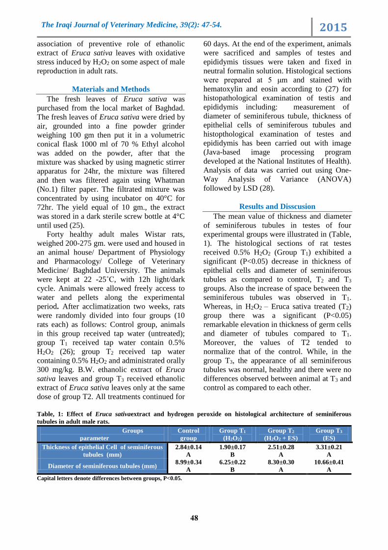
Results and Disscusion
The mean value of thickness and diameter
of seminiferous tubules in testes of four
experimental groups were illustrated in (Table,
1). The histological sections of rat testes
received 0.5% H2O2 (Group T1) exhibited a
significant (P<0.05) decrease in thickness of
epithelial cells and diameter of seminiferous
tubules as compared to control, T2 and T3
groups. Also the increase of space between the
seminiferous tubules was observed in T1.
Whereas, in H2O2 – Eruca sativa treated (T2)
group there was a significant (P<0.05)
remarkable elevation in thickness of germ cells
and diameter of tubules compared to T1.
Moreover, the values of T2 tended to
normalize that of the control. While, in the
group T3, the appearance of all seminiferous
tubules was normal, healthy and there were no
differences observed between animal at T3 and
control as compared to each other.
Table, 1: Effect of Eruca sativaextract and hydrogen peroxide on histological architecture of seminiferous
tubules in adult male rats.
3Group T
(ES) 2Group T
+ ES) 2O2(H 1Group T
)2O2(H
Control
group
Groups
parameter
3.31±0.21
A
2.51±0.28
A
1.90±0.17
B
2.84±0.14
A
Thickness of epithelial Cell of seminiferous
tubules (mm)
10.66±0.41
A
8.30±0.30
A
6.25±0.22
B
8.99±0.34
A Diameter of seminiferous tubules (mm)
Capital letters denote differences between groups, P<0.05.

The Iraqi Journal of Veterinary Medicine, 39(2): 47-54. 2015
74
Histological sections obtained from rat
testis exposed to 0.5% hydrogen peroxide
(group T1) at the end of the experiment
revealed decrease in the thickness of basement
membrane of seminiferous tubules (Table, 1
and Fig. 1), incomplete spermatogenesis, cell
debris, vaculation of Sertoli cells and sperms
in the lumen of seminiferous tubules (Fig. 2)
comparing to control group (Fig. 3), while
testes of rats treated with 0.5% H2O2 plus
ethanolic extract of (300 mg/kg B.W) E. sativa
(T2) showed complete spermatogenesis with
presence of large number of sperms in the
lumen of seminiferous tubule and active
Leydig cells (Fig. 4). Normal obvious
histological architecture of seminiferous
tubules and complete spermatogenesis were
shown in sections of testis of group T3 (Fig, 5),
as well as, the absence of main differences
between seminiferous tubules morphology and
thickness of basement membrane in
comparison to control (Table, 1 and Fig. 1).
Rats of group T1 showed few sperms in the
lumen of epididymis with round dark nuclei
cells (Fig. 6) as compared to control group
(Fig. 7). Whereas, the epididymis of group T2
showed normal structure and the lumen filled
with sperms (Fig. 8 and 9) as compared with
control group (Fig. 7). Meanwhile, the
histopathological sections of epididymis of
groupT3 showing normal structure and the
lumen compact with sperms (Fig.10)
comparing to group T1 (Fig. 6).
Figure, 1: Cross section of the rat testes group T1 showing
decrease in thickness of basement of seminiferous tubules
with in complete spermatogenesis (H and E stain 40 x).
Exposure of rats to hydrogen peroxide
(group T1) results in the appearance of
thickness of basement membrane of
seminiferous tubules (Table, 1) with
incomplete spermatogenesis and cellular
debris (Fig. 2 and 4) as well as few sperms in
the lumen of epididymis with round nuclei
cells (Fig. 8). It has been proven that H2O2
induces oxidative stress in animal models, by
forming potent ROS and nitrogen–oxygen
species (29 and 30).
Figure, 2: Cross section of the rat testes of group T1
showing incomplete spermatogenesis with cellular debris
in the lumen of seminiferous tubules (H and E stain 40 x).
Figure, 3: Cross section of the rat testes control group
showing normal structure of seminiferous tubule (H and E
stain 40x).
Figure, 4: Cross section of the rat testes of group T2
showing complete spermatogenesis with presence large
number of sperm in the lumen of seminiferous tubules (H
and E stain 40x).

The Iraqi Journal of Veterinary Medicine, 39(2): 47-54. 2015
05
Figure, 5: Cross section of the rat testes of group T3
showing complete spermatogenesis (H and E stain 40x).
Figure, 6: Cross section of rat epididymis of group T1
showing few sperms in the lumen with round dark nuclei
cells (H and E stain 40x).
Figure, 7: Cross section of the rat epididymis of control
group showing normal structure (H and E stain 10x).
Figure, 8: Cross section of rat epididymis of group T2
showing the lumen was overfilled with sperms (H and E
stain 10x).
Figure, 9: Cross section of rat epididymis of group T2
showing the lumen was overfilled with sperms (H and E
stain 40x).
Figure, 10: Cross section of rat epididymis of group T3
showing the lumen filled with sperms (H and E stain 40x).
Oxidative stress augmented production of
ROS overwhelms the body’s antioxidant
defenses (31-33). Therefore, the increase in
thickness of basal membrane in seminiferous
tubules which accompanied with incomplete
spermatogenesis may be due to an increase in
production of ROS that damage normal
spermatozoa by inducing LPO and DNA
damage (6 and 34). Several investigators
showed that severe oxidative stress would
causes damage to DNA, proteins, and
enzymes, including lipid peroxidation
enzymes. Such degeneration leads to cell death
(35-37) thereby demonstrating the pathological
role of ROS on the male reproductive system
(38 and 39). Research suggested that the
oxidation of the proteins leads to the loss of
function or to the degradation in the
peroxisomes, whereas the lipid peroxidation
affects the biological function of the
membrane. However, the most serious damage
is detected at DNA level, as this may lead to
mutation of genes, inducing translation of
defective proteins, in addition to alteration in

The Iraqi Journal of Veterinary Medicine, 39(2): 47-54. 2015
05
gene expression and eruption of apoptosis
(40).
Whereas, other studies (37 and 41) explained
that the most of the deleterious effects of H2O2
on tissues, including lipid peroxidation,
depend on the conversion of the compound
into OH•, which is catalyzed by iron and
copper through the Fenton reaction in cells.
Furthermore, (42 and 43) explained that
testicular oxidative stress, will lead to an
increase in germ cell apoptosis and subsequent
hypospermatogenesis. Such stress conditions
can cause changes in the dynamics of
testicular microvascular blood flow, endocrine
signaling, and germ cell apoptosis. Many
investigators have demonstrated that testicular
testosterone production is acutely reduced in a
number of conditions associated with ROS
production and oxidative stress in the testis
(42, 44 and 45). It is also true that
steroidogenesis itself produces ROS, largely
from mitochondrial respiration and the
catalytic reactions of the steroidogenic
cytochrome P450 enzymes (46-48). The ROS
produced by spermatogenesis, if unchecked by
intracellular antioxidants, can also damage
mitochondrial membranes and contribute to
the inhibition of subsequent steroid production
(49).
From the data obtained in the current study,
oral administration of Eruca sativa leaves
extract for 60 days to male rats in groups T2
show an ameliorating the histopathological
changes in testis and epididymis which is
created by H2O2. Meanwhile, testis and
epididymis of rats in group T3 orally
administrated Eruca sativa extract show the
same histological architecture as in the control
group which was compatible with (50) who
explained that low dose of Eruca sativa seed
oil caused an increase the diameter of
seminiferous tubules, may be due to the high
rate of proliferation of haploid cell resulting in
stimulation of spermatogenesis and increase
sperm count. It has been proven that Eruca
sativa leaves extract may be capable in
improving healing sperm parameter and
fertility with increasing the diameter of
seminiferous tubules (51). As explained
earlier, the presence of bio-active
isthiocyanates (ITCs) in ES extract which
results from glucosinolate upon myrosinase
hydrolysis (15) have antibacterial properties
and induce metabolizing enzymes such as
glutathione-s-transferase, NADPH, which play
an important role in detoxification and
protection against oxidative stress (16 and 52).
The results of the current study ascertained
that ethanolic extract of E. sativa leaves
alleviated the harmful effect of oxidative stress
induced by H2O2. Thus, it can be suggested
that the component of E. sativa leaves extract
have the potential to improve male
reproductive functions attributed to its
antioxidant and androgenic properties.
References
1. Dalle-Donne, I.; Scaloni, Giustarini, A.;
Cavarra, D.; Tell, E. and Lungarella, G.
(2005). Proteins as biomarkers of oxidative
stress in diseases: the contribution of redox
proteomics, Mass Spectrom Rev., 24: 55-99,
2005.
2. Henkel, R. R. (2011). Leukocytes and
oxidative stress: dilemma for sperm function
and male fertility. Asian J. Androl., 13: 43-52.
3. Trussell, J. C. (2013). Optimal diagnosis and
medical treatment of male infertility. Semen
Reprod. Med., 31: 235-6.
4. Grriveau, J. F. and Le Lannou, D. (1997):
Reactive oxygen species and human
spermatozoa: physiology and pathology. Int.,
J. Androl., 20: 61-69.
5. Agarwal, A. and Said, T. M. (2003). Role of
sperm chromatin abnormalities and DNA
damage in male infertility. Hum. Reprod.
Update. 9: 331-45.
6. Alvarez, J. G.; Sharma, R. K.; Ollero, M.;
Saleh, R. A. and Lopez, M. C. (2002).
Increased DNA damage in sperm from
leukocytospermic semen samples as
determined by the sperm chromatin structure
assay. Fertil Steril., 78: 319-329.
7. Saleh, R. A.; Agarwal, A.; Kandirali, E.;
Sharma, R. K. and Thomas, A. J. (2002).
leukocytospermic is associated with increased
reactive oxygen species production by human
spermatozoa. Fertil. Steril., 78:1215-1224.
8. Pasqualotto, F. F.; Sharma, R. K.; Nelson, D.
R.; Thomas, A. J. and Agarwal, A. (2000).
Relationship between oxidative stress, semen
characteristic, and clinical diagnosis in men
undergoing infertility investigation. Fertil.
Steril., 73: 459-464.

The Iraqi Journal of Veterinary Medicine, 39(2): 47-54. 2015
05
9. Venkatesh, S.; Gupta, N. P.; Kumar, R.;
Deecaraman, M. and Dada, R. (2009).
Correlation of sperm morphology and
oxidative stress in infertile men. Iran J.
Repord. Med., 7: 29-34.
10. Hamada, A.; Esteves, S. C. and Agarwal, A.
(2011). Unexplained male infertility: potential
causes and management. Hum. Androl., 1: 2-
16.
11. Padulosi, S. and Pignone, D. (1997). Rocket :
Mediterranean crop for the world .
International plant genetic resources institute,
Rome, Italy.
12. Font, R.; Galan, S.; Ruiz, P.; Villatoro, P. and
Delrio, C. (2003). Characterization of the
sensorial, morphological and agronomic
attributes of a world collection of rocket
Brassica, 5th international symposium on
brassica and the 16th crucifer genetic work
shop.
13. Alam, M.; kaur, G.; Jabbar, Z.; Javed, K. and
Athar, M. (2006). Eruca sativa seeds possess
antioxidant activity and exert a protective
effect on chloride mercuric induced renal
toxicity Food Chem. Toxicol., 29 : 172.
14. Al-Qurainy, F.; Alameri, A. A. and Khan, S.
(2010). Rapid profile for the assessment of
genotoxicity on a medicinal plant Eruca
sativa. J. Med. Plants. Res., 4(7): 579-586.
920.
15. Al-Qasoumi, S.; Al-Sohaibani, M.; Al-
Howiriny, T.; Al-Yahya, M. and Rafatullah,
S. (2009). Rocket "Eruca sativa": a salad herb
with potential gastric anti-ulcer activity.
World J. Gastroenterol., 15(16): 1958-1965.
16. Khoobchandani, M.; Ojeswi, B. K.; Ganesh,
N.; Srivastava, M. M. and Gabbanini, S.
(2010). Antimicrobial properties and
analytical profile of traditional Eruca sativa
seed oil: Comparison with various aerial and
root plant extracts. Food Chem., 120: 217-
224.
17. Bukhashi, E.; Maliki, S. A. and Ahmed, S. S.
(2007). Estimation of nutritional value and
trace elements content of Carthamus
oxycantha, Eruca sativa and Plantago ovanta.
Pak. J. Bot., 30(4): 1181-1187.
18. Kazemi, M.; Tahmasbi, A. M.; Naserian, A.
A.; Valizadeh, R. and Moheghi, A. (2012).
Potential nutritive value of some forage
species as ruminants feed in Iran. African J.
Biotech., 11(57): 12110-12117.
19. Kim, S. J.; Jin, S. and Ishii, G. (2004).
Isolation and structural elucidation of 4-
(beta-D-glucopyranosyldisulfanyl) butyl
glucosinolate from leaves of rocket salad
(Eruca sativa L.) and its antioxidative
activity. Biosci. Biotechnol. Biochem., 68:
2444-2450.
20. Yehuda, H.; Khatib, S.; Sussan, I.; Musa, R.;
Vaya, J. and Tamir, S. (2009). Potential skin
anti-inflammatory effects of 4- methyl-
thiobutylisothiocyanate (MTBI) isolated from
rocket (Eruca sativa) seeds. Biofactors,
35(3): 295-305.
21. Michael, H. N.; Shafik, R. E. and Rasmy, G.
E. (2011). Studies on the chemical
constituents of fresh leaf of Eruca sativa
extract and its biological activity as anticancer
agent in vitro. J. Medicinal Plants Res.,
5(7):1184-1191.
22. Martinez-Sanchez, A.; Liorach, R.; Gil, M. I.
and Ferreres, F. (2007). Identification of new
flavonoid glycosides and flavonoid profiles to
characterize rocket leafy salads. J. Agric.
Food Chem., 55(4): 1356-1363.
23. Barillari, J.; Canistro, D.; Paolini, M.;
Ferroni, F. and Pedulli, G. F. et al., (2005).
Direct antioxidant activity of purified
glucoerucin, the dietary secondary metabolite
contained in rocket (Eruca sativa Mill.) seeds
and sprouts. J. Agric. Food Chem., 53: 2475-
2482.
24. Homady, M. H.; Hussain, H. H.; Tarawaneh,
K. A. and Shakhanbeh, J. M. (2000). Effects
of medicinal plant extracts used in Jordan on
social aggression as well as testicular and
preputial gland structure in male mice. J. Bio.
Sci., 3(3): 389-402.
25. Jin, J.; Koroleva, O. A.; Gibson, T.;
Swanston, J. and Magan, J. et al., (2009).
Analysis of phytochemical composition and
chemoprotective capacity of rocket (Eruca
sativa and Diplotaxis tenuifolia) leafy salad
following cultivation in different
environments. J. Agric. Food Chem., 57(12):
5227-5234.
26. Khudier, K. K. (2000). The role of aqueous
extraction of Olive leaves (Olea europaea L.)
and Garlic (Allium sativum L.) in
ameliorating the effect of experimentally
induced atherosclerosis in rats. Ph.D. Thesis/
College of Veterinary Medicine/ University
of Baghdad.

The Iraqi Journal of Veterinary Medicine, 39(2): 47-54. 2015
05
27. Luna, L. G. (1968). Manual of Histology
Staining. Methods of Armed Forces. Institute
of Pathology. 3rd edition. McGraw-Hill Book
Company, New York and London.
28. Snedecor, G.W. and Cochran, W. G. (1973).
Statistical Methods. 6th ed. the Iowa state
University press., Pp:238-248.
29. Imlay, J. A.; Chin, S. M. and Linn, S. (1988).
Toxic DNA damage by hydrogen peroxide
through the Fenton reaction invivo and
invitro. Sci., 240: 640–642.
30. Sisein, E. A.; Ayakeme, T.; Ebiagbe Ebiere, J.
and Adegi, A. A. (2013). Melatonin and
vitamin E reduces the effect of H2O2 induced
oxidative stress in male albino rats in vitro. J.
Biol. Food Sci. Res., 2(8): 92-96.
31. Cardenas-Rodriguez, N.; Huerta-Gertrudis,
B.; Rivera-Espinosa, L.; Montesinos-Correa,
H. and Bandala, C. et al., (2013). Role of
oxidative stress in refractory epilepsy:
Evidence in patients and experimental
models. Int. J. Mol. Sci., 14: 1455-1476.
32. Ruttkay-Nedecky, B.; Nejdl, L.; Gumulec, J.;
Zitka, O. and Masarik, M. et al., (2013). The
role of metallothionein in oxidative stress. Int.
J. Mol. Sci., 14: 6044-6066.
33. Agarwal, A.; Virk, G.; Ong, C.; Stefan, S. and
du Plessis (2014). Effect of oxidative stress
on male reproduction .World J. Mens Health.,
32(1): 1-17.
34. Aitken, R. J.; Gordon, E.; Harkiss, D.; Twigg,
J. P. and Milne, P. (1998).Relative impact of
oxidative stress on the functional competence
and genomic integrity of human spermatozoa.
Biol. Reprod., 59:1037-1046.
35. Ward, J. F.; Blakely, W. and Joner, E. I.
(1985). Mammalian cells are not killed by
DNA single-strand breaks caused by hydroxyl
radicals from hydrogen peroxide. Radiation
Res., 103: 383–392.
36. Sharma, R. K. and Agarwal, A. (1996). Role
of reactive oxygen species in male infertility.
Urology, 48: 835–85.
37. Ganie, S. A.; Haq, E.; Hamid, A.; Masood, A.
and Zargar, M. A. (2011). Long dose
exposure of hydrogen peroxide (H2O2) in
albino rats and effect of Podophyllum
hexandrum on oxidative stress. Eur. Rev.
Med. Pharmacol. Sci., 15: 906–915.
38. Agarwal, A.; Saleh, R. A. and Bedaiwy, M.
R. (2003). Role of reactive oxygen species in
the pathophysilogy of human reproduction.
Fertil. Steril., 79: 829-843.
39. Agarwal, A.; Gupta, S. and Sikka, S. (2006).
The role of free radicals and antioxidant in
reproduction. Curr Opin Obstet Gynecol,
18(3): 325-32.
40. Sorg, O. (2004). Oxidative stress: A
theoretical model or a biological reality?
Biol., 327: 649–662.
41. Park, K. J.; Kim, Y. J.; Kim, J.; Kim, S. M.
and Bae, J.W. et al., (2012). Protective effects
of peroxiredoxin on hydrogen peroxide
induced oxidative stress and apoptosis in
Cardiomyocytes. Korean Circ. J., 42: 23–32.
42. Turner, T. T. and Lysiak, J. J. (2008).
Oxidative stress: a common factor in
testicular dysfunction. J. Androl., 29: 488–
498.
43. Rahim, S. M.; Taha, E. M.; Mubark, Z. M.;
Aziz, S. S. and Simon, K. D. et al., (2013).
Protective effect of Cymbopogon citratus on
hydrogen peroxide-induced oxidative stress in
the reproductive system of male rats. Syst.
Biol. Reprod. Med., 59(6): 329-336.
44. Ismail, H. and Al-Nahari, H. (2009).
Therapeutic and protective role of Panax
ginseng on pituitary and testicular axis in
male rats treated with carbon tetrachloride.
Ale. J. Agric. Res., 54(1):1-12.
45. Tijani, A. S.; Ukwenya, V. O.; Sodunke, G.
A. and Fakunle, J. B. (2010). Acute
administration of co-artesiane® induces
oxidative stress in the testes of adult male
Wistar rats. Biosci. Res. Commun., 22(5):
259-265.
46. Peltola, V.; Huhtaniemi, I.; Metsa-Ketela and
Ahotupa, M. (1996). Induction of lipid
peroxidation during steroidogenesis. Endoc.,
137: 105– 112.
47. Hales, B. D. (2002). Testicular macrophage
modulation of Leydig cell steroidogenesis. J.
Reprod. Immunol., 57:3–18.
48. Hanukoglu, I. (2006). Antioxidant protective
mechanisms against reactive oxygen species
(ROS) generated by mitochondrial P450
systems in steroidogenic cells. Drug. Meta.
Rev., 38: 171–196.
49. Luo, L.; Chen, H.; Trush, M. A.; Show, M. D.
and Anway, M. D. et al., (2006). Aging and
the Brown Norway rat Leydig cell antioxidant
defense system. J. Androl., 27: 240–247.

The Iraqi Journal of Veterinary Medicine, 39(2): 47-54. 2015
07
50. Salem, M. A. R. and Moustafa, N. A. (2001).
Histological and quantitative study of the
effect of Eruca sativa seed oil on the testis of
albino Rat. Egyptian J. Hosp. Med., 2: 148-
162.
51. Hussein, Z. F. (2013). Study the effect of
Eruca sativa leaves extract on male fertility in
albino mice, J. Al-Nahrain Uni., 16(1): 143-
146.
52. Fahey, J. W. and Talalay, P. (1999).
Antioxidant function of sulphoraphane: a
potent inducer of phase II detoxication
enzymes. Food Chem. Toxicol., 37: 973-979.
الجرذان المستحدث فيها الكرب وراق نبات الجرجير في الجهاز التكاثري لذكور دور المستخلص الكحولي لأ
كسدي تجريبياً أالت أحمد جاسم نوفل و بــراء نجـم العقيلي
.العراق، جامعة بغداد، كلية الطب البيطري ،فرع الفسلجه والأدويه
E-Mail: [email protected]
الخلاصة
حد من الأذى المستحدث ببيروكسيد للتخلص الكحولي لأوراق الجرجير التحري عن الدور الوقائي للمسإلى الدراسة هدفت
أوزانها يتراوح معدل التي من ذكور الجرذان البالغة 04ل ستعمذكور الجرذان البالغة. الجين في كفاءة الجهاز التكاثري الهيدرو
،عدت كمجموعة سيطرةأعطيت ماء الحنفية ووالتي إلى أربعة مجاميع متساوية: المجموعة الأولى غم وقسمت عشوائيا 044-072
فقد أعطيت (2Tأما المجموعة الثالثة ) ،بيروكسيد الهيدروجين %4.2أعطيت ماء الحنفية الحاوي على T)1المجموعة الثانية )
خلص الكحولي كغم من وزن الجسم من المست م/ملغ044مع تجريعها بيروكسيد الهيدروجين %4.2 ماء الحنفية الحاوي على
كغم من وزن الجسم من المستخلص الكحولي لأوراق نبات م/ملغ044جرعت (3Tالمجموعة الرابعة )، لأوراق نبات الجرجير
دراسة لالبربخ أخذت عينات من الخصية وبالحيوانات وحي ض في نهاية التجربة و يوما 04 المجاميع لمدة جميعالجرجير. وعوملت
القياسات النسجية لمقاطع أوضحت نتائج عن قياس الخليا المبطنة للنبيبات مع أقطار النبيبات المنوية. جية فضلا التغيرات النس
منوية أنخفاضا معنويا في معدل أقطار النبيبات ال T)1(البربخ لذكور الجرذان للمجموعة المعاملة ببيروكسيد الهيدروجين الخصى و
لكن هذه القياسات أظهرت أرتفاعا معنوياا في سمك الخليا الطلئية ومعدل اقطار النبيبات المنوية في ،وسمك الخليا الطلئية لها
بمجموعة السيطرة. كما مقارنة 3Tوجود فرق بين مجموعة لم يلحظ. في حين 1Tبالمقارنة مع مجموعة 3T و 2T مجموعتي
( إلى قله عملية تصنيع 1T) البربخ لذكورالجرذان المعاملة ببيروكسيد الهيدروجينالتغيرات النسجية لمقاطع الخصى وأشارت
تجاويف النبيبات وجود حطام الخليا ووجود عدد قليل من النطف في و ،جوات واسعة بين النبيبات المنويةالنطف مع وجود ف
عملية تصنيع النطف وأمتلء التجاويف بالنطف مقارنة مع لأرتفاع معنوي 3T و 2T في حين أظهرت المجموعتين ،والبربخالمنوية
إن بيروكسيد الهيدروجين يضعف عملية تخليق النطف فضل عن ذلك، أكدت النتائج الدور الوقائي ،ستنتج الدراسهت. 1Tالمجموعة
ذكور الجرذان.لالأجهاد التأكسدي الجرجير ضد للمستخلص الأثيلي لأوراق
النبيبات المنويه. الخصى، البربخ، : بيروكسيد الهيدروجين، أوراق الجرجير،مفتاحيةالكلمات ال