Embed Size (px)
Citation preview

I
THE ISOLATION AND CHARACTERIZATION OF A GROWTH FACTORIN RUMEN FLUID FOR A STRAIN OF BUTYRIVIBRIO
by
Gale Ross Gqrdon
Thesis submitted to the Graduate Faculty of the
Virginia Polytechnic Institute
in canidacy for the degree of
MASTER OF SCIENCE
in
Biochemistry
May, 1961 IBlacksburg, Virginia I
II

-2..
TABLE OF CONTENTS
Page
INTRODUCTION AND REVIEW OF LITERATURE ............ 5
MATERIALS AND METHODS . . . . . .......... . .... 11• • • • • • • • • • • • • • • • • • • •
• • • • • • • • • • • • • • • • • • •
• • • • • • • • • • • • • • • • • • •
•Dialyßis• • • • • • • • • • • • • • • • • • • •
•Bj-Üassay• • • • • • • • • • • • • • • • • • • •
•Liquid•LiquidExtraction (G,H) . ........ . . . . 26Amcnium Sulfete Precipitation (1,3) .......... 26Ion Exchange Chromatography (K,L,M,N) ......... 27Acid and Base Hydrolysis (0,P) ............. 27
(Q, • • • • • • • • • • • • •
•EnzymaticHydrolysis (S) ................ 28Studies on Resin Eluates ................ 29
• • • • • • • • • • • • • • • • • • • • • • • • • • •
• • • • • • • • • • • • • • • • • • • • • • • • •
• • • • • • • • • • • • • • • • • • • • • • • • • •
•ACKNOWLEDGEMENTS...................... 41
• • • • • • • • • • • • • • • • • • • • • • • • •
• • • • • • • • • • • • • • • • • • • • • • • • • • • •
. g ·3·LIST OF FIGURES
Page
1. Schematie Diagram of Fractionation of Rumen Fluid .... 16
2. Growth Curves of a Strain of Butzrivibrio withIncreasing Levels of Ruen Fluid ............ 18
3. Response of a Strain of Butgrivibrio to VariousLevels of Rumen Fluid When Measured at the 22nd Hour . . 21
lI

-4-
LIST OF TABLES
Page
1. Composition of the Basal Medium ............. 12
2. Fractionation of Growth Stimulatiug Materialin Rumeu Fluid . . . . . . .... . . . . . ...... 24
3. Ninhydriu Positive Spots of Growth StimulatiugFractions ....................... 34
4. Carbohydrate amd Reduciug Substances of IRCG·40OI I I I I I I I I I I I I I I I I I I II5.
Compounds Screened for Stimulatory Activity ....... 36
l _
I-5-
INTRODUCTION AND REVIEW OF LITERATURE
The development ef isolation and cultivation procedures which have
allowed characterization of the ruminal flora, has led to investigations
of the nutritional requirements of several ruminal bacteria. Among these
procedures, those of Hugate (23,24), Bryant (6), Bryant and Burkey (9,10)
and Doetsch g£_g1. (17) were found by King and Smith (25) to be more
valuable than the procedures of Gall and Huhtanen (21) for estimation of
both total bacterial counts and counts of the cellulolytic flora. The
Hungate—type medium consisted essentially of inorganic sa1ts·sugar—rumen
fluid incubated in roll tubes as contrasted with the Ga1l·type medium of
a highly organic mixture cultured in Brewer anaerobic plates. The medium
contained no rumen
fluid.Sijpesteijn(29), Bryant and Smll (13,14), and Hungate (24),
described several new genera which have been observed to be indigenous
to the rumen. These included Ruminococcus {lavefaciens (29), Succinivibrio
dextrinosolvens (14), Lachnogpira multiparus (14), Bacteroides succinogenes
(24), and Butyrivibrio fibrisolvens (13).
Descriptions of the physiological and nutritlonal characteristics
of the rumen microflora have appeared only recently in the literature
and relatively little lnformaticn is available. In practically all werk
involving the culture of rumen bacteria, rumen fluid has been incorporated
in the basel medium to stimulate growth ad has been found to be essential
for growth in many cases (1,8,11,12).
Medium containing ruen fluid is chemically undefined since rumen
liquor is a heterogenous mixture of simple and complex biological compounds
-5.l
I
derived from the animal, its diet and the microbial population of the
rumen. In attempts to provide chemlcally defined media which will
satisfy the growth requirements of fastidious organisms, lt has long been
recognized that such media could not be autoclaved and remain unchanged.
Carbohydrates are often hydrolyzed and for example, Wblin and Weinberg (33)
found that a substance produced as a result of autoclaving glucose with
phosphate was required for growth of the rumen bacterium Streptococcus
gggig when inorganic nitrogen was supplied as the sole nitrogen source.
The product of autoclaving was not required when glutamine was used as the
sole nitrogen source in the culture medium. Acetaldehyde, pyruvate, or
acetoln could be used to replace the substance produced by autoclaving
gluose with phosphate.
The requirement for ruen fluid in certain pure cultures of rumen
bacteria such as Bacteroides succinogenes and some members of the genus
Borrelia, has been noted by Hungate (24) and by Bryant (7). Hungate (24)
showed that materials quite similar to the natural food of ruminants,
such as dried grass and malt Sprßutß, failed to suport growth of the
cellulolytic forms. Hungate inferred that it was highly probable that
in the normal rumen the factors required for growth of the cellulolytlc
forms are in part produced by the flora indigenous to the rumen.
Ruf g£_gl. (28) tested several of the more common feeds for cattle
and sheep including soybean oil eal, linseed oil meal, and wheat bran
using an artiflcial rumen technique to determine the presence or absence
of an unidentified factor stimlatory to rumen microorganisms in the
digestion of cellulose. Cow manure extract and yeast were found to be
I. _M g_____________________..._................................................---------¤¤¤-a
.7-
active in stimulating the digestion of cellulose. Fractionation of
these materials indicated the factor to be heat stable, and soluble in
water and low concentrations of ethanol. It was absorbed on Norite and
could be eluted with acetone and ethanol. Absorption did not occur on
Amberlite 400 resin (anion exchanger) and ashing destroyed the factor,
thus indicating that the active material was an organic substance.
Precipitation of protein with hydrochloric acid did not remove the un-
identified factor and various B·vitamin and amino acid supplements failed
to stimulate cellulose digestion. Burroughs gg gl. (15) found that the
ig xitra digestion of cellulose was increased upon the addition of theash from molasses, immature clover hay or mature timothy hay. Bentley
e£_gl. (4) using ig_!£££g_studies, found that the addition of autoclavedrumen juice, extracts of various plant materials, molasses, or yeast
extract, markedly increased the rate of cellulose digestion. An active
material, which increased the rate of cellulose hydrolysis, was concentrated
from rumen juice by lead acetate precipitation followed by extraction of
the dried filtrate residue. This material could be adsorbed on Norite A
and eluted, but the eluate was less active than the original rumen juice.
Dehority gg gl. (16) used a similar ig_gi££g technique for assaying
cellulolytic activity of rumen microorganisms to isolate a factor which
stimulated cellulolysis from autolyzed yeast, casein hydrolysate, and alfalfa
hay extract. The factor was isolated by Dowex-50 (cation exchanger) ion
exchange resin treatment, charcoal (Darco G·60) treatment and large scale
paper chromatography. Further fractionation indicated that the factors
which stimulated cellulolytic activity were valine, proline, leucine,
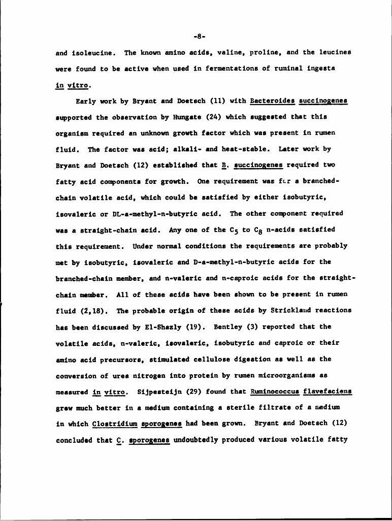
-3-and isoleucine. The known amino acids, valine, proline, and the leucines
were found to be active when used in fermentations of ruminal ingesta
Lexma-Early work by Bryant and Doetsch (11) with Bacteroides succinogenes
supported the observation by Hungate (24) which suggested that this
organism required an unknown growth factor which was present in ruen
fluid. The factor was acid; a1ka1i• and heat-stable. Later work by
Bryant and Doetsch (12) established that Q, succinogenes required two
fatty acid components for growth. One requirement was far a branched-
chain volatile acid, which could be satisfied by either isobutyric,
isovaleric or DL·a•methy1-n·butyric acid. The other component required
was a straight—chain acid. Any one of the C5 to C8 n-acids satisfied
this requirement. Under normal conditions the requirements are probably
met by isobutyric, isovaleric and D-a·methyl·n~butyric acids for the
hranched-chain member, and n·valeric and n-caproic acids for the straight-
chain member. All of these acids have been shown to be present in rumen
fluid (2,18). The probable origin of these acids by Strickland reaetions
has been discusaed by El-Shazly (19). Bentley (3) reported that the
volatile acids, n·va1eric, isovaleric, isobutyric and caproic or their
amino acid precursors, stimulated cellulose digestion as well as the
conversion of urea nitrogen into protein by rumen microorganisms as
measured Sijpesteijn (29) found that Ruminococcus flavefaciens
grew much better in a medium containing a sterile filtrate of a medium
in which Clostridiu gporogenes had been grown. Bryant and Doetsch (12)
concluded that Q, gporogenes undoubtedly produced various volatile fatty

-9-4
aclds from amino acids in the medium and these may have been responsible
for the improved growth of g, flavefaciens. Investigations by Allison
g£_gl. (1) with a strain of_§. flavefaciens revealed the presence of asteam-distillable factor in rumen fluid which stimulated growth. This
factor seems to be distinct from that required by the_g. glbgg strain 69of Fletcher (20). Strain 69 of Fletcher required two yellow acidic fractions
for maximal growth which were isolated from rumen fluid by chromatography
on celite columns with chloroform and n-butanol in chloroform. The yellow
substancea were not extraeted from chloroform by water, but were soluble
in dilute sodium hydroxide. Tha growth factor was soluble in diethyl ether
from concentrates extracted at pH 4, but was not extracted from concentrates
at pH 8. It was non-volatile with steam, but was dialyzable, orgenic, and
extremely stable to heat, acid and alkali.
Bryan: and Small (13) established the genus Butygivibrio and designated
the type species Butyrlwibrio fibrisolvens. The genus includes anaerobic,
non—sporeforming, monotrichous, grampnegative, curved rods that ferment
glucose with the production of butyric acid. Members of the genus
Butgrivibrio have been found among the most numerous orgsnisms cultured
from rmen contents of animals maintained on roughage rations (22,26).
They are of considerable importance in the breakdown of fibrous constituents
of cattle feed es shown by the ability of some strains to digest cellulose,
and most strains to digest xylan. Also, the genus Butyrivibrio probably
contrlbutes to the breakdown of starch and protein in the rumen.
Gill an King (22) described the B-vitamin, amino acid, CO2, and
purine and pyrimidine requirements of a strain of Butyrlvibrio isolated
.1g- 1
from bovine rumen ingesta. They observed, however, a pronounced stimula-
tion of growth when various preparations of rumen ingesta were added tc
the best chemically defined medium available. None of 68 compounds
which are comnly stimulatory to microbial growth could replace the
active principal in rumen fluid. The activity was non~dialyzable, some·
what stable to acid, and retained on anion exchange resins.
The aim of the present research was an attempt to isolate the
components from rumen ingesta which are stimulatcry for the growth of
Butyrivibrio.
11
1

1 ·ll-
1 MATERIALS AND METHODSCultural Methods. The organism used in the microbial assays of the
various fractions of rumen fluid conformed to the criteria established
for the genus Butyrivibrio as described by Bryant and Small (13). It
was an anaerobic, monotrichous, gram·negative, curved rod about 0.7 by
2.1 microns in size, that produced predominant quantities of butyric acid
from the fermentation of glucose. The method for flagella staining was
that of Leifson as described by Skerman (30). Fatty acids were determined
by the method described by Strong (31) and also by the method described
by Bruno (5). The production of butyric acid as a fermentatlon product
from glucose by Butyrivibrio may possibly vary since Gill and King (22)
found that uon decreasing the percentage of rumen fluid in the basal
medium, the production of acid shifted from butyric to succinic and lactic.
Lee and Moore (26) reported the shift of fermentation products with changes
in pH and concentration of products.
The bacterium was isolatedl from bovine rumen ingesta and was
initially cultured in a salts·glucose—skimmed milk medium incubated in
roll tubes. It was later cultured in the medium of Gill and King (22)
with the following modification: the equivalent of five grams of vitamin·
free hydrolyzed casein was replaced with chromatographically pure L•amino
acids in the same proportion as they occur in casein (32). The composition
of the resulting medium is shown in Table 1. The amino acids were weighed
out in l0X the amunts required for one liter and shaken vigorously in a
Robert S. Fulghum anddesignated culture No. 70. He also confirmed the cultural characteristics.
1

11-12-X Table 1 .
V
Composition of the Basel Medium.
Component Amount per Liter
1. KHZPOA 4.24 3.
2. K2H04 3.26 3.
3. NaHC03 2.50 3.4 . M3S0(* O . 02 3 .
5. CaC12 0.02 3.6. MnC12 ' 4H20 0.02 3.7. FeS04 ' 7H2O 0.02 3.8. CoC12 ° 6H20 0.02 3.9. (NH4)6Mo7024 ° 4H20 0.0023.
10. ZnC12 , 0.0023.
1 1 . CUSOA ° BHZÜ Ü . ÜÜZQ •
12. Glucose 15.00 3.
13. (NH4)gS04 2.00 3.
14. Amino acid mixture 5.59 3.
15. Pyridoxal hydrochloride 0.02 mg.
16. Folie acid 0.05 mg. b17. Biotin 0.01 mg.
i18. Resazurine 1.00 mg.
19. Cysteine 0.50 3. 1
I
·l3•jarwith several clean glass marbles for 20 hours. The correct amount was ,
then taken for one liter or for any fraction of a liter. A glucose·cysteine
mixture was prepared the same way. The vitamine were weighed out in 10,000X
the amunts required for the basel concentration and thoroughly mixed with
10 g. of glucose by shaking with marbles as above.
The basel salts were made up to 100X the basal level and stored in
the following combinations since precipitation and microbial growth
occurred if stored as a mixture. With reference to Table l, salt 1 was
made u alone; salts 2 and 3 were mixed together; salt 4 was made up alone;
salts S, 6, 9, 10, and ll were mixed together and salts 7 and 8 were made up
separately.
All media were autoclaved in test tubes under carbon dioxide for ten
minutes at l2l°C. After autoclaving, the tubes were immediately placed
in cold tap water. Matched test tubes (13 x 100 mm.) were used for all
assays and the rubber stoppers were held in place during autoclaving by
a special hinged cover on a test tube rack. The cover had screws which
allowed for individual adjustment on each rubber stopper.
Growth measurements were made turbidimetrically with a Klett·Summerson
colorimeter equipped with a Number 66 red filter. Four tubes of media
were made up for each treatment assay and three of them were inoculated.
The uninoculated tube was used as a blank to set the instrument.
The standard inoculum used throughout the experimental work was from
a l2•hour culture that had been incubated inthe basel medium plus 20
percent rumen fluid. The culture was centrifuged for five minutes at
2000 RCF, the suernatant was decanted and the residual cells resuspended

V
V-14-
in three mls. of the basal medium. Frem this final volume, ü.05 mls. wasI
pipetted into the assay medium. A11 assays were incubated at 39°C.
Aseptic techniques were practiced during all transfers and the individual I
tubes were gassed by passing oxygen-free, carbon dioxide through sterile
glass nozzles filled with glass weol. The carbon dioxide was passed
through a large tube filled with cotton which was periodically autoclaved
throughout the course of the experimental work. 0xygen·free carbon dioxide
was prepared by bubbling the gas through a series of suction flasks
containing a chromcus acid·amalgamated zinc mixture. The general anaerobic
techniques and deoxygenating trap have been fully described by Hungate (24).
-15-EXPERIMENTAL PROCEDURE
The procedures for the fractionation of rumen fluid to determine
the characteristics of the constituents which stimulate the growth of
cultures of Butyrivibrio are outlined on the flow sheet of Figure l.
Rumen Fluid Preparation (A,B). The rumen fluid used in these experiments
was collected from mature, fistulated Holstein steers which were
maintained on an alfalfa hay and grain supplement diet. The crude rumen
fluid (A) was strained through two layers of cheese cloth into separatory
funnels where, upon standing at room temperature for 15 minutes, further
separation of particulate matter and protozoan cells from the rumen fluid
took place. The fluid was then autoclaved for 15 minutes at l2l°C. in
stoppered 2S0·ml. Florence flasks. The fluid was then centrifuged at
20,000 X G for 30 minutes. The decanted supernatant was then stored in
stoppered bottles at S°C. All further references to rumen fluid will
concern samples prepared in this manner (Item B, Fig. 1).
V Assgy Procedure. Increasing amounts of ruen fluid (B) were added to a
series of culture tubes. The stimulation cf growth of a strain of
Bugyrivibrio by prepared rumen fluid (B) is evident in Figure 2. Each
point in these curves represents the average of triplicate observations.
Both the length of the lag phase and the maximum amount of growth of
this strain of Butyrivibrio were changed by the rumen fluid which was
added to the basal medium. At all levels of rumen fluid except the 40
percent level, the maximal growth is approached at about the 22nd hour
of incubation. The 40 percent level shows an actual decrease in turbidity
from the maximal value which occurs around the l6·l8th hour. whether this

I
•16•
Figure 1. Schematic Diagram cf Fractionation of Emmen Fluid.

I·l7· ICrude Rumen Fluid (A)
FilteredAutoclavedCentrifuged
Prepared Human Fluid (B)
Dialxsis Ion Exchange Resins
Dialyzable Non-dialyzabls Amberlite 400 Amberlite 120<¤> <¤> I I I II H20 HC1 H20 HC1
Bioassay eluate eluate eluate eluate(E) (F) (K) (L) (M) (N)
Liguid·Liguid ExtractionHydrolysis
Aqueous AqueousI Iphase phase
from from H2SO4 Ba(OH)2ether butanol 0) (P)extraction extraction(G) (H)
Cold AeetoneDialyzed against I Isat’d. Precipitate SuernatantI”l—__—”I (Q) (R)
Non·dia1yzable Non·dialyzableprecipltate supernatsnt
(I) Trypsin HydrolysisI (S)Dialyzed againstrunning tap H20
Nou·dialyzab1esupernatant(J) I

-13. I
I
I
Figure 2. Growth Curves of a Straiu of Butxrivibrio with IncreasiugLevels of Rumen Fluid.
I

Q9 ¤ 5ää 3 5 5
..1 .J g, L': kIL LL z z ZE 5 w *·* ‘“2 2 5 ’
'3 3 Z? D Ügz m ct cz tt
3 3 3 3 :5O Q O '¢ :0 N •0 O
NNQ\ ON
• \ $K \ .—• \\
‘ SQ" \_( \I vvI
_<!
Q I •:tI Ou Q1
>( x' z .\: "OLIJ¤ —§
I •-\I‘\• wg
V;x\ gx “ __‘
\\ . (S-\= I {2)~. -\ I
\ |¢x ‘ Ixx I
\ I\\
O O OO O O O Q Q O O:2.* 32 sägg 3 «>··~
SLINII NOSUBNNDS ·.Li3'I>I N! ALICIQUILL

> ·20·
was due to clumping or autolysis of the cells is not known.
To eliminate the possibility that rumen fluid could dilute som
necessary factor in the basal medium, the basel nutrients were added
in concentrated form, the necessary amount of rumen fluid was added
and the resulting mixture was made up to 20 mls. with water. Five mls.
of the resulting mixture were then pipetted into each of four tubes.
The same procedure was used for each of the reconstituted fractions
of rumen fluid. Theoretically, the turbidimeteric reading obtained
after inoculation and incubation should be due to the inoculum and
cellular division. A sterile tube of medium was used as a blank to
cancel any differences in color between levels or treatments. To
rule out any error due to a change in color of the medium as a result
of cultural metabolism, a comparison was made between the sterile blank
and centrifuged inoculated tubes after maximal growth had been reaehed.
These comparisons failed to reveal any marked differences between the
Klett·Sumerson readings.
Figure 3 shows the curve that is obtained when the percent rumen fluid
which was added to the basel medium is plotted against the turbidimetric
reading obtained at the 22nd hour of incubation. The value for the 15
percent level was obtained from six observations, three on two separate
days. Each of the other points represents an average of nine observatlons.
Three observations for each point were determined on each of three separate
days. The standard errors of the means for the values were: 0%--6.50;
52-•4.49; 102--11.06; 15%--5.4; 201--2.39; 30Z·*5.04; 402--3.42. The 95
percent confidence range, which takes into account all variations observed
between, as well as, within days, is indicated by the vertical bars at each

11 11Q
.21,
Figure 3. Response cf a Strain ef gutyrivibrio to Various Levels ofRuen Fluid When Measured at the 22nd Hour.
1
}1

.-22.-
O¢
0F7
O10
I)N
ED..1L;
ON li
}
E2(3 •»——" 2
uJU
KLU(J.
9
10
‘ 0O O O O O O O O O O ON o <¤ •0 wr N O cn co ~1· NN N ·· ·· " " '
SLIN0 NOSUBVIVCOS ·.L.L3'l)| NI ALIGIBEHLL
1
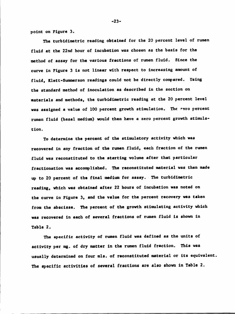
1-23-
polnt on Figure 3.I
The turbidimetric reading obtained for the 20 percent level of rumenL
fluid at the 22nd hour of incubation was chosen as the basis for the L
method of assay for the various fractions of rumen fluid. Since the L
curve in Figure 3 is not linear with respect to increasing amount of L
fluid, Klett-Sumerson readings could not be directly compared. Using L
the standard method of inoculation es described in the section on
materials and methods, the turbidimetric reading at the 20 percent level
was assigned a value of 100 percent growth stimulation. The rero percent
rumen fluid (basal medium) would then have a zero percent growth stimula·
tion.
To determine the percent of the stimulatory activity which was
recovered in any fraction of the rumen fluid, each fraction of the ruen
fluid was reconstltuted to the starting volue after that particular
fractionation was accomplished. The reconstltuted material was then made
up to 20 percent of the final mediu for assay.· The turbidimetric
reading, which was obtalned after 22 hours of incubatlon was noted on
the curve in Figure 3, and the value for the percent recovery was taken
from the absclssa. The percent of the growth stimulating activity which
was recovered in each of several fractions of rumen fluid is shown in
Table 2.
The specific activity of rumen fluid was defined as the units of
activity per mg. of dry matter in the ruen fluid fraction. This was
usually determlned on four mls. of reconstituted material or its equlvalent.
The specific activities of several fractions are also shown in Table 2. 1
1
1 1

I
Table 2. II
Fractionation of Growth Stimulating Material in Runen Fluid. I
II
Specific IPercent of Growth ActivitY
Stimuleting Activity Treatment of Mgs. dry ut. Units/mg.Recovered Runen Fluid in 4 mls. dry wt. I
1. 100 None (B) 68.2 1.46
2. 69 Non·dia1yzab1e (D) 30.9 2.22
I3. 39 Dialyzable (C) 42.7 0.91
4. 72 Recombine 2 6 3 (C,D) 61.0 1.18IAqueous phase from
5. 65 butanol extraction (H) 51.2 1.26 I
Aqueous phase from6. 80 ether axtraction (G) 57.1 1.40
H20 eluate from7. 52 IRCG·400 resin (K) 50.4 1.03
HC1 eluate from8. 36 lRCG·400 resin (L) 6.1 5.85
9. 42 Recombins 7 6 8 (K,L) 56.5 0.74IH20 eluate from I
10. 45 CG·120 resin (M) 40.4 1.11I
HC1 eluate from11. 45 CG•120 resin (N) 8.8 5.08 I
12. 53 Recombine 10 6 11 (M,N) 49.3 1.07 I
13. 35 (NHa)2S04 pp't. (I) 47.0 0.74 I
Supernatant fromI
14. 16 (NH4)280a pp':. (J) 203.0 0.07 „
15. 10 Recombine 13 6 14 (1,J) 250.0 0.03 I
I
I
I
I

-25- ‘
Dialysis (GID). One volume of ruen fluid was dialyzed in a cellulose
mmbrane against six volumes of distilled water for 24 hours at5°C.Thedialysis bath was replaced by fresh water at the 12th hour. At the
end of the dialysis period, the two dialyzable fractions (C) were combined
and concentrated on a vacuum flash evaporator to the volume of the I
original rumen fluid. The non·dia1yzab1e fraction (D) increased slightly i
in volume during dialysis. This increase was taken into consideration iwhen the fraction was asaayed for activity. I
Bioassay (E,F). From rmen fluid that had been dialyzed, 1.4 mls. of the
non·dialyzable fraction (D) containing four mgs. of dry matter were
applied to whatman 3•MM paper at an origin approximetely four inches wide.
The chromatogram (E) was developed in a descending system using the same
solvents as for the study of the enzymatic hydrolysis described later for
fraction S. Upon drying, the paper was cut into one inch strips parallel
to the origin and beginning one-half inch back of the point of application.
These strips were folded and placed in 13 X 100 mm. test tubes. Five mls.
of the basel medium were added and the tubes were sterilized for 10 minutesI
at 121°C. The assay tubes, containing the chromatogram strips (E), were {inoculated with Butyrivibrio. The paper strips were remved from the test ;
tubes after incubation. A control chromatogram was run in the same manner I
without the application of rumen fluid.
Four more chromatograms (F) (18 x 22 1/2 inches) were prepared byI
applying 5 mls. of the non-dialyzable fraction (D). They were developed ¤
using a descending system of n•butanol, water and glaeial acetic acid in I
e ratio of 5:4:1. The aqueous phase was used to equilibrate the chamber I

-26- 1and the organic phase was used to develop the chromatograms. Upon
1drying, the chromatograms were cut into one inch strips as was done with
1the previous chromatograms and each strip was eluted with approximately 1
30 mls. of 10 petcent acetic acid. The acetic acid eluates from the 1
strip: of one chromatogram were pooled with the eluates from the corre-
sponding strips of one of the other chroatograms. The remaining pair
of chromatograms were treated in a similar manner. The eluates were
concentrated to dryness by bubbling warm air through them. One set (F) of
eluates was assayed qualitatively for activity and the other set was
reserved for chemical study.
Liguid-Liguid Extraction (G·H). The pH of 100 mls. of rumen fluid was
adjusted with concentrated HC1 to approximately pH 5.5 using narrow range
pH paper. Fifty mls. of the acidified fluid were extracted continuously
for 24 hours with 100 mls. of diethyl ether in a liquid-liquid extractor (G).
The remaining 50 mls. were extracted using the same method with n·hutanol (H).
At the end of the extraction, both organic phases were removed and the
aqueous phases evaporated to dryness on a hot plate. Water was added
to the aqueous phase residue, the pH was adjusted to 7 with ammonium
hydroxide and then each was made u to a final volume of 50 mls. for assay
of the stimulatory material.
Ammonium Sulfate Precipitation (I·J). One volume of rumen fluid was 1dialyzed against six volumes of aaturated ammonium sulfate at 5°C. for 1
10 hours. The non·dialyzable precipitate (I) which formed was centrifuged, 1
washed with saturated ammonium sulfete and made up to the original volume 1
with distilled water. The non·dialyzab1e supernatant was further dialyzed 1

I
-g7-against running tap water to free the solution of ammnium sulfate (J)
and then concentrated ig_ggggg to the volue of the original sample.
Ion Exchagge Chromatogrghy (K,L,g,N). Two ion exchange resins were used
to attempt fractionation of the active material. One resin was Amberlite
IRCG-400 (a strong anion exchanger), the other was Amberlite CG·l20 (a strong
cation exchanger). The resins were slurried with aproximately 100 mls.
of distilled water, poured into 1.9 cm. diameter glass coluns which had
one end loosely plugged with a small amount of glass wool. The resins
were allowed to settle by gravity to a depth of 12 cm. A circular piece
of filter paper was placed on top of the settled coluns. Both resins
were put into their respective phases by passing 250 mls. of 8N H1 through
the columns. Following the acid wash, several volumes of distilled, ion-
free water were passed through both resins until the effluent gave a
negative silver chloride test. Fifty mls. of rumen fluid were adjusted
to pH 5 with HC1 and 25 mls. were put on each resin colun in small
quantities until the samples were washed well into the columns. Each
column was then eluted with approximately 500 mls. of ion•free water
to give the first fractions (K,M), after which the same volume of 8N HC1
eluted the remaining material (L,N). The water eluates were concentrated to25 mls. on a vacuum flash evupcratut. The HC1 fractions were dialyzed
against running tap water until the fractions were free of chloride ion
as determined by the silver chloride test. Each of these fractions was P
concentrated to 25 mls. on a vacuum flash evsporator previous toassay.Acidand Base gydrolysis (0,P). Concentrated sulfuric acid and barium ,hydroxide were added toseparate samples of rumen fluid to yield an E
I
I
'
II
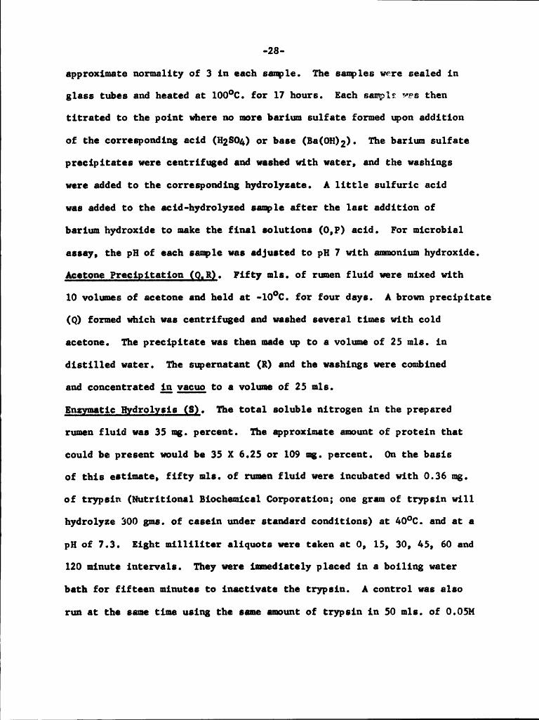
-23-approximate normality of 3 in each sample. The samples were sealed in
glass tubes and heated at 100°C. for 17 hours. Each sample was then
titrated to the point where no more bariu sulfate formed upon addition
cf the correspondiag acid (142804) er base (Ba(0u)2). The barium sulfate
pracipitates were centrifuged and washed with water, and the washings
were added to the corresponding hydrolyzate. A little sulfuric acidN
was added to the cid·hydro1yzed sample after the last addition ofN
barium hydroxide to make the final solutions (0,P) acid. For microbial
assay, the pH of each sample was adjusted to pH 7 with smonium hydroxide.
Aeetone Precipitation SQIRQ. Fifty mls. of rumen fluid were mixed with
10 volumes of acetone and held at -l0°C. for four days. A brown precipitate
(Q) formed which was centrifuged and washed several times with cold N
acetone. The precipitate was then made u to a volume of 25 mls. in
distilled water. The supernatant (R) and the washings were combined
and concentrated ;g,ggggg to a volume of 25 mls.
Eggyggtic gydrolysis (S). The total soluble nitrogen in the prepared
rumen fluid was 35 mg. percent. The aproximate amount of protein that
could be present would be 35 X 6.25 or 109 mg. percent. On the basis
of this estimate, fifty mls. of rumen fluid were incubated with 0.36 mg. N
of trypsin (Nutritional Biochemical Corporation; one gram of trypsin will
hydrolyze 300 gms. of casein under standard conditions) at ao°c. and at a
pH of 7.3. Eight milliliter aliquots were taken at 0, 15, 30, 45, 60 and
120 minute intervals. They were immediately placed in a boiling water N
bath for fifteen minutes to inactivate the trypsin. A control was also
run at the same time using the sam amount of trypsin in 50 mls. of 0.05M
1

-29.
phosphate buffer at pH 7.3. Fifty microliters of the rumen fluid·trypsin
mixture from each time interval were chromatographed on Whatman No. 1paper with methyl·ethyl ketone, isopropyl alcohol, ammonium hydroxideand water in the ratio of 3:5:1:1 in a descending system. The chromatogramswere sprayed with 0.3 percent ninhydrin in scétone. From each of theeight ml. rumen fluid·trypsin aliquots above, an additional one ml. wasdiluted to 10 mls. with distilled water. One ml. of 10 percent trichloro-acetic acid was then added to precipitate the protein. The optical
density of the supernatant was determined at a wave length of 280
millimicrons. The activity of the trypsin preparation was determined
by incubating the same amount of trypsin in 50 mls. of a one percentcasein solution which was buffered with 0.05H phosphate at pH 7.3. The
optical density was determined at fifteen minute intervals using thesame procedure as for the rumen fluid•trypsin mixture. ·
Studies on Resin Eluates. To one ml. of each eluate (K,L,M,N) from the
Amberlite resins was added one ml. of concentrated HC1 to yield a finalsolution of approximately 6N. The first four fractions from the second
set of acetic acid eluates (F) that had been reserved for chemical study
were combined and dried ig_ggggg. These four fractions corresponded tothe first four inches of two of the n·butano1 developed chromatograms.
The residue from these fractions was taken up in one ml. of water. One·half ml. of this solution was mixed with 0.5 ml. of concentrated HC1. Oneml. of rumen fluid (B) was also mixed with one ml. of concentrated HC1.The above acid solutions of the resin eluates, the chromatogram extract
and ruen fluid were hydrolyzed in sealed test tubes for 24 hours at l00°C.
11
‘ .
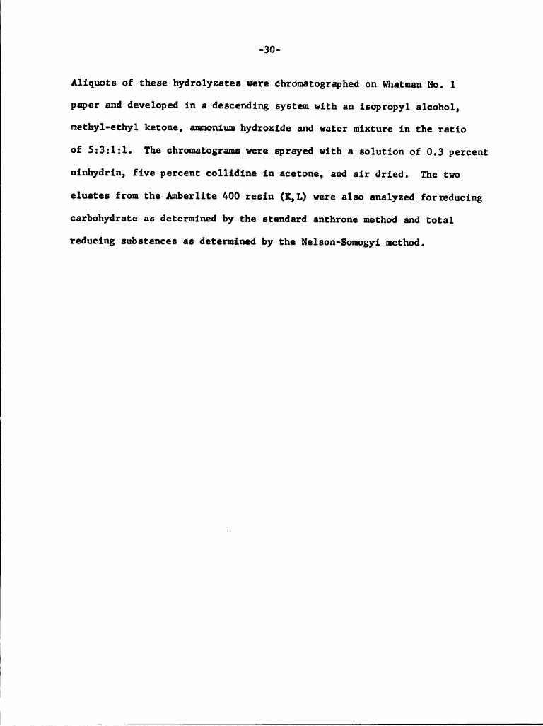
I-60- IAliquots of these hydrolyzates were ehromatographed on Whatman No. 1 I
paer and developed in a descending system with an isopropyl alcohol, I
methyl-ethyl ketone, ammonium hydroxide and water mixture in the ratioI
of 5:3:1:1. The chromatograma were sprayed with a solution of 0.3 percent 1ninhydrin, five percent collidine in acetone, and air dried. The two ,eluates from the Amberlite 400 resin (K,L) were also analyzed forreducing Icarbohydrate as determined by the standard anthrone method and total I
reducing substances as determined by the Nelson·Somogyi method.
I
III
I
I
I II .I

I
-31-RESULTS
The data presented in Table 2 demonstrates that both of the acid
eluates (L,N) from the resins had a higher specific activity than that of
rumen fluid. This indicates the active factor(s) underwent a four·fold
purification although there was considerable apparent loss of total
active material. The loss of active material may have occurred during
dialysis of the acid eluates to free them of hydrochloric acid. It is
also possible that more than one stimmlatory factor ispresent or that
a single factor exhibits an amphoteric nature. The large amount of dry
matter in the ammonium sulfate·treated rumen fluid (J) is probably due
to excess ammonium sulfate that remained despite dialysis. This large
weight of dry material would make the specific activity appear low even if
the percent recovery had been high.
Had the factor(s) responsible for the growth stimulation of this
strain of Butyrivibrio been freely dialyzable and had equilibrium been
reached in l2 hours, the dialyzable fraction (C) should have contained
forty-nine times the amount of stimlatory material that the non-
dialyzable fraction (D) contained. The results of the dialysis in
Table 2 show that the active material is not freely dialyzable. Further
information about the response of the stimulatory material to dialysis
is found in the acid eluates (L,N) from the resin coluns. These eluates, I
even though dialyzed until ch1oride•free, still showed a high specific
activity.
When rumen fluid was ashed at 600°C. for two hours, the stimulatory 'factor(s) was destroyed. Hydrolysis with barium hydroxide (P) or sulfuric I
II II
I I

-32-
acid (C) also destroyed the factor(s). The precipitate (Q) that formed
when rumen fluid was treated with cold acetone could not be solubilized
entirely with acid, basic or neutral aqueous solutions. When assayed
for activity, the acetone precipitate (Q) and the suernatant (R) yielded
no growth stimulation.
When the aliquots taken from the enzymatically hydrolyzed rumen
fluid (S) were assayed for activity, 100 percent growth stimulation
was recovered in all cases. No increase in the intensity of any
ninhydrin positive spots could be detected with increasing hydrolysis
time, nor was there an increase in the optical density of the inoubation
mixture when the amount of tyrosiue was determined. When the optical
density of the trypsin·casein mixture was determined under the same
conditions, a linear increase was obtained with increasing hydrolysis
time.
The results of the first chromatographic separation and bioassey (E)
indicete that the greatest amount of stimulatory activity remained in
the area between the Rf values of 0.028 and 0.086. An area bounded by
Rf values of 0.144 and 0.318 showed no growth at all, indicating the
presence of an inhibitor since growth is present in the basel medium.
The presence of an inhibitor is further indicated by the presence of
uniform growth throughout the control chromatogram. The acetic acid
eluate of two of the chromatograms that had been developed with n—butanol,
water and glacial acetic acid (5:4:1), showed the greatest activity
at or near the origin. This is not too surprising since the liquid- h
liquid extraction of rumn fluid with n-butanol (H) failed to remove
theactiveprinciple(s).,
l n

-33-
Since ninhydrin·positive spots were detected in the active resin
column eluates (K,L,M,N) (Table 3) and in the acetic acid eluate of a
rumen fluid chromatogram (F), several common lnamino acids as indicated
in Table 5 were screened for stimulatory activity. Because carbohydrate
and reducing substances were also detected in the IRCG-400 resin eluates
(Table 4), some d·sugars (Table 5) were also included in the list of
compounds that were screened for activity. The amino acids were added
to the basel medium which already contained free Leamino aeids. No
growth stimulation was noted when acid·hydrolysed or enzymatic·hydrolyzed
casein was added at a level of two percent to the basal medium. None of
the compounds tested in Table S could replace the factor(s) responsible
for the growth stimulating property of rumen fluid. When all of the
vitamine were added to the basel medium, a two·fo1d response in growth
occurred. This would be equivalent to aproximately 20 percent of the
stimulation which ie obtained with rumen fluid. When the same vitamine
were added to a 20 percent rumen fluid medium, the growth was unaffected.
Of special interest is the fact that granular mncin, (Type 1701-W;
Wilson Lab., Chicago, Ill.), when assayed at final concentrations of
0.5, 1.0, 2.0 mg. per ml. gave percent growth stimulations equivalent to
36, 51 and 48, respectively. When mncin was added at a final concentretion
of 1.0 mg. per ml. in 20 percent rumen fluid mediu, the growth stimulation Ä
did not significantly change from 100 percent. The mucin is prepared I
from hog atomach linings end is sometimes used to increase the virulence Ä
of certain pathogenic organisms. Fresh bovine saltva also gave growth i
stimulation equivalent to 50 an 37 petccut when assayed at final Ä
Ä
concentrations of one and two percent in the basel mediu. Ä

I
-34-Table3.INinhydrin·PositiveSpots of Growth Stimmlatiug Fractions.*
I
Percent of GrowthStimulating Activity Treatment of
Recovered Ruen Fluid Rf Value
CG•120 resin45 H20 eluate (M) 0.07 0.17 0.28 0.33 0.53 0.64
45 HC1 eluate (N) 0.10 0.35 0.52
IRCG—400 resin52 H20 eluate (K) 0.09 0.18 0.29 0.52
36 HC1 eluate (L) 0.09 0.24 0.32 0.49 0.64
Crude Rumn100 Fluid (B) 0.08 0.20 0.32 0.51 0.64
HA: eluate ofbutanol·deve1opedchromatogram 0.10 0.17 0.53 0.66
* All have been hydrolyzed for 24 hr. at 100°C. in 6N Hl.
1I I1
1

*35* 1
Table 4.1
Carbohydrate and Reducing Subatances of IRCG·400 Resin
Eluates.Treatmentof TotalReducingRunenFluid Anthrone CH* Substances* ,
Crude Runen Fluid 10,750 32,000
H20 eluate 4,375 _ 1,800
Cl eluate** 4,125 4,600
* Values expressed as ugs. of glucose per 100 mls. of eluate.
** Has been dialyzed to free eluate ef HC1.
1
1111.

Table 5.
Compounds Screened for Stimulatory Activity.
Sugars* Amino Acids* Vitam1ns** Other*
Ribose Arginine Riboflavin Sodium citrateSorbitol Histidine Niacin Glutaric acidXylose Lysine Choline A Succinic acidCellobiose Tyrosine Thiamine , a•Kctog1utaric acidMannose Tryptophan Cobalamine Sodium oxalateFructose Cystine Folie acid Ascorbic acidInulin Methionine Mcnadione Calciu gluconateLactose Serine Biotin -
Threonine CalciumLeucine pantothenateIsoleucineValineGlutamic acidAspartic acid
GlycineAlauineProliueHydroxyproline
* Final concentration was 0.1 mg./ml. of basal medium.
** Final concentration was 10 ug./ml. of basal medium.
Natural forms of the sugars an the amino acida ware used.
III
I I

-37-mscussxou (
The requirement for volatile fatty acids by rumen organisms has
been reported by Bryant and Doetsch (12) and also by Allison gg gl. (1).L
The factor(s) responsible for growth stimulation for a strain of
Bugyrivibrio did not appear to be of lipid character since organicsolvents auch as diethyl ether and n·butanol did not extract the Lactive material from rumen fluid under acid conditions. Nor was thestimulatory activity that remained in the aqueous phase destroyed to any
great extent by the extraction process. A high degree of activity wasL
retained when the factor(s) was dialyzed against distilled water and (
also when the acid eluates (L,N) from the resin columns were dialyzed (
against running ta water. When subjected to ashing and acid and base
hydrolysis, the stimulatory material was destroyed. These characteristics
indicate the factor(s) to be a relatively large organic molecule. The
factor(s) is stable to autoclaving at l2l°C. Any fraction that was
assayed had been autoclaved twice; once when the run fluid was
initially preared, and also when the essay medium was sterilized.
The loss of activity that occurred when rumen fluid wastreatedwith
cold acetone (Q,R), and the low recovery or activity of the amenium Lsulfate treated rumen fluid (I,J), seems unusual since neither treatmentis considered to be chemically reactive in nature.. Since a partially
Linsoluble preeipitate was formed in the acetone treatnt, it is possible
that the active material was in the insoluble portion. Another possibility
may be that acetone destroyed the stimulatory material. The supernatant
(R) from the acetone treatment may have contained traces of solvent after
Lconcentration which were toxic to Butgrivibrio. The precipitate (I) that
( (

I
-33-formed when rumen fluid was treated with saturated ammenium sulfate did I
contain 35 percent of the growth stimulation, but the purification step
was not enhanced since the specific activity was low. The supernatant (J) I
had a low growth stimlation which could be due to a toxic excess ofammonium sulfate that remained despite dialysis. I
The stimulatory material was not destroyed by treatment with trypsin.Allthe aliquots taken at different time intervals from a trypsin-rumen Ifluid mixture yielded 100 patccut growth stimulation. Chromatographic and I
colorimetric absorption data inicate that no enzymatic hydrolysis tookplace. Reid and Huffman (27) reported that bovine sallva contains protein,mucinates and ascorbic acid. It is known that egg white contains a water-soluble mucoprotein which is a powerful inhibitor of trypsin. Incubationof a trypsin-casein mixture in the presence of added rumen fluid, Type
170l-w mcin or bovine saliva, wes not tried. Of the constituents listedabove that Reid and Huffman (27) reported to be in bovine saliva, onlymucin gave growth stimulation. Acid- or enzymatic·hydrolyzed caaein andascorbic acid, when added te the basel medium separately, did not stimulatethe growth of Butyrivibrio. Mncin that is found in huan saliva is notcoagulated by heat in neutral solution. It gives the usual protein colorreactiona and is precipitated by saturated ammnium sulfate. It is
insoluble in water and dilute acid, but soluble in dilute alkali. upsn I
hydrolyais it yields protein, HZSOA, acetic acid, glucuronic acid and I
glucosamine. It is significant to note that mucin from two different Isources, commercial mncin and possibly bovine saliva mucin, gave good
growth stimulation. I
I II I

I.39.
The acid eluates (L,N) from the resin colums contained ninhydrin· I
positive spots (Table 3) whose Rf values corresponded to several known
amino acids in that solvent system. Adding free Leamino ecids and casein I
in separate experiments to the basel medium did not support the possibility
that the factor(s) were amino acids. Beveral reducing end non-reducing I
sugar sprays were tried on chromatograms of the same eluates, but no I
definite spots could be detected. It does not seem likely that any Inon·conjugated sugar or amino acid would remain in the HC1 eluates after
dialysis. It is apparent from Table 4 that the total redueing content of
rumen fluid was reduced by dialysis or that the material responsible
for the reducing property was not completely removed from the resin column.
The amount of anthrone carbohydrate was not greatly changed by passage
through the resin.
It is seen from Table 2, that recombination experiments of the resin
eluates (K,L,M,N) did not yield a growth response that was equal to the
sum of the two separate fractions. This evidence and the paper chromato-
graphic results indicate that rumen fluid also contains material that
is inhibitory towards the growth of Butyrivibrio.
From the data discussed, several interesting possibilities are
presented. A mixture of vitamine and mncin or bovine saliva in the Ibasel medium was not tried. It is possible that in combinations the
above materials may eccout for all the stimulatory activity of rumen I
fluid. I
I
IIII

ÄÄ•40· ÄSUMMARY
One or ore factors which occur in bovtne rumen fluid stimulateÄ
the growth of a strain of Butyrtvtbrto. The stimlating material is Ä
heat stable, organic in nature and non-dtalyzable. It cannot beÄextracted from rumen flutd with liptd solvents and ts retatned tn part Ä
on anton and catton exchange resin:. It can be eluted from the resin: Äwith strong actd. It ts stable to enzymattc hydrolysts by trypstn.
Granular muctn or bovtne saltva wtll parttally replace the sttmulatory
acttvtty. The part of the matertal which was not replaced by muctn dtd
not appear to be any compoumd that ts commonly used to stimulate bactertal
growth. The presence of a poastble tnhtbttor for the growth of a strain
of Butgrivtbrio was demonatrated.
ÄÄ
Ä
Ä ÄÄ Ä

-41-
ACKNOWLEDGEQENTS
111
1 1111 11

-42-REFBRBNCE8
Acid Growth Factor for Cellulolytic Cocci of the Bovine Runen. IScience, 128: 474-475. 1958.2. Annison, B. F. 8oe Observation: on Volatile Fatty Acids in the
Sheep's Runen. Biochem. J., 57: 400-405. 1954.3. Bentley, 0. G. The "Cellulolytic Factor" Activity of Certain Short 4Chained Patty Acids. J. Am. Chem. Soc., 76: 5000•5001. 1954.4. Bentley, 0. G., Johnson, R. R., Vanecko, S., and Hunt, C. H. Studies
on Factors Needed by Ruen Hicroorganisms for Cellulose Digestion;g_vitro. J. An. Sci., 13: 581-593. 1954.
S. Bruno, C. F. The Hetabolic Fate of Lactic Acid in the Bovine Ruen.M. S. Thesis, Virginia Polytechnic Institute. 1960. V
6. Bryant, M. P. Some Characteristics of the Different Bacteria Presentin the Ruen of Cattle on a Constant Ration. Bureau of DairyInd. Inf. Bul., 132. 1951.
7. Bryant, M. P. The Isolation an Characterization of a Spirochaetefrom the Bovine Runen. J. Bact., 64: 325-335. 1952.
8. Bryant, N. P., and Burkey, L. A. The Bacterial Flora in the Runen ofHelfers Fed a Ration of Alfalfa Silage. Bureau of Dairy Ind.Inf. Bul., 151. 1953.
9. Bryant, M. P., and Burkey, L. A. Nnbers and Some Predominant Grousof Bacteria in the Ruen of Cons Fed Different Rations. J. DairySci., 36: 218-224. 1953.
10. Bryant, H. P., and Burkey, L. A. Cultural Methods and Some Character-istics of Some of the More Numerous Groups of Bacteria in theBovine Runen. J. Dairy Sci., 36: 205-217. 1953.
ll. Bryant, M. P., and Boetsch, R. N. A Study of Actively CellulolyticRod-Shaed Bacteria of the Bovine Rmen. J. Dairy Sci., 37:1176-1183. 1954. y
12. Bryant, H. P., and Doetsch, R. N. Factors Necessary for the Growth of \Bacteroides succinogenes in the Volatile Acid Fraction of RunenFluid. J. Dairy Sci., 38: 340-350. 1955.
13. Bryant, H. P., and Small, N. The Anaerobic, Mbnotrichous, ButyricAcid-Producing, Curved, Rod-Shaped Bacteria of the Runen. J. 4Bact., 72: 16-21. 1957.

N•l•·3•
14. Bryant, M. P., and Small, N. Characteristics of Two New Genera ofAnaerobic Curved Rods Isolated from the Runen of Cattle. J. Bact.,72: 22-26. 1956.
15. Burroughs, W., Latona, A., DePau1, P., Gerlaugh, P., and Bethke, R. M.Mineral Influenees Upon Urea Utilisation and Cellulose Digestion Nby Rmen Microorganisms Using the Artificial Ruen Technique.J. An. Sci., 10: 693-705. 1951.
16. Dehority, B. A., Bentley, 0. G., Johnson, R. R., and Moxon, A. L. NIsolation and Identification of Compounds from Autolyzed Yeast,Alfalfa Meal, and Casein Hydrolyaete with Cellulolytic FactorActivity for Runen Microorganisms ;g_vitro. J. An. Sci.,16: 502-514. 1957. N
17. Doetsch, R. N., Robinson, R. Q., and Shaw, J. C. Techniques Employed Nin Cultural Investigationa of the Bacteriology of Bovine RunenContents. J. An. Scl., 11: 536-544. 1952.
18. E1-Shazly, K. Degradation of Protein in the Runen of the Sheep.I. Some Volatile Fatty Acids, Inclulng Branched Chain Isomere,Found ig_vivo. Biochem. J., 51: 640-647. 1952.
19. E1·Shaz1y, K. Degrdation of Protein in the Runen of the Sheep.II. The Action of Runen Microorganisns on Amino Acids. Biochem.J., 51: 647-653. 1952.
20. Fletcher, D. W., Jr. Ph. D. Thesis. State College of Washington.Dissertation Abstracts, 17: 722. No. 4. 1957.
21. Gall, L. 8., and Huhtanen, C. N. Criteria for Judging a True RuenOrganism and e Description of Five Rnen Bacteria. J. DairySci., 34: 353-362. 1951.
22. Gill, J. W., and King, K. W. Nutritional Characteristics of aBugyrivibrio. J. Bact., 75: 666-673. 1958.
23. Hugate, R. F. The Cellulose Deconposing Bacteria in the Rmen ofCattle. J. Beet., 51: 589. 1946.
24. Hungate, R. F. The Anaerobie osohilic Cellulolytic Bacteria.Bact. Rev., 14: 1-49. 1950.
25. King, K. W., and Smith, P. H. Comparisons of Two Media Proposed forthe Isolation of Bacteria from the Rumen. J. Bact., 70: 726-729.1955.
26. Lee, H. C., and Moore, W. E. C. Isolation and Fermntation Character-istics of Strains of Butzgivibrio from Rumlnal Ingesta. J. Bact.,77: 741-747. 1959. N
NN

J-44·
27. Reid, J. T., and Buffman, C. F. Some Physiological and ChemicalProperties of Bovine Saliva Which May Affect Runen Digestionand Synthesis. J. Dairy Sci., 32: 123-132. 1949. [
28. Ruf, E. W., Bale, W. H., and Burroughs, W. Observations Upon anUnidentified Factor in Feedstuffs Stimulatory to CellnloseDigestion in the Runen and Improved Liveweight Gains in Lambs.J. An. Sci., 12: 731-739. 1953. g
29. Sijpesteljn, A. K. On Ruinococcus flavefaciens, A Cellulose- [Deconposing Bacterium fro the Runn of Sheep and Cattle.J. Gen. Mierobiol., 5: 869-879. 1951.
30. Skerman, V. B. D. A Guide gg ghg Identification gg ggg Genera ggBacteria. The Williams and Wilkins Company. Baltimore, Md.pp. 198-199. 1959.
[31. Strong, F. M. Biochemical Techni nes. g;Laborato anual.Burgess Publishing Co. Minneapolis, Minnesota. pp. 51-54.1957.
32. West, E. S., and Todd, W. R. Textbook gg Biochemistg. MacmillanConany. New York. 2nd Edition. p. 1211. 1952.
33. Wblin, M. J., and Weinberg, G. Some Factors Affecting the Growthof Stregtococcua bovis on Chenically Defined edia. J. Dairy
:—
Oe[
[J


I
ABSTRAGT
One or more factors which occur in bovine rumen fluid stimulatethe growth of a strain of Butgrivibrio. The stimulating material is
heat stable, organic in nature and non-dialyzable. It cannot be
extracted from rumen fluid with lipid solvents and is retained in part
Ion anion and cation exchange resins. It can be eluted from the resins
iwith strong acid. It is stable to enzymatic hydrolysis by trypsin.
Granulat mucin or bovine salivs will partially replace the stimulatory
activity. 'l‘he part of the material which was not replaced by mucin did
not appear to be any conpound that is comnonly used to stimulate bacterial
growth. The presence of a possible inhibitor for the growth of a strain
of Butyrivibrio was demonstrated.
II
II