Embed Size (px)
Citation preview

The Gene Encoding Disabled-1 (DAB1), the Intracellular Adaptorof the Reelin Pathway, Reveals Unusual Complexity inHuman and Mouse*
Received for publication, July 17, 2002, and in revised form, November 4, 2002Published, JBC Papers in Press, November 21, 2002, DOI 10.1074/jbc.M207178200
Isabelle Bar‡§, Fadel Tissir¶�, Catherine Lambert de Rouvroit‡, Olivier De Backer‡,and Andre M. Goffinet¶
From the ‡Neurobiology Unit, University of Namur Medical School, 61, rue de Bruxelles, B5000 Namur, Belgium andthe ¶Developmental Genetics Unit, University of Louvain Medical School, Avenue E. Mounier, B1200 Brussels, Belgium
The Disabled-1 (Dab1) gene encodes a key regulator ofReelin signaling. Reelin is a large glycoprotein secretedby neurons of the developing brain, particularly Cajal-Retzius cells. The DAB1 protein docks to the intracellu-lar part of the Reelin very low density lipoprotein recep-tor and apoE receptor type 2 and becomes tyrosine-phosphorylated following binding of Reelin to corticalneurons. In mice, mutations of Dab1 and Reelin gener-ate identical phenotypes. In humans, Reelin mutationsare associated with brain malformations and mental re-tardation; mutations in DAB1 have not been identified.Here, we define the organization of Dab1, which is sim-ilar in human and mouse. The Dab1 gene spreads over1100 kb of genomic DNA and is composed of 14 exonsencoding the major protein form, some alternative in-ternal exons, and multiple 5�-exons. Alternative poly-adenylation and splicing events generate DAB1 iso-forms. Several 5�-untranslated regions (UTRs)correspond to different promoters. Two 5�-UTRs (1A and1B) are predominantly used in the developing brain.5�-UTR 1B is composed of 10 small exons spread over 800kb. With a genomic length of 1.1 Mbp for a coding regionof 5.5 kb, Dab1 provides a rare example of genomic com-plexity, which will impede the identification of humanmutations.
Neuronal migration is a complex process that is affected in avariety of human disorders such as periventricular heteroto-pias and different types of lissencephalies (1–3). The Dis-abled-1 (Dab1) gene belongs to the Reelin signaling pathwaythat plays a key role during brain development in mouse andhuman (4–6). Inactivation of Dab1, either by homologous re-combination (7) or by spontaneous mutations in scrambler oryotari mutant mice (8, 9), generates a phenotype similar to thatof Reelin-deficient mice. This phenotype is characterized by apoor organization of architectonic patterns at the end of radial
neuronal migration (reviewed in Ref. 10). The neurons that arethe most affected include those of the cortical plate in the cortexand hippocampus, Purkinje cells, and inferior olivary neurons. Inhuman, mutations in Reelin result in a specific lissencephalywith mental retardation and severe abnormalities of the cerebel-lum, hippocampus, and brain stem (Norman-Roberts type,OMIM257320) (11), a phenotype that shows similarity to itsmouse counterpart. Cognitive development is delayed, with littleor no language acquisition and no ability to sit or stand unsup-ported. Thus far, no human disease associated with mutations inDAB1 or other genes in the Reelin pathway has been identified.
Reelin is an extracellular protein secreted by some neuronssuch as Cajal-Retzius cells in the marginal zone of the embry-onic cerebral cortex and hippocampus, external granule cells inthe cerebellum, olfactory mitral cells, and ganglion and ama-crine cells in the mouse retina (10, 12) and in the spinal cord(13, 14). The response of target neurons to Reelin requires theexpression of at least one of two surface receptors that belong tothe lipoprotein receptor family, viz. the very low density li-poprotein receptor and apoE receptor type 2, as well as thepresence of the intracellular adaptor DAB1. The DAB1 proteincontains a 180-amino acid N-terminal protein interaction/phosphotyrosine-binding (PTB)1 domain that docks to the shortcytoplasmic tail of the very low density lipoprotein receptor orapoE receptor type 2 at the level of NPXY motifs, with a prefer-ence for unphosphorylated motifs (15–19). Potential tyrosinephosphorylation sites and a 310-amino acid C-terminal region ofunknown function follow the PTB domain. The binding of Reelinto the extracellular part of both receptors induces phosphoryla-tion of tyrosine residues of DAB1, particularly Tyr198 and Tyr220
(20, 21). Mice expressing a mutant form of DAB1 in which all thepotential tyrosine phosphorylation sites are mutated have a phe-notype similar to reeler mice (4), and mice expressing a truncatedDAB1 protein missing the C-terminal part have an almost nor-mal phenotype (22). This shows that the PTB domain and tyro-sine phosphorylation are both necessary and sufficient to fulfillmost of the DAB1 functions. Cytoplasmic tyrosine kinases of theSrc family are able to phosphorylate DAB1 in vitro, but thekinase(s) involved in DAB1 phosphorylation in vivo remain to beidentified (23). Similarly, the other downstream effectors of theReelin signal are not known.
* This work was supported in part by Grant 3.4533.95 from the Fondsde la Recherche Scientifique et Medicale, Grants 186 and 248 from theActions de Recherches Concertees, and by the Fondation MedicaleReine Elisabeth (Belgium). The costs of publication of this article weredefrayed in part by the payment of page charges. This article musttherefore be hereby marked “advertisement” in accordance with 18U.S.C. Section 1734 solely to indicate this fact.
The nucleotide sequence(s) reported in this paper has been submittedto the GenBankTM/EBI Data Bank with accession number(s) AF525763,AY131331, AY172736, and AY174214–AY174232.
§ Chargee de Recherche of the Fonds National de la Recherche Sci-entifique. To whom correspondence should be addressed. Tel.: 32-81-724-274; Fax: 32-81-724-280; E-mail: [email protected].
� Postdoctoral Fellow supported by European Union Program CON-CORDE Grant QLG3-CT-2000-00158.
1 The abbreviations used are: PTB, phosphotyrosine-binding; RACE,rapid amplification of cDNA ends; P, postnatal day; E, embryonic day;PAC, P1 artificial chromosome; YAC, yeast artificial chromosome; con-tig, group of overlapping clones; UTR, untranslated region; DMEM,Dulbecco’s modified Eagle’s medium; RA, all-trans-retinoic acid; RT,reverse transcription; IAP, intracisternal A particle; ORF, open readingframe; uORF, upstream open reading frame; EST, expressed sequencetag.
THE JOURNAL OF BIOLOGICAL CHEMISTRY Vol. 278, No. 8, Issue of February 21, pp. 5802–5812, 2003© 2003 by The American Society for Biochemistry and Molecular Biology, Inc. Printed in U.S.A.
This paper is available on line at http://www.jbc.org5802
by guest on January 2, 2020http://w
ww
.jbc.org/D
ownloaded from

Various isoforms of the mouse DAB1 protein have been de-scribed. The main form contains an open reading frame of 555amino acids encoding a 80-kDa protein, the predominant formexpressed in the brain. Another form, 555*, contains an addi-tional exon inserted in-frame between codons 241 and 242.Form 217 results from alternative polyadenylation, whereasisoform 271 is similar to form 555, except that an additionalexon of 270 bp containing a stop codon is inserted betweencodons 241 and 242. Upon Northern blotting, three transcriptsof 5.5, 4.0, and 1.8 kb have been detected with a probe coveringthe PTB domain, and protein isoforms of 36, 45, 60, 80, and 120kDa have been observed on Western blots (23).
In this work, we defined the genomic organization of thehuman and mouse Dab1 genes. The structure is highly complexand similar in both species. The gene extends over �1 Mbp ofgenomic DNA due to the presence of large introns and the widedispersion of several alternative transcription initiation sites.The presence of several alternative promoters and alterna-tively spliced forms points to a fine regulation of Dab1 expres-sion and further emphasizes the key position of this gene as aswitch in the Reelin signaling pathway. The complexity of thegene may explain why no human disease associated with DAB1mutations could be identified thus far.
EXPERIMENTAL PROCEDURES
Rapid Amplification of cDNA Ends (RACE)—Information on theprimers used is provided in Table I. First-strand cDNA synthesis wasperformed at 60 °C on 1 �g of DNase I-treated RNA (embryonic humanbrain and P0 mouse brain) using Dab1-specific primer in exon 2 (Table
I) and Thermoscript reverse transcriptase (Invitrogen). After RNase Hdigestion, the cDNA was purified with a GlassMAX spin cartridge(Invitrogen) and modified by adding a polydeoxycytidine sequence tothe 5�-end using terminal deoxynucleotidyltransferase (Invitrogen).cDNAs were amplified by PCR with an abridged anchor primer (5�-G-GCCACGCGTCGACTAGTACGGGIIGGGIIGGGIIG-3�), which hybrid-ized to the poly(dC) sequence, and antisense Dab1-specific primer 2using Taq DNA polymerase (Biotools). Nested PCRs were performedwith the abridged universal amplification primer (5�-GGCCACGCGT-CGACTAGTAC-3�) and antisense Dab1-specific primer 3. The PCRproducts were cloned into the pCRII vector (Invitrogen) and sequencedusing the BigDye terminator cycle sequencing kit (PE Biosystems) andan ABI Prism 377 sequencer.
3�-RACE was carried out on 2 �g of total RNA from E15 mouse brain.First-strand cDNA synthesis was performed using an adaptor oligo(dT)primer (5�-GGCCACGCGTCGACTAGTACTTTTTTTTTTTTTTTTT-3�)with Superscript II reverse transcriptase (Invitrogen). cDNA was am-plified using Dab1-specific primer 4 in exon 15 and the abridged uni-versal amplification primer. An aliquot of the reaction was subjected tonested PCR using downstream Dab1-specific primer 5 and the adaptorprimer. The 3�-RACE products were subcloned into the pCRII vector forsequencing.
5�-RNA Ligase-mediated RACE—5�-RNA ligase-mediated RACE wasperformed using the GeneRacer kit (Invitrogen). 250 ng of mRNA fromE17 mouse brain was dephosphorylated using calf intestinal phospha-tase and decapped using tobacco acid pyrophosphatase to target full-length mRNAs. An RNA oligonucleotide was ligated to the decappedmRNA, and reverse transcription was performed at 60 °C using randomprimers and Thermoscript reverse transcriptase. PCR was done toamplify the resultant cDNAs using the GeneRacer 5�-primer and Dab1-specific primer 6 in exon 3 or Dab1-specific primer 7 in exon 1B1.Nested PCR was done using the GeneRacer 5�-nested primer and Dab-specific primer 8 in exon 2, primer 9 in exon 1B1, primer 10 in exon 1A,
TABLE IPrimers used in this study
5�–3� sequence Orientation Exon Position on mouse contigNW_000211.1
Position onY08379
1 GATCCTGACCTTTCTTCCTGGAG Antisense 2 and 3 4683618–4683610 and4557515–4557502
339–317
2 TTCTGTCTCAGTTGACATCCTACT Antisense 2 4557466–4557443 281–2583 TCTCCAAGAGAAAGGCTCCT Antisense 2 4557385–4557366 200–1814 AATCAACAGACCTAAGAAATAGC Sense 15 4820540–4820562 1971–19925 GGCAGATACCTATGGCAGCACA Sense 15 4820721–4820743 2151–21726 CGGACACTTCATCAATCCCAATCAG Antisense 3 4683702–4683678 423–3997 CCCATCAGCATTTCGCCACCTAAC Antisense 1B1 3690493–3690470 Not present8 CAGAAAGGACACCCAACACCGTGC Antisense 2 4557360–4557337 175–1529 ACTCCCTCCTAAATCACCACAGCATC Antisense 1B1 3690379–3690354 Not present
10 GAGCATCCTCCTTGGCTGCCGCA Antisense 1A 4445613–4445591 115–9311 AGACCGATCGGAGCGAAGAGCTTG Antisense 1D 4445169–4445146 Not present12 ACTTCAACAAAGTCGGGGTTG Sense 12 4806938–4806956 1600–161813 TGAAGTCAGATGTGTGAGAGG Antisense Not present Not present14 TCGGGGAGACAAGTTATGTC Sense 3 4683707–4683726 428–44715 GATTCCTCCAAAGGAGATGG Antisense 4 4691555–4691536 545–52616 AGGGAGGAGCCTTTCTCTTG Sense 2 4557362–4557381 177–19617 TTTGGCACTGGGAGGCATTGT Antisense 1C 4447139–4447139 Not present18 GAAGGGAGGGAGGAGAGAAA Sense 1C 4446989–4447008 Not present19 TAGCCCATTGACTCTAGAGGT Sense 4444294–4444314 Not present20 GGTTCTAATTTACAGGAGGGTC Sense 4444731–4444752 Not present21 CACTCAAACGCGCTCTCCAG Antisense 1A 4445550–4445531 52–3322 CGGCGCTCACCCGGGCTT Sense 1A 4445506–4445523 8–2523 TCTACTTACTGACCTCTGTGG Sense 3689101–3689121 Not present24 GTGTGGGCTCTGCTCAGGAA Antisense 1B1 3690422–3690403 Not present25 GGGGAGGATGGACCCAGCTC Sense 1A 4445487–4445506 Not present26 GGAGCATCCTCCTTGGCTGC Antisense 1A 4445614–4445595 116–9727 TTCCTGAGCAGAGCCCACAC Sense 1B1 3690403–3690422 Not present28 CTCCACCATCACGAGTGACAT Antisense 1B8 4357551–4357531 Not present29 CACTGAGCGTCCCAAGCCCTT Antisense 1D 4445374–4445355 Not present30 AATTAGCAGAGTCCTAGAGGG Sense 1D 4445039–4445059 Not present31 AGAAGGAAGGTGTTTATGATG Sense 9 4767121–4767141 943–96332 CAGGAGGGGTGGACATGTCT Antisense 10 4797192–4797173 1032–101333 TGGCCACAAATCTGTGATTCC Antisense 5 4757538–4757518 684–66434 AGGAACCGAACCCACGGAGTA Antisense 15 4823607–4823587 Not present35 CTGGTACAATTCTGGTAATGTG Sense 15 4821574–4821595 Not present36 TGGAGAAGGCCTCTGAGGTA Antisense 12 4806934–4806915 1596–157737 ACTAGAGCTGCCGGGAGTGA Sense 1B1 3690622–3690641 Not present38 ACTCCGCTGAGCTGTCGCT Sense 1D 4445279–4445297 Not present39 TGGCCCTGGCAATCTCAGAA Sense 1B2 4026628–4026647 Not present
The Dab1 Gene 5803
by guest on January 2, 2020http://w
ww
.jbc.org/D
ownloaded from

and primer 11 in exon 1D. Products were cloned into pCRII andsequenced.
Genomics—PACs containing parts of the human and mouse Dab1genes were obtained by PCR screening of the RZPD Deutsches Ressour-cenzentrum fuer Genomforschung libraries 711 (RPCI21; constructedby Dr. Pieter de Jong, Roswell Park Cancer Institute) and 709 (RPCI6).Human PACs were isolated as follows: PACs RPCI6-239D12 andRPCI6-225E22 with primers 12 and 13; PAC RPCI6-65F20 with prim-ers 14 and 15; and PAC RPCI6-102O10 with primers 16 and 1. MousePACs RPCI21-97L11 and RPCI21-31E11 were isolated with primers 17and 18. Intron sizes were determined by PCR on genomic or PAC DNA.Amplification of large fragments was carried out with the Elongaseamplification system (Invitrogen). The sequences of the exon-intronjunctions and of upstream genomic regions were determined by directsequencing of PAC DNA or of PCR products. Exon-intron boundarieswere determined by aligning cDNA and genomic sequences. Resultswere confirmed by comparison with the human and mouse genomesequences (human contig, NCBI accession number NT_029223.8 (build30); and mouse contig, NCBI accession number NW_000211.1).
Reporter Plasmid Constructs—Fragments were cloned into thepGL3-Basic promoterless vector (Promega). A 2.9-kb mouse fragmentnamed ACD (containing exons 1A–1D) was amplified from genomicDNA by PCR using primers 19 and 17. The PCR product was cloned intopCRII and cloned in the forward and reverse orientations into the SacIsite of pGL3-Basic. Fragment AD (821 bp) was amplified using primers20 and 21, cloned into pCRII, and cloned (SpeI-XbaI) in the forward andreverse orientations into the NheI site of pGL3-Basic. Construct A (248bp) was derived from construct AD by MluI digestion and ligation. Toobtain construct D (574 bp), fragment AD in pCRII was digested withKpnI and MluI and cloned into the corresponding sites of pGL3-Basicfor the forward construct and digested with HindIII and MluI andcloned into the corresponding sites for the reverse construct. FragmentC (1641 bp) was amplified from genomic DNA using primers 22 and 17,cloned into pCRII, and cloned in the forward and reverse orientationsinto the SacI site of pGL3-Basic. Fragment B (1.3 kb) was amplifiedwith primers 23 and 24, cloned into pCRII, and cloned into the KpnI-XhoI site of pGL3-Basic. A PmlI-EcoRV digestion/ligation of this con-struct was carried out to remove three ATG codons present in the5�-UTR.
Cell Culture and Transient Transfections—Undifferentiated P19,HepG2, and HEK293 cells were grown in Dulbecco’s modified Eagle’smedium (DMEM) (Invitrogen) supplemented with 10% fetal bovineserum and 100 units/ml penicillin and streptomycin. Confluent cells(80–90%) were plated into 24-well tissue culture plates 24 h prior totransfection. Recombinant pGL3-Basic reporter constructs (950 ng/well) containing the firefly luciferase gene and a pRL-TK vector con-taining the Renilla luciferase reporter (50 ng/well; to normalize fortransfection efficacy) were introduced into cells using LipofectAMINE2000 (Invitrogen) at 1 �l/well according to the manufacturer’s recom-mendations. After 24 h, the cells were harvested, and luciferase activ-ities were determined using the Dual-Luciferase Reporter assay system(Promega). Data are expressed as means � S.E. of at least threeindependent measurements.
Culture and Transfection of Primary Neuronal Cultures and P19Cells—Primary neuronal cultures were prepared from embryonic brainhemispheres of 15-day mouse fetuses of the BALB/c strain. The cortexwas dissected on ice in 2 ml of phosphate-buffered saline. Tissue frag-ments (1 mm3) were added to 5 ml of phosphate-buffered saline con-taining 0.25% trypsin and incubated for 15 min at 37 °C with occasionalshaking. Trypsin digestion was terminated by three washes in Neuro-basal medium (Invitrogen) and suspension in Neurobasal medium sup-plemented with 0.4 mg/ml soybean trypsin inhibitor (Invitrogen) and0.25 mg/ml DNase I. Cells were dissociated by gentle pipetting, filteredthrough Falcon cell strainer sieves (70 �m), and counted. Following afinal centrifugation, cells were seeded in poly-L-lysine-coated 12-welltissue culture plates at a density of 8 � 105 cells/well in Neurobasalmedium containing 2% B27 supplement (Invitrogen), 500 �M gluta-mine, and penicillin/streptomycin.
P19 embryonic carcinoma cells were cultivated at a density of 1 � 106
cells/10 ml of medium in non-adhesive 10-cm diameter dishes in DMEMwith 5% fetal bovine serum and all-trans-retinoic acid (RA) (Sigma R2625) at a final concentration of 5 � 10�7 M. After 2 days, the mediumwas replaced, and fresh RA was added. At day 4, aggregates wereharvested, washed once in DMEM without serum, and suspended in 2ml of 0.25% trypsin/EDTA and DNase I (50 �g/ml). Cells were collectedby centrifugation and suspended in 5 ml of DMEM and 10% fetal bovineserum. They were triturated to yield a single cell suspension andcounted. Dissociated cells were plated at a density of 6 � 105 cells in 1
ml of DMEM and 10% fetal bovine serum in 12-well poly-L-lysine-coatedplates. The day following plating, arabinosylcytosine (Sigma C 1768)was added at a final concentration of 5 �g/ml. Both primary corticalneurons and P19 cells were transfected on day 4 after plating. 1 dayprior to transfection, one-half of the medium was replaced. For trans-fection, 4 �g of LipofectAMINE 2000 (Invitrogen)/well in 50 �l of Opti-MEM (Invitrogen) was mixed with 1 �g of DNA in 50 �l of Opti-MEMand incubated for 20 min at room temperature prior to addition to cellsin 400 �l of culture medium. Following 5 h of incubation at 37 °C, thesupernatant was removed and replaced with 1 ml of medium. Assay forluciferase activity was done 24 h after transfection.
In Situ Hybridization and Northern Blotting—The probe for exon 1A(130 bp) was amplified with primers 25 and 26. The probe for exon 1Bwas amplified by RT-PCR from E15 mouse brain using primers 27 and28 and cloned into pBluescript. This 713-bp fragment contains fourexons of 5�-UTR 1B (see Fig. 1): 1B1, 1B2, 1B4, and 1B8. The probe forexon 1D (343 bp) was amplified with primers 29 and 30. The probe foralternative exon 555* was a 189-bp fragment amplified with primers 31and 32 and cloned into pCRII. It contains both exons 555* plus 27 bp ofexon 9 and 47 bp of exon 10. A 480-bp probe covering most of the PTBregion was amplified with primers 16 and 33 and cloned into pCRII. Insitu hybridization was carried out as explained in the legend to Fig. 5.The probe for the mouse Dab1 3�-UTR was a 2-kb fragment amplifiedwith primers 34 and 35.
RESULTS
The published physical map of the mouse Dab1 gene con-tains inconsistencies (8, 9). We therefore reconsidered thephysical map and genomic organizations of the human andmouse Dab1 genes as summarized in Fig. 1 and Table II.
Physical Maps—The mouse Dab1 gene maps to chromosome4 at 52.7 centimorgans, whereas human DAB1 maps to chro-mosome 1p32-p31 (24). In both species, the 5�-end is located onthe centromeric side. The gene is flanked by the mouseAK008020 locus (identical to human XM_055482 or LOC115209)on the centromeric side and by the complement factor 8B (C8B)gene on the telomeric side. As shown in Fig. 1A, Dab1 is locatedbetween microsatellites D4Mit118 and D4Mit75, but is at least 3Mbp away from marker D4Mit176, confirming the data publishedby Ware et al. (9). D4Mit331 is located between Dab1 exons 1B1and 1B2 (described below), 565 kb upstream of the Dab1 ATGcodon and 700 kb centromeric to D4Mit29, in agreement with thereported genetic distance of 0.6 centimorgans between D4Mit29and D4Mit331 (9). D4Mit29 is located in intron 4, 1.5 kb from exon4 of Dab1 (Fig. 1B). D4Mit75 maps 440 kb distal to the ATG codonand 300 kb telomeric to D4Mit29.
In scrambler mutant mice, a portion of an intracisternal Aparticle (IAP) sequence is inserted in the antisense orientationin the Dab1 mRNA by aberrant splicing. The mutation resultsin production of an enlarged transcript of �7 kb, with theintroduction of multiple stop codons. The defect results fromthe use of a cryptic splice acceptor site in intron 4 coupled witha cryptic donor site in the IAP element. In the scramblermRNA, 28 bases unrelated to Dab1 or to the IAP are insertedbetween Dab1 exon 4 and the IAP sequence. BLAST alignmentagainst the mouse genome localized these 28 bases in intron 4,11 kb distal to exon 4. No IAP sequence is present in this regionin the C57BL/6 DNA. Although we cannot exclude that the IAPelement was present in the DC/le strain in which the scramblermutation arose, it appears more likely that an IAP insertioncaused the mutation. In yotari mutant mice, 357 nucleotidescorresponding to exons 5–8 are missing from the Dab1 mRNA,and the open reading frame is maintained. At the genomiclevel, this deletion in the mRNA is due to the insertion of a962-bp L1 element. This insertion starts at the junction of exon5-intron 5 and ends in the middle of exon 8 (25).
Genomic Organization—The mouse genomic organizationwas derived from direct sequencing on PACs and sequencingof PCR products derived from YACs and genomic DNA. Datafrom the NCBI mouse genome sequence (accession number
The Dab1 Gene5804
by guest on January 2, 2020http://w
ww
.jbc.org/D
ownloaded from

NW_000211.1) confirmed the structure of the gene. The humangenomic organization was defined by direct sequencing on PACclones and confirmed by the NCBI human genome sequence(accession number NT_029223.8). As explained below, severalalternative first exons named A–F were identified in both themouse and human genes and are dispersed over large genomicregions. Genomic YAC clones covering the mouse Dab1 regionwere described previously (9) and are shown in Fig. 1B. YAC37G4 (500–800 kb) contains exons 2–15, and YAC 175A2(1220–1500 kb) extends from the complex 5�-UTR 1B to exon 9.Mouse PACs RPCI21-97L11 and RPCI21-31E11 contain up-stream exons 1A–1D and 1B8. The sequence-tagged site con-tent of PACS and YACs was not analyzed in detail. However,the fact that the mouse and human genomic organization issimilar suggests that the clones were not rearranged.
The entire human DAB1 ORF (Fig. 1B) is contained in fourPAC clones covering �300 kb of genomic DNA. Clone RPCI6-102O10 (132 kb; accession number AL390243) contains 120 kb ofintron 1, exon 2, and 15 kb of intron 2; clone RPCI6-65F20 (107kb; accession number AL138779) contains exons 3–6; cloneRPCI6-239D12 (175 kb; accession number AL161740) containsexons 10–15 and the C8B gene; and clone RPCI6-225E22 (notsequenced) contains exon 5 up to at least exon 15. The humanPACs have been characterized by the Sanger Center using fluo-
rescent in situ hybridization and sequence determination and arenot chimeric.
The Dab1 coding regions (from exon 2 containing the ATGcodon to exon 14 containing the stop codon) are spread over 254kb of genomic DNA for the mouse gene and over 294 kb for thehuman gene. The size of the major DAB1 protein is 555 aminoacids, which corresponds to a coding capacity of 0.2% comparedwith a mean genomic coding density of �10%. The organizationof the mouse and human genes is conserved, and all exon-intron splice junctions conform to the GT/AG rule (Fig. 1C andTable II). With the exception of exons 12 and 15 (549 and�3300 bp, respectively), exons are relatively small, ranging insize from 39 to 140 bp. Introns in the ORF region range in sizefrom 89 bp to 146 kb. The PTB domain is encoded in exons 3–6.Important tyrosine residues are encoded in exons 6 (Tyr185), 7(Tyr198), 8 (Tyr200 and Tyr220), and 9 (Tyr232) (4, 21).
The 3�-end of the mouse Dab1 mRNA was determined using3�-RACE. Using a primer in exon 15, we found a Dab1 3�-untranslated segment that extends 1252 bp downstream fromthe stop codon. This sequence contains several putative poly-adenylation signals and aligns with several ESTs. Another setof ESTs align with genomic sequences 1 kb farther downstream(Fig. 2A). RT-PCR with primer 34 defined in this downstreamEST set and other primers in exons 14 and 15 showed that the
FIG. 1. Organization of the human and mouse Dab1 genes. A, the human and mouse Dab1 genes map in a large block of cosynteny betweenhuman chromosome 1 (HSA1) and mouse chromosome 4 (MMU4). The human DAB1 gene maps to 1p32-p31 at 57.39 Mbp from the centromere,and the mouse Dab1 gene maps to chromosome 4 at 102.19 Mbp from the centromere (left, Ensembl program). In both species (right), Dab1 isflanked by C8B and hypothetical XM_055482 gene (similar to mouse AK008020); the centromere is represented by a filled oval. D4MitNNmicrosatellites previously used to clone the mouse Dab1 gene (9) are represented. LEPR, leptin receptor; JUN, jun oncogene; AK2, adenylatekinase-2 pseudogene; TACST2, tumor-associated calcium signal transducer-2; C8A and C8B, complement factor 8A and 8B genes, respectively;Ak3, adenylate kinase-3. B, shown is an overview of the mouse Dab1 gene (the human gene is similar). Dab1 is shaded in gray, and neighboringgenes in are black; Dab1 exons are represented by vertical lines. The 5�-UTR is not to scale. PAC and YAC clones used to define the exon-intronorganization are schematized by horizontal lines (solid lines for human clones and dashed lines for mouse clones). IAP is the locus of insertion ofan IAP retrotransposon in scrambler mutant mice. C, shown are the exons and introns of the human and mouse Dab1 genes. The organization andsequence of coding exons 2–15 are highly conserved in human and mouse. The complex human and mouse 5�-UTRs are schematized separately.Conserved 5�-UTR sequences in human and mouse are shown as gray boxes. Note the complexity of UTR 1B. Tyrosine residues important for DAB1function are indicated.
The Dab1 Gene 5805
by guest on January 2, 2020http://w
ww
.jbc.org/D
ownloaded from

TABLE IINucleotide sequences of exon-intron boundaries of the human and mouse Dab1 genes
The upper part concerns the 5�-UTRs for the mouse (Mm) and human (Hs) genes. The methods of validation were RACE, RT-PCR, and/orGenBank™/EBI Data Bank entries as indicated. The ATG column indicates the number of ATG codons present in each 5�-UTR exon. The lower partindicates the exon-intron organization of the conserved part, with human and mouse in the top and bottom lines, respectively. RLM, RNAligase-mediated.
The Dab1 Gene5806
by guest on January 2, 2020http://w
ww
.jbc.org/D
ownloaded from

3�-UTR extends at least until another polyadenylation signallocated 3325 bp from the stop codon. To verify that the Dab13�-UTR is �3 kb long, we performed Northern blot analysis ofmouse brain mRNA using a 2-kb probe corresponding to this3�-sequence (Fig. 2B). This probe revealed a single band of 5.5kb, whereas probes corresponding to the Dab1 PTB codingregion revealed a band of similar size plus two additional bandsof lower size (see Fig. 4). In human, four EST clones (accessionnumbers AA541650, AI799728, R52905, and R67274) contain apolyadenylation signal (followed by a poly(A) tail) localized3344 bp downstream from the DAB1 stop codon.
Alternative First Exons and Organization of the 5�-Region—Comparison of the different Dab1 sequences revealed extensivevariation in the 5�-regions. In mouse, the Dab1 cDNA sequenceinitially described (accession number Y08379) (23) contains263 nucleotides of 5�-UTR encoded by exon 1A and 136 bp of5�-UTR encoded by exon 2, which contains the ATG codon. Themacaque AB05528 and human AK095513 DAB1 cDNAs containanother 5�-UTR (1B) of 497 and 532 bp, respectively. The humanAF263547 DAB1 cDNA contains a different 5�-UTR region of 629bp, and the human XM_010707 cDNA sequence contains yetanother different first exon of 508 bp.
As these data suggest the presence of alternative first exons,we performed 5�-RACE and RNA ligase-mediated RACE onembryonic human and mouse brain RNAs. Using embryonicmouse brain RNA, four different products named 1A–1D wereobtained (Fig. 1C and Table II). Mouse fragment 1A corre-sponds to Dab1 exon 1 in sequence Y083379 (23) and is foundin 10 mouse and two rat ESTs. RACE product 1B is similar tohuman DAB1 cDNA sequences AF263547, AB05528, andAK095513 and is present in three mouse ESTs and one humanEST. In mouse, RT-PCR and RACE experiments with primersspecific for this product 1B revealed that it does not correspondto a single exon, but is composed of combinations of 10 differentexons, 1B1–1B10 (Fig. 1C). Exon 1C does not correspond to anypublished sequence, but could be amplified by RT-PCR, whereasexon 1D is novel and is present in one mouse and one rat ESTsequence; it is conserved in human and mouse and was amplifiedfrom human brain RNA by RT-PCR. Attempts to map the tran-scription initiation sites by primer extension were unsuccessful,possibly because of high GC content and secondary structures ofthe alternative first exons. Using repeated RNA ligase-mediatedRACE reactions on poly(A) RNA from E17 mouse brain, we wereunable to extend the Dab1 UTR sequences farther and thereforeconsidered them close to full-length. All the sequences obtainedby RNA ligase-mediated RACE were also obtained using classical
RACE reactions, and all RACE products were shown to be con-nected to Dab1 by RT-PCR.
RACE reactions on human brain RNA yielded four differentproducts. One is similar to mouse exon 1A, with no match inhuman EST data bases. A second RACE product is similar tothe highly complex mouse fragment 1B and is present in hu-man cDNA clones AF263547, AB055282, and AK095513.RACE and RT-PCR experiments revealed that 5�-UTR 1B iscomposed of combinations of a least seven different exons. Wewere not able to clone the 5�-end of the reconstituted RNAsequence XM_010707 using RACE or to connect it to DAB1exon 2 using RT-PCR on embryonic human brain RNA. Exon Eis not present in EST data bases, but can be connected to DAB1exon 2 by RT-PCR. Exon F is similar to bases 608–701 ofsequence XM_060465, the rest of which is unrelated to DAB1.
Among the novel 5�-UTRs, three are conserved between hu-man and mouse (Fig. 1C), viz. exons 1A (90% identity), 1B(mouse 1B1/human 1B1, 65% identity; mouse 1B2/human 1B4,67% identity; and mouse 1B4/human 1B7, 79% identity), and1D (55% identity). The sequences are present in EST databases and have been isolated as cDNA by others, thus confirm-ing their expression.
Genomic Organization of the 5�-Region—The Dab1 5�-UTRspreads over 850 kb in mouse and 961 kb in human (Fig. 1, Band C). Whereas exons 1A, 1C, and 1D in mouse and exons 1Aand 1D–1F in human are clustered in a 1.5-kb fragment, thecomplex 5�-UTR 1B has a highly unusual structure. It is com-posed of 10 exons (1B1–1B10) in mouse and seven exons (1B1–1B7) in human, with sizes ranging from 63 to �675 nucleotides,separated by introns with sizes between a few hundred nucle-otides and �300 kb. The sequence of UTR 1B is dispersed over�800 kb of genomic DNA, which is consistent with physicalmapping data. YAC 175A2, which is 1500 kb in length, containsthe region between exon 1B8 and coding exon 9, but does notcontain exons 1B1 and 1B2 (Fig. 1C). Exons 1B1–1B10 areflanked with consensus splice sites and obey the GT/AG rule.Both in mouse and man, the alternative exons that composeUTR 1B contain numerous ATG codons and upstream ORFs(uORFs) (Table II). For example, exon 1B1 contains one uORFof 87 codons in human and of 101 codons in mouse, and the first55 encoded amino acids are highly conserved. uORFs are com-mon in certain genes that are involved in the control of cellulargrowth and differentiation. This may have implications for thecontrol of DAB1 mRNA translation, as many examples havebeen described in which ORFs present in the 5�-UTR influenceexpression levels (26, 27).
FIG. 2. Mouse Dab1 3�-UTR. A, shownis a schematic representation of themouse Dab1 3�-UTR region. Exon 14 con-tains the stop codon and part of the 3�-UTR. Exon 15 is 3.3 kb in length andcontains the rest of the 3�-UTR. The endof the published Dab1 cDNA and frag-ment obtained by RACE are represented.Potential AATAAA polyadenylation sig-nals are also indicated. A cluster of ESTswas found 1 kb farther downstream. ForNorthern blot analysis, a 2-kb probe wasamplified with primers 34 and 35. B, 2 �gof P0 mouse brain poly(A) RNA was hy-bridized to randomly primed 32P-labeledprobe as illustrated in A. This probe re-vealed a single band of 5.5 kb, whereasprobes corresponding to the Dab1 PTBcoding region revealed a band of similarsize plus two additional bands of 4 and 1.3kb (see Fig. 4).
The Dab1 Gene 5807
by guest on January 2, 2020http://w
ww
.jbc.org/D
ownloaded from

Expression of Alternative First Exons—To assess whetherthe different 5�-exons have different expression patterns, PCRswere carried out on mouse brain cDNA at different stages from
E11 to E18 and at postnatal stages from P0 to adult. We usedforward primers in the alternative first exons and reverseprimers in exons 2 and 5 (Fig. 3A). As shown in Fig. 3B, exons1A and 1D were expressed in all stages tested. Amplification ofexon 1C was weak, indicating low level expression in the tis-sues examined (data not shown). The complex UTR 1B wasbarely detectable at E11 and E12, whereas two main bandswere amplified in RNA isolated from brain at E15 and later,including adult (Fig. 3B). We tested the expression of the al-ternative first exons in P19 cells, which differentiate into neu-rons in the presence of RA (Fig. 3C). Exons 1A and 1D weredetected in undifferentiated and differentiated P19 cells. Incontrast, the expression of UTR 1B was complex. Multiplebands were amplified in undifferentiated cells and up to 4 daysafter RA induction. When neural induction was complete, onlytwo bands were visible. This developmental regulation wasconfirmed in vivo. As shown in Fig. 3 (C and D), a pattern ofmultiple bands amplified from E11 mouse RNA becomes re-stricted to two main amplicons at P0 and adult. Sequencing ofthe two main amplicons showed that they are formed of frag-ments 1B1, 1B2, and 1B4, with alternative inclusion of frag-ment 1B8. Interestingly, the sequences of fragments 1B1, 1B2,and 1B4 are conserved in mouse, man, and macaque. Sequenc-
FIG. 3. Expression of alternative 5�-UTRs. A, schematic representation of 5�-UTR exons (boxes) and primers used inRT-PCR, with orientation indicated by ar-rows. All reactions were carried out using30 cycles of PCR and 25 ng of cDNA. UTRs1A, 1B, 1C, and 1D are shaded differently.Exons 2–5 are coding exons. Note the com-plexity of UTR 1B, also shown in C–E. B,RT-PCR analysis of UTR 1A, 1B, and 1Dexpression during mouse brain develop-ment from E11 to adult. The followingprimer combinations were used: for exon1A, primers 33 and 22 (amplicon of 666 bp);for UTR 1B, primers 33 and 37 (ampliconsof 974 and 1085 bp corresponding to exons1B1, 1B2, and 1B4 and exons 1B1, 1B2,1B4, and 1B8, respectively); and for exon1D, primers 33 and 38 (amplicon of 663 bp).Glyceraldehyde-3-phosphate dehydrogen-ase (G3PDH) was used as a reference gene.C, RT-PCR analysis of exons 1A, 1B, and1D during P19 cell differentiation inducedby RA from days 0 to 9 and in control P0brain. The following primer combinationswere used: for exon 1A, primers 2 and 22(375-bp product); for exon 1D, primers 38and 2 (351 bp); and for UTR 1B, primers39 and 2 (450 and 560 bp, respectively,and several larger products). Hypoxan-thine-guanine phosphoribosyltransferase(HPRT) was used as a reference gene. D,illustration of the complexity of UTR 1Busing RT-PCR (primers 39 and 2). Shownis a comparison of undifferentiated (�RA)versus differentiated (�RA) P19 cells andof developing (E11) versus mature (adult(Ad)) mouse brain. Note the simplificationof the UTR 1B amplification pattern inparallel to neural maturation. E, organi-zation of UTR 1B. PCR products in C andD were cloned and sequenced (exon ter-minology as described for Fig. 1C). RT-PCR on non-neural tissues showed theinclusion of numerous alternative exonsof UTR 1B.
FIG. 4. Expression of alternative 5�-UTR 1A. Poly(A)� RNA (2 �g)from P0 mouse brain was analyzed by Northern blotting using 32P-labeled probes. Transcripts of three different sizes (�5.5, 4, and 1.3 kb,indicated by arrowheads) were detected with a probe for the PTBdomain (left). Three bands of similar sizes plus a band of 1.8 kb weredetected with a probe for exon 1A (right).
The Dab1 Gene5808
by guest on January 2, 2020http://w
ww
.jbc.org/D
ownloaded from

ing of the other amplicons confirmed that they correspond todifferent combinations of exons 1B1–1B10, as shown in Fig. 3E.These fragments contain variable numbers of ATG codons,suggesting that some uORFs are excluded from segment 1B inparallel to neuronal differentiation (Fig. 3E and Table II). Theability of uORFs to down-regulate translation is documented inmammals, as mentioned above (26, 27). Although the brain isthe major site of Dab1 expression, low mRNA levels are alsodetected in the kidney, liver, and uterus (23). By RT-PCR,exons 1A and 1D were amplified from the brain, testis, kidney,and liver, but not from the spleen, heart, or thymus. By con-trast, the mature forms of exon 1B were solely amplified frombrain cDNA, confirming its neuron-specific expression in theadult.
Northern blot analysis was performed using probes corre-sponding to the different alternative first exons and a probecovering the PTB domain-encoding region as a control. Withthe PTB domain probe, three bands of �5.5, 4, and 1.3 kb wererevealed in poly(A) RNAs from E17 and P0 brain and corre-spond to the pattern described previously (23, 9). Satisfactoryresults were achieved with the exon 1A probe, which revealedthree bands with similar sizes and an additional band of �1.8kb (Fig. 4). No signal could be detected with a probe for exon1D. A probe for UTR 1B revealed a major 5.5-kb band andseveral smaller and fainter bands, all of which are absent in
RNA extracted from scrambler mouse brain (data not shown).No clear correspondence between the bands revealed on North-ern blots and the alternative forms of the Dab1 RNA could beestablished.
We analyzed the expression of the different 5�-first exons byin situ hybridization with 33P-labeled riboprobes. The shortsequence of the exons and their high GC content (�75% forexons 1A and 1D) made it difficult to obtain a signal, andsatisfactory results could be obtained only with the complexUTR 1B. As shown in Fig. 5 (D–F), the UTR 1B expressionpattern at E14 and in the newborn (P0) brain was very similarto that observed with the PTB probe (A–C), with the exceptionthat the ventricular zone appeared to be very weakly labeled atboth stages. Both signals were also similar in other parts of thebrain. This suggests that 5�-UTR 1B and the form containingexon 1A are the two major Dab1 forms in the brain. This wasconfirmed using multiplex RT-PCR on adult brain RNA reac-tions, which yielded approximate proportions of messages con-taining exons 1A, 1B, and 1D in the brain of 47, 33, and 20%,respectively.
Mouse Dab1 Promoter Activity in Neurons and Cell Lines—The presence of multiple 5�-exons suggests that the transcrip-tion of Dab1 is regulated by different promoters. Promoteractivity was assessed by transient transfection of HEK293 andHepG2 cells, which do not express Dab1, as well as undiffer-
FIG. 5. Expression of Dab1 iso-forms: in situ hybridization. 33P-La-beled riboprobes were hybridized to cryo-stat sagittal sections of E14 (A, B, D, E,and G, and H) and P0 (C, F, and I) mousebrain, and the signal was revealed by dip-ping in LM1 emulsion (Amersham Bio-sciences) as described (40, 41). The threefollowing probes were used. The PTBprobe (A–C) covers most of the Dab1 PTBcoding region and is expected to reveal allDab1 isoforms. Probe 1B (D–F) corre-sponds to exons 1B1, 1B2, 1B4, and 1B8(see Fig. 1) and revealed expression ofalternative 5�-exon 1B. Probe 555* (G–I)covers alternative exons 555* plus someflanking cDNA sequences. The PTB probe(A–C) confirmed very strong expression inthe cortical plate at E14 and P0. At P0,the signal was stronger in the outer thanin the inner tiers of the cortical plate (CP).There was also moderate expression inthe ventricular zone (VZ), which wasmore evident at E14 than at P0. Theprobe covering exons 1B (D–F) revealedan expression similar to that obtainedwith the PTB probe, with maximal ex-pression in the cortical plate and weakexpression in the ventricular zone. Probe555* (G–I) revealed a pattern differentfrom that obtained with the PTB probe.The signal was the strongest in the ven-tricular zone, particularly at early stages(E14), and decreased at P0. This probealso revealed a signal in the cortical plate,but part of this signal may be related tothe parts of the probe that correspond toDab1 cDNA sequences adjacent to exons555*. V, ventricle. Bars � 100 �m.
The Dab1 Gene 5809
by guest on January 2, 2020http://w
ww
.jbc.org/D
ownloaded from

entiated P19 embryonic carcinoma cells and embryonic mouseprimary neuronal cultures, which express Dab1 (Fig. 6) (23). A150-bp fragment upstream of exon 1A (construct A� in Fig. 6A)was active in all cells tested (25-fold in neurons, 8-fold in P19,12-fold in HEK293, and 7-fold in HepG2). A comparable orhigher activity was observed when this sequence was cloned inthe reverse orientation (construct A in Fig. 6B), suggesting thatthis region may function as a bidirectional promoter, at least invitro. This may be related to its high GC content (75% in mousewith 32 CpG dinucleotides and 80% in human with 38 CpGdinucleotides), with three SP1-binding sites conserved betweenmouse and human coupled with the absence of TATA andCAAT sequences. Construct C�, containing the region up-stream of exon 1C, showed weak promoter activity in all celllines tested. This segment has a lower GC content of �56%.The promoter prediction programs Promoter Inspector,TSSW, and TSSG detected a promoter, a degenerate TATAbox, and a transcription initiation site in this region. Con-struct D�, corresponding to the 500-bp region upstream ofexon 1D, was 6-fold more active than the promoterless vectorin HEK293 cells and neurons and 3-fold more active inHepG2 and P19 cells. This region had no promoter activitywhen cloned in the reverse orientation. The programs alsopredicted a promoter and a degenerate TATA box in thissegment. Construct AD�, which contains both regions up-stream of exons 1A and 1D, showed promoter activity com-parable to that of fragment A�. Construct ACD�, whichcontains exons 1A, 1C, and 1D, showed promoter activitycomparable to that of fragment C�. There was no activity ofthis segment when cloned in the reverse orientation. Twoconstructs were used to assay the promoter activity of regionsupstream of exon 1B1 (data not shown). A 1.3-kb constructthat includes 350 nucleotides of exon 1B1 and three ATGcodons was inactive in primary cortical neurons. To avoidpossible interference of the ATG codons with translation of
the luciferase reporter, another construct was derived bydeleting these ATG triplets. However, no promoter activitywas detected in primary cortical neurons, indicating that thepromoter of form 1B may be located farther upstream in the260-kb genomic interval between exon 1B1 and theAK008020 gene.
Internal Alternative Splicing Events—Using PCR on humanbrain cDNA and alignment of genomic and EST sequences, wewere unable to identify exons corresponding to mouse Dab1forms 217 and 271 in human. Using RT-PCR, the presence offragment 555* was confirmed in mouse and man. In bothspecies, this sequence corresponds to two exons of 51 and 48 bpseparated by an intron of 91 bp in mouse and of 89 bp in human(Fig. 7A). Both exons were consistently co-amplified. Interest-ingly, an alternatively spliced product of 57 bp was detected inthe corresponding location in the Dab1 cDNA in lizard andchick (Fig. 7B). As shown in Fig. 7C, the peptide sequencesencoded by the two small exons that form fragment 555* inmouse and man and by the single 57-nucleotide exon in lizardand chick are conserved, suggesting a duplication event duringevolution. Upon Northern blotting using a probe that includesexons 555* and some adjacent sequences, a major RNA speciesof �5.5 kb, presumably corresponding to the longest form of theDab1 mRNA, was detected in poly(A) RNA from E17 mousebrain (data not shown). In undifferentiated P19 cells, the Dab1cDNA did include fragment 555*. However, when differentia-tion of P19 cells was induced with RA, a proportion of Dab1cDNA without fragment 555* appeared at day 2 and increasedprogressively to become the major form at day 9 (Fig. 7D). Inearly embryonic mouse brain (E11 and E12), the Dab1 isoformwith fragment 555* was predominant, but RNAs from laterdevelopmental stages (E12 and later) and from primary neu-ronal cultures did not contain this fragment (Fig. 7B). In non-neural tissues such as liver and kidney, the Dab1 mRNA con-tained fragments 555* (data not shown). A similar pattern was
FIG. 6. Promoter activities of se-quences upstream of exons 1A, 1C,and 1D in mouse. A, shown is a sche-matic representation of the genomic re-gion with the constructs used to test pro-moter activity in the luciferase reportersystem. B, reporter activity was tested inP19 cells, primary neuronal cultures, andHepG2 and HEK293 cells. The promoteractivity is expressed relative to that of thepromoterless control plasmid pGL3-Ba-sic. � and � refer to constructs tested inthe forward and reverse orientations. Val-ues correspond to the means � S.D. of atleast three experiments.
The Dab1 Gene5810
by guest on January 2, 2020http://w
ww
.jbc.org/D
ownloaded from

found in chick, with inclusion of the small 57-nucleotide exon inRNA from E6 or adult eye, but exclusion of that exon frombrain RNA at E20 (Fig. 7B).
Using in situ hybridization with a cDNA probe coveringfragments 555* and adjacent segments (Figs. 5 (G–I) and 7A),a strong signal was detected in ventricular zones of precursorproliferation; the moderate labeling of post-migratory fieldscould be related to the parts of the probe adjacent to exons 555*(Fig. 5, G–I). The predominance of the exon 555*-related signalin ventricular zones was also noted in other parts of the brain.Altogether, these observations suggest that the exclusion ofexons 555* parallels neural differentiation.
DISCUSSION
Both in man and mouse, the Dab1 gene reveals an unusualcomplexity that leaves ample room for subtle regulation of itsexpression and function. Examples of such highly complexgenomic organization are few and include the metabotropic glu-tamate receptor GRM8 gene, which spans �800 kb of genomicDNA for a coding length of 2.3 kb (28), and the human neurotro-phin receptor genes NTRK2 and NTRK3 (29), which extend over�350 and 380 kb for coding lengths of 3.7 and 2.8 kb, respec-tively. Intriguingly, the Dab1 paralogous gene Dab2 (namedDAB2 or DOC2 in human) is much simpler than Dab1, with anORF extending over 50 kb of genomic sequence compared with300 kb for Dab1 (30, 31). Apparently, this situation is not un-usual. For example, the NTRK1 gene, closely related to NTRK2and NTRK3, spreads over only 20 kb (29). A similar feature isfound in the two mouse paralogous phospholipase D genes Pld1and Pld2. Whereas Pld1 contains 28 exons and spans �147 kb,the whole Pld2 gene is contained in 17 kb of genomic DNA (32).From an evolutionary standpoint, it would be interesting to knowwhether such huge differences in the genomic complexity ofparalogous genes result from extension or contraction of the set of
introns in one of the genes after duplication. Like most Drosoph-ila genes, the fly Disabled gene has small introns and extendsover 12 kb of genomic DNA (33), suggesting that the large size ofDab1 might result from intron extension.
Our results also reveal a remarkable diversity in the 5�-UTRof both the human and mouse Dab1 genes. We have identifiedsix alternative 5�-UTRs in human and four in mouse, three ofwhich are conserved. Fragments with promoter activity weredefined for two of them, but we were unable to clone thepromoter for 5�-UTR 1B. This 5�-UTR is unusually complex andspreads over 1 Mbp of genomic DNA. It is composed of sevenexons in human and 10 exons in mouse, with three exonsconserved between both species and always included togetherin the mRNA. This results in a 5�-UTR of 1 kb or more (with theinclusion of alternative exons), which is much larger than theaverage size of 210 bp (34, 35). In situ hybridization using aprobe specific for UTR 1B and RT-PCRs clearly showed that itis part of the Dab1 mRNA. The long 5�-UTR of Dab1 containssmall uORFs and numerous upstream ATG codons that pre-cede the major translation initiation site, some of which areconserved between human and mouse. The ability of uORFs todown-regulate translation of mRNA in mammals is well docu-mented. For example, in mice, this phenomenon is implicatedin the 50-fold increase in the concentration of the cyclin-de-pendent kinase inhibitor p18INK4c during differentiation ofskeletal muscle cells. In proliferating myoblasts, this gene isabundantly transcribed, but not detectably translated, becausethe mRNA carries a 1115-nucleotide-long 5�-UTR with fiveupstream ATG codons. During differentiation, a downstreampromoter produces a second form of mRNA with a much shorter5�-UTR that efficiently supports translation (36). It would beinteresting to test whether mutations in the long 5�-UTR ofDab1 affect the expression of the major ORF, resulting in
FIG. 7. Alternative exons 555* are excluded from neurons. A, shown is the genomic localization of alternative exons 555* in the mouse Dab1gene. Form 555* is composed of two exons of 51 (555*1) and 48 (555*2) bp, located between alternative exon 271 and exon 10. Boxes indicate exons,and horizontal lines indicate introns. Primers are represented by arrows. B, using primers 31 and 36, two products of 746 and 644 bp were obtained,respectively, with and without exons 555*. Alternative exon 271 was never included in the amplified fragments. Exons 555* were expressed duringearly mouse brain development, but not in P0 brain or primary cortical neuronal cultures. An alternative exon was also included in RT-PCRproducts amplified from chick (early stage E6 and eye, but not E20) or lizard RNA using the same primers. C, in human (Homo sapiens (Hs)) andmouse (Mus musculus (Mm)), form 555* is composed of two exons, 555*1 and 555*2. In chick and lizard, this alternative form is composed of onesmall exon. The amino acid sequences coded by these exons are well conserved, and this suggests a possible duplication in mammals. D, shown isthe alternative splicing of exons 555* during P19 cell differentiation induced by RA from days 0 to 9. cDNAs prepared from undifferentiated anddifferentiated P19 cells and control P0 mouse brain RNA were amplified using primers 31 and 36. During neuronal maturation, the larger productof 746 bp containing exon 555* was progressively replaced with a product of 644 bp lacking exon 555*.
The Dab1 Gene 5811
by guest on January 2, 2020http://w
ww
.jbc.org/D
ownloaded from
altered regulation of gene expression in vivo.Large gene size and complexity may be important for the
production and processing of the transcripts. Based on a tran-scription rate of �1.4 kb/min (37) and data on the dystrophingene (38), transcription of Dab1 would require at least 13 h.This is close to or larger than the estimated division time ofneuronal precursors (39), suggesting that the promoter associ-ated with form 1B could not be utilized in proliferating cells. Insummary, our data show that Dab1 is far more complex thanexpected and that further work is needed to understand betterthe control of Dab1 expression and the molecular machinery bywhich it exerts its powerful activity. The detailed genomicstructure reported here should facilitate the study of humanDAB1 mutations, which are predicted to yield abnormal brainphenotypes similar to those related to Reelin deficiency.
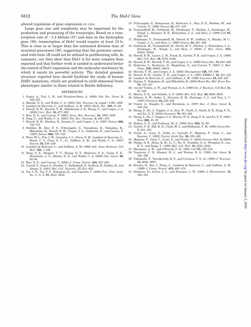
REFERENCES
1. Gupta, A., Tsai, L. H., and Wynshaw-Boris, A. (2002) Nat. Rev. Genet. 3,342–355
2. Monuki, E. S., and Walsh, C. A. (2001) Nat. Neurosci. 4, (suppl.) 1199–12063. Lambert de Rouvroit, C., and Goffinet, A. M. (2001) Mech. Dev. 105, 47–564. Howell, B. W., Herrick, T. M., Hildebrand, J. D., Zhang, Y., and Cooper, J. A.
(2000) Curr. Biol. 10, 877–8855. Rice, D. S., and Curran, T. (2001) Annu. Rev. Neurosci. 24, 1005–10396. Feng, Y., and Walsh, C. A. (2001) Nat. Rev. Neurosci. 2, 408–4167. Howell, B. W., Hawkes, R., Soriano, P., and Cooper, J. A. (1997) Nature 389,
733–7378. Sheldon, M., Rice, D. S., D’Arcangelo, G., Yoneshima, H., Nakajima, K.,
Mikoshiba, K., Howell, B. W., Cooper, J. A., Goldowitz, D., and Curran, T.(1997) Nature 389, 730–733
9. Ware, M. L., Fox, J. W., Gonzalez, J. L., Davis, N. M., Lambert de Rouvroit, C.,Russo, C. J., Chua, S. C., Jr., Goffinet, A. M., and Walsh, C. A. (1997)Neuron 19, 239–249
10. Lambert de Rouvroit, C., and Goffinet, A. M. (1998) Adv. Anat. Embryol. CellBiol. 150, 1–106
11. Hong, S. E., Shugart, Y. Y., Huang, D. T., Shahwan, S. A., Grant, P. E.,Hourihane, J. O., Martin, N. D., and Walsh, C. A. (2000) Nat. Genet. 26,93–96
12. Rice, D. S., and Curran, T. (2000) J. Comp. Neurol. 424, 327–33813. Carroll, P., Gayet, O., Feuillet, C., Kallenbach, S., de Bovis, B., Dudley, K., and
Alonso, S. (2001) Mol. Cell. Neurosci. 17, 611–62314. Yip, J. W., Yip, Y. P., Nakajima, K., and Capriotti, C. (2000) Proc. Natl. Acad.
Sci. U. S. A. 97, 8612–8616
15. D’Arcangelo, G., Homayouni, R., Keshvara, L., Rice, D. S., Sheldon, M., andCurran, T. (1999) Neuron 24, 471–479
16. Trommsdorff, M., Gotthardt, M., Hiesberger, T., Shelton, J., Stockinger, W.,Nimpf, J., Hammer, R. E., Richardson, J. A., and Herz, J. (1999) Cell 97,689–701
17. Hiesberger, T., Trommsdorff, M., Howell, B. W., Goffinet, A., Mumby, M. C.,Cooper, J. A., and Herz, J. (1999) Neuron 24, 481–489
18. Gotthardt, M., Trommsdorff, M., Nevitt, M. F., Shelton, J., Richardson, J. A.,Stockinger, W., Nimpf, J., and Herz, J. (2000) J. Biol. Chem. 275,25616–25624
19. Howell, B. W., Lanier, L. M., Frank, R., Gertler, F. B., and Cooper, J. A. (1999)Mol. Cell. Biol. 19, 5179–5188
20. Howell, B. W., Herrick, T. M., and Cooper, J. A. (1999) Genes Dev. 13, 643–64821. Keshvara, L., Benhayon, D., Magdaleno, S., and Curran, T. (2001) J. Biol.
Chem. 276, 16008–1601422. Herrick, T. M., and Cooper, J. A. (2002) Development 129, 787–79623. Howell, B. W., Gertler, F. B., and Cooper, J. A. (1997) EMBO J. 16, 121–13224. Lambert de Rouvroit, C., and Goffinet, A. M. (1998) Genomics 53, 246–24725. Kojima, T., Nakajima, K., and Mikoshiba, K. (2000) Brain Res. Mol. Brain Res.
75, 121–12726. van der Velden, A. W., and Thomas, A. A. (1999) Int. J. Biochem. Cell Biol. 31,
87–10627. Morris, D. R., and Geballe, A. P. (2000) Mol. Cell. Biol. 20, 8635–864228. Scherer, S. W., Soder, S., Duvoisin, R. M., Huizenga, J. J., and Tsui, L. C.
(1997) Genomics 44, 232–23629. Valent, A., Danglot, G., and Bernheim, A. (1997) Eur. J. Hum. Genet. 5,
102–10430. Sheng, Z., He, J., Tuppen, J. A., Sun, W., Fazili, Z., Smith, E. R., Dong, F. B.,
and Xu, X. X. (2000) Genomics 70, 381–38631. Sheng, Z., He, J., Tuppen, J. A., Martin, W. D., Dong, F. B., and Xu, X. X. (2001)
Gene 268, 31–3932. Redina, O. E., and Frohman, M. A. (1998) Gene 222, 53–6033. Gertler, F. B., Hill, K. K., Clark, M. J., and Hoffmann, F. M. (1993) Genes Dev.
7, 441–45334. Pesole, G., Liuni, S., Grillo, G., Licciulli, F., Mignone, F., Gissi, C., and
Saccone, C. (2002) Nucleic Acids Res. 30, 335–34035. Mignone, F., Gissi, C., Liuni, S., and Pesole, G. (2002) Genome Biol. 3, S000436. Phelps, D. E., Hsiao, K. M., Li, Y., Hu, N., Franklin, D. S., Westphal, E., Lee,
E. Y., and Xiong, Y. (1998) Mol. Cell. Biol. 18, 2334–234337. Shermoen, A. W., and O’Farrell, P. H. (1991) Cell 67, 303–31038. Tennyson, C. N., Klamut, H. J., and Worton, R. G. (1995) Nat. Genet. 9,
184–19039. Takahashi, T., Nowakowski, R. S., and Caviness, V. S., Jr. (1995) J. Neurosci.
15, 6046–605740. Bernier, B., Bar, I., Pieau, C., Lambert de Rouvroit, C., and Goffinet, A. M.
(1999) J. Comp. Neurol. 413, 463–47941. Simmons, D., Arriza, J. L., and Swanson, L. W. (1989) J. Histotechnol. 12,
169–181
The Dab1 Gene5812
by guest on January 2, 2020http://w
ww
.jbc.org/D
ownloaded from

M. GoffinetIsabelle Bar, Fadel Tissir, Catherine Lambert de Rouvroit, Olivier De Backer and André
Pathway, Reveals Unusual Complexity in Human and MouseThe Gene Encoding Disabled-1 (DAB1), the Intracellular Adaptor of the Reelin
doi: 10.1074/jbc.M207178200 originally published online November 21, 20022003, 278:5802-5812.J. Biol. Chem.
10.1074/jbc.M207178200Access the most updated version of this article at doi:
Alerts:
When a correction for this article is posted•
When this article is cited•
to choose from all of JBC's e-mail alertsClick here
http://www.jbc.org/content/278/8/5802.full.html#ref-list-1
This article cites 41 references, 11 of which can be accessed free at
by guest on January 2, 2020http://w
ww
.jbc.org/D
ownloaded from