Embed Size (px)
Citation preview
J Physiol 000.0 (2016) pp 1–22 1
The
Jou
rnal
of
Phys
iolo
gy
Neuroscience The incretin hormone glucagon-like peptide 1 increases
mitral cell excitability by decreasing conductanceof a voltage-dependent potassium channel
Nicolas Thiebaud1, Ida J. Llewellyn-Smith2, Fiona Gribble3, Frank Reimann3, Stefan Trapp4,5
and Debra Ann Fadool1,6
1The Florida State University, Department of Biological Science, Program in Neuroscience, Tallahassee, FL, USA2Cardiovascular Medicine and Human Physiology, School of Medicine, Flinders University, Bedford Park, SA, Australia3Cambridge Institute for Medical Research, Addenbrooke’s Hospital, Cambridge, UK4Centre for Cardiovascular and Metabolic Neuroscience, Department of Neuroscience, Physiology, and Pharmacology, University College London,London, UK5Department of Surgery and Cancer, Imperial College London, London, UK6The Florida State University, Institute of Molecular Biophysics, Tallahassee, FL, USA
Key points
� The gut hormone called glucagon-like peptide 1 (GLP-1) is a strong moderator of energyhomeostasis and communication between the peripheral organs and the brain.
� GLP-1 signalling occurs in the brain; using a newly developed genetic reporter line of mice, wehave discovered GLP-synthesizing cells in the olfactory bulb.
� GLP-1 increases the firing frequency of neurons (mitral cells) that encode olfactory informationby decreasing activity of voltage-dependent K channels (Kv1.3).
� Modifying GLP-1 levels, either therapeutically or following the ingestion of food, could alter theexcitability of neurons in the olfactory bulb in a nutrition or energy state-dependent mannerto influence olfactory detection or metabolic sensing.
� The results of the present study uncover a new function for an olfactory bulb neuron (deepshort axon cells, Cajal cells) that could be capable of modifying mitral cell activity through therelease of GLP-1. This might be of relevance for the action of GLP-1 mimetics now widely usedin the treatment of diabetes.
Abstract The olfactory system is intricately linked with the endocrine system where it may serveas a detector of the internal metabolic state or energy homeostasis in addition to its classicalfunction as a sensor of external olfactory information. The recent development of transgenicmGLU-yellow fluorescent protein mice that express a genetic reporter under the control ofthe preproglucagon reporter suggested the presence of the gut hormone, glucagon-like peptide(GLP-1), in deep short axon cells (Cajal cells) of the olfactory bulb and its neuromodulatoryeffect on mitral cell (MC) first-order neurons. A MC target for the peptide was determinedusing GLP-1 receptor binding assays, immunocytochemistry for the receptor and injection offluorescence-labelled GLP-1 analogue exendin-4. Using patch clamp recording of olfactory bulbslices in the whole-cell configuration, we report that GLP-1 and its stable analogue exendin-4increase the action potential firing frequency of MCs by decreasing the interburst interval ratherthan modifying the action potential shape, train length or interspike interval. GLP-1 decreasesKv1.3 channel contribution to outward currents in voltage clamp recordings as determinedby pharmacological blockade of Kv1.3 or utilizing mice with Kv1.3 gene-targeted deletion asa negative control. Because fluctuations in GLP-1 concentrations monitored by the olfactory
C© 2016 The Authors. The Journal of Physiology published by John Wiley & Sons Ltd on behalf of The Physiological Society DOI: 10.1113/JP272322
This is an open access article under the terms of the Creative Commons Attribution License, which permits use, distributionand reproduction in any medium, provided the original work is properly cited.
2 N. Thiebaud and others J Physiol 000.0
bulb can modify the firing frequency of MCs, olfactory coding could change depending uponnutritional or physiological state. As a regulator of neuronal activity, GLP-1 or its analogue maycomprise a new metabolic factor with a potential therapeutic target in the olfactory bulb (i.e. viaintranasal delivery) for controlling an imbalance in energy homeostasis.
(Resubmitted 15 January 2016; accepted after revision 25 February 2016; first published online 2 March 2016)Corresponding author N. Thiebaud: Florida State University, 319 Stadium Drive, King Life Sciences Building, Suite3007, Tallahassee, FL 32306, USA. Email: [email protected]
Abbreviations aCSF, artificial cerebral spinal fluid; AP, action potential; AVMA, American Veterinary MedicineAssociation; ASPA, Animals Scientific Procedures Act; DAPI, 4′,6-diamidino-2-phenylindole; dSAC, deep short axoncell; Ex4, exendin-4; EPL, external plexiform layer; GLP-1, glucagon-like peptide-1; GLP-1R, glucagon-like peptide 1receptor; GCL, granule cell layer; GFP, green fluorescent protein; ISI, interspike interval; IACUC, Institutional AnimalCare and Use Committee; Kv1.3−/−, Kv1.3 gene-targeted deletion; MGTX, margatoxin; MC, mitral cell; MCL, mitralcell layer; OB, olfactory bulb; OMP, olfactory marker protein; PBS, phosphate-buffered saline; PFA, paraformaldehyde;PPG, preproglucagon; RMP, resting membrane potential; WT, wild-type; YFP, yellow fluorescent protein.
Introduction
Energy homeostasis is dependent upon neuronal andhormonal communication between the peripheral organsand the brain. Glucagon-like peptide 1 (GLP-1) is anincretin hormone secreted from intestinal L-cellsfollowing a meal (Holst, 2007). Peripheral GLP-1 exertsstrong effects on the control of energy metabolismby regulating glycaemia, stimulating glucose-dependentinsulin secretion and proinsulin gene expression, andinhibiting glucagon release and gastric emptying (Holst,2007). In the central nervous system, cell bodies of GLP-1producing neurons have been identified in the caudalnucleus of the solitary tract and intermediate reticularnucleus (Jin et al. 1988; Larsen et al. 1997; Merchenthaleret al. 1999; Hisadome et al. 2010; Trapp & Richards,2013). These neurons send axons to several areas in thebrain, including the paraventricular nucleus, the dorso-medial hypothalamus and the nucleus accumbens (Larsenet al. 1997; Tauchi et al. 2008; Llewellyn-Smith et al.2011). Direct central infusion of GLP-1 has been shown toinhibit food and water intake and to reduce blood glucose(Tang-Christensen et al. 1996; Kinzig et al. 2002; Dossatet al. 2011).
The receptor for GLP-1 (GLP-1R) is a seven trans-membrane domain, G-protein coupled receptor that isexpressed in a wide variety of tissues, including thehypothalamus and the nucleus accumbens, where GLP-1has been demonstrated to play a role in the controlof food intake (Dossat et al. 2011). Ligand bindingof the peptide to GLP-1R triggers activation of theGαs protein, which leads to the production of cAMPthrough the activation of the enzyme adenylate cyclase.Elevation of cAMP concentration in the cell increasesprotein kinase A activity, which results in the inhibitionof potassium channels, KATP, Kv2.1 and Kv1.5, as wellas the auxiliary Kvβ2 protein subunit (Gromada et al.1998a; Gromada et al. 1998b; MacDonald et al. 2003;
Kim et al. 2012; Kim et al. 2013) by phosphorylation oracetylation. This results in a reduction of the Kv currentamplitude or reduced Kv surface trafficking, either ofwhich would cause increased depolarization. In pancreaticβ-cells, GLP-1R activation also induces an elevation ofintracellular Ca2+ concentrations through modificationof L-type Ca2+ channels and the oscillation of Ca2+through Ca2+-induced Ca2+ release from intracellularstores (Holst, 2007).
Most studies investigating the second messengersinvolved in GLP-1 signalling have been performedin pancreatic β-cells where GLP-1 stimulates glucose-dependent insulin release. GLP-1, however, has alsobeen shown to increase membrane excitability ofnodose ganglion neurons by decreasing the potassiumconductance of both inactivating and delayed-rectifierpotassium currents (Gaisano et al. 2010). Nodose neuronsare no longer modulated by the peptide following blockadeof K+ currents with the relatively non-selective antagonist,4-AP.
Because the olfactory system is intricately linked withthe endocrine system, it may not only serve as anexternal sensor of olfactory information, but also as aninternal sensor of metabolic state or energy homeo-stasis (Palouzier-Paulignan et al. 2012). It has beenreported previously that hormonal and metabolic factorsmodulate the excitability of olfactory sensory neurons,as well as mitral cells (MCs) representing the first-orderneurons in the olfactory bulb (OB) (Fadool et al. 2000;Fadool et al. 2011; Aime et al. 2012; Soria-Gomez et al.2014). Initial electrophysiological studies exploring neuro-endocrine modulation focused upon the hormone insulin,which has the highest binding affinity and receptor densityreported for the OB compared to all other brain regions(Hill et al. 1986; Banks et al. 1999). Other factors, suchas orexins, have been shown to modulate MC firingfrequency (Apelbaum et al. 2005; Hardy et al. 2005).
C© 2016 The Authors. The Journal of Physiology published by John Wiley & Sons Ltd on behalf of The Physiological Society

J Physiol 000.0 GLP-1 modulation in the olfactory bulb 3
Orexins originate from neurons in the lateral hypo-thalamic area that send centrifugal projections to the OB(Gascuel et al. 2012). Prud’homme et al. (2009) havedemonstrated that the release of the orexigenic peptidewithin the OB is highly dependent upon nutritional status(fasted vs. satiated). Recently, our group demonstratedthat other metabolic factors, including glucose itself,modulate the firing frequency of MCs (Tucker et al. 2013),suggesting that there is a glucose-sensitive populationof MCs detecting changes in glucose homeostasis. InMCs, glucose and insulin use a mechanism that involveschanges in excitability driven by a voltage-dependentpotassium channel, Kv1.3, as demonstrated by the lackof response to these metabolic factors in MCs from micewith gene-targeted deletion of Kv1.3 (Kv1.3−/−) (Fadoolet al. 2011; Tucker et al. 2013).
Hormonal regulation occurring within the olfactorysystem is hypothesized to modulate olfactory sensitivityand the hedonic representation of odours followingchanges in nutritional state. For example, the endo-canabinoid system has recently been shown to be involvedin the modulation of olfactory sensitivity and food intakethrough activation of CB1 receptors in the olfactorysystem (Soria-Gomez et al. 2014). Moreover, dysregulationof energy metabolism affects the olfactory system.Rodent models of genetic- or diet-induced obesity havedemonstrated both anatomical and functional changesin the olfactory system (Badonnel et al. 2012; Tuckeret al. 2012; Thiebaud et al. 2014). Such changes havean impact on olfactory-linked food behaviours or foodintake.
Although a single previous study has used in situhybridization to characterize the expression of pre-proglucagon (PPG) and the GLP-1 receptor in theolfactory system (Merchenthaler et al. 1999), the physio-logical functions of the GLP-1 pathway, as well asprotein expression and localization, remain completelyunexplored. The recent development of transgenicmGLU-yellow fluorescent protein (YFP) mice, expressingthe yellow fluorescent protein, Venus, under the control ofthe glucagon promoter (Reimann et al. 2008), allowed usto confirm the presence of GLP-1 producing neurons in theOB. Following GLP-1 receptor binding assays and labellingexperiments in mice with a genetic reporter for MCs, weconjectured that MCs were a target for the endogenouspeptide. Accordingly, we performed a biophysical analysisof GLP-1 modulation on these first-order neurons. Usingthe whole-cell configuration of the patch clamp in OBslices, we determined the mechanism of action of GLP-1 orits stable analogue exendin-4 (Ex4) on MCs. Fluctuationsin GLP-1 concentrations were able to modify the intrinsicfiring frequency of MCs and alter the olfactory coding inresponse to a changed physiological state, such as followinga meal or after fasting.
Methods
Ethical approval
All experiments described in the present study wereapproved by the Florida State University InstitutionalAnimal Care and Use Committee (IACUC) underprotocols #1124 and #1427, and were conducted inaccordance with the American Veterinary MedicineAssociation (AVMA), the National Institutes of Health,and the UK Home Office Regulations under the AnimalsScientific Procedures Act (ASPA) 1986. In preparationfor OB slice electrophysiology, mice were anaesthetizedwith isoflurane (Aerrane; Baxter, Deerfield, IL, USA)using the IACUC-approved drop method and then werekilled by decapitation (AVMA Guidelines on Euthanasia,June 2007). These procedures are contained in the ASPASchedule 1 (Wolfenshon & Lloyd, 2003) for the species,stage of development, and size of our mice (see below). Allauthors understood the ethical principles that The Journalof Physiology operates under and the work complied withthe animal ethics checklist reported by Grundy (2015).
Animal care and mouse lines
All mice were housed at the Florida State University,Imperial College London, or University College Londonvivaria in accordance with the institutional requirementsfor animal care. All mice used in the present study (Musmusculus, C57BL/6 background strain; Jax Laboratories,Bar Harbor, ME, USA) were maintained under a standard12:12 h light/dark cycle and were allowed ad libitum accessto 5001 Purina Chow (Purina, Richmond, VA, USA) andwater. Mice of both sexes were aged from postnatal day21 to 35 at the time of the experiment unless otherwisenoted, and had a body weight in the range 8.4–20.0 g(mean ± SD: 15.9 ± 2.5 g). A total of 62 wild-type(WT), 12 Kv1.3−/−, three olfactory marker protein-greenfluorescent protein (OMP-GFP), nine PPG-YFP, threeGLP-1R-tdRFP, three Thy1-YFP and three GLP-1R−/−mice were used in the present study. Kv1.3−/− mice wereproduced previously by excision of the Kv1.3 promoterregion and one-third of the 5′ coding region (Koni et al.2003; Xu et al. 2003) and were a generous gift from DrsLeonard Kaczmarek and Richard Flavel (Yale University,New Haven, CT, USA). Mice with an olfactory markerprotein GFP transgene (OMP-GFP) (Potter et al. 2001)were used to identify all mature olfactory sensory neuronsand were a gift from Dr Peter Mombaerts (Max PlankInstitute, Frankfort, Germany). PPG-YFP mice, expressingthe YFP variant Venus (Nagai et al. 2002) under thecontrol of the mouse PPG promotor (mGLU-124 line)(Reimann et al. 2008), were used to identify proglucagonexpressing neurons. Briefly, mice were generated usingmouse bacterial artificial chromosomes using the RedEt
C© 2016 The Authors. The Journal of Physiology published by John Wiley & Sons Ltd on behalf of The Physiological Society
4 N. Thiebaud and others J Physiol 000.0
technique (Zhang et al. 2000). Pronuclear injection ofbacterial artificial chromosome constructs containingthe Venus sequence in place of the coding region ofPPG resulted in the generation of founder mice withthe transgene (mGLU-124 line). GLP-1R Cre mice thatexpress Cre-recombinase under the GLP-1R promoterwere generated as described previously (Richards et al.2014). These mice were crossed with ROSA26-tdRFPreporter strains (Luche et al. 2007) to enable fluorescencedetection of cells expressing GLP-1R by cytosolic tdRFPexpression. Thy1-YFP mice were used to identify MCs andwere a gift from Dr Guoping Feng (MIT Broad Institute,Boston, MA, USA) (Feng et al. 2000). GLP-1R−/− micewere a generous gift from Dr Julio E. Ayala (Diabetesand Obesity Research Centre, Orlando, FL, USA) (Ayalaet al. 2009).
Solutions, reagents and antisera
Margatoxin (MGTX) was purchased from Sigma-Aldrich(St Louis, MO, USA) and used to selectively block thevestibule of the Kv1.3 channel (Knaus et al. 1995).TTX was purchased from Ascent Scientific (Princeton,NJ, USA) or Abcam Biochemicals (Cambridge, MA,USA) to block voltage-gated sodium channels. GLP-1,glucagon-like peptide 1 (7-36)-lys (biotin) (Ref46-1-65) and GLP-1-(7-36) amide were purchased fromAmerican Peptide Company (Sunnyvale, CA, USA).Phosphate-buffered saline (PBS) was made as describedpreviously (Tucker & Fadool, 2002). Slice intracellularpipette solution contained (in mM): 135 potassiumgluconate, 10 KCl, 10 Hepes, 10 MgCl2, 0.4 NaGTPand 2 NaATP (pH 7.3; 280–285 mOsm). Artificialcerebral spinal fluid (aCSF) contained (in mM): 119 NaCl,26.2 NaHCO3, 1 NaH2PO4, 2.5 KCl, 1.3 MgCl2, 2.5 CaCl2and 22 D-glucose (pH 7.3; 310–315 mOsm). In experi-ments where the external concentration of potassium wasadjusted to alter the calculated reversal potential for K+,KCl was modified to 2.5 or 8.5 mM. Sucrose-modifiedaCSF was used for sectioning and contained (in mM):83 NaCl, 26.2 NaHCO3, 1 NaH2PO4, 3.3 MgCl2,0.5 CaCl2, 72 sucrose and 22 D-glucose (pH 7.3;315 mOsm) (De Saint Jan & Westbrook, 2007). All saltsand sugars were purchased from Sigma-Aldrich or FisherScientific (Pittsburgh, PA, USA).
The GLP-1R antibody (dilution 1:3000) was agenerous gift from Dr Joel Habener (Harvard Catalyst,Cambridge, MA, USA) and Dr Scott Heller (HagedornResearch Institute, Denmark) and was generated againstthe N-terminal epitope (CQHRYERWKQVTESLSVT)in rabbits (Heller et al. 1996). Chicken anti-GFPwas purchased from Abcam Biochemicals (dilution1:50,000, catalogue number ab13970) and was designedto recognize the enhanced form of GFP, and all of thefluorescent proteins made by Aequorea victoria, including
YFP. The insulin receptor (IR) kinase antibody wasa mouse monoclonal directed against the β subunit(dilution 1:1000, Clone CT-3; catalogue number 05-1104;Millipore, Billerica, MA, USA). Host-specific secondaryantibodies were purchased from Jackson Immuno-Research (West Grove, PA, USA) and used in accordancewith the manufacturer’s instructions. GLP1-R expressingcells from GLP-1R transgenic line were detected usinga far red polyclonal antibody, DsRed (dilution 1:1000,catalogue number 632496; Clontech, Palo Alto, CA, USA)using protocols described previously (Cork et al. 2015).
Immunocytochemistry
YFP-immunoreactivity in GLP-1 neurons was visua-lized in PPG-YFP mice as described previously (Llewellyn-Smith et al. 2011; Llewellyn-Smith et al. 2013). Briefly,a total of five mice aged 12–16 weeks and weighingbetween 25 and 35 g were perfused intracardiallywith 4% paraformaldehyde (PFA). The head was sub-sequently postfixed in 4% PFA/PBS overnight anddecalcified for 3–5 days in 0.3 M EDTA and prepared forcryosectioning (30 μm) as described in Thiebaud et al.(2014). YFP-immunoreactivity was confirmed in parallelexperiments using an antibody to GFP (see antibodies).Here, an immunoperoxidase protocol in conjunctionwith a metal-intensified diaminobenzidine reaction wasused to optimally visualize the morphology of GLP-1neurons (Llewellyn-Smith et al. 2011). A total of four micefor the immunoperioxidase studies were perfused with4% PFA/PBS. Brains with attached OBs were removedand post-fixed for 3 days at room temperature in thesame fixative. OBs were infiltrated with sucrose andcryosectioned (30 μm). A BH-2 microscope (Olympus,Tokyo, Japan) equipped with a SPOT Insight Model 18.2firewire colour camera and SPOT, version 4.6 (DiagnosticInstruments, Inc., Sterling Heights, MI, USA) was used tocapture images.
To localize GLP-1R by immunocytochemicalapproaches, a total of three adult mice were used. Micewere intracardially perfused with 4% paraformaldehydein PBS (PFA/PBS). The OBs were postfixed overnight in4% PFA, decalcified for 3–5 days in 0.3 M EDTA, andthen prepared for cryosectioning (12 μm) as described inThiebaud et al. (2014). Prepared OB frozen sections wereair-dried for 30 min, fixed in 1% PFA/PBS, washed threetimes in PBS and then incubated for 30 min in 3% bovineserum albumin/PBS to block non-specific binding. Foruse of the IR kinase antibody, it was also necessaryto perform a demasking step by boiling sections (i.e.microwave) in 10 mM citrate buffer (pH 6.0) for 4 min forepitope retrieval prior to the blocking step in accordancewith the manufacturer’s instructions. After the blockingstep, the tissue sections were incubated overnight withthe primary antiserum (dilution in PBS + 3% BSA) at
C© 2016 The Authors. The Journal of Physiology published by John Wiley & Sons Ltd on behalf of The Physiological Society
J Physiol 000.0 GLP-1 modulation in the olfactory bulb 5
the same time as protecting from light and maintainingsections at 4°C. The immunofluorescence signal wasdetected after a 2 h incubation with host-specific Cy3 orCy2 secondary antisera diluted in PBS (dilution 1:200;Jackson ImmunoResearch). After three washes in PBS,nuclei were additionally stained by incubating slidesfor 5 min in 4′,6-diamidino-2-phenylindole (DAPI)nuclear stain diluted in PBS (dilution 1:15,000; LifeTechnologies, Carlsbad, CA, USA), washing again inPBS and coverslipping with Fluoromount G (SouthernBiotechnology, Birmingham, AL, USA) to prevent photo-bleaching. OB sections were examined by brightfield andfluorescence using an Axiovert S100 inverted microscope(Carl Zeiss Microimaging, Thornwood, NY, USA). Digitalimages were captured with an Axiocam digital cameraand AxioVision, version 4.8 (Carl Zeiss Microimaging).
GLP-1 binding assay
The GLP-1-biotin binding assay was performed on threeWT mice as adapted from Cowley et al. 2003. Briefly,150 μm vibratome sections of OB were obtained inice-cold, sucrose-modified aCSF. The oxygenated (95%O2/5% CO2) sections recovered for 1 h in an inter-face chamber in normal aCSF (Fadool et al. 2011) andwere then incubated with either 1 μM GLP-1-biotinconjugate or a mix of 1 μM GLP-1-biotin conjugatewith 2 μM of ‘cold’ GLP-1 (unconjugated) for 20 minat +4ºC. The sections were subsequently fixed with 4%PFA/PBS for 10 min, washed in PBS and incubated in1:200 Cy3-streptavidin (Life Technologies) for 30 min.The sections were stained with DAPI nuclear stain(dilution 1:15,000), washed in PBS, and then mountedand visualized as described above.
In another experiment, Thy-1-YFP mice that expressYFP in MCs or GLP-1R−/− mice received a S.C. injection(120 nmol kg−1) of fluorescence-labelled Ex4 [Cys(HiLyteFluor647 C2 maleimide)]-exendin-4 (Ref AS-63714;AnaSpec, Fremont, CA, USA). The animals were trans-cardially perfused with 4% PFA 4 h after injection and theOBs were processed and cryosectioned at a thickness of12 μM as described previously (Thiebaud et al. 2014).
OB slice electrophysiology
A total of 79 mice were used for 197 patch clampelectrophysiology recordings from OB slices as describedpreviously (Fadool et al. 2011). Following isofluraneanaesthesia, the mice were killed by decapitation. TheOBs were rapidly exposed by removing the dorsal andlateral portions of the cranium between the cribriformplate and the lambda suture as described previously(Nickell et al. 1996). After removing the dura, the OBs(when still attached to the forebrain) were quicklyremoved, glued to a sectioning block with Superglue
(Lowe’s Home Improvement, Tallahassee, FL, USA) andsubmerged in oxygenated, ice-cold, sucrose-modifiedaCSF to prepare the tissue for sectioning (De Saint Jan& Westbrook, 2007). Coronal sections (275 μm) were cutin oxygenated, ice-cold, sucrose-modified aCSF using aSeries 1000 Vibratome (Leica, Wetzlar, Germany). Thesections were allowed to recover in an interface chamber(Krimer & Goldman-Rakic, 1997) with oxygenated,sucrose-modified aCSF at 33°C for 30 min and thenmaintained at room temperature in oxygenated normalaCSF until needed (De Saint Jan & Westbrook, 2007;Fadool et al. 2011). Neuronal slices were visualized at 10×and 40× using an Axioskop 2FS Plus microscope (CarlZeiss Microimaging) equipped with infrared detectioncapabilities (CCD100; Dage-MTI, Michigan City, IN,USA).
Membrane voltage and current properties weregenerated using pCLAMP, version 9 or 10, in conjunctionwith an Axopatch 200B amplifier (Axon Instruments,Foster City, CA, USA). The analogue signal was filteredat 2 kHz and minimally digitally sampled every 100 μs.Electrodes were fabricated from borosilicate glass(#1405002; Hilgenberg GmbH, Malsfeld, Germany) to adiameter of �2 μm to yield pipette resistances rangingfrom 4 to 7 M�. Positive pressure was retained whennavigating through the OB laminae until a high resistanceseal (1.5–10 G�) was obtained on a positionally-identifiedMC in the slice (Fadool et al. 2011). The morphologyand biophysical properties of the neurons were used todistinguish MCs from tufted cells. In addition, thy1-YFPtransgenic mice comprised a good tool to secondarilyconfirm cell identity as in the study by Fadool et al. 2011(Fig. 1). The whole-cell configuration was established byapplying gentle suction to the lumen of the pipette atthe same time as monitoring resistance. Each MC wasfirst sampled for adequate resting potential (less than–55 mV) and proper series resistance (less than 40 M�)prior to initiating a series of current clamp recordings.Only current clamp recordings with a sustained restingmembrane potential (RMP) of at least –50 mV or aninput resistance (Rin) of at least 150 M� were accepted.During recordings, the membrane potential (Em) wasadjusted to –65 mV by injecting a few pA of current sothat data could be statistically evaluated from the samepotential across cells.
For current clamp recordings, the perithreshold currentlevel was determined by incrementally injecting 1 s long,25 pA steps of current every 10 s, starting at –50 pA.Following the determination of spike threshold, cells werethen stimulated with a long, perithreshold current step(typically ranging from 25 to 100 pA) of 5000 ms atan interpulse interval of 10 s to acquire spike frequencydata before and after peptide or drug application. Ingeneral, recordings were acquired for a minimum of30 min following application of peptide. Latency to first
C© 2016 The Authors. The Journal of Physiology published by John Wiley & Sons Ltd on behalf of The Physiological Society
6 N. Thiebaud and others J Physiol 000.0
spike, spike event frequency (calculated throughout stepdepolarization), interburst interval (calculated betweenspike cluster), interspike interval (ISI) (calculated within aspike cluster) and action potential (AP) cluster length weremeasured as described previously (Balu et al. 2004; Fadoolet al. 2011; Mast & Fadool, 2012). A cluster was definedas three or more consecutive spikes with an ISI of 100 msor less as established by Balu et al. (2004). Because MCfiring is intrinsically intermittent and is characterized byvariable spike clusters, classical means of computing spiketiming variability, such as peristimulus time histograms,were less suitable for the behaviour of these neurons andtherefore alternative means of spike analysis were appliedas described previously (Balu et al. 2004).
For voltage clamp recordings, voltage-activatedoutward currents were elicited using a 400 ms voltagestimulation protocol with a holding potential (Vh) of–80 mV and command potential (Vc) steps from –100to +40 mV in 10 mV increments. Interpulse intervalswere taken at 45 s to prevent cumulative inactivation ofthe Kv1.3 current that is expressed in MCs (Marom et al.1993; Fadool & Levitan, 1998).
Statistical analysis
All electrophysiological data were analysed usingpClamp, version 10 (Clampfit 10.2; Axon Instruments),Prism, version 5 (GraphPad Software, Inc., La Jolla,CA, USA) and Igor Pro, version 6.12A (Wave-Metrics Inc., Portland, OR, USA) with the plug-inNeuroMatic, version 2 (written by Jason Rothman;http://www.neuromatic.thinkrandom.com). The pipettecapacitance was electrically compensated through thecapacitance neutralization circuit of the Axopatch 200Bamplifier. Similarly, series resistance compensation wasused to electrically reduce the effect of pipette resistance.
Voltage clamp traces were subtracted linearly for leakageconductance. Resting membrane potentials were correctedfor a –14 mV junction potential offset.
For voltage clamp experiments, the peak transientcurrent was defined as the greatest current evoked aftervoltage activation. Current–voltage relationships wereplotted by normalizing the peak transient current to eithercell capacitance or to the maximum evoked current at+40 mV. The family of current–voltage relations wascompared (via Prism) using a repeated measures two-wayANOVA (and a Bonferoni post hoc test) with peptide andvoltage step as factors. For current clamp experiments,change in the AP frequency, ISI or interburst interval wereplotted as the mean percentage change compared to thecontrol condition prior to bath application of the peptide.Baseline, treatment and washout values were calculatedfrom the mean of 10 consecutives traces generatedfrom a 5 s duration current injection. For statisticalanalyses, Prism was used to perform the Friedmantest (non-parametric alternative for repeated measuresANOVA) followed by Bonferoni’s post hoc comparisontest. All data are reported as the mean ± SEM. Differentletters denote statistically different mean values with thecorresponding P value as specified. Statistical differencewas defined at the 95% confidence interval (or α � 0.05),unless otherwise specified.
Results
Localization of PPG-neurons in the OB
Immunocytochemical localization of GLP-1 peptide canyield precarious results as a result of non-specific labellingby commercially-available antibodies. We avoided thisissue by using transgenic PPG-YFP mice and tookadvantage of the YFP expression that occurs throughout
GCL
MCL
EPL
GLM100μm 100μm 50μm
A B
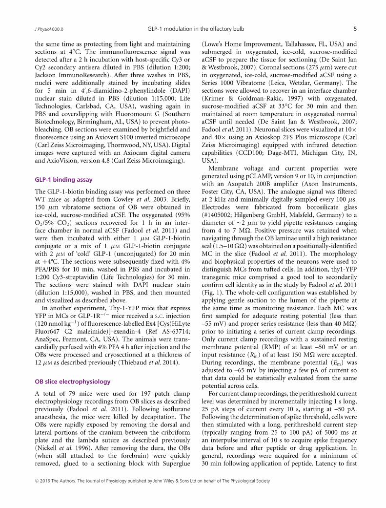
Figure 1. Preproglucagon positive (PPG+) short axon cells are visualized in the OBA, photomicrograph of a representative coronal section of the mouse OB of a PPG-YFP mouse demonstratingnative YFP labelling of dSAC within the GCL. Also visible in the section are other neurolamina, MCL, EPL andthe glomerular cell layer (GLM). Note the position of the soma in the upper portion of the GCL and the axonprojections into the MCL and EPL. B, same as in (A) but labelled using a metal-intensified diaminobenzidinereaction in conjunction with an anti-GFP antibody to optimally visualize the short axon morphology. Inset: highermagnification of the boxed area is shown on the right. Note that the YFP-immunoreactive cells are stellate withdendrites containing spine-like structures or a varicose appearance.
C© 2016 The Authors. The Journal of Physiology published by John Wiley & Sons Ltd on behalf of The Physiological Society

J Physiol 000.0 GLP-1 modulation in the olfactory bulb 7
the cytoplasm of these neurons, including their terminals(Hisadome et al. 2010). Immunohistochemical detectionusing an anti-GFP antiserum was found to significantlyenhance the native fluorescence signal of the YFPas described previously (Llewellyn-Smith et al. 2011;Llewellyn-Smith et al. 2013). YFP fluorescence revealeda large population of PPG-neurons located in the granulecell layer (GCL) of the OB (Fig. 1A). The somatawere positioned in the upper portion of the GCL withaxonal arbors reaching into the mitral cell layer (MCL)and external plexiform layer (EPL). In the GCL, therefined peroxidase labelling strategy revealed that thePPG-neurons had stellate dendrites covered with manyspines (Fig. 1B) that are characteristic of deep short-axoncells (dSACs), also named Cajal cells as described pre-viously (Ramon y Cajal S., 1911; Price & Powell, 1970;Eyre et al. 2008; Eyre et al. 2009; Nagayama et al.2014). At higher magnification (Fig. 1B), we coulddiscern that the size of the soma was slightly larger
(10.20 ± 0.21 μm, n = 73 from three mice) comparedto that reported for granule cells (Woolf et al. 1991; Eyreet al. 2009).
GLP-1R is expressed in the MCL and the GCL
Following the identification of the PPG neurons in theOB, we next investigated the presence of the potentialtarget receptor, or GLP-1R, by immunolocalization.We used a home-made antisera (generously providedby Scott Heller) to reveal immunoreactivity mainly inthe MCL, as well as in sparse cells within the GCL(Fig. 2A). Using a recently developed transgenic mousemodel expressing Cre-recombinase under the control ofthe GLP-1R promoter with a ROSA26-EYFP reporter(GLP-1R-Cre mice; Richards et al. 2014), we confirmedGLP-1R expression in the same MCL and GCL regions(data not shown). These results are in accordance witha previous study reporting GLP-1R mRNA in the MCL
GLM
MCL
452 bp
A
B
C D
GCL
GLP-1R
IRβ
Merge
50 μmDAPI
1μM
GLP
-1-b
iotin
1μM
GLP
-1-b
iotin
+ 2
μM
GLP
-1 c
old
GLM
EPL
MCL
IPL
GCL
50 μm
100 μm
100 μm
50 μm
50 μm
0.5 mm
Figure 2. GLP-1R is expressed in the granularcell and MCLs of the mouse OBA, left: photomicrographs of representative coronalsections of the OB of an OMP-GFP mouse carryinga transgene for olfactory marker protein, OMP. Thesix-panel composite demonstrates fluorescencelabelling using an antibody directed against GLP-1R(left; red) with no signal in the GLM, definedlabelling in the MCL and scattered labelling in theGCL. Merged image on the right usingdouble-colour fluorescence strategy to visualize theGFP (green) and receptor (red) overlay. DAPI nuclearstain (blue). Entire OB for perspective shown on theright. B, RT-PCR agarose electrophoresis gel usingwhole OB tissue as the template yields theanticipated size product (452 bp) for the GLP-1R.C, representative photomicrograph composites as in(A) where a peptide binding assay was performedto visualize (top) GLP-1 biotin conjugate binding(GLP-1-biotin) competing with combined (bottom)GLP-1-biotin plus GLP-1 unconjugated binding(GLP-1 cold). D, representative photomicrographcomposite in which the section was co-labelledwith anti-GLP-1R (red, top), anti-IR kinase (green,middle) with the merged image indicating MCs thatputatively exhibit both co-labelled proteins (yellow,bottom). DAPI nuclear stain (blue) is cropped below.
C© 2016 The Authors. The Journal of Physiology published by John Wiley & Sons Ltd on behalf of The Physiological Society
8 N. Thiebaud and others J Physiol 000.0
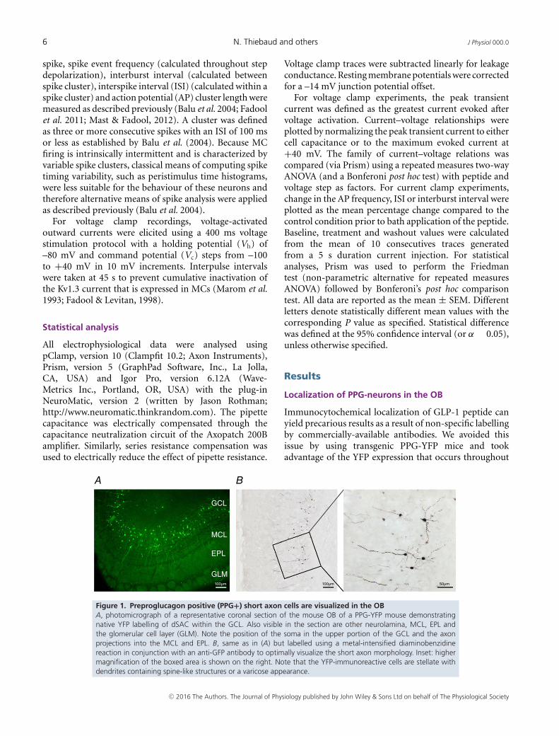
(Merchenthaler et al. 1999). RT-PCR strategies produceda product of anticipated size for the receptor (452 bp) asharvested from whole OB tissue (Fig. 2B). Because antiseradirected against G-protein coupled receptors, includingGLP1-R, can often create false positives, we furtherconfirmed GLP-1R protein expression, as well as thelocation, with a binding essay. Here, GLP-1 was conjugatedwith biotin and detection employed streptavidin coupledto a fluorescent dye (Fig. 2C). The binding assay confirmedthe receptor immunolabelling, demonstrating strongbinding of GLP-1-biotin in the MCL and GCL (Fig. 2C,upper). The labelling in these layers was abolished whenthe OB slice was alternatively incubated in the pre-sence of excess unbiotinylated (cold) GLP-1 (Fig. 2C,lower). It was previously demonstrated that IR kinasewas expressed in the MCL (Fadool et al. 2000; Lacroixet al. 2008; Marks et al. 2009) and so we were interested indetermining whether it might be co-expressed with that ofthe GLP-1R. Double-colour immunofluorescence imagesof the OB lamina demonstrated that IR and GLP-1Rwere co-expressed in an overlapping population of MCswithin the MCL (Fig. 2D). Finally, as parallel support forthe identity of the GLP-1R-expressing neurons across theMCL, we performed S.C. injection of fluorescence-labelledEx4 in a transgenic line, Thy1-YFP, which expresses thefluorescent protein in MCs (Feng et al. 2000), as previouslyused to localize the distribution of the GLP-1 receptor inthe brain (Secher et al. 2014). Double-colour immuno-fluorescence images demonstrated that all Thy1-YFPpositive MCs also reveal intracellular labelling for thefluorescent Ex4 (Fig. 3, top). Consistent with previousstudies, GLP-1 analogues are internalized in neurons afterbinding to the receptor (Roed et al. 2014; Secher et al.
2014; Roed et al. 2015). No fluorescence was observed inMCs of mice deficient for GLP-1R (GLP-1R–/–) (Fig. 3,bottom).
GLP-1 and Ex4 increase evoked AP firing frequencyin MCs
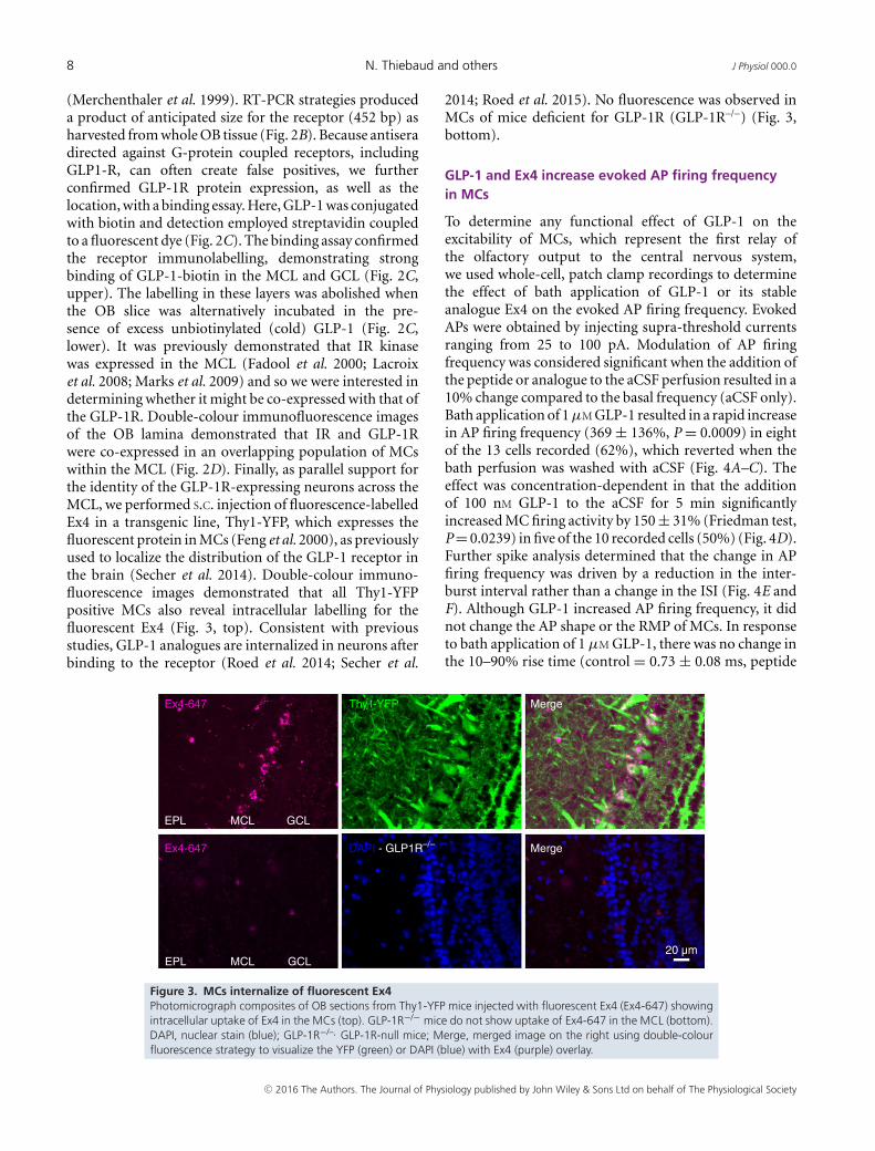
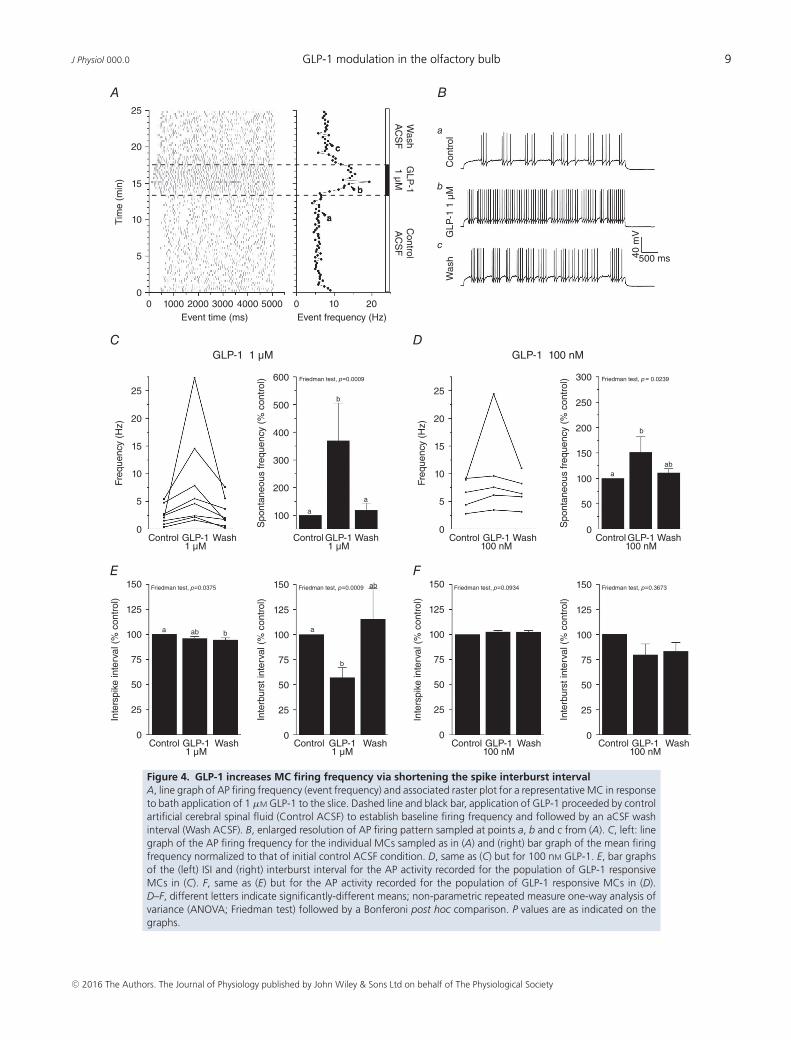
To determine any functional effect of GLP-1 on theexcitability of MCs, which represent the first relay ofthe olfactory output to the central nervous system,we used whole-cell, patch clamp recordings to determinethe effect of bath application of GLP-1 or its stableanalogue Ex4 on the evoked AP firing frequency. EvokedAPs were obtained by injecting supra-threshold currentsranging from 25 to 100 pA. Modulation of AP firingfrequency was considered significant when the addition ofthe peptide or analogue to the aCSF perfusion resulted in a10% change compared to the basal frequency (aCSF only).Bath application of 1 μM GLP-1 resulted in a rapid increasein AP firing frequency (369 ± 136%, P = 0.0009) in eightof the 13 cells recorded (62%), which reverted when thebath perfusion was washed with aCSF (Fig. 4A–C). Theeffect was concentration-dependent in that the additionof 100 nM GLP-1 to the aCSF for 5 min significantlyincreased MC firing activity by 150 ± 31% (Friedman test,P = 0.0239) in five of the 10 recorded cells (50%) (Fig. 4D).Further spike analysis determined that the change in APfiring frequency was driven by a reduction in the inter-burst interval rather than a change in the ISI (Fig. 4E andF). Although GLP-1 increased AP firing frequency, it didnot change the AP shape or the RMP of MCs. In responseto bath application of 1 μM GLP-1, there was no change inthe 10–90% rise time (control = 0.73 ± 0.08 ms, peptide
Ex4-647
Ex4-647
Thy1-YFP
DAPI - GLP1R–/– Merge
EPL MCL GCL
EPL MCL GCL20 μm
Merge
Figure 3. MCs internalize of fluorescent Ex4Photomicrograph composites of OB sections from Thy1-YFP mice injected with fluorescent Ex4 (Ex4-647) showingintracellular uptake of Ex4 in the MCs (top). GLP-1R−/− mice do not show uptake of Ex4-647 in the MCL (bottom).DAPI, nuclear stain (blue); GLP-1R−/–, GLP-1R-null mice; Merge, merged image on the right using double-colourfluorescence strategy to visualize the YFP (green) or DAPI (blue) with Ex4 (purple) overlay.
C© 2016 The Authors. The Journal of Physiology published by John Wiley & Sons Ltd on behalf of The Physiological Society
J Physiol 000.0 GLP-1 modulation in the olfactory bulb 9
25
A B
C D
E F
Wash
AC
SF
GLP
-11 μM
Control
AC
SF
20
15
10
5
0
25
150
125
100
75
50
25
0
150
125
100
75
50
25
0
150
125
100
75
50
25
0
150
125
100
75
50
25
0
600 Friedman test, p=0.0009
Friedman test, p=0.0375 Friedman test, p=0.0009 Friedman test, p=0.0934 Friedman test, p=0.3673
Friedman test, p= 0.0239
b
a
a ab
b
ab
ab
a
b
ab
a
300
250
200
150
100
50
0
500
400
300
200
100
20
15
10
5
0
25
20
15
10
5
0
0 0 10 20Event frequency (Hz)Event time (ms)
GLP-1 1 μM GLP-1 100 nM
1000
Control WashGLP-11 μM
Control WashGLP-11 μM
Control WashGLP-1100 nM
Control WashGLP-1100 nM
Control WashGLP-11 μM
Control WashGLP-11 μM
Control WashGLP-1100 nM
Control WashGLP-1100 nM
2000 3000 4000 5000
Tim
e (m
in)
Freq
uenc
y (H
z)In
ters
pike
inte
rval
(%
con
trol
)
Inte
rbur
st in
terv
al (
% c
ontr
ol)
Inte
rbur
st in
terv
al (
% c
ontr
ol)
Inte
rspi
ke in
terv
al (
% c
ontr
ol)
Spo
ntan
eous
freq
uenc
y (%
con
trol
)
Freq
uenc
y (H
z)
Spo
ntan
eous
freq
uenc
y (%
con
trol
)
Con
trol
40 m
VGLP
-1 1
μM
Was
h 500 ms
a
a
bb
c
c
Figure 4. GLP-1 increases MC firing frequency via shortening the spike interburst intervalA, line graph of AP firing frequency (event frequency) and associated raster plot for a representative MC in responseto bath application of 1 μM GLP-1 to the slice. Dashed line and black bar, application of GLP-1 proceeded by controlartificial cerebral spinal fluid (Control ACSF) to establish baseline firing frequency and followed by an aCSF washinterval (Wash ACSF). B, enlarged resolution of AP firing pattern sampled at points a, b and c from (A). C, left: linegraph of the AP firing frequency for the individual MCs sampled as in (A) and (right) bar graph of the mean firingfrequency normalized to that of initial control ACSF condition. D, same as (C) but for 100 nM GLP-1. E, bar graphsof the (left) ISI and (right) interburst interval for the AP activity recorded for the population of GLP-1 responsiveMCs in (C). F, same as (E) but for the AP activity recorded for the population of GLP-1 responsive MCs in (D).D–F, different letters indicate significantly-different means; non-parametric repeated measure one-way analysis ofvariance (ANOVA; Friedman test) followed by a Bonferoni post hoc comparison. P values are as indicated on thegraphs.
C© 2016 The Authors. The Journal of Physiology published by John Wiley & Sons Ltd on behalf of The Physiological Society

10 N. Thiebaud and others J Physiol 000.0
0.76 ± 0.09 ms; n = 8), width at half-maximum amplitude(control = 1.64 ± 0.17 ms, peptide 1.63 ± 0.12 ms;n = 8) or RMP (control = –51.4 ± 0.8 mV, peptide–51.0 ± 1.6 mV; n = 8) (paired t test, all P � 0.05).
Because GLP-1 is metabolized by dipeptidylpeptidase-IV to yield a putatively inactive metabolite[GLP-1 (9-36)-amide] (Sharma et al. 2013), resultingin a plasma half-life of several minutes in vivo, highaffinity analogues (GLP-1 mimetics) have been developedand therapeutically approved (Exenatide; Briones & Bajaj,2006) that have indistinguishable pharmacology (Gokeet al. 1993; Thorens & Waeber, 1993) and the ability tostimulate insulin secretion in the treatment of diabetes(Eng et al. 1992; Donnelly, 2012). Therefore, we applied1 μM Ex4, a stable analogue of GLP-1, to the slice usinga paradigm identical to that for GLP-1, which resulted ina significant increase in AP firing frequency (172 ± 15%,Friedman test, P = 0.0084) (Fig. 5A–C) in 64% of therecorded cells (seven of 11 cells) (Fig. 5C). At 100 nM, no
significant change in the firing frequency was observed(n = 6; data not shown). Similar to GLP-1, the observedincrease in firing frequency with 1 μM Ex4 was driven bya reduction in interburst interval (Fig. 5D).
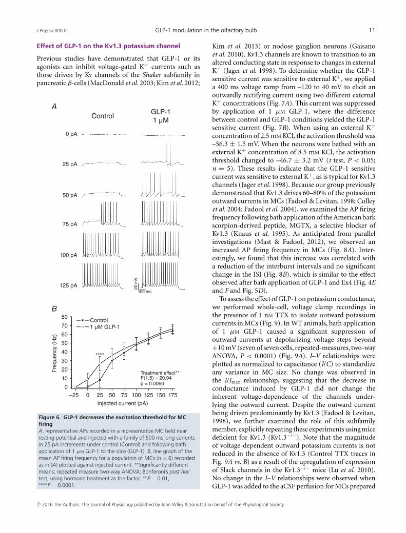
GLP-1 might increase not only the firing frequency,but also the AP threshold to enhance MC excitability.Although the stochastic firing pattern of MCs makes thedetermination of a precise rheobase challenging (Balu et al.2004), we were able to determine the current thresholdto elicit APs before and after GLP-1 perfusion (Fig. 6A).GLP-1 significantly decreased the excitation threshold asdetermined by the increased firing frequency observedacross the family of injected currents that demonstrateda significant effect of peptide treatment (F1,5 = 20.94;repeated-measures, two-way ANOVA, P < 0.0060; n = 6).A Bonferroni’s post hoc test showed that the differencelay with the lower current injection steps and not thehigher current injections that are sensitive to changes inISI (Fig. 6B).
25
20
15
10
5
0
Tim
e (m
in)
0 0 10 20Event frequency (Hz)Event time (ms)
1000 2000 3000 4000 5000
Wash
AC
SF
Ex4
1 μMC
ontrolA
CS
F
Con
trol
Ex4
1 μ
M
Was
h
a
b
c
40 m
V
500 ms
Ex4 1 μM Ex4 1 μM
Friedman test, p=0.0084 Friedman test, p=0.9640 Friedman test, p=0.003620
15
10
5
0
250
200
150
100
50
0
150
125
100
75
50
25
0
150
125
100
75
50
25
0Control WashEx4
1 μMControl WashEx4
1 μMControl WashEx4
1 μMControl WashEx4
1 μM
Freq
uenc
y (H
z)
Spo
ntan
eous
freq
uenc
y (%
con
trol
)
Inte
rbur
st in
terv
al (
% c
ontr
ol)
Inte
rspi
ke in
terv
al (
% c
ontr
ol)
a
b
ab
a
bab
A B
C D
a
b
c
Figure 5. GLP-1 analogue, Ex4, increases MC firing frequency via shortening the spike interburst intervalA and B, same experimental paradigm as in Fig. 4A and B but for bath application of 1 μM Ex4. C–D, same spikeanalysis, statistical metric and notations as in Fig. 4C and E in the analysis of Ex4 as opposed to natural peptideGLP-1.
C© 2016 The Authors. The Journal of Physiology published by John Wiley & Sons Ltd on behalf of The Physiological Society
J Physiol 000.0 GLP-1 modulation in the olfactory bulb 11
Effect of GLP-1 on the Kv1.3 potassium channel
Previous studies have demonstrated that GLP-1 or itsagonists can inhibit voltage-gated K+ currents such asthose driven by Kv channels of the Shaker subfamily inpancreatic β-cells (MacDonald et al. 2003; Kim et al. 2012;
0 pA
ControlGLP-11 μM
1 μM GLP-1
25 pA
50 pA
75 pA
100 pA
125 pA 20 m
V
100 ms
80
70
60
50
40
30
10
20
0–25 0 25 50 75 100 125 150 175
Injected current (pA)
Treatment effect**
****
Control
F(1,5) = 20.94p = 0.0060
Freq
uenc
y (H
z)
A
B
Figure 6. GLP-1 decreases the excitation threshold for MCfiringA, representative APs recorded in a representative MC held nearresting potential and injected with a family of 500 ms long currentsin 25 pA increments under control (Control) and following bathapplication of 1 μM GLP-1 to the slice (GLP-1). B, line graph of themean AP firing frequency for a population of MCs (n = 6) recordedas in (A) plotted against injected current. ∗∗Significantly differentmeans; repeated measure two-way ANOVA; Bonferoni’s post hoctest, using hormone treatment as the factor. ∗∗P � 0.01,∗∗∗∗P � 0.0001.
Kim et al. 2013) or nodose ganglion neurons (Gaisanoet al. 2010). Kv1.3 channels are known to transition to analtered conducting state in response to changes in externalK+ (Jager et al. 1998). To determine whether the GLP-1sensitive current was sensitive to external K+, we applieda 400 ms voltage ramp from –120 to 40 mV to elicit anoutwardly rectifying current using two different externalK+ concentrations (Fig. 7A). This current was suppressedby application of 1 μM GLP-1, where the differencebetween control and GLP-1 conditions yielded the GLP-1sensitive current (Fig. 7B). When using an external K+concentration of 2.5 mM KCl, the activation threshold was–56.3 ± 1.5 mV. When the neurons were bathed with anexternal K+ concentration of 8.5 mM KCl, the activationthreshold changed to –46.7 ± 3.2 mV (t test, P < 0.05;n = 5). These results indicate that the GLP-1 sensitivecurrent was sensitive to external K+, as is typical for Kv1.3channels (Jager et al. 1998). Because our group previouslydemonstrated that Kv1.3 drives 60–80% of the potassiumoutward currents in MCs (Fadool & Levitan, 1998; Colleyet al. 2004; Fadool et al. 2004), we examined the AP firingfrequency following bath application of the American barkscorpion-derived peptide, MGTX, a selective blocker ofKv1.3 (Knaus et al. 1995). As anticipated from parallelinvestigations (Mast & Fadool, 2012), we observed anincreased AP firing frequency in MCs (Fig. 8A). Inter-estingly, we found that this increase was correlated witha reduction of the interburst intervals and no significantchange in the ISI (Fig. 8B), which is similar to the effectobserved after bath application of GLP-1 and Ex4 (Fig. 4Eand F and Fig. 5D).
To assess the effect of GLP-1 on potassium conductance,we performed whole-cell, voltage clamp recordings inthe presence of 1 nM TTX to isolate outward potassiumcurrents in MCs (Fig. 9). In WT animals, bath applicationof 1 μM GLP-1 caused a significant suppression ofoutward currents at depolarizing voltage steps beyond+10 mV (seven of seven cells, repeated-measures, two-wayANOVA, P < 0.0001) (Fig. 9A). I–V relationships wereplotted as normalized to capacitance (I/C) to standardizeany variance in MC size. No change was observed inthe I/Imax relationship, suggesting that the decrease inconductance induced by GLP-1 did not change theinherent voltage-dependence of the channels under-lying the outward current. Despite the outward currentbeing driven predominantly by Kv1.3 (Fadool & Levitan,1998), we further examined the role of this subfamilymember, explicitly repeating these experiments using micedeficient for Kv1.3 (Kv1.3−/−). Note that the magnitudeof voltage-dependent outward potassium currents is notreduced in the absence of Kv1.3 (Control TTX traces inFig. 9A vs. B) as a result of the upregulation of expressionof Slack channels in the Kv1.3−/− mice (Lu et al. 2010).No change in the I–V relationships were observed whenGLP-1 was added to the aCSF perfusion for MCs prepared
C© 2016 The Authors. The Journal of Physiology published by John Wiley & Sons Ltd on behalf of The Physiological Society

12 N. Thiebaud and others J Physiol 000.0
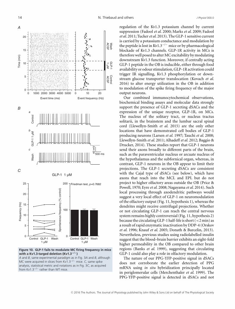
from these mice (eight of eight cells, P = 0.8089) (Fig. 9B).In a parallel current clamp study (Fig. 10A, B), GLP-1failed to significantly increase the AP firing frequencyin Kv1.3−/− animals in seven of the eight recorded cells(Fig. 10C). These data strongly support a GLP-evokedneuromodulation of MC excitability that is attributed inpart to an underlying Kv1.3 conductance. A plausiblemodel of how GLP-1 signalling may operate in the OBis discussed below and is modelled in Fig. 11.
Discussion
The present study is the first to report neuromodulation byincretin hormones in the olfactory system. We have usedan advantageous transgenic marker to identify the PPGneurons responsible for local processing in the OB. Thereceptor for GLP-1 is localized to the first-order neurons,the MCs, which are known to be modulated by a gamutof other metabolically-important molecules linked to the
[K+]ext = 2.5 mM [K+]ext = 8.5 mM
45I/C (pA/pF)
A
BVoltage (mV) Voltage (mV)
Voltage (mV) Voltage (mV)
GLP-1 current (control - GLP-1)
a - Control
a-b
c-d
b - GLP-1
I/C (pA/pF)
I/C (pA/pF) I/C (pA/pF)
c - Control
d - GLP-1403530252015
5
20 40 600–5–10
–20–40–60–80–100–120
20 40 600–20–40–60–80–100–120
10
10
12
8
6
4
–2
2
20 40 600–20–40–60–80–100–120
10
12
14
8
6
4
–2
2
404550
3530252015
5
20 40 600–5–10
–20–40–60–80–100–120
10
Figure 7. The threshold for activation ofthe GLP-1 sensitive current shifts with achange in the equilibrium potential for K+(EK) generated by altering the externalconcentration for K+A, representative I/C plotted relationship ofcurrents evoked using a 400 ms voltage rampfrom –120 to +40 mV before (a, Control, blackline) and after (b, GLP-1, grey line) bathapplication of 1 μM GLP-1 to the slice underconditions of 2.5 mM (left) vs. 8.5 mM (right)external KCl concentration. B, the GLP-1sensitive current is calculated by a subtractionof a – b to yield an activation threshold of–56.3 ± 1.5 mV using a 2.5 mM external K+concentration (left) compared with theright-shifted activation threshold of–46.7 ± 3.2 mV using a 8.5 mM external K+concentration (right).
300 160
140
120
100
80
60
40
20
0
250
200
150
100
50
00 5 10 15 20 25 30 0 5 10 15 20 25 30
40 m
V
MG
TX
1 n
M15
min
MG
TX
1 n
M5
min
Con
trol
500 ms Eve
nt fi
ring
freq
uenc
y (%
of c
ontr
ol)
Inte
rval
(%
of c
ontr
ol)
Interspike intervalInterburst interval
Time after 1 nM Margatoxintreatment (min)
Time after 1 nM Margatoxintreatment (min)
a
b
c
A B
Figure 8. MGTX increases AP firing frequency in MCs by decreasing the interburst interval and not theISIA, representative APs recorded in a representative MC under (a) baseline ASCF control bath conditions (Control),(b) after 5 min of 1 nM MGTX and (c) after 15 min of 1 nM MGTX stimulation, reflecting the slow Kon reportedfor this small peptide molecule that blocks the vestibule of the Kv1.3 channel. B, left: line graph of the meanfiring frequency over time for five sampled MCs normalized to initial AP firing rate before MGTX treatment (time0 min). Right: same population of MCs plotted to examine AP mean ISI (closed symbol) and interburst interval(open symbols) over time.
C© 2016 The Authors. The Journal of Physiology published by John Wiley & Sons Ltd on behalf of The Physiological Society

J Physiol 000.0 GLP-1 modulation in the olfactory bulb 13
Control1 nM TTX
A
B
Control
Wildtype
+ 1 μM GLP-1
GLP-1 1 μM
Voltage step (mV) Voltage step (mV)
50 ms
12I/C (pA/pF) I/Imax
10
8
6
**
* *
4
2
–2–100 –80 –60 –40 –20 0 20 40
Kv1.3–/–
1.2
1.0
0.8
0.6
0.4
0.2
–0.2–100 –80 –60 –40 –20 0 20 40
500
pA
Control1 nM TTX
Control
+ 1 μM GLP-1
GLP-1 1 μM
Voltage step (mV) Voltage step (mV)
50 ms
28I/C (pA/pF) I/Imax
20
24
16
12
8
4
–4–100 –80 –60 –40 –20 0 20 40
1.2
1.0
0.8
0.6
0.4
0.2
–0.2–100 –80 –60 –40 –20 0 20 40
500
pAFigure 9. The magnitude of MCvoltage-activated outward currents aredecreased following GLP-1 application inWT mice but not for mice with aKv1.3-targeted deletion (Kv1.3–/–)A, top: representative family ofvoltage-activated currents elicited bystepping the command voltage (Vc) in10 mV increments (–100 to +40 mV) from aholding voltage (Vh) of –80 mV using a400 ms pulse duration (Pd) and a 45 sinterpulse interval. MC recordings wereacquired from WT mice. TTX was applied tothe bath to isolate outward potassiumconductances (Control, 1 nM TTX) before(left traces) and after (right traces) bathapplication of the peptide (+ 1 μM GLP-1).Bottom left: plotted I/C relationship for fiveMCs recorded as in (A). Solid symbols, before(Control); open symbols, after GLP-1(GLP-11 μM) bath application. Significantly differentmeans; repeated measures two-wayANOVA; Bonfernoni’s post hoc test,∗P � 0.05; ∗∗P � 0.001. Right, same as leftI/C but normalized to that of the +40 mVvoltage step. B, as in (A), except MCs wererecorded from Kv1.3−/− mice (Kv1.3−/−).
C© 2016 The Authors. The Journal of Physiology published by John Wiley & Sons Ltd on behalf of The Physiological Society
14 N. Thiebaud and others J Physiol 000.0
Event time (ms)
GLP-1 1 μM
Tim
e (m
in)
20
A
B
C
15
Wash
AC
SF
GLP
-11 μ
MC
ontrolA
CS
F
Was
hG
LP-1
1 μ
MC
ontr
ol
WashGLP-11 μM
Control WashGLP-11 μM
Control
10
5
00
a
b
c
1000 2000 3000 4000 5000 0
500 ms40 m
V10
a
b
c
20
Event frequency (Hz)
Freq
uenc
y (H
z)
Spo
ntan
eous
freq
uenc
y (%
con
trol
)
200
100
0
25 Friedman test, p=0.7682
20
15
10
5
0
Figure 10. GLP-1 fails to modulate MC firing frequency in micewith a Kv1.3-targed deletion (Kv1.3−/−)A and B, same experimental paradigm as in Fig. 3A and B, althoughMC were acquired in slices from Kv1.3−/− mice. C, same spikeanalysis, statistical metric and notations as in Fig. 3C, as acquiredfrom Kv1.3−/− rather than WT mice.
regulation of the Kv1.3 potassium channel by currentsuppression (Fadool et al. 2000; Marks et al. 2009; Fadoolet al. 2011; Tucker et al. 2013). The GLP-1 sensitive currentis carried by a potassium conductance and modulation bythe peptide is lost in Kv1.3−/− mice or by pharmacologicalblockade of Kv1.3 channels. GLP-1R activity in MCs istherefore well posed to alter MC excitability by modulatingdownstream Kv1.3 function. Moreover, if centrally actingGLP-1 peptide in the OB is inducible, either through foodavailability or odour stimulation, GLP-1R activation couldtrigger IR signalling, Kv1.3 phosphorylation or down-stream glucose transporter translocation (Kovach et al.2016) to alter energy utilization in the OB in additionto modulation of the spike firing frequency of the majoroutput neurons.
Our combined immunocytochemical observations,biochemical binding assays and molecular data stronglysupport the presence of GLP-1 secreting dSACs and theexpression of the unique receptor, GLP-1R, on MCs.The nucleus of the solitary tract, or nucleus tractussolitarii, in the brainstem and the lumbar sacral spinalcord (Llewellyn-Smith et al. 2015) are the only otherlocations that have demonstrated cell bodies of GLP-1producing neurons (Larsen et al. 1997; Tauchi et al. 2008;Llewellyn-Smith et al. 2011; Alhadeff et al. 2012; Baggio &Drucker, 2014). These studies report that GLP-1 neuronssend their axons broadly to different parts of the brain,such as the paraventricular nucleus or arcuate nucleus ofthe hypothalamus and the subfornical organ, whereas, incontrast, GLP-1 neurons in the OB appear to limit theirprojections. The GLP-1 secreting dSACs are consistentwith the Cajal type of dSACs (see below), which haveaxons that reach into the MCL and EPL but do notproject to higher olfactory areas outside the OB (Price &Powell, 1970; Eyre et al. 2008; Nagayama et al. 2014). Suchlocal processing through axodendritic pathways wouldsuggest a very local effect of GLP-1 on neuromodulationof the olfactory output (Fig. 11, hypothesis 1), whereas thedendrites might receive centrifugal projections. Whetheror not circulating GLP-1 can reach the central nervoussystem remains highly controversial (Fig. 11, hypothesis 2)because the circulating GLP-1 half-life is short (<2 min) asa result of rapid enzymatic inactivation by DDP-4 (Orskovet al. 1996; Knauf et al. 2005; Donath & Burcelin, 2013).Nevertheless, previous studies using radiolabelled insulinsuggest that the blood–brain barrier exhibits an eight-foldhigher permeability in the OB compared to other brainregions (Banks et al. 1999), suggesting that circulatingGLP-1 could also play a role in olfactory modulation.
The nature of our PPG-YFP-positive signal in dSACsdoes not corroborate the earlier detection of PPGmRNA using in situ hybridization principally locatedin periglomerular cells (Merchenthaler et al. 1999). ThePPG-YFP-positive signal is detected in dSACs and not
C© 2016 The Authors. The Journal of Physiology published by John Wiley & Sons Ltd on behalf of The Physiological Society
J Physiol 000.0 GLP-1 modulation in the olfactory bulb 15
superficial short axon cells surrounding the glomeruli(juxtaglomerular neurons) (Kiyokage et al. 2010). ThedSACs are distinguished morphologically from granulecells as a result of their intermediate size betweengranule cells and MCs, and their cell bodies are contained
in the GCL or internal plexiform layer (Eyre et al. 2008).With well-impregnated Golgi sections, Ramon y Cajal(1911) and subsequent investigators distinguished seventypes of non-granule cells in the GCL, based upon somasize, shape and location; orientation of dendrites; and
Centrifugal inputMetabolic control
Hormonal Control?(CCK, Leptin)
Mitral Cell
dSAC
Blood VesselLeaky Blood Brain Barrier
GLM
MCL
GCL
1
2
3
4
GLP-1R
Kv1.3
GLP-1
GLP-1
InsR
?
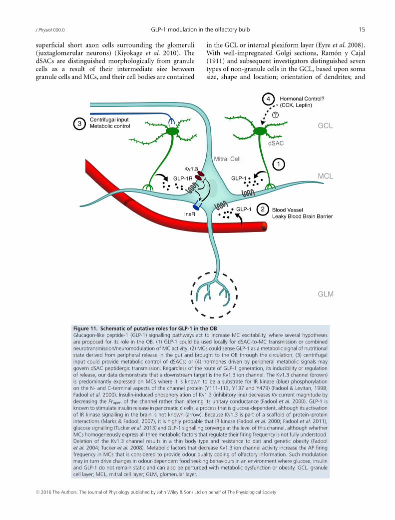
Figure 11. Schematic of putative roles for GLP-1 in the OBGlucagon-like peptide-1 (GLP-1) signalling pathways act to increase MC excitability, where several hypothesesare proposed for its role in the OB. (1) GLP-1 could be used locally for dSAC-to-MC transmission or combinedneurotransmission/neuromodulation of MC activity; (2) MCs could sense GLP-1 as a metabolic signal of nutritionalstate derived from peripheral release in the gut and brought to the OB through the circulation; (3) centrifugalinput could provide metabolic control of dSACs; or (4) hormones driven by peripheral metabolic signals maygovern dSAC peptidergic transmission. Regardless of the route of GLP-1 generation, its inducibility or regulationof release, our data demonstrate that a downstream target is the Kv1.3 ion channel. The Kv1.3 channel (brown)is predominantly expressed on MCs where it is known to be a substrate for IR kinase (blue) phosphorylationon the N- and C-terminal aspects of the channel protein (Y111-113, Y137 and Y479) (Fadool & Levitan, 1998;Fadool et al. 2000). Insulin-induced phosphorylation of Kv1.3 (inhibitory line) decreases Kv current magnitude bydecreasing the Propen of the channel rather than altering its unitary conductance (Fadool et al. 2000). GLP-1 isknown to stimulate insulin release in pancreatic β cells, a process that is glucose-dependent, although its activationof IR kinase signalling in the brain is not known (arrow). Because Kv1.3 is part of a scaffold of protein–proteininteractions (Marks & Fadool, 2007), it is highly probable that IR kinase (Fadool et al. 2000; Fadool et al. 2011),glucose signalling (Tucker et al. 2013) and GLP-1 signalling converge at the level of this channel, although whetherMCs homogeneously express all three metabolic factors that regulate their firing frequency is not fully understood.Deletion of the Kv1.3 channel results in a thin body type and resistance to diet and genetic obesity (Fadoolet al. 2004; Tucker et al. 2008). Metabolic factors that decrease Kv1.3 ion channel activity increase the AP firingfrequency in MCs that is considered to provide odour quality coding of olfactory information. Such modulationmay in turn drive changes in odour-dependent food seeking behaviours in an environment where glucose, insulinand GLP-1 do not remain static and can also be perturbed with metabolic dysfunction or obesity. GCL, granulecell layer; MCL, mitral cell layer; GLM, glomerular layer.
C© 2016 The Authors. The Journal of Physiology published by John Wiley & Sons Ltd on behalf of The Physiological Society

16 N. Thiebaud and others J Physiol 000.0
the presence or absence of dendritic spines, for whichmembers of a subdivision of interneuron types wereclassified as dSACs (Ramon y Cajal S., 1911; Price & Powell,1970; Schneider & Macrides, 1978). A recent review byNagayama et al. (2014) provides an excellent summary ofthe traditional morphological and synaptic distinctionsacross the dSACs compared to recent reclassificationsusing a different nomenclature (Eyre et al. 2008; Eyreet al. 2009; Nagayama et al. 2014). The new classificationsuggests that there are three subtypes of dSACs inthe inframitral layers: (1) the largest population withcolumn-like axonal arbors confined predominantly tothe external plexiform layer, or EPL-dSACs, and witha soma location in the GCL (also referred to as Blanescells and Cajal cells); (2) a smaller population where thesoma is retained in the internal plexiform layer with ahorizontal axonal extension to the glomerular layer, orGL-dSACs (also referred to as Horizontal cells and Golgicells); and (3) the smallest population with axonal arborsconstrained to the GCL only, or GCL-dSAC (also referredto as Horizontal cells and Golgi cells). Our combinedmorphological data (Fig. 1) and biophysical evidence(Pressler et al. 2013; Fadool & Thiebaud, 2015) narrowsdown the identification of the PPG-YFP-positive cells tothe EPL-dSAC classification and, in particular, to Cajalcells. To our knowledge, this is the first report of aneuromodulatory effect linked to Cajal cells. Boyd et al.(2012) report that all dSACs are GABAergic and that Cajalcells, in particular, participate in disynaptic relays (Boydet al. 2012). Understanding all of the contributions tothe synaptic network and the capacity for modulation isfundamental to clarifying how Cajal cells might participatein sensory processing by controlling the main OB networkvia inhibition of other interneurons. Our data do notsimplify the potential synaptic function of these dSACs.The present study shows that Cajal cells contain notonly GABA, but also GLP-1, and could release both toinfluence synaptic transmission within the OB. Moreover,GLP-1 release from Cajal cells could be modulated by thenutritional state to fine-tune communication between OBlaminae. We also cannot exclude the possibility that locallyreleased GLP-1 has a solely intrinsic or paracrine neuro-modulatory effect within the defined OB circuit withouta metabolic-linked function. Whether GLP-1 levels in theOB are modified in a nutritional state-dependent fashionis completely unknown and deserves further study.
Our mapped distribution of GLP-1R in MCs (Fig. 2),combined with our injection of fluorescently-conjugatedEx4 that binds selectively to GLP-1R (Fig. 3), providesparallel support for evoked GLP-1 release activating MCsby binding to receptors on these cells. The biotinylationresults, along with the Ex4 binding assay, suggest thatthe receptor distribution along MCs is homogeneous.These findings are of interest given the recent studiesreporting a molecular and physiological diversity of MCs
that may not be a homogeneous population of neuronswith respect to voltage-gated ion channel expression(Angelo et al. 2012; Padmanabhan & Urban, 2014).Although our earlier immunocytochemical data (Fadool &Levitan, 1998; Fadool et al. 2000) support a homogeneousexpression for Kv1.3 ion channel, GLP-1R activation mayhave other downstream targets to mediate AP firingfrequency. We also cannot exclude the possibility thatthere are preparation factors explaining why 50–64% ofMCs exhibited enhanced firing frequency in response toGLP-1 or its analogue. Sectioning of the lateral dendriticarbors during the slice preparation could result in lessthan 100% of cells being responsive in our currentclamp recordings. Our electrophysiological findings showthat most (but not all) of the sampled MCs exhibita rapid and reversible increase in AP firing frequencyin response to GLP-1 or its analogue; therefore, theydo not unambiguously reveal whether all MCs haveidentical neuromodulatory pathways. There are also adiversity of adaptor proteins interacting with K+ channelsacross the OB neurolamina (Marks & Fadool, 2007)that could refine membrane excitability and olfactorycoding capacity. Finally, post-translational modification ofdelayed rectifiers or their auxillary β subunits has involvedGLP-1 evoked Ser/Thr phosphorylation and acetylation(Kim et al. 2012). Because Kv1.3 has been well studiedand is known to be a substrate for Tyr phosphorylationby several neuromodulators of MC AP firing frequency(Fadool & Levitan, 1998; Fadool et al. 2000; Tucker &Fadool, 2002; Colley et al. 2004; Fadool et al. 2011; Mast &Fadool, 2012), it would be intriguing to explore whetherGLP-1 stimulation of the OB can phosphorylate Kv1.3 oneither Ser or Thr residues or use a cAMP independentmechanism to phosphorylate Kv1.3 on Tyr residues linkedto MAPK activation (Egan et al. 2003).
As a post-prandial anorexigenic signal, GLP-1modulation of MC excitability might be expected to becorrelated with a reduction in the sensitivity to foododours. However, the change in GLP-1 concentration atthe level of the OB following a meal is unknown andan increase in MC excitability does not necessitate again in olfactory function. Indeed, MC intrinsic firingis complex and variable, and may promote faster odourdiscrimination with increased inhibition. From our earlierwork (Fadool et al. 2000), we have demonstrated thatthe concentration of another neuromodulatory moleculein the OB (insulin) does not follow what is anticipatedfrom measured levels in the plasma. For example, in theseexperiments, an overnight fast induced a plummeting ofplasma insulin, whereas that in the OB remained high.As a post-prandial anorexigenic signal, we do not knowwhat explicitly activates PPG neurons, nor are we certainthat an elevation of GLP-1 in the periphery translates toa linear change centrally at the OB. Second, inhibition ofMCs can be as critical to olfactory coding as excitation,
C© 2016 The Authors. The Journal of Physiology published by John Wiley & Sons Ltd on behalf of The Physiological Society

J Physiol 000.0 GLP-1 modulation in the olfactory bulb 17
and MC intrinsic firing properties can be modulateddifferentially to several odours, being state- or sensoryexperience-dependent (Kato et al. 2012). Some intriguingwork by Nunes & Kuner (2015) demonstrated that tuningMC inhibition via disinhibition of granule cells resulted inmice with faster odour discrimination without disruptionof olfactory learning. Therefore the inhibitory network inthe OB is working to adjust the speed of odour sensoryprocessing. Gschwend et al. (2015) report that neuro-nal pattern separation is critical for odour discriminationlearning, where enhancing the inhibition of MCs increasesodour-evoked pattern separation of activated glomeruliand improves odour discrimination learning. Lastly, Liet al. (2015) use a go no–go association task to reveal thatchanges in MC firing frequency encode information aboutthe rewarding nature of the odour, rather than the odouridentity. GLP-1 modulation of MC firing frequency maytherefore serve roles beyond that of a changed sensitivityto food odours to include how (context and circuitry) MCfiring becomes changed as a critical variable influencingodour discrimination (Gschwend et al. 2015; Li et al. 2015).
Neuromodulation by GLP-1 or its analogue has beencharacterized in central neurons, such as hypothalamichypocretin/orexin neurons, or within vagal neural andmotor pathways (Acuna-Goycolea & van den Pol, 2004;Wan et al. 2007a; Wan et al. 2007b; Gaisano et al. 2010).Similar to our findings in the OB, GLP-1 can inhibitvoltage-gated potassium channels leading to an increase inevoked Aps, such as observed in nodose ganglion neurons(Gaisano et al. 2010). We have previously demonstrated,both by pharmacological and genetic strategies, that Kv1.3drives 60–80% of the outward potassium currents in theMCs (Fadool & Levitan, 1998; Colley et al. 2004; Fadoolet al. 2004; Fadool et al. 2011); therefore, we investigatedthe effect of GLP-1 stimulation in mice deficient forthis Kv family member. Similar to that found for insulin(Fadool et al. 2011) and glucose (Tucker et al. 2013),GLP-1 failed to decrease outward currents in voltage clamprecordings and failed to modulate AP firing frequencyin current clamp experiments in which Kv1.3–/– micewere substituted for WT mice. These data, combinedwith the fact that a Kv1.3 pore blocker selectively alteredthe spike train interburst but not ISI (Fig. 8), stronglysupport the involvement of the Kv1.3 ion channel asa downstream signalling component of GLP-1 neuro-modulation of MCs (Fig. 11). Interestingly, although allthree metabolically-important molecules affect MC firingin a Kv1.3-dependent manner, the mechanism shows adifference in onset latency. Insulin elicits an increase inAP firing frequency that is much slower to develop (inthe order of minutes) and difficult to wash out, whichmay be the result of the Tyr-dependent phosphorylationobserved for Kv1.3 (Fadool & Levitan, 1998; Marks et al.2009; Fadool et al. 2011). Modulation by glucose is rapidand reversible (Tucker et al. 2013) and on the same
time course as that for GLP-1; however, glucose can beexcitatory or inhibitory, suggesting a heterogeneity in thetype of MC that exhibits glucose modulation or sensing.Although we have not yet pursued strict co-localizationof Kv1.3 and GLP-1R, our current immunocytochemicaldata suggest that IR kinase and GLP-1R are co-localized,and our previously reported findings demonstrate that IRkinase and Kv1.3 are not only co-localized, but also canbe co-immunoprecipitated in the presence of a varietyof adaptor proteins present in the OB (Marks & Fadool,2007; Marks et al. 2009). There is certainly a glucosedependence of GLP-1 evoked insulin release. The inter-action of GLP-1 with glucose and insulin sensing pathwayshas been well studied in pancreatic β-cells (Holst, 2007).One could speculate an active interaction of GLP-1, insulinand glucose in neurons such as MCs that might co-expressGLP-1R, IR kinase (Fadool et al. 2000) and glucosetransporters such as sodium-glucose transport protein-1or insulin sensitive, glucose transporter 4 (Al Koborssyet al. 2014), of which the latter is known to beregulated by Kv1.3 (Xu et al. 2004). For conditionsduring which insulin, glucose or GLP-1 peptide werepresent, phosphorylation or other means of decreasingKv conductance would shift glucose transporter 4 trans-location to the membrane (Kovach CP, Al KoborssyD, Huang Z, Chelette B, Fadool JM, & Fadool DA,unpublished) as a positive feedback for continued glucoseutilization in MCs to yield an ATP substrate for continuedactivation of GLP-1R.
It is interesting that another class of chemosensoryreceptors has been linked to the operation of GLP-1signalling. The L cells of the gut have been found toexpress α-gustducin, sweet taste receptors (T1R2/T1R3)and PLCβ2 (Jang et al. 2007), and dietary sugar hasthe capacity to increase sodium-dependent glucose trans-porter isoform 1 in the presence of the taste trans-duction machinery (Margolskee et al. 2007). Jang et al.(2007) demonstrate that T1R3 and gustducin have arole in glucose-mediated GLP-1 release and may serveas a gut-derived lumenal glucose sensor. Reciprocally,GLP-1 was found to be expressed in the tongue in twodistinct classes of taste cells and the receptor is foundon intragemmal afferent nerve fibres (Shin et al. 2008).Gene-targeted deletion of the receptor reduced sweet tastesensitivity in behaving mice as determined using briefaccess gustatory tests. In light of these gut/chemosensoryparallels with the taste system, it is not unexpected thatthe olfactory system could have the ability to regulateglucose-dependent incretin release or the ability to senseGLP-1 hormone.
In summary, our findings generate several new hypo-theses (Fig. 11) concerning the role of GLP-1 in theOB, each of which is important to consider within theframework of future investigations: (1) GLP-1 could beused locally for dSAC-to-MC transmission or combined
C© 2016 The Authors. The Journal of Physiology published by John Wiley & Sons Ltd on behalf of The Physiological Society
18 N. Thiebaud and others J Physiol 000.0
neuromodulation of MC activity; (2) MCs could senseGLP-1 as a metabolic signal of nutritional state derivedfrom peripheral release in the gut and brought to theOB through the circulation; (3) centrifugal input couldprovide metabolic control of dSACs; or (4) hormonesdriven by peripheral metabolic signals may govern dSACpeptidergic transmission. Activation of the dSAC inter-neuron population to co-release GLP-1 peptide and aneurotransmitter (hypothesis 1) could be explored usingselect synaptic and GLP-1 receptor inhibitors. GLP-1 isknown to be an inducible peptide and changes in thehomeostatic state of the organism could be perceivedthrough circulating GLP-1 across a leaky blood–brainbarrier at the level of the OB (hypothesis 2). If peripheralchanges in GLP-1 are detected at the OB following a meal,fasting or as a consequence of a metabolic disease state(hypothesis 2–3), or if odour signalling pathways releaseGLP-1 in the OB locally (hypothesis 1), GLP-1 sensitivepotassium currents are capable of enhancing the APfiring frequency of the major output neurons or loweringtheir threshold for excitability. Although we cannot yetdiscriminate between these alternative models, the presentstudy definitively demonstrates that the GLP-1 signallingpathway is present in the OB and can be a regulator ofneuronal activity. Taken together, the uncovered GLP-1signalling and the highly-expressed glucose and insulinmetabolic factors in this region are especially interestingin the context of the modulation of olfactory perceptionand food intake. The reported discovery of this incretinhormone in the OB challenges future investigationsto examine the olfactory system as a new potentialtherapeutic target for controlling metabolic imbalance.
References
Acuna-Goycolea C & van den Pol A (2004). Glucagon-likepeptide 1 excites hypocretin/orexin neurons by direct andindirect mechanisms: implications for viscera-mediatedarousal. J Neurosci 24, 8141–8152.
Aime P, Hegoburu C, Jaillard T, Degletagne C, Garcia S,Messaoudi B, Thevenet M, Lorsignol A, Duchamp C, MoulyAM, & Julliard AK (2012). A physiological increase of insulinin the olfactory bulb decreases detection of a learned aversiveodor and abolishes food odor-induced sniffing behavior inrats. PLoS ONE 7, e51227.
Al Koborssy D, Palouzier-Paulignan B, Salem R, Thevenet M,Romestaing C, & Julliard AK (2014). Cellular and molecularcues of glucose sensing in the rat olfactory bulb. FrontNeurosci 8, 333.
Alhadeff AL, Rupprecht LE, & Hayes MR (2012). GLP-1neurons in the nucleus of the solitary tract project directly tothe ventral tegmental area and nucleus accumbens to controlfor food intake. Endocrinology 153, 647–658.
Angelo K, Rancz EA, Pimentel D, Hundahl C, Hannibal J,Fleischmann A, Pichler B, & Margrie TW (2012). Abiophysical signature of network affiliation and sensoryprocessing in mitral cells. Nature 488, 375–378.
Apelbaum AF, Perrut A, & Chaput M (2005). Orexin A effectson the olfactory bulb spontaneous activity and odorresponsiveness in freely breathing rats. Regul Pept 129,49–61.
Ayala JE, Bracy DP, James FD, Julien BM, Wasserman DH, &Drucker DJ (2009). The glucagon-like peptide-1 receptorregulates endogenous glucose production and muscleglucose uptake independent of its incretin action.Endocrinology 150, 1155–1164.
Badonnel K, Lacroix MC, Monnerie R, Durieux D, Caillol M, &Baly C (2012). Chronic restricted access to food leading toundernutrition affects rat neuroendocrine status andolfactory-driven behaviors. Horm Behav 62, 120–127.
Baggio LL & Drucker DJ (2014). Glucagon-like peptide-1receptors in the brain: controlling food intake and bodyweight. J Clin Invest 124, 4223–4226.
Balu R, Larimer P, & Strowbridge BW (2004). Phasic stimulievoke precisely timed spikes in intermittently dischargingmitral cells. J Neurophysiol 92, 743–753.
Banks WA, Kastin AJ, & Pan W (1999). Uptake anddegradation of blood-borne insulin by the olfactory bulb.Peptides 20, 373–378.
Boyd AM, Sturgill JF, Poo C, & Isaacson JS (2012). Corticalfeedback control of olfactory bulb circuits. Neuron 76,1161–1174.
Briones M & Bajaj M (2006). Exenatide: a GLP-1 receptoragonist as novel therapy for type 2 diabetes mellitus. ExpertOpin Pharmacother 7, 1055–1064.
Colley BS, Tucker K, & Fadool DA (2004). Comparison ofmodulation of Kv1.3 channel by two receptor tyrosinekinases in olfactory bulb neurons of rodents. ReceptorsChannels 10, 25–36.
Cork SC, Richards JE, Holt MK, Gribble FM, Reimann F, &Trapp S (2015). Distribution and characterisation ofglucagon-like peptide-1 receptor expressing cells in themouse brain. Mol Metab 4, 718–731.
Cowley MA, Smith RG, Diano S, Tschop M, Pronchuk N,Grove KL, Strasburger CJ, Bidlingmaier M, Esterman M,Heiman ML, Garcia-Segura LM, Nillni EA, Mendez P, LowMJ, Sotonyi P, Friedman JM, Liu H, Pinto S, Colmers WF,Cone RD & Horvath TL (2003). The distribution andmechanism of action of ghrelin in the CNS demonstrates anovel hypothalamic circuit regulating energy homeostasis.Neuron 37, 649–661.
De Saint Jan D & Westbrook GL (2007). Disynapticamplification of metabotropic glutamate receptor 1responses in the olfactory bulb. J Neurosci 27, 132–140.
Donath MY & Burcelin R (2013). GLP-1 effects on islets:hormonal, neuronal, or paracrine? Diabetes Care 36 Suppl 2,S145–S148.
Donnelly D (2012). The structure and function of theglucagon-like peptide-1 receptor and its ligands. Br JPharmacol 166, 27–41.
Dossat AM, Lilly N, Kay K, & Williams DL (2011).Glucagon-like peptide 1 receptors in nucleus accumbensaffect food intake. J Neurosci 31, 14453–14457.
Egan JM, Bulotta A, Hui H, & Perfetti R (2003). GLP-1receptor agonists are growth and differentiation factors forpancreatic islet beta cells. Diabetes Metab Res Rev 19,115–123.
C© 2016 The Authors. The Journal of Physiology published by John Wiley & Sons Ltd on behalf of The Physiological Society
J Physiol 000.0 GLP-1 modulation in the olfactory bulb 19
Eng J, Kleinman WA, Singh L, Singh G, & Raufman JP (1992).Isolation and characterization of exendin-4, an exendin-3analogue, from Heloderma suspectum venom. Furtherevidence for an exendin receptor on dispersed acini fromguinea pig pancreas. J Biol Chem 267, 7402–7405.
Eyre MD, Antal M, & Nusser Z (2008). Distinct deepshort-axon cell subtypes of the main olfactory bulb providenovel intrabulbar and extrabulbar GABAergic connections.J Neurosci 28, 8217–8229.
Eyre MD, Kerti K, & Nusser Z (2009). Molecular diversity ofdeep short-axon cells of the rat main olfactory bulb. Eur JNeurosci 29, 1397–1407.
Fadool DA & Levitan IB (1998). Modulation of olfactory bulbneuron potassium current by tyrosine phosphorylation.J Neurosci 18, 6126–6137.
Fadool DA & Thiebaud N (2015). Optogenetic activation ofpre-proglucagon neurons in the mouse olfactory bulbcontrols mitral cell activity. Chem. Senses 40, 603.
Fadool DA, Tucker K, & Pedarzani P (2011). Mitral cells of theolfactory bulb perform metabolic sensing and are disruptedby obesity at the level of the Kv1.3 ion channel. PLoS One 6,e24921.
Fadool DA, Tucker K, Perkins R, Fasciani G, Thompson RN,Parsons AD, Overton JM, Koni PA, Flavell RA, & KaczmarekLK (2004). Kv1.3 channel gene-targeted deletion produces‘Super-Smeller Mice’ with altered glomeruli, interactingscaffolding proteins, and biophysics. Neuron 41, 389–404.
Fadool DA, Tucker K, Phillips JJ, & Simmen JA (2000). Braininsulin receptor causes activity-dependent currentsuppression in the olfactory bulb through multiplephosphorylation of Kv1.3. J Neurophysiol 83, 2332–2348.
Feng G, Mellor RH, Bernstein M, Keller-Peck C, Nguyen QT,Wallace M, Nerbonne JM, Lichtman JW, & Sanes JR (2000).Imaging neuronal subsets in transgenic mice expressingmultiple spectral variants of GFP. Neuron 28, 41–51.
Gaisano GG, Park SJ, Daly DM, & Beyak MJ (2010).Glucagon-like peptide-1 inhibits voltage-gated potassiumcurrents in mouse nodose ganglion neurons.Neurogastroenterol Motil 22, 470–9, e111.
Gascuel J, Lemoine A, Rigault C, Datiche F, Benani A, PenicaudL & Lopez-Mascaraque L (2012). Hypothalamus-olfactorysystem crosstalk: orexin a immunostaining in mice. FrontNeuroanat 6, 44.
Goke R, Fehmann HC, Linn T, Schmidt H, Krause M, Eng J, &Goke B (1993). Exendin-4 is a high potency agonist andtruncated exendin-(9-39)-amide an antagonist at theglucagon-like peptide 1-(7-36)-amide receptor ofinsulin-secreting beta-cells. J Biol Chem 268, 19650–19655.
Gromada J, Anker C, Bokvist K, Knudsen LB, & Wahl P(1998a). Glucagon-like peptide-1 receptor expression inXenopus oocytes stimulates inositol trisphosphate-dependentintracellular Ca2+ mobilization. FEBS Lett 425, 277–280.
Gromada J, Bokvist K, Ding WG, Holst JJ, Nielsen JH, &Rorsman P (1998b). Glucagon-like peptide 1 (7-36) amidestimulates exocytosis in human pancreatic beta-cells by bothproximal and distal regulatory steps in stimulus-secretioncoupling. Diabetes 47, 57–65.
Grundy D (2015). Principles and standards for reportinganimal experiments in The Journal of Physiology andExperimental Physiology. J Physiol 593, 2547–2549.
Gschwend O, Abraham NM, Lagier S, Begnaud F, Rodriguez I,Carleton A (2015). Neuroanl pattern segretation in theolfactory bulb improves odor discrimination learning. NatNeurosci 18, 1474–1482.
Hardy AB, Aioun J, Baly C, Julliard KA, Caillol M, Salesse R, &Duchamp-Viret P (2005). Orexin A modulates mitral cellactivity in the rat olfactory bulb: patch-clamp study on slicesand immunocytochemical localization of orexin receptors.Endocrinology 146, 4042–4053.
Heller RS, Kieffer TJ, & Habener JF (1996). Point mutations inthe first and third intracellular loops of the glucagon-likepeptide-1 receptor alter intracellular signaling. BiochemBiophys Res Commun 223, 624–632.
Hill JM, Lesniak MA, Pert CB, & Roth J (1986).Autoradiographic localization of insulin receptors in ratbrain: prominence in olfactory and limbic areas.Neuroscience 17, 1127–1138.
Hisadome K, Reimann F, Gribble FM, & Trapp S (2010).Leptin directly depolarizes preproglucagon neuronsin the nucleus tractus solitarius: electrical properties ofglucagon-like peptide 1 neurons. Diabetes 59,1890–1898.
Holst JJ (2007). The physiology of glucagon-like peptide 1.Physiol Rev 87, 1409–1439.
Jager H, Rauer H, Nguyen AN, Aiyar J, Chandy KG, & GrissmerS (1998). Regulation of mammalian Shaker-related K+channels: evidence for non-conducting closed andnon-conducting inactivated states. J Physiol 506,291–301.
Jang HJ, Kokrashvili Z, Theodorakis MJ, Carlson OD, Kim BJ,Zhou J, Kim HH, Xu X, Chan SL, Juhaszova M, Bernier M,Mosinger B, Margolskee RF, & Egan JM (2007).Gut-expressed gustducin and taste receptors regulatesecretion of glucagon-like peptide-1. Proc Natl Acad Sci USA104, 15069–15074.
Jin SL, Han VK, Simmons JG, Towle AC, Lauder JM, & LundPK (1988). Distribution of glucagonlike peptide I (GLP-I),glucagon, and glicentin in the rat brain: animmunocytochemical study. J Comp Neurol 271,519–532.
Kato HK, Chu MW, Isaacson JS, Komiyama T (2012). Dynamicsensory representations in the olfactory bulb: modulation bywakefulness and experience. Neuron 76, 962–975.
Kim SJ, Ao Z, Warnock G, & McIntosh CH (2013).Incretin-stimulated interaction between beta-cell Kv1.5 andKvbeta2 channel proteins involves acetylation/deacetylationby CBP/SirT1. Biochem J 451, 227–234.
Kim SJ, Widenmaier SB, Choi WS, Nian C, Ao Z, Warnock G,& McIntosh CH (2012). Pancreatic beta-cell prosurvivaleffects of the incretin hormones involve post-translationalmodification of Kv2.1 delayed rectifier channels. Cell DeathDiffer 19, 333–344.
Kinzig KP, D’Alessio DA, & Seeley RJ (2002). The diverseroles of specific GLP-1 receptors in the control of food intakeand the response to visceral illness. J Neurosci 22,10470–10476.
Kiyokage E, Pan YZ, Shao Z, Kobayashi K, Szabo G, YanagawaY, Obata K, Okano H, Toida K, Puche AC, & Shipley MT(2010). Molecular identity of periglomerular and short axoncells. J Neurosci 30, 1185–1196.
C© 2016 The Authors. The Journal of Physiology published by John Wiley & Sons Ltd on behalf of The Physiological Society

20 N. Thiebaud and others J Physiol 000.0
Knauf C, Cani PD, Perrin C, Iglesias MA, Maury JF, Bernard E,Benhamed F, Gremeaux T, Drucker DJ, Kahn CR, Girard J,Tanti JF, Delzenne NM, Postic C, & Burcelin R (2005). Brainglucagon-like peptide-1 increases insulin secretion andmuscle insulin resistance to favor hepatic glycogen storage.J Clin Invest 115, 3554–3563.
Knaus HG, Koch RO, Eberhart A, Kaczorowski GJ,Garcia ML, & Slaughter RS (1995). [125I]margatoxin, anextraordinarily high affinity ligand for voltage-gatedpotassium channels in mammalian brain. Biochemistry 34,13627–13634.
Koni PA, Khanna R, Chang MC, Tang MD, Kaczmarek LK,Schlichter LC, & Flavella RA (2003). Compensatory anioncurrents in Kv1.3 channel-deficient thymocytes. J Biol Chem278, 39443–39451.
Krimer LS & Goldman-Rakic PS (1997). An interface holdingchamber for anatomical and physiological studies of livingbrain slices. J Neurosci Methods 75, 55–58.
Lacroix MC, Badonnel K, Meunier N, Tan F, Schlegel-Le PC,Durieux D, Monnerie R, Baly C, Congar P, Salesse R, &Caillol M (2008). Expression of insulin system in theolfactory epithelium: first approaches to its role andregulation. J Neuroendocrinol 20, 1176–1190.
Larsen PJ, Tang-Christensen M, Holst JJ, & Orskov C (1997).Distribution of glucagon-like peptide-1 and otherpreproglucagon-derived peptides in the rat hypothalamusand brainstem. Neuroscience 77, 257–270.
Li A, Gire DH, Restrepo D (2015). ϒ spike-field coherence in apopulation of olfactory bulb neurons differentiates betweenodors irrespective of associated outcome. J Neurosci 35(14),5808–5822.
Llewellyn-Smith IJ, Gnanamanickam GJ, Reimann F, GribbleFM, & Trapp S (2013). Preproglucagon (PPG) neuronsinnervate neurochemically identified autonomicneurons in the mouse brainstem. Neuroscience 229,130–143.
Llewellyn-Smith IJ, Marina N, Manton RN, Reimann F,Gribble FM, & Trapp S (2015). Spinally projectingpreproglucagon axons preferentially innervatesympathetic preganglionic neurons. Neurosci 284,872–887.
Llewellyn-Smith IJ, Reimann F, Gribble FM, & Trapp S (2011).Preproglucagon neurons project widely to autonomiccontrol areas in the mouse brain. Neuroscience 180,111–121.
Lu S, Das P, Fadool DA, & Kaczmarek LK (2010). The slacksodium-activated potassium channel provides a majoroutward current in olfactory neurons of Kv1.3–/–
super-smeller mice. J Neurophysiol 103, 3311–3319.Luche H, Weber O, Nageswara RT, Blum C, & Fehling HJ
(2007). Faithful activation of an extra-bright redfluorescent protein in ‘knock-in’ Cre-reporter miceideally suited for lineage tracing studies. Eur J Immunol 37,43–53.
MacDonald PE, Wang X, Xia F, El-kholy W, Targonsky ED,Tsushima RG, & Wheeler MB (2003). Antagonism of ratbeta-cell voltage-dependent K+ currents by exendin 4requires dual activation of the cAMP/protein kinase A andphosphatidylinositol 3-kinase signaling pathways. J BiolChem 278, 52446–52453.
Margolskee RF, Dyer J, Kokrashvili Z, Salmon KS, Ilegems E,Daly K, Maillet EL, Ninomiya Y, Mosinger B, &Shirazi-Beechey SP (2007). T1R3 and gustducin in gut sensesugars to regulate expression of Na+-glucose cotransporter1. Proc Natl Acad Sci USA 104, 15075–15080.
Marks DR & Fadool DA (2007). Post-synaptic density perturbsinsulin-induced Kv1.3 channel modulation via a clusteringmechanism involving the SH3 domain. J Neurochem 103,1608–1627.
Marks DR, Tucker K, Cavallin MA, Mast TG, & Fadool DA(2009). Awake intranasal insulin delivery modifies proteincomplexes and alters memory, anxiety, and olfactorybehaviors. J Neurosci 29, 6734–6751.
Marom S, Goldstein SA, Kupper J, & Levitan IB (1993).Mechanism and modulation of inactivation of the Kv3potassium channel. Receptors Channels 1, 81–88.
Mast TG & Fadool DA (2012). Mature and precursorbrain-derived neurotrophic factor have individual roles inthe mouse olfactory bulb. PLoS ONE 7, e31978.
Merchenthaler I, Lane M, & Shughrue P (1999). Distribution ofpre-pro-glucagon and glucagon-like peptide-1 receptormessenger RNAs in the rat central nervous system. J CompNeurol 403, 261–280.
Nagai T, Ibata K, Park ES, Kubota M, Mikoshiba K, &Miyawaki A (2002). A variant of yellow fluorescent proteinwith fast and efficient maturation for cell-biologicalapplications. Nat Biotechnol 20, 87–90.
Nagayama S, Homma R, & Imamura F (2014). Neuronalorganization of olfactory bulb circuits. Front Neural Circuits8, 98.
Nickell WT, Shipley MT, & Behbehani MM (1996).Orthodromic synaptic activation of rat olfactory bulb mitralcells in isolated slices. Brain Res Bull 39, 57–62.
Nunes D & Kuner T (2015). Disinhibition of olfactory bulbgranule cells accelerates odour discrminiation in mice. NatCommun 6, 8950.
Orskov C, Poulsen SS, Moller M, & Holst JJ (1996).Glucagon-like peptide I receptors in the subfornical organand the area postrema are accessible to circulatingglucagon-like peptide I. Diabetes 45, 832–835.
Padmanabhan K & Urban NN (2014). Disrupting informationcoding via block of 4-AP-sensitive potassium channels.J Neurophysiol 112, 1054–1066.
Palouzier-Paulignan B, Lacroix MC, Aime P, Baly C, Caillol M,Congar P, Julliard AK, Tucker K, & Fadool DA (2012).Olfaction under metabolic influences. Chem Senses 37,769–797.
Potter SM, Zheng C, Koos DS, Feinstein P, Fraser SE, &Mombaerts P (2001). Structure and emergence of specificolfactory glomeruli in the mouse. J Neurosci 21, 9713–9723.
Pressler RT, Rozman PA, & Strowbridge BW (2013).Voltage-dependent intrinsic bursting in olfactory bulb Golgicells. Learn Mem 20, 459–466.
Price JL & Powell TP (1970). The mitral and short axon cells ofthe olfactory bulb. J Cell Sci 7, 631–651.
Prud’homme MJ, Lacroix MC, Badonnel K, Gougis S, Baly C,Salesse R, & Caillol M (2009). Nutritional status modulatesbehavioural and olfactory bulb Fos responses to isoamylacetate or food odour in rats: roles of orexins and leptin.Neuroscience 162, 1287–1298.
C© 2016 The Authors. The Journal of Physiology published by John Wiley & Sons Ltd on behalf of The Physiological Society

J Physiol 000.0 GLP-1 modulation in the olfactory bulb 21
Ramon y Cajal S (1911). Histologie du systeme nerveux de1’homme et des vertebres. Maloine, Paris.
Reimann F, Habib AM, Tolhurst G, Parker HE, Rogers GJ, &Gribble FM (2008). Glucose sensing in L cells: a primary cellstudy. Cell Metab 8, 532–539.
Richards P, Parker HE, Adriaenssens AE, Hodgson JM, CorkSC, Trapp S, Gribble FM, & Reimann F (2014).Identification and characterization of GLP-1receptor-expressing cells using a new transgenic mousemodel. Diabetes 63, 1224–1233.
Roed SN, Nohr AC, Wismann P, Iversen H, Brauner-OsborneH, Knudsen SM, & Waldhoer M (2015). Functionalconsequences of glucagon-like peptide-1 receptor cross-talkand trafficking. J Biol Chem 290, 1233–1243.
Roed SN, Wismann P, Underwood CR, Kulahin N, Iversen H,Cappelen KA, Schaffer L, Lehtonen J, Hecksher-Soerensen J,Secher A, Mathiesen JM, Brauner-Osborne H, Whistler JL,Knudsen SM, & Waldhoer M (2014). Real-time traffickingand signaling of the glucagon-like peptide-1 receptor. MolCell Endocrinol 382, 938–949.
Schneider SP & Macrides F (1978). Laminar distributions ofinterneurons in the main olfactory bulb of the adulthamster. Brain Res Bull 3, 73–82.
Secher A, Jelsing J, Baquero AF, Hecksher-Sorensen J, CowleyMA, Dalboge LS, Hansen G, Grove KL, Pyke C, Raun K,Schaffer L, Tang-Christensen M, Verma S, Witgen BM,Vrang N, & Bjerre KL (2014). The arcuate nucleus mediatesGLP-1 receptor agonist liraglutide-dependent weight loss.J Clin Invest 124, 4473–4488.
Sharma R, McDonald TS, Eng H, Limberakis C, Stevens BD,Patel S, & Kalgutkar AS (2013). In vitro metabolism of theglucagon-like peptide-1 (GLP-1)-derived metabolitesGLP-1(9-36)amide and GLP-1(28-36)amide in mouse andhuman hepatocytes. Drug Metab Dispos 41, 2148–2157.
Shin YK, Martin B, Golden E, Dotson CD, Maudsley S, Kim W,Jang HJ, Mattson MP, Drucker DJ, Egan JM, & Munger SD(2008). Modulation of taste sensitivity by GLP-1 signaling.J Neurochem 106, 455–463.
Soria-Gomez E, Bellocchio L, & Marsicano G (2014). Newinsights on food intake control by olfactory processes: theemerging role of the endocannabinoid system. Mol CellEndocrinol 397, 59–66.
Tang-Christensen M, Larsen PJ, Goke R, Fink-Jensen A, JessopDS, Moller M, & Sheikh SP (1996). Central administrationof GLP-1-(7-36) amide inhibits food and water intake inrats. Am J Physiol Regul Integr Comp Physiol 271, R848–R856.
Tauchi M, Zhang R, D’Alessio DA, Stern JE, & Herman JP(2008). Distribution of glucagon-like peptide-1immunoreactivity in the hypothalamic paraventricular andsupraoptic nuclei. J Chem Neuroanat 36, 144–149.
Thiebaud N, Johnson MC, Butler JL, Bell GA, Ferguson KL,Fadool AR, Fadool JC, Gale AM, Gale DS, & Fadool DA(2014). Hyperlipidemic diet causes loss of olfactory sensoryneurons, reduces olfactory discrimination, and disruptsodor-reversal learning. J Neurosci 34, 6970–6984.
Thorens B & Waeber G (1993). Glucagon-like peptide-I and thecontrol of insulin secretion in the normal state and inNIDDM. Diabetes 42, 1219–1225.
Trapp S & Richards JE (2013). The gut hormone glucagon-likepeptide-1 produced in brain: is this physiologically relevant?Curr Opin Pharmacol 13, 964–969.
Tucker K, Cho S, Thiebaud N, Henderson MX, & Fadool DA(2013). Glucose sensitivity of mouse olfactory bulb neuronsis conveyed by a voltage-gated potassium channel. J Physiol591, 2541–2561.
Tucker K & Fadool DA (2002). Neurotrophin modulation ofvoltage-gated potassium channels in rat through TrkBreceptors is time and sensory experience dependent. J Physiol542, 413–429.
Tucker KR, Godbey SJ, Thiebaud N, & Fadool DA (2012).Olfactory ability and object memory in three mouse modelsof varying body weight, metabolic hormones, and adiposity.Physiol Behav 107, 424–432.
Tucker K, Overton JM & Fadool DA (2008). Kv1.3gene-targeted deletion alters longevity and reduces adiposityby increasing locomotion and metabolism in melanocortin-4receptor-null mice. Int J Obes (Lond) 32, 1222–1232.
Wan S, Browning KN, & Travagli RA (2007a). Glucagon-likepeptide-1 modulates synaptic transmission to identifiedpancreas-projecting vagal motoneurons. Peptides 28,2184–2191.
Wan S, Coleman FH, & Travagli RA (2007b). Glucagon-likepeptide-1 excites pancreas-projecting preganglionic vagalmotoneurons. Am J Physiol Gastrointest Liver Physiol 292,G1474–G1482.
Wolfenshon, S & Lloyd M (2003). Handbook of LaboratoryAnimal Management and Welfare. Blackwell Publishing,Oxford.
Woolf TB, Shepherd GM, & Greer CA (1991). Localinformation processing in dendritic trees: subsets of spinesin granule cells of the mammalian olfactory bulb. J Neurosci11, 1837–1854.
Xu J, Koni PA, Wang P, Li G, Kaczmarek L, Wu Y, Li Y, FlavellRA, & Desir GV (2003). The voltage-gated potassiumchannel Kv1.3 regulates energy homeostasis and bodyweight. Hum Mol Genet 12, 551–559.
Xu J, Wang P, Li Y, Li G, Kaczmarek LK, Wu Y, Koni PA, FlavellRA, & Desir GV (2004). The voltage-gated potassiumchannel Kv1.3 regulates peripheral insulin sensitivity. ProcNatl Acad Sci USA 101, 3112–3117.
Zhang Y, Muyrers JP, Testa G, & Stewart AF (2000). DNAcloning by homologous recombination in Escherichia coli.Nat Biotechnol 18, 1314–1317.
Additional information
Competing interests
The authors declare that they have no competing interests.
Author contributions
NT was responsible for the collection of electrophysiologicaldata and the receptor binding assays. IJL was responsible forimmunocytochemistry in the PPG-YFP mouse line. FG, ST and
C© 2016 The Authors. The Journal of Physiology published by John Wiley & Sons Ltd on behalf of The Physiological Society
22 N. Thiebaud and others J Physiol 000.0
FR designed and generated the PPG-YFP mice. NT and DFdesigned the experiments, assembled and interpreted the data,and wrote the manuscript. All authors shared in revising themanuscript and contributing important intellectual content. Allauthors have approved the final version of the manuscript andagree to be accountable for all aspects of the work. All personsdesignated as authors qualify for authorship, and all those whoqualify for authorship are listed.
Funding
This work was supported by NIH R01 DC013080 and DC003387from the NIDCD, an American Heart Association (AHA) Post-doctoral Grant Award 14POST20380615, a Creative ResearchCouncil (CRC) award from FSU, a grant from the Medical
Research Council, UK (MR/J013293/1) and support from theNational Health and Medical Research Council of Australia,Project Grant #1025031. PPG-YFP mice, expressing the YFPvariant Venus under the control of the mouse proglucagonpromoter (mGLU124 line), were generated with grant supportfrom the Wellcome Trust.
Acknowledgements
We thank Mr Christopher Cibrowski and Mr WesleyJoshua Earl for routine technical assistance and mousehusbandry/genotyping. We thank Mr Charles Badland forassistance in the generation of artwork for Fig. 11. We thankDr Diana Williams for providing helpful suggestions and insightinto our experiments in the early phases of project design.
C© 2016 The Authors. The Journal of Physiology published by John Wiley & Sons Ltd on behalf of The Physiological Society