Embed Size (px)
Citation preview
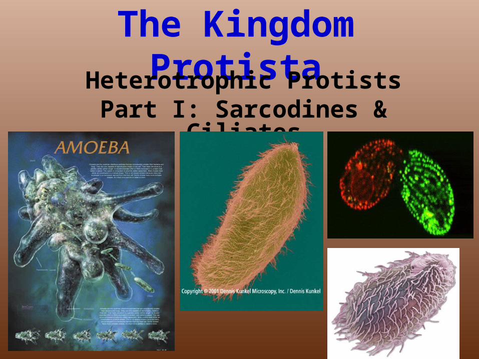
The Kingdom ProtistaHeterotrophic Protists
Part I: Sarcodines & Ciliates
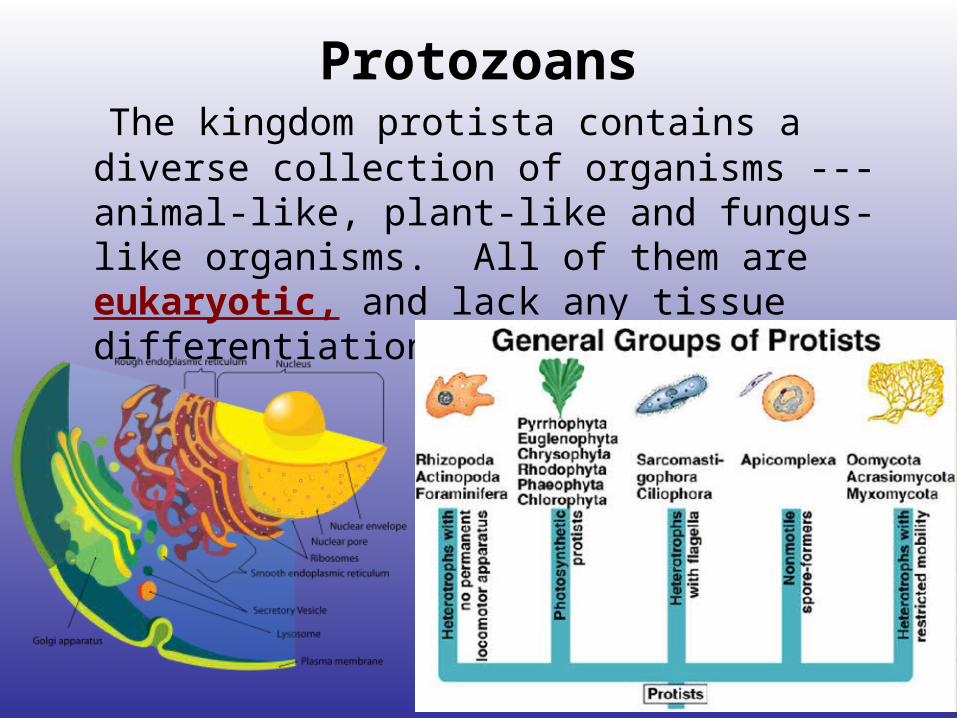
Protozoans The kingdom protista contains a diverse
collection of organisms ---animal-like, plant-like and fungus-like organisms. All of them are eukaryotic, and lack any tissue differentiation.
A) General Characteristics: Protozoa are single-celled &
microscopic. Many have a means of locomotion or motility. 1) They live in a variety of environments: freshwater rivers and
ponds, some drift in the ocean, some live in soil, & some live in the bodies of other organisms.
2) Some are free-living, while some are parasitic.
3) They are heterotrophic, obtaining nutrients by ingesting small molecules or cells.
4) Many species make up zooplankton, which are a primary
food source of aquatic ecosystems.

5) They can reproduce by binary fission, (some by multiple fission), while can reproduce sexually by conjugation.

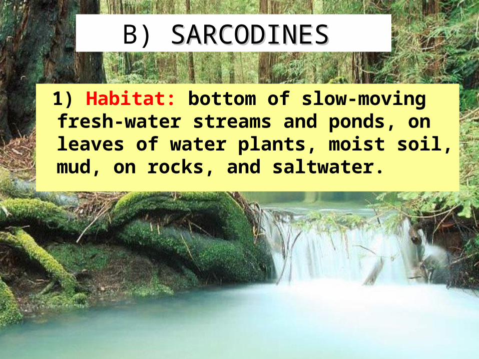
B) SARCODINESSARCODINES
1) Habitat: bottom of slow-moving fresh-water streams and ponds, on leaves of water plants, moist soil, mud, on rocks, and saltwater.
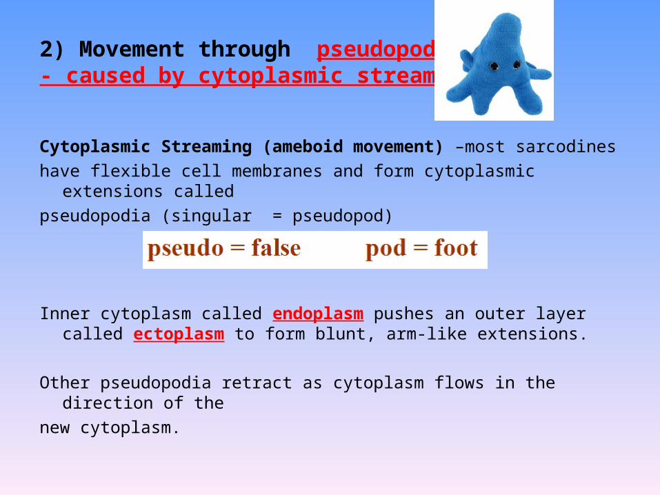
2) Movement through pseudopodia:- caused by cytoplasmic streaming
Cytoplasmic Streaming (ameboid movement) –most sarcodines
have flexible cell membranes and form cytoplasmic extensions called
pseudopodia (singular = pseudopod)
Inner cytoplasm called endoplasm pushes an outer layer called ectoplasm to form blunt, arm-like extensions.
Other pseudopodia retract as cytoplasm flows in the direction of the
new cytoplasm.
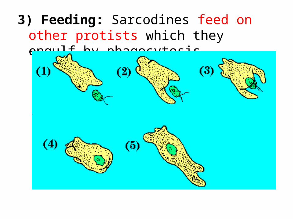
3) Feeding: Sarcodines feed on other protists which they engulf by phagocytosis.
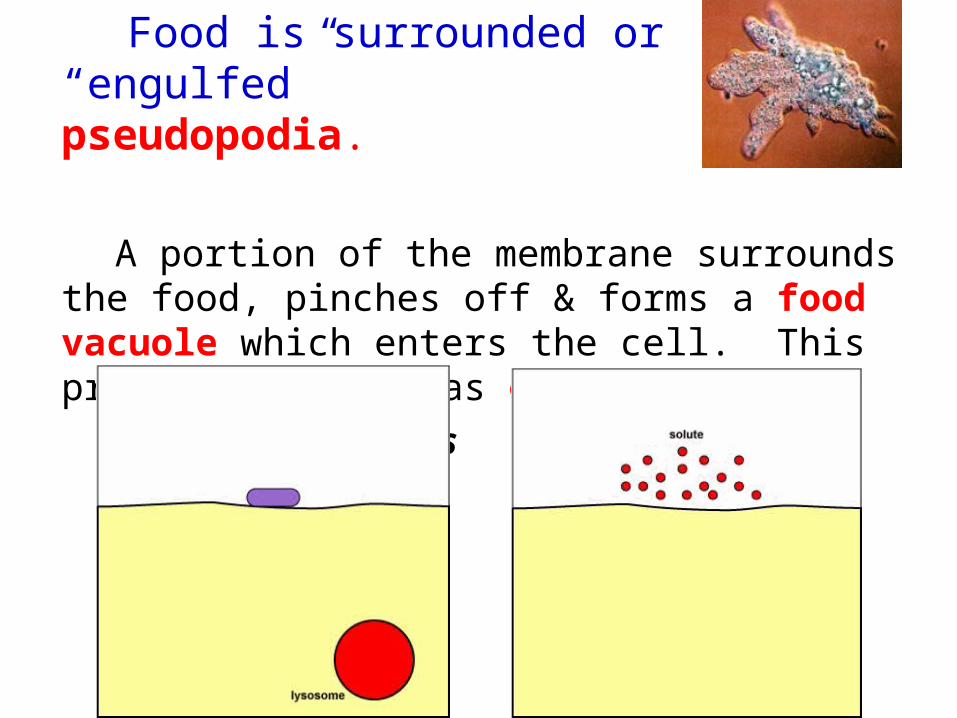
Food is surrounded or “engulfed” by pseudopodia.
A portion of the membrane surrounds the food, pinches off & forms a food vacuole which enters the cell. This process is known as endocytosis.
phagocytosis pinocytosis
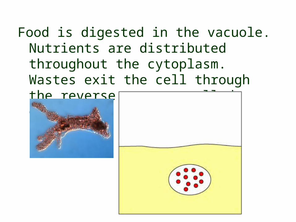
Food is digested in the vacuole. Nutrients are distributed throughout the cytoplasm. Wastes exit the cell through the reverse process called exocytosis.
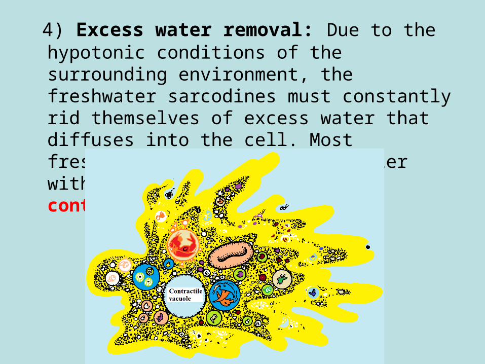
4) Excess water removal: Due to the hypotonic conditions of the surrounding environment, the freshwater sarcodines must constantly rid themselves of excess water that diffuses into the cell. Most freshwater sarcodines expel water with an organelle called a contractile vacuole.
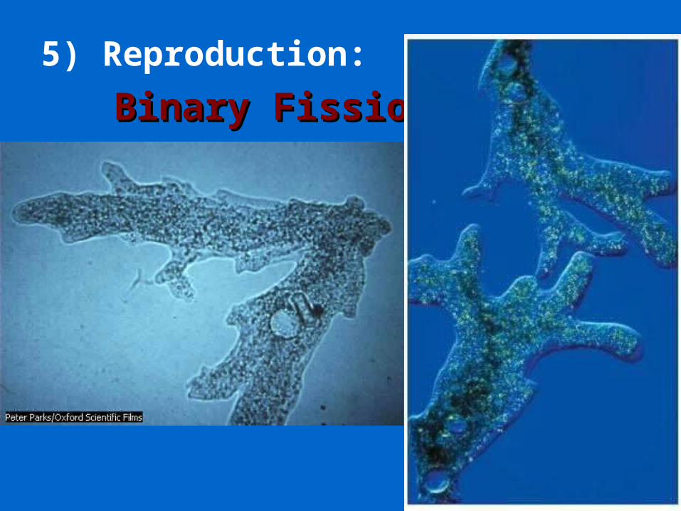
5) Reproduction:
Binary FissionBinary Fission
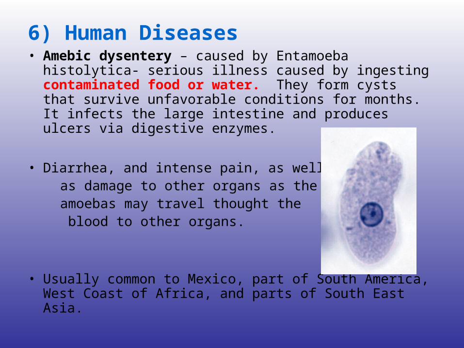
6) Human Diseases• Amebic dysentery – caused by Entamoeba histolytica-
serious illness caused by ingesting contaminated food or water. They form cysts that survive unfavorable conditions for months. It infects the large intestine and produces ulcers via digestive enzymes.
• Diarrhea, and intense pain, as well as damage to other organs as the amoebas may travel thought the blood to other organs.
• Usually common to Mexico, part of South America, West Coast of Africa, and parts of South East Asia.

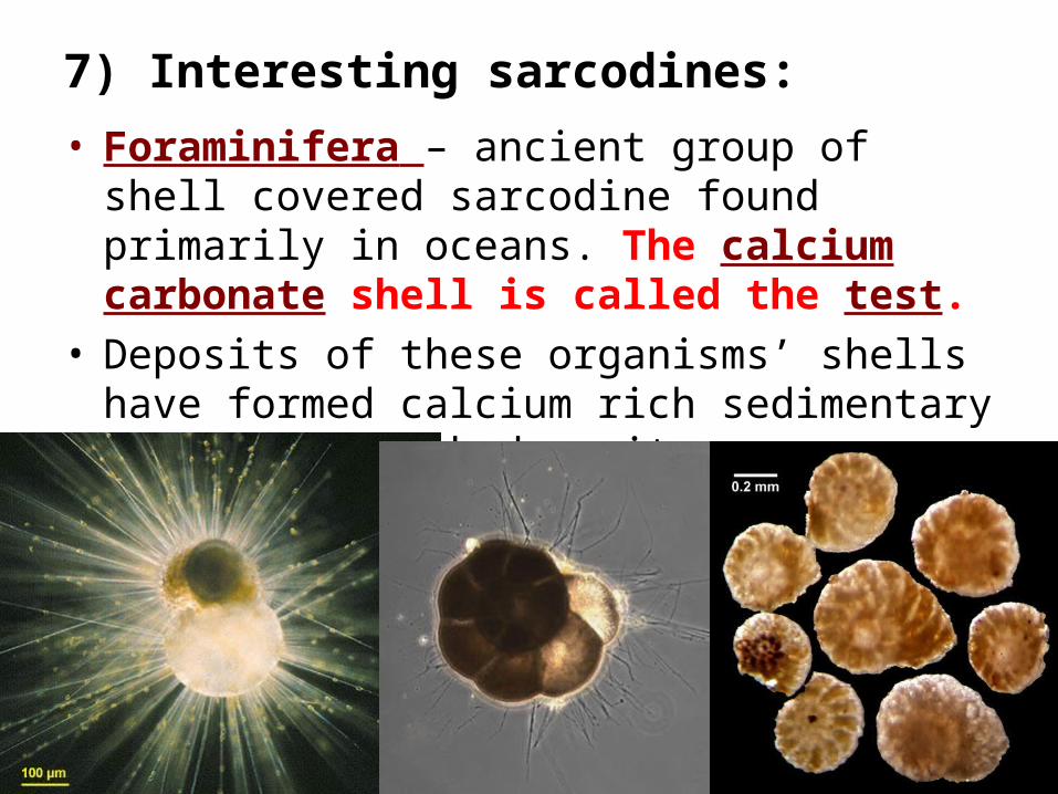
7) Interesting sarcodines:
• Foraminifera – ancient group of shell covered sarcodine found primarily in oceans. The calcium carbonate shell is called the test.
• Deposits of these organisms’ shells have formed calcium rich sedimentary layers of chalk deposits.

Millions of years of deposits have formed the “white cliffs of Dover” in England, and were a source of limestone for the Great Pyramids of Egypt.

• Radiolarians – the oldest group which have a silicon dioxide test with a radial arrangement of spines that extend from the shell.
• “With their glassy skeleton of almost perfect geometric form and symmetry, they are among the most beautiful of all protists.” ---
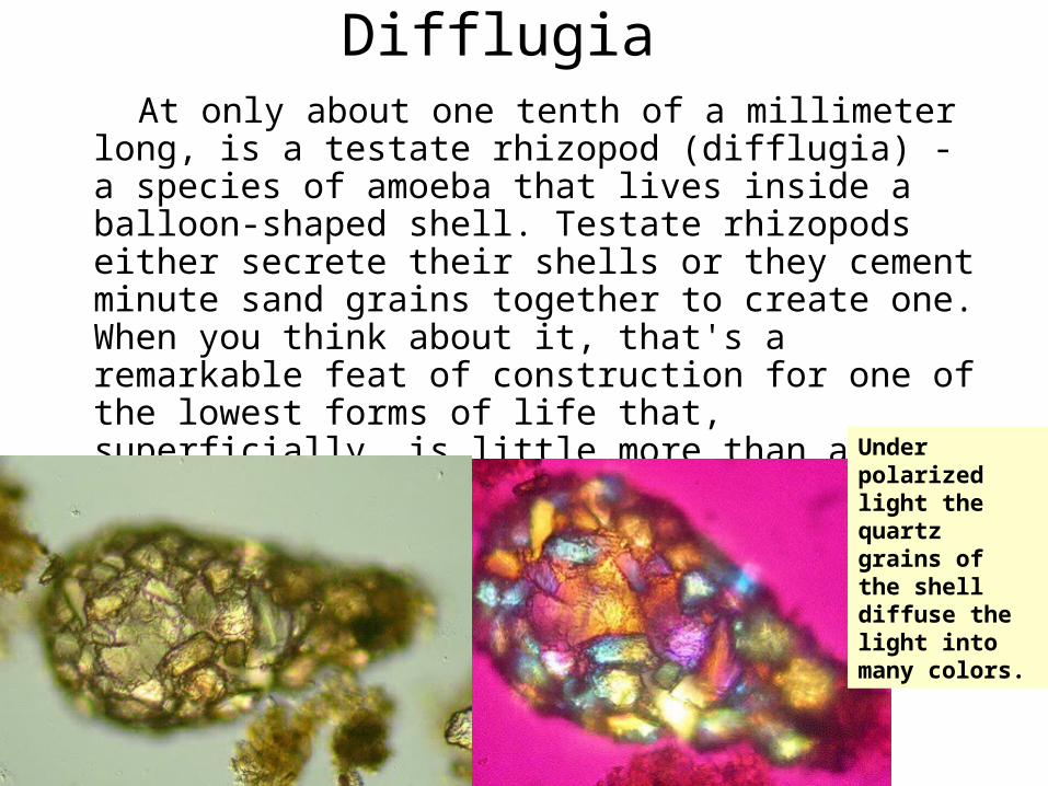
Difflugia At only about one tenth of a millimeter long, is a testate
rhizopod (difflugia) - a species of amoeba that lives inside a balloon-shaped shell. Testate rhizopods either secrete their shells or they cement minute sand grains together to create one. When you think about it, that's a remarkable feat of construction for one of the lowest forms of life that, superficially, is little more than a slithering blob of cytoplasm.
normal light polarized light microscopy
Under polarized light thequartz grains of the shelldiffuse the light into many colors.

The Brain Eating Amoeba• (9/29/07) 6 die from brain-eating amoeba in warm
lake waters (Arizona)• Doctors said Aaron Evans, a 17 year-old who died
after swimming, probably picked up the amoeba in the balmy shallows of Lake Havasu, a popular man-made lake on the Colorado River between Arizona and California.
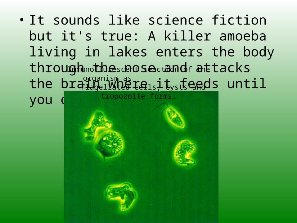
• It sounds like science fiction but it's true: A killer amoeba living in lakes enters the body through the nose and attacks the brain where it feeds until you die. Immunoflurescent reaction of the organism as
flagellated cells, cysts and tropozoite forms.

• People become infected when they wade through shallow water and stir up the bottom. If someone allows water to shoot up the nose - say, by doing a somersault in chest-deep water - the amoeba can latch onto the olfactory nerve. The amoeba destroys tissue as it makes its way up into the brain, where it continues the damage, "basically feeding on the brain cells," Beach said.
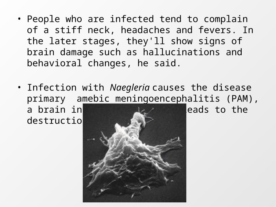
• People who are infected tend to complain of a stiff neck, headaches and fevers. In the later stages, they'll show signs of brain damage such as hallucinations and behavioral changes, he said.
• Infection with Naegleria causes the disease primary amebic meningoencephalitis (PAM), a brain inflammation, which leads to the destruction of brain tissue.
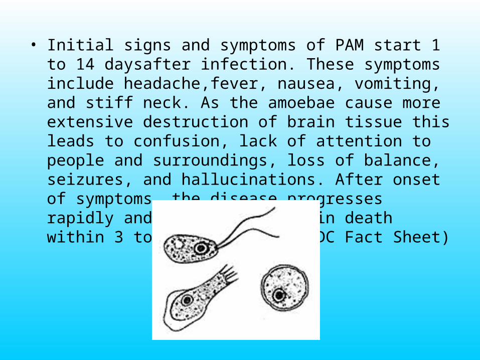
• Initial signs and symptoms of PAM start 1 to 14 daysafter infection. These symptoms include headache,fever, nausea, vomiting, and stiff neck. As the amoebae cause more extensive destruction of brain tissue this leads to confusion, lack of attention to people and surroundings, loss of balance, seizures, and hallucinations. After onset of symptoms, the disease progresses rapidly and usually results in death within 3 to 7 days. (from CDC Fact Sheet)
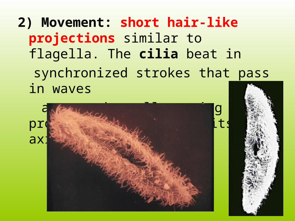
2) Movement: short hair-like projections similar to flagella. The cilia beat in
synchronized strokes that pass in waves
across the cell causing the protozoan to rotate on its axis.
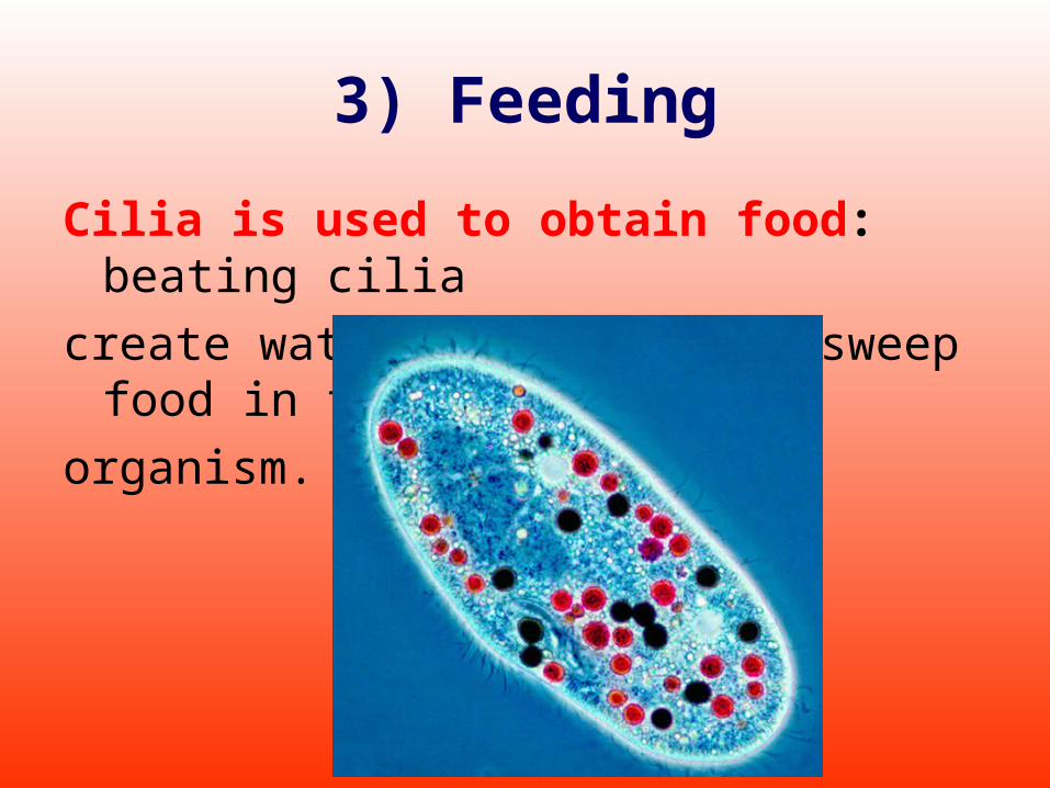
3) Feeding
Cilia is used to obtain food: beating cilia
create water currents that sweep food in the
organism.

Paramecium Feeding: 1) A funnel-shaped depression in the pellicle layer called the oral groove, is lined with cilia the bring in food.(pellicle layer - a clear elastic layer of protein that surrounds the cell membrane to help maintain shape)
2) Food moves down the groove to the mouth pore, into the gullet.
3) Food vacuoles form at the gullet which circulate through the cytoplasm. Organic molecules are digested and absorbed.
4) Waste moves to the anal pore and is expelled.
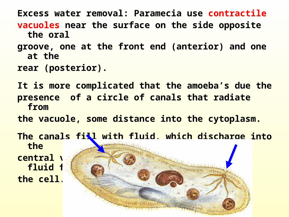
Excess water removal: Paramecia use contractile vacuoles near the surface on the side opposite the oral groove, one at the front end (anterior) and one at therear (posterior).
It is more complicated that the amoeba’s due thepresence of a circle of canals that radiate fromthe vacuole, some distance into the cytoplasm.
The canals fill with fluid, which discharge into thecentral vacuole. The vacuole then ejects the fluid fromthe cell.
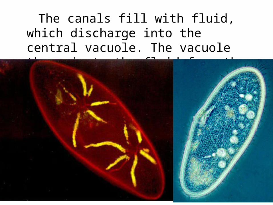
The canals fill with fluid, which discharge into the central vacuole. The vacuole then ejects the fluid from the cell.
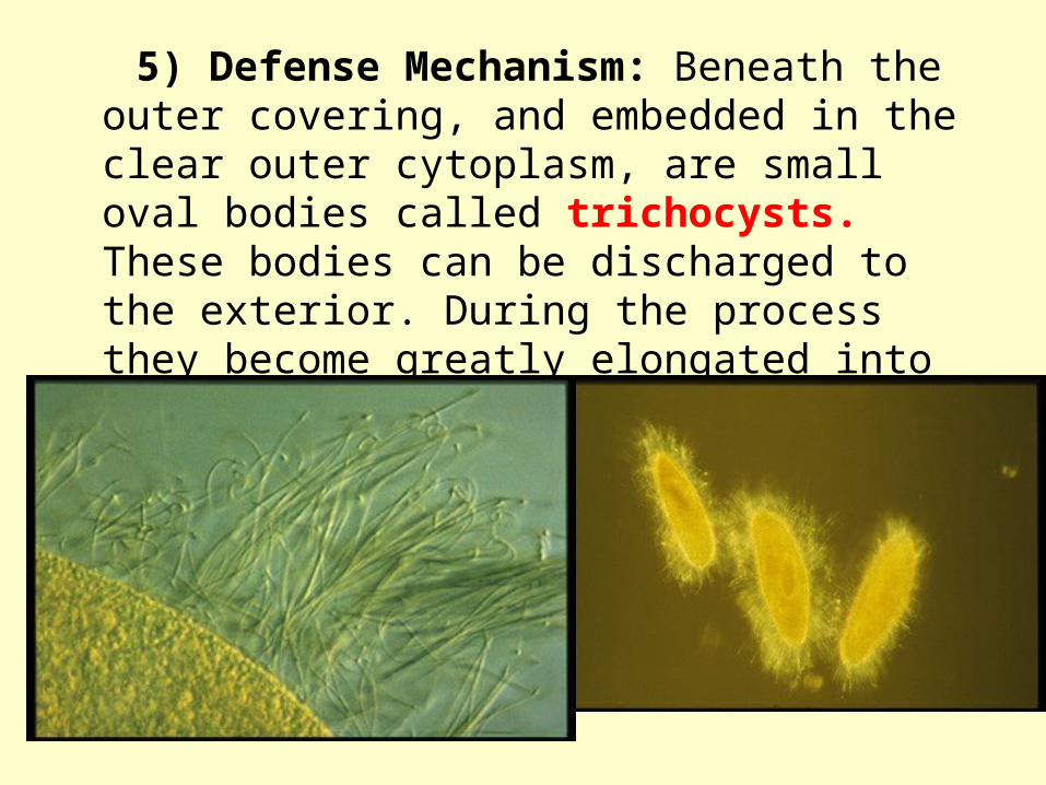
5) Defense Mechanism: Beneath the outer covering, and embedded in the clear outer cytoplasm, are small oval bodies called trichocysts. These bodies can be discharged to the exterior. During the process they become greatly elongated into fine threads.
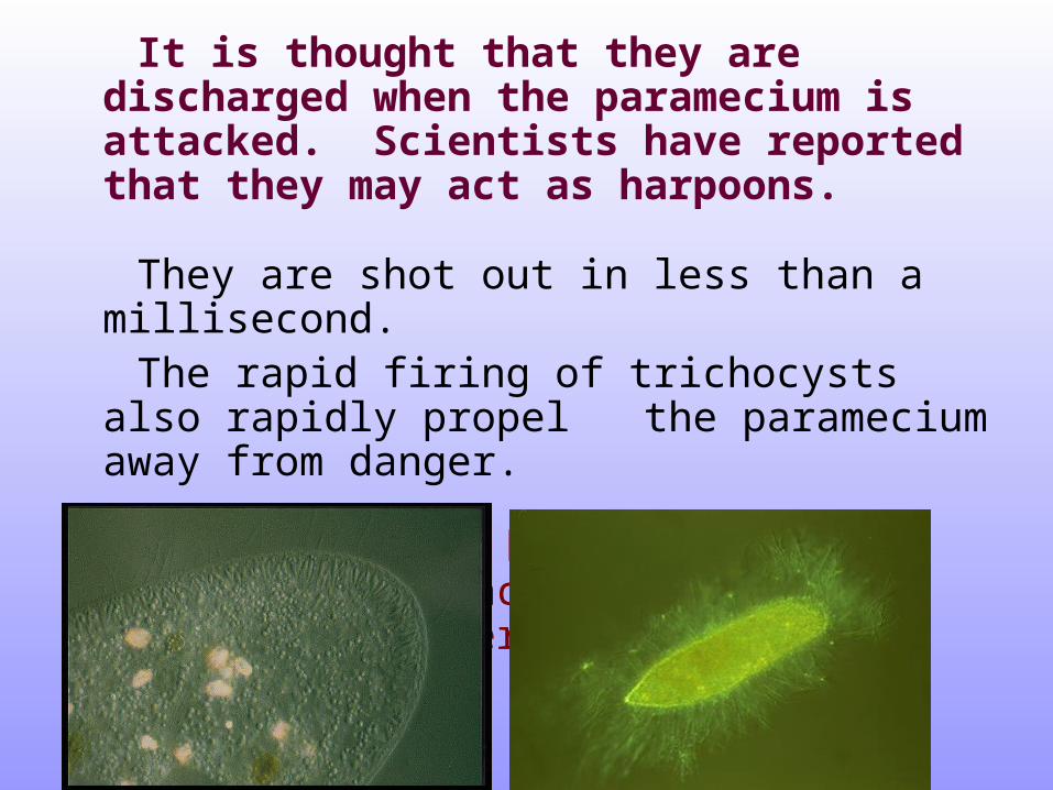
It is thought that they are discharged when the paramecium is attacked. Scientists have reported that they may act as harpoons.
They are shot out in less than a millisecond. The rapid firing of trichocysts also rapidly propel
the paramecium away from danger.
They also may be used by the paramecium to anchor itself while feeding on bacteria.
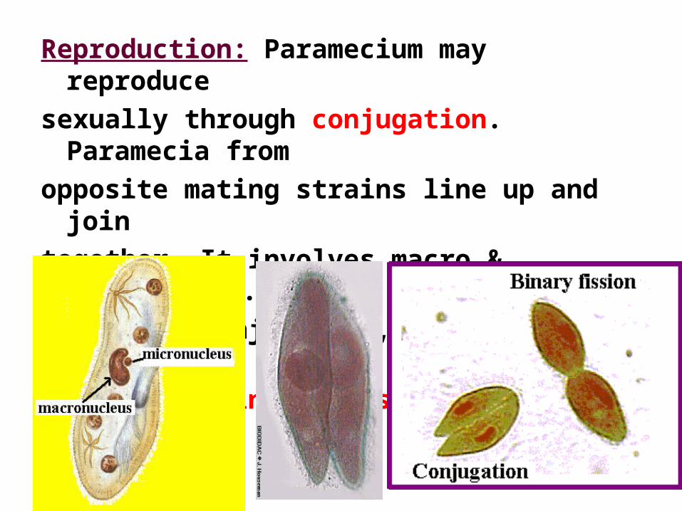
Reproduction: Paramecium may reproduce
sexually through conjugation. Paramecia from
opposite mating strains line up and join
together. It involves macro & micronuclei.
Following conjugation, each paramecium
divides by binary fission.

Paramecia conjugating