Embed Size (px)
Citation preview

1The Knee
Parks_ch01_p001-p042.indd 1 9/13/17 7:03 PM
SAMPLE ONLY. NOT FOR DISTRIBUTION.

2 CHAPTER 1
One of the best ways to understand how something, anything, works is to take it apart and put it back together again. For the knee, this is fairly simple. We need a relatively short list of parts: 4 bones, 2 tendons, 4 ligaments, and 2 types of cartilage.
To start, we place the femur, tibia, and fibula bones in their proper positions (Figure 1-1). Next, we need a system of ligaments
Femur
Fibula
Tibia
Figure 1-1. Building a knee: the femur, tibia, and fibula bones.
THEY ARE NOT CUSHIONS!I can’t tell you how many times I’ve heard it said that the meniscus cartilages are the “cushions” that reside between the femur and tibia. This isn’t true. They do reside between the tibia and femur bones, and it is true that, if a meniscus cartilage is damaged or surgically removed, the articular surfaces adjacent to it will wear out faster. But, the meniscus cartilages are peripheral to the load-bearing contact surfaces in the knee. This deserves a little further explanation. Illustrations like the drawing in Figure 1-4 are misleading. To enable you to see the meniscus cartilages, I have distracted the femur and tibia apart, opening the joint space much wider than it is anatomically. A more accurate repre-sentation would be what is illustrated in Figure 1-A. To better understand the rela-tionship between the meniscus, articular cartilage surfaces, and bones, let’s study
(continued on following page)
to hold them together (Figure 1-2) and a coating of articular cartilage on the surface of the femur and tibia, two of the three bones that will articulate against each other (Figure 1-3). Of all the structures used to assemble the knee we are building, this thin layer of glistening articular cartilage tissue is probably the most important and the most interesting (please read the sidebar on articular cartilage on page 18). Now we are ready to add the meniscus cartilages, which sit like two rubbery, horseshoe-shaped pads on the surface of the tibia (Figure 1-4). The exact role that the meniscus cartilages play in the function of the knee is poorly understood, but they do not act as a “cushion” between the femur and tibia, as many of us were taught (see sidebar). The last bone we need to add if we are building a knee is the patella. The patella is a link in the chain of structures known as the extensor mecha-nism (Figure 1-5). These structures—the quadriceps muscle, the quadriceps tendon, the patella, and the patellar tendon—allow us to forcibly straighten (extend) our knees. When it contracts, the quad-riceps muscle (via its quadriceps tendon attachment to the patella) pulls the patella proximally. As it is pulled proximally, the patella (via its patella tendon attachment to the tibia) pulls the anterior tibia proximally, which rotates the knee into extension.
Figure 1-A. A drawing of the knee showing how the meniscus cartilages are hidden from view by the femur.
Parks_ch01_p001-p042.indd 2 9/13/17 7:03 PM
SAMPLE ONLY. NOT FOR DISTRIBUTION.

THE KnEE 3
Having completed the simple “build-a-knee” exercise, it is time to study the different knee conditions seen in a typical out-patient clinic.
LIGAMENT INJURIESPatients with ligament injuries are usually easy to separate from other patients with knee complaints. The role of the knee cruciate and collateral ligaments is to stabilize the joint. These structures
a series of cross-sectional lateral views (Figure 1-B). Here, we can see that the dimensions and location of the contact patch between the tibia and femur are not affected by the presence or absence of the meniscus. While the exact function of the meniscus cartilages is not known, they do not function as cushions inter-posed between the articular surfaces of
(continued on following page)
Figure 1-2. A. The medial and lateral collateral ligaments. B. The anterior and posterior cruciate ligaments.
Medial collateralligament (MCL)
Lateral collateralligament (LCL)
A
Posterior cruciateligament (PCL)
Anterior cruciateligament (ACL)
B
Figure 1-B. A cross-sectional lateral view of the knee 1. showing the layer of articular carti-lage on the surface of the femur and tibia; 2. showing the anterior and posterior horn of the medial meniscus; 3. after removing the anterior and posterior horns of the meniscus; 4. showing the contact patch between the tibia and the femur and how its dimensions are not changed when the meniscus is removed.
Articularcartilage
1
Meniscuscartilage(posterior horn)
Meniscuscartilage
(anterior horn)
2
Parks_ch01_p001-p042.indd 3 9/13/17 7:03 PM
SAMPLE ONLY. NOT FOR DISTRIBUTION.

4 CHAPTER 1
connect the bones in a way that allows normal motion (flex-ion and extension) but resists the forces that create abnormal motion (hyperextension; varus/valgus [see further discussion]; anteroposterior translation and rotation). The knee ligaments of
Lateral meniscus Medial meniscus
Figure 1-4. The medial and lateral meniscus cartilages.
Articularcartilage
Figure 1-3. The articular cartilage coatings on the surfaces of the femur and tibia.
the tibia and femur. Figure 1-C shows how this same anatomy appears on a sagittal MRI image of the knee.
Figure 1-B. (Continued)
3
4
Figure 1-C. How the anatomy in Figure 1-B appears on a corresponding MRI image (Repro-duced with permission from Ross Goldstein, MD).
Parks_ch01_p001-p042.indd 4 9/13/17 7:03 PM
SAMPLE ONLY. NOT FOR DISTRIBUTION.

THE KnEE 5
a given patient are about the same length and diameter as that patient’s pinky finger, so they are essentially impossible to tear without substantial trauma. A patient with knee pain but no his-tory of trauma or injury is not likely to have a ligament injury, at least not a recent ligament injury. You will encounter patients who tell you that they had a “knee sprain” years ago that seemed to heal well, but ever since, they’ve had a “trick knee” that will give out on them once or twice a year if they twist just right. These episodes of instability are usually followed by a few days to a week of pain and swelling, then the knee returns to normal. This is a classic history for a patient with a chronic ligament-deficient knee. In these patients, the pain and swelling from the initial injury have resolved, but, because the ligament did not heal, they are prone to intermittent episodes of instability. With very few exceptions, trauma, even remote trauma, is requisite in the history for a patient to have a knee ligament injury.
Some patients will offer that they felt, or even heard, a “pop” when the ligament was injured. Knee ligaments are very strong structures. They can store a tremendous amount of energy before failing. If the load is big enough to fail the ligament, then the liga-ment will rupture, and that stored energy is released suddenly, creat-ing what the patient perceives as a pop. Though not pathognomonic,
Quadriceps muscle
Quadriceps tendon
Patella
Patellar tendon
Figure 1-5. The extensor mechanism.
Parks_ch01_p001-p042.indd 5 9/13/17 7:03 PM
SAMPLE ONLY. NOT FOR DISTRIBUTION.

6 CHAPTER 1
when patients report a “pop,” this important clue strongly suggests a knee ligament injury. An effusion and the timing of its onset can also be important clues, especially when trying to distinguish liga-ment injuries from meniscal tears. Ligaments are more vascular than meniscal tissue, and patients with ligament injuries tend to develop effusions within an hour of their injury. In patients with meniscus tears, effusions usually develop much more slowly.
▶ Physical ExamThe four knee ligaments, the two collaterals and the two cruci-ates, each provide a unique and specific aspect of knee stabil-ity. Think of the knee as a hinge connecting an upper segment (the femur) to a lower segment (the tibia and fibula). The hinge-like knee joint enables us to flex (bend the knee) and extend (straighten the knee). If the lower segment deviates toward the midline, we call that a varus deformity. If the lower segment deviates away from the midline, we call that a valgus deformity (Figure 1-6). The collateral ligaments are designed to prevent the
Varus
R L R L
ValgusM
idlin
e
Mid
line
Figure 1-6. Varus and valgus are terms used in orthopedics to describe angu-lar deformities in the coronal plane. In a varus deformity, the distal segment of the articulation (the tibia in the case of a knee joint) deviates toward the mid-line. In valgus deformities, the distal segment deviates away from the midline.
EXAMINING LIGAMENTSTo better understand the knee ligament exam, think of the ligaments as ropes or chains that span the joint like a bridge connecting one bone to the other. The ligaments are positioned not only to allow normal motion but also to resist abnormal motion. When we test a liga-ment, we apply a force to the knee that attempts to create an abnormal motion and then measure what happens. Specifi-cally, we try to measure two things: the amount of displacement and the quality of the end point. The ligaments are not perfectly rigid; they have a slight amount of elasticity, so they stretch a bit under an applied load. The elasticity of human ligaments varies a great deal from person to person. This helps explain why I have a hard time reaching down to touch my toes, while a contortionist can cross their ankles behind their head. Because of this variability, there is no standard, “normal” amount of displacement to expect when we test a patient’s ligaments. The medial side of one patient’s knee may open
(continued on following page)
lower segment (the tibia and fibula) from swinging back and forth like a pendulum. The medial collateral ligament (MCL) prevents the lower segment from swinging away from the midline creating a valgus deformity (Figure 1-7A). The lateral collateral ligament (LCL) prevents the lower segment from swinging toward the midline, creating a varus deformity (Figure 1-7B). When the distal segment deviates toward the midline, it is called a varus deformity.
Parks_ch01_p001-p042.indd 6 9/13/17 7:03 PM
SAMPLE ONLY. NOT FOR DISTRIBUTION.

THE KnEE 7
Knowing this, we can easily invent the physical exam tests for the collateral ligaments (Figure 1-8). Both tests are done with the patient lying supine, with muscles relaxed and both knees out in full or near-full extension on the exam table. To test the MCL, place one hand on the lower leg and pull it away from the midline while using the other hand to push the thigh toward the midline. Test the LCL by doing the opposite, using one hand to push the lower leg toward the midline while the other hand is pulling the thigh away from the midline. Try to estimate how many millimeters the joint opens and the quality of the “end point” you feel when the liga-ment stops the knee from moving (see sidebar). If you want to be
A B
C D
Figure 1-7. A. The medial collateral ligament (MCL) prevents valgus deformi-ties. B. The lateral collateral ligament prevents varus deformities. C. While the collateral ligaments prevent varus and valgus deformities, the cruciate liga-ments prevent anterior and posterior translation tibia. The anterior cruciate ligament prevents anterior tibial translation. D. The posterior cruciate ligament prevents posterior tibial translation.
2 mm on the MCL test, while another patient’s might open a centimeter, and both ligaments could be perfectly nor-mal. To know whether the amount of opening we feel on the ligament exam is normal or abnormal, we have to compare our findings on the knee we are examin-ing to the gold standard: the patient’s other, uninjured knee. While there is con-siderable variability in the elasticity of the ligaments from one person to another, there is little variability when we compare the elasticity of the ligaments of one knee to the other in the same person. A dif-ference of 3 mm or greater between the right and left knees of the same patient suggests a ligament injury.
The “end point” we refer to in the ligament physical exam test is the ces-sation of motion that occurs during the test when the ligament reaches its elastic limit and displacement stops. In the clinic, I illustrate this to patients by holding my necktie between my two hands. I let it sag a bit, then abruptly tug it tight. The sudden stop that occurs when the tie snaps taut is the end point.
Based on the amount of displace-ment and the quality of the end point, we can report the findings of the liga-ment exam using the orthopedic three-grade classification (see Figure 1-9). A grade I ligament injury is one where the ligament is strained, but there is no macroscopic fiber damage. On physical exam, we won’t detect any increase in displacement compared to the opposite knee, and there will be a normal, firm end point. The only finding that differentiates a grade I injury from a normal knee on exam is that the patient will experience pain when the ligament is stretched during the test. In a grade II injury, there is a partial tear of the ligament, with some fibers torn and some still intact. In this case, the exam will show increased displacement but a firm end point. In a grade III injury, there is complete rupture of all fibers of the ligament. The ligament exam will demonstrate increased dis-placement and a soft, mushy end point.
Parks_ch01_p001-p042.indd 7 9/13/17 7:03 PM
SAMPLE ONLY. NOT FOR DISTRIBUTION.

8 CHAPTER 1
fancy, you can use the ligament-grading system described in the sidebar and illustrated in Figure 1-9.
The function of the cruciate ligaments is very different from the function of the collateral ligaments. While the collateral liga-ments resist varus/valgus angular deformities in the coronal plane (varus/valgus deformities), the cruciate ligaments resist transla-tional motion, specifically anterior and posterior translation of the tibia (Figure 1-7C,D). The examination used to assess the anterior cruciate ligament (ACL) has evolved some in recent years. The
Figure 1-8. A. Testing the medial collateral ligament. B. Testing the lateral collateral ligament.
A
B
HOW THE ILIOTIBIAL BAND KILLED THE ANTERIOR DRAWER TESTThe ITB is a long, dense, firm band of con-nective tissue that runs down the side of the thigh. Technically, it is the tendon that connects the gluteus maximus and tensor fascia lata muscles of the pelvis to the lateral side of the tibia just below the knee (Figure 1-D). In the time-honored
anterior drawer test, the knee is placed in 90 degrees of flexion, the examiner sits on the patient’s foot to stabilize it, and then the examiner pulls the tibia anteriorly to apply a load to the ACL (Figure 1-E).
Figure 1-D. The iliotibial band.
Iliotibialband
(continued on following page)
Parks_ch01_p001-p042.indd 8 9/13/17 7:03 PM
SAMPLE ONLY. NOT FOR DISTRIBUTION.

THE KnEE 9
anterior drawer test is slowly being replaced by a more accurate test called Lachman’s test. In both tests, the examiner tests the ACL by pulling the tibia anteriorly, which pulls the ACL tight; however, what makes Lachman’s test superior to the anterior drawer test is that the observed changes in displacement are greater (and there-fore easier to detect) using Lachman’s test. The reason for the dif-ference is the iliotibial band (ITB), which suppresses anterior tibial translation when the knee is in 90 degrees of flexion (see sidebar).
The posterior drawer test is still valid and popular for assessing the posterior cruciate ligament (PCL) because the ITB shortens when the tibia is pushed posteriorly (Figure 1-10).
Grading ligament injuries
Normal Grade I Grade II Grade III
Figure 1-9. The three grades of ligament injury. In grade I injuries, there is no visable loss of continuity of the tendon tissue, just bleeding/bruising. Grade II injuries are partial tears, and grade III tears are complete tears that result in two un-connected “stumps” of tendon tissue.
Posterior drawer
Figure 1-10. The posterior drawer test. The knee is placed in 90 degrees of flexion and the tibia is pushed posteriorly.
In 90 degrees of flexion, the ITB is in a position to resist anterior tibial transla-tion, so the amount of translation isn’t as obvious. Lachman studied ACL-deficient knees in many different flexion angles and found that the best knee flexion angle for optimizing anterior tibial translation is 30 degrees. In 30 degrees of flexion, the ITB is not in a position to mute anterior tibial translation, so there is greater displacement, which is easier to detect (Figure 1-F).
Figure 1-F. Lachman’s test for the ACL.
Anterior drawer
Lachman’s
Iliotib
ial band
Iliotibial band
Figure 1-E. The location of the ITB in 90 degrees of flexion (the anterior drawer test for a torn ACL).
Iliotibial band
Parks_ch01_p001-p042.indd 9 9/13/17 7:03 PM
SAMPLE ONLY. NOT FOR DISTRIBUTION.
10 CHAPTER 1
Note that it can be difficult, even impossible due to pain and swelling of the acutely injured knee joint, to perform a meaning-ful ligament exam on an acutely injured knee. If the patient is too uncomfortable to relax for the exam, the options are to repeat the exam in 1-2 weeks when the pain has decreased or to obtain a magnetic resonance image (MRI).
▶ Imaging StudiesIf the mechanism of injury is trauma, which it usually is for knee ligament injuries, x-rays are probably warranted to rule out a frac-ture. This trauma may be contact trauma (i.e., two football players colliding) or noncontact trauma (a soccer player running down the field makes a cutting move and twists his or her knee). Any-time there is a history of significant trauma, an x-ray should be taken to rule out a fracture. Another imaging option is an MRI. An MRI is a very specific and sensitive test for evaluating liga-ment injuries, and it will pick up fractures as well. For suspected ligament injuries, the MRI can help diagnose patients who are too uncomfortable to be examined acutely or too impatient to be reex-amined a week or two later to confirm the diagnosis on physical exam.
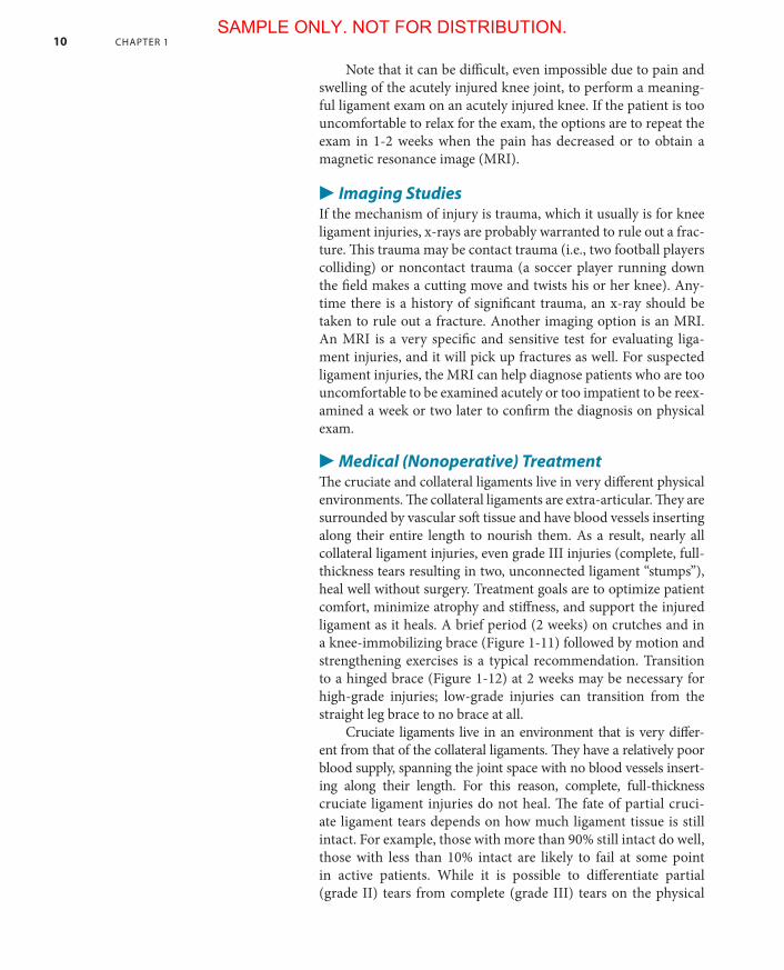
▶ Medical (Nonoperative) TreatmentThe cruciate and collateral ligaments live in very different physical environments. The collateral ligaments are extra-articular. They are surrounded by vascular soft tissue and have blood vessels inserting along their entire length to nourish them. As a result, nearly all collateral ligament injuries, even grade III injuries (complete, full-thickness tears resulting in two, unconnected ligament “stumps”), heal well without surgery. Treatment goals are to optimize patient comfort, minimize atrophy and stiffness, and support the injured ligament as it heals. A brief period (2 weeks) on crutches and in a knee-immobilizing brace (Figure 1-11) followed by motion and strengthening exercises is a typical recommendation. Transition to a hinged brace (Figure 1-12) at 2 weeks may be necessary for high-grade injuries; low-grade injuries can transition from the straight leg brace to no brace at all.
Cruciate ligaments live in an environment that is very differ-ent from that of the collateral ligaments. They have a relatively poor blood supply, spanning the joint space with no blood vessels insert-ing along their length. For this reason, complete, full-thickness cruciate ligament injuries do not heal. The fate of partial cruci-ate ligament tears depends on how much ligament tissue is still intact. For example, those with more than 90% still intact do well, those with less than 10% intact are likely to fail at some point in active patients. While it is possible to differentiate partial (grade II) tears from complete (grade III) tears on the physical
Parks_ch01_p001-p042.indd 10 9/13/17 7:03 PM
SAMPLE ONLY. NOT FOR DISTRIBUTION.
THE KnEE 11
Figure 1-11. The straight leg knee immobilizer brace. This brace is rigid and does not allow any type of knee motion (Licensed from Shutterstock).
exam (see the sidebar in this chapter on examining ligaments), it is impossible to use the physical exam to determine exactly how much of the ligament remains intact in partial tears. Some have advocated obtaining an MRI on suspected partial tears so that those with high-grade partial tears can be identified and consid-ered for surgical treatment.
▶ Surgical TreatmentGrade III (complete) cruciate ligament injuries (and the rare grade III collateral ligament injuries that don’t heal) result in lig-ament-deficient knees. In general, ligament-deficient knees are not well tolerated by patients and require surgical reconstruction. As mentioned, collateral ligament injuries, even complete tears, typically heal without surgery. The few torn collateral ligaments that don’t heal can often be successfully repaired (their torn ends sutured back together) or reconstructed with a graft. Because of their poor blood supply, torn cruciate ligaments will not heal with suture repair and have to be replaced with a graft. Grafts can be
THE THEORY OF “THE NEUROMUSCULARLY ELITE”The dogma as it relates to ligament-deficient knees, specifically ACL-deficient knees, is that ligament deficiency results in arthritis. While this is true in many, perhaps even most, patients, it is not true for all patients. An ACL-deficient knee will usu-ally have a specific pattern of instability: In certain sports or activities, the knee will experience a force that tries to drive the tibia anteriorly with respect to the femur above it. Without an ACL to counteract this force, the tibia slides farther forward
(continued on following page)
Parks_ch01_p001-p042.indd 11 9/13/17 7:04 PM
SAMPLE ONLY. NOT FOR DISTRIBUTION.

12 CHAPTER 1
either from the patient’s own tissue (autograft) or from cadaveric donors (allograft). Artificial (synthetic) grafts and grafts from nonhuman animals have been tried and have not worked well.
Recovery after ACL surgery is long, 6-12 months, but results are generally very good, with high rates of return to sports and strenu-ous activities. Keep in mind that not all ligament-deficient knees require surgery. The goals of ligament reconstruction surgery are a) to eliminate symptoms of joint instability and b) to help prevent the pattern of arthritis that these patients typically experience years or decades after their injury. Rarely, patients with ligament-deficient knees do not have instability (see sidebar). Patients who don’t have instability do not need surgical reconstruction to stabilize their knees … or do they? This is somewhat controversial. There is a body of evidence that suggests that some of these patients who have no subjective sense of instability have a pattern of “micromo-tion” instability that, over the course of decades, results in destruc-tive arthritis. If this is true, then the only ACL-deficient patients who should be treated nonoperatively are those who both (a) have no instability symptoms and (b) are old enough that, for these patients, the development of arthritis 20 or 30 years down the road is inconsequential.
Figure 1-12. The hinged knee brace. There are hinges built into the medial and lateral sides of this brace that allow flexion/extension, but not varus/ valgus or hyperextension.
than it should, allowing the condyles of the femur to strike the posterior horns of the meniscus cartilages. This results in meniscus tears, and meniscus tears accelerate the rate of wear of the knee’s articular surfaces, which in turn results in arthritis. But, patients who are sedentary are unlikely to apply the knee stresses necessary to create instability. A simple and brief course of muscle strengthen-ing in physical therapy can give them the stability they require for the modest demands of daily activities. These patients can do well treated nonoperatively. What’s interesting is that there are a few humans out there who aren’t sedentary, who can still be quite active on their lig-ament-deficient knees without experienc-ing any instability! (Figure 1-G) Although it is controversial, one explanation is that
these patients have a better proprio-ceptive system than the rest of us, and, when the tibia starts to translate too far anteriorly, they fire a compensatory ham-string muscle contraction that arrests the pathologic motion of the tibia. Best esti-mates are that these individuals account for less than 5% of the population, and, as of right now, we don’t have any reli-able way to test for this gift. For now, the consensus recommendation favors ACL reconstruction in active patients because 95% of them will experience a pattern of instability that eventually results in arthritis.
Figure 1-G. The relationships between ACL deficiency and the development of arthritis. The accepted fact is that ACL tears result in arthritis. This is because, in most patients, ACL tear result in a pattern of instability that creates meniscus tears, which leads to arthritis. For some patients, ACL tears do not result in instability, so they don’t get instability.
ACL tears Arthritis
ACL tears
ACL tears
Instability Meniscus tears Arthritis
Instability Meniscus tears Arthritis
Parks_ch01_p001-p042.indd 12 9/13/17 7:04 PM
SAMPLE ONLY. NOT FOR DISTRIBUTION.


![Zimmer PCL Project - Stanford University · Figure 1. Zimmer Inc. NexGen CR PCL -sparing total knee prosthesis. [9 ] Figure 2. Implanted Zimmer NexGen CR prosthesis. [9 ] Currently,](https://img.pdfslide.net/doc/110x75/5f15debe8ee78072f07aa605/zimmer-pcl-project-stanford-university-figure-1-zimmer-inc-nexgen-cr-pcl-sparing.jpg)