Embed Size (px)
Citation preview
• Recognize and use terms related to the anatomy and physiology of the musculoskeletal system.
• Recognize and use terms related to the pathology of the musculoskeletal system.
• Recognize and use terms related to the diagnostic procedures for the musculoskeletal system.
• Recognize and use terms related to the therapeutic interventions for the musculoskeletal system.
CHAPTER OUTLINE
OBJECTIVES
Functions of the
Musculoskeletal System
Musculoskeletal Specialists
Anatomy and Physiology
Axial Skeleton
Appendicular Skeleton
Pathology
Diagnostic Procedures
Therapeutic Interventions
Pharmacology
Chapter Review
Case Study With Accompanying
Medical Report
“The leadership instinct you are born with is the backbone. You develop the funny bone and the wishbone that go with it.”—Elaine Agather
3
Musculoskeletal System
CHAPTER AT A GLANCE
KEY TERMS
KEY WORD PARTS
ANATOMY AND PHYSIOLOGY
appendicular skeleton
articulation
axial skeleton
bone depression
bone process
bursa
cartilage
fascia
ligament
muscle
tendon
PREFIXES
dia-
endo-, end-
epi-
inter-
peri-
syn-
SUFFIXES
-centesis
-desis
-graphy
-listhesis
-malacia
-physis
-plasia
-plasty
-trophy
COMBINING FORMS
arthr/o
articul/o
burs/o
chondr/o
ligament/o
my/o
myel/o
oste/o
spondyl/o
tendin/o
arthrocentesis
arthroplasty
arthroscopy
carpal tunnel syndrome (CTS)
electromyography (EMG)
herniated intervertebral disk
lumbago
muscular dystrophy (MD)
osteoarthritis (OA)
osteomalacia
osteomyelitis
osteoporosis
pathologic fractures
prosthesis
rheumatoid arthritis (RA)
scoliosis
spinal stenosis
sprain
strain
subluxation
62 Chapter 3
FUNCTIONS OF THE MUSCULOSKELETAL SYSTEM
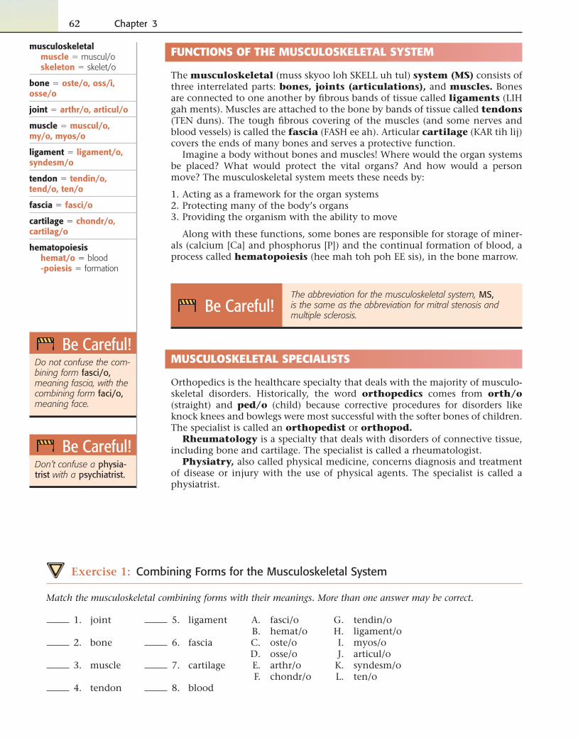
The musculoskeletal (muss skyoo loh SKELL uh tul) system (MS) consists of three interrelated parts: bones, joints (articulations), and muscles. Bones are connected to one another by fi brous bands of tissue called ligaments (LIH gah ments). Muscles are attached to the bone by bands of tissue called tendons (TEN duns). The tough fi brous covering of the muscles (and some nerves and blood vessels) is called the fascia (FASH ee ah). Articular cartilage (KAR tih lij) covers the ends of many bones and serves a protective function.
Imagine a body without bones and muscles! Where would the organ systems be placed? What would protect the vital organs? And how would a person move? The musculoskeletal system meets these needs by:
1. Acting as a framework for the organ systems 2. Protecting many of the body’s organs 3. Providing the organism with the ability to move
Along with these functions, some bones are responsible for storage of miner-als (calcium [Ca] and phosphorus [P]) and the continual formation of blood, a process called hematopoiesis (hee mah toh poh EE sis), in the bone marrow.
Be Careful! The abbreviation for the musculoskeletal system, MS,
is the same as the abbreviation for mitral stenosis and multiple sclerosis.
0
MUSCULOSKELETAL SPECIALISTS
Orthopedics is the healthcare specialty that deals with the majority of musculo-skeletal disorders. Historically, the word orthopedics comes from orth/o (straight) and ped/o (child) because corrective procedures for disorders like knock knees and bowlegs were most successful with the softer bones of children. The specialist is called an orthopedist or orthopod.
Rheumatology is a specialty that deals with disorders of connective tissue, including bone and cartilage. The specialist is called a rheumatologist.
Physiatry, also called physical medicine, concerns diagnosis and treatment of disease or injury with the use of physical agents. The specialist is called a physiatrist.
Be Careful!Do not confuse the com-bining form fasci/o, meaning fascia, with the combining form faci/o, meaning face.
Be Careful!Don’t confuse a physia-trist with a psychiatrist.
musculoskeletalmuscle � muscul/oskeleton � skelet/o
bone � oste/o, oss/i, osse/o
joint � arthr/o, articul/o
muscle � muscul/o, my/o, myos/o
ligament � ligament/o, syndesm/o
tendon � tendin/o, tend/o, ten/o
fascia � fasci/o
cartilage � chondr/o, cartilag/o
hematopoiesishemat/o � blood-poiesis � formation
Exercise 1: Combining Forms for the Musculoskeletal System
Match the musculoskeletal combining forms with their meanings. More than one answer may be correct.
5. ligament
6. fascia
7. cartilage
8. blood
A. fasci/o B. hemat/o C. oste/o D. osse/o E. arthr/o F. chondr/o
1. joint
2. bone
3. muscle
4. tendon
G. tendin/o H. ligament/o I. myos/o J. articul/o K. syndesm/o L. ten/o
Musculoskeletal System 63
Decode the following terms using your knowledge of musculoskeletal word parts and suffi xes learned in Chapter 1.
9. articular
10. tendinous
11. muscular
12. syndesmal
13. chondral
14. osseous
appendicular � appendic/o
skeleton � skelet/o
ANATOMY AND PHYSIOLOGY
BONES
Types of Bones
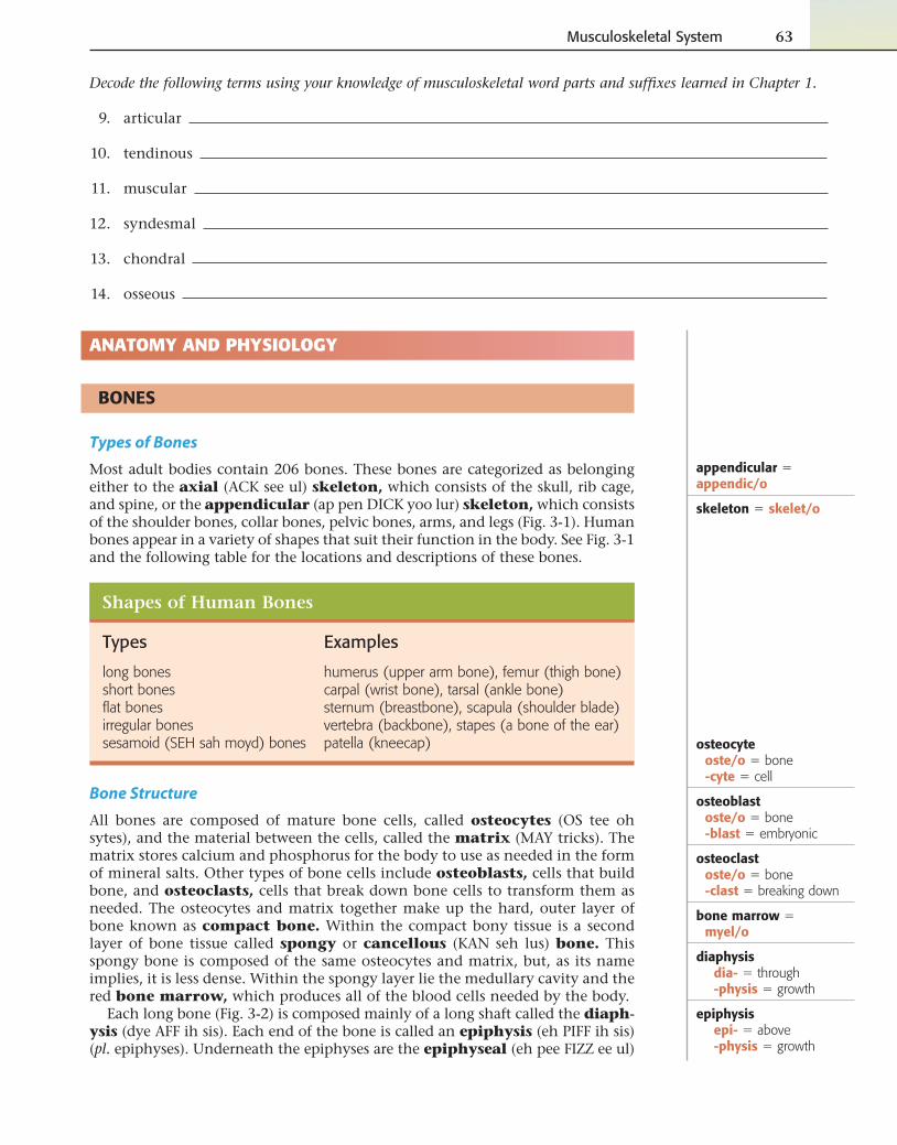
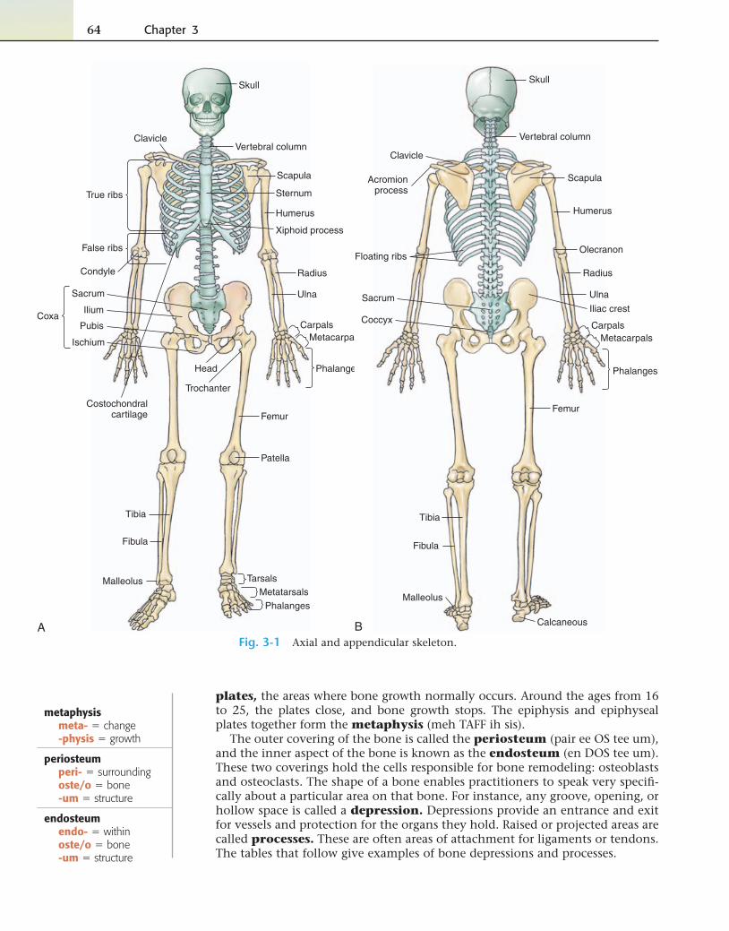
Most adult bodies contain 206 bones. These bones are categorized as belonging either to the axial (ACK see ul) skeleton, which consists of the skull, rib cage, and spine, or the appendicular (ap pen DICK yoo lur) skeleton, which consists of the shoulder bones, collar bones, pelvic bones, arms, and legs (Fig. 3-1). Human bones appear in a variety of shapes that suit their function in the body. See Fig. 3-1 and the following table for the locations and descriptions of these bones.
Shapes of Human Bones
Types Examples
long bones humerus (upper arm bone), femur (thigh bone)short bones carpal (wrist bone), tarsal (ankle bone)fl at bones sternum (breastbone), scapula (shoulder blade)irregular bones vertebra (backbone), stapes (a bone of the ear)sesamoid (SEH sah moyd) bones patella (kneecap)
Bone Structure
All bones are composed of mature bone cells, called osteocytes (OS tee oh sytes), and the material between the cells, called the matrix (MAY tricks). The matrix stores calcium and phosphorus for the body to use as needed in the form of mineral salts. Other types of bone cells include osteoblasts, cells that build bone, and osteoclasts, cells that break down bone cells to transform them as needed. The osteocytes and matrix together make up the hard, outer layer of bone known as compact bone. Within the compact bony tissue is a second layer of bone tissue called spongy or cancellous (KAN seh lus) bone. This spongy bone is composed of the same osteocytes and matrix, but, as its name implies, it is less dense. Within the spongy layer lie the medullary cavity and the red bone marrow, which produces all of the blood cells needed by the body.
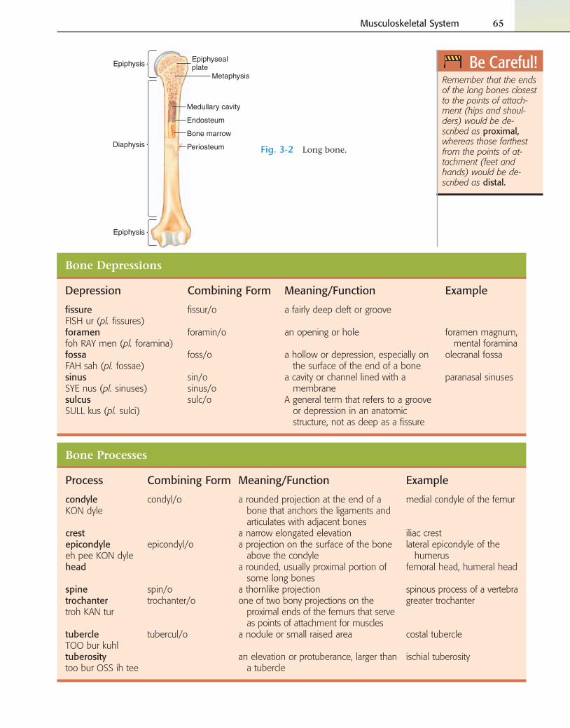
Each long bone (Fig. 3-2) is composed mainly of a long shaft called the diaph-ysis (dye AFF ih sis). Each end of the bone is called an epiphysis (eh PIFF ih sis) (pl. epiphyses). Underneath the epiphyses are the epiphyseal (eh pee FIZZ ee ul)
osteocyteoste/o � bone-cyte � cell
osteoblastoste/o � bone-blast � embryonic
osteoclastoste/o � bone-clast � breaking down
bone marrow � myel/o
diaphysisdia- � through-physis � growth
epiphysisepi- � above-physis � growth
64 Chapter 3
A
Skull
Scapula
Humerus
Radius
Ulna
CarpalsMetacarpals
Phalanges
Femur
Patella
Tibia
Fibula
TarsalsMetatarsals
Phalanges
Pubis
Ilium
Clavicle
Sternum
Vertebral column
Ischium
Sacrum
Malleolus
True ribs
False ribs
Condyle
Coxa
Costochondralcartilage
Head
Trochanter
Xiphoid process
Skull
Scapula
Humerus
Radius
Ulna
CarpalsMetacarpals
Phalanges
Femur
Tibia
Fibula
Clavicle
Vertebral column
Sacrum
Malleolus
Floating ribsOlecranon
Coccyx
Acromionprocess
Iliac crest
CalcaneousBFig. 3-1 Axial and appendicular skeleton.
plates, the areas where bone growth normally occurs. Around the ages from 16 to 25, the plates close, and bone growth stops. The epiphysis and epiphyseal plates together form the metaphysis (meh TAFF ih sis).
The outer covering of the bone is called the periosteum (pair ee OS tee um), and the inner aspect of the bone is known as the endosteum (en DOS tee um). These two coverings hold the cells responsible for bone remodeling: osteoblasts and osteoclasts. The shape of a bone enables practitioners to speak very specifi -cally about a particular area on that bone. For instance, any groove, opening, or hollow space is called a depression. Depressions provide an entrance and exit for vessels and protection for the organs they hold. Raised or projected areas are called processes. These are often areas of attachment for ligaments or tendons. The tables that follow give examples of bone depressions and processes.
metaphysismeta- � change-physis � growth
periosteumperi- � surroundingoste/o � bone-um � structure
endosteumendo- � withinoste/o � bone-um � structure
Musculoskeletal System 65
Be Careful!Remember that the ends of the long bones closest to the points of attach-ment (hips and shoul-ders) would be de-scribed as proximal, whereas those farthest from the points of at-tachment (feet and hands) would be de-scribed as distal.
Periosteum
Bone marrow
Epiphysis
Epiphysis
Epiphysealplate
Diaphysis
Medullary cavity
Endosteum
Metaphysis
Fig. 3-2 Long bone.
Bone Processes
Process Combining Form Meaning/Function Example
condyleKON dyle
condyl/o a rounded projection at the end of a bone that anchors the ligaments and articulates with adjacent bones
medial condyle of the femur
crest a narrow elongated elevation iliac crestepicondyleeh pee KON dyle
epicondyl/o a projection on the surface of the bone above the condyle
lateral epicondyle of the humerus
head a rounded, usually proximal portion of some long bones
femoral head, humeral head
spine spin/o a thornlike projection spinous process of a vertebratrochantertroh KAN tur
trochanter/o one of two bony projections on the proximal ends of the femurs that serve as points of attachment for muscles
greater trochanter
tubercleTOO bur kuhl
tubercul/o a nodule or small raised area costal tubercle
tuberositytoo bur OSS ih tee
an elevation or protuberance, larger than a tubercle
ischial tuberosity
Bone Depressions
Depression Combining Form Meaning/Function Example
fi ssureFISH ur (pl. fi ssures)
fi ssur/o a fairly deep cleft or groove
foramenfoh RAY men (pl. foramina)
foramin/o an opening or hole foramen magnum, mental foramina
fossaFAH sah (pl. fossae)
foss/o a hollow or depression, especially on the surface of the end of a bone
olecranal fossa
sinusSYE nus (pl. sinuses)
sin/osinus/o
a cavity or channel lined with a membrane
paranasal sinuses
sulcusSULL kus (pl. sulci)
sulc/o A general term that refers to a groove or depression in an anatomic structure, not as deep as a fi ssure
66 Chapter 3
Fill in the blank.
16. Osteoblasts bone, whereas osteoclasts bone.
17. The shaft of a long bone is called the ; the ends of a long bone are
called (plural!).
18. The outer covering of bone is the , whereas the inner lining is the
.
19. A foramen, a sulcus, and a fossa are examples of bone . A condyle, a
trochanter, and a tuberosity are examples of bone .
20. A synonym for a sinus is a/an .
9. -blast
10. epi-
11. foss/o
12. endo-
13. trochanter/o
14. -cyte
15. -clast
A. trochanter B. foramen, hole C. above, upon D. cell E. bone marrow F. surrounding, around G. embryonic H. spine I. breaking down J. growth K. hollow, depression L. condyle, knob M. within N. sinus, cavity O. structure
1. myel/o
2. -physis
3. peri-
4. condyl/o
5. spin/o
6. sin/o
7. foramin/o
8. -um
Exercise 2: Bone Basics
Match the bone word parts with their meanings.
Musculoskeletal System 67
AXIAL SKELETON
The axial skeleton includes the skull, spine, and rib cage (see Fig. 3-1).
Skull
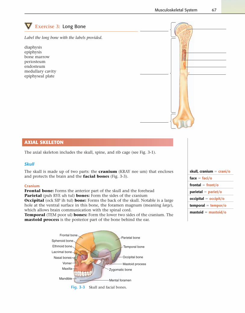
The skull is made up of two parts: the cranium (KRAY nee um) that encloses and protects the brain and the facial bones (Fig. 3-3).
Cranium
Frontal bone: Forms the anterior part of the skull and the foreheadParietal (puh RYE uh tul) bones: Form the sides of the craniumOccipital (ock SIP ih tul) bone: Forms the back of the skull. Notable is a large hole at the ventral surface in this bone, the foramen magnum (meaning large), which allows brain communication with the spinal cord.Temporal (TEM poor ul) bones: Form the lower two sides of the cranium. The mastoid process is the posterior part of the bone behind the ear.
Zygomatic bone
Lacrimal bone
Maxilla
Mandible
Vomer
Frontal boneParietal bone
Occipital bone
Temporal boneEthmoid bone
Sphenoid bone
Nasal bones
Mastoid process
Mental foramen
Fig. 3-3 Skull and facial bones.
skull, cranium � crani/o
face � faci/o
frontal � front/o
parietal � pariet/o
occipital � occipit/o
temporal � tempor/o
mastoid � mastoid/o
Exercise 3: Long Bone
Label the long bone with the labels provided.
diaphysisepiphysisbone marrowperiosteumendosteummedullary cavityepiphyseal plate
68 Chapter 3
Ethmoid (EHTH moyd) bone: Forms the roof and walls of the nasal cavitySphenoid (SFEE noyd) bone: Anterior to the temporal bones and the basilar
part of the occipital bone
The last three bones of the skull, the ossicles, are tiny bones within the ear. These will be discussed in Chapter 14.
Facial Bones
Use Fig. 3-3 to locate the names and locations of the majority of the following facial bones:
Zygoma (zye GOH mah): Cheekbone. Also called the zygomatic (zye goh MAT tick) bone
Lacrimal (LACK rih mul) bones: Paired bones at the corner of each eye that cradle the tear ductsMaxilla (MACK sill ah): Upper jaw bone. Also called the maxillary boneMandible (MAN dih bul): Lower jaw bone. Also called the mandibular boneVomer (VOH mur): Bone that forms the posterior/inferior part of the nasal sep-tal wall between the nostrilsPalatine (PAL eh tyne) bones: Make up part of the roof of the mouthInferior nasal conchae (KON kee): Make up part of the interior of the nose
Rib Cage
The ribs consist of 12 pairs of thin, fl at bones attached to the thoracic vertebrae in the back and to costochondral (kost toh KON drul) tissue in the front (see Fig. 3-1). The ribs can be categorized as follows:
• True ribs: Seven pairs attached directly to the breastbone (sternum) in the front of the body
• False ribs: Five pairs attached to the sternum by cartilage• Floating ribs: Two pairs of false ribs not attached in the front of the body
at all
In addition to ribs, the rib cage includes the sternum (STUR num), also known as the breastbone. The sharp point at the most inferior aspect of the ster-num is called the xiphoid (ZIH foyd) process.
Spine
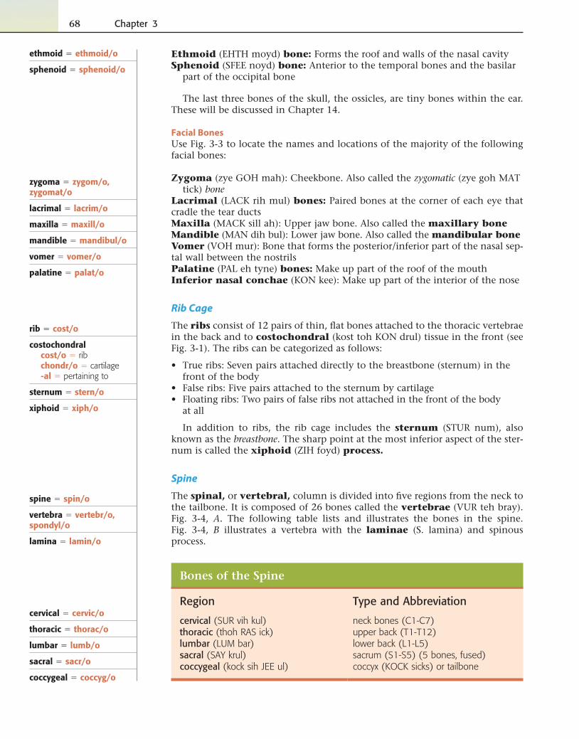
The spinal, or vertebral, column is divided into fi ve regions from the neck to the tailbone. It is composed of 26 bones called the vertebrae (VUR teh bray). Fig. 3-4, A. The following table lists and illustrates the bones in the spine. Fig. 3-4, B illustrates a vertebra with the laminae (S. lamina) and spinous process.
Bones of the Spine
Region Type and Abbreviation
cervical (SUR vih kul) neck bones (C1-C7)thoracic (thoh RAS ick) upper back (T1-T12)lumbar (LUM bar) lower back (L1-L5)sacral (SAY krul) sacrum (S1-S5) (5 bones, fused)coccygeal (kock sih JEE ul) coccyx (KOCK sicks) or tailbone
ethmoid � ethmoid/o
sphenoid � sphenoid/o
zygoma � zygom/o, zygomat/o
lacrimal � lacrim/o
maxilla � maxill/o
mandible � mandibul/o
vomer � vomer/o
palatine � palat/o
rib � cost/o
costochondralcost/o � ribchondr/o � cartilage-al � pertaining to
sternum � stern/o
xiphoid � xiph/o
spine � spin/o
vertebra � vertebr/o, spondyl/o
lamina � lamin/o
cervical � cervic/o
thoracic � thorac/o
lumbar � lumb/o
sacral � sacr/o
coccygeal � coccyg/o
Musculoskeletal System 69
7 cervicalvertebrae
12 thoracicvertebrae
5 lumbarvertebrae
Spinousprocess
Vertebralbody
Sacrum
Coccyx
Transverseprocess
Columnfor spinal
cord
A
Lamina
Spinous process
Vertebral body
Spinal nerves
TransverseprocessSpinal cord
BFig. 3-4 A, Spine. B, Vertebra.
Decode the following terms below using your knowledge of word parts.
17. submandibular
18. costochondral
19. lumbosacral
20. thoracic
21. substernal
Exercise 4: Axial Skeletal Combining Forms
Match each axial skeletal term with its correct combining form.
9. zygomat/o
10. lumb/o
11. mandibul/o
12. coccyg/o
13. vertebr/o
14. palat/o
15. maxill/o
16. stern/o
A. lower jaw bone B. rib C. backbone D. lower back E. cheekbone F. roof and walls of nasal cavity G. cartilage H. roof of mouth I. neck J. skull K. lamina of vertebra L. upper jaw bone M. back of skull N. chest O. tailbone P. breastbone
1. cervic/o
2. lamin/o
3. ethmoid/o
4. chondr/o
5. thorac/o
6. crani/o
7. occipit/o
8. cost/o
70 Chapter 3
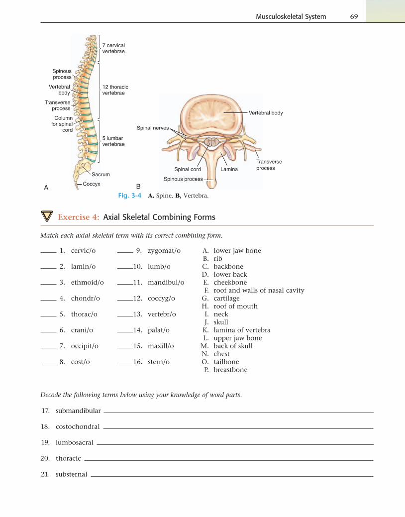
Exercise 5: Bones of the Cranium and Face
Using the diagram provided below, label the bones of the cranium and face with their combining forms where appropriate.
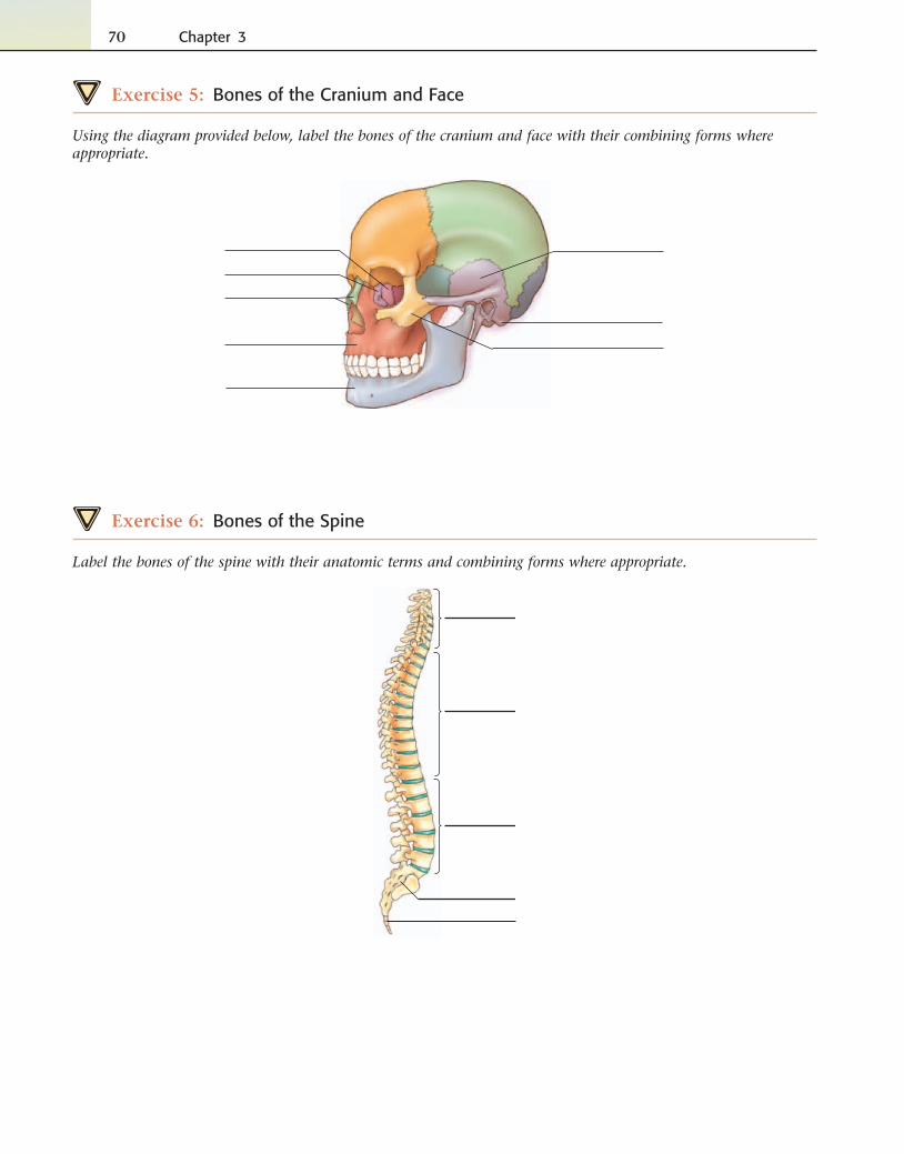
Exercise 6: Bones of the Spine
Label the bones of the spine with their anatomic terms and combining forms where appropriate.
Musculoskeletal System 71
scapula � scapul/o
clavicle � clavicul/o, cleid/o
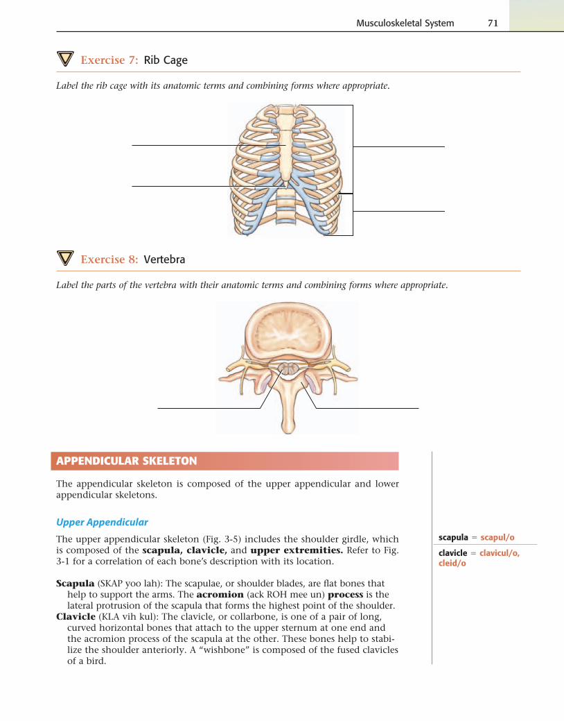
Exercise 7: Rib Cage
Label the rib cage with its anatomic terms and combining forms where appropriate.
Exercise 8: Vertebra
Label the parts of the vertebra with their anatomic terms and combining forms where appropriate.
APPENDICULAR SKELETON
The appendicular skeleton is composed of the upper appendicular and lower appendicular skeletons.
Upper Appendicular
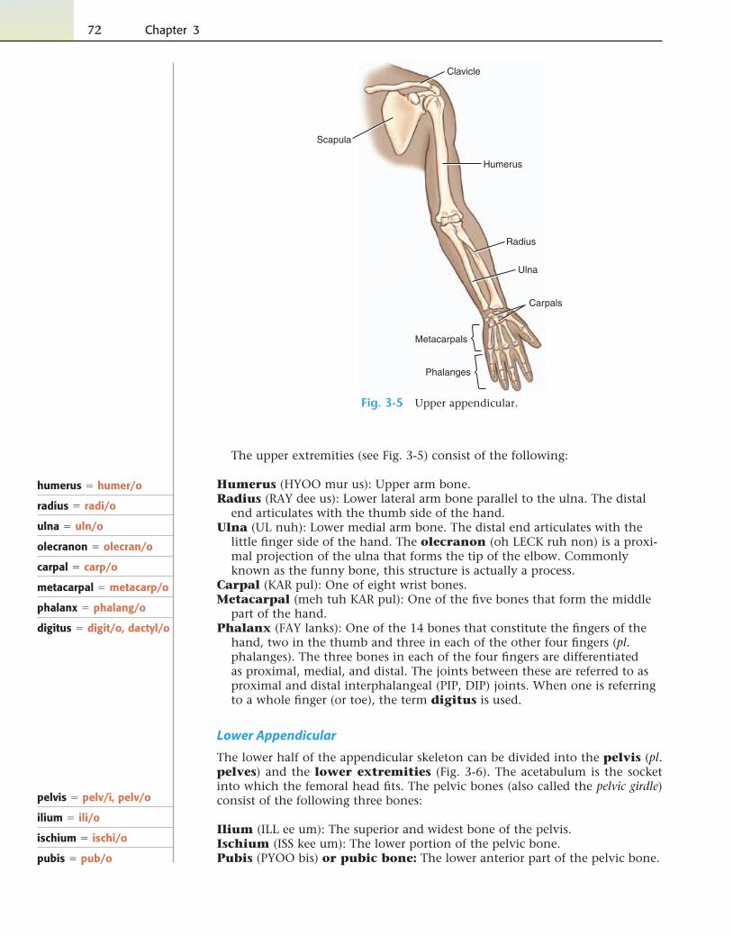
The upper appendicular skeleton (Fig. 3-5) includes the shoulder girdle, which is composed of the scapula, clavicle, and upper extremities. Refer to Fig. 3-1 for a correlation of each bone’s description with its location.
Scapula (SKAP yoo lah): The scapulae, or shoulder blades, are fl at bones that help to support the arms. The acromion (ack ROH mee un) process is the lateral protrusion of the scapula that forms the highest point of the shoulder.
Clavicle (KLA vih kul): The clavicle, or collarbone, is one of a pair of long, curved horizontal bones that attach to the upper sternum at one end and the acromion process of the scapula at the other. These bones help to stabi-lize the shoulder anteriorly. A “wishbone” is composed of the fused clavicles of a bird.
72 Chapter 3
The upper extremities (see Fig. 3-5) consist of the following:
Humerus (HYOO mur us): Upper arm bone.Radius (RAY dee us): Lower lateral arm bone parallel to the ulna. The distal
end articulates with the thumb side of the hand.Ulna (UL nuh): Lower medial arm bone. The distal end articulates with the
little fi nger side of the hand. The olecranon (oh LECK ruh non) is a proxi-mal projection of the ulna that forms the tip of the elbow. Commonly known as the funny bone, this structure is actually a process.
Carpal (KAR pul): One of eight wrist bones.Metacarpal (meh tuh KAR pul): One of the fi ve bones that form the middle
part of the hand.Phalanx (FAY lanks): One of the 14 bones that constitute the fi ngers of the
hand, two in the thumb and three in each of the other four fi ngers (pl. phalanges). The three bones in each of the four fi ngers are differentiated as proximal, medial, and distal. The joints between these are referred to as proximal and distal interphalangeal (PIP, DIP) joints. When one is referring to a whole fi nger (or toe), the term digitus is used.
Lower Appendicular
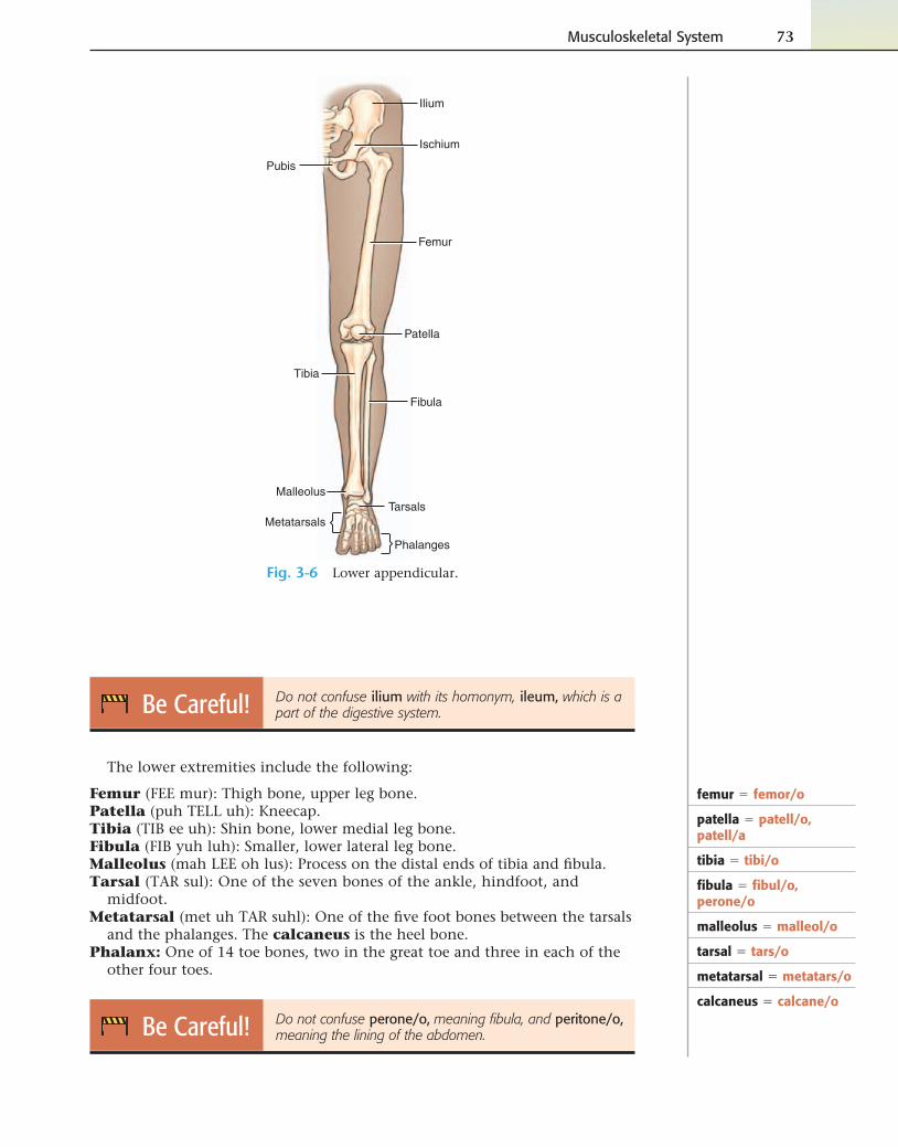
The lower half of the appendicular skeleton can be divided into the pelvis (pl. pelves) and the lower extremities (Fig. 3-6). The acetabulum is the socket into which the femoral head fi ts. The pelvic bones (also called the pelvic girdle) consist of the following three bones:
Ilium (ILL ee um): The superior and widest bone of the pelvis.Ischium (ISS kee um): The lower portion of the pelvic bone.Pubis (PYOO bis) or pubic bone: The lower anterior part of the pelvic bone.
humerus � humer/o
radius � radi/o
ulna � uln/o
olecranon � olecran/o
carpal � carp/o
metacarpal � metacarp/o
phalanx � phalang/o
digitus � digit/o, dactyl/o
Clavicle
Scapula
Humerus
Radius
Ulna
Carpals
Metacarpals
Phalanges
Fig. 3-5 Upper appendicular.
pelvis � pelv/i, pelv/o
ilium � ili/o
ischium � ischi/o
pubis � pub/o
Musculoskeletal System 73
Be Careful! Do not confuse ilium with its homonym, ileum, which is a part of the digestive system.
The lower extremities include the following:
Femur (FEE mur): Thigh bone, upper leg bone.Patella (puh TELL uh): Kneecap.Tibia (TIB ee uh): Shin bone, lower medial leg bone.Fibula (FIB yuh luh): Smaller, lower lateral leg bone.Malleolus (mah LEE oh lus): Process on the distal ends of tibia and fi bula.Tarsal (TAR sul): One of the seven bones of the ankle, hindfoot, and
midfoot.Metatarsal (met uh TAR suhl): One of the fi ve foot bones between the tarsals
and the phalanges. The calcaneus is the heel bone.Phalanx: One of 14 toe bones, two in the great toe and three in each of the
other four toes.
Be Careful! Do not confuse perone/o, meaning fi bula, and peritone/o, meaning the lining of the abdomen.
femur � femor/o
patella � patell/o, patell/a
tibia � tibi/o
fi bula � fi bul/o, perone/o
malleolus � malleol/o
tarsal � tars/o
metatarsal � metatars/o
calcaneus � calcane/o
Ilium
Ischium
Pubis
Femur
Patella
Tibia
Fibula
MalleolusTarsals
Metatarsals
Phalanges
Fig. 3-6 Lower appendicular.
74 Chapter 3
Decode the terms.
23. interphalangeal
24. humeroulnar
25. infrapatellar
26. femoral
27. supraclavicular
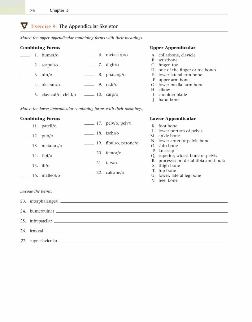
Exercise 9: The Appendicular Skeleton
Match the upper appendicular combining forms with their meanings.
6. metacarp/o
7. digit/o
8. phalang/o
9. radi/o
10. carp/o
Upper Appendicular
A. collarbone, clavicle B. wristbone C. fi nger, toe D. one of the fi nger or toe bones E. lower lateral arm bone F. upper arm bone G. lower medial arm bone H. elbow I. shoulder blade J. hand bone
Combining Forms
1. humer/o
2. scapul/o
3. uln/o
4. olecran/o
5. clavicul/o, cleid/o
Match the lower appendicular combining forms with their meanings.
17. pelv/o, pelv/i
18. ischi/o
19. fi bul/o, perone/o
20. femor/o
21. tars/o
22. calcane/o
Lower Appendicular
K. foot bone L. lower portion of pelvis M. ankle bone N. lower anterior pelvic bone O. shin bone P. kneecap Q. superior, widest bone of pelvis R. processes on distal tibia and fi bula S. thigh bone T. hip bone U. lower, lateral leg bone V. heel bone
Combining Forms
11. patell/o
12. pub/o
13. metatars/o
14. tibi/o
15. ili/o
16. malleol/o
Musculoskeletal System 75
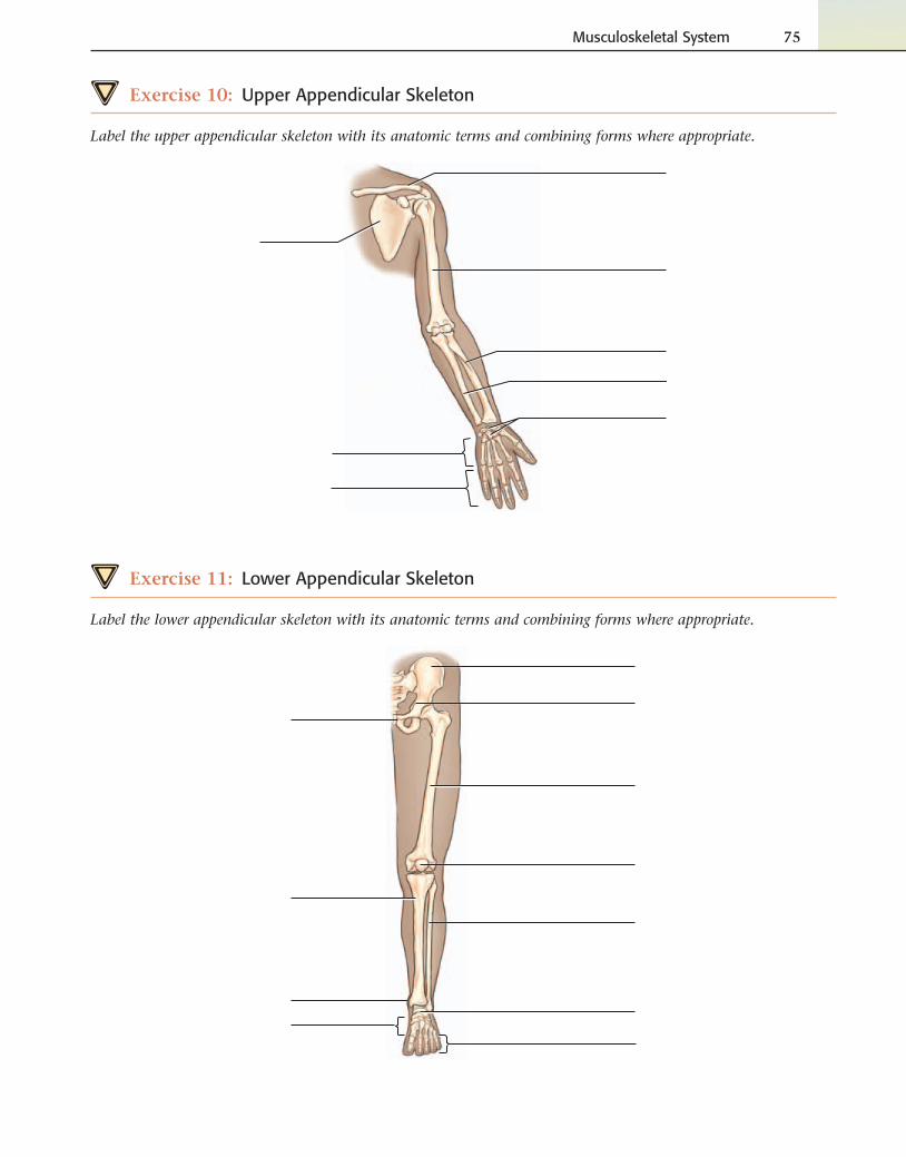
Exercise 10: Upper Appendicular Skeleton
Label the upper appendicular skeleton with its anatomic terms and combining forms where appropriate.
Exercise 11: Lower Appendicular Skeleton
Label the lower appendicular skeleton with its anatomic terms and combining forms where appropriate.
76 Chapter 3
Joints
Joints, or articulations as they are sometimes called, are the parts of the body where two or more bones of the skeleton join. Examples of joints include the knee, which joins the tibia and the femur, and the elbow, which joins the hu-merus with the radius and ulna. Joints provide range of motion (ROM), the range through which a joint can be extended and fl exed. Different joints have different ROM, ranging from no movement at all to full range of movement. Categorized by ROM, they are as follows:
No ROM: Most synarthroses (sin ar THROH sees) are immovable joints held together by fi brous cartilaginous tissue. The suture lines of the skull are ex-amples of synarthroses.
Limited ROM: Amphiarthroses (am fee ar THROH sees) are joints joined together by cartilage that are slightly movable, such as the vertebrae of the spine or the pubic bones.
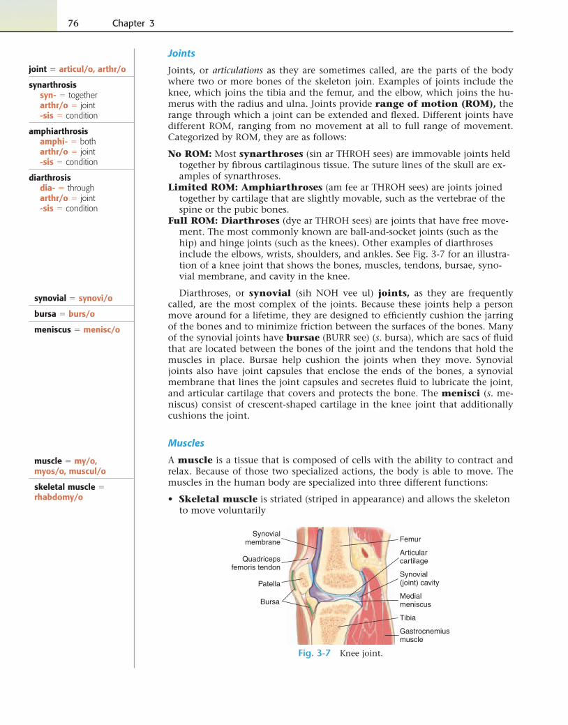
Full ROM: Diarthroses (dye ar THROH sees) are joints that have free move-ment. The most commonly known are ball-and-socket joints (such as the hip) and hinge joints (such as the knees). Other examples of diarthroses include the elbows, wrists, shoulders, and ankles. See Fig. 3-7 for an illustra-tion of a knee joint that shows the bones, muscles, tendons, bursae, syno-vial membrane, and cavity in the knee.
Diarthroses, or synovial (sih NOH vee ul) joints, as they are frequently called, are the most complex of the joints. Because these joints help a person move around for a lifetime, they are designed to effi ciently cushion the jarring of the bones and to minimize friction between the surfaces of the bones. Many of the synovial joints have bursae (BURR see) (s. bursa), which are sacs of fl uid that are located between the bones of the joint and the tendons that hold the muscles in place. Bursae help cushion the joints when they move. Synovial joints also have joint capsules that enclose the ends of the bones, a synovial membrane that lines the joint capsules and secretes fl uid to lubricate the joint, and articular cartilage that covers and protects the bone. The menisci (s. me-niscus) consist of crescent-shaped cartilage in the knee joint that additionally cushions the joint.
Muscles
A muscle is a tissue that is composed of cells with the ability to contract and relax. Because of those two specialized actions, the body is able to move. The muscles in the human body are specialized into three different functions:
• Skeletal muscle is striated (striped in appearance) and allows the skeleton to move voluntarily
joint � articul/o, arthr/o
synarthrosissyn- � togetherarthr/o � joint-sis � condition
amphiarthrosisamphi- � botharthr/o � joint-sis � condition
diarthrosisdia- � througharthr/o � joint-sis � condition
Femur
Quadricepsfemoris tendon
Synovialmembrane
Articularcartilage
Medialmeniscus
Tibia
Synovial(joint) cavityPatella
Bursa
Gastrocnemiusmuscle
Fig. 3-7 Knee joint.
synovial � synovi/o
bursa � burs/o
meniscus � menisc/o
muscle � my/o, myos/o, muscul/o
skeletal muscle � rhabdomy/o
Musculoskeletal System 77
• Smooth muscle that is responsible for involuntary movement of the organs
• Heart muscle that pumps blood to the circulatory system
Muscles are attached to bones by strong fi brous bands of connective tissue called tendons. The bone that is at the end of the attachment that does not move and is nearest to the trunk is termed the origin (O); the bone that is at the end that does move and is farthest from the trunk is termed the insertion (I). The function of a muscle is its action (A). For example, a fl exor muscle bends a joint, and an extensor muscle stretches out a joint. These muscle pairs are termed antagonistic muscles. Synergistic muscles work together to refi ne a movement.
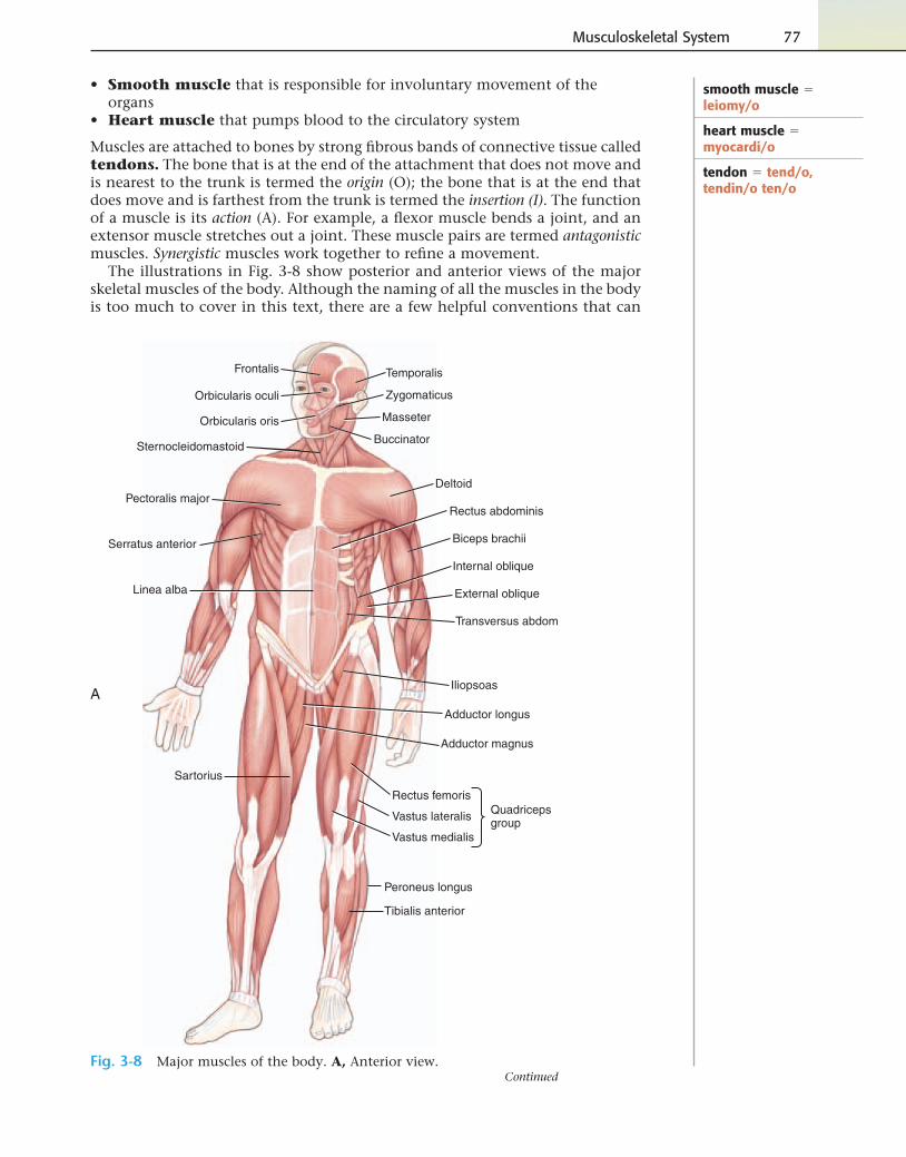
The illustrations in Fig. 3-8 show posterior and anterior views of the major skeletal muscles of the body. Although the naming of all the muscles in the body is too much to cover in this text, there are a few helpful conventions that can
smooth muscle � leiomy/o
heart muscle � myocardi/o
tendon � tend/o, tendin/o ten/o
Temporalis
Orbicularis oculi Zygomaticus
Buccinator
Orbicularis oris
Deltoid
Biceps brachii
Rectus abdominis
Internal oblique
External oblique
Transversus abdom
Iliopsoas
Adductor longus
Adductor magnus
Rectus femoris
Vastus lateralis
Vastus medialis
Tibialis anterior
Peroneus longus
Quadricepsgroup
Sartorius
Linea alba
Serratus anterior
Pectoralis major
Sternocleidomastoid
Masseter
Frontalis
A
Fig. 3-8 Major muscles of the body. A, Anterior view.Continued
78 Chapter 3
Deltoid
Trapezius
Triceps brachii
Latissimus dorsi
Gluteus maximus
Biceps femoris
Semitendinosus
Semimembranosus
Gracilis
Adductor magnus
Gluteus medius
Hamstringgroup
Gastrocnemius
Soleus
Achilles tendon(calcaneal tendon)
B
Sternocleidomastoidmuscle
Origin
Insertion
Sternum
Clavicle
Mastoidprocess
C
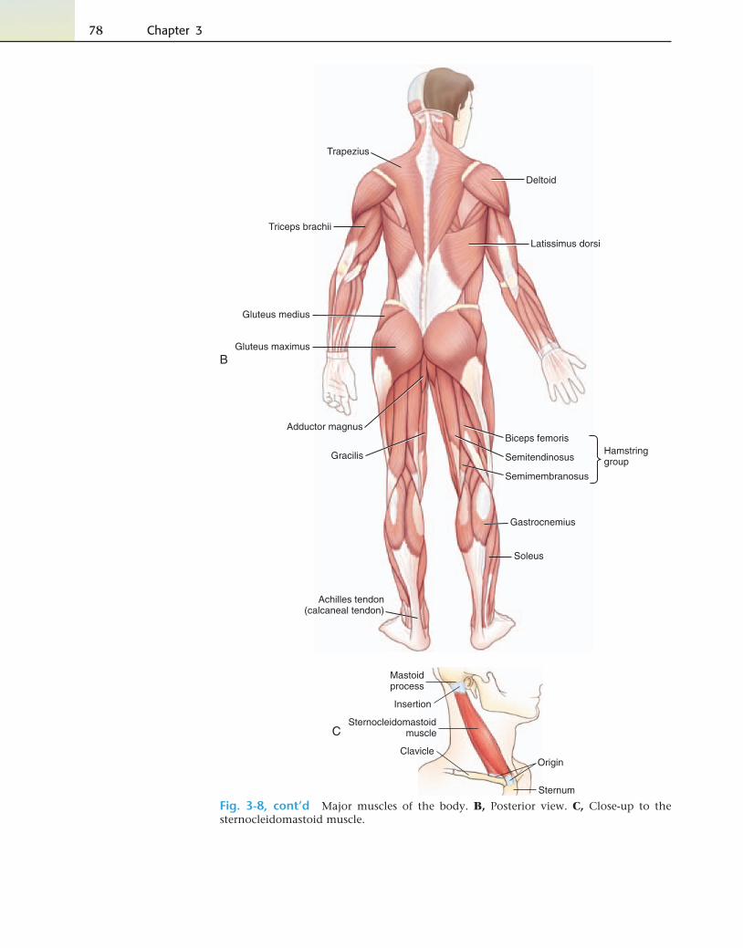
Fig. 3-8, cont’d Major muscles of the body. B, Posterior view. C, Close-up to the sternocleidomastoid muscle.
Musculoskeletal System 79
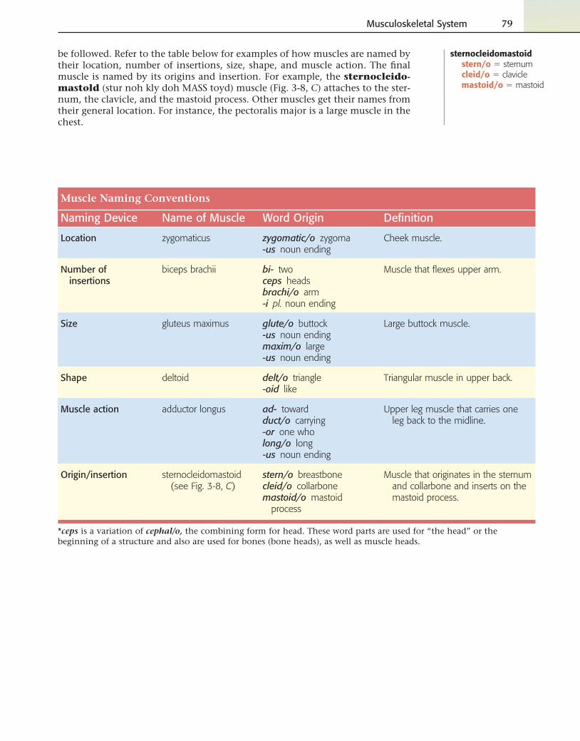
be followed. Refer to the table below for examples of how muscles are named by their location, number of insertions, size, shape, and muscle action. The fi nal muscle is named by its origins and insertion. For example, the sternocleido-mastold (stur noh kly doh MASS toyd) muscle (Fig. 3-8, C) attaches to the ster-num, the clavicle, and the mastoid process. Other muscles get their names from their general location. For instance, the pectoralis major is a large muscle in the chest.
Muscle Naming Conventions
Naming Device Name of Muscle Word Origin Defi nition
Location zygomaticus zygomatic/o zygoma-us noun ending
Cheek muscle.
Number of insertions
biceps brachii bi- twoceps headsbrachi/o arm-i pl. noun ending
Muscle that fl exes upper arm.
Size gluteus maximus glute/o buttock-us noun endingmaxim/o large-us noun ending
Large buttock muscle.
Shape deltoid delt/o triangle-oid like
Triangular muscle in upper back.
Muscle action adductor longus ad- towardduct/o carrying-or one wholong/o long-us noun ending
Upper leg muscle that carries one leg back to the midline.
Origin/insertion sternocleidomastoid (see Fig. 3-8, C)
stern/o breastbonecleid/o collarbonemastoid/o mastoid
process
Muscle that originates in the sternum and collarbone and inserts on the mastoid process.
*ceps is a variation of cephal/o, the combining form for head. These word parts are used for “the head” or the beginning of a structure and also are used for bones (bone heads), as well as muscle heads.
sternocleidomastoidstern/o � sternumcleid/o � claviclemastoid/o � mastoid
80 Chapter 3
Exercise 12: Joints and Muscles
Match the joint and muscle combining forms with their meanings.
7. my/o, myos/o, muscul/o
8. leiomy/o
9. tendin/o, ten/o, tend/o
10. burs/o
11. glute/o
A. smooth muscle B. triangle C. sac of fl uid to cushion joints D. skeletal muscle E. joint F. crescent-shaped cartilage G. heart muscle H. synovial I. muscle J. connects bone to muscle K. buttock
1. arthr/o, articul/o
2. myocardi/o
3. delt/o
4. rhabdomy/o
5. synovi/o
6. menisc/o
Build the terms.
12. pertaining to within the muscle
13. pertaining to buttocks
14. pertaining to the synovium
Musculoskeletal System 81
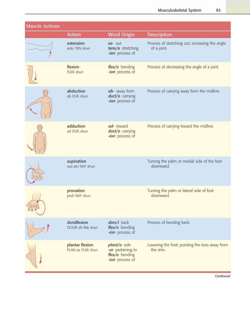
Muscle Actions
Action Word Origin Description
extensionecks TEN shun
ex- outtens/o stretching-ion process of
Process of stretching out; increasing the angle of a joint.
fl exionFLEK shun
fl ex/o bending-ion process of
Process of decreasing the angle of a joint.
abductionab DUK shun
ab- away fromduct/o carrying-ion process of
Process of carrying away from the midline.
adductionad DUK shun
ad- towardduct/o carrying-ion process of
Process of carrying toward the midline.
supinationsoo pin NAY shun
Turning the palm or medial side of the foot downward.
pronationproh NAY shun
Turning the palm or lateral side of foot downward.
dorsifl exionDOOR sih fl ek shun
dors/i backfl ex/o bending-ion process of
Process of bending back.
plantar fl exionPLAN tar FLEK shun
plant/o sole-ar pertaining tofl ex/o bending-ion process of
Lowering the foot; pointing the toes away from the shin.
Continued
82 Chapter 3
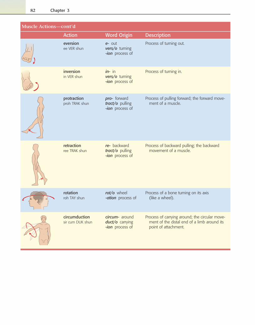
Muscle Actions
Action Word Origin Description
eversionee VER shun
e- outvers/o turning-ion process of
Process of turning out.
inversionin VER shun
in- invers/o turning-ion process of
Process of turning in.
protractionproh TRAK shun
pro- forwardtract/o pulling-ion process of
Process of pulling forward; the forward move-ment of a muscle.
retractionree TRAK shun
re- backwardtract/o pulling-ion process of
Process of backward pulling; the backward movement of a muscle.
rotationroh TAY shun
rot/o wheel-ation process of
Process of a bone turning on its axis (like a wheel).
circumductionsir cum DUK shun
circum- aroundduct/o carrying-ion process of
Process of carrying around; the circular move-ment of the distal end of a limb around its point of attachment.
—cont’d
Musculoskeletal System 83
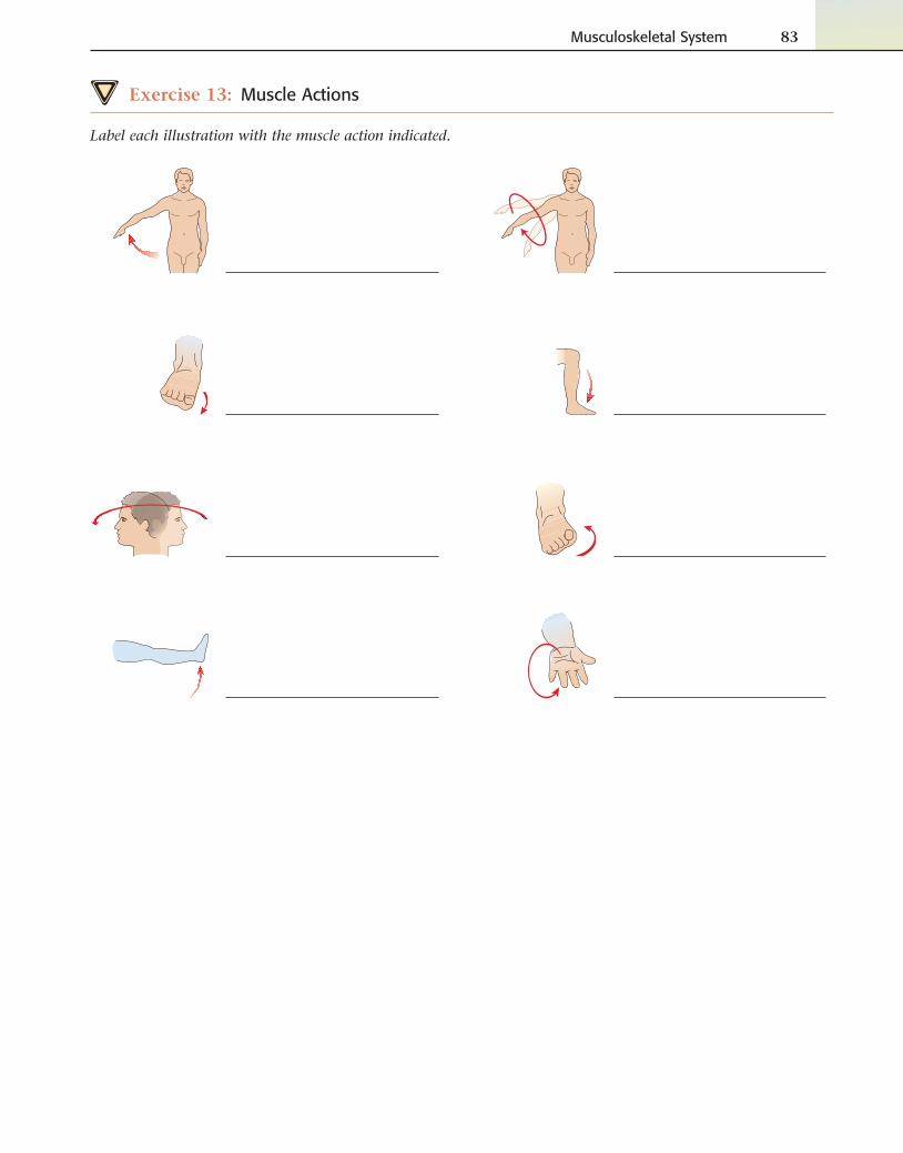
Exercise 13: Muscle Actions
Label each illustration with the muscle action indicated.
84 Chapter 3
Exercise 14: Muscle Actions
Match the muscle action with the correct defi nition.
8. rotation
9. supination
10. eversion
11. dorsifl exion
12. abduction
13. fl exion
14. retraction
A. process of turning in B. process of bending C. process of bone turning on its axis D. process of bending back E. process of pulling backward F. turning the palm downward G. process of carrying away from (the midline) H. turning the palm upward I. process of turning out J. process of pulling forward K. process of stretching out L. process of carrying around M. process of carrying toward (the midline) N. lowering the foot
1. plantar fl exion
2. circumduction
3. pronation
4. inversion
5. adduction
6. extension
7. protraction
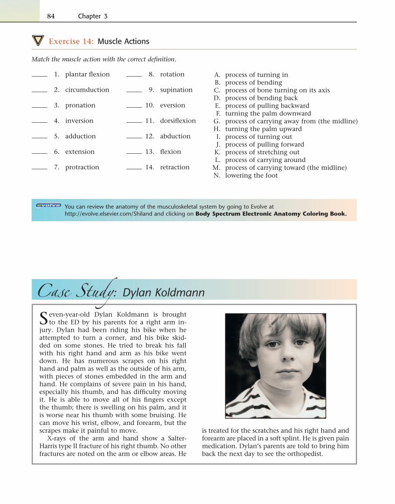
Case Study: Dylan Koldmann
Seven-year-old Dylan Koldmann is brought to the ED by his parents for a right arm in-
jury. Dylan had been riding his bike when he attempted to turn a corner, and his bike skid-ded on some stones. He tried to break his fall with his right hand and arm as his bike went down. He has numerous scrapes on his right hand and palm as well as the outside of his arm, with pieces of stones embedded in the arm and hand. He complains of severe pain in his hand, especially his thumb, and has diffi culty moving it. He is able to move all of his fi ngers except the thumb; there is swelling on his palm, and it is worse near his thumb with some bruising. He can move his wrist, elbow, and forearm, but the scrapes make it painful to move.
X-rays of the arm and hand show a Salter-Harris type II fracture of his right thumb. No other fractures are noted on the arm or elbow areas. He
is treated for the scratches and his right hand and forearm are placed in a soft splint. He is given pain medication. Dylan’s parents are told to bring him back the next day to see the orthopedist.
You can review the anatomy of the musculoskeletal system by going to Evolve at http://evolve.elsevier.com/Shiland and clicking on Body Spectrum Electronic Anatomy Coloring Book.
Musculoskeletal System 85
Case Study: Dylan Koldmann
…
KOLDMANN, DYLAN M. - 507940Task Edit View
Flowsheet: Level:ED ED Table Group List
Time Scale Options Help
KOLDMANN, DYLAN M. Age: 7 yearsDOB: 1/27/2002
Sex: MaleMRN: 507940
Loc: ARHFIN: 3506004
** Allergies **Outpatient [2/20/2009]
Navigator
PROD MAHAFC
As Of 16:10
26 March 2008 16:10
ED
Reference Text Browser
Orders EDED Lab Surgery Pt. Info Pt. Schedule Task List I & O MARClinical NotesAssessmentsRadiologyLast 48 Hours
Form Browser Medication Profile
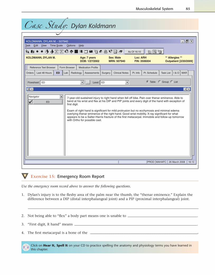
7-year-old sustained injury to right hand when fell off bike. Pain over thenar eminence. Able to bend at his wrist and flex at his DIP and PIP joints and every digit of the hand with exception of first digit.
Exam of right hand is significant for mild protrusion but no ecchymosis and minimal edema overlying thenar eminence of the right hand. Good wrist mobility. X-ray significant for what appears to be a Salter-Harris fracture of the first metacarpal. Immobile and follow-up tomorrow with Ortho for possible cast.
Exercise 15: Emergency Room Report
Use the emergency room record above to answer the following questions.
1. Dylan’s injury is to the fl eshy area of the palm near the thumb, the “thenar eminence.” Explain the difference between a DIP (distal interphalangeal joint) and a PIP (proximal interphalangeal) joint.
2. Not being able to “fl ex” a body part means one is unable to .
3. “First digit, R hand” means .
4. The fi rst metacarpal is a bone of the ______________________________________________________________________________________________________.
Click on Hear It, Spell It on your CD to practice spelling the anatomy and physiology terms you have learned in this chapter.
86 Chapter 3
Combining and Adjective Forms for the Musculoskeletal System
Meaning Combining Form Adjective Formbone marrow myel/o
bone oste/o, osse/o, oss/i osseous, osteal
bursa burs/o bursal
calcaneus (heel bone) calcane/o calcaneal
carpal bone carp/o carpal
cartilage chondr/o, cartilag/o cartilaginous, chondral
clavicle (collarbone) clavicul/o, cleid/o clavicular, cleidal
coccyx (tailbone) coccyg/o coccygeal
condyle condyl/o condylar
elbow (olecranon) olecran/o olecranal
epicondyle epicondyl/o epicondylar
ethmoid ethmoid/o ethmoidal
fascia fasci/o fascial
femur (thigh bone) femor/o femoral
fi bula (lower lateral leg bone) fi bul/o, perone/o fi bular, peroneal
fi nger, toe, (whole), digitus dactyl/o, digit/o digital
foramen foramin/o foraminal
frontal bone front/o
humerus (upper arm bone) humer/o humeral
ilium ili/o iliac
ischium ischi/o ischial
jaw gnath/o
joint (articulation) arthr/o, articul/o articular
lacrima lacrim/o lacrimal
lamina lamin/o laminar
ligament ligament/o, syndesm/o ligamentous, syndesmal
lower back lumb/o lumbar
malleolus malleol/o malleolar
mandible (lower jaw bone) mandibul/o mandibular
mastoid process mastoid/o
maxilla (upper jaw bone) maxill/o maxillary
meniscus menisc/o menisceal
metacarpus (hand bone) metacarp/o metacarpal
Practice pronouncing anatomy and physiology terms! Click Hear It, Say It on your CD.
Musculoskeletal System 87
Combining and Adjective Forms for the Musculoskeletal System
Meaning Combining Form Adjective Formmetatarsus (foot bone) metatars/o metatarsal
muscle (heart) myocardi/o, cardiomy/o myocardial
muscle (smooth) leiomy/o
muscle (skeletal) rhabdomy/o
muscle my/o, myos/o, muscul/o muscular
neck cervic/o cervical
occiput occipit/o occipital
palatine bone palat/o
parietal bone pariet/o parietal
patella (kneecap) patell/o, patell/a patellar
pelvis pelv/i, pelv/o pelvic
phalanx (one of the bones of the fi ngers or toes)
phalang/o phalangeal
pubis (pubic bone) pub/o pubic
radius (lower lateral arm bone) radi/o radial
rib (costa) cost/o costal
sacrum sacr/o sacral
scapula (shoulderblade) scapul/o scapular
skeleton skelet/o skeletal
skull (cranium) crani/o cranial
sole plant/o plantar
sphenoid sphenoid/o sphenoidal
spinal column, spine spin/o, rachi/o, vertebr/o spinal, vertebral, rachial
sternum, breastbone stern/o sternal
tarsus (anklebone) tars/o tarsal
temporal bone tempor/o
tendon tendin/o, tend/o, ten/o tendinous
thorax (chest) thorac/o thoracic
tibia (shinbone) tibi/o tibial
ulna uln/o ulnar
vertebra (backbone) vertebr/o, spondyl/o vertebral
vomer vomer/o
xiphoid process xiph/o xiphoid
zygoma (cheekbone) zygomat/o zygomatic
—cont’d
88 Chapter 3
PATHOLOGY
Terms Related to Congenital Conditions
Term Word Origin Defi nition
achondroplasiaa kon droh PLAY zha
a- withoutchondr/o cartilage-plasia development
Disorder of the development of cartilage at the epiphyses of the long bones and skull, resulting in dwarfi sm.
■ ICD-9-CM code 756.4
muscular dystrophyMUSS kyoo lurDISS troh fee
muscul/o muscle-ar pertaining todys- bad, abnormaltroph/o development-y process of
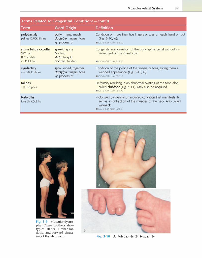
Group of disorders characterized as an inherited progressive atrophy of skeletal muscle without neural involvement (Fig. 3-9).
■ ICD-9-CM code 359.1
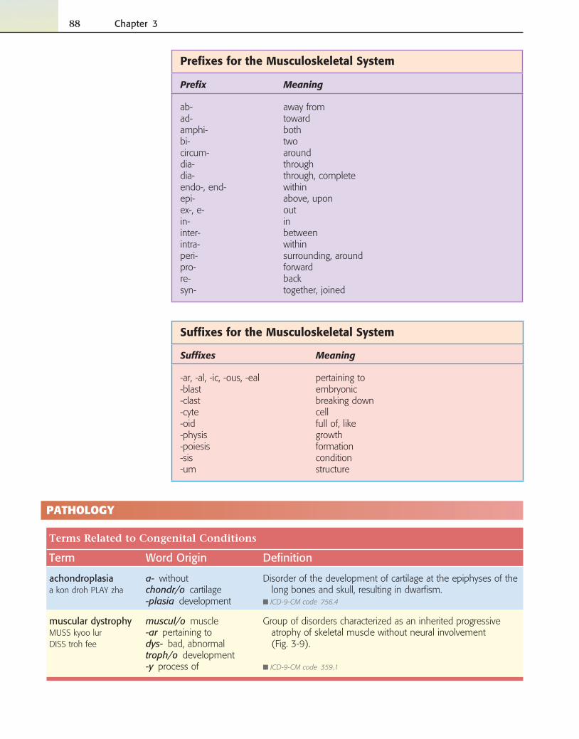
Prefi xes for the Musculoskeletal System
Prefi x Meaning
ab- away fromad- towardamphi- bothbi- twocircum- arounddia- throughdia- through, completeendo-, end- withinepi- above, uponex-, e- outin- ininter- betweenintra- withinperi- surrounding, aroundpro- forwardre- backsyn- together, joined
Suffi xes for the Musculoskeletal System
Suffi xes Meaning
-ar, -al, -ic, -ous, -eal pertaining to-blast embryonic-clast breaking down-cyte cell-oid full of, like-physis growth-poiesis formation-sis condition-um structure
Musculoskeletal System 89
A
B
Fig. 3-10 A, Polydactyly. B, Syndactyly.
Fig. 3-9 Muscular dystro-phy. These brothers show typical stance, lumbar lor-dosis, and forward thrust-ing of the abdomen.
Terms Related to Congenital Conditions
Term Word Origin Defi nition
polydactylypall ee DACK tih lee
poly- many, muchdactyl/o fi ngers, toes-y process of
Condition of more than fi ve fi ngers or toes on each hand or foot (Fig. 3-10, A).
■ ICD-9-CM code 755.00
spina bifi da occultaSPY nahBIFF ih dahah KULL tah
spin/o spinebi- two-fi da to splitocculta hidden
Congenital malformation of the bony spinal canal without in-volvement of the spinal cord.
■ ICD-9-CM code 756.17
syndactylysin DACK tih lee
syn- joined, togetherdactyl/o fi ngers, toes-y process of
Condition of the joining of the fi ngers or toes, giving them a webbed appearance (Fig. 3-10, B).
■ ICD-9-CM code 755.10
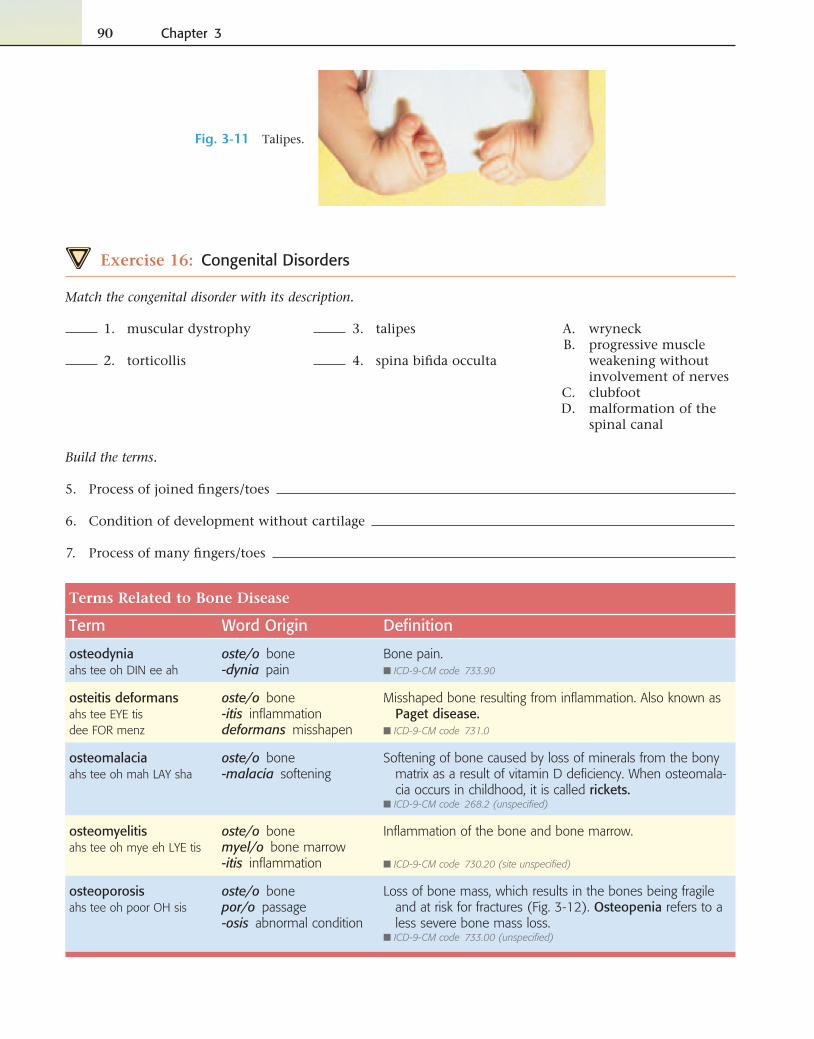
talipesTALL ih peez
Deformity resulting in an abnormal twisting of the foot. Also called clubfoot (Fig. 3-11). May also be acquired.
■ ICD-9-CM code 754.70
torticollistore tih KOLL lis
Prolonged congenital or acquired condition that manifests it-self as a contraction of the muscles of the neck. Also called wryneck.
■ ICD-9-CM code 723.5
—cont’d
90 Chapter 3
Terms Related to Bone Disease
Term Word Origin Defi nition
osteodyniaahs tee oh DIN ee ah
oste/o bone-dynia pain
Bone pain.■ ICD-9-CM code 733.90
osteitis deformansahs tee EYE tisdee FOR menz
oste/o bone-itis infl ammationdeformans misshapen
Misshaped bone resulting from infl ammation. Also known as Paget disease.
■ ICD-9-CM code 731.0
osteomalaciaahs tee oh mah LAY sha
oste/o bone-malacia softening
Softening of bone caused by loss of minerals from the bony matrix as a result of vitamin D defi ciency. When osteomala-cia occurs in childhood, it is called rickets.
■ ICD-9-CM code 268.2 (unspecifi ed)
osteomyelitisahs tee oh mye eh LYE tis
oste/o bonemyel/o bone marrow-itis infl ammation
Infl ammation of the bone and bone marrow.
■ ICD-9-CM code 730.20 (site unspecifi ed)
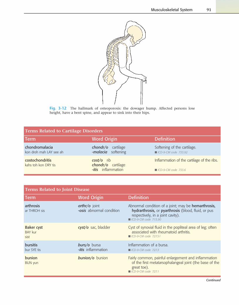
osteoporosisahs tee oh poor OH sis
oste/o bonepor/o passage-osis abnormal condition
Loss of bone mass, which results in the bones being fragile and at risk for fractures (Fig. 3-12). Osteopenia refers to a less severe bone mass loss.
■ ICD-9-CM code 733.00 (unspecifi ed)
Fig. 3-11 Talipes.
Exercise 16: Congenital Disorders
Match the congenital disorder with its description.
3. talipes
4. spina bifi da occulta
A. wryneck B. progressive muscle
weakening without involvement of nerves
C. clubfoot D. malformation of the
spinal canal
1. muscular dystrophy
2. torticollis
Build the terms.
5. Process of joined fi ngers/toes
6. Condition of development without cartilage
7. Process of many fi ngers/toes
Musculoskeletal System 91
Fig. 3-12 The hallmark of osteoporosis: the dowager hump. Affected persons lose height, have a bent spine, and appear to sink into their hips.
Terms Related to Cartilage Disorders
Term Word Origin Defi nition
chondromalaciakon droh mah LAY see ah
chondr/o cartilage-malacia softening
Softening of the cartilage.■ ICD-9-CM code 733.92
costochondritiskahs toh kon DRY tis
cost/o ribchondr/o cartilage-itis infl ammation
Infl ammation of the cartilage of the ribs.
■ ICD-9-CM code 733.6
Terms Related to Joint Disease
Term Word Origin Defi nition
arthrosisar THROH sis
arthr/o joint-osis abnormal condition
Abnormal condition of a joint; may be hemarthrosis, hydrarthrosis, or pyarthrosis (blood, fl uid, or pus respectively, in a joint cavity).
■ ICD-9-CM code 715.90
Baker cystBAY kur sist
cyst/o sac, bladder Cyst of synovial fl uid in the popliteal area of leg; often associated with rheumatoid arthritis.
■ ICD-9-CM code 727.51
bursitisbur SYE tis
burs/o bursa-itis infl ammation
Infl ammation of a bursa.■ ICD-9-CM code 727.3
bunionBUN yun
bunion/o bunion Fairly common, painful enlargement and infl ammation of the fi rst metatarsophalangeal joint (the base of the great toe).
■ ICD-9-CM code 727.1
Continued
92 Chapter 3
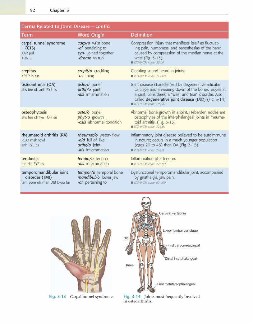
Fig. 3-13 Carpal tunnel syndrome.
Cervical vertebrae
Hip
First carpometacarpal
Distal interphalangeal
Knee
First metatarsophalangeal
Lower lumbar vertebrae
Fig. 3-14 Joints most frequently involved in osteoarthritis.
Terms Related to Joint Disease
Term Word Origin Defi nition
carpal tunnel syndrome (CTS)
KAR pulTUN ul
carp/o wrist bone-al pertaining tosyn- joined together-drome to run
Compression injury that manifests itself as fl uctuat-ing pain, numbness, and paresthesias of the hand caused by compression of the median nerve at the wrist (Fig. 3-13).
■ ICD-9-CM code 354.0
crepitusKREP ih tus
crepit/o crackling-us thing
Crackling sound heard in joints.■ ICD-9-CM code 719.60
osteoarthritis (OA)ahs tee oh arth RYE tis
oste/o bonearthr/o joint-itis infl ammation
Joint disease characterized by degenerative articular cartilage and a wearing down of the bones’ edges at a joint; considered a “wear and tear” disorder. Also called degenerative joint disease (DJD) (Fig. 3-14).
■ ICD-9-CM code 715.90
osteophytosisahs tee oh fye TOH sis
oste/o bonephyt/o growth-osis abnormal condition
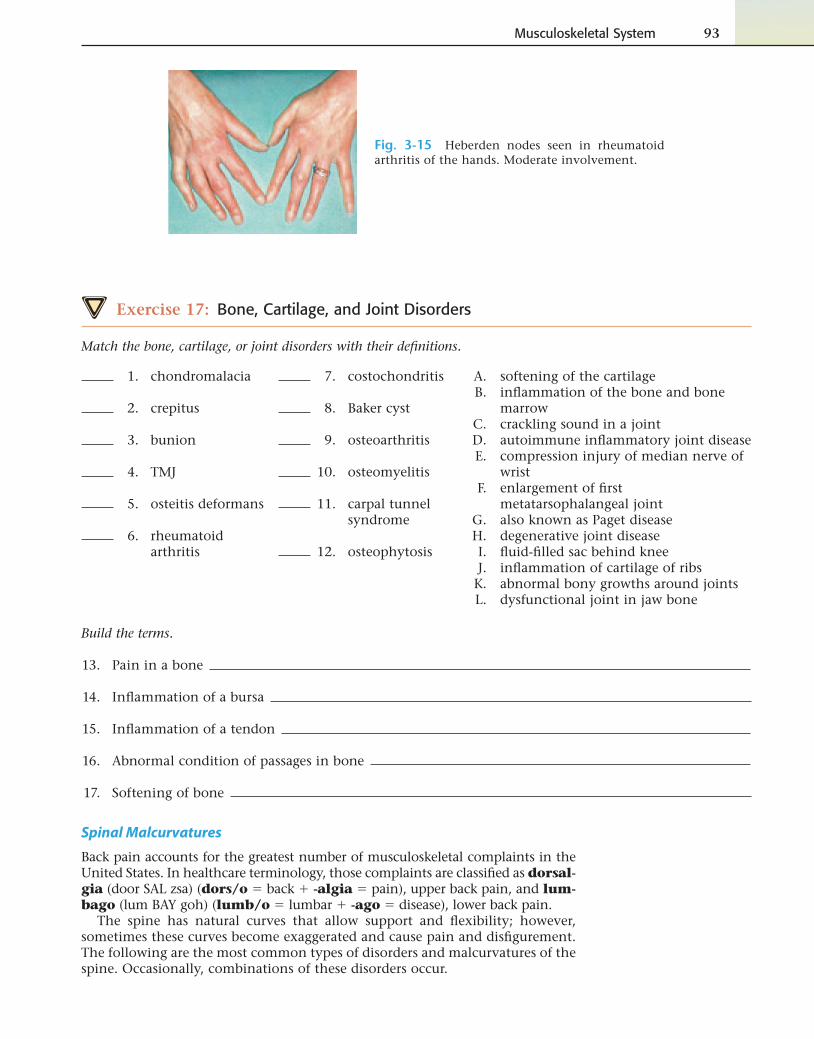
Abnormal bone growth in a joint. Heberden nodes are osteophytes of the interphalangeal joints in rheuma-toid arthritis. (Fig. 3-15).
■ ICD-9-CM code 726.91
rheumatoid arthritis (RA)ROO mah toydarth RYE tis
rheumat/o watery fl ow-oid full of, likearthr/o joint-itis infl ammation
Infl ammatory joint disease believed to be autoimmune in nature; occurs in a much younger population (ages 20 to 45) than OA (Fig. 3-15).
■ ICD-9-CM code 714.0
tendinitisten din EYE tis
tendin/o tendon-itis infl ammation
Infl ammation of a tendon.■ ICD-9-CM code 726.90
temporomandibular joint disorder (TMJ)
tem pore oh man DIB byoo lur
tempor/o temporal bonemandibul/o lower jaw-ar pertaining to
Dysfunctional temporomandibular joint, accompanied by gnathalgia, jaw pain.
■ ICD-9-CM code 524.60
—cont’d
Musculoskeletal System 93
Exercise 17: Bone, Cartilage, and Joint Disorders
Match the bone, cartilage, or joint disorders with their defi nitions.
7. costochondritis
8. Baker cyst
9. osteoarthritis
10. osteomyelitis
11. carpal tunnel syndrome
12. osteophytosis
A. softening of the cartilage B. infl ammation of the bone and bone
marrow C. crackling sound in a joint D. autoimmune infl ammatory joint disease E. compression injury of median nerve of
wrist F. enlargement of fi rst
metatarsophalangeal joint G. also known as Paget disease H. degenerative joint disease I. fl uid-fi lled sac behind knee J. infl ammation of cartilage of ribs K. abnormal bony growths around joints L. dysfunctional joint in jaw bone
1. chondromalacia
2. crepitus
3. bunion
4. TMJ
5. osteitis deformans
6. rheumatoid arthritis
Build the terms.
13. Pain in a bone
14. Infl ammation of a bursa
15. Infl ammation of a tendon
16. Abnormal condition of passages in bone
17. Softening of bone
Fig. 3-15 Heberden nodes seen in rheumatoid arthritis of the hands. Moderate involvement.
Spinal Malcurvatures
Back pain accounts for the greatest number of musculoskeletal complaints in the United States. In healthcare terminology, those complaints are classifi ed as dorsal-gia (door SAL zsa) (dors/o � back � -algia � pain), upper back pain, and lum-bago (lum BAY goh) (lumb/o � lumbar � -ago � disease), lower back pain.
The spine has natural curves that allow support and fl exibility; however, sometimes these curves become exaggerated and cause pain and disfi gurement. The following are the most common types of disorders and malcurvatures of the spine. Occasionally, combinations of these disorders occur.
94 Chapter 3
Terms Related to Spinal Disorders
Term Word Origin Defi nition
ankylosing spondylitisang kih LOH singspon dill LYE tis
ankyl/o stiffeningspondyl/o vertebra-itis infl ammation
Chronic infl ammatory disease of idiopathic origin, which causes a fusion of the spine.
■ ICD-9-CM code 720.0
herniated intervertebral disk
inter- betweenvertebr/o vertebra-al pertaining to
Protrusion of the central part of the disk that lies between the vertebrae, resulting in compression of the nerve root and pain.
■ ICD-9-CM code 722.2
kyphosiskye FOH sis
kyph/o round back-osis abnormal condition
Extreme posterior curvature of the thoracic area of the spine.■ ICD-9-CM code 737.10
lordosislore DOH sis
lord/o swayback-osis abnormal condition
Swayback; exaggerated anterior curve of the lumbar verte-brae (lower back).
■ ICD-9-CM code 737.20
scoliosisskoh lee OH sis
scoli/o curvature-osis abnormal condition
Lateral S curve of the spine that can cause an individual to lose inches in height.
■ ICD-9-CM code 737.30
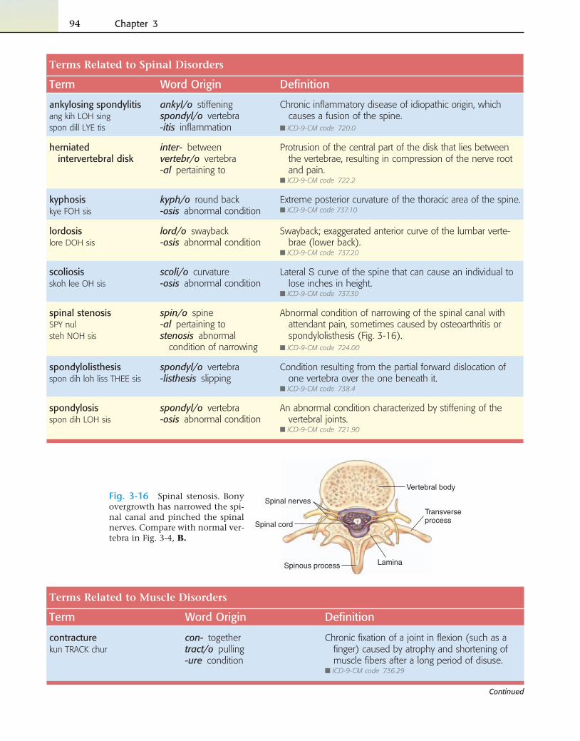
spinal stenosisSPY nulsteh NOH sis
spin/o spine-al pertaining tostenosis abnormal
condition of narrowing
Abnormal condition of narrowing of the spinal canal with attendant pain, sometimes caused by osteoarthritis or spondylolisthesis (Fig. 3-16).
■ ICD-9-CM code 724.00
spondylolisthesisspon dih loh liss THEE sis
spondyl/o vertebra-listhesis slipping
Condition resulting from the partial forward dislocation of one vertebra over the one beneath it.
■ ICD-9-CM code 738.4
spondylosisspon dih LOH sis
spondyl/o vertebra-osis abnormal condition
An abnormal condition characterized by stiffening of the vertebral joints.
■ ICD-9-CM code 721.90
LaminaSpinous process
Vertebral body
Spinal nervesTransverseprocessSpinal cord
Fig. 3-16 Spinal stenosis. Bony overgrowth has narrowed the spi-nal canal and pinched the spinal nerves. Compare with normal ver-tebra in Fig. 3-4, B.
Terms Related to Muscle Disorders
Term Word Origin Defi nition
contracturekun TRACK chur
con- togethertract/o pulling-ure condition
Chronic fi xation of a joint in fl exion (such as a fi nger) caused by atrophy and shortening of muscle fi bers after a long period of disuse.
■ ICD-9-CM code 736.29
Continued
Musculoskeletal System 95
Exercise 18: Spinal and Muscle Disorders
Match the muscle and spinal disorders with their defi nitions.
7. contracture
8. fi bromyalgia
9. lordosis
10. scoliosis
11. dorsalgia
12. myasthenia gravis
A. upper back pain B. infl ammation of the fascia of the foot C. chronic fl exion of a joint caused by
muscle atrophy D. muscle disorder characterized by
musculoskeletal pain, fatigue, and sleep disorders
E. lateral S curve of the spine F. protrusion of the central part of the
vertebral disk G. narrowing of the spinal canal H. chronic infl ammatory disease of idiopathic
origin, which causes fusion of the spine I. usually severe disease characterized by
muscular weakness J. symptoms that occur after removal of the
lamina K. swayback L. lower back pain
1. ankylosing spondylitis
2. spinal stenosis
3. herniated intervertebral disk
4. plantar fasciitis
5. postlaminectomy syndrome
6. lumbago
Terms Related to Muscle Disorders
Term Word Origin Defi nition
fi bromyalgiafye broh mye AL jah
fi br/o fi bermy/o muscle-algia pain
Disorder characterized by musculoskeletal pain, fatigue, muscle stiffness and spasms, and sleep disturbances.
■ ICD-9-CM code 729.1
myasthenia gravismye ah STHEE nee ah GRAV us
my/o musclea- without, no-sthenia condition of
strengthgravis severe
Usually severe condition characterized by fatigue and progressive muscle weakness, especially of the face and throat.
■ ICD-9-CM code 358.00
plantar fasciitisPLAN turfass ee EYE tis
plant/o sole-ar pertaining tofasci/o fascia-itis infl ammation
Infl ammation of the fascia on the sole of the foot.
■ ICD-9-CM code 728.71
polymyositispahl ee my oh SYE tis
poly- manymyos/o muscle-itis infl ammation
Chronic, idiopathic infl ammation of a number of voluntary muscles.
■ ICD-9-CM code 710.4
postlaminectomy syndrome
post lam in ECK tuh mee
post- afterlamin/o lamina-ectomy removal
Group of symptoms that occur together after the removal of a lamina to correct a spinal disorder.
■ ICD-9-CM code 722.80
rhabdomyolysisrab doh mye AL ih sis
rhabdomy/o striated muscle-lysis breakdown, destruction
Breakdown of striated/skeletal muscle.■ ICD-9-CM code 728.88
—cont’d
96 Chapter 3
Build the terms.
13. Condition of slipping of the vertebrae
14. Abnormal condition of the vertebra
15. Abnormal condition of curvature
16. Pertaining to infl ammation of the fascia of the sole
17. Breakdown of striated muscle
18. Infl ammation of many muscles
Trauma
Fractures
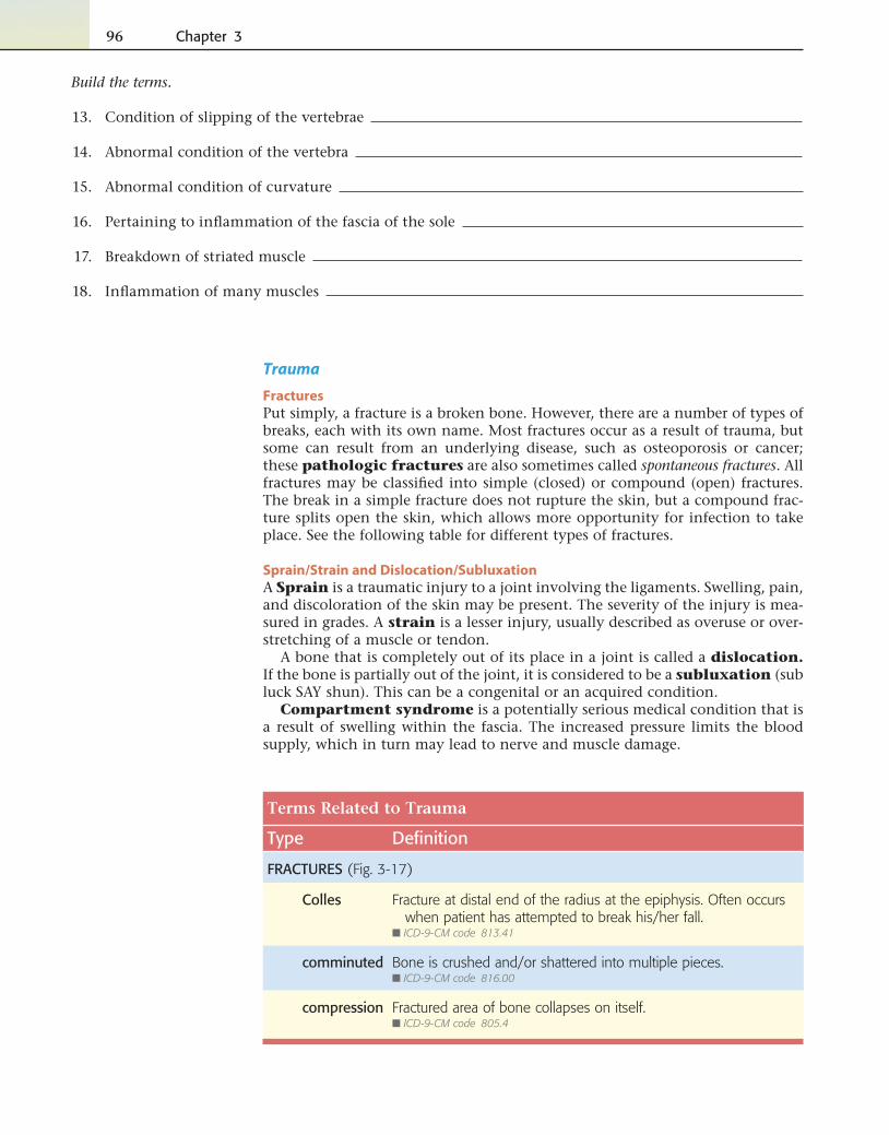
Put simply, a fracture is a broken bone. However, there are a number of types of breaks, each with its own name. Most fractures occur as a result of trauma, but some can result from an underlying disease, such as osteoporosis or cancer; these pathologic fractures are also sometimes called spontaneous fractures. All fractures may be classifi ed into simple (closed) or compound (open) fractures. The break in a simple fracture does not rupture the skin, but a compound frac-ture splits open the skin, which allows more opportunity for infection to take place. See the following table for different types of fractures.
Sprain/Strain and Dislocation/Subluxation
A Sprain is a traumatic injury to a joint involving the ligaments. Swelling, pain, and discoloration of the skin may be present. The severity of the injury is mea-sured in grades. A strain is a lesser injury, usually described as overuse or over-stretching of a muscle or tendon.
A bone that is completely out of its place in a joint is called a dislocation. If the bone is partially out of the joint, it is considered to be a subluxation (sub luck SAY shun). This can be a congenital or an acquired condition.
Compartment syndrome is a potentially serious medical condition that is a result of swelling within the fascia. The increased pressure limits the blood supply, which in turn may lead to nerve and muscle damage.
Terms Related to Trauma
Type Defi nition
FRACTURES (Fig. 3-17)
Colles Fracture at distal end of the radius at the epiphysis. Often occurs when patient has attempted to break his/her fall.
■ ICD-9-CM code 813.41
comminuted Bone is crushed and/or shattered into multiple pieces.■ ICD-9-CM code 816.00
compression Fractured area of bone collapses on itself.■ ICD-9-CM code 805.4
Musculoskeletal System 97
Comminuted Compression
Colles Complicated
Impacted Hairline
Greenstick Salter-Harris
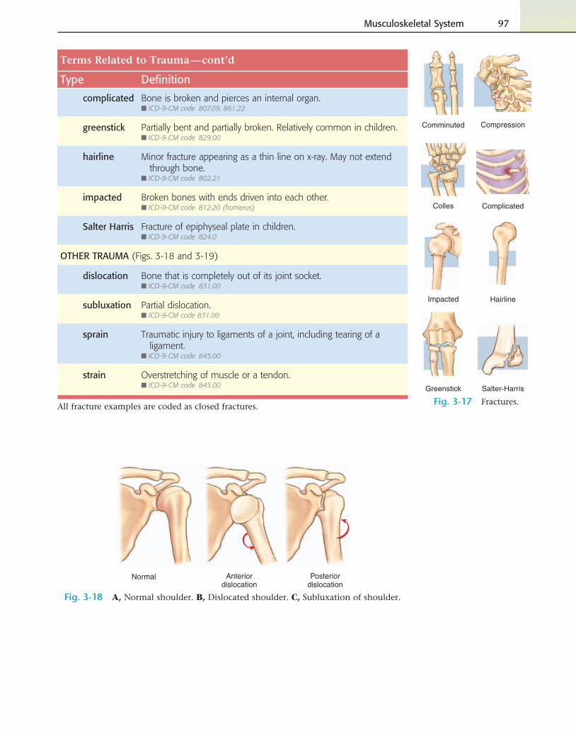
Fig. 3-17 Fractures.
Normal Anteriordislocation
Posteriordislocation
Fig. 3-18 A, Normal shoulder. B, Dislocated shoulder. C, Subluxation of shoulder.
Terms Related to Trauma
Type Defi nition
complicated Bone is broken and pierces an internal organ.■ ICD-9-CM code 807.09, 861.22
greenstick Partially bent and partially broken. Relatively common in children.■ ICD-9-CM code 829.00
hairline Minor fracture appearing as a thin line on x-ray. May not extend through bone.
■ ICD-9-CM code 802.21
impacted Broken bones with ends driven into each other.■ ICD-9-CM code 812.20 (humerus)
Salter Harris Fracture of epiphyseal plate in children.■ ICD-9-CM code 824.0
OTHER TRAUMA (Figs. 3-18 and 3-19)
dislocation Bone that is completely out of its joint socket.■ ICD-9-CM code 831.00
subluxation Partial dislocation.■ ICD-9-CM code 831.00
sprain Traumatic injury to ligaments of a joint, including tearing of a ligament.
■ ICD-9-CM code 845.00
strain Overstretching of muscle or a tendon.■ ICD-9-CM code 845.00
All fracture examples are coded as closed fractures.
—cont’d
98 Chapter 3
Exercise 19: Fractures
Match the fractures with their defi nitions.
6. simple/closed
7. compound/open
8. hairline
9. pathologic
A. broken bone pierces internal organ. B. broken bone pierces skin. C. spontaneous fracture as a result of disease. D. bone is partially bent and partially broken. E. bone is broken, skin is closed. F. distal end of radius is broken. G. ends of broken bone are driven into each
other. H. fracture appears as a line on the bone and
fracture may not be completely through bone.
I. bone is crushed.
1. complicated
2. greenstick
3. Colles
4. impacted
5. comminuted
Exercise 20: Other Trauma
1. A partial displacement of a bone at a joint is a ; full displacement is a
.
2. An injury that can be described in grades and involves the soft tissue of a joint is a .
3. An overstretching of a muscle is a .
4. Swelling within the confi nes of a muscle fascia can lead to .
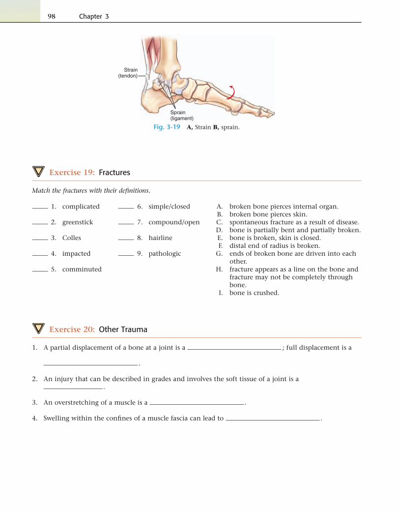
Strain(tendon)
Sprain(ligament)
Fig. 3-19 A, Strain B, sprain.
Musculoskeletal System 99
Terms Related to Benign Neoplasms
Term Word Origin Defi nition
chondromakon DROH mah
chondr/o cartilage-oma tumor
Benign tumor of the cartilage, usually occurring in children and adolescents.
■ ICD-9-CM code 213.9
exostosiseck ahs TOH sis
ex- outoste/o bone-osis abnormal condition
Abnormal condition of bony growth. Also called hyper-ostosis and osteochondroma.
■ ICD-9-CM code 726.91
leiomyomalye oh mye OH mah
leiomy/o smooth muscle-oma tumor
Benign tumor of smooth muscle. The most common leiomyoma is in the uterus and is termed a fi broid.
■ ICD-9-CM code 215.9
osteomaahs tee OH mah
oste/o bone-oma tumor
Benign bone tumor, usually of compact bone.■ ICD-9-CM code 213.9
rhabdomyomarab doh mye OH mah
rhabdomy/o skeletal muscle-oma tumor
Benign tumor of striated/voluntary/skeletal muscle.■ ICD-9-CM code 215.9
Terms Related to Malignant Neoplasms
Term Word Origin Defi nition
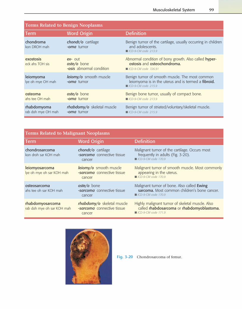
chondrosarcomakon droh sar KOH mah
chondr/o cartilage-sarcoma connective tissue
cancer
Malignant tumor of the cartilage. Occurs most frequently in adults (Fig. 3-20).
■ ICD-9-CM code 170.9
leiomyosarcomalye oh mye oh sar KOH mah
leiomy/o smooth muscle-sarcoma connective tissue
cancer
Malignant tumor of smooth muscle. Most commonly appearing in the uterus.
■ ICD-9-CM code 170.9
osteosarcomaahs tee oh sar KOH mah
oste/o bone-sarcoma connective tissue
cancer
Malignant tumor of bone. Also called Ewing sarcoma. Most common children’s bone cancer.
■ ICD-9-CM code 170.9
rhabdomyosarcomarab doh mye oh sar KOH mah
rhabdomy/o skeletal muscle-sarcoma connective tissue
cancer
Highly malignant tumor of skeletal muscle. Also called rhabdosarcoma or rhabdomyoblastoma.
■ ICD-9-CM code 171.9
Fig. 3-20 Chondrosarcoma of femur.
100 Chapter 3
Exercise 21: Neoplasms
Match the neoplasms with their defi nitions.
3. leiomyosarcoma
4. chondrosarcoma
A. connective tissue cancer of bone B. connective tissue cancer of cartilage C. connective tissue cancer of skeletal
muscle D. connective tissue cancer of smooth
muscle
1. rhabdomyosarcoma
2. osteosarcoma
Build the term.
5. Benign tumor of skeletal muscle
6. Benign bone tumor
7. Benign tumor of smooth muscle
8. Benign tumor of cartilage
9. An abnormal condition of out(growth) of bone ______________________________________________________________________________________
Case Study: Jean Herold
Jean Herold is a 54-year-old nurse’s aide who ex-ercises three to four times a week at her local fi t-
ness center. As she is leaving the center one night, she slips on some ice and falls heavily on her right upper arm and shoulder. Jean drove herself home but spends the night in a great deal of pain, and the next day her friend drives her to the hospital. She has x-rays and a CT scan of her right arm and shoulder. She is diagnosed with a fracture to the top of her upper arm bone and is given pain and nausea medication. She is admitted to the hospital and has surgery the next day.
Click on Hear It, Spell It on your CD to practice spelling the pathology terms you have learned in this chapter.
To see how well you can pronounce the pathology terms in the chapter, click on Hear It, Spell It on your CD.
Musculoskeletal System 101
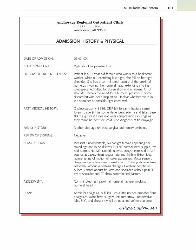
Anchorage Regional Outpatient Clinic1247 Inuit Blvd.
Anchorage, AK 99506
ADMISSION HISTORY & PHYSICAL
DATE OF ADMISSION: 03/01/XX
CHIEF COMPLAINT: Right shoulder pain/fracture
HISTORY OF PRESENT ILLNESS: Patient is a 54-year-old female who works as a healthcare worker. While out exercising last night, she fell on her right shoulder. She has a comminuted fracture of the proximal humerus involving the humeral head, extending into the joint space. Admitted for observation and analgesia. CT of shoulder reveals the need for a humeral prosthesis. Some discomfort with deep inspiration. Unclear whether this is in the shoulder or possible right chest wall.
PAST MEDICAL HISTORY: Cholecystectomy 1986. ORIF left forearm, fracture same forearm, age 9. Has some dependent edema and takes Lasix 80 mg qd for it. Does not wear compression stockings as they make her feet feel cold. Also diagnosis of fi bromyalgia.
FAMILY HISTORY: Mother died age 64 post surgical pulmonary embolus.
REVIEW OF SYSTEMS: Negative.
PHYSICAL EXAM: Pleasant, uncomfortable, overweight female appearing her stated age and in no distress. HEENT normal, neck supple, thy-roid normal. No JVD, carotids normal. Lungs decreased breath sounds at bases. Heart regular rate and rhythm. Extremities: normal range of motion of lower extremities. Motor sensory deep tendon refl exes are normal in arm. Trace pretibial edema bilaterally without venostasis changes. Excellent peripheral pulses. Cannot adduct her arm and shoulder without pain. X-ray of shoulder and CT show comminuted fracture.
ASSESSMENT: Comminuted right proximal humeral fracture involving humeral head.
PLAN: Admit for analgesia, IV fl uids. Has a little nausea probably from analgesics. Won’t have surgery until tomorrow. Preoperative labs, EKG, and chest x-ray will be obtained before that time.
Melissa Landrey, MD
102 Chapter 3
Exercise 22: Admission Record
Using the admission record on p. 101, answer the following questions:
1. Which bone did she break while exercising? Give the medical and English names.
2. Did she fracture the area closest to her shoulder or farther from her shoulder? Circle one.
3. Describe the type of fracture sustained.
4. What other MS disorder does she currently have?
5. How was her previous fracture of her left forearm treated?
6. What does “cannot adduct her arm and shoulder without pain” mean?
Age MattersPediatrics
As can be seen from our table of congential disorders, there are several musculoskeletal conditions that a child may be born with: achondroplasia, muscular dystrophy, two disorders of the phalanges (syndactyly and polydactyly), spina bifi da occulta, talipes, and congential torticollis. Although not ex-clusive to childhood, pediatric statistics reveal high numbers of children treated each year for the ef-fects of physical trauma. Fractures are common: beginning with clavicular fractures (the result of birth trauma) to fractures of the arms (humerus, radius, and ulna) and the legs (femur, tibia, and fi bula). Sprains, strains, dislocations, and subluxations are other pediatric diagnoses that appear with regularity for this system.
Geriatrics
Statistics collected on the geriatric population of patients also report a high number of fractures. These, however, are mainly fractures of the hip and femur and are often preceded by bone loss caused by osteoporosis or cancer. Osteoarthritis, often referred to as “wear and tear disease,” is another disorder that affl icts many patients as they age. The high number of total knee replacement surgeries today are often the result of this disease.
Term Word Origin Defi nition
arthrographyar THRAH gruh fee
arthr/o joint-graphy process of recording
X-ray recording of a joint.
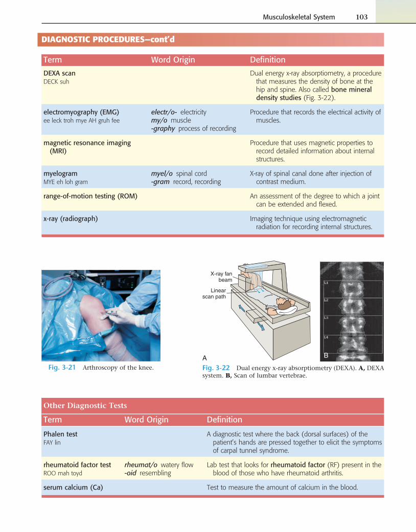
arthroscopyar THRAHS kuh pee
arthr/o joint-scopy process of viewing
Visual examination of a joint, accomplished by use of an arthroscope (Fig. 3-21).
computed tomography (CT) scan
tom/o section-graphy process of recording
Imaging technology that records transverse planes of the body for diagnostic purposes.
DIAGNOSTIC PROCEDURES
You can review the pathology terms you’ve learned in this chapter by playing Medical Millionaire on your CD.
Musculoskeletal System 103
Term Word Origin Defi nition
DEXA scanDECK suh
Dual energy x-ray absorptiometry, a procedure that measures the density of bone at the hip and spine. Also called bone mineral density studies (Fig. 3-22).
electromyography (EMG)ee leck troh mye AH gruh fee
electr/o- electricitymy/o muscle-graphy process of recording
Procedure that records the electrical activity of muscles.
magnetic resonance imaging (MRI)
Procedure that uses magnetic properties to record detailed information about internal structures.
myelogramMYE eh loh gram
myel/o spinal cord-gram record, recording
X-ray of spinal canal done after injection of contrast medium.
range-of-motion testing (ROM) An assessment of the degree to which a joint can be extended and fl exed.
x-ray (radiograph) Imaging technique using electromagnetic radiation for recording internal structures.
DIAGNOSTIC PROCEDURES—cont’d
Fig. 3-21 Arthroscopy of the knee.
X-ray fanbeam
Linearscan path
A B
Fig. 3-22 Dual energy x-ray absorptiometry (DEXA). A, DEXA system. B, Scan of lumbar vertebrae.
Other Diagnostic Tests
Term Word Origin Defi nition
Phalen testFAY lin
A diagnostic test where the back (dorsal surfaces) of the patient’s hands are pressed together to elicit the symptoms of carpal tunnel syndrome.
rheumatoid factor testROO mah toyd
rheumat/o watery fl ow-oid resembling
Lab test that looks for rheumatoid factor (RF) present in the blood of those who have rheumatoid arthritis.
serum calcium (Ca) Test to measure the amount of calcium in the blood.
104 Chapter 3
Exercise 23: Diagnostic Procedures
Match the diagnostic tests to their defi nitions.
5. RF
6. serum calcium
7. EMG
8. CT scan
A. test for rheumatoid arthritis B. imaging technique using electromagnetic
radiation C. imaging of a plane of the body D. blood test for Ca E. test to measure bone density, using dual energy
x-ray F. imaging using magnetic resonance G. range of motion H. electromyography
1. MRI
2. DEXA scan
3. x-ray
4. ROM
Build the terms.
9. Process of viewing a joint
10. Process of recording a joint
11. Process of recording the electrical (activity) of a muscle _______________________________________________________________________
12. Process of recording the spinal cord
THERAPEUTIC INTERVENTIONS
Setting Fractures
Broken bones must be “set”—that is, aligned and immobilized; the most com-mon method is with a plaster cast. If a bone does not mend and realign cor-rectly, it is said to be a malunion. If no healing takes place, it is a nonunion. A piece of bone that does not have a renewed blood supply will die; this tissue then is called a sequestrum (seh KWES trum). Removal of dirt, damaged tissue, or foreign objects from a wound is one of the fi rst steps in repairing an open fracture. This removal of debris is called débridement (de breed MON). Methods of fi xation and alignment are described as follows:
External fi xation: (EF) Noninvasive stabilization of broken bones in which no opening is made in the skin; instead, the stabilization takes place mainly through devices external to the body that offer traction.
Internal fi xation: (IF) Stabilization of broken bones in their correct posi-tion, using pins, screws, plates, and so on, which are fastened to the bones to maintain correct alignment.
Reduction: Alignment and immobilization of the ends of a broken bone. Open reduction (OR) requires incision of the skin; closed reduction (CR) does not require incision.
Go to your CD to view animations of an open reduction internal fi xation (ORIM) of an ankle fracture and a closed reduction (CR) and pinning of a hip fracture.
Musculoskeletal System 105
Terms Related to Therapeutic Interventions
Term Word Origin Defi nition
amputationam pyoo TAY shun
Removal of a limb when there are no feasible options to save it.
arthrocentesisar throh sen TEE sis
arthr/o joint-centesis surgical puncture
Surgical puncture of a joint to remove fl uid.
arthrodesisar throh DEE sis
arthr/o joint-desis binding
Binding or stabilization of a joint by operative means.
arthroplastyAR throh plas tee
arthr/o joint-plasty surgical repair
General term meaning surgical repair of a joint.
bunionectomybun yun ECK tuh mee
bunion/o bunion-ectomy excision, resection
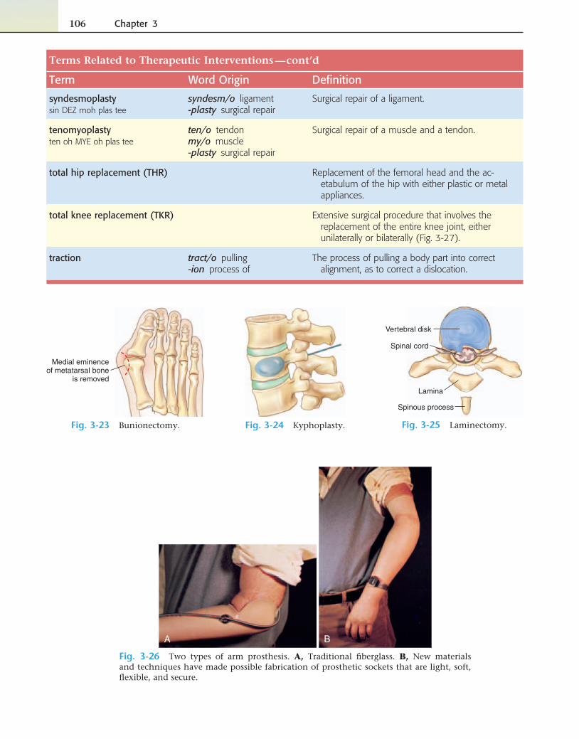
Removal of a bunion (Fig. 3-23).
kyphoplastyKYE foh plas tee
kyph/o round back-plasty surgical repair
Minimally invasive procedure designed to address the pain of fractured vertebrae resulting from os-teoporosis or cancer (Fig. 3-24). A balloon is used to infl ate the area of fracture before a cementlike substance is injected. The substance hardens rap-idly, and pain relief is immediate in most patients.
laminectomylam ih NECK tuh mee
lamin/o lamina-ectomy excision, resection
Removal of the bony arches of one or more ver-tebrae to relieve compression of the spinal cord (Fig. 3-25).
meniscectomymen iss ECK tuh mee
menisc/o meniscus-ectomy removal
Removal of a meniscus such as in the knee.
myorrhaphymye ORE rah fee
my/o muscle-rrhaphy suture
Suture of a muscle.
operative ankylosisAH pur ah tivang kih LOH sis
ankyl/o stiffening-osis abnormal condition
Procedure used in the treatment of spinal fractures or after diskectomy or laminectomy for the cor-rection of a herniated vertebral disk; also used to describe surgical fi xation of a joint. Also called arthrodesis.
osteoclasisAHS tee oh klay sis
oste/o bone-clasis intentional breaking
Refracture of a bone, usually done if a bone has a malunion.
osteoplastyAHS tee oh plas tee
oste/o bone-plasty surgical repair
Surgical repair of a bone.
prosthesisprahs THEE sis
prosthes/o addition-is thing
An artifi cial body part that is constructed to replace missing limbs, eyes, and other body parts (pl. prostheses) (Fig. 3-26).
spondylosyndesisspon dih loh sin DEE sis
spondyl/o vertebrasyn- together-desis binding
Fixation of an unstable segment of the spine by skeletal traction, immobilization of the patient in a body cast, or stabilization with a bone graft or synthetic device. Also called spinal fusion and spondylodesis.
Continued
106 Chapter 3
Terms Related to Therapeutic Interventions
Term Word Origin Defi nition
syndesmoplastysin DEZ moh plas tee
syndesm/o ligament-plasty surgical repair
Surgical repair of a ligament.
tenomyoplastyten oh MYE oh plas tee
ten/o tendonmy/o muscle-plasty surgical repair
Surgical repair of a muscle and a tendon.
total hip replacement (THR) Replacement of the femoral head and the ac-etabulum of the hip with either plastic or metal appliances.
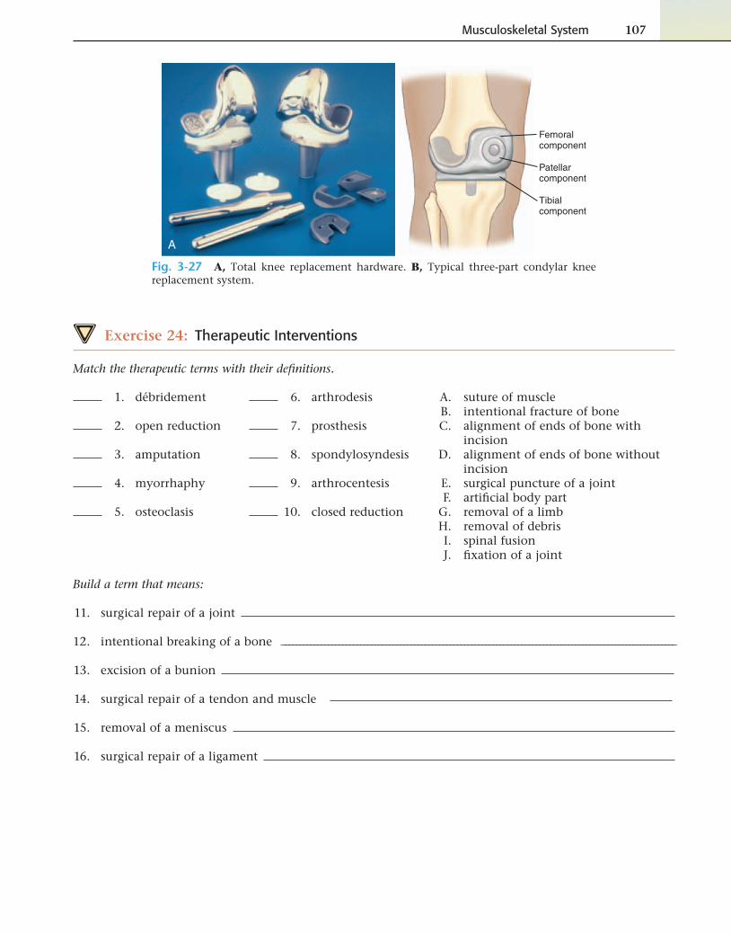
total knee replacement (TKR) Extensive surgical procedure that involves the replacement of the entire knee joint, either unilaterally or bilaterally (Fig. 3-27).
traction tract/o pulling-ion process of
The process of pulling a body part into correct alignment, as to correct a dislocation.
—cont’d
Medial eminenceof metatarsal bone
is removed
Fig. 3-23 Bunionectomy. Fig. 3-24 Kyphoplasty.
Spinal cord
Vertebral disk
Lamina
Spinous process
Fig. 3-25 Laminectomy.
A B
Fig. 3-26 Two types of arm prosthesis. A, Traditional fi berglass. B, New materials and techniques have made possible fabrication of prosthetic sockets that are light, soft, fl exible, and secure.
Musculoskeletal System 107
A
Femoralcomponent
Patellarcomponent
Tibialcomponent
Fig. 3-27 A, Total knee replacement hardware. B, Typical three-part condylar knee replacement system.
Exercise 24: Therapeutic Interventions
Match the therapeutic terms with their defi nitions.
6. arthrodesis
7. prosthesis
8. spondylosyndesis
9. arthrocentesis
10. closed reduction
A. suture of muscle B. intentional fracture of bone C. alignment of ends of bone with
incision D. alignment of ends of bone without
incision E. surgical puncture of a joint F. artifi cial body part G. removal of a limb H. removal of debris I. spinal fusion J. fi xation of a joint
1. débridement
2. open reduction
3. amputation
4. myorrhaphy
5. osteoclasis
Build a term that means:
11. surgical repair of a joint
12. intentional breaking of a bone ______________________________________________________________________________________________________________
13. excision of a bunion
14. surgical repair of a tendon and muscle
15. removal of a meniscus
16. surgical repair of a ligament
108 Chapter 3
PHARMACOLOGY
Analgesics: Reduce pain. Examples include morphine (MS Contin), hydroco-done (Vicodin or Lortab, in combination with acetaminophen), sumatriptan (Imitrex), acetaminophen (Tylenol), and naproxen (Anaprox).
Antiinfl ammatories: Used to reduce infl ammation and pain. Examples include steroidal and nonsteroidal antiinfl ammatory drugs (NSAIDs). Prednisolone (Delta-Cortef) is an example of a steroid; ibuprofen (Advil, Motrin) and celecoxib (Celebrex) are examples of NSAIDs.
Antirheumatics: Manage symptoms of rheumatoid arthritis. Methotrexate, hydroxychloroquine (Plaquenil), and gold sodium thiomalate (Aurolate) are common examples.
Bisphosphonates: Affect bone formation to treat diseases such as osteoporo-sis, Paget disease, or bone cancer. Examples include alendronate (Fosamax) and zoledronic acid (Zometa).
Disease-modifying antirheumatic drugs (DMARDs): Slow progression of rheumatoid arthritis while also reducing signs and symptoms. Examples include lefl unomide (Arava), etanercept (Enbrel), and infl iximab (Remicade).
Muscle relaxants: Relieve pain caused by muscle spasms by relaxing the skeletal muscles. Examples include cyclobenzaprine (Flexeril) and metaxa-lone (Skelaxin).
Exercise 25: Pharmacology
1. Osteoporosis may be treated pharmacologically with or .
2. Rheumatoid arthritis may be treated with .
3. NSAIDs are used to treat what kinds of symptoms?
4. are used to treat muscle spasms.
Go to your CD and play Triage to practice sorting into anatomic, pathologic, diagnostic and therapeutic categories. Keep in mind that if you recognize the suffi xes in each term, you will be able to categorize most of the terms correctly.
Click on Hear It, Spell It on your CD to practice spelling the diagnostic and therapeutic terms you have learned in this chapter.
To hear how well you pronounce the terms in the chapter, click on Hear It, Spell It .
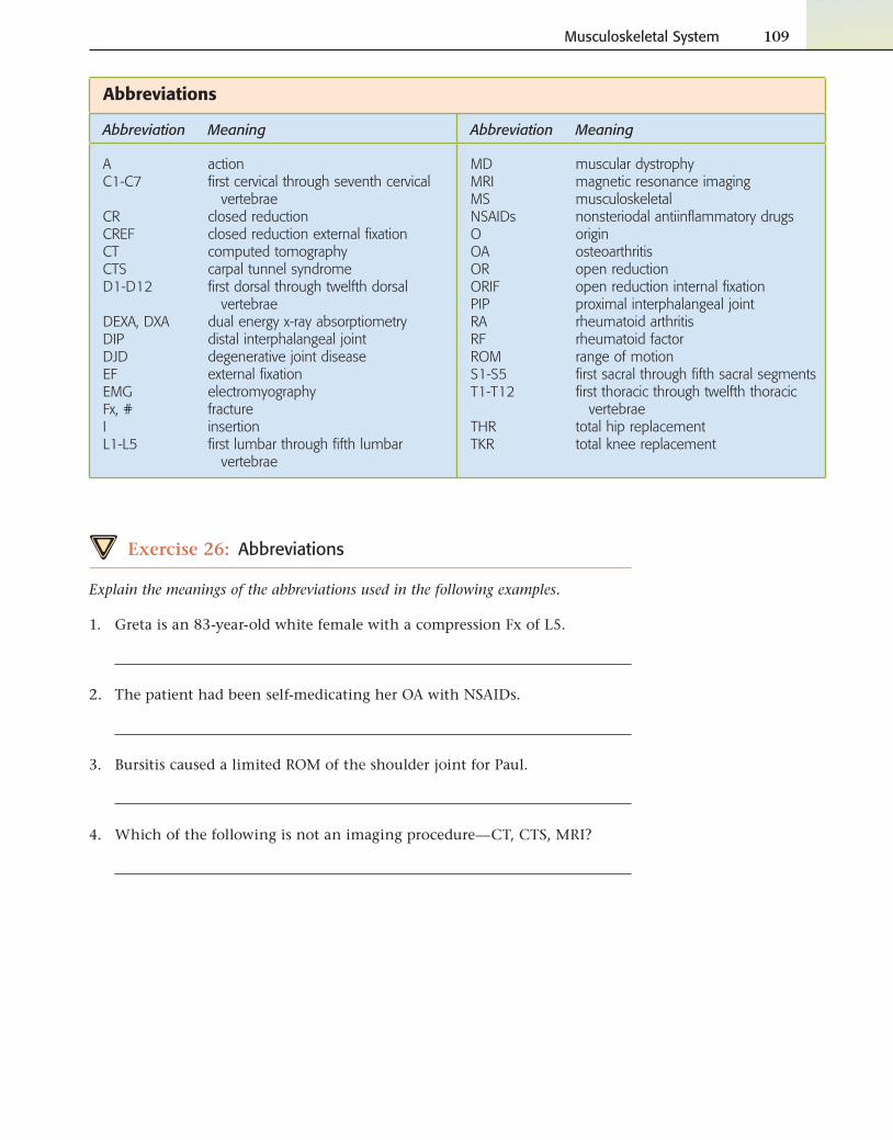
Musculoskeletal System 109
Abbreviations
Abbreviation Meaning Abbreviation Meaning
AC1-C7
CRCREFCTCTSD1-D12
DEXA, DXADIPDJDEFEMGFx, #IL1-L5
actionfi rst cervical through seventh cervical
vertebraeclosed reductionclosed reduction external fi xationcomputed tomographycarpal tunnel syndromefi rst dorsal through twelfth dorsal
vertebraedual energy x-ray absorptiometrydistal interphalangeal jointdegenerative joint diseaseexternal fi xationelectromyographyfractureinsertionfi rst lumbar through fi fth lumbar
vertebrae
MDMRIMSNSAIDsOOAORORIFPIPRARFROMS1-S5T1-T12
THRTKR
muscular dystrophymagnetic resonance imagingmusculoskeletalnonsteriodal antiinfl ammatory drugsoriginosteoarthritisopen reductionopen reduction internal fi xationproximal interphalangeal jointrheumatoid arthritisrheumatoid factorrange of motionfi rst sacral through fi fth sacral segmentsfi rst thoracic through twelfth thoracic
vertebraetotal hip replacementtotal knee replacement
Exercise 26: Abbreviations
Explain the meanings of the abbreviations used in the following examples.
1. Greta is an 83-year-old white female with a compression Fx of L5.
2. The patient had been self-medicating her OA with NSAIDs.
3. Bursitis caused a limited ROM of the shoulder joint for Paul.
4. Which of the following is not an imaging procedure—CT, CTS, MRI?
Chapter ReviewA. Functions of the Musculoskeletal System
1. In your own words, explain what functions may be lost or disrupted when the musculoskeletal system is diseased or injured.
B. Build a Term
Build and defi ne the terms below using the word parts given.Example: Combining oste/o with -itis builds the term osteitis.
110 Chapter 3
2. oste/o
A. -penia ________________________________
B. -porosis _______________________________
C. -sarcoma ______________________________
D. -clasis ________________________________
E. -malacia ______________________________
F. myel/o, -itis ______________________________________________
3. my/o
A. -rrhaphy ______________________________
B. a-, -sthenia _____________________ gravis
C. fi br/o, -algia ___________________________
D. electr/o, -graphy ______________________
4. arthr/o
A. -centesis ___________________________________________________
B. oste/o, -itis _______________________________________________
C. -desis ________________________________________________________
D. -scopy ______________________________________________________
E. -plasty ______________________________________________________
5. dactyl/o
A. syn-, -y _____________________________________________________
B. poly-, -y ___________________________________________________
6. chondr/o
A. -malacia ___________________________________________________
B. a-, -plasia _________________________________________________
C. cost/o, -itis _______________________________________________
7. spondyl/o
A. -listhesis ___________________________________________________
B. syn-, -desis _______________________________________________
C. -osis _________________________________________________________
Musculoskeletal System 111
C. Fill in the blank with the type of trauma described.
8. Martin broke his collarbone while jumping on the bed. It did not pierce the skin, so what type of
fracture is it? (give two names)
9. After dropping a bowling ball on her foot, Rena was treated for metatarsal bones that were shattered.
What kind of fracture is this?
10. Darnell fell off a piece of playground equipment and sustained a fracture in which his bones were
partially bent and partially broken. What type of fracture is this classifi ed as?
11. Advanced cancer of the bone caused a spontaneous hip fracture in a patient living in an assisted care
facility. This patient has which type of fracture?
12. Slipping on a spilled soda that hadn’t been cleaned up, Javier tried to catch himself and ended up
fracturing the distal end of his right radius. This is called which type of fracture?
13. Catching a football improperly may result in a fi nger bone that is partially disarticulated. In this case,
the injury would be described as a/an .
14. During the playoffs, the point guard ruptured a ligament in his ankle and damaged the soft tissue
around that joint. The injury would be termed a/an .
15. Rickets is osteomalacia that occurs during which stage of life?
16. A data input clerk and a bus driver who had just visited their doctors discovered that they both had
the same diagnosis—pain and numbness in their fi ngers caused by compression of the median nerve
of the wrist. This diagnosis was .
17. The newly hired coder at a local community hospital was surprised to see the large number of patients
with DJD. He knows that this is an abbreviation for , which also
means the same thing as .
18. Infl ammation of the joints is a characteristic of the autoimmune disorder
arthritis.
19. Ms. Ralston had an enlargement and infl ammation of the joint at the base of her great toe on her left
foot. This was diagnosed as a/an .
20. The name of the joint affected by the disorder described in question 18 is the _____________________________________
joint.
21. Pain in the lower back is called .
22. An imperfect healing of a broken bone is a .
23. A piece of dead bone is called .
24. Because Raymond had been having trouble with weakness in one arm, he had a procedure that
records the electrical activity of muscles called a/an .
25. One of the patients had an x-ray of the spinal canal using a contrast medium to assess damage
sustained during a car accident. The procedure is termed a/an .
26. The soccer player had an x-ray of his shoulder joint after injuries sustained in the championship
match. The procedure is termed a/an .
27. Ms. Wright was suspected to have osteoporosis. A procedure that measures density of bone is
.
28. Tyara had a lab test that reveals the presence or absence of a substance found in the blood of those
with rheumatoid arthritis. The substance is called the .
29. Moving the ends of broken bones into alignment is called .
30. If an incision is necessary, the process above is described as .
31. Fastening sections of bone with pins is known as .
32. External fi xation is considered a non procedure, meaning that an
incision is not necessary.
33. Removal of a limb is called , and the artifi cial appliance that replaces
the limb is called a/an .
34. The synonyms for spinal fusion are and .
35. Anna was prescribed Fosamax to treat her .
36. Methotrexate is used to treat which type of arthritis?
37. Celebrex is used to treat .
38. Analgesics are used to treat .
112 Chapter 3
D. Abbreviations
Spell out the abbreviation in the following sentences.
39. Mrs. Jones was advised to eat more Ca-rich foods.
40. Johnna had an Fx of one of her metatarsals. _________________________________________________________________________________________
41. Jason fell off a horse and sustained a fracture of his C2.
42. Painful bursitis resulted in limited ROM of the patient’s left shoulder.
43. Robert was treated for DJD with a regimen of weight loss and NSAIDs before surgery was discussed.
E. Singulars and Plurals
Change the following singular terms to plural.
44. foramen
45. bursa
46. prosthesis
47. phalanx
48. sulcus
Musculoskeletal System 113
49. vertebra
50. ilium
51. pelvis
52. arthroscopy
53. costa
F. Translations
Rewrite the following to explain the underlined terms.
54. An x-ray revealed a greenstick fracture of the child’s right humerus.
55. Ms. Burton-Smith was treated for bursitis with heat, rest, and NSAIDs.
56. The basketball player had an osteoclasis for a malunion of one of his metacarpals.
57. The patient was sent for a sonography of her calcaneus to assess her osteoporosis.
58. The patient complained of lumbago resulting from his spinal stenosis.
59. Electromyography was used to confi rm the child’s muscular dystrophy.
G. Be Careful
Circle the correct answer.
60. Is the suturing of a muscle spelled myorhaphy or myorrhaphy?
61. In this chapter, does the abbreviation MS refer to multiple sclerosis, musculoskeletal, or mitral stenosis?
62. Is part of the hip bone called the ileum or the ilium?
63. It is possible to break which kind of bone—the peritoneal or peroneal bone?
64. Is the tough, outer covering of the bone called the paraosteum, perosteum, or periosteum?
65. Do the minerals in bone consist of calcium and potassium or calcium and phosphorus?
66. Is the socket in the hip the acromion or the acetabulum?
67. A Colles fracture occurs to which bone: radius, ulna, or humerus?
68. The bone marrow is key in hematopoiesis or hematoporosis?
114 Chapter 3
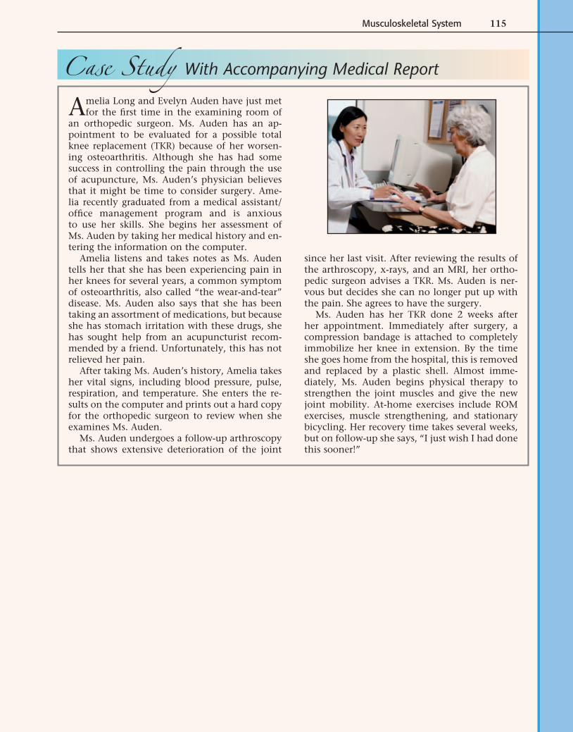
Musculoskeletal System 115
Case Study With Accompanying Medical Report
Amelia Long and Evelyn Auden have just met for the fi rst time in the examining room of
an orthopedic surgeon. Ms. Auden has an ap-pointment to be evaluated for a possible total knee replacement (TKR) because of her worsen-ing osteoarthritis. Although she has had some success in controlling the pain through the use of acupuncture, Ms. Auden’s physician believes that it might be time to consider surgery. Ame-lia recently graduated from a medical assistant/offi ce management program and is anxious to use her skills. She begins her assessment of Ms. Auden by taking her medical history and en-tering the information on the computer.
Amelia listens and takes notes as Ms. Auden tells her that she has been experiencing pain in her knees for several years, a common symptom of osteoarthritis, also called “the wear-and-tear” disease. Ms. Auden also says that she has been taking an assortment of medications, but because she has stomach irritation with these drugs, she has sought help from an acupuncturist recom-mended by a friend. Unfortunately, this has not relieved her pain.
After taking Ms. Auden’s history, Amelia takes her vital signs, including blood pressure, pulse, respiration, and temperature. She enters the re-sults on the computer and prints out a hard copy for the orthopedic surgeon to review when she examines Ms. Auden.
Ms. Auden undergoes a follow-up arthroscopy that shows extensive deterioration of the joint
since her last visit. After reviewing the results of the arthroscopy, x-rays, and an MRI, her ortho-pedic surgeon advises a TKR. Ms. Auden is ner-vous but decides she can no longer put up with the pain. She agrees to have the surgery.
Ms. Auden has her TKR done 2 weeks after her appointment. Immediately after surgery, a compression bandage is attached to completely immobilize her knee in extension. By the time she goes home from the hospital, this is removed and replaced by a plastic shell. Almost imme-diately, Ms. Auden begins physical therapy to strengthen the joint muscles and give the new joint mobility. At-home exercises include ROM exercises, muscle strengthening, and stationary bicycling. Her recovery time takes several weeks, but on follow-up she says, “I just wish I had done this sooner!”
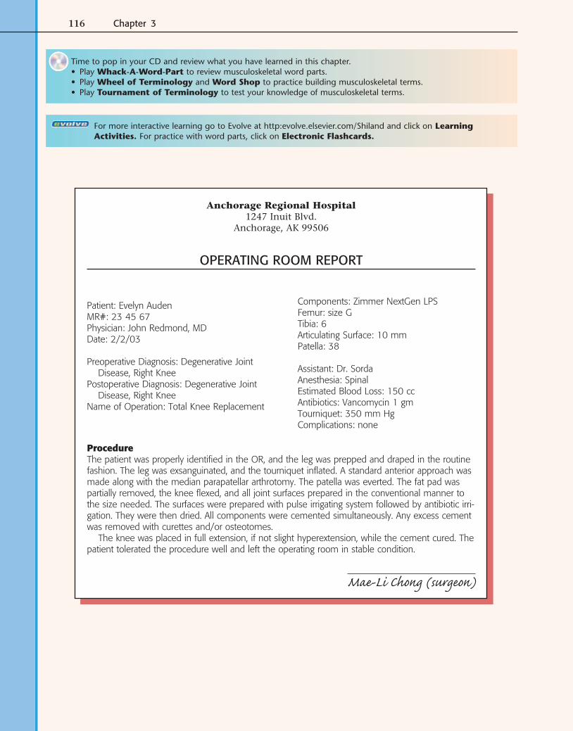
116 Chapter 3
Patient: Evelyn AudenMR#: 23 45 67Physician: John Redmond, MDDate: 2/2/03
Preoperative Diagnosis: Degenerative Joint Disease, Right Knee
Postoperative Diagnosis: Degenerative Joint Disease, Right Knee
Name of Operation: Total Knee Replacement
Components: Zimmer NextGen LPSFemur: size GTibia: 6Articulating Surface: 10 mmPatella: 38
Assistant: Dr. SordaAnesthesia: SpinalEstimated Blood Loss: 150 ccAntibiotics: Vancomycin 1 gmTourniquet: 350 mm HgComplications: none
ProcedureThe patient was properly identifi ed in the OR, and the leg was prepped and draped in the routine fashion. The leg was exsanguinated, and the tourniquet infl ated. A standard anterior approach was made along with the median parapatellar arthrotomy. The patella was everted. The fat pad was partially removed, the knee fl exed, and all joint surfaces prepared in the conventional manner to the size needed. The surfaces were prepared with pulse irrigating system followed by antibiotic irri-gation. They were then dried. All components were cemented simultaneously. Any excess cement was removed with curettes and/or osteotomes.
The knee was placed in full extension, if not slight hyperextension, while the cement cured. The patient tolerated the procedure well and left the operating room in stable condition.
Mae-Li Chong (surgeon)
Anchorage Regional Hospital1247 Inuit Blvd.
Anchorage, AK 99506
OPERATING ROOM REPORT
Time to pop in your CD and review what you have learned in this chapter.• Play Whack-A-Word-Part to review musculoskeletal word parts.• Play Wheel of Terminology and Word Shop to practice building musculoskeletal terms.• Play Tournament of Terminology to test your knowledge of musculoskeletal terms.
For more interactive learning go to Evolve at http:evolve.elsevier.com/Shiland and click on Learning Activities. For practice with word parts, click on Electronic Flashcards.
Musculoskeletal System 117
H. Healthcare Report
69. A synonym for the preoperative diagnosis of degenerative joint disease is .
70. An “anterior approach” to the knee would be through which part of the knee?
71. What is the patella?
72. To what does the term parapatellar refer?
73. What is an arthrotomy?
74. If the patella was everted, how would it be placed?
75. What is an osteotome?
76. What would hyperextension be?


![[RFF2008] Serial Instinct](https://img.pdfslide.net/doc/110x75/54826c57b4af9f7d408b48dd/rff2008-serial-instinct.jpg)