Embed Size (px)
Citation preview

1
Homeostatic regulation of synaptic strength andthe safety factor for neuromuscular transmission
1. Synaptic transmission, safety factor and size-strength relationships at NMJ
2. Quantal analysis
3. Pathophysiology
The The ‘‘Life CycleLife Cycle’’ of Neuromuscular Synapses of Neuromuscular Synapses
Synaptic transmission
Desaki & Uehara, 1981, J Neurocytol 10,101
MEPPs
Synaptic recordings from the frog NMJ: B. Katz et al.

2
Schwann cell Nerve terminal Motor end-plate
Transmission electron micrographs of the principal features of neuromuscular synapses.
Junctional Fold
Basal lamina
Synaptic vesicle
Pre
Post
http://neuromuscular.wustl.edu/musdist/dag2.htm
Neuromuscular Junction: postsynaptic
Each nAChR contains two α subunits, giving anoverall stoichiometry of α2βγδ (fetal form) or α2βδε(adult form). Each of the subunits contains fourhydrophobic transmembrane domains.
6 nm
3 nm
2 nm
2nm
9 nm
Fetal Adult
γ−subunit ε−subunit
BovineMuscle
XenopusOocyte
End-Plate Current (EPC)
2 ms
200,000 channels
20 mV
End-Plate Potential (EPP)
http://neuromuscular.wustl.edu/pathol/snare.htm
Neuromuscular Junction: presynaptic (vesicle proteins)
http://www.hhmi.org/research/investigators/sudhof.html

3
Desaki & Uehara, 1981
Wood & Slater (1997)
q = MEPP
m =EPP
q
Quantal Size:
Quantal Content:
MEPPEPP
Stim.
MEPPs
EPPs
Quantal analysis
!
Px
=e
mm
x
x! Actual m
Threshold m
Threshold
The ʻSafety Factorʼ for transmission
Wood SJ, Slater CR. The contribution ofpostsynaptic folds to the safety factor forneuromusculartransmission in rat fast-and slow-twitch muscles.J Physiol. 1997Apr 1;500 ( Pt 1):165-76.PMID: 9097941

4
Factors affecting safety factor for synaptictransmission
-Probability of release
-Transmitter store size and mobilisation
-Cholinesterase activity
-ACh receptor density
-Muscle fibre diameter and ‘input resistance’
-Nerve terminal size/strength
-Junctional fold density (Na channel density)
Gillingwater D. Thomson
5 ms
EPPs - Facilitation
300 ms
10 mV
EPPs - Short-term Depression
250 ms
10 mV
Synaptic depression
Vm
Ch.2
2.5 mV
1 mV
10.00 ms
Vm
Ch.2
2.5 mV
1 mV
10.00 ms
Vm
Ch.2
2.5 mV
1 mV
10.00 ms
10 mV
2 nA
mf
0
-2
-4
-6
-8
-10
mV
AC
1
190 200 210 220 230 240 250 260 270 280 290
s
Keyboard31
6
5
4
3
2
mV
AC
1
85 90 95 100 105 110 115 120 125 130 135 140 145
s
Ch.2
10 mV
5.00 ms
Ch.2
10 mV
5.00 ms
Rin
MEPPs
EPPs
ntSynaptic size-strength regulation maintains safety factor
20 ms
NMJ size and muscle fibre diameter co-vary
Kuno et al., 1971

5
20 µm
10 ms
10 mv
fiber diameter (µm)
0end
plat
e ar
ea (µ
m2 )
300
600
900
1200
10 20 4030 50 7060
Harris JB, Ribchester RR. The relationship between end-plate size and transmitter release in normalanddystrophic muscles of the mouse.J Physiol. 1979 Nov;296:245-65.PMID: 231101
Costanzo EM, Barry JA, Ribchester RR. Co-regulation of synaptic efficacy at stable polyneuronallyinnervated neuromuscular junctions in reinnervated rat muscle. J Physiol. 1999 Dec 1;521 Pt 2:365-74.PMID: 10581308
0 10 20 30 40 50 60
0
20
40
60
Occupancy%
Quantal Content (variancemethod) at NMJ of rat HD
0 100 200 300 4000
100
200
300
400
500First EPPPlateau EPP (10Hz)
Age
(Based on Kelly & Roberts, 1977 and Kelly, 1978)
Frog 200
Rat, mouse 50-75
Man 20-30
Species Quantal content
Frog
Rat
Man
Frog
Rat
Man
The size of NMJ and the extent of junctional folding vary between species
Frog
Rat
Man
0 250 500 750 1000 1250 15000
50
100
150
200
Synaptic area
Frog
Rat
Man

6
Quantal Analysis
Ch0
-5 mV 5.00 ms
1
2
3
4
0
Binomial model:
Let: n=3p= 0.17(q=1-p)
m=n.p
P(0) = ?P(1) = ?P(2) = ?P(3) = ?
Binomial model:
Let: n=3p= 0.1(q=1-p)
m=n.p
P(0) = q3
P(1) = 3pq2
P(2) = 3p2qP(3) = p3
P(x) =n!
x!(n ! x)!px.q(n! x)
Let :x<<np<<1
Thenq(n-x) ~ exp(-np)
andn!
(n ! x)!" n
x
P(x) = exp(!m).m
x
x!
P(0) = ?P(1) = ?P(2) = ?P(3) = ?
Poisson Distribution

7
P(x) = exp(!m).m
x
x!Poisson Distribution
P(0) = exp(-m)P(1) = m.exp(-m)P(2) = m2.exp(-m)/2P(3) = m3.exp(-m)/6
Freq
uenc
y
Poisson distribution of QuantalContents of EPPs (n=100 trials)
0 1 2 3 4 5 6 7 8 9 10 11 12
0
10
20
30
40
m=1
Quantal content
Freq
uenc
y
Poisson distribution of QuantalContents of EPPs (n=100 trials)
0 1 2 3 4 5 6 7 8 9 10 11 12
0
10
20
30
40
m=2
Quantal content
Freq
uenc
y
Poisson distribution of QuantalContents of EPPs (n=100 trials)
0 1 2 3 4 5 6 7 8 9 10 11 12
0
10
20
30
40
m=3
Quantal content
Freq
uenc
y
Poisson distribution of QuantalContents of EPPs (n=100 trials)
0 1 2 3 4 5 6 7 8 9 10 11 12
0
10
20
30
40
m=4
Quantal content
Freq
uenc
y
Poisson distribution of QuantalContents of EPPs (n=100 trials)
0 1 2 3 4 5 6 7 8 9 10 11 12
0
10
20
30
40
m=5
Quantal content

8
Methods of quantal analysis:
1. Direct method : m=EPP/MEPP (better, EPC/MEPPC)
2. Failures method: P(0)=exp(-m); m=Ln(Tests/Failures) ( for binomial: P(0)=(1-p)n)
3. Variance method: m = 1/(C.V.)2 i.e. m=EPP2 /var(EPP) (for binomial: var(m)=npq)
Problems
- MEPP variance
- Non-linear summation
- Non-Poisson conditions
y = exp(!(x ! µ)2 / 2" 2 ) /(" 2# )
The Normal (Gaussian) Distribution
x
yy 5
x2!( )
2 0.25"exp# $% &
0.5 2'=
(µ = 0; σ =0.5)
P(x) = exp(!m)m
x
x!k =1
n
" .1
2#k$ 2
! x ! kx ( )2
2k$ 2
%
& ' '
(
) * *
+
, - -
.
/ 0 0
m=3 quantaσ= 0.2 mvx =1.1mv
y 153!( )exp 3
x"
x!# $% &' ( 1
0.2 2)k
x 1.1k!( )2!
2k0.22# $
% &' (
exp# $% &' (
# $% &' (
k 1=
10
*=
q = MEPP
m =EPP
q
Quantal Size:
Quantal Content:
MEPPEPP
Stim.
MEPPs
EPPs
Quantal analysis
!
Px
=e"mm
x
x!

9
McLachlan EM, Martin AR. Non-linear summation of end-plate potentials in the frog andmouse. J Physiol. 1981 Feb;311:307-24.PMID: 6267255
v' = v /(1! v /(Em! E
r)
m =v!
q(1 ! v!
(Em ! Er )
v' = v /(1! fv(Em ! Er )
Correction Factors
Martin (1955):
v= EPP amplitudeq= MEPP amplitudem = quantal content
McLachlan & Martin (1981)
Where f = an empirically determined ('fudge’) factor
For mouse muscle, long fibres: f=0.8For frog muscle, long fibres: f=0.55
For short muscle fibres (e.g. FDB) the correction is unknown, butf=0.3 gives a good fit to our data.
Methods of quantal analysis:
1. Direct method : m=EPP/MEPP (better, EPC/MEPPC)
2. Failures method: P(0)=exp(-m); m=Ln(Tests/Failures) ( for binomial: P(0)=(1-p)n)
3. Variance method: m = 1/(C.V.)2 i.e. m=EPP2 /var(EPP) (for binomial: var(m)=npq)
4. Convolutions; graphical methods (e.g. see Clements & Silver, TINS 23, 105-113.)
Note: For all methods except the Failures Method, it is necessary toassess and correct if required for non-linear summation of synapticpotentials. Synaptic currents sum linearly.
Pathophysiology
Myasthenia GravisBefore
After edrophonium(Tensilon Test)
Case 1•Bilateral ptosis•Double vision in all directions•Fatiguable weakness•Reflexes disappear after exercise•Sensation normal
10
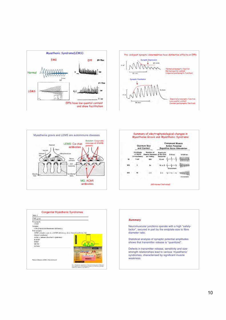
Myasthenic Syndrome(LEMS):
EMG
EPPs have low quantal contentand show facilitation
EPP
Normal
LEMS
Pre- and post-synaptic abnormalities have distinctive effects on EPPs
- Normal presynaptic function Normal quantal content (impaired postsynaptic function)
- Impaired presynaptic function Low quantal content (normal postsynaptic function)
Synaptic Depression
Synaptic Facilitation
MG: AChR antibodies
X X
Myasthenia gravis and LEMS are autoimmune diseases
LEMS: Ca channelantibodies
X X
Botulism: Enzymaticcleavage of SNAREproteins
Summary of electrophysiological changes inMyasthenia Gravis and Myasthenic Syndrome
(NI=Normal Individual)
Congenital Myasthenic Syndromes
Palace & Beeson (2008) J Neuroimmunol
Summary
Neuromuscular junctions operate with a high “safety-factor”, secured in part by the endplate-size to fibrediameter ratio.
Statistical analysis of synaptic potential amplitudesshows that transmitter release is “quantized”.
Defects in transmitter release, sensitivity and size-strength relationships lead to various ʻmyasthenicʼsyndromes, characterised by significant muscleweakness.


![Physiology and Pathophysiology of Neuromuscular Transmission€¦ · 1 Physiology and Pathophysiology of Neuromuscular Transmission “The neuromuscular junction... [is] an experimentally](https://img.pdfslide.net/doc/110x75/5b1acdb37f8b9a41258e0a00/physiology-and-pathophysiology-of-neuromuscular-1-physiology-and-pathophysiology.jpg)