Embed Size (px)
Citation preview

http://wrap.warwick.ac.uk/
Original citation: Kreikemeyer-Lorenzo, D., Unterberger, W., Duncan, David A., Lerotholi, T. J. and Woodruff, D. P.. (2013) The local structure of the azobenzene/aniline reaction intermediate on TiO2(110). Surface Science, Volume 613 . pp. 40-47. Permanent WRAP url: http://wrap.warwick.ac.uk/56072 Copyright and reuse: The Warwick Research Archive Portal (WRAP) makes this work of researchers of the University of Warwick available open access under the following conditions. Copyright © and all moral rights to the version of the paper presented here belong to the individual author(s) and/or other copyright owners. To the extent reasonable and practicable the material made available in WRAP has been checked for eligibility before being made available. Copies of full items can be used for personal research or study, educational, or not-for-profit purposes without prior permission or charge. Provided that the authors, title and full bibliographic details are credited, a hyperlink and/or URL is given for the original metadata page and the content is not changed in any way. Publisher statement: NOTICE: this is the author’s version of a work that was accepted for publication in Surface Science. Changes resulting from the publishing process, such as peer review, editing, corrections, structural formatting, and other quality control mechanisms may not be reflected in this document. Changes may have been made to this work since it was submitted for publication. A definitive version was subsequently published in http://dx.doi.org/10.1016/j.susc.2013.03.009 A note on versions: The version presented here may differ from the published version or, version of record, if you wish to cite this item you are advised to consult the publisher’s version. Please see the ‘permanent WRAP url’ above for details on accessing the published version and note that access may require a subscription. For more information, please contact the WRAP Team at: [email protected]

1
The local structure of the azobenzene/aniline reaction intermediate on
TiO2(110)
D. Kreikemeyer-Lorenzo1 W. Unterberger1, D.A. Duncan2, T.J. Lerotholi3, D.P.
Woodruff2*
1 Fritz-Haber-Institut der Max-Planck-Gesellschaft, Faradayweg 4-6, 14195, Berlin,
Germany
2 Physics Department, University of Warwick, Coventry CV4 7AL, UK
3 School of Chemistry, University of Witwatersrand, PO Wits, Johannesburg, 2050, South
Africa
Abstract
Scanned-energy mode photoelectron diffraction (PhD) and near-edge X-ray absorption
fine structure (NEXAFS) have been used to study the surface species, previously
proposed to be phenyl imide, C6H5N-, on rutile TiO2(110) following exposure to either
azobenzene or aniline. All measurements are consistent with the two reactants forming a
common surface species in the same local adsorption site. N K-edge NEXAFS confirms
the scission of the N=N bond in azobenzene, while C K-edge NEXAFS shows the phenyl
ring to be intact with the molecular plane tilted relative to the surface normal and not
aligned in either principle azimuth of the surface. N 1s PhD data indicate that the N atom
bonds atop a surface five-fold-coordinated Ti atom, most probably at a Ti-N bondlength
of 1.770.05 Å, and not bridging two such atoms, as had been suggested. This atop
geometry is favoured by recent density functional theory (DFT) calculations, but more
quantitative aspects of the DFT result are not in agreement with the conclusions of our
experimental study.
Keywords: surface structure, TiO2; azobenzene; aniline; photoelectron diffraction;
molecular adsorption
* Corresponding author. Email [email protected]
2
1. Introduction
The rutile-phase TiO2(110) surface is the most-studied of all oxide surfaces (e.g. [1,2, 3]),
in large part motivated by the known importance of titania in photocatalysis, gas sensors
and as a support in a range of heterogeneous catalysts. Particular interest in recent years
has centred on the properties of Au nanoparticles on titania following the discovery that
this system is an effective catalysis for low-temperature oxidation of CO [4]. This
combination has also been shown more recently to act as a high-yield catalyst in the
synthesis of aromatic compounds [5, 6] including the synthesis of azobenzene
(C6H5N=NC6H5) via oxidation of aniline (C6H5NH2) (see Fig. 1). Somewhat intriguingly,
it was found [6] that TiO2 in the absence of the Au nanoparticles was also effective, and
highly selective, in azobenzene synthesis. This led to a careful UHV surface science
study of the interaction of azobenzene and aniline with both the rutile-phase TiO2(110)
and anatase-phase TiO2(101) surfaces by Li, Diebold and coworkers [7, 8] who found
evidence for a single surface reaction intermediate, produced by interaction with either
azobenzene or aniline on both surfaces. On the rutile TiO2(110) surface they found that
adsorption of either molecule at full coverage leads to the formation of a c(2x2) ordered
molecular phase, as seen both with low energy electron diffraction (LEED) and scanning
tunnelling microscopy (STM). The STM images appear to be independent of the initial
reactant molecule, as do the X-ray photoelectron spectroscopy (XPS) data from both the
substrate and the molecular constituent atoms. The common molecular adsorbate species
was thus assigned to phenyl imide (C6H5N-), produced either by N=N bond scission of
azobenzene, or by dehydrogenation of aniline.
Here we present the results of further investigations of this system using the techniques of
scanned-energy mode photoelectron diffraction (PhD) and near-edge X-ray absorption
fine structure (NEXAFS). The PhD technique [9, 10] exploits the coherent interference of
the directly emitted component of a photoelectron wavefield from an adsorbate atom with
other components of the same wavefield elastically scattered by the surrounding atoms.
Scanning the photon energy leads to changes in the photoelectron energy, and thus the

3
photoelectron wavelength, causing different scattering paths to switch in and out of phase
with the directly-emitted wavefield. The resulting modulations in the measured intensity
can be interpreted in terms of the different scattering paths associated with a particular
adsorption geometry, through the use of multiple scattering simulations for different trial
structures. In the present case, the use of N 1s PhD data allows us to determine the
adsorption site of the expected bonding N atom on the TiO2(110) surface and the
associated Ti-N bondlength. We can also compare the local adsorption geometry of the
molecular species formed by exposure to the two different reactant molecules,
azobenzene and aniline. C K-edge NEXAFS provides information on the integrity of the
aromatic ring in the adsorbed molecular species, and on its orientation. N K-edge
NEXAFS allows us to establish independently whether the N=N bond scission occurs
following exposure to azobenzene.
2. Experimental and computational details
The experiments were conducted in an ultra-high vacuum surface science chamber
equipped with facilities for sample cleaning, heating and cooling. This instrument was
installed on the UE56/2-PGM-2 beamline of BESSY II which comprises a 56 mm period
undulator followed by a plane grating monochromator [11]. Sample characterisation in
situ was achieved by low energy electron diffraction (LEED) and by soft-X-ray
photoelectron spectroscopy (SXPS) using the incident synchrotron radiation. These wide-
scan SXPS spectra, and the narrow-scan N 1s spectra used in the PhD measurements,
were obtained using an Omicron EA-125HR 125 mm mean radius hemispherical
electrostatic analyser, equipped with seven-channeltron parallel detection, which was
mounted at a fixed angle of 60 to the incident X-radiation in the same horizontal plane
as that of the polarisation vector of the radiation.
A clean well-characterised rutile TiO2(110) surface was prepared by briefly bombarding
with Ar+ ions at an energy of 500 eV, followed by annealing in UHV at ~800 K for 10
minutes. The sample was then annealed at ~700 K in an oxygen atmosphere of 2x10⁻⁷
mbar for 10 minutes followed by a brief flash to ~1000 K. Oxygen dosing is known to

4
heal oxygen surface vacancies [1] that can arise from multiple re-cleaning cycles using
ion bombardment and UHV annealing alone. This treatment led to a sharp (1x1) LEED
pattern and a Ti 2p photoemission spectrum showing no detectable high kinetic energy
shoulder, indicative of a low concentration of surface oxygen vacancies. The sample
colour was dark blue, consistent with a concentration of bulk defects sufficient to render
the sample conducting. Aniline and azobenzene (Sigma-Aldrich, 99.5% and 99% purity,
respectively), subjected to freeze-thaw pumping cycles in situ, were dosed from the
vapour phase at 300 K for all the preparations studied in UHV, although low temperature
(150 K) dosing was also used to provide additional system characterisation. The
azobenzene was heated to ~370 K to achieve a sufficiently high vapour pressure for the
dosing, with the sample facing the gas inlet. Dosing of both species was checked using a
mass spectrometer mounted on the chamber to monitored the gas phase components.
NEXAFS spectra from the dosed surfaces were measured in the Auger electron mode by
setting the electron energy analyser to an energy in the range of the KLL Auger peak of
the relevant element (C and N) and measuring the yield as a function of photon energy.
N 1s PhD data were obtained from the adsorbed species by recording a sequence of
photoelectron energy distribution curves (EDCs) around this photoemission peak, at
equal steps in photon energy, in the photoelectron kinetic energy range of ~ 60-320 eV.
These data were measured at several different polar emission angles in the range 0° to
60°, and in the two principal azimuths, [001] and [110] (see Fig. 2), although at polar
angle greater than 30° no clear modulations above the noise level could be discerned.
Data reduction followed our general PhD methodology (e.g. [9, 10]), in which each of the
individual N 1s EDCs was fitted by the sum of a Gaussian peak, a step and a template
background. The integrated peak areas were then plotted as a function of photoelectron
energy, I(E), and each final PhD modulation spectrum, (E), was obtained by subtraction
of, and normalisation by, a smooth spline function, I0(E), representing the non-diffractive
intensity and instrumental factors.

5
3. Results
3.1 SXPS and NEXAFS
Fig. 3 shows the SXP spectra in the energy ranges of the Ti 2p, O 1s, N 1s and C 1s
emission peaks from the clean surface and following nominal saturation doses at room
temperature of the two reactant molecules. The spectra recorded following aniline
exposure are almost identical to those obtained following azobenzene exposure; the
absence of chemical shifts in these pairs of spectra indicates that the same surface species
is formed by the two molecules. In common with the results of Li and Diebold [7],
however, we note a very small shift (~0.1 eV) of all peaks to lower kinetic energy (higher
binding energy) after reaction with aniline. These authors suggest that this is due to band
bending at the surface. While scission of the N=N bond in azobenzene would lead to only
phenyl imide fragments, dehydrogenation of aniline leads to the release of two H atoms,
and if these lead to hydroxylation of the surface bridging oxygen atoms (Fig. 2), some
charge transfer is to be expected. We note, however, that our O 1s spectra (recorded at a
much lower kinetic energy, and thus under much more surface-specific conditions than
the conventional laboratory-source XPS used in the earlier study) show no evidence of
the appearance of a low kinetic energy shoulder with a chemical shift of ~1.3 eV
expected for hydroxyl species on this surface (e.g. [12]). As the STM and LEED results
of Li and Diebold show a c(2x2) molecular overlayer, consistent with a coverage of the
molecular species of 0.5 ML, achieving this coverage of phenyl imide from aniline would
release 1 ML of atomic hydrogen. The absence of any significant OH feature in the O 1s
spectrum indicates that the hydroxyl coverage must be very significantly less than 1 ML,
so only a small fraction of the released H atoms can be bonded to the surface oxygen
atoms and we can only surmise that most (or all) of the H atoms are desorbed as H2 or are
absorbed below the surface; on the basis of previous studies of hydrogen absorption and
desorption [13, 14], the latter option seems the more probable.
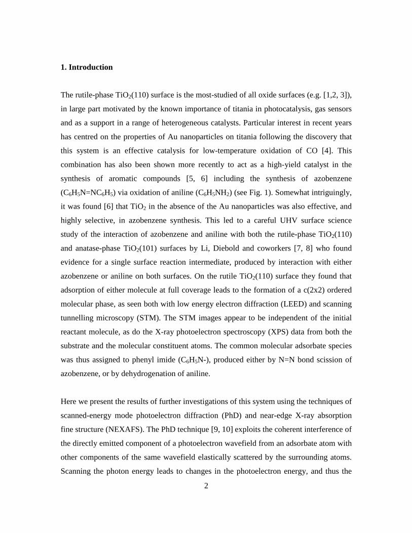
Fig. 4 shows the C K-edge NEXAFS spectra (measured in the Auger electron detection

6
mode) obtained from these same surface preparation treatments. The spectra are rather
noisy (particularly from azobenzene, for which a lower coverage was achieved) and there
are significant variations in the background shape, but it is clear that the main features of
the spectra recorded from surfaces exposed to azobenzene or to aniline occur at the same
energies with similar relative intensities. In all of these spectra the main features are the
three peaks at photon energies of ~285 eV, ~286 eV and ~288.5 eV, although the third of
these peaks occurs in an energy range of varying background and is more or less clearly
visible in the various spectra. The appearance of two peaks at energies of 284.9-285.5 eV
and 288.6-289.4 eV, the exact energies being dependent on whether the molecule is in the
gas phase or in monolayer or multilayer adsorption, is seen in NEXAFS spectra from
benzene; both peaks are attributed to transitions from the C 1s state to final states of -
symmetry, and are thus characteristic of an intact aromatic ring [15]. The splitting of the
lower-energy peak into two components separated by ~ 1 eV is also seen in molecular
aniline [15, 16] and is attributed to the inequivalence of the C atoms within the aromatic
ring that are, or are not, bonded to N. The appearance of these three peaks is thus entirely
consistent with the surface molecular species arising from both reactants having both the
aromatic ring and the attached N atom, as in phenyl imide.
NEXAFS data also provides information on molecular orientation. In particular,
transitions from the 1s state to final states of -symmetry in a planar molecule are only
possible when there is a component of the electric vector, A, of the incident radiation
perpendicular to the molecular plane. Indeed, the intensity of the relevant peak in the
NEXAFS spectrum is proportional to cos2(), where is the angle between A and the
normal to the molecular plane. For example if the molecular plane were to be
perpendicular to the surface and aligned in the [001] azimuth, then at normal incidence
with linearly-polarised radiation (i.e. at a grazing incidence angle, p, of 90°, which also
corresponds to the angle between the surface normal and A), there should be no -
resonance visible with incidence in the [001] azimuth, but a -resonance of maximum
intensity with incidence in the [110] azimuth. Indeed, the absence of a -resonance peak
in the spectrum for incidence in the [001] azimuth should also be true if the molecular

7
plane is tilted but remains aligned in the [001] azimuth. The absence of any obvious
dependence of the NEXAFS spectra in Fig. 4 on the incidence azimuth thus shows that
the phenyl imide is not aligned in either of the principle azimuths; either it is aligned in
an intermediate low-symmetry direction, or is randomly oriented (possible rotating about
the N-surface bond(s)). The selection rule also shows that if the molecular plane is
parallel to the surface, no -resonance should be detected with normal incidence (A-
vector in the surface plane) in either azimuthal direction. As this is clearly not the case,
the molecule, is not lying flat on the surface. Finally, we note that if the molecular plane
is perpendicular to the surface, no -resonance should be detected with a grazing
incidence angle of 0° (A-vector perpendicular to the surface). Of course, true zero
grazing-angle incidence is not achievable in practice, but the spectra of Fig. 4 clearly
show that at an angle of 10° the -resonances are clearly visible, so this perpendicular
orientation is unlikely. A more quantitative determination of the tilt angle requires
quantification of the polarisation-angle dependence of the -resonance intensities, which
must be normalised to the underlying edge-jump in each spectrum. Unfortunately, as seen
in Fig. 4, the background shape in the various spectra is such that, in at least several of
the spectra (particular those measured at normal incidence) it is not really possible to
identify a clear edge jump.
We therefore conclude simply that the C K-edge NEXAFS data show the adsorbed
species contains an intact phenyl ring, with some inequivalence of C atoms in the ring
that give rise to spectra essentially identical to those of molecular aniline, that the
molecular plane is not aligned along either principle azimuth and is probably randomly
oriented azimuthally, and that this plane is tilted relative to the surface normal, by an
unspecified (but probably significant) amount.
As it is the presence and polarisation-orientation dependence of -resonance features that
are most informative in NEXAFS, N K-edge NEXAFS from phenyl imide is expected to
be relatively featureless. By contrast, however, intact azobenzene contains a N=N double
bond, so in this species a -resonance should be observed, and indeed has been reported

8
previously in measurements from more complex azo-biphenyl derivatives [17]. An
absence of this -resonance in spectra recorded from the room-temperature deposition of
azobenzene is therefore a clear spectral signature of the scission of the N=N bond. Fig. 5
shows these data, and indeed the spectra from the room-temperature deposition are
dominated by broad peaks that are due not to true NEXAFS features, but to the fact that,
at the energy window values set on the electron spectrometer (377 eV and 379 eV for the
spectra recorded at 90° and 10° grazing incidence, respectively), photoemission peaks
from the shallow core levels of TiO2 pass through this window. In the data collected at
10° grazing incidence (in which this spurious broad peak is at a higher energy by 2 eV), a
weak edge jump is visible at a photon energy of ~398.2 eV, but there is clearly no -
resonance peak here. In order to provide an internal consistency check on the significance
of this absence, similar measurements were also made from a multilayer film of
azobenzene deposited at a sample temperature of 140 K, and these results are also shown
in Fig. 5. In this case an intense -resonance peak is seen at the edge jump as expected,
providing a clear contrast with the equivalent spectrum from the surface exposed at room
temperature. Evidently room temperature exposure to near-saturation coverage does lead
to N=N bond scission, as proposed by Li and Diebold. However, also shown in the
bottom panel of Fig. 5 is a series of NEXAFS spectra obtained after the multilayer was
heated briefly to increasing temperatures. In these spectra a residual -resonance remains
even after heating to significantly above room temperature. The reason for this difference
from the spectra obtained following room temperature deposition is unclear, but it seems
that the high-coverage adsorbed layer obtained from multilayer desorption is not
completely dissociated in the same way as it is following room temperature deposition.
Interestingly, Li and Diebold report evidence of adsorbed intact azobenzene at low
coverage following room temperature exposure, so there appear to be some
circumstances under which azobenzene does not dissociate on TiO2(110).
3.2 PhD structure determination
Based on the results of Li and Diebold, reinforced by our own NEXAFS results
summarised above, there is strong evidence that exposure of the TiO2(110) surface to

9
either azobenzene or aniline leads to a common surface species which seems to be most
likely to be phenyl imide, and we may anticipate that this must chemisorb through the N
atom to the surface, most probably to one or two surface Ti atoms that are five-fold
(under-) coordinated (Fig. 2). Analysis of N 1s PhD modulation spectra should therefore
allow us to determine explicitly and quantitatively this adsorption geometry.
Fig. 6 shows a comparison of a subset of the PhD spectra, recorded in three different
emission directions, after exposure to each of the reactant molecules. While there are
clear differences in the fine structure, due in large part to experimental scatter (especially
from the lower-coverage azobenzene exposure), the dominant long-period modulations,
characterised particularly by the energies of the main minima, which arise from near-
neighbour substrate scattering, are clearly closely similar in spectra recorded from the
surface exposed to the two different reactants. This subjective judgement is supported by
a calculated value of the R-factor between the two experimental data sets of 0.30; such a
value in a experiment/theory comparison would indicate generally good agreement, and a
higher value might be expected in an experiment/experiment comparison due to the role
of noise in both sets of spectra. These data demonstrate that the local adsorption
geometry of the N atom bonded to the surface, in the surface species produced by the two
different reactants, is the same. Combined with the NEXAFS data indicating both
reactants lead to a surface species with a similarly-oriented phenyl ring and also
demonstrating the scission of the S=S bond in azobenzene, together with the XPS results
and the earlier STM images [7], there can be little doubt that the two reactant molecules
do lead to a common surface molecular species. Moreover, the PhD data show that if
coadsorbed (or absorbed) atomic hydrogen is present following reaction with aniline, this
must have little or no effect on the local adsorption geometry of the adsorbed phenyl
imide species.
In order to extract quantitative structural information from PhD data it is necessary to
perform multiple scattering simulations of the experimental data for different structural
models, adjusting the models until the best agreement is achieved. In many cases,
however, some initial indication of the structure can be obtained from visual inspection of

10
the PhD modulation spectra. In particular, it is generally found that if the emission
direction corresponds to near-180 backscattering from a nearest-neighbour substrate
atom, the PhD spectrum is dominated by a single long-period modulation associated with
the short scattering path from this one neighbouring atom. Thus, if the emitter atom is
atop its nearest-neighbour substrate atom, this condition is met for emission along the
surface normal, and this simple dominance of a single period is lost if the polar emission
angle is increased substantially. The spectra shown in Fig. 6, all recorded near normal
emission, do show this kind of underlying long-period modulation, while spectra
recorded at much larger emission angles lose this character, and indeed are in most cases
too noisy to be useful in the full analysis. This preliminary inspection, therefore, suggests
that the molecule may be bonding to the surface with the N in, or close to, a site atop a
surface (5-fold coordinated) Ti atom.
In order to achieve a proper quantitative analysis of the PhD data, however, we have
undertaken a search of possible structures with the aid of multiple scattering calculations
using the computer codes developed by Fritzsche [18, 19, 20]. These are based on the
expansion of the final state wave-function into a sum over all scattering pathways that
the electron can take from the emitter atom to the detector outside the sample. The
agreement between theory and experiment was quantified using an objective reliability
factor (R-factor) [9, 10], similar to that defined by Pendry for quantitative LEED studies
[21]. This approach also allows us to define a criterion for the precision of the analysis
and the relatively acceptability of different possible structural solutions. Specifically,
we define a variance in the minimum value of the R-factor, Rmin, (obtained for the best-fit
structure) that depends on the size of the data-set used in the analysis [9, 10]. We then
regard all structures having values of R < Rmin + var(Rmin) as falling within the limits of
our precision.
The calculations explored two basic structures, namely N bonding to a single 5-fold
coordinated Ti surface atom in a near-atop site, or N bonding to two such surface Ti
atoms in or near a bridging site. Optimisation of the fit for each of these models involved
adjusting the exact location of the N emitter both perpendicular and parallel to the

11
surface, and varying the relaxation of the near-surface Ti and O atoms, the orientation of
the attached phenyl ring (but omitting the very weakly-scattering H atoms), and the
vibrational amplitudes of the surface and bulk atoms.
Bearing in mind the N bonding in the intact azobenzene (N=N) and aniline (NH2)
molecules, it seems reasonable to anticipate that the N atom of phenyl imide may bond to
the TiO2 surface in a bridging site to create two Ti-N bonds, and indeed this configuration
was depicted in the cartoon of the reaction by Li and Diebold [7], although these authors
made no claim to have determined this geometry. However, our attempts to fit the PhD
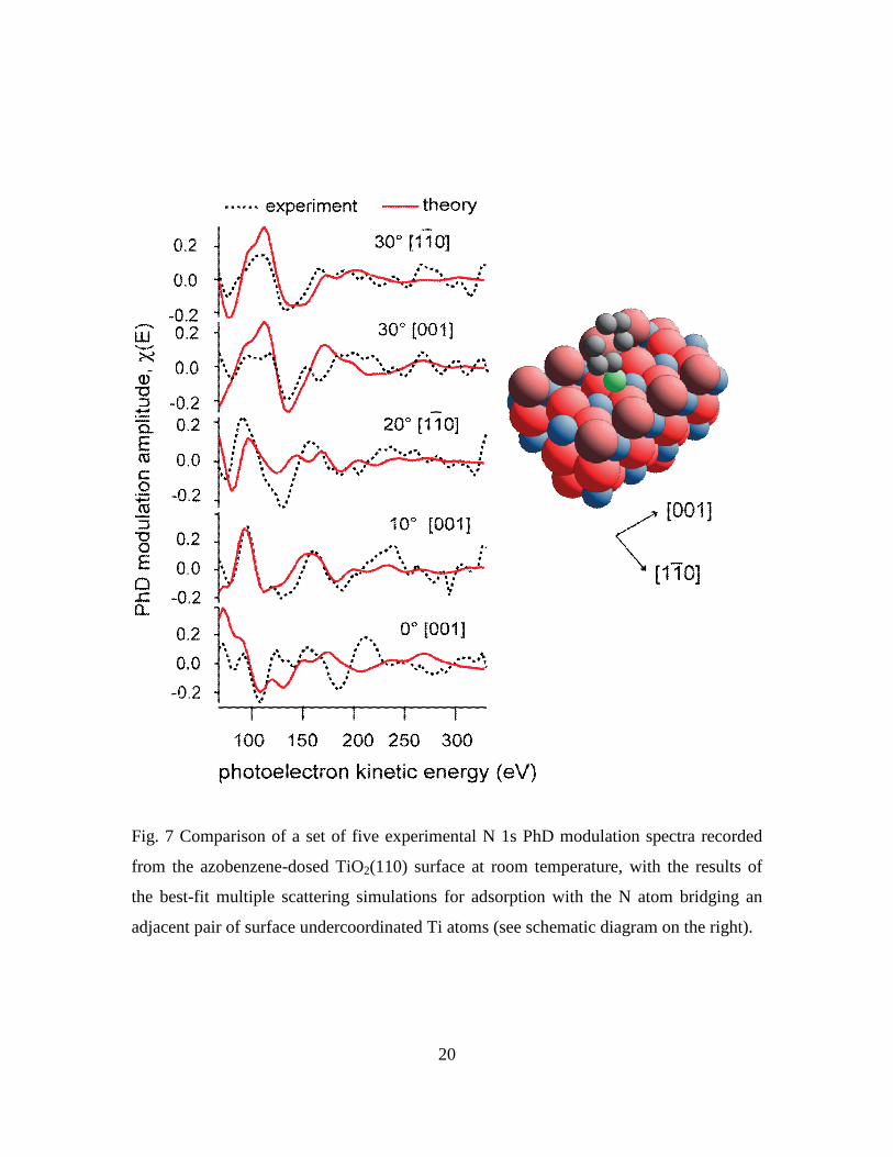
data using this model failed to find a satisfactory solution. Fig. 7 shows a comparison of
the set of five experimental N 1s PhD modulation spectra selected for the full analysis
(on the basis of the size of the modulations and therefore data reliability), with the
multiple scattering simulations for the best-fit bridging geometry found. The
corresponding Ti-N bondlength is found to be 2.14 Å, but the overall R-factor value of
0.48 is rather large, consistent with the visual appearance of a poor fit. The fit to the
normal emission spectrum is particularly bad, but lesser differences in the experimental
and theoretical spectra at a polar emission angle of 30 in the [001] azimuth are also
significant. In particular, a characteristic of the PhD technique is that in, or close to, an
emission geometry corresponding to 180° scattering from a nearest-neighbour substrate
atom, the spectrum is dominated by a single long-period modulation corresponding to
backscattering from this atom. If the N emitter occupies this bridging site, this effect
should be seen at a polar angle of ~40 in [001]. While we have no measurement in this
exact geometry, a polar emission angle of 30° is sufficiently close for this effect to be
seen in the simulated spectrum, which is dominated by a single period leading to three
broad peaks that are damped at higher energies. By contrast, the experimental spectrum
shows only the first of these peaks and is characterised by weak finer structure at higher
energies. The absence of this clear long-period modulation in the experimental spectrum
recorded at 30° is thus further evidence that N is not in this bridge-bonded site.
By contrast, exploration of the optimised geometry associated with the N atom near an
atop site led to much better agreement between simulations and experiment, but with fits

12
of closely comparable quality at two quite different Ti-N bondlengths. These results are
shown in Fig. 8. The two alternative values of the Ti-N bondlength are 1.770.05 Å and
2.270.04 Å, with associated R-factor values of 0.36 and 0.31, respectively. Visual
inspection of the comparison of the simulated PhD spectra with the experimental data
shows that both solutions are greatly superior to that achieved for the bridging site (Fig.
7), and indeed that these two alternative atop geometries give comparably-good
agreement overall, the simulated spectra for the shorter bondlength being slightly
preferred at the higher emission angles, while those of the longer bondlength give slightly
superior fits near normal emission. The bottom panel of Fig. 8 shows how the R-factor
varies as a function of the Ti-N bondlength if the remainder of the structural parameters
(notably the near-surface relaxations) are fixed at the values corresponding to each of two
best-fit structures. Notice that, depending on the values of these secondary structural
parameters, either bondlength can yield the lower R-factor, but if we compare the best set
of all parameters for each solution, that with the longer bondlength gives the lowest
value, as given above. Unfortunately, the variance calculated for this value of Rmin is
0.06, so the R-factor value for the short bondlength solution of 0.36 is below Rmin +
var(Rmin) =0.37 and must also be regarded as acceptable. This problem of multiple
coincidences at different bondlengths is well-documented in PhD [22], and also in the
closely related method of quantitative LEED [23]; the general solution to the problem is
to enlarge the data-set which lowers and variance and generally also increases the
difference in the R-factors of the alternative solutions. In the present case the weak
modulations and thus relatively poor signal-to-noise ratio of the PhD spectra recorded at
other emission angles meant that we were unable to adopt this solution.
4. Discussion and theoretical results
Based on this analysis of the PhD data alone we must conclude that the phenyl imide
bonds through the N atom in an atop site, and not a bridging site, relative to the
undercoordinated Ti atoms in the surface, but that we are unable to distinguish between
solutions with two very different Ti-N bondlengths. So far, however, we have not applied
any constraints to the possible solutions based on other information, but the application of

13
such constraints is often a necessary component of all methods of structure
determination. In the present case, a key question is whether the two alternative Ti-N
bondlengths, 1.770.05 Å and 2.270.04 Å, are equally reasonable. In fact, particularly
as we may expect this Ti-N bond to have a bond order of ~2, the longer bondlength
seems quite unphysical. For example, NH bonded to a Ti atom in TiCl2(NH)(OPPh3)2
has a Ti-N bondlength as short as 1.63 Å [24], while NH2 bonded to a Ti atom in
Ti(NH2)(Me5C5)2 has a Ti-N bondlength of 1.95 Å [25], similar to that (1.92 Å) in
N(CH3)2 bonded to Ti in azatitanatranes [26]. Longer Ti-N bondlengths in the range 2.02-
2.11 Å are found in titanium complexes bearing 2-pyrazolato ligands in which a pair of
adjacent N atoms in a five-membered (N2C3R2) ring bond to a single Ti atom [27], while
even longer Ti-N bonds of 2.33-2.40 Å have been reported, these are found in complexes
in which the N atom that is bonded to the Ti atom is also bonded to three other atoms
[28]. Comparison with these bondlengths in molecular clusters does suggest, therefore,
that if phenyl imide is bonded to a single undercoordinated Ti atom on the TiO2(110)
surface, the Ti-N bondlength may be expected to be very significantly less than 2.27 Å,
and probably less than 2.00 Å. We therefore favour the solution in which the Ti-N
bondlength is 1.770.05 Å.
It is interesting to compare these conclusions with the results of a recent density
functional theory (DFT) investigation [29, 30] of the adsorption and dissociation of
azobenzene on TiO2(110). The lowest-energy structure for phenyl imide on the surface
found in this investigation does correspond to an atop adsorption site with the N bonding
to a 5-fold coordinated Ti surface atom, in agreement with the conclusions of our PhD
investigation. However, the Ti-N bondlength found in the DFT study of 2.07 Å is
approximately midway between the two possible solutions found in the PhD analysis, and
therefore clearly not consistent with our results. Moreover, in the DFT structure the
phenyl ring is found to be perpendicular to the surface and parallel to the [011] azimuth,
quite inconsistent with the results of our C K-edge NEXAFS measurements that indicate
a tilted species not aligned along either principle azimuth. It is important to note,
however, that this DFT study leads to relative energies that indicate that the dissociation

14
of azobenzene into two phenyl imide species on TiO2(110) involves a very significant
energy cost and should therefore not occur spontaneously. This has led the authors to
concluded that this process ‘still requires further investigation’. One possibility is that the
common surface intermediate formed by reaction of azobenzene or aniline on TiO2(110)
is not phenyl imide but some other species that still retains a phenyl ring and bonds to the
surface through a N atom. New DFT calculations aimed at exploring this possibility are
currently under way [31].
Acknowledgements
The authors acknowledge the partial support of the Engineering and Physical Sciences
Research Council (UK) for this work, and the award of synchrotron radiation beamtime
at the BESSY facility. The computing facilities were provided by the Centre for
Scientific Computing of the University of Warwick with support from the Science
Research Investment Fund.

15
Figure Captions
Fig. 1 the azobenzene and aniline molecules used in this study.
Fig. 2 Schematic diagram of the TiO2(110) surface showing the different surface atoms
and the azimuthal directions within the surface.

16
Fig. 3 SXP spectra from the clean TiO2(110) surface and following exposure to aniline,
and to azobenzene, at room temperature, recorded at normal emission.
17
Fig. 4 C K-edge NEXAFS spectra from the TiO2(110) surface following exposure to
azobenzene, and to aniline, at room temperature. Spectra are shown for measurements in
two different incidence azimuths (see Fig. 2), at each of two different incidence angle. p
is the grazing incidence angle (i.e. relative to the surface plane) and thus also the angle
between the surface normal and the electric vector of the incident linearly-polarised
radiation.

18
Fig. 5 N K-edge NEXAFS spectra from the TiO2(110) surface following azobenzene
exposure. The upper two panels show data obtained following exposure at room
temperature, including one spectrum measured after aniline exposure. The panel below
shows the spectrum recorded from an azobenzene multilayer deposited at 140 K, together
spectra recorded after subsequent heating to increasing temperatures.

19
Fig. 6 Comparison of experimental N 1s PhD modulation spectra recorded in three
different emission directions from the surface species formed by exposure of TiO2(110)
at room temperature to either azobenzene or aniline.
20
Fig. 7 Comparison of a set of five experimental N 1s PhD modulation spectra recorded
from the azobenzene-dosed TiO2(110) surface at room temperature, with the results of
the best-fit multiple scattering simulations for adsorption with the N atom bridging an
adjacent pair of surface undercoordinated Ti atoms (see schematic diagram on the right).

21
Fig. 8 Comparison of a set of five experimental N 1s PhD modulation spectra recorded
from the azobenzene-dosed TiO2(110) surface at room temperature, with the results of
the best-fit multiple scattering simulations for adsorption with the N atom atop an
undercoordinated surface Ti atom (see schematic diagram on the right). Results are
shown for two different values of the Ti-N bondlength, while the bottom panel shows
how the R-factor depends on this bondlength for the two alternative structural solutions.

22
References
1 U. Diebold, Surf. Sci. Rep. 48 (2003) 53.
2 M.A. Henderson, Surf. Sci. Rep. 46 (2002) 1.
3 C.L. Pang, R. Lindsay, and G. Thornton, Chem. Soc. Rev. 37 (2008) 2328.
4 M. Haruta, Catal. Today 36 (1997) 153.
5 A. Corma, P. Serna, Science 313 (2006) 332.
6 A. Grirrane, A. Corma, H. Garcia, Science 322 (2008) 1661.
7 S.-C. Li, U. Diebold, J. Am. Chem. Soc. 132 (2010) 64.
8 S.-C. Li, Y. Losovyj, V.K. Paliwal, U. Diebold, J. Phys. Chem. C 115 (2011) 10173.
9 D.P. Woodruff, A.M. Bradshaw, Rep. Prog. Phys. 57 (1994) 1029 .
10 D.P. Woodruff, Surf. Sci. Rep. 62 (2007) 1.
11 K.J.S. Sawhney, F. Senf, M. Scheer, F. Schäfers, J. Bahrdt, A. Gaupp, and W. Gudat,
Nucl. Instrum. Methods A 390 (1997) 395.
12 W. Unterberger, T. J. Lerotholi, E. A. Kröger, M. J. Knight, D. A. Duncan, D.
Kreikemeyer-Lorenzo, K. A. Hogan, D. C. Jackson, M. Sierka, R. Włodarczyk, J.
Sauer, D. P. Woodruff, Phys. Rev. B, 84 (2011) 115461.
13 X.-L. Yin, M. Calatayud, H. Qiu, Y. Wang, A. Birkner, C. Minot, Ch. Wöll,
ChemPhysChem 9 (2008) 253.
14 D.A.Panayotov, J.T. Yates, Jr., Chem. Phys. Lett. 436 (2007) 204.
15 J.L. Solomon, R.J. Madix, J. Stöhr, Surf. Sci. 255 (1991) 12.
16 S.X. Huang, D.A. Fischer, J.L. Gland, J. Phys. Chem. 100 (1996) 10233.
17 M. Elbing, A. Blaszczyk, C. von Hänisch, M. Mayor, V. Ferri, C. Grave, M.A. Rampi,
G. Pace, P. Samori, A. Shaporenko, M. Zharnikov, Ad. Funct. Mater. 18 (2008) 2972.
18 V. Fritzsche, Surf. Sci., 265 (1992) 187.
19 V. Fritzsche, J. Phys.: Condens. Matter 2 (1990) 1413.
20 V. Fritzsche, Surf. Sci., 213 (1989) 648.
21 J.B. Pendry, J. Phys. C: Solid State Physics, 13 (1980) 937.
22 R. Dippel, K-U. Weiss, K-M. Schindler, D.P. Woodruff, P. Gardner, V. Fritzsche,
A.M. Bradshaw, M.C. Asensio, Surf. Sci. 287/288 (1993) 465.
23 S. Andersson, J.B. Pendry, Solid State Commun. 16 (1975) 563.

23
24 P.J. McKarns, G.P.A. Yap, A.L. Rheingold, C.H. Winter, Inorg. Chem. 35 (1996)
5968.
25 E. Brady, J.R. Telford, G. Mitchell, Acta Cryst. C 51 (1995) 558.
26 F. Rioux, M.W. Schmidt, M.S. Gordon, Organometallics, 16 (1997) 158.
27 I.A. Guzei, A.G. Baboul, G.P.A. Yap, A.L. Rheingold, H.B. Schlegel, C.H. Winter, J.
Am. Chem. Soc. 119 (1997) 3387.
28 Y. Sarazin, R.H. Howard, D.L. Hughes, S.M. Humphrey, M. Bochmann, Dalton
Trans. (2006) 340
29 J.P. Prates Ramalho, F. Illas, Chem. Phys. Lett. 501 (2011) 379.
30 J.P. Prates Ramalho, F. Illas, Chem. Phys. Lett. 545 (2012) 60.
31 M.K. Bradley, J. Robinson, D.P. Woodruff, to be published.