Embed Size (px)
Citation preview

The Long Arms of Anencephaly: A Refutation
Mason Barr Jr.*
Teratology Unit, Department of Pediatrics, Pathology, and Obstetrics, University of Michigan, Ann Arbor, Michigan
Received 20 February 2009; Revised 19 March 2009; Accepted 25 March 2009
A paper published in 1925 reported that human fetuses with anencephaly have arms that are longer thannormal. This finding was accepted as true through the early 1990s. An analysis of body dimensions done in1996 and enlarged and updated here shows that the arms of human fetuses with anencephaly are appropri-ate for gestational age and normal in proportion to their leg lengths. A subtle difference in measurementtechnique was found to explain the discordant findings. Birth Defects Research (Part A) 85:710–714,2009. � 2009 Wiley-Liss, Inc.
INTRODUCTION
In 1925 Juan Nanagas published an elegant study ofexternal dimensions of 57 anencephalic human fetuses(Nanagas, 1925). The one remarkable finding of thisstudy was that the arms of anencephalics were consider-ably longer (by 21.8 mm or 11.9% on average) thanexpected. This finding has been cited a number of timesin the subsequent literature (Lemire et al., 1972, 1975,1978; Goodman and Gorlin, 1983; Gorlin et al., 1990). In1996, in a study of body dimensions and visceral weightsof 54 anencephalic fetuses, I found that their arms andlegs showed no significant deviation from those of age-matched normal fetuses. This was reported at a SmithWorkshop (Barr, 1977) but not presented in a formal pa-per. At the urging of two different citers of Nanagas’swork, I have repeated parts of the 1996 study with alarger sample and have confirmed my earlier finding thatthe arms of anencephalics are no longer than they shouldbe for either gestational age or leg length. Furtherevaluation disclosed the reason for the discrepancy fromNanagas’s findings.
MATERIALS AND METHODS
From a sample of 131 anencephalic fetuses, 16 wereexcluded because of incomplete measurement, advanceddeterioration, trisomy 18, or combinations of these three.The sample size used was 115 (35 meroacrania and 80holoacrania, 31 of the latter with rachischisis below mid-neck). All fetuses were fresh and measured by the sameprosector using the same technique.
Measurements, in millimeters, of both anencephalicsand controls were done as follows: (1) Arm length: fromthe tip of the acromion to the tip of the third digit, withthe arm in full extension and parallel to the long axis ofthe body. (2) Leg length: from the perineal surface to thesole of the foot, with the leg fully extended in the long
axis of the body and the foot at 90 degrees to the axis ofthe leg; this is identical to crown-heel length (CHL)minus crown-rump length (CRL).Control standards (expected mean and SD) were
derived from 1155 morphologically normal, autopsiedfetuses. These fetuses were from a larger sample that hadbeen screened for outliers (z-scores on preliminary curvefitting >13.5 or <23.5). Any fetus with two or more out-lying z-scores was excluded from the analysis (Barr et al.,1994). For the linear measures considered here, curve fit-ting for means and 95% confidence limits was by best fitusing power equations. Since Nanagas relied primarilyon arm length versus leg length to reach his conclusions,that comparison was used in this study. Here z-score dis-tributions were determined for both normal and anence-phalic fetuses using the same power equation derivedfrom normal fetuses.Age data for most of the normal fetuses were validated
by early ultrasonography. In the absence of early ultra-sonic validation, the given dates were accepted if thegrowth parameters were consistent with those fetuseswhose age had been confirmed by early ultrasonography.The normal measurements used by Nanagas were
obtained from the work of Scammon and Calkins (1929,hereafter referred to as S&C). For linear versus linearmeasurements, both straight-line regressions (as used inthe Minnesota studies of S&C) and more highly corre-lated Michigan power equations were used, enablingcomparison of the Minnesota and Michigan data withreasonable confidence. For linear versus age measure-ments, second-degree polynomial equations had a better
In honor of Thomas H. Shepard, mentor, colleague, and friend.*Correspondence to: Mason Barr Jr., MD, Teratology Unit, Pediatric Genetics,D5240 MPB/5718, University of Michigan Medical Center, Ann Arbor, MI48109-5718. E-mail: [email protected] online 13 May 2009 in Wiley InterScience (www.interscience.wiley.com).DOI: 10.1002/bdra.20591
Birth Defects Research (Part A): Clinical and Molecular Teratology 85:710�714 (2009)
� 2009 Wiley-Liss, Inc. Birth Defects Research (Part A) 85:710�714 (2009)

fit to the scatterplots than did the linear equations usedby S&C. When comparisons between the Michigan andMinnesota data were attempted, linear equations wereused on both data sets to demonstrate the degree of cor-respondence between the two.
RESULTS
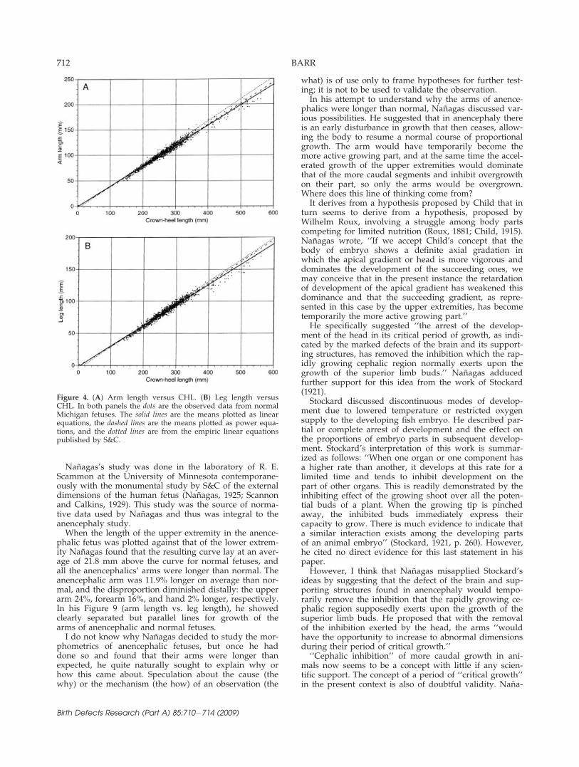
Scatterplots of arm length and leg length versus post-menstrual age showed no meaningful difference betweenanencephalic and normal fetuses (Fig. 1). When the datawere fitted for means, the normal and anencephalicfetuses were virtually indistinguishable. Plotting armlength versus leg length, with incorporation of the meanand 95% confidence limits, showed the anencephalicfetuses well within the confidence limits, except six thatwere high and three that were low (Fig. 2). Again themeans were virtually identical. The z-score distributionsdemonstrated the similarity of the two populations (Fig.3). To validate that measurements from the Minnesotastudies of S&C were concordant with the measurementobtained in the Michigan studies, the empiric formulaepublished by S&C were graphed on the scatterplots ofthe Michigan data from normal fetuses (Fig. 4). Leglength of the Minnesota sample was adjusted to themethod used for the Michigan sample by using the S&Cempiric formulae for CHL minus CRL. The resulting
plots of arm length versus CHL showed great similaritywhen curve fittings were by linear equations and notsubstantively different from more highly correlatedpower curves (Fig. 4).
DISCUSSION
I decided to explore these morphometrics because, onencountering the assertion that the arms of anencephalicfetuses were unduly long, my initial impression was thatthis was likely an illusion: an illusion due to the shorten-ing of their vertebral columns by kyphotic and lordoticcurvatures. The requisite information for this explorationwas at hand, since limb measurements had been rou-tinely obtained on many both normal and abnormalfetuses.
Figure 1. Lengths of arm (A) and leg (B) versus gestational age,fitted with second-degree polynomial curves. Normal fetuses:dots and solid line. Anencephalic fetuses: open diamonds and dottedline.
Figure 2. Arm length versus leg length. Solid lines: mean and95% confidence limits of 1155 normal fetuses. Open diamonds: 115anencephalic fetuses. Dotted line: mean of anencephalics
Figure 3. Arm versus leg z-score distribution. Calculated bythe formula derived from normal fetuses: Expected arm ¼ðlegð0:9745�ð3:423310�43legÞþð1:836310�63leg2ÞÞÞ31:5127; SD ¼ðlegð0:9745�ð3:423310�43legÞþð1:836310�63leg2ÞÞÞ3 5:687310�2;z-score:(observed arm 2 expected arm) / SD.
711THE LONG ARMS OF ANENCEPHALY
Birth Defects Research (Part A) 85:710�714 (2009)
Nanagas’s study was done in the laboratory of R. E.Scammon at the University of Minnesota contemporane-ously with the monumental study by S&C of the externaldimensions of the human fetus (Nanagas, 1925; Scannonand Calkins, 1929). This study was the source of norma-tive data used by Nanagas and thus was integral to theanencephaly study.
When the length of the upper extremity in the anence-phalic fetus was plotted against that of the lower extrem-ity Nanagas found that the resulting curve lay at an aver-age of 21.8 mm above the curve for normal fetuses, andall the anencephalics’ arms were longer than normal. Theanencephalic arm was 11.9% longer on average than nor-mal, and the disproportion diminished distally: the upperarm 24%, forearm 16%, and hand 2% longer, respectively.In his Figure 9 (arm length vs. leg length), he showedclearly separated but parallel lines for growth of thearms of anencephalic and normal fetuses.
I do not know why Nanagas decided to study the mor-phometrics of anencephalic fetuses, but once he haddone so and found that their arms were longer thanexpected, he quite naturally sought to explain why orhow this came about. Speculation about the cause (thewhy) or the mechanism (the how) of an observation (the
what) is of use only to frame hypotheses for further test-ing; it is not to be used to validate the observation.In his attempt to understand why the arms of anence-
phalics were longer than normal, Nanagas discussed var-ious possibilities. He suggested that in anencephaly thereis an early disturbance in growth that then ceases, allow-ing the body to resume a normal course of proportionalgrowth. The arm would have temporarily become themore active growing part, and at the same time the accel-erated growth of the upper extremities would dominatethat of the more caudal segments and inhibit overgrowthon their part, so only the arms would be overgrown.Where does this line of thinking come from?It derives from a hypothesis proposed by Child that in
turn seems to derive from a hypothesis, proposed byWilhelm Roux, involving a struggle among body partscompeting for limited nutrition (Roux, 1881; Child, 1915).Nanagas wrote, ‘‘If we accept Child’s concept that thebody of embryo shows a definite axial gradation inwhich the apical gradient or head is more vigorous anddominates the development of the succeeding ones, wemay conceive that in the present instance the retardationof development of the apical gradient has weakened thisdominance and that the succeeding gradient, as repre-sented in this case by the upper extremities, has becometemporarily the more active growing part.’’He specifically suggested ‘‘the arrest of the develop-
ment of the head in its critical period of growth, as indi-cated by the marked defects of the brain and its support-ing structures, has removed the inhibition which the rap-idly growing cephalic region normally exerts upon thegrowth of the superior limb buds.’’ Nanagas adducedfurther support for this idea from the work of Stockard(1921).Stockard discussed discontinuous modes of develop-
ment due to lowered temperature or restricted oxygensupply to the developing fish embryo. He described par-tial or complete arrest of development and the effect onthe proportions of embryo parts in subsequent develop-ment. Stockard’s interpretation of this work is summar-ized as follows: ‘‘When one organ or one component hasa higher rate than another, it develops at this rate for alimited time and tends to inhibit development on thepart of other organs. This is readily demonstrated by theinhibiting effect of the growing shoot over all the poten-tial buds of a plant. When the growing tip is pinchedaway, the inhibited buds immediately express theircapacity to grow. There is much evidence to indicate thata similar interaction exists among the developing partsof an animal embryo’’ (Stockard, 1921, p. 260). However,he cited no direct evidence for this last statement in hispaper.However, I think that Nanagas misapplied Stockard’s
ideas by suggesting that the defect of the brain and sup-porting structures found in anencephaly would tempo-rarily remove the inhibition that the rapidly growing ce-phalic region supposedly exerts upon the growth of thesuperior limb buds. He proposed that with the removalof the inhibition exerted by the head, the arms ‘‘wouldhave the opportunity to increase to abnormal dimensionsduring their period of critical growth.’’‘‘Cephalic inhibition’’ of more caudal growth in ani-
mals now seems to be a concept with little if any scien-tific support. The concept of a period of ‘‘critical growth’’in the present context is also of doubtful validity. Nana-
Figure 4. (A) Arm length versus CHL. (B) Leg length versusCHL. In both panels the dots are the observed data from normalMichigan fetuses. The solid lines are the means plotted as linearequations, the dashed lines are the means plotted as power equa-tions, and the dotted lines are from the empiric linear equationspublished by S&C.
712 BARR
Birth Defects Research (Part A) 85:710�714 (2009)

gas posited an early growth disturbance, ‘before thelower extremity is fully differentiated.’’ The long-termeffect of transiently arrested development would be morerelevant to hypodevelopment, as in restriction of cellnumber, than to hyperdevelopment. I think overgrowthis more likely to be a late event (fetal as opposed to em-bryonic), and the genesis of anencephaly is clearly anembryonic event.
Nanagas concluded, ‘‘In the teratological conditionknown as anencephaly there is an excessive developmentof the superior extremity. This hyperdevelopment is mostprominent proximally and decreases distally in accord-ance with the general law of cephalocaudal growth.’’S&C comment on a ‘‘law of developmental direction,’’which they found preferable to the ‘‘law of cephalocau-dal development’’ used by others. Their reasoning wasthat their terminology covered not only cephalocaudalbut mediodistal gradients as well. Be that as it may, I donot think either concept reaches the stature of ‘‘law.’’ Aless all-embracing term would at least admit the obvi-ously small arms of T. rex and the greatly elongated‘‘fingers’’ of bats. Also, as S&C note, even in humans thedistally placed legs end up outgrowing the proximallyplaced arms in length.
This hypothesizing harkens back to the era followingthe introduction of evolutionary theory when there was agrand search for overarching laws of development. Theideas discussed were not considered exceptional at thetime. That the proposal of ‘‘laws’’ sometimes went toofar is evident now, but at the time these proposalsseemed to make sense (provided one did not find real-life variation too unsettling). We can now recognize thatthe ‘‘laws’’ of cephalocaudal development or develop-mental direction have too many exceptions to be consid-ered laws. These days, by parsing the details of develop-mental schedules, we find deeper and deeper layers ofcomplexity that were unimagined in the late nineteenthand early twentieth centuries.
Aside from current rejection of such conjectures, thereis a more compelling reason to reject the application ofsuch suppositions to explain the long arms of anence-phalic fetuses. That reason is that the arms of anence-phalics are not unduly long. If my claim is that the armsof anencephalics are not longer than normal, it is incum-bent on me to try to discover why Nanagas found other-wise. His study appeared to be carefully done and clearlydescribed. That his proposals for the why of his findingsare incorrect is irrelevant.
There are some differences in the measurement meth-ods used in the Minnesota and Michigan studies. TheMinnesota fetuses were all formalin fixed, while the Mich-igan fetuses were unfixed. However, S&C in a pilot studyfound minimal shrinkage of extremity measurements afterprolonged fixation, on the order of 0.3%, not 12%.
For leg measurements, the methods of S&C and Nana-gas are concordant. S&C sum the leg segments as fol-lows: from the greater trochanter to the center of theknee joint, from there to the center of the ankle joint, andfrom there to the sole of the foot. Nanagas sums the legsegments as follows: from the greater trochanter to thelateral condyle of the tibia, from there to the most promi-nent part of the lateral maleolus, and thence to the soleof the foot. The lateral condyle of the tibia is just belowthe center of knee joint and the lateral maleolus is justabove the center of the ankle joint, so these two misplace-
ments result in shortened foreleg measurements, but thetotal leg lengths agree with those of S&C.It must be noted that the legs of Minnesota fetuses
were measured from the greater trochanter, while thelegs of the Michigan fetuses were measured from theperineum. I find the latter technique more reproduciblethan the S&C method, especially in the young fetuswhose trochanter is minimally developed. The leglengths reported by S&C are approximately 26% greateron average than those of age-matched Michigan fetuses.However, if the calculation CHL minus CRL equals leglength is applied to the S&C data, there are negligibledifferences from my observations of leg lengths (Fig. 4).This indicates that there was actually no substantive dif-ference in leg lengths between the Minnesota and Michi-gan fetuses.S&C’s graphs and tables record extremity measure-
ments for CHL and gestational age (in lunar months).Nanagas did not have age data for his specimens, andthe CHL in anencephaly is obviously not to be consid-ered normal. He estimated CHL from his measured leglengths: ‘‘When the crown-heel length of the body is cal-culated from this [leg length] with the empiric formula[leg length 5 0.43 CHL 2 7.0 mm], the resulting valuesrange from 279 to 500 mm, which may be regarded aswithin reasonable limits for normal material.’’ (Themethod of estimating arm length from CHL was armlength 5 0.43 CHL 2 4.0 mm).In his Figure 9, Nanagas showed what appear to be
parallel ascending lines denoting the arm length versusleg length for normal fetuses and for anencephalicfetuses. Since Nanagas and I did not measure leg lengthsin the same way, I cannot construct a graph of arm ver-sus leg that is directly comparable to his Figure 9. How-ever, if the difference between anencephalic and normalfetuses were a constant average of 11.9%, these lineswould clearly diverge with continued growth. If the dif-ference between anencephalic and normal arms were aconstant 21.8 mm, the lines would be parallel, as shownby Nanagas and imply a decreasing percentage differ-ence with continued growth.The upper extremity length is commonly defined as the
distance in a straight line from the lateral tip of the acro-mial process to the end of the middle finger. The arms ofthe Michigan fetuses were measured from the acromion tothe tip of the third finger as a single, straight-line mea-surement. The Minnesota fetuses were fixed specimenswith flexed limbs. The method used by S&C was ‘‘thelength of the superior extremity is the sum of the distan-ces from the acromial process to the angle of the elbow,from the angle of the elbow to the base of the palm of thehand, and from the base of the palm of the hand to thetip of the middle finger.’’ When the S&C data for armlength versus CHL are contrasted with the comparableMichigan data there is close agreement (Fig. 4).None of this so far provides an adequate explanation
for the difference between Nanagas’s findings and mine.However, a close reading of the methods of arm mea-
surement published by S&C and by Nanagas does pro-vide the answer. For S&C the focal point at the elbowwas ‘‘the angle of the elbow joint.’’ As shown in theirFigure 1, the angle of the elbow is the antecubital crease,which is coincident with the center of elbow joint of theextended arm. If they measured from the antecubitalcrease with the arm flexed, their forearm measurement
713THE LONG ARMS OF ANENCEPHALY
Birth Defects Research (Part A) 85:710�714 (2009)

would have been too short. I think they actually did mea-sure to the center of the elbow joint either by extendingthe arm or by making the adjustment to the joint center,because we get very similar readings of total arm length.But Nanagas, claiming identical measuring technique toS&C, gave the following: ‘‘1. UEL, length of the upper ex-tremity; the sum of the separate lengths of arm, forearm,and hand. 2. AL, arm length; from the tip of the acro-mium [sic] to the tip of the olecranon process. 3. FML,forearm length; from the tip of the olecranon process tothe middle of the wrist joint. 4. HL, hand length; from themiddle of the wrist joint to the tip of the middle finger.’’
The tip of the olecranon process is not coincident withthe center of the elbow joint. So, rather than measuringthe true length of the arm, Nanagas’s summation ofupper arm 1 forearm 1 hand has overstated arm length,predominantly of the proximal segment, but also of thedistal limb to a lesser degree. By measuring arm lengthby both of these methods (assuming S&C meant the mid-dle of the elbow joint) and by my linear method in asample of eight normal fetuses, I found that Nanagas’smethod produced lengths that were 12.25% greater thanthe S&C and Michigan methods, which agreed with eachother. This excess is so close to Nanagas’s findings thatthe arms of anencephalics were on average 11.9% longerthan normal that I believe the mystery is solved.
REFERENCES
Barr M. 1997. Is it true what they say about anencephalics? A morpho-metric analysis. Proc Greenwood Genet Ct 16:201–202.
Barr M, Blackburn WR, Cooley NR. 1994. Human fetal somatic and vis-ceral morphometrics. Teratology 49:487–496.
Child CM. 1915. Senescence and rejuvenescence. Chicago: University ofChicago Press.
Goodman RM, Gorlin RJ. 1983. The malformed infant and child. NewYork: Oxford University Press (illustration on p. 4 labeled with ‘‘longarms’’).
Gorlin RJ, Cohen Jr. MM, Levin LS. 1990. Syndromes of the head andneck. 3rd ed. New York: Oxford University Press (p. 566).
Lemire RJ, Beckwith JB, Shepard TH. 1972. Iniencephaly and anencephalywith spinal retroflexion. A comparative study of eight human speci-mens. Teratology 6:27–36.
Lemire RJ, Beckwith JB, Warkany J. 1978. Anencephaly. New York: RavenPress (p. 63).
Lemire RJ, Loeser JD, Leech RW, Alvord Jr. EC. 1975. Normal and abnor-mal development of the human nervous system. Hagerstown, MD:Harper & Row (p. 307, Table 18-4).
Nanagas JC. 1925. A comparison of the growth of the body dimensions ofanencephalic human fetuses with normal growth as determined bygraphical analysis and empirical formulae. Am J Anat 35:455–494.
Roux W. 1881. Der Kampf der Teile im Organismus [The Battle of Partsin the Organism]. Leipzig.
Scammon RE, Calkins LA. 1929. The development and growth of theexternal dimensions of the human body in the fetal period. Minneap-olis: University of Minnesota Press.
Stockard CR. 1921. Developmental rate and structural expression: an ex-perimental study of twins, ‘double monsters’, and single deformitiesand the interaction among embryonic organs during their origin anddevelopment. Am J Anat 28:115–277.
714 BARR
Birth Defects Research (Part A) 85:710�714 (2009)