Embed Size (px)
Citation preview
T H E L Y M P H A T I C STATUS OF H A M S T E R C H E E K P O U C H T I S S U E I N R E L A T I O N TO ITS P R O P E R T I E S
AS A G R A F T A N D AS A G R A F T SITE*
BY CLYDE F. BARKER,~ M.D., A~'D R. E. BILLINGHAM, F.R.S.
(From the Departments of Medical Genetics and Surgery, School of Medicine, University of Pennsylvania, Philadelphia, Pennsylvania 19104)
(Received for publication 28 October 1970)
While there is an impressive body of empirical evidence that homografts placed in the highly vascular walls, i.e. the skin, of the hamster's cheek pouch constitute provocative exceptions to the rule that transplantation of genetically alien tissue is followed by rejection (1), the physiological basis of the immunologically privileged status of the cheek pouch as a graft site awaits clarification. Besides demonstrating that intact pouches consistently protect implanted homografts of trunk skin from rejection for long periods, Billingham and Silvers (2, 3) noted that pouch skin be- haved as an immunologically privileged tissue when transplanted to the trunks of homologous hosts. I t significantly outlived homografts of ordinary skin. Furthermore, they found that even when cheek pouch skin was transposed to a hamster's trunk, it retained its capacity to protect small homografts of ordinary skin, if these were inlaid in shallow beds prepared in its center. Skin homografts protected in the milieu of the cheek pouch or heterotopic homografts of pouch skin were shown to owe their survival to inability to sensitize their hosts. These and other observations were explained provisionally by the hypothesis that the areolar connective tissue of pouch skin pre- vents the escape of antigen, though allowing the graft to acquire a rich blood supply.
Reports that the brain can prevent implanted homografts from inciting sensitivity because it lacks a lymphatic drainage, thus having no afferent pathway of the im- munological reflex, are more equivocal (4-7). Indeed a critical reevaluation of this organ as a privileged site is long overdue.
Our recent demonstration that interruption of the afferent lymphatic drainage but not of the blood supply of flaps raised from guinea pigs' trunk skin produces graft sites capable of sustaining inlaid skin homografts for long periods (8), in conjunc- tion with the findings of others (9), provides an experimental basis for the thesis that vascularized tissues or organs within the body, devoid of a lymphatic drainage, should protect implanted homografts from rejection.
Most of the work to be described in this communication represents a critical evaluat ion of the premise tha t the longevity of homografts in the hamster ' s
* The expenses of this work were defrayed in part by U. S. Public Health Service Grant AI-07001.
~C Markle Scholar in Academic Medicine.
620
Dow
nloaded from http://rupress.org/jem
/article-pdf/133/3/620/1084052/620.pdf by guest on 08 January 2022
CLYDE F. BARKER AND R. E. BILLINGtIA1V~ 621
cheek pouch depends upon its lack of a func t iona l l y m p h a t i c dra inage sys tem.
Other exper iments repor ted on were designed to eva lua te the capac i ty of homo-
grafts long-sus ta ined in a pr iv i leged site to affect the immunolog ic r eac t i v i t y of
the hos t (3), and to find ou t whe ther a brief init ial t r e a t m e n t of a hos t wi th
an t i - l ymphocy te se rum (10, 11) can improve the ou tcome of t r ansp lan t ing
homogra f t s to pr iv i leged sites or of t r ansp lan t ing homogra f t s of an immuno-
logically pr ivi leged tissue to a conven t iona l site.
Materials and Methods
Adult hamsters, from domestically maintained sublines of the CB (agouti) and MHA (albino) isogenic strains, were used respectively as donors and recipients of homografts. (CB X MHA) F1 hybrid animals served as hosts in experiments in which lymphoid cells were used to study lymphatic drainage.
Grafts of cheek pouch skin were obtained by everting the pouches of anesthetized animals and excising them from a point close to their union with the buccal mucosa. An incision along one side of an excised pouch produced a large, fiat sheet of pouch skin. Mter trimming off some of the excess loose connective tissue we cut from it approximately circular grafts 2.5-3.0 cm in diameter, taking care to exclude areas where muscle fibers were present.
The grafts were transplanted to beds of appropriate size cut down to the level of the highly vascular panniculus carnosus in the close-clipped skin of the host's chest, according to our standard procedure for grafting ordinary skin (12).
Ear skin grafts, 1.2-1.8 cm in diameter were prepared from the skin which can easily be peeled away from the median ear cartilage of excised pinnae. Such grafts are preferable to those of trunk skin because they are much thinner and become vascularized more rapidly.
Inlay of ear skin grafts in established pouch skin grafts entailed the injection of 2-3 ml of physiological saline solution as superficially as possible, via a No. 30 gauge needle, into the centers of pouch skin isografts that had been in residence on the sides of MHA hamsters' chests for at least 30 days. The saline distended the connective tissue of the pouch skin, facilitating the cutting with fine curved scissors of very shallow, split-thickness beds 1.2-1.5 cm in diameter that received the ear skin grafts. The latter healed in just as well in these sites as in beds of comparable depth prepared in normal trunk skin.
Viable suspensions of lymph node cells were prepared from the pooled axillary, brachial, inguinal, and mesenteric nodes and dispensed in Hanks' solution as described elsewhere (12).
Sensitization of hamsters was accomplished by first grafting them bilaterally with skin from the alien donor strain and then, about 12 days later, injecting them intraperitoneally with about 100 million spleen cells from this strain. Immune lymph nodes for the preparation of cell suspensions were harvested 8-10 days later.
Suspensions of cheek pouch epidermal cells were prepared by the tryptic digestion of small pieces of pouch skin (13).
Rabbit anti-hamster lymphocyte serum, (ALS) 1 the generous gift of Dr. C. F. Shaffer, was prepared according to the method of Gray et al. (14) and administered intramuscularly.
Visualization of lymphatic vessds and their draining nodes was accomplished by superficial intracutaneous injection, via a No. 30 gauge needle, of a mixture of equal volumes of 2% aqueous solutions of Berlin Blue and Patent Blue V. Previous experience had shown that whereas the former dye is very effective in outlining the lymphatic vessels, it passes through the nodes very rapidly. Berlin Blue, on the other hand, stains the lymphatic vessels less
1 Abbreviations used in this paper: ALS, anti-hamster lymphocyte serum; ILT, Immune lymphocyte transfer; MST, median survival time.
Dow
nloaded from http://rupress.org/jem
/article-pdf/133/3/620/1084052/620.pdf by guest on 08 January 2022
622 H A M S T E R C H E E K P O U C H T I S S U E AS G R A F T A N D G R A F T S I T E
effectively but is retained in the nodes for a longer time. The combination of dyes has the advantages of both.
O B S E R V A T I O N S
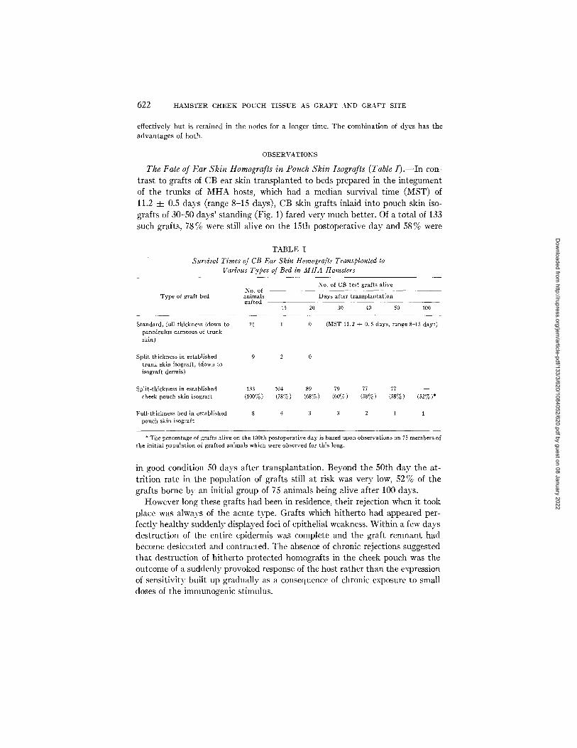
The Fate of Ear Skin Homografts in Pouch Skin Isografts (Table I).--In con- trast to grafts of CB ear skin transplanted to beds prepared in the integument of the trunks of MHA hosts, which had a median survival time (MST) of l l .2 4- 0.5 days (range 8-15 days), CB skin grafts inlaid into pouch skin iso- grafts of 30-50 days' standing (Fig. 1) fared very much better. Of a total of 133 such grafts, 78% were still alive on the 15th postoperative day and 58% were
T A B L E I
Snrz4val Times of CB Ear Skin Homografts Transplanted to Various Types of Bed in IklHA Hamsters
No. of CB test grafts alive No. of
Type of graf t bed animals Days after transplantation gaffed
15 20 30 40 50 100
Standard, full-thickness (down to panniculus carnosus of t runk skin)
Split-thickness in established trunk skin isograft, (down to isograft dermis)
21 l
9 2 0
0 (MST 11.2 ± 0.5 days, range 8-15 days)
Split-thickness in established 133 104 89 79 77 77 - - cheek pouch skin isograft (100%) (78%) (68%) (60~) (58%) (58%) (52%)*
Full-thickness bed in established pouch skin isograft
8 4 3 3 2 1 1
* The percentage of grafts alive on the 100th postoperative day is based upon observations on 75 members of the initial population of grafted animals which were observed for this long,
in good condition 50 days after transplantation. Beyond the 50th day the at- trition rate in the population of grafts still at risk was very low, 52 % of the grafts borne by an initial group of 75 animals being alive after 100 days.
However long these grafts had been in residence, their rejection when it took place was always of the acute type. Grafts which hitherto had appeared per- fectly healthy suddenly displayed foci of epithelial weakness. Within a few days destruction of the entire epidermis was complete and the graft remnant had become desiccated and contracted. The absence of chronic rejections suggested that destruction of hitherto protected homografts in the cheek pouch was the outcome of a suddenly provoked response of the host rather than the expression of sensitivity built up gradually as a consequence of chronic exposure to small doses of the immunogenic stinmlus.
Dow
nloaded from http://rupress.org/jem
/article-pdf/133/3/620/1084052/620.pdf by guest on 08 January 2022
CLYDE F. BARKER AND R. E. BILLINGHAM 623
Despite attempts to obtain grafts of standard size, there was in fact consider- able variation in the size of grafts after the healing in phase, from about 4 to 18 mm in diameter. I t was noted, however, that the size or dosage of a graft had no influence on its longevity, larger grafts tending to fare just as well as the much smaller ones reported previously (2).
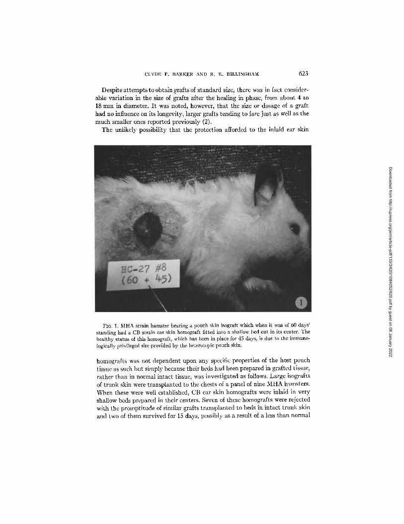
The unlikely possibility that the protection afforded to the inlaid ear skin
FIG. 1. MHA strain hamster bearing a pouch skin isograft which when it was of 60 days' standing had a CB strain ear skin homograft fitted into a shallow bed cut in its center. The healthy status of this homograft, which has been in place for 45 days, is due to the immuno- logically privileged site provided by the heterotopic pouch skin.
homografts was not dependent upon any specific properties of the host pouch tissue as such but simply because their beds had been prepared in grafted tissue, rather than in normal intact tissue, was investigated as follows. Large isografts of trunk skin were transplanted to the chests of a panel of nine MHA hamsters. When these were well established, CB ear skin homografts were inlaid in very shallow beds prepared in their centers. Seven of these homografts were rejected with the promptitude of similar grafts transplanted to beds in intact trunk skin and two of them survived for 15 days, possibly as a result of a less than normal
Dow
nloaded from http://rupress.org/jem
/article-pdf/133/3/620/1084052/620.pdf by guest on 08 January 2022
624 HAMSTER CHEEK POUCI-I TISSUE AS GRAFT AND GRAFT SITE
lymphatic circulation from the body skin isografts that furnished their beds (see below).
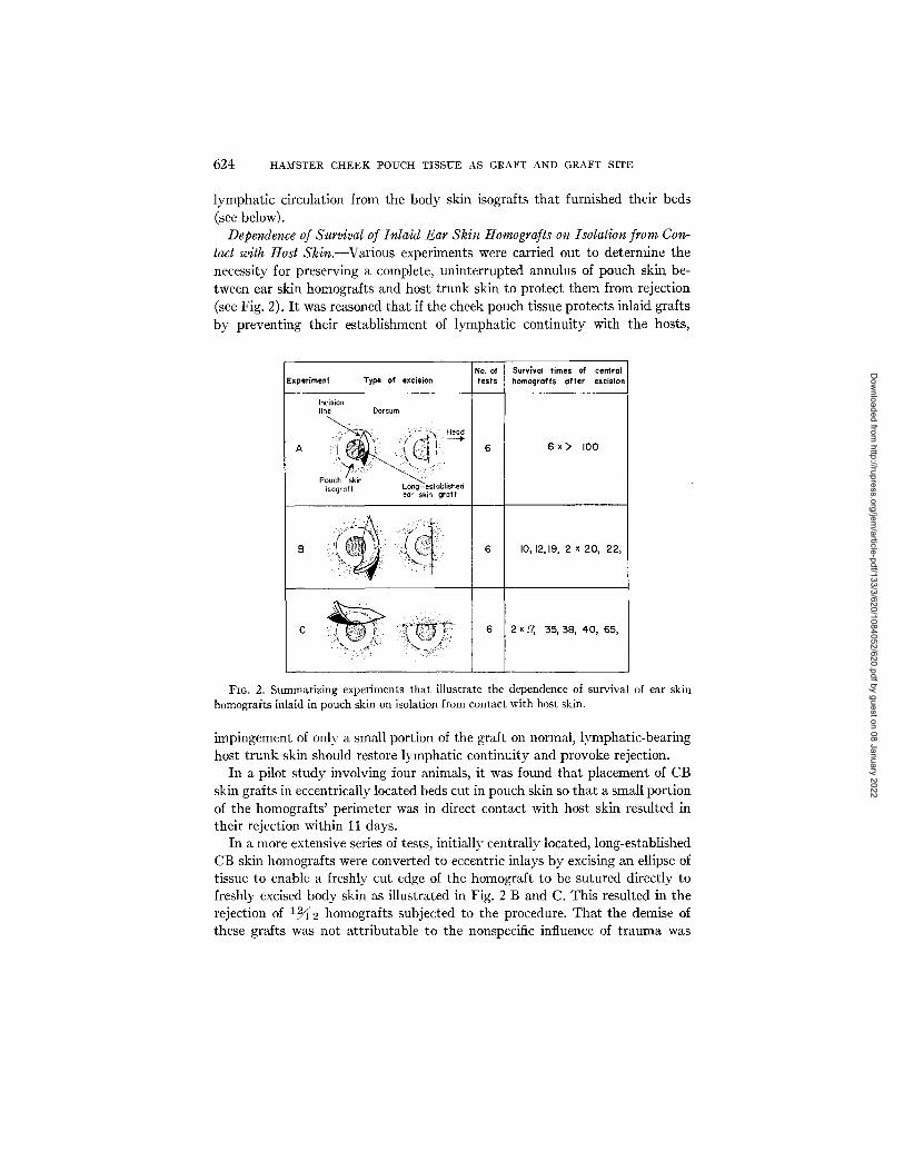
Dependence of Survival of Inlaid Ear Skin Homografts on Isolation from Con- tact with Host Skin.--Various experiments were carried out to determine the necessity for preserving a complete, uninterrupted annulus of pouch skin be- tween ear skin homografts and host trunk skin to protect them from rejection (see Fig. 2). I t was reasoned that if the cheek pouch tissue protects inlaid grafts by preventing their establishment of lymphatic continuity with the hosts,
NO. of Surv;val times of central Experiment Type of excision tests homografts af ter excision
Incision line Dorsum
isogr aft Long - establisnea ear skin graft
C >i( ~!! i ~ 6 2x]~ 35~ 381 40~ 65 ~
FIG. 2. Summarizing experiments that illustrate the dependence of survival of ear skin homografts inlaid in pouch skin on isolation from contact with host skin.
impingement of only a small portion of the graft on normal, lymphatic-bearing host trunk skin should restore lymphatic continuity and provoke rejection.
In a pilot study involving four animals, it was found that placement of CB skin grafts in eccentrically located beds cut in pouch skin so that a small portion of the homografts' perimeter was in direct contact with host skin resulted in their rejection within 11 days.
In a more extensive series of tests, initially centrally located, long-established CB skin homograffs were converted to eccentric inlays by excising an ellipse of tissue to enable a freshly cut edge of the homograft to be sutured directly to freshly excised body skin as illustrated in Fig. 2 B and C. This resulted in the rejection of 12/~2 homografts subjected to the procedure. That the demise of these grafts was not attributable to the nonspecific influence of trauma was
Dow
nloaded from http://rupress.org/jem
/article-pdf/133/3/620/1084052/620.pdf by guest on 08 January 2022
CLYDE F. B A R K E R AND R. E. BILLINGHAN[ 625
established as follows. Full-thickness elliptical areas, including portions of both skin homograft and pouch skin, were excised in such a way that the entire area of excision was located well within the perimeter of the pouch skin graft (Fig. 2 A). Five of the defects were then closed by suture and two additional ones were allowed to granulate and reepithelialize of their own accord. Neither pro- cedure caused rejection of the affected homograft.
As the data summarized in Fig. 2 indicate, the location of the site of juxta- position of the inlaid homografts with host skin seemed to determine the dura- tion of their subsequent survival times. When the line of union was dorsally located, the approximate median survival time of the grafts was 35 days; whereas, when it was located cephalad, it was only about 20 days. These findings are consistent with the anatomical distribution of the major lymphatic vessels on the side of a hamster's trunk which run principally in the cephalocaudad direction (15).
Despite the considerable variation in survival times of individual homografts in these excisional experiments, rejection when it occurred was always of the acute type.
None of the experiments so far described has shed any light on the necessity for isolating intrapouch skin homografts from host tissue beneath them to procure their exemption from rejection. The standard beds were prepared in such a way as to ensure that an intact layer of pouch skin connective tissue separated or insulated homograft dermis from direct contact with the host's panniculus carnosus. Accordingly, in a panel of eight MHA hamsters, central beds were deliberately cut down through the pouch skin isograft and under- lying panniculus to expose the deep fascia of the host which constituted the bed for the CB inlay ear skin grafts. The extended survival of 50% of these grafts (Table I) is consistent with the fact that muscle has a relatively poor lymphatic supply as compared with skin (16) and lends further credence to the premise that lymphatic continuity between skin homograft and host is the optimum circumstance for instigating the rejection process.
Dye Injection Studies
To try and obtain more direct evidence concerning the lymphatic drainage status of the skin of intact cheek pouches and of skin homografts protected by residence within pouch skin isografts on hamsters' chests, dye injection experiments were performed.
First, the consistency with which fine lymphatics could be revealed in hamsters' skin by this means had to be evaluated. 0.1-0.2 ml of dye was in- jected as superficially as possible into the close-clipped, intact skin of the sides of animals' trunks. Mter about 30 sec delay an incision was made in the long axis of the body, 2-3 cm ventral to the injection site. The injected area was then approached by sharp dissection and the undersurface of the intra-
Dow
nloaded from http://rupress.org/jem
/article-pdf/133/3/620/1084052/620.pdf by guest on 08 January 2022
626 HAMSTER CHEEK POUCH TISSUE AS GRAFT AND GRAFT SITE
cutaneous bleb of dye visualized. In every instance a fine network of intra- cutaneous lymphatics was seen, apparently radiating from the injection site and emptying into one or more larger lymphatic channels which usually drained in a cephalad direction, terminating in the ipsilateral axillary node, which was deeply stained. Frequently the nearby brachial node was stained as well. In a few cases, especially when the injections had been made more caudally, evidence of drainage of dye towards and into the inguinal node was obtained.
Confidence in the usefulness of this procedure for revealing patent lymphatic drainage pathways was increased when it was found that dye injected super- ficially into established isografts of ordinary trunk skin on six hamsters promptly and conspicuously passed to the regional nodes, revealing the vessels which it traversed.
Dye Injection into Intact Cheek Pouches.--Five hamsters were anesthetized, their pouches everted, injected as superficially as possible with dye, and then replaced in their natural positions. A few minutes later a careful dissection was made to display the entire course of the pouches, from their openings into the buccal cavity and backwards along the neck and over the shoulder, as well as the cervical and other regional nodes. In no case were any dye-filled lymphatic vessels seen, nor was there any evidence of passage of the dye into a lymph node. Despite one report (17) that parts of the cheek pouch do have sparsely distributed lymphatics, the present observation is consonant with findings of other workers of the inability or great reluctance of the connective tissue of the intact cheek pouch to allow the passage of colloidal or particulate matter to the putative regional nodes (18-21).
However, if instead of injecting the dye into the mucosa of the pouch it were injected into that of the lip or buccal cavity, cervical lymphatics and their draining nodes would be immediately and consistently outlined, again corroborating the findings of previous studies.
Injection of Dye into Pouch Skin Isografts and Inlaid Ear Skin Grafts.- When dye was injected into pouch skin isografts of 30-50 days' standing, although it always displayed some tendency to spread horizontally in the graft's connective tissue, it never entered regional lymphatics of the host's skin or its nodes. Dye injected into ear skin isografts of at least 30 days' stand- ing in pouch skin isografts revealed a rich lymphatic plexus within them, but there were no connecting lymph vessels within the host pouch skin, and dis- section failed to reveal stained regional lymphatics.
If the protection occurring in ear skin homografts inlaid in p~uch skin is due to their lack of a patent lymphatic drainage, then it follows that grafts which do undergo rejection must fortuitously have established some func- tional lymphatic connections. To explore this possibility, CB inlay homografts in pouch skin isografts that displayed signs of rejection 13-15 days after trans- plantation as well as the surrounding pouch skin were injected with dye. In
Dow
nloaded from http://rupress.org/jem
/article-pdf/133/3/620/1084052/620.pdf by guest on 08 January 2022
CLYDE F. B A R K E R AND R. E . B I L L I N G H A M 627
none of six grafts thus treated did any dye enter the host's lymphatic system. Since this result might have been due to breakdown of lymphatic vessels as- sociated with the advanced state of rejection of the grafts concerned, another approach was followed. Some hamsters received juxtaposed ear skin isografts and homografts in common shallow beds in pouch skin isografts. Apart from being vascularized at the same time, it was hoped that if lymphatic connec-
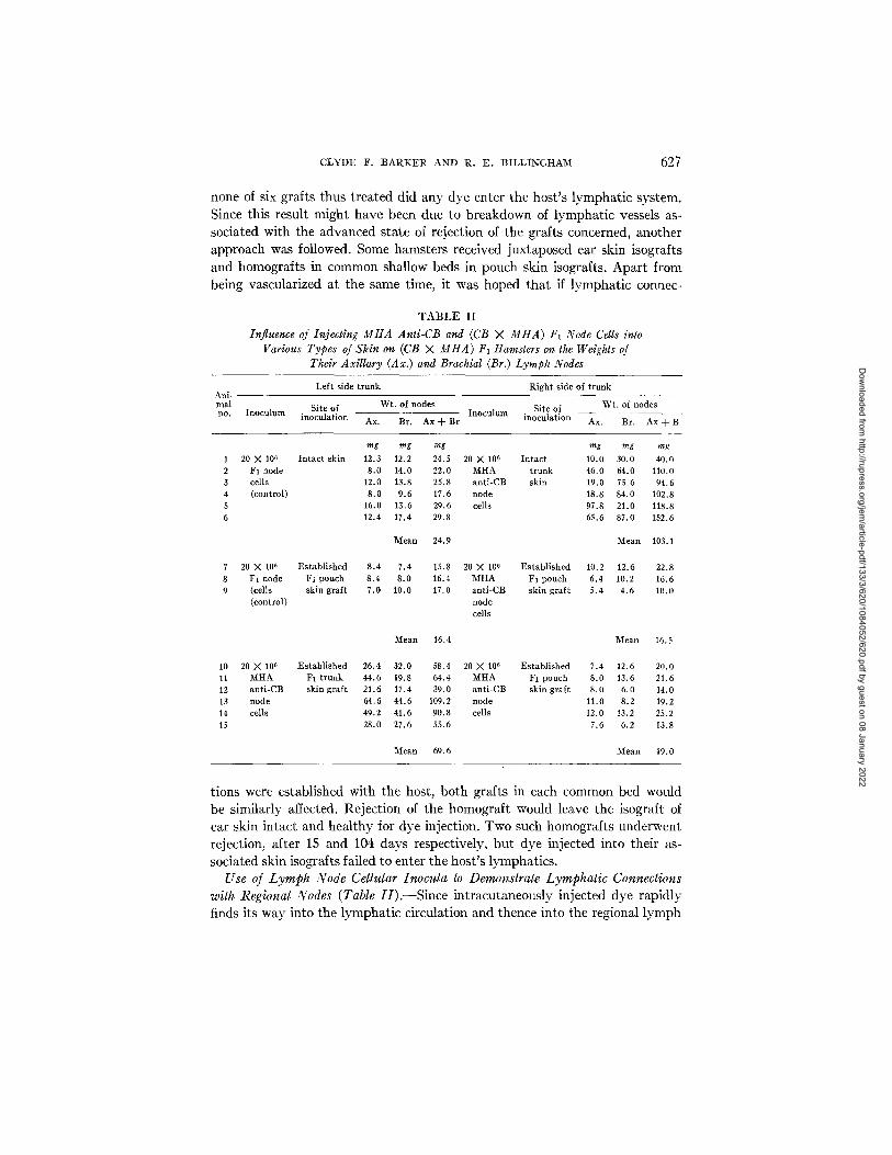
T A B L E I I
Influence of Injecting MIIA Anti-CB and (CB X Mt tA) F1 Node Cells into Various Types of Skin on (CB X Mt tA) F1 Hamsters on the Weights of
Their Axillary (Ax.) and Brachial (Br.) Lymph Nodes
Ani- mal no.
Left side trunk Right side of trunk
Site of Wt. of nodes Site of Wt. of nodes Inoculum inoculation Inoculum inoculation Ax. Br. Ax q- Br Ax. Br. Ax ~ B
mg mg mg mg mg mg 20 X l0 s Intact skin 12.3 12.2 24.5 20 >( 106 Intact 10.0 30.0 40.0
F1 node 8.0 14.0 22.0 MHA trunk 46.0 64.0 110.0 cells 12.0 13.8 25.8 anti-CB skin 19.0 75.6 94.6 (control) 8.0 9.6 17.6 node 18.8 84.0 102.8
16.0 13.6 29.6 cells 97.8 21.0 118.8 12.4 17.4 29.8 65.6 87.0 152.6
Mean 24.9 Mean 103.1
7 20 X 10~ Established 8.4 7.4 15.8 20 X 106 Established 10.2 12.6 22.8 8 F1 node F1 pouch 8.4 8.0 16.4 MHA F1 pouch 6.4 10.2 16.6 9 (cells skin graft 7.0 10.0 17.0 anti-CB skin graft 5.4 4.6 10.0
(control) node cells
Mean 16.4 Mean 16.5
10 20 X 106 Established 26.4 32.0 58.4 20 X 106 Established 7.4 12.6 20.0 11 MHA F1 trunk 44.6 19.8 64.4 MHA F1 pouch 8.0 13.6 21.6 12 anti-CB skin graft 21.6 17.4 39.0 anti-CB skin graft 8.0 6.0 14.0 13 node 64.6 44.6 109.2 node 11.0 8.2 19.2 14 cells 49.2 41.6 90.8 cells 12.0 13.2 25.2 15 28.0 27.6 55.6 7.6 6.2 13.8
Mean 69.6 Mean 19.0
tions were established with the host, both grafts in each common bed would be similarly affected. Rejection of the homograft would leave the isograft of ear skin intact and healthy for dye injection. Two such homografts underwent rejection, after 15 and 104 days respectively, but dye injected into their as- sociated skin isografts failed to enter the host's lymphatics.
Use of Lymph Node Cellular Inocula to Demonstrate Lymphatic Connections with Regional Nodes (Table//).--Since intracutaneously injected dye rapidly finds its way into the lymphatic circulation and thence into the regional lymph
Dow
nloaded from http://rupress.org/jem
/article-pdf/133/3/620/1084052/620.pdf by guest on 08 January 2022
628 HAMSTER C H E E K POUCH TISSUE AS GRAFT AND GRAFT SITE
nodes, it seemed likely that at least some cells from a monodisperse suspension would do likewise. This should certainly apply to small lymphocytes of the long-lived variety, which include antigen-sensitive cells, since there are good grounds for belief that these cells do leave the blood circulation in the peripheral tissues, including skin, passing to afferent lymphatic vessels and thence to the regional nodes, eventually to return to the circulation via the thoracic duct (22). With lymph node cell inocula from a donor isogenic with the host, passage of cells to the regional node is unlikely to be detectable. However, there is evidence that if lymphoid cells from a parental strain donor are injected into or caused to enter the parenchyma of some tissues, such as the skin, kidney, or the uterus of an F1 hybrid offspring which is unable to reject them and at the same time confronts them with an alien transplantation antigen deter- mined by a major H-locus, some of them will react against host antigens giving rise to local graft-versus-host reactions (23) and others will pass to the regional nodes. In the latter sites graft-versus-host reactions also occur, re- flected in hypertrophy of these organs (23). Indeed this forms a sensitive means of assaying graft-versus-host reactivity (24). Lymph node cell inocula from donors presensitized against the alien tissue antigens of the host are more effective in this respect than cells from normal donors.
To explore the feasibility of employing graft-versus-host reactions to reveal patent lymphatic pathways, six adult (CB X MHA) F1 hamsters were in- jected intracutaneously at a single site on the right thoracic wall with 20 X 106 lymph node cells, suspended in 0.1 ml of Hanks' solution, prepared from an MHA hamster which had been sensitized against the tissue antigens of the CB strain. A similar number of cells from an isologous, (CB X MHA) F1 donor were injected in the skin on the contralateral side, as a control. In every instance an intense local inflammatory immune lymphocyte transfer (ILT) reaction (25) developed at the injection site which attained its zenith within 48-72 hr. On the 7th day after inoculation the animals were killed and the axillary and brachial nodes on both sides of the body were carefully dissected out and weighed. The results (see Table II) show that in every animal there was a striking hypertrophy of either the axillary or the brachial node and in some cases both nodes draining the skin site injected with MHA anti-CB lymphoid cells, as compared with the nodes on the contralateral side of the body which received the immunologically nonreactive FI hybrid cells. I t may be noted that although the inguinal nodes were also weighed they were never found to be enlarged. Accordingly the weights of these nodes are not recorded in Table II. I t is important to realize that some of the lymph node hypertrophy observed in the experiments to be described might have been incited by non- specific, diffusible products of the local graft-versus-host reactions which oc- curred at the inoculation sites of the lymph node cells in the skin. However, even if this were the case, its occurrence would still be indicative of a patent anatomical drainage pathway from the inoculation site to the affected nodes.
Dow
nloaded from http://rupress.org/jem
/article-pdf/133/3/620/1084052/620.pdf by guest on 08 January 2022
CLYDE F . B A R K E R AND R. E . BILLINGHA1V£ 629
To test the permeability of cheek pouch skin to superficially injected lymph node cells, three (CB X MHA) F1 hybrid hamsters bearing large, long-es- tablished pouch skin isografts on each side of the thorax were employed. The grafts on the right side were injected with 20 X 106 MHA anti-CB node cells, while those on the left side received a similar number of cells from an isologous hybrid donor. Although striking ILT reactions developed on the former inocu- lation sites, when the nodes from these animals were harvested and weighed 7 days after injection, there was not the slightest evidence of hypertrophy in those draining the pouch skin that received the immune cells, the mean weight of the combined left axillary and brachial nodes being almost identical with that for the corresponding nodes on the opposite side of the body.
The second experiment was carried out on a panel of six (CB X MHA) F1 animals, each of which bore a large pouch skin isograft on the right side of its chest and an isograff of ordinary trunk skin of comparable size on its left side. Both grafts were inoculated with 20 X 106 CB anti-MHA node cells. Although intense ILT reactions developed at all inoculation sites, there was no evidence of hypertrophy in the nodes draining the areas that included the pouch skin grafts. I t is interesting to note that although there was hypertrophy of the nodes draining the trunk skin isografts, it was not as great as that caused by similar inocula placed in intact trunk skin. This suggests that the lymphatic drainage of a well-healed in skin isograft is inferior to that of intact skin.
Finally, to evaluate the lymphatic drainage from intact pouches, a group of six (CB X MHA) F1 hamsters received an inoculum of 20 X 106 MHA anti-CB node cells in the mucosa of their right cheek pouch and 20 X 106 isologous hybrid cells in their left cheek pouch. Again, strong ILT reactions developed at the inoculation sites of the immune cells. 7 days later the animals were killed and careful dissections made to reveal all the nodes draining the cephalad half of the body. No significant difference could be discerned between the appearance and size of the nodes on the two sides of the body.
The most reasonable interpretation of these findings, which are in accord with those of Chadwick and Blarney (20) on the intact cheek pouch, is that, unlike intact skin or established isografts of ordinary skin, the skin of the intact pouches and pouch skin isografts of long standing prevent lymphoid cells inoculated into them from gaining access to the regional node system in detectable numbers.
Influence of ALS on Survival of Pouch Skin ttomografts (Table III).--One of the incentives to study privileged sites and tissues is the possibility that homologous tissue grafts which enjoy prolonged survival, either on account of some special intrinsic property or because of transplantation to an unusual site, may in time induce some degree of specific unresponsiveness or tolerance in the host in respect to the alien antigens involved (3).
Experiments were therefore carried out to confirm previous observations (2, 3) that heterotopic homografts of pouch skin live much longer than homo-
Dow
nloaded from http://rupress.org/jem
/article-pdf/133/3/620/1084052/620.pdf by guest on 08 January 2022
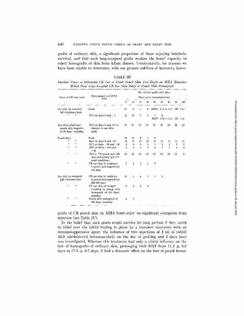
630 H A M S T E R C H E E K P O U C H T I S S U E AS G R A F T AND G R A F T SITE
grafts of ordinary skin, a significant proportion of them enjoying indefinite survival, and that such long-accepted grafts weaken the hosts' capacity to reject homografts of skin from infant donors. Unfortunately, for reasons we have been unable to determine, with our present sublines of hamsters, homo-
T A B L E I I I
Survival Times of Orthotopic CB Ear or Cheek Pouch Skin Test Grafts on M H A Hamsters Which Have Long-Accepted CB Ear Skin Inlay or Pouch Skin Homografts
No. of test grafts still alive
Type of CB test graft Pretreatment of MHA Days after transplantation host
0 12 15 18 20 30 40 50 100
Ear skin in standard None 21 6 i 0 (MST 11.2 ~ 0.5; SD 1.2) full-thickness beds
" " ALS on days 0 and 4-2 21 18 I7 6 1 0 (MST 17.0 -4- 0.7; SD 1.1)
Ear skin inlaid into ALS on days 0 and 4-2 in 25 24 24 24 23 23 23 22 15 pouch skin isografts relation to ear skin of 60 days' standing graft
Pouch skin None 34 23 8 1 0 " ALS on days 0 and 4-2 33 33 33 33 33 33 33 33 31 " " ALS on days --50 and --48 6 6 6 6 6 3 3 2 2 " ALS on days --100 and 6 6 6 6 6 5 5 4 4
--98 ALS 4- CB pouch skin 120 13 13 13 13 13 13 11 11 9
days previously (graft in good condition)
CB ear skin in residence 2 2 2 1 0 in pouch skin isograft for 110 days
Ear skin in standard CB ear skin in residence 15 8 4 1 1 0 full-thickness bed in pouch skin isograft for
100-200 days " CB ear skin of 60 days' 8 4 2 0
standing in pouch skin homograft of 120 days' standing
" " Pouch skin homograft of 4 0 100 days' standing
grafts of CB pouch skin on MHA hosts enjoy no significant exemption from rejection (see Table IV).
In the belief that such grafts might survive for long periods if they could be tided over the initial healing in phase by a transient treatment with an immunosuppressive agent, the influence of two injections of 2 ml of rabbit ALS administered intramuscularly on the day of grafting and 2 days later was investigated. Whereas this treatment had only a trivial influence on the fate of homografts of ordinary skin, prolonging their MST from 11.2 -4- 0.5 days to 17.0 :t= 0.7 days, it had a dramatic effect on the fate of pouch homo-
Dow
nloaded from http://rupress.org/jem
/article-pdf/133/3/620/1084052/620.pdf by guest on 08 January 2022
C L Y D E F . B A R K E R A N D R . E . B I L L I N G H A M 631
grafts, 94% of which still survived in good condition 100 days after trans- plantation.
Before exploring the tolerogenic capacity of these long-accepted highly vascular pouch skin homografts, the possible long-term effect of the brief ex- posure to ALS had to be evaluated. To do this two panels of MHA hamsters which respectively had received two pulses of ALS 50 and 100 days before- hand were challenged with pouch skin homografts from CB donors. The re- sults show that brief exposure of hamsters to ALS does indeed cause a long- lasting impairment of their ability to reject subsequent pouch grafts. This was not demonstrable in the case of homografts of trunk skin.
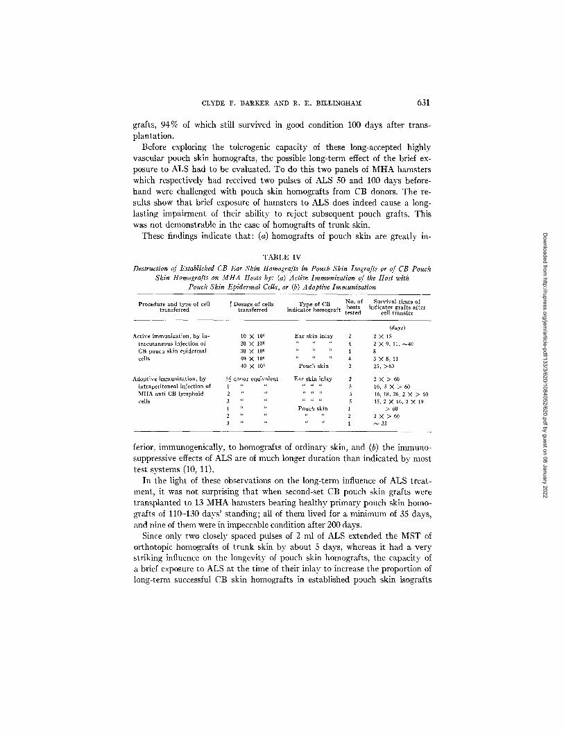
These findings indicate that: (a) homografts of pouch skin are greatly in-
TABLE IV Destruction of Established CB Ear Skin Homografts in Pouch Skin Isografts or of CB Pouch
Skin Homografts on MHA Hosts by: (a) Active Immunization of the Host with Pouch Skin Epidermal Cells, or (b) Adoptive Immunization
P r o c e d u r e a n d t y p e of cell [ D o s a g e of cel ls T y p e of CB N o . of S u r v i v a l t i m e s of ' ho s t s i n d i c a t o r g r a f t s a f t e r
t r a n s f e r r e d t r a n s f e r r e d i n d i c a t o r h o m o g r a f t t e s t e d cel l t r a n s f e r
( d a y s )
A c t i v e i m m u n i z a t i o n , b y in- 10 ) 106 E a r sk in i n l a y 2 2 X 15
t r a c u t a n e o u s i n j e c t i o n of 20 X 10~ " " 4 2 X 9, 11, ~ 4 0 CB p o u ch s k i n e p i d e r m a l 30 X 106 " " " 1 8
cel ls 40 X 106 " 4 3 X 8, 11 40 X 106 P o u c h s k i n 2 25, > 6 0
A d o p t i v e i m m u n i z a t i o n , b y ~ d o n o r e q u i v a l e n t E a r sk in i n l a y 2 2 X > 60
i n t r a p e r i t o n e a l i n j e c t i o n of 1 " " " " 5 16, 5 X 2> 60 M H A a n t i CB l y m p h o i d 2 " " " " 5 16, 18, 26, 2 X > 60
cel ls 3 . . . . . . . ' " 5 15, 2 X 16, 2 X 19 1 " " P o u c h sk in 1 > 60
2 " " " " 2 2 X > 60 3 " " " 1 ~ 33
ferior, immunogenically, to homografts of ordinary skin, and (b) the immuno- suppressive effects of ALS are of much longer duration than indicated by most test systems (10, 11).
In the light of these observations on the long-term influence of ALS treat- ment, it was not surprising that when second-set CB pouch skin grafts were transplanted to 13 MHA hamsters bearing healthy primary pouch skin homo- grafts of 110-130 days' standing; all of them lived for a minimum of 35 days, and nine of them were in impeccable condition after 200 days.
Since only two closely spaced pulses of 2 ml of ALS extended the MST of orthotopic homografts of trunk skin by about 5 days, whereas it had a very striking influence on the longevity of pouch skin homografts, the capacity of a brief exposure to ALS at the time of their inlay to increase the proportion of long-term successful CB skin homografts in established pouch skin isografts
Dow
nloaded from http://rupress.org/jem
/article-pdf/133/3/620/1084052/620.pdf by guest on 08 January 2022
632 HAMSTER CHEEK POUCH TISSUE AS GRAFT AND GRAFT SITE
was investigated. The finding that 22/~5 (88%) central inlay CB ear skin grafts survived for 50 days or more on MHA hosts treated in this way suggests that this procedure may have general application in facilitating homograft survival in so-called privileged sites. Evidently there is synergy between the protection afforded a homograft by an incompletely privileged site and that provided by threshold doses of ALS.
However, in this experiment only 15~5 (60%) of the CB inlay grafts were still alive 100 days after the grafting operation, as compared with 52 % of similar inlay grafts on non-ALS treated hosts. Evidently, the ALS treatment affects only those rejections that would normally occur relatively early on after implantation.
Investigation of Possible Tolerogenic Influence of Skin Homografts in a Privi- leged Site and of Long-Accepted Pouch Skin Homografts (Table I[I).--The first series of tests to seek evidence of a possible tolerogenic influence of long- accepted homografts in a privileged site entailed challenging a group of 15 MHA hamsters that had borne healthy, CB ear skin grafts inlaid in pouch skin isografts for 100-200 days with CB ear skin grafts transplanted to the opposite side of the trunk. The findings that of these test grafts four were alive on the 15th day and two of these survived for 16 and 18 days respectively, the remainder being rejected within 9-14 days, are at best indicative of only a very trivial weakening of the hosts' capacity to respond. In all cases it was noted that rejection of the hitherto healthy, long-accepted intrapouch skin homograft occurred almost simultaneously with that of the challenge graft, indicating the completeness of the efferent path of the immunological reflex so far as intrapouch grafts are concerned.
In another series of experiments a group of eight MHA hamsters bearing CB pouch skin homografts of 120 days' standing with inlaid CB ear skin grafts that had been in residence for 60 days were challenged with orthotopic honlo- grafts of CB ear skin. Again the results provide no grounds for belief that the long-accepted homografts had significantly weakened the host's capacity to react against the antigens concerned.
Evidence of the Immunogenicity of Pouch Skin Epidermal Cells (Table IV).-- A possible contributory factor to the apparent relatively weak and easily overcome immunogenicity of pouch skin homografts is inadequate expression of transplantation antigens on the part of their epidermal cells, i.e., they may behave like trophoblast cells (26). That this is unlikely is suggested by ob- servations that pouch skin epidermal cells and extracts prepared from them are capable of inciting direct hypersensitivity reactions (25) when injected into the skins of hamsters presensitized by means of skin homografts from the donor strain (13). 2
2 Zakarian, S. 1971. Personal communication.
Dow
nloaded from http://rupress.org/jem
/article-pdf/133/3/620/1084052/620.pdf by guest on 08 January 2022
CLYDE F. B A R K E R AND R. E . BILLINGt IAM 633
As a direct test of the immunogencity of pouch skin epidermal cells, doses of these cells ranging from 10 to 40 X 106 were injected in subdoses at multiple sites in the skins of MHA hamsters bearing long-accepted CB ear skin grafts in pouch skin isografts. As the findings set out in Table IV show, an inocnlum of 3040 X 106 epidermal cells consistently procured the rejection of the target CB grafts within 8 days and inocula of 10-15 X 106 cells were only slightly less effective. On the basis of only three tests it would appear that long-established pouch skin homografts are less vulnerable to the sensitivity evoked by pouch skin epidermal cells than ear skin homografts inlaid in established pouch skin isografts, a provisional conclusion consonant with previous findings that when hamsters bearing CB ear skin homografts in established pouch skin homografts were subsequently challenged with orthotopic CB ear skin grafts, destruction of the inlaid ear skin homograffs frequently preceded that of the surrounding homologous pouch skin.
Adoptive Immunization of Hamsters Bearing Intrapouch Skin Grafts of CB Ear Skin (Table IV) . - -The capacity of lymphoid cells from specifically sensi- tized animals to transfer transplantation immunity between members of an inbred strain is demonstrable in three ways: (a) in terms of their ability to enable their unsensitized host to reject in an accelerated manner a test skin homograft from a donor against which the sensitivity was directed; (b)to cause the destruction of healthy, well-established skin homografts on hosts previously rendered tolerant of the tissue antigens of the alien donor strain more rapidly than this could be caused by a similar number of lymphoid cells from a normal isologous donor; and (c) in the guinea pig, to procure the re- jection of a target skin homograft implanted into a skin flap having a blood supply but no lymphatic drainage (8, 22). The tests now to be described were performed to evaluate the usefulness of inlaid CB ear skin homografts in pouch skin isografts for studies on adoptive immunization.
A known number of donor equivalents of a coarse suspension of lymphoid cells in Hanks' solution, prepared from the accessible nodes and spleens of MHA hamsters sensitized against CB tissue antigens, were transferred intra- peritoneally to MHA hosts bearing inlaid CB ear skin homografts of long standing as indicators. In a few additional tests, established CB pouch skin homografts furnished the indicators. The results show that although the present system is capable of revealing the transfer of sensitivity, it is very insensitive in that a minimal dosage of three donor equivalents of immune cells was required consistently to procure rejection of the target graft within 15-19 days. Clearly adoptive immunization of these animals is a much less efficient means of destroying the target grafts than active immunization by means of orthotopic skin homografts or suspensions of homologous epidermal cells injected intracutaneously, either on account of different levels of sensi- tivity in the host produced by these two different procedures and/or a slight
Dow
nloaded from http://rupress.org/jem
/article-pdf/133/3/620/1084052/620.pdf by guest on 08 January 2022
634 HAMSTER CHEEK POUCH TISSUE AS GRAFT AND GRAFT SITE
impairment in the efferent pathway of the immunological reflex sufficient to impair the fulfillment of adoptive immunization.
DISCUSSION AND CONCLUSIONS
On the basis of a large-scale confirmation and extension of a previous finding that long-standing isografts of full-thickness cheek pouch skin on hamsters' chests afford a high degree of protection from rejection to inlaid homografts of ordinary skin, a variety of critical experiments have been carried out to analyze the mechanism of the privilege afforded. This system was selected for analysis rather than that presented by the intact cheek pouch because of accessibility for experimentation and inspection. Since skin homografts sustained within the milieu of cheek pouch tissue rapidly acquire a good blood supply, yet for periods exceeding 200 days consistently retain their susceptibility to a specific state of sensitivity actively or adoptively acquired by their hosts, some form of interruption of the afferent pathway of the immunological reflex seems to be involved. The existence of several recent reports that experimental disrup- tion of the continuity of lymphatic drainage systems from beds of orthotopic skin homografts in trunk skin greatly delays the development of the host response (8, 9) prompted us to evaluate the lymphatic drainage status of established pouch skin grafts.
Although not completely conclusive, the following findings lend strong support to the thesis that the distinctive physiological property of pouch skin responsible for its capacity to protect homografts is its lack of, or at least great paucity of, lymphatics:
(a) Despite the facility and consistency with which dye injected super- ficially into intact trunk skin or into established isografts in trunk skin pzsses via the lymphatic system of the skin to the draining nodes, in no instance was evidence obtained of the ability of dye injected either into the intact cheek pouch or into established isografts of this tissue on hamster's chests to gain access to the lymphatic system. Furthermore, although dye injected into well- established, inlaid isografts of ear skin in pouch skin isografts revealed a plexus of lymphatic vessels within the ear skin, it failed to enter lymph vessels of the host.
(b) This inability of pouch tissue to transmit colloidal dyes injected into its stroma to the lymphatic drainage system of the host was paralleled by its apparent failure to provide inoculated lymphoid cells with channels of access to the regional nodes. Whereas parental strain lymphocytes injected into either the intact skin of FI hybrid hamsters or into established isografts of hybrid trunk skin on such animals rapidly gained access to the regional nodes and, through graft-versus-host reactivity, caused them to hypertrophy by a factor of 3-4, the injection of similar cells into established pouch skin isografts on the chests of Fi hosts consistently failed to affect the size of the draining nodes.
Dow
nloaded from http://rupress.org/jem
/article-pdf/133/3/620/1084052/620.pdf by guest on 08 January 2022
CLYDE F. BARKER AND R. E. BILLINGHAM 635
(c) Deliberate surgical restoration of contact of a hitherto protected intra- pouch skin homograft with host trunk skin inevitably led to its rejection. The time taken for this to occur was related to the pattern of distribution of lym- phatics in the skin that provides the bridge.
There are, however, certain findings difficult to reconcile with our thesis. The most serious is the inability to account for the rejection in an abrupt manner of about 50 % of all centrally inlaid grafts over a 100 day observation period, which suggests that a random event of sudden onset enabled them to sensitize the host. Various kinds of dye injection experiments designed to show that grafts undergoing rejection had acquired a lymphatic drainage were un- successful, possibly because the procedure was unable to reveal very fine vessels.
However, the possibility cannot entirely be dismissed that the apparent sudden evocation of sensitivity in some of the hosts was not dependent upon the passage of antigenic material or of antigenically stimulated lymphocytes from the graft via lymphatics to the host's nodes.
Intrapouch skin homografts are perfused by host blood, which includes antigen-sensitive small lymphocytes, and there is also evidence indicating reutilization of the original vasculature of the grafts (27). Perhaps some ran- dom event within the graft, e.g. a small focus of inflammation, may lead to activation of host lymphocytes peripherally as they pass through its vascula- ture. Such cells may then be carried via the venous circulation to a component of the host's lymphohematopoietic system where they settle out and generate an effector cell population (28, 29). Indirect support for this interpretation comes from the observation that the regional nodes were not enlarged in hamsters whose intrapouch skin homografts were undergoing or had just undergone rejection.
The finding that treatment with two pulses of ALS given 2 days apart at the time of transplantation of orthotopic homografts of skin only extended their MST from about 11.2 to 17.0 days, but the same treatment enabled the ma- jority of much larger homografts of pouch skin to survive for at least 100 days, is consonant with previous evidence of feeble immunogenicity of this tissue. This must surely be attributed to the same intrinsic property that enables pouch skin to protect inlaid skin homografts. The finding that pouch skin epidermal ceils are effective incitors of transplantation immunity refutes the notion that feeble antigenicity of its epidermis is responsible for the weak immunogenicity of pouch skin.
One finding from the various experiments carried out with ALS deserves special comment. If the administration of ALS is followed 50 or even 100 days later by the grafting of pouch skin homografts, these grafts survive for much longer than those grafted on untreated animals, indicating that the immuno- suppressant effects of this agent are very long lasting when the test system
Dow
nloaded from http://rupress.org/jem
/article-pdf/133/3/620/1084052/620.pdf by guest on 08 January 2022
636 :HAMSTER C H E E K POUCH TISSUE AS GRAFT AND GRAFT SITE
comprises a feeble immunogen. This hints that transient treatment of patients with ALS may cause a long-lasting impairment of their postulated immunologic tumor surveillance mechanisms (30) since only weak immunogens are in- volved here.
Only very marginal evidence has been obtained that long-term exposure of hosts to skin homografts in vascularized by privileged sites can induce tol- erance in respect to the antigens concerned. Possibly the degree of histoin- compatibility involved in the present experiments was too strong to be effective in this regard or the graft dosage was insufficient.
One conclusion that can be drawn from the indefinite retention of vul- nerability of the intracheek pouch skin homografts to sensitivity of active or adoptive origin is that such grafts do not undergo any kind of antigenic adapta- tion, resulting in an apparent loss of antigencity of the kind that was noted (31) in intraocular homografts of thyroid tissue in guinea pigs. Indeed, their long retention of susceptibility to rejection and capacity to incite sensitization of their host suggests that the progressive loss of passenger leukocytes of donor origin (32) does not affect the immunological properties of skin homografts as it does those of kidney (33, 34).
Clinical Bearing
At present the results of hormone therapy for certain endocrine deficiency diseases, notably diabetes and parathyroprival disease, leave much to be de- sired. Surgical replacement seems to offer better prospects of therapy though the undesirable side effects of immunosuppression needed to sustain homo- grafts of these tissues would appear to negate their advantages. An alternative approach would be to implant functional endocrine tissue into a vascularized but alymphatic site, if one can be found or created surgically. As the present findings indicate, a short period of immunosuppression at the time of the grafting operation may then facilitate the acceptance of such grafts, especially if they are derived from a well-matched donor.
SUMMARY
Hamster cheek pouch skin, transplanted to the side of an isogenic host's chest wall, retains its immunologically privileged status as evidenced by the prolonged survival of inlaid homografts of ordinary skin.
Various findings sustain the premise that exemption from rejection by other- wise susceptiblc homografts in both intact pouch tissue and in established pouch skin isografts is due to an impediment in the afferent pathway of the immunologic reflex, i.c., to deficient lymphatic drainage. Although lymphatics were not apparent when dye was injected into pouch skin grafts or into grafts of ordinary skin sustained by them, lymph vessels were readily and con- sistently revealed by dye injected into intact trunk skin or established iso-
Dow
nloaded from http://rupress.org/jem
/article-pdf/133/3/620/1084052/620.pdf by guest on 08 January 2022
CLYDE F. BARKER AND R. E. BILLINGHAM 637
grafts of trunk skin. When suspensions of viable lymph node cells from spe- cifically sensitized parental strain donors were injected superficially into either the intact skin or established grafts of normal skin on F1 hybrid hamsters, a striking hypertrophy of the regional lymph nodes occurred, due to graft- versus-host reactivity. However, similar cell suspensions inoculated into intact pouch tissue or into pouch skin grafts on F1 hamsters incited no regional 1;yTnphadenopathy, indicating the lack of appropriate pathways to the nodes.
When skin homografts were inlaid eccentrically into pouch skin isografts, so that they were in contact with host skin at one edge, rejection occurred. Furthermore, rejection of long-established intrapouch skill homografts resulted if the hosts received: (a) small homografts of ordinary skin transplanted to conventional beds; (b) suspensions of donor strain pouch skin epidermal cells, injected intracutaneously; (c) lymph node cells from specifically sensitized donors of the same strain, i.e. adoptive immunization; or, (d) if a portion of the target homograft 's perimeter was surgically approximated to body skin.
Treatment of normal hamsters with two closely spaced pulses of ALS, al- though only marginally effective in prolonging the lives of homografts of trunk skin, enabled pouch skin homografts to survive for very long periods. The influence of this brief treatment with immunosuppressant was still demon- strable if challenge of hosts with the weakly immunogenic pouch skin homo- grafts was delayed for 100 days.
The authors are indebted to Dr. Willys K. Silvers for his invaluable criticism of the manu- script, to Dr. Charles F. Shaffer for providing the ALS, and to Mr. George H. Sawchuck for expert technical assistance.
BIBLIOGRAPHY
1. Handler, A. H., and D. Shepro. 1968. Cheek pouch technology: Uses and appli- cations. In The Golden Hamster. R. A. Hoffman, P. F. Robinson, and H. Magalhaes, editors. Iowa State University Press, Iowa City, Iowa. 195.
2. Billingham, R. E., and W. K. Silvers. 1962. Studies on cheek pouch skin homo- grafts in the Syrian hamster. In Ciba Foundation Symposium on Transplanta- tion. G. E. W. Wolstenholme and M. P. Cameron, editors. J. and A. Churchill Ltd., London, England. 90.
3. Billingham, R. E., and W. K. Silvers. 1964. Studies on homografts of foetal and infant skin and further observations on the anomalous properties of pouch skin grafts in hamsters. Proc. Roy. Soc. Set. B. Biol. Sci. 161:168.
4. Medawar, P. B. 1948. Immunity to homologous grafted skin. III . The fate of skin homografts transplanted to the brain, to subcutaneous tissue, and to the anterior chamber of the eye. Brit. J. Exp. Pathol. 9.9:58.
5. Scheinberg, L. C., F. L. Edelman, and W. A. Levy. 1964. Is the brain "an im- munologically privileged site"? Arch. Neurol. 11:248.
6. Lance, E. M. 1967. A functional and morphological study of intracranial thyroid allografts in the dog. Surg. Gynecol. Obstet. Int. Abstr. Surg. 128:529.
Dow
nloaded from http://rupress.org/jem
/article-pdf/133/3/620/1084052/620.pdf by guest on 08 January 2022
638 HAMSTER CHEEK POUCH TISSUE AS GRAFT AND GRAFT SITE
7. Billingham, R. E., and W. K. Silvers. 1971. The Immunobiology of Transplanta- tion. Prentice-Hall Inc., Englewood Cliffs, N. J.
8. Barker, C. F., and R. E. Billingham. 1968. The role of afferent lymphatics in the rejection of skin homografts. J. Exp. Med. 19.8:197.
9. Lambert, P. B., H. A. Frank, S. Bellman, and D. Farnsworth. 1965. The role of the lymph trunks in the response to allogeneic skin transplants. Trans- plantation. 3:62.
10. Medawar, P. B. 1968. Biological effects of heterologous antilymphocyte sera. I n Human Transplantation. F. T. Rapaport and J. Dausset, editors. Grune and Stratton Inc., New York. 501.
11. James, K. 1969. The preparation and properties of anti-lymphocyte sera. Progr. Surg. 7:140.
12. Billingham, R. E., and W. K. Silvers, editors. 1961. Transplantation of Tissues and Cells. The Wistar Institute Press, Philadelphia, Pa.
13. Ramseier, H., and R. E. Billingham. 1966. Studies on delayed cutaneous in- flammatory reactions elicited by inoculation of homologous cells into hamsters' skin. J. Exp. Med. 19.3:629.
14. Gray, J. G., A. P. Monaco, M. L. Wood, and P. S. Russell. 1966. Studies on heterologous anti-lymphocyte serum in mice, in vitro and in vivo. J. Immunol. 96:217.
15. Miotti, R. 1961. Die Lymphknoten und Lymphgefasse des Syrischen Gold- hamsters. Acla. Anat. 46:192.
16. Godart, S. 1968. Studies of the physiology of lymphatic vessels by microcircu- lation methods. Lymphology. 1:80.
17. Lindenmann, R., and P. StrAuli. 1968. Lymphatic vessels in the cheek pouch of the golden hamster. Transplantation. 6:557.
18. Shepro, D., N. Kula, and J. A. E. Halkett. 1963. The role of the cheek pouch in effecting transplantation immunity in the hamster. J. Exp. Med. 117:749.
19. Witte, S., D. M. Goldenberg, and K. T. Schricker. 1965. Mangel an Lymph- gafassen als Ursache der immunologischen Privilegierung der Hamsterbacken- tasche. Klin. Wochenschr. 4,3:1182.
20. Chadwick, D. E., and R. W. Blarney. 1968. Observations on the removal of cells and particulate matter from the hamster cheek pouch. Transplantation. 6:544.
21. Goldenberg, D. M., and W. Steinborn. 1970. Reduced lymphatic drainage from hamster cheek pouch. Proc. Soc. Exp. Biol. Meet. 135:724.
22. Wilson, D. B., and R. E. Billingham. 1967. Lymphocytes and transplantation immunity. Advan. Immunol. 7:189.
23. Billingham, R. E. 1968. The biology of graft-versus-host reactions. In The Harvey Lectures. Series 62. Academic Press Inc., New York. 21.
24. Levine, S. 1968. Local and regional forms of graft-versus-host disease in lymph nodes. T~ansplantation. 6:799.
25. Brent, L., J. B. Brown, and P. B. Medawar. 1962. Quantitative studies on tissue transplantation immunity. VI. Hypersensitivity reactions associated with the rejection of homografts. Proc. Roy. Soc. Set. B. Biol. Sci. 156:187.
26. Kirby, D. R. S. 1968. Transplantation and pregnancy. In Human Transplanta-
Dow
nloaded from http://rupress.org/jem
/article-pdf/133/3/620/1084052/620.pdf by guest on 08 January 2022
CLYDE F. B A R K E R AND R. E. B I L L I N G H A M 639
tion. F. T. Rapaport and J. Dausset, editors. Grune and Stratton Inc., New York. 565.
27. Haller, J. A., and R. E. Billingham. 1967. Studies of the origin of the vasculature in free skin grafts. Ann. Surg. 166:896.
28. Medawar, P. B. 1957. The homograft reaction. Proc. Roy. Soc. Set. B. Biol. Sci. 149:145.
29. Strober, S., and J. L. Gowans. 1965. The role of lymphocytes in the sensitization of rats to renal homografts. J. Exp. Med. 19.2:347.
30. Burnet, F. M. 1970. Immunological Surveillance. Pergamon Press Ltd., Oxford, England.
31. Woodruff, M. F. A., and H. G. Woodruff. 1950. The transplantation of norms] tissues: with special reference to auto- and homotransplants of thyroid and spleen in the anterior chamber of the eye, and subcutaneously, in guinea pigs. Phil. Trans. Roy. Soc. London Ser. B. Biol. Sci. 234:559.
32. Steinmuller, D. 1968. Allograft immunity produced with skin isografts from immunologically tolerant mice. Transplant. Proc. 1:593.
33. Guttman, R. D., R. R. Lindquist, S. A. Ockner, and J. P. Merrill. 1968. Mecha- nism of long-term survival of renal allografts after treatment with antilvmoho- cyte antibody. Transplant. Proc. 1:463.
34. Stuart, F. P., E. Bastien, A. Holter, F. W. Fitch, and W. L. Elkins. 1971. Role of passenger leucocytes in the rejection of renal allografts. Transplant. Proc. In press.
Dow
nloaded from http://rupress.org/jem
/article-pdf/133/3/620/1084052/620.pdf by guest on 08 January 2022