Embed Size (px)
Citation preview

Histochem Cell Biol (2008) 129:667–686
DOI 10.1007/s00418-008-0427-6REVIEW
The mammalian centrosome and its functional signiWcance
Heide Schatten
Accepted: 2 April 2008 / Published online: 24 April 2008© Springer-Verlag 2008
Abstract Primarily known for its role as major microtu-bule organizing center, the centrosome is increasinglybeing recognized for its functional signiWcance in key cellcycle regulating events. We are now at the beginning ofunderstanding the centrosome’s functional complexitiesand its major impact on directing complex interactions andsignal transduction cascades important for cell cycle regu-lation. The centrosome orchestrates entry into mitosis, ana-phase onset, cytokinesis, G1/S transition, and monitorsDNA damage. Recently, the centrosome has also been recog-nized as major docking station where regulatory com-plexes accumulate including kinases and phosphatases aswell as numerous other cell cycle regulators that utilize thecentrosome as platform to coordinate multiple cell cycle-speciWc functions. Vesicles that are translocated alongmicrotubules to and away from centrosomes may also carryenzymes or substrates that use centrosomes as main dock-ing station. The centrosome’s role in various diseases hasbeen recognized and a wealth of data has been accumulatedlinking dysfunctional centrosomes to cancer, Alstrom syn-drome, various neurological disorders, and others. Centro-some abnormalities and dysfunctions have been associatedwith several types of infertility. The present review high-lights the centrosome’s signiWcant roles in cell cycle eventsin somatic and reproductive cells and discusses centrosomeabnormalities and implications in disease.
Keywords Centrosomes · Microtubules · Cell cycle · Mitosis · Centrosome proteins · Centrosome functions · MTOC
Introduction
Over 120 years have passed since the discovery of centro-somes (Flemming 1875; Van Beneden 1876) and yet, weare only at the beginning of understanding the multiple andcentral roles of this intriguing cellular organelle. Boveri(1901) who coined the name centrosomes had produced aremarkable wealth of new information characterizing andanalyzing centrosomes during fertilization and cell division(Boveri 1901) by using iron hematoxylin as a major cyto-logical staining technique, and he brilliantly recognized thevery important role of centrosomes in cancer (1914). Flem-ming had called the discovery of centrosomes as importantas the discovery of the nucleus (Flemming 1891) yet,research on centrosomes stagnated for decades for a num-ber of reasons including war events in Europe that slowedall research and because studies using the electron micro-scope did not reveal a structure that could be easily deWned.Centrosome structure was described as amorphous osmio-philic material surrounding a pair of well-structured centri-oles, which stimulated and generated new research oncentrioles while research on centrosomes became lessappreciated. With the development of immunologicalprobes and molecular techniques coupled with immuno-Xuorescence microscopy centrosomes reclaimed their statusand Boveri’s earlier work was vigorously pursuit by repro-ductive biologists, and by cellular and cancer biologistswho were able to conWrm and build on Boveri’s remarkablydetailed and accurate data and visionary research. Ourunderstanding of centrosomes has advanced enormouslyand very rapidly in recent years and its central role in cellu-lar biology has clearly been recognized. It can be predictedthat a number of diseases other than cancer are linkeddirectly or indirectly to centrosome dysfunctions. Centro-some isolations, forward and reverse genetics, RNA-mediated
H. Schatten (&)Department of Veterinary Pathobiology, University of Missouri, 1600 E Rollins Street, Columbia, MO 65211, USAe-mail: [email protected]
123

668 Histochem Cell Biol (2008) 129:667–686
interference, mass-spectrometry-based proteomics, live cellimaging particularly with GFP-tagged proteins, and lasermicrosurgery are some of the newer more recently employedtechniques that have been applied to centrosome researchand rapidly moved the Weld into new frontiers. The presentpaper will review the state of our knowledge of centrosomesand analyze its impact on cellular functions.
DeWnition of the centrosome and its key functions
The centrosome is a subcellular non-membrane bound semi-conservative organelle, approximately 1 �m in size, thatserves as the cell’s primary microtubule organizing center(MTOC) and plays a major role in numerous cellular func-tions. Through its microtubule organizing functions the cen-trosome facilitates many cellular activities including cellmotility, polarity, maintenance of cell shape, cell division,transport of vesicles, and targeting of numerous signalingmolecules. A typical mammalian centrosome consists of aproteinaceous scaVold containing a large number of centro-some proteins including �-tubulin and the gamma-tubulincomplex (�-TuRC) that typically surround a pair of perpen-dicularly oriented cylindrical centrioles, therefore referred toas pericentriolar material (PCM). The PCM scaVold under-goes shape changes throughout the cell cycle to producecell-cycle-speciWc microtubule organizations for speciWccellular functions. Lacking a deWned membrane boundary,the centrosome’s three-dimensional architecture is main-tained through speciWc protein–protein interactions. Micro-tubules are anchored at the centrosome with their minusends (Bornens 2002) and microtubule growth is regulated bydistal plus-end addition of tubulin subunits (McIntosh andEuteneuer 1984). In interphase, the centrosome is juxtaposi-tioned and closely associated with the nucleus. At the G2/Mtransition it undergoes signiWcant cell cycle-speciWc reorga-nizations and matures into the division-competent center ofthe mitotic poles. Because the centrosome nucleates micro-tubules and controls microtubule numbers and lengths itdirects most microtubule-related functions including trans-port of macromolecular complexes, positioning of cellorganelles, cell motility, cell shape, polarity, and segrega-tion of chromosomes during cell division. The mitotic cen-trosomes are critically important to mediate the strictlybalanced bipolar separation of chromosomes. In addition tothe regular function as MTOC the centrosome orchestratesvarious major important cell cycle events including entryinto mitosis, anaphase onset, cytokinesis, G1/S transition,and monitoring of DNA damage (Kramer et al. 2004).
While a typical centrosome consists of a pair of centriolessurrounded by PCM, centrioles are not always presentwithin the centrosome structure as will be detailed later inthis paper. In general, centrioles are important for centro-
some integrity and to aid in the recruitment of centrosomalproteins into the PCM. The PCM is a Wbrous scaVoldinglattice that consists of a large amount of coiled-coil centro-some proteins and anchors signaling molecules and compo-nents of the �-TuRC (Fig. 1). Some centrosome proteins arepermanently associated with the centrosome core structuresuch as �-tubulin, the �-TuRC, and centrin (addressed belowin more detail) while several other centrosomal proteins aretemporarily associated with the centrosome core structureand include centrosome proteins such as the Nuclear MitoticApparatus protein (NuMA). Such centrosomal proteinsserve cell-cycle-speciWc functions and are needed for spe-ciWc cell cycle activities. The cell cycle-speciWc centrosomeproteins are diVerent from those proteins or enzymes(kinases, phosphatases and others) that may be colocalizedwith centrosomes and may utilize centrosomes as a dockingstation but are not centrosome proteins that are directlyinvolved in performing centrosome-speciWc functions. Sofar, about 500 diVerent proteins have been associated withthe centrosome by mass spectroscopy (Andersen et al. 2003)although it is not clear whether all of those proteins are cen-trosome proteins or proteins or enzymes that use centro-somes as docking station. Regulators of various cell cycleevents have been co-localized with centrosomes and includethe mitogen activated protein kinase (MAPK) (Sun et al.2002) which may use centrosomes as a docking station toperform and coordinate multiple cell cycle-speciWc func-tions. Vesicles that are translocated along microtubules toand away from centrosomes may also carry enzymes or sub-strates that use centrosomes as docking platform (Fig. 2a, b).
Functional homologues of centrosomes are spindle polebodies of yeast (Knop et al. 1999), the nucleus-associatedbody of the cellular slime mold (Daunderer et al. 1999),and nucleating sites in plants (Chan et al. 2003). In addi-tion, multiple plasma membrane-associated sites in Dro-sophila epidermal cells (Mogensen 1999) and in Xenopusoocytes (PfeiVer and Gard 1999) have also been identiWedto serve as MTOCs. New research has established thatvirus, bacteria, and parasites exploit the host cell’s centro-somal capabilities and recruit centrosomal material for theirown survival within host cells (reviewed in Scaplehorn andWay 2004; Coppens et al. 2006).
Taken together, the centrosome is a truly central andmain cellular station that directs, coordinates, and regulatesmost cellular functions either directly or indirectly throughits microtubule organizing capabilities.
Centrosome proteins and their speciWc functions
Numerous centrosome proteins have been described andtheir functions have been determined but the list of newlydiscovered centrosome proteins continues to grow and
123

Histochem Cell Biol (2008) 129:667–686 669
includes centrosome proteins that have multifunctionalcapabilities. Furthermore, the list of diseases in which cen-trosome protein dysfunctions play a role also continues togrow as research on centrosomes has progressed rapidlyand new research teams have formed around the growingcentrosome Weld. The initial discovery of centrosomal pro-teins in an autoimmune antibody has provided a most pow-erful tool to stain centrosomes with immunological probes(Calarco-Gillam et al. 1983) and since then, a large numberof centrosomal proteins have been identiWed based on puri-Wcation of autoimmune sera and subsequent production ofspeciWc centrosome antibodies.
The reported number of centrosome proteins variesdepending on the methods applied. Because the centro-some is a non-membrane-bound organelle it does not havedeWned borders and isolation methods can be Xawed byremoving critical centrosome proteins or including con-taminants that are not centrosome proteins. In addition,because the centrosome is a Xexible structure that under-goes cell cycle-speciWc changes the number of centrosomeproteins varies in diVerent cell cycle stages. Functionalscreens have been used but because of the nature of centro-somes may not detect all centrosome proteins. Mass spec-troscopy has identiWed as many as 500 centrosomalproteins (Andersen et al. 2003) while a more conservative
number of 100 have been identiWed with diVerent methodsat various cell cycle stages with about 60 being present inthe interphase centrosome (reviewed by Wilkinson et al.2004) and may reXect the average centrosome proteinquantities that are characteristic for interphase centrosomesin typical mammalian somatic cells. Various deWnitionshave been used to describe the centrosome. It is generallyagreed that the centrosome core structure consists of per-manently associated centrosome proteins that remain aftertreatment of the centrosome complex with microtubuledepolymerizing agents such as cold, nocodazole, colchi-cine derivatives, and others. The well-studied �-tubulin is aprotein of the centrosome core structure. Centrosome coreproteins are permanently associated with the centrosome
Fig. 1 A typical mammalian centrosome is composed of two centri-oles surrounded by a meshwork of a proteins embedded in matrixcalled the pericentriolar material (PCM). Gamma-tubulin and the gam-ma-tubulin ring complex that nucleate microtubules along with associ-ated proteins are embedded in the PCM. Highlighted in this diagramare two centrosomal complexes, the microtubule nucleating complexand the microtubule anchoring complex
Nucleating complex
Anchoring complex
Fig. 2 a The centrosome serves as central station to mediate substrateand enzyme activities along microtubules towards the minus ends ofmicrotubules driven by dynein (white circles) and toward the plus endsdriven by kinesin (black circles). b Through its microtubule-organizingcapabilities the centrosome mediates distribution of cell organellessuch as mitochondria, Golgi, and vesicles of various sizes, many con-taining regulatory enzymes for cell cycle regulation
plasma membrane
nuclear membrane
centrosome
mitochondria
mitochondria
microtubule
A
B
centrosome
microtubule
123

670 Histochem Cell Biol (2008) 129:667–686
structure throughout cell cycle-dependent structuralchanges in most systems. Such cell cycle-speciWc changesare particularly apparent in reproductive cells after fer-tilization in, which rapid changes of centrosomes andmicrotubule formations are required for rapid cell cyclefunctions. Other centrosome proteins associate with thecore structure during diVerent cell cycle stages to performcell cycle-speciWc functions (discussed below). Such pro-teins include NuMA (reviewed in Sun and Schatten 2006)that performs essential functions in mitosis. The analysis ofdetermining the number of centrosome proteins is furthercomplicated by the great number of proteins that use cen-trosomes as a central docking station or platform whereregulatory complexes accumulate and communicate by sig-naling through the microtubule network. Such complexesmay co-localize with centrosomes but should not beincluded in the deWnition of the centrosome. Known andnewly identiWed centrosome proteins that localize tohuman centrosomes are reviewed in Wilkinson et al.(2004). Centrosome proteins that are known to be presentin puriWed centrosomes and identiWed by mass spectro-metric analysis include the structural proteins alpha tubu-lin, beta tubulin, gamma-tubulin, gamma-tubulin complexcomponents 1–6, centrin 2 and 3, AKAP450, pericentrin/kendrin, ninein, pericentriolar material 1 (PCM1), ch-TOGprotein, C-Nap1, Cep250, Cep2, centriole-associated pro-tein CEP110, Cep1, centriolin, centrosomal P4.1 associatedprotein (CPAP), CLIP-associating proteins CLASP1 andCLASP 2, ODF2, cenexin, Lis1, Nudel, EB1, centractin,myomegalin; the regulatory molecules cell division protein2 (Cdc2), Cdk1, cAMP-dependent protein kinase type II—alpha regulatory chain, cAMP-dependent protein kinase—alpha catalytic subunit, serine/threonine protein kinasePlk1, serine/threonine-protein kinase Nek2, serine/threo-nine-protein kinase Sak, Casein kinase I, delta and epsilonisoforms, protein phosphatase 2A, protein phosphatase 1alpha isoform, 14-3-3 proteins, epsilon and gamma iso-forms; the motor and related proteins dynein heavy chain,dynein intermediate chain, dynein light chain, dynactin 1,p150 Glued, dynactin 2, p50, dynactin 3; and the heatshock proteins heat shock protein Hsp90, TCP subunits,and heat shock protein Hsp73.
Centrosomes can be depleted of many PCM componentsby treatment with high salt which also extracts �-tubulin(Moritz et al. 1998) leaving behind centrosome scaVolds. Acentrosome matrix has been described in several studiesand has been visualized in the classic sea urchin inverte-brate model system with low voltage Weld emission scan-ning electron microscopy (LVFESEM) on cold treated andisolated centrosomes (Thompson-CoVe et al. 1996;reviewed in Schatten et al. 2000a; Schatten and Chakrabarti2004), which revealed a particle structure of 1–2 �m andrepeating subunits.
Centrosome proteins that have been studied more fullyin various cell systems include �-tubulin, pericentrin, cen-trin, and NuMA.
Gamma-tubulin is an essential centrosome protein whichhad been discovered through fungal genetics (Oakley andOakley 1989) and since then described in all eukaryotic cellgroups. Gamma tubulin is not only found in centrosomesbut it also can serve as nucleating sites in animal cells thatdo not display typical centrosomes (Tulu et al. 2003).Gamma-tubulin is localized at the oocyte cortex in amphibianswhich has been shown by PfeiVer and Gard (1999). Inplants that do not have typical centrosomes, �-tubulin isthought to nucleate unfocused microtubules within the celland it is found along the length of microtubules (Canadayet al. 2000). Gamma-tubulin is a highly conserved proteinin eukaryotes. It is vital for centrosome functions and elim-ination of �-tubulin is lethal to cells and to the organism(Oakley et al. 1990; Joshi et al. 1992) while elimination ofcomponents that bind to �-tubulin has little eVect on cellviability. The function of �-tubulin was clearly determinedby puriWcation of the �-tubulin ring complex (�-TuRC) thatrevealed a ring shape and a substructure that were able tonucleate microtubule polymerization in vitro (Zheng et al.1995). The ca. 2.2-MDa �-TuRC consists of 12 or 14�-tubulin molecules and is the nucleating complex formicrotubules in all cells studied so far (Hannak et al. 2002).Gamma tubulins may laterally interact with each other.Several accessory proteins are associated with this complex(Tassin and Bornens 1999; Gunawardane et al. 2000; Mur-phy et al. 2001) providing new challenges for further inves-tigations. The �-TuRC has been well characterized byelectron microscopy including immunoelectron tomogra-phy showing an open ring structure of ca. 25 nm diameter(Moritz et al. 1995; Zheng et al. 1995; Oegema et al. 1999;Moritz et al. 2000). The �-TuRC is capable of functionallycapping the minus ends of microtubules facilitating growthof microtubule protoWlaments. Six components of the�-TuRC in addition to �-tubulin have been identiWed so far(Gunawardane et al. 2003; Murphy et al. 2001; Tassin et al.1998). Gamma-tubulin complexes are required for microtu-bule assembly which has been shown by using salt-extracted centrosomes that lack �-TuRCs and are not ableto recruit microtubules while addition of �-tubulin com-plexes from cytoplasmic extracts restored nucleation com-petency although various other components are needed toanchor the �-TuRC to the centrosome structure includingthe large coiled-coil A-kinase anchoring proteins (Tassinet al. 1998; Murphy et al. 1998; Dictenberg et al. 1998;Doxsey et al. 1994; Flory and Davis 2003; Gillingham andMunro 2000; Kawaguchi and Zheng 2004; Keryer et al.2003; Steadman et al. 2002; Takahashi et al. 2002) andCep135 (Ohta et al. 2002). The microtubule-minus-end-binding proteins including �-TuRC are concentrated at the
123

Histochem Cell Biol (2008) 129:667–686 671
proximal ends of centrioles while tubulin polyglutamyla-tion of the centriole walls modulates interaction betweentubulin and microtubule associated proteins. The centriole-interacting proteins can bind and aid in the recruitment ofmatrix proteins.
A cell cycle-dependent regulation of �-TuRC is thoughtto account for diVerences between interphase and mitoticmicrotubule structure formations and activities. In inter-phase �-TuRC nucleates fewer but longer microtubuleswhile in mitosis, more �-TuRC is associated with the cen-trosome, which is part of the centrosome maturation pro-cess that takes place from interphase to mitosis (discussedbelow). Mitotic microtubules are shorter, larger in number,and highly dynamic. They are regulated by a number of cellcycle-speciWc proteins that participate in centrosome regu-lation such as the small GTPase Ran, Aurora A kinase,polo-like kinases and others.
Pericentrin is a centrosome protein that plays a role incentrosome and spindle organization (Doxsey et al. 1994;Dictenberg et al. 1998; Young et al. 2000). It forms a ca. 3MDa-complex with �-tubulin and depends on dynein forassembly onto centrosomes (Young et al. 2000). Pericentrinplays a role in recruiting �-tubulin to centrosomes in verte-brate cells (Dictenberg et al. 1998) along with several otherproteins; it is part of the pericentrin/AKAP450 centrosomaltargeting (PACT) domain (Gillingham and Munro 2000).Mutation of the pericentrin gene results in loss of recruit-ment of several other centrosomal proteins aside from�-tubulin, which argues for its essential role in the centro-some complex; it may provide a link between centriolesand centrosomes (Martinez-Campos et al. 2004).
Centrins are small proteins and members of a highlyconserved subgroup of the EF-hand superfamily of Ca2+-binding proteins that is associated with centrioles (Bornens2002; Jurczyk et al. 2004). Centrin is also an intrinsic com-ponent of centrosomes that has an essential role in theduplication of centrosomes (Levy et al. 1996; Salisbury1995; Lutz et al. 2001; reviewed in Manandhar et al. 2005)which has been clearly determined in mutation experiments(Salisbury et al. 2002).
NuMA (Nuclear Mitotic Apparatus protein) is an essen-tial cell cycle-dependent centrosome-associated protein thatplays a signiWcant role in the organization of the mitoticapparatus during mitosis. It is a multifunctional protein(reviewed in Sun and Schatten 2006, 2007) that performsnuclear functions as nuclear matrix protein during inter-phase and translocates from the nucleus to spindle poles atthe onset of mitosis to form a crescent-shaped complexaround centrosomes tethering microtubules into the accu-rate bipolar organization (addressed below in more detail).
Many of the other centrosomal proteins also play signiW-cant roles in centrosome functions but are not discussed indetail in this section. Several centrosomal proteins have
been implied in microtubule anchoring to the centrosomeincluding ninein that has strongly been implied to serve asmicrotubule minus-end anchoring protein (Mogensen et al.2000) and dynactin that has a major role in microtubuleanchorage at centrosomes as well as at non-centrosomalanchorage sites. It is preferentially localized to the mothercentriole and plays a role in microtubule organization(Quintyne et al. 1999; Schroer 2001; Quintyne and Schroer2002).
Centrosome cycle within the cell cycle
Live cell imaging and immunoXuorescence microscopy haveclearly shown the remarkable shape changes of the centro-some which is best visualized in reproductive cells after fer-tilization requiring rapid centrosome and microtubuledynamics to position the pronuclei and prepare the zygotecell for mitosis. Figure 3 shows stages of the centrosomecycle in a sea urchin egg which is the model system used byBoveri (1901) for many of his centrosome studies. Centro-somal material disperses around the zygote nucleus (Fig. 3a)to become bipolar in late prophase (Fig. 4c). It becomes max-imally condensed during metaphase (Fig. 3e) and expandswithin the anaphase spindle poles (Fig. 3g) before it becomescompacted again when telophase nuclei form. Figure 3b, d, f,and h are images of microtubule labeling for either the samecell or cells of equivalent cell cycle stages.
In interphase, a single centrosome containing a pair ofcentrioles is juxta-positioned and closely connected to thenucleus as shown in Fig. 4 in an LNCaP prostate cancer cell(Fig. 4b), mouse 3T3 cells containing GFP-centrin label todetect centrosomes and labeled with �-tubulin to detectmicrotubules (Fig. 4a, c, d), and a pig Wbroblast cell labeledwith �-tubulin to detect the centrosome and MitotrackerRosamine to detect mitochondria (Fig. 4e). Centrosomesduplicate shortly before the G2 stage of the cell cycle in aprecisely orchestrated program to ensure precise duplica-tion into two and not more centrosomes. This processbegins with disorientation of the pair of centrioles, centrioleduplication, centrosome disjunction, and results in sistercentrosome separation as reviewed in Mack et al. (2000)and Ou et al. (2004). While a wealth of data has been accu-mulated on the duplication and separation of centrioles dataon the molecular events underlying centrosome duplication,disjunction and separation are only slowly accumulating.Centrosome separation refers to the spatial separation ofcentrosome material around the nucleus which is driven byplus- and minus-end directed microtubule motor proteins.The Nek2 kinase is involved in centrosome disjunction(Meraldi and Nigg 2001; reviewed by Fry 2002). Phosphor-ylation of the centrosomal protein centrin plays a role incentrosome disjunction at the G2/prophase transition.
123
672 Histochem Cell Biol (2008) 129:667–686
Polo like kinases are required for multiple stages ofmitotic progression and have clearly been implicated incentrosome separation. SpeciWc kinases have been linked tocentrosome and microtubule dynamics and Plk1 plays acritical role in centrosome and microtubule organization(Sun et al. 2001a, b, c, d, 2002; Tong et al. 2002, 2003; Fanet al. 2003). Plk1 and Plk3 both have been implicated inmicrotubule and centrosome functions in interphase and inmitosis (Fenton and Glover 1993; Donaldson et al. 2001;Wang et al. 2002). Loss of Plk3 function has also beenassociated with loss of cell shape which indicates loss ofmicrotubule functions underneath the plasma membrane(Wang et al. 2002).
While the role of centrosomes in microtubule nucleationand chromosome separation has been studied in great detail,centrosomes have only more recently been implicated incytokinesis. Khodjakov and Rieder (2001) showed that 30–50% of cells with laser-ablated centrosomes failed to com-plete cytokinesis perhaps because spindles formed withoutorientation and did not have attachment to the cell cortexresulting in incomplete chromosome separation. The molec-ular mechanisms by which centrosomes exert an eVect oncytokinesis are not clear but their role as docking station forcytokinesis-required regulatory proteins is implied and mayinclude Plk and microtubule motor proteins.
The role of centrosomes as docking station where regu-latory complexes accumulate for cell cycle activities hasonly been studied in recent years. The centrosome serves as
a platform for a wide variety of regulatory molecules thatplay a role in cell cycle-speciWc functions but are not them-selves centrosome proteins. These complexes regulateperipheral events through microtubule-dependent transportwhich indicates a role of the centrosome as command cen-ter that integrates signals using microtubules as signalingroutes. As such, centrosomes play critical roles in cell cycleregulation in both mitosis and interphase by providing acentral signaling station and controls to trigger the nextphases of the cell cycle by integrating, regulating, andamplifying signaling pathways.
Phosphorylation and posttranslational modiWcations areother important aspects in centrosome regulation requiredfor cell cycle-speciWc changes which has been reviewed inWilkinson et al. (2004).
Centrosome duplication in a regular cell cyle is typicallywell synchronized with the DNA cycle. Studies on duplica-tion of centrosomes have been performed in recent years toreveal that there is a block to reduplication which assuresthat centrosomes are duplicated accurately only oncewithin a normal cell cycle. Elegant studies to determine theblock to reduplication have been performed by Wong andStearns (2003) who fused G1 phase cells containing onecentrosome with G2 phase cells containing two centro-somes to determine if an already duplicated centrosomewould re-duplicate under these conditions. Interestingly,the G1 centrosome duplicated while the G2 centrosome didnot which led to the conclusion that there is an intrinsic
Fig. 3 The structural cell cycle-dependent changes of centrosomes areshown here in sea urchin eggs during the Wrst cell cycle after fertiliza-tion. This system had been used by Theodor Boveri for the majority ofhis classic studies on centrosomes. Centrosome material dispersesaround the zygote nucleus (a arrows) and separates to the poles duringprophase (c arrows). Centrosomes become densely compacted inmetaphase (e arrows) and disperse during anaphase (g arrows). The
correlated images for microtubules either from the same cell or fromcorresponding cell cycle stages are shown in b, d, f, and h. Immuno-Xuorescence microscopy of centrosomes, microtubules, and DNA(blue). Centrosomes are displayed in green; microtubules are displayedin green (b, d) or red (f, h). Reprinted with permission from Schattenet al. 2000a
123

Histochem Cell Biol (2008) 129:667–686 673
block to reduplication within the centrosome. Initiation ofcentrosome duplication is under cytoplasmic control anddriven by cyclin-dependent kinase 2 (Cdk2) complexedwith cyclin E or cyclin A that rises during the late G1 stage(reviewed in Sluder 2004). These results were compiled byseveral laboratories in the late 1990s using Xenopus eggs(HinchcliVe et al. 1999; Lacey et al. 1999) and mammaliansomatic cells (Mantel et al. 1999; Matsumoto et al. 1999).It was later shown that initiation of centrosome duplicationalso requires calcium/calmodulin-dependent kinase II(CaMKII) (Matsumoto and Maller 2002). CaMKII phos-phorylates centrosome proteins in vitro (Pietromonacoet al. 1995) and is localized to spindle poles (Ohta et al.
1990). Ubiquitin-mediated proteolysis of centrosomal pro-teins may be involved in the block to reduplication as pro-posed by several investigators (Tugendreich et al. 1995;Freed et al. 1999; Gstaiger et al. 1999) who showed locali-zation of a variety of components of the SFC proteolysispathway as well as the 26-S proteasome to centrosomes inhuman cells.
Centrosome duplication and DNA replication bothrequire hyperphosphorylation of the retinoblastoma (RB)protein and activation of Cdk2. Although the centrosomecycle and DNA cycle are normally coupled through cellcycle-dependent checkpoints it has been possible to disso-ciate the centrosome cycle from other cell cycle events.
Fig. 4 In interphase, a single centrosome is juxta-positioned to the nucleus. a, c, and d show small GFP-centrin-labeled cen-trosomes in mouse 3T3 cells. Microtubules are detected with �-tubulin and shown in red; b is of an LNCaP prostate cancer cell labeled with human autoimmune antibody SPJ displaying multi-ple centrosomal foci perhaps indicating centrosome abnor-malities; e shows a porcine Wbro-blast cell labeled with �-tubulin to detect the centrosome and Mi-totracker Rosamine to detect mitochondria. Microtubules are shown in green; b reprinted with permission from Schatten et al. 2000b
123

674 Histochem Cell Biol (2008) 129:667–686
Application of cycloheximide to Xenopus oocytes hasresulted in centrosome duplication while other cell cycleevents were inhibited (Gard et al. 1990). Hydroxyurea oraphidicolin also resulted in continuation of the centrosomecycle in CHO cells producing supernumerary centrosomeswhile DNA replication was inhibited (Balczon et al. 1995).
Normally, the DNA cycle and the centrosome cycle areintimately coupled and synchronized in a regular cell cycle.When the nucleus or DNA is damaged, centrosomesbecome inactivated in a well-orchestrated process. The�-TuRC dissociates from centrosomes, the spindle fails toassemble resulting in failure of chromosome segregation.Figure 5 is a schematic representation of a typical centro-some cycle within a typical cell cycle. Please see Wgure leg-end for description.
Centrosome maturation into mitotic centrosomes
One of the most important functions of the centrosome is itsrole in establishing the bipolar mitotic spindles that orches-
trate the precisely balanced segregation of chromosomes.To accomplish this important function a major reorganiza-tion of centrosomal material occurs at the G2/M transitionin vertebrate cells which is referred to as centrosome matu-ration. During this time some centrosomal proteins becomediminished while others such as the �-TuRC becomeenriched (Khodjakov and Rieder 1999) and �-tubulinincreases three to Wve-fold (Khodjakov and Rieder 1999).
Cyclin-dependent kinases play a signiWcant role in cen-trosome cycle progression. The G2/M transition requiresCdk1/cyclin B as well as Cdk1/cyclin A, which has beenreviewed in detail by Fry and Hames (2004). Cdk1 is localizedto centrosomes at the onset of mitosis (Bailly et al. 1989;Pockwinse et al. 1997) and it has been shown that Cdk1/cyclin B activation is detected in centrosomes duringprophase (Jackman et al. 2003).
The centrosome cycle is tied to the centriole cycle andboth are closely coupled with the DNA cycle. At the transi-tion from the G1 to S phase centriole duplication begins andcontinues through G2 when the replicating pro-centrioleelongates. Two duplicated juxta-positioned centrioles are
Fig. 5 A typical mammalian centrosome cycle within the cell cycle.The single interphase centrosome (a) is closely associated with the nu-cleus and nucleates an array of interphase microtubules. Centrosomeduplication occurs during the S phase in synchrony with DNA dup-liction (b). Centrosome separation of the duplicated centrosome to-ward opposite poles takes place in the early prophase stage (c). Thebipolar mitotic apparatus becomes established when centrosomes havereached the opposite poles and the nuclear envelope has broken down(d). During this stage interphase centrosomes mature into mitotic cen-trosomes acquiring mitosis-associated centrosomal proteins including
NuMA that moves out of the nucleus to become a mitotic centrosome-associated protein. The metaphase centrosome (e) becomes highlycompacted to organize the metaphase spindle with microtubulesattached to the kinetochores. Anaphase is the stage when centrosomalmaterial becomes decompacted again (f) before reorganizing intointerphase centrosomes that associate with the nuclei of the separatingdaughter cells (g). Centrosomes shaded yellow with centrioles andPCM displayed in black; nuclei shaded orange; microtubules dis-played as black rods; chromosomes displayed in black. ModiWed fromSun and Schatten 2007
A
B
C
GD
EF
123

Histochem Cell Biol (2008) 129:667–686 675
clearly apparent at the G2 stage. The two centrosome com-plexes are held to each other throughout interphase andundergo disjunction prior to entry into mitosis (Mayor et al.1999). Centrosome maturation takes place by acquiringmitotic centrosome proteins including Plk1 (Golsteyn et al.1995), NuMA (Merdes et al. 1996) and others while inter-phase centrosome proteins such as C-Nap1 (Fry et al. 1998)or Nlp (Casenghi et al. 2003) are removed. Increased recruit-ment of �-TuRC to the centrosome assures increased nucle-ation of microtubules for spindle formation. While centrioleseparation has been studied in great detail in recent years, ourknowledge about centrosome separation is still incompleteand we do not yet fully understand how centrosomes are sep-arated into two. Separation is complete when cells re-enterG1 with a single centrosome surrounding a pair of centrioles.
One of the well-studied essential mitotic centrosome-associated proteins is NuMA. After nuclear envelopebreakdown (NEB), NuMA becomes a most signiWcant cen-trosome-associated protein (reviewed in Sun and Schatten2006, 2007). NuMA does not associate with interphase cen-trosomes. It serves as nuclear matrix protein in the inter-phase nucleus and becomes dispersed into the cytoplasmduring NEB, a process in which cyclin B plays a criticalrole. Cdk1/cyclin B dependent phosphorylation is impor-tant for translocation of NuMA from the nucleus into thecytoplasm (Saredi et al. 1997; and references therewithin)where it associates with microtubules using a dynein–dynactin-mediated mechanism to be translocated alongmicrotubules to the centrosome area. Next, to become afunctional mitotic protein NuMA needs to be translocatedfrom the cytoplasm to the mitotic centrosomes. In thishighly regulated process microtubules and dynein–dynactinplay crucial roles in transporting NuMA to the mitotic poleswhere it forms an insoluble crescent around centrosomesthat tethers microtubules precisely into the bipolar con-Wguration that forms the mitotic apparatus (Merdes andCleveland 1998). NuMA is distributed to the separatingcentrosomes during early mitosis. It ensures minus-endbinding and stabilization of microtubules on the centro-some area that faces chromosomes. NuMA facilitates cross-linking of spindle microtubules, aiding in the organizationand stabilization of spindle poles from early mitosis to ana-phase. To exit its mitotic functions and to relocate to thenucleus NuMA needs to be dissociated from the mitoticspindle poles through a process that requires cdc1/cyclin B(Gehmlich et al. 2004). Destruction of cyclin B promotesexit from mitosis, which starts at the mitotic poles and islinked to disassembling the mitotic centrosome complex.Failure of NuMA to relocate to the nucleus will result incytoplasmic NuMA spots that organize abnormal microtubuleformations (Gehmlich et al. 2004).
As nuclear matrix protein NuMA is involved in DNAreplication and transcription. It displays distinct staining
patterns in the nucleus (Merdes and Cleveland 1998; Zeng2000; Gobert et al. 2001) and stains heavily in mitosiswhen using anti-NuMA antibodies (Saredi et al. 1997; Mer-des et al. 2000; Gobert et al. 2001; Zhong et al. 2005; Liuet al. 2006). Inaccurate NuMA regulation and atypicalNuMA distribution will result in abnormal mitosis as hasbeen reported for cancer cells (Saunders et al. 2000).
Some of the important mitotic cell cycle regulators areconcentrated at the centrosome and include Polo andAurora A kinases (Barr and Gergely 2007), and cdc2/cyclinB kinase (Jackman et al. 2003). Some of these proteins bindto the anaphase promotion complex/cyclosome (APC/C).The activated APC/CCdc20 degrades cyclin B and securin toallow cell cycle exit from mitosis (Kramer et al. 2000;Huang and RaV 1999; WakeWeld et al. 2000).
Microtubule motor proteins are essential for the composi-tion of a functional mitotic centrosome as many centrosomeproteins are shuttled along microtubules to the centrosomecore structure including pericentrin and NuMA. Pericentrio-lar satellites (or PCM-1 granules) may function as cargovehicle for various proteins including ninein, centrin, andpericentrin (Dammermann and Merdes 2002). These PCM-1granules move along microtubules in a dynein-dependentprocess (Kubo et al. 1999; Kubo and Tsukita 2003).
The microtubule motor proteins dynein and kinesin arecrucial for transport of cargo along microtubules and bal-anced transport is important to maintain cellular homeosta-sis. In most cells, transport of cellular organelles such asmitochondria and vesicles along microtubules is mediatedby the kinesin family of motor proteins to the plus end ofmicrotubules and by cytoplasmic dyneins and its co-factordynactin to the minus ends (reviewed by Welte 2004). Bidi-rectional transport of cell organelles and vesicles is alsopossible (Haggeness et al. 1978). Imbalanced transport canlead to pathologies related to centrosome and microtubulefunctions as well as to failures in organelle and vesicle dis-tribution. Centrosome and microtubule pathologies are theresult of disrupted transport. Disruption of transport is thecause or eVect in a chain of signal transduction events; dis-ruption depletes the resources of proteins and generates sig-nals regulating transcription. Following disruption is acascade of declines resulting in secondary pathologies.Microtubule-based transport processes are vital for cellfunctions and dysfunctional transport along microtubuleshas been implicated in cellular deterioration and diseasesuch as Alzheimer’s.
As is the case for transport toward the minus end trans-port of microtubules toward centrosomes dynein is involvedin the mediation of transloction along microtubules to theplus ends towards the cell periphery. Dynein motors maybe linked to actin or intermediate Wlaments as has beenshown for neurons in which dynein and dynactin areresponsible for translocation of microtubules from the
123

676 Histochem Cell Biol (2008) 129:667–686
centrosome in the cell body into the axon (Baas 1998;Ahmad et al. 1999). Myosin may play a role in connectingmicrotubules to actin Wlaments (Cao et al. 2003).
Supernumerary centrosomes: role in genomic instability and cancer
Boveri’s visionary book on centrosome abnormalities incancer had originally been translated from German intoEnglish as its signiWcance and powerful implications hadbeen recognized from the onset (Boveri 1914). This bookhas recently been re-edited, again because of its signiW-cance that continues to fascinate investigators in the centro-some Weld as well as others. The visionary research andintellectually remarkable interpretations implied one cellwith abnormal centrosomes as the primordial tumor cell.
Boveri’s observations of dispermic eggs developingmultipolar mitoses and his brilliant thought process toimply supernumerary centrosomes in tumor has foundenormous recognition during the past decade when severalgroups followed up on Boveri’s initial Wndings and ana-lyzed archived tumor tissues of various types. Since thenmultipolar mitoses have been identiWed in a large numberof cancers as hallmarks of tumor tissue with direct implica-tions in the causes for aneuploidy (Lingle and Salisbury1999, 2000; Lingle et al. 1998; Pihan et al. 2003; Pihan andDoxsey 1999; Schatten et al. 1998a, b, 1999a, b, 2000a, b,c; Brinkley and Goepfert 1998; and others; reviewed inGoepfert and Brinkley 2004). Moreover, overexpression ofspeciWc centrosome proteins resulted in abnormal centro-some conWgurations and aneuploidy (Lingle et al. 2002;Katayama et al. 2003) conWrming the important role of cen-trosomes in cancer development and progression. Increasedcentrosome number and volume, supernumerary centrioles,accumulation of increased PCM, and abnormal phosphory-lation of centrosomes have all been associated with cancercell centrosomes followed by loss of cell polarity (Lingleet al. 1998; Schatten et al. 2000c).
Deregulation of centrosome duplication and genes impli-cated in centrosome ampliWcation are processes that lead tocascades of cell cycle-related abnormalities. The loss ofp53 is associated with multiple cycles of centrosome dupli-cation in one S phase resulting in multiple centrosomenumbers (Pihan and Doxsey 1999). P53 might play a role inthe block to reduplication in synchrony with the DNAcycle. Viral oncoproteins that inactivate p53 also result incells with supernumerary centrosomes as has been shownfor the papilloma virus (reviewed in Münger and Duensing2004). However, diVerent investigators have challengedthat loss of p53 directly aVects centrosome duplication(Meraldi et al. 2002) and attribute centrosome abnormali-ties to missing checkpoint functions after loss of p53.
The breast cancer suppressor gene BRCA1 may alsoplay a role in deregulation of centrosome duplication as tar-geted deletion of BRCA1 exon11 leads to centrosomeampliWcation (Xu et al. 1999). However, the mechanism bywhich BRCA1 aVects centrosome duplication remains to befully investigated. BRCA1 plays a role in G2/M checkpointfunctions which may result in loss of the block to centro-some reduplication.
Aurora kinases play central roles in the mitotic processand cell division and Aurora A has been implicated in cen-trosome ampliWcation in breast cancer. Aurora A localizesto centrosomes and overexpression of Aurora A causesmultipolar mitotic spindles and is implicated in early devel-opment of mammary tumors.
Oncogenic insults may result in a high number of muta-tion rates in cells predispositioned to tumor and the mutationrate increases in cells that have reached replicative senes-cence. In contrast, cervical carcinogenesis has strongly beenassociated with infections by high-risk human papillomavirus(HPVs). It has been suggested that the HPV E7 oncoproteinmay induce primary centrosome duplication errors and actas mitotic mutator (reviewed in Münger and Duensing2004). As with other cancers, abnormal multipolar mitosesresulting from supernumerary centrosomes have clearlybeen associated with HPV-associated lesions and centro-some abnormalities are already detected in early stages oftumor development. Studies have shown that uncoupling ofthe cell division cycle from the centrosome cycle subvertscentrosome homeostasis (Duensing et al. 2000).
An emerging Weld in centrosome research is their role inaging. Supernumerary centrosomes are thought to be associ-ated with aging and have been shown in senescing cells (Sch-atten et al. 1999a). Many of the cell cycle regulators that playa role at the transition from G2 to M phases are downregu-lated in aging cells (Ly et al. 2000), which is the most signiW-cant phase for reorganizations of microtubules andcentrosomes. As mentioned above, centrosomes undergomajor changes in phosphorylation at the G2/M transition andit has been shown that centrosomes in aging cells have loweractivity in centrosome-associated protein kinases (Cande1990; Huang 1990). Plk has been identiWed as one of the geneproducts that are down-regulated in aging cells (Ly et al.2000), which will have signiWcant consequences for centro-some and microtubule organization. This is important con-sidering that many diseases of aging involve inaccuratemicrotubule organization coupled with transport dysfunctions.
Centrosome-independent pathways and non-centrosomal microtubule arrays
Typical centrosomes are absent in most plant cells and cen-trosome material without centrioles compose the meiotic
123

Histochem Cell Biol (2008) 129:667–686 677
spindle in animal oocyte cells. Somatic cells also canassemble bipolar spindles in the absence of typical centro-somes (Meraldi and Nigg 2002; Manandhar et al. 2005). Inanimal cells, microsurgery to remove the centrosome fromtransformed mouse Wbroblast cells (L929 cells) resulted inreorganization of microtubules without a centrosome butabsence of a mitotic spindle and cell cycle progressionarrest implying that the centrosome is important for cellcycle progression. Although the centrosome is vital for ani-mal cells and there are no viable mutants in animal cellsthat lack centrosomes there are centrosome-independentpathways that have recently been explored by destroyingcentrosomes in situ with focused pulses of high-energy(laser) light, termed the “ablative photo-decomposition”approach (reviewed in Khodjakov and Rieder 2004). Thesestudies used Green Fluorescent Protein (GFP) fusion fordetection of centrosomes for precise centrosome targetingand to follow the extent of centrosome destruction in livecells. These studies showed that some acentrosomal spindleformation is possible when centrosomes are destroyed.
In parthenogenetic human, rhesus, and bovine embryos abipolar spindle is formed at the time of Wrst mitosis withoutapparent �-tubulin and pericentrin foci at the spindle poleswhich argues that mitotic spindles can self-assemble with-out functional MTOCs. Acentrosomal meiotic and mitoticspindles have been described (Lee et al. 2000; Megrawet al. 2001; Bonaccorsi et al. 2000; Khodjakov et al. 2000;Khodjakov and Rieder 2001; RaV 2001). It is not entirelyclear how the spindle poles are formed but microtubulemotor activities have been implicated in this process (Walc-zak et al. 1998).
Release of entire microtubules from the centrosomecomplex has been well documented as the main mechanismto produce non-centrosomal, free microtubules in neurons(Ahmad et al. 1999). The microtubule-severing proteinkatanin has been implicated in this process (Ahmad et al.1999; Baas 1999; McNally and Vale 1993; McNally et al.2000). Katanin may also play a role in M-phase severingprocesses at the spindle poles in mammalian cells whenspindle pole microtubules become depolymerized (McNallyet al. 1996; McNally and Thomas 1998). Katanin also playsa role in severing of axonemal microtubules (Lohret et al.1998) and katanin-like proteins have been identiWed inplants (Burk et al. 2001).
Non-centrosomal microtubule arrays have been describedfor polarized epithelial cells (reviewed by Morgensen 2004)to serve various specialized functions of diVerentiated cells.In these cells centrosomes are present but organize fewermicrotubules while most of the cellular microtubules arenon-centrosomal and the minus ends are localized toward theplasma membrane without �-tubulin anchorage. Such organi-zation is seen in rat Sertoli cells (Vogl et al. 1995). Otherexamples of non-centrosomal microtubules with signiWcant
cellular functions are found in the organ of Corti in the innerear, and during vertebrate myogenesis. The microtubuleminus end-anchoring protein ninein is localized to the non-centrosomal microtubule minus-end sites in MDCK polarizedepithelial cells while �-tubulin or pericentrin are absent.
Microtubule-associated proteins (MAPs) at the minusends may play a role in minus end-capping of microtubulespreventing their depolymerization. Plus-end cappinginvolves Rho GTPases and the downstream eVector mDia(mouse diaphanous-related formins) (Gundersen 2002;Palazzo et al. 2001). Other plus-end capping candidates areCLIP-170, EB1, dynein/dynactin, and APC (adenomatispolyposis coli) protein (reviewed in Mogensen 2004). It isthought that these non-centrosomal microtubules originatefrom centrosomes. Dynactin also localizes to the non-cen-trosomal microtubule minus-end sites in MDCK polarizedepithelial cells in further support for anchorage sites at thecell apex. A microtubule release and capture mechanismhas been implicated in the generation of non-centrosomalapico-basal microtubule formations in polarized epithelialcells. The non-centrosomal microtubule organization pro-vides support for the idea that two functionally distinctmicrotubule minus-end-associated complexes are indepen-dent from each other and comprise the nucleating complexand the anchoring complex. The role of ninein in micro-tubule anchorage at the centrosomal PCM has beenreported by Mogensen et al. (2000). The minus end micro-tubule anchoring capabilities by ninein have been demon-strated by overexpression of ninein causing an increase ofmicrotubule anchorage and decrease in microtubule release.The loss of microtubule radial organization has been dem-onstrated by using either anti-ninein antibodies or RNAidepletion of ninein (Dammermann and Merdes 2002).
Centrosome abnormalities and mitotic cell death
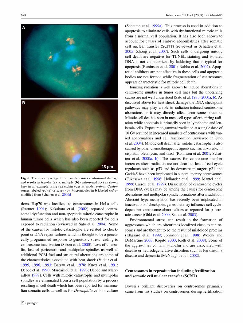
Alterations in centrosome organization and function areassociated with cellular stress and may be part of themitotic catastrophe response characteristic for programmedmitotic cell death. Centrosome structure and function aredirectly aVected by chaotropic agents such as formamide(Schatten et al. 2000d) and in response to heat shock andDNA damage. While mild heatshock treatment triggersrepair mechanisms and centrosome thermotolerance severeheat shock leads to cell death by a mitotic catastrophemechanism. Formamide induces the formation of multipo-lar spindles perhaps as a result of centrosomal protein dena-turation (Schatten et al. 2000d). Tripolar and fractionatedcentrosomes after treatment of cells with formamide areshown in Fig. 6a and b, respectively. The eVects of heatshock and other environmental stresses have been studiedin various cell systems that all displayed centrosome altera-
123
678 Histochem Cell Biol (2008) 129:667–686
tions. Hsp70 was localized to centrosomes in HeLa cells(Rattner 1991); Nakahata et al. (2002) reported centro-somal dysfunction and non-apoptotic mitotic catastrophe inhuman tumor cells which has also been reported for cellsexposed to radiation (reviewed in Sato et al. 2004). Someof the causes for mitotic catastrophe are related to check-point or DNA repair failures which is thought to be a geneti-cally programmed response to genotoxic stress leading tocentrosome inactivation (Sibon et al. 2000). Loss of �-tubu-lin, loss of pericentrin and multipolar spindles as well asadditional PCM foci and structural alterations are some ofthe characteristics associated with heat shock (Vidair et al.1995, 1996, 1993; Barrau et al. 1978; Knox et al. 1991;Debec et al. 1990; Marcaillou et al. 1993; Debec and Marc-aillou 1997). Cells with mitotic catastrophe and multipolarspindles are eliminated from a cell population by a processresulting in cell death which has been reported for mamma-lian somatic cells as well as for Drosophila cells in culture
(Schatten et al. 1999a). This process is used in addition toapoptosis to eliminate cells with dysfunctional mitotic cellsfrom a normal cell population. It has also been shown toaccount for causes of embryo abnormalities after somaticcell nuclear transfer (SCNT) (reviewed in Schatten et al.2005; Zhong et al. 2007). Such cells undergoing mitoticcell death are negative for TUNEL staining and isolatedDNA is not characterized by laddering that is typical forapoptosis (Roninson et al. 2001; Nabha et al. 2002). Apop-totic inhibitors are not eVective in these cells and apoptoticbodies are not formed while fragmentation of centrosomesappears characteristic for mitotic cell death.
Ionizing radiation is well known to induce aberrations incentrosome number in tumor cell lines but the underlyingcauses are not well understood (Sato et al. 1983, 2000a, b). Asdiscussed above for heat shock damage the DNA checkpointpathways may play a role in radiation-induced centrosomeaberrations or it may directly aVect centrosome structure.Mitotic cell death is seen in most cell types after ionizing radi-ation while apoptosis is primarily seen in lymphoma and leu-kemia cells. Exposure to gamma-irradiation at a single dose of10 Gy resulted in increased numbers of centrosomes with var-ied abnormalities and cell fractionation (reviewed in Satoet al. 2004). Mitotic cell death after mitotic catastrophe is alsocaused by other chemotherapeutic agents such as doxorubicin,cisplatin, bleomycin, and taxol (Roninson et al. 2001; Schat-ten et al. 2000a, b). The causes for centrosome numberincreases after irradiation are not clear but loss of cell cycleregulators such as p53 and its downstream targets p21 andGadd45 have been implicated in supernumerary centrosomes(Fukasawa et al. 1996; Hollander et al. 1999; Mantel et al.1999; Carroll et al. 1999). Dissociation of centrosome cyclesfrom DNA cycles may be among the causes for centrosomeaberrations and multipolar spindle formations after irradiation.Aberrant hypermethylation has recently been implicated ininactivation of checkpoint genes that may inXuence cell cycle-dependent centrosome abnormalities as reported for pancre-atic cancer (Ohki et al. 2000; Sato et al. 2003).
Environmental stress can result in the formation ofaggresomes which are oftentimes localized close to centro-somes and are thought to be the result of misfolded proteins(Ellgaard et al. 1999; Johnston et al. 1998; Wojcik andDeMartino 2003; Kopito 2000; Roth et al. 2008). Some ofthe aggresomes contain �-tubulin and are associated withdisease or neurodegenerative disorders such as Parkinson’sdisease and dementia (McNaught et al. 2002).
Centrosomes in reproduction including fertilization and somatic cell nuclear transfer (SCNT)
Boveri’s brilliant discoveries on centrosomes primarilycame from his studies on centrosomes during fertilization
Fig. 6 The chaotropic agent formamide causes centrosomal damageand results in tripolar (a) or multiple (b) centrosomal foci as shownhere in an example using sea urchin eggs as model system. Centro-somes labeled red (a) or green (b). Microtubules in b labeled red a=modiWed from Schatten et al. 2000d
123

Histochem Cell Biol (2008) 129:667–686 679
and cell division in sea urchin eggs (Boveri 1901), whichresulted in a wealth of profound information on the impor-tant contribution of sperm centrosomes for successful fer-tilization. His work on reproduction also resulted in theremarkable insights that supernumerary centrosomes are atthe core of malignant tumors (Boveri 1914) which wasbased on his observations that dispermic eggs (eggs fertil-ized with two sperm) developed multipolar mitotic poles.An example of a dispermic sea urchin egg is seen in Fig. 7.Boveri had recognized that sperm contribute the dominantcentrosomal material that was detected by staining withiron hematoxylin and could be traced throughout cell divi-sion and development. In more recent years, staining ofcentrosomes with immunological probes conWrmed that inmost animal species except for the mouse (Schatten et al.1986, 1987) dominant centrosomal material is contributedby sperm (reviewed in Manandhar et al. 2005; Sun andSchatten 2006, 2007) and biparental centrosome contribu-tions to the zygote are typical for most species. Moredetailed studies using speciWc immunoXuorescent probes tospeciWc centrosome proteins provided additional insightsinto centrosomal contributions during gametogenesis andfertilization (reviewed by Manandhar et al. 2005). Thesestudies showed that the sperm retains its proximal centriolewhile losing most of the PCM. The oocyte, on the otherhand degenerates centrioles while retaining centrosomalproteins. The sperm’s proximal centriole recruits egg pro-teins shortly after sperm incorporation including �-tubulin,centrin, pericentrin, and NuMA to the sperm centriolarcomplex. The recruitment of maternal �-tubulin to thesperm’s centrosomal �-tubulin results in a signiWcant accu-
mulation of �-tubulin. The blended centrosome after fertil-ization also contains centrin from both sperm and egg. Thezygotic centrosome that is closely associated with thedecondensing male pronucleus then organizes a radial asterthat grows toward the female pronucleus. The centriole-centrosome complex duplicates during the pronuclearstage. A zygote aster forms around the appositioned (orfused) male and female pronuclei (zygote nucleus) and cen-trosomes separate to form the bipolar mitotic apparatus inpreparation for cell division (reviewed in Sun and Schatten2006, 2007). Dysfunctional sperm centrosomes, dysfunc-tional zygote centrosomes, and polyspermy have beenimplicated in causes for male-derived infertility (reviewedin Schatten 1994) while aging of oocyte centrosomes isamong the major causes for infertility in older women (Bat-taglia et al. 1996) as dysfunctional centrosomes are impliedin the causes for aneuploidy. It is estimated that the rates ofaneuploidy in preimplantation human embryos is as high as52-61% (Munne and Cohen 1998).
Centrosome remodeling after nuclear transfer: communication between embryonic and somatic cells
Cloning of animals by somatic cell nuclear transfer (SCNT)involves cellular communication of embryonic cells withsomatic cell nuclei and somatic cell centrosomes fromdonor cells. In recent years, cloning of animals by SCNThas opened up new avenues to produce genetically modi-Wed species with higher nutritional value or improved traitsthat are useful in agriculture. Genetically modiWed pigshave been produced as models for human disease and forvarious other biomedical applications (reviewed in Prather2000, 2007; Schatten et al. 2005). However, one of themajor diYculties to overcome in cloning is the low cloningeYciency that ranges from 1 to 5% in diVerent animals.Centrosomal dysfunctions have been identiWed as possiblecauses for low cloning eYciencies in several speciesincluding the pig (Zhong et al. 2007) and rhesus monkeys(Simerly et al. 2003) and centrosomal dysfunctions mayalso play a role in developmental abnormalities that are fre-quently associated with cloning.
SCNT involves transferring a somatic cell nucleus withknown genetic value into an enucleated oocyte from adiVerent animal followed by electrical activation. Subse-quently, SCNT requires communication of enucleatedoocytes with the donor cell nucleus and its associated cen-trosome. The complex molecular regulation and functionsof centrosomes in reproductive cells are complicated by thefact that somatic cell centrosomes need to be remodeled bythe enucleated oocyte (reviewed in Sun and Schatten 2007),which is crucially important as the donor cell’s centrosomeis required to perform all functions that are normally ful-Wlled by the blended sperm and egg centrosomal material.
Fig. 7 A tetrapolar spindle displaying microtubules (green) and chro-mosomes (red) as a result of dispermy in a sea urchin egg. Such resultson dispermic eggs had stimulated Boveri to propose that centrosomeabnormalities are at the core of malignant tumors. Image produced incollaboration at the Integrated Microscopy Resource at the Universityof Wisconsin
123

680 Histochem Cell Biol (2008) 129:667–686
Under these conditions, the enucleated oocyte’s cytoplas-mic regulatory machinery is required to regulate the donorcell centrosome for embryo-speciWc functions. Centrosomedysfunctions may occur as the oocyte’s cell cycle regula-tion systems are diVerent from those used by somatic cells.Indeed, multipolar mitoses resulting from supernumerarycentrosomes have been reported in reconstructed pigoocytes (Zhong et al. 2007) which may be among theunderlying causes for decreased cell numbers as a result ofincreased mitotic cell death.
While blending of sperm and egg centrosomal materialinto a functional centrosome after fertilization is necessaryfor embryo-speciWc functions, it is not known whether thedonor cell centrosome also attracts centrosomal compo-nents from the oocyte or whether the somatic cell centro-some is able to nucleate and organize microtubules foroocyte-speciWc functions without oocyte centrosomal com-ponents. Aberrant composition of centrosome proteins willresult in abnormal microtubule organizations as has beendiscussed for cancer cells. As the donor cell centrosomecontains �-tubulin and �-TuRC it is not clear whether itattracts additional �-tubulin from the oocyte as is the casefor sperm centrosomes. Analysis of cancer cell centrosomesrevealed increased �-tubulin associated with centrosomesresulting in more than the normal number of microtubulesleading to aneuploidy (Lingle et al. 1998; Schatten et al.2000a, b). Increased accumulation of microtubules in thereconstructed egg may also play a role in centrosome dys-functions resulting in abnormal microtubule organizationand aneuploidy (Zhong et al. 2007; Martin et al. 2007). Thecomposition of the centrosome in reconstructed oocytes isnot known and needs further investigation to determinewhether oocyte-stored centrosome proteins are recruited tothe donor cell centrosome.
SCNT also challenges nucleo-cytoplasmic interactionsas several centrosome proteins such as NuMA are localizedin the nucleus, adding further complexities to centrosomeremodeling. The oocyte’s regulatory systems have to com-municate with the donor cell’s nuclear and centrosomalmaterial as many of the cell-cycle-dependent centrosomeproteins are of nuclear origin and depend on nucleo-cyto-plasmic interactions for cell cycle-speciWc and develop-mentally regulated functions. Such functions includegoverning microtubule-mediated translocation of mito-chondria for spatio-temporal requirements for ATP, trans-location of macromolecular complexes, vesicles that maycontain enzymes, and translocation of transient centro-some-associated proteins that are needed for molecular cen-trosome restructuring to perform cell cycle-speciWccentrosome functions. We do not yet have information onthe role of cyclin B in centrosome remodeling in recon-structed embryos which will be important information toobtain as cyclin B is one of the major regulators of centro-
somes and is crucial for centrosome maturation and for cellcycle-dependent molecular centrosome dynamics. Cyclin Bis a critical regular of NuMA association with centrosomesand its release from spindle poles during exit from mitosis(Saredi et al. 1997; Merdes and Cleveland 1998; Gehmlichet al. 2004). In contrast to mammalian somatic cells inwhich cyclin B synthesis increases during G2 and Mmainly as the result of cyclin B gene transcription increase,in embryonic cells cyclin B synthesis is constant through-out the cell cycle and cyclin B accumulation is the result ofdecrease in its degradation rate.
Taken together, studies of centrosomes in reconstructedembryos will provide new avenues to obtain information oncentrosomal regulation by cytoplasmic factors.
Concluding remarks and future directions
Renewed interest in centrosome research started about25 years ago when it was shown that certain autoimmunesera from CREST patients would reliably label centrosomematerial in animal cells. Since then, rapid progress has beenmade to identify speciWc centrosome proteins and deter-mine their functions, which led to renewed appreciation ofthe enormous signiWcance that centrosomes carry as cellu-lar organelles and as major platform for cellular regulation.Centrosome dysfunctions have been linked to varioushuman genetic diseases (reviewed in Badano et al. 2005)and centrosomes can directly be aVected by environmentaland genotoxic stresses. Adverse eVects on centrosomes willresult in multiple cascades of cellular dysfunctions as cen-trosomes regulate distribution of cellular organelles includ-ing mitochondria which are the major source for ATPproduction and depend on microtubules for distribution totheir cell cycle-speciWc functional destinations. Based onthese new insights it is easy to understand that centrosomesare linked to many diseases in which transport along micro-tubules is impaired which includes Alzheimer’s and severalneurological diseases in which extensive traYcking alongmicrotubules takes place. This area of research with a newfocus on centrosomes is just at the beginning.
The entire Weld of aging research is open to investiga-tions of centrosomes in senescing cells. Our previous stud-ies on Drosophila cells in culture have linked multipolarmitoses to cellular senescence (Schatten et al. 1999a) andfurther studies are needed to explore centrosome regulationin aging cells. Other new areas of research include theeVects of environmental stresses on centrosomes andmanipulation and exploitation of centrosomes and the asso-ciated microtubule cytoskeleton by pathogens which hasbeen explored for Toxoplasma parasites (Coppens et al.2006) and has been reviewed by Scaplehorn and Way(2004) in a recent book dedicated to various aspects of cen-trosome research (Nigg 2004). Taken together, we are in
123

Histochem Cell Biol (2008) 129:667–686 681
the middle but also at the beginning of new appreciation forthe centrosome as key organelle and as main station fordirecting and regulating cellular processes through itsmicrotubule highway system and further research willundoubtedly result in uncovering and perhaps also repair-ing pathologies related to centrosome dysfunctions.
Acknowledgments The present article remembers the late DanielMazia for his inspiration, friendship, and dissemination of an ever-expanding research area in cellular and molecular biology as well as inmicrobiology. Daniel Mazia’s passionate lecture on centrosomes dur-ing a 1984 UNESCO-ICRO course in Palermo/Italy along with numer-ous subsequent fascinating discussions on centrosomes Wrst inspiredthe author’s research focus on centrosomes. Appreciation is extendedto Donald Connor for excellent help with the schematic diagrams andassembly of images. Figure 4E was kindly supplied by Dr. MikaKatayama resulting from a collaborative project (Katayama et al. 2006).Figure 4 a, c, d were kindly provided by Dr. Zhisheng Zhong resultingfrom a collaborative project (Zhong et al. 2007). Figure 7 had beenproduced in collaborations at the University of Wisconsin–Madisonand the results using formamide had been obtained in collaborationwith the late Daniel Mazia.
References
Ahmad FJ, Yu W, McNally FJ, Baas PW (1999) An essential role forkatanin in severing microtubules in the neuron. J Cell Biol145:305–315
Andersen JS, Wilkinson CJ, Mayor T, Mortensen P, Nigg EA, MannM (2003) Proteomic characterization of the human centrosome byprotein correlation proWling. Nature 426:570–574
Baas PW (1998) The role of motor proteins in establishing the microtu-bule arrays of axons and dendrites. J Chem Neuroanat 14:175–180
Baas PW (1999) Microtubules and neuronal polarity: lessons frommitosis. Neuron 22:23–31
Badano JL, Teslovich TM, Katsanis N (2005) The centrosome inhuman genetic disease. Nat Rev Genet 6:194–205
Bailly E, Dorée M, Nurse P, Bornens M (1989) P34cdc2 is located inboth nucleus and cytoplasm; part is centrosomally associated atG2/M and enters vesicles at anaphase. EMBO J 8:3985–3995
Balczon R, Bao L, Zimmer WE, Brown K, Zinkowski RP, BrinkleyBR (1995) Dissociation of centrosome replication events fromDNA synthesis and mitotic division in hydroxyurea-arrested Chi-nese hamster ovary cells. J Cell Biol 130:105–115
Barr AR, Gergely F (2007) Aurora A: the maker and breakerof spindlepoles. J Cell Sci 120:2987–2996
Barrau MD, Blackburn GR, Dewey WC (1978) EVects of heat on the cen-trosomes of Chinese hamster ovary cells. Cancer Res 38:2290–2294
Battaglia DE, Klein NA, Soules MR (1996) Changes in centrosomaldomains during meiotic maturation in the human oocyte. MolHum Reprod 2(11):845–851
Bonaccorsi S, Giansanti MG, Gatti M (2000) Spindle assembly in Dro-sophila neuroblasts and ganglion mother cells. Nat Cell Biol2:54–56
Bornens M (2002) Centrosome composition and microtubule anchor-ing mechanisms. Curr Opion Cell Biol 14:25–34
Boveri T (1901) Zellen-Studien: Über die Natur der Centrosomen.Jena. Germany: Fisher Z Med Naturw 28:1–220
Boveri T (1914) Zur Frage der Entstehung maligner Tumoren. G. Fish-er, Jena, Germany
Brinkley BR, Goepfert TM (1998) Supernumerary centrosomes andcancer: Boveri’s hypothesis resurrected. Cell Motil Cytoskeleton41:281–288
Burk DH, Liu B, Zhong R, Morrison WH, Ye ZH (2001) A katanin-like protein regulates normal cell wall biosynthesis and cell elon-gation. Plant Cell 13:807–827
Calarco-Gillam PC, Siebert MC, Hubble R, Mitchison T, Kirschner M(1983) Centrosome development in early mouse embryos as deW-ned by an autoantibody against pericentriolar material. Cell 35(3Pt 2):621–629
Canaday J, Stoppin-Mellet V, Mutterer J, Lambert A, Schnit A-C(2000) Higher plant cells: gamma-tubulin and microtubule nucle-ation in the absence of centrosomes. Microsc Res Tech 49:487–495
Cande Z (1990) Centrosomes: composition and reproduction. CurrOpin Cell Biol 2:301–305
Cao K, Nakajima R, Meyer HH, Zheng Y (2003) The AAA-ATPaseCdc48/p97 regulates spindle disassembly at the end of mitosis.Cell 115:355–367
Carroll E, Okuda M, Horn HF, Biddinger P, Stambrook PJ, Gleich LL,Li YQ, Tarapore P, Fukasawa K (1999) Centrosome hyperampli-Wcation in human cancer: chromosome instability induced by p53mutation and/or Mdm2 overexpression. Oncogene 18:1935–1944
Casenghi M, Meraldi P, Weinhart U, Duncan PI, Korner R, Nigg EA(2003) Polo-like kinase 1 regulates Nlp, a centrosome protein in-volved in microtubule nucleation. Dev Cell 5:113–125
Chan J, Calder GM, Doonan JH, Lloyd CW (2003) EB1 reveals mobilemicrotubule nucleation sites in Arabidopsis. Nat Cell Biol 5:967–971
Coppens I, Dunn JD, Romano JD, Pypaert M, Zhang H, Boothroyd JC(2006) Toxoplasma gondii sequesters lysosomes from mamma-lian hosts in the vacuolar space. Cell 125:261–274
Dammermann A, Merdes A (2002) Assembly of centrosomal proteinsand microtubule organization depends on PCM–1. J Cell Biol159:255–266
Daunderer CC, Schliwa MM, Gräf RR (1999) Dictyostelium discoide-um: a promising centrosome model system. Biol Cell 91:313–320
Debec A, Marcaillou C (1997) Structural alterations of the mitoticapparatus induced by the heat shock response in Drosophila cells.Biol Cell 89:67–78
Debec A, Courgeon A, Maingourd M, Maisonhaute C (1990) The re-sponse of the centrosome to heat shock and related stresses in aDrosophila cell line. J Cell Sci 96:403–412
Dictenberg J, Zimmerman W, Sparks C, Young A, Vidair C, Zheng Y,Carrington W, Fay F, Doxsey SJ (1998) Pericentrin and gammatubulin form a protein complex and are organized into a novel lat-tice at the centrosome. J Cell Biol 141:163–174
Donaldson MM, Tavares AAM, Hagan IM, Nigg EA, Glover DM(2001) The mitotic roles of polo-like kinase. J Cell Sci 114:2357–2358
Doxsey SJ, Stein P, Evans L, Calarco P, Kirschner M (1994) Pericen-trin, a highly conserved protein of centrosomes involved in micro-tubule organization. Cell 76:639–650
Duensing S, Lee LY, Duensing A, Basile J, Piboonniyom S, Gonz-alez S, Crum CP, Munger K (2000) The human papillomavirustype 16 E6 and E7 oncoproteins cooperate to induce mitoticdefects and genomic instability by uncoupling centrosomeduplication from the cell division cycle. Proc Natl Acad SciUSA 97:10002–10007
Ellgaard L, Molinari M, Helenius A (1999) Setting the standards: qual-ity control in the secretory pathway. Science 286:1882–1888
Fan H-Y, Tong C, Teng C-B, Lian L, Li S-W, Yang Z-M, Chen D-Y,Schatten H, Sun Q-Y (2003) Characterization of polo-like kinase-1 in rat oocytes and early embryos implies its functional roles inthe regulation of meiotic maturation, fertilization and cleavage.Mol Reprod Dev 65:318–329
Fenton B, Glover DM (1993) A conserved mitotic kinase active at lateanaphase-telophase in syncytial Drosophila embryos. Nature363:637–640
123
682 Histochem Cell Biol (2008) 129:667–686
Flemming W (1875) Studien über die Entwicklungsgeschichte der Na-jaden. Sitzungsber Akad Wissensch Wien 71:81–147
Flemming W (1891) Verhandlungen der anatomischen Gesellschaft,Jahrg. 6, München (found as item 60 vol II in Collected Papers ofWalther Flemming in M.B.L. Library, reprint collection)
Flory MR, Davis TN (2003) The centrosomal proteins pericentrin andkendrin are encoded by alternatively spliced products of one gene.Genomics 82:401–405
Freed E, Lacey KR, Huie P, Lyapina SA, Deshaies RJ, Stearns T, Jack-son PK (1999) Components of an SCF ubiquitin ligase localize tothe centrosome and regulate the centrosome duplication cycle.Genes Dev 13:2242–2257
Fry AM (2002) The Nek2 protein kinase: a novel regulator of centro-some structure. Oncogene 21:6184–6194
Fry AM, Hames RS (2004) The role of the centrosome in cell cycleprogression. In: Nigg E (ed) Centrosomes in development anddisease. Wiley-VCA Verlag GmbH & CoKGaG, Weinheim, pp143–166
Fry AM, Mayor T, Meraldi P, Stierhof YD, Tanaka K, Nigg EA (1998)C-Nap1, a novel centrosomal coiled-coil protein and candidatesubstrate of the cell cycle-regulated protein kinase Nek2. J CellBiol 141:1563–1574
Fukasawa K, Choi T, Kuriyama R, Rulong S, Vande Woude GF (1996)Abnormal centrosome ampliWcation in the absence of p53. Sci-ence 271:1744–1747
Gard DL, Hafezi S, Zhang T, Doxsey SJ (1990) Centrosome duplica-tion continues in cycloheximide-treated Xenopus blastulae in theabsence of a detectable cell cycle. J Cell Biol 110:2033–2042
Gehmlich K, Haren L, Merdes A (2004) Cyclin B degradation leads toNuMA release from dynein/dynactin and from spindle poles.EMBO Rep 5:97–103
Gillingham AK, Munro S (2000) The PACT domain, a conserved cen-trosomal targeting motif in the coiled-coil proteins AKAP450 andpericentrin. EMBO Rep 1:524–529
Gobert GN, Hueser CN, Curran E, Sun Q-Y, Glinsky VV, WelshonsW, Eisenstark A, Schatten H (2001) Immunolocalization ofNuMA and phosphorylated proteins during the cell cycle in hu-man breast and prostate cancer cells as analyzed by immunoXuo-rescence and postembedding immunoelectron microscopy.Histochem Cell Biol 115:381–395
Goepfert TM, Brinkley WR (2004) Centrosome anomalities in cancer:from early observations to animal models. In: Nigg E (ed) Centro-somes in development and disease. Wiley-VCA Verlag GmbH &CoKGaG, Weinheim, pp 323–336
Golsteyn RM, Mundt KE, Fry AM, Nigg EA (1995) Cell cycle regula-tion of the activity and subcellular localization of Plk1, a humanprotein kinase implicated in mitotic spindle function. J Cell Biol129:1617–1628
Gstaiger M, Marti A, Krek W (1999) Association of humanSCF(SKP2) subunit p19(SKP1) with interphase centrosomes andmitotic spindle poles. Exp Cell Res 247:554–562
Gundersen GG (2002) Evolutionary conservation of microtubule-cap-ture mechanisms. Nat Rev Mol Cell Biol 3:296–304
Gunawardane RN, Martin OC, Cao K, Zhang L, Dej K, Iwamatu A,Zheng Y (2000) Characterization and reconstitution of Drosoph-ila �-tubulin ring complex subunits. J Cell Biol 151:1513–1523
Gunawardane RN, Martin OC, Zheng Y (2003) Characterization of a new�-TuRC subunits with WD repeats. Mol Biol Cell 14:1017–1026
Haggeness MH, Simon M, Singer SJ (1978) Association of mitochon-dria with microtubules in cultured cells. Proc Natl Acad Sci U SA 75:3863–3866
Hannak E, Oegema K, Kirkham M, Gonczy P, Habermann B, HymanAA (2002) The kinetically dominant assembly pathway for cen-trosomal asters in Caenorhabditis elegans is {gamma}-tubulindependent. J Cell Biol 157:591–602
HinchcliVe EH, Li C, Thompson EA, Maller J, Sluder G (1999)Requirement of Cdk2-cyclinE activity for repeated centrosomere-production in Xenopus egg extracts. Science 283:851–854
Hollander MC, Sheik MS, Bulavin DV, Lundgren K, Augeri-Henmu-eller L, Shehee R, Molinaro TA, Kim KE, Tolosa E, Ashwell JD,Rodenber MP, Zhan Q, Fernandez-Salguero PM, Morgan WF,Deng CX, Fornace AJ Jr (1999) Genomic instability in Gadd45a-deWcient mice. Nat Genet 23:176–184
Huang B (1990) Genetics and biochemistry of centrosomes and spin-dle poles. Curr Opin Cell Biol 2:28–32
Huang J, RaV JW (1999) The disappearance of cyclin B at the end ofmitosis is regulated spatially in Drosophila cells. EMBO J18:2184–2195
Jackman M, Lindon C, Nigg E, Pines J (2003) Active cyclin B1-Cdk1Wrst appears on centrosomes in prophase. Nat Cell Biol 5:143–148
Johnston JA, Ward CL, Kopito RR (1998) Aggresomes: a cellular re-sponse to misfolded proteins. J Cell Biol 143:1883–1898
Joshi HC, Palacios MJ, McNamara L, Cleveland DW (1992) �-Tubulinis a centrosomal protein required for cell cycle-dependent micro-tubule nucleation. Nature 356:8083
Jurczyk A, Gromley A, Redick S et al (2004) Pericentrin forms a com-plex with intraXagellar transport proteins and polycystin-2 and isrequired for primary cilia assembly. J Cell Biol 166(5):637–643
Katayama H, Brinkley WR, Sen S (2003) The Aurora kinases: role incell transformation and tumorigenesis. Cancer Metastasis Rev22:451–464
Katayama M, Zhong Z-S, Lai L, Sutovsky P, Prather RS, Schatten H(2006) Mitochondria distribution and microtubule organization infertilized and cloned porcine embryos: implications for develop-mental potential. Dev Biol 299:206–220
Kawaguchi S, Zheng Y (2004) Characterization of a Drosophila cen-trosome protein CP309 that shares homology with Kendrin andCG-NAP. Mol Biol Cell 15:37–45
Keryer G, Di Fiore B, Celati C, Lechtreck KF, Mogensen M, DelouveeA, Lavia P, Bornens M, Tassin AM (2003) Part of Ran is associ-ated with AKAP450 at the centrosome: involvement in microtu-bule-organizing activity. Mol Biol Cell 14:4260–4271
Khodjakov A, Rieder CL (1999) The sudden recruitment of gamma-tubulin to the centrosome at the onset of mitosis and its dynamicexchange throughout the cell cycle, do not require microtubules.J Cell Biol 146:585–596
Khodjakov A, Rieder CL (2001) Centrosomes enhance the Wdelity ofcytokinesis in vertebrates and are required for cell cycle progres-sion. J Cell Biol 153:237–242
Khodjakov A, Rieder CL (2004) A synergy of technologies: usingGreen Fluorescent Protein tagging and laser microsurgery tostudy centrosome function and duplication in vertebrates. In:Nigg E (ed) Centrosomes in development and disease. Wiley-VCA Verlag GmbH & CoKGaG, Weinheim, pp 191–210
Khodjakov A, Cole RW, Oakley BR, Rieder CL (2000) Centrosome-independent mitotic spindle formation in vertebrates. Curr Biol10:59–67
Knop M, Pereira G, Schiebel E (1999) Microtubule organization by thebudding yeast spindle pole body. Biol Cell 91:291–304
Knox JD, Mitchell RE, Brown DL (1991) EVects of hyperthermia onmicrotubule organization and cytolyic activity of murine cyto-toxic T lymphocytes. Exp Cell Res 194:275–283
Kopito RR (2000) Aggresomes, inclusion bodies and protein aggrega-tion. Trends Cell Biol 10:524–530
Kramer ER, Scheuringer N, Podtelejnikov AV, Mann M, Peters JM(2000) Mitotic regulation of the APC activator proteins CDC20and CDH1. Mol Biol Cell 11:1555–1569
Kramer A, Lukas J, Bartek J (2004) Checking out the centrosome. CellCycle 3(11):1390–1393
123

Histochem Cell Biol (2008) 129:667–686 683
Kubo A, Tsukita S (2003) Non-membranous granular organelle con-sisting of PCM–1, subcellular distribution and cell-cycle-depen-dent assembly/disassembly. J Cell Sci 116:919–928
Kubo A, Sasaki H, Yuba-Kubo A, Tsukita S, Shiina N (1999) Centri-olar satellites: molecular characterization, ATP-dependent move-ment toward centrioles and possible involvement in ciliogenesis.J Cell Biol 147:969–980
Lacey KR, Jackson PK, Stearns T (1999) Cyclin-dependent kinasecontrol of centrosome duplication. Proc Natl Acad Sci USA96:2817–2822
Lee J, Miyano T, Moor RM (2000) Spindle formation and dynamics of�-tubulin and nuclear mitotic apparatus protein distribution dur-ing meiosis in pig and mouse oocytes. Biol Reprod 62:1184–1120
Levy YY, Lai EY, Remillard SP, Heintzelman MB, Fulton C (1996)Centrin is a conserved protein that forms diverse associationswith centrioles and MTOCs in Naegleria and other organisms.Cell Motil Cytoskeleton 33:298–323
Lingle WL, Salisbury JL (1999) Altered centrosome structure is asso-ciated with abnormal mitoses in human breast tumors. Am JPathol 155:1941–1951
Lingle WL, Salisbury JL (2000) The role of the centrosome in thedevelopment of malignant tumors. Curr Top Dev Biol 49:313–329
Lingle WL, Lutz WH, Ingle JN, Maihle NJ, Salisbury JL (1998) Cen-trosome hypertrophy in human breast tumors: implications forgenomic stability and cell polarity. Proc Natl Acad Sci USA95:2950–2955
Lingle WL, Barrett SL, Negron VC, D’Assoro AB, Boeneman K, LiuW, Whitehead CM, Reynolds C, Salisbury JL (2002) CentrosomeampliWcation drives chromosomal instability in breast tumordevelopment. Proc Natl Acad Sci USA 99:1978–1983
Liu ZH, Schatten H, Hao YH, Lai L, Wax D, Samuel M, Zhong Z-S,Sun Q-Y, Prather RS (2006) The nuclear mitotic apparatus (Nu-MA) protein is contributed by the donor cell nucleus in clonedporcine embryos. Front Biosci 11:1945–1957
Lohret TA, McNally FJ, Quarmby LM (1998) A role for katanin-med-iated axonemal severing during Chlamydomonas deXaggelation.Mol Biol Cell 9:1195–1207
Lutz W, Lingle WL, McCormick D, Greenwood TM, Salisbury JL(2001) Phosphorylation of centrin during the cell cycle and itsrole in centriole separation preceding centrosome duplication.J Biol Chem 276:20774–20780
Ly DH, Lockhart DJ, Lerner RA, Schultz PG (2000) Mitotic misregu-lation and human aging. Science 287:2486–2492
Mack GJ, Ou Y, Rattner JB (2000) Integrating centrosome structurewith protein composition and function in animal cells. MicroscRes Tech 49:409–419
Manandhar G, Schatten H, Sutovsky P (2005) Centrosome reductionduring gametogenesis and its signiWcance. Biol Repro 72:2–13
Mantel C, Braun SE, Reid S, Henegariu O, Liu L, Hangoc G, Brox-meyer HE (1999) p21cip¡1/waf¡1-deWciency causes deformed nu-clear architecture, centriole overduplication, polyploidy, andrelaxed microtubule damage checkpoints in human hematopoieticcells. Blood 93:1390–1398
Marcaillou C, Debec A, Lauverjat S, Saihi A (1993) The eVect of heatshock response on ultrastructure of the centrosome of Drosophilacultured cells in interphase: possible relation with changes in thechemical state of calcium. Biochem Cell Biol 71:507–517
Martinez-Campos M, Basto R, Baker J, Kernan M, RaV JW (2004) TheDrosophila pericentrin-like protein is essential for cilia/Xagellafunction, but appears indispensable for mitosis. J Cell Biol165(5):673–678
Martin L, Besch-Williford C, Lai L, Cheong H-T, Im G-S, Park K-W,Murphy C, Hao Y, Ellersieck MR, Keisler DH, Schatten H, GreenA, Prather RS (2007) Morphologic and histologic comparisons
between in vivo and nuclear transfer derived porcine embryos.Mol Reprod Devel 74:952–960
Matsumoto Y, Maller JL (2002) Calcium, calmodulin, and CaMKIIrequirement for initiation of centrosome duplication in Xenopusegg extracts (comment). Science 295:499–502
Matsumoto Y, Hayashi K, Nishida E (1999) Cyclin-dependent kinase2 (Cdk2) is required for centrosome duplication in mammaliancells. Curr Biol 9:429–432
Mayor T, Meraldi P, Stierhof YD, Nigg EA, Fry AM (1999) Protein ki-nases in control of the centrosome cycle. FEBS Lett 452:92–95
McIntosh JR, Euteneuer U (1984) Tubulin hooks as probes for micro-tubule polarity: an analysis of the method and an evaluation ofdata on microtubule polarity in the mitotic spindle. J Cell Biol98:525–533
McNally FJ, Vale RD (1993) IdentiWcation of katanin, an ATPase thatsevers and disassembles stable microtubules. Cell 75:419–429
McNally FJ, Thomas S (1998) Katanin is responsible for the M-phasemicrotubule-severing activity in Xenopus eggs. Mol Biol Cell9:1847–1861
McNally FJ, Okawa K, Iwamatsu A, Vale RD (1996) Katanin, themicrotubule-severing ATPase, is concentrated at centrosomes.J Cell Sci 109:561–567
McNally KP, Bazirgan OA, McNally FJ (2000) Two domains of p80katanin regulate microtubule severing and spindle pole targetingby p60 katanin. J Cell Sci 113:1623–1633
McNaught KS et al (2002) Impairment of the ubiquitin-proteasomesystem causes dopaminergic cell death and inclusion body forma-tion in ventral mesencephalic cultures. J Neurochem 81:301–306
Megraw TL, Kao LR, Kaufman TC (2001) Zygotic development with-out functional mitotic centrosomes. Curr Biol 11:116–120
Meraldi P, Nigg EA (2001) Centrosome cohesion is regulated by a bal-ance of kinase and phosphatase activities. J Cell Sci 114:3749–3757
Meraldi P, Nigg EA (2002) The centrosome cycle. FEBS Lett 521:9–13
Meraldi P, Honda R, Nigg EA (2002) Aurora-A overexpression revealstetraploidization as a major route to centrosome ampliWcation inp53–/–cells. EMBO J 21:483–492
Merdes A, Cleveland DA (1998) The role of NuMA in the interphasenucleus. J Cell Sci 111:71–79
Merdes A, Ramyar K, Vechio JD, Cleveland DW (1996) A complex ofNuMA and cytoplasmic dynein is essential for mitotic spindleassembly. Cell 87:447–458
Merdes A, Heald R, Samejima K, Earnshaw WC, Cleveland D (2000)Formation of spindle poles by dynein/dynactin-dependent trans-port. J Cell Biol 149:851–861
Mogensen MM (1999) Microtubule release and capture in epithelialcells. Biol Cell 91:331–341
Mogensen MM (2004) Microtubule organizing centers in polarizedepithelial cells. In: Nigg E (ed) Centrosomes in developmentand disease. Wiley-VCA Verlag GmbH & CoKGaG, Weinheim,pp 299–319
Mogensen MM, Malik A, Piel M, Bouckson-Castaing V, Bornens M(2000) Microtubule minus-end anchorage at centrosomal andnon-centrosomal sites: the role of ninein. J Cell Sci 113:3013–3023
Moritz M, Braunfeld MB, Sedat JW, Alberts B, Agard DA (1995)Microtubule nucleation by gamma-tubulin-containing rings in thecentrosome. Nature 378:638–640
Moritz M, Zheng Y, Alberts BM, Oegema K (1998) Recruitment of the�-tubulin ring complex to Drosophila salt-stripped centrosomescaVolds. J Cell Biol 142:775–786
Moritz M, Braunfeld MB, Guenebaut V, Heuser J, Agard DA (2000)Structure of the gamma-tubulin ring complex: a template formicrotubule nucleation. Nat Cell Biol 2:365–370
123

684 Histochem Cell Biol (2008) 129:667–686
Münger K, Duensing S (2004) Radiation therapy and centrosome ano-malities in pancreatic cancer. In: Nigg E (ed) Centrosomes indevelopment and disease. Wiley-VCA Verlag GmbH & CoK-GaG, Weinheim, pp 353–370
Murphy SM, Preble AM, Patel UK, O’Connell KL, Dias DP, MoritzM, Agard D, Stults JT, Stearns T (2001) GCP5 and GCP6: twonew members of the human gamma-tubulin complex. Mol BiolCell 12:3340–3352
Murphy S, Urbani L, Stearns T (1998) The mammalian gamma-tubulincomplex contains homologues of yeast spindle pole body compo-nent sspc97p and spc98p. J Cell Biol 141:663–674
Nabha SM, Mohammed RM, Dandashi MH, Coupaye-Gerard B,Aboukameel A, Pettit GR, Al-Katib AM (2002) Combretastatin-A4 prodrug induces mitotic catastrophe in chronic lymphocyticleukemia cell line independent of caspase activation andpoly(ADP-ribose) polymerase cleavge. Clin Cancer Res 8:2735–2741
Nakahata K, Miyakoda M, Suzuki K, Kodama S, Watanabe M (2002)Heat shock induces centrosomal dysfunction, and causes non-apoptotic mitotic catastrophe in human tumour cells. Int J Hyper-thermia 18:332–343
Nigg EA (2004) Centrosomes in development and disease. Wiley-VCH, Weinheim
Oakley CD, Oakley BR (1989) IdentiWcation of �-tubulin, a new mem-ber of the tubulin superfamily encoded by mipA gene of Aspergil-lus nidulans. Nature 338:662–664
Oakley BR, Oakley CE, Yoon Y, Jung MK (1990) Gamma-tubulin isa component of the spindle pole body that is essential for micro-tubule function in Aspergillus nidulans. Cell 61:1289–1301
Oegema K, Wiese C, Martin OC, Milligan RA, Iwamatsu E, MitchisonTJ, Zheng Y (1999) Characterization of two related Drosophila �-tubulin complexes that diVer in their ability to nucleate microtu-bules. J Cell Biol 144:721–733
Ohki R, Nemoto J, Murasawa H, Oda E, Inazawa J, Tanaka N, Tanig-uchi T (2000) Reprimo, a new candidate mediator of the p53-mediated cell cycle arrest at the G2 phase. J Biol Chem275:22627–22630
Ohta Y, Ohba T, Miyamoto E (1990) Ca2+/calmodulin-dependent pro-tein kinase II: localization in the interphase nucleus and the mitot-ic apparatus of mammalian cells. Proc Natl Acad Sci USA87:5341–5345
Ohta T, Essner R, Ryu JH, Palazzo RE, Uetake Y, Kuriyama R (2002)Characterization of Cep135, a novel coiled-coil centrosomalprotein involved in microtubule organization in mammalian cells.J Cell Biol 156:87–99
Ou Y, Rattner JB (2004) The centrosome in higher organisms: struc-ture, composition and duplication. Int Rev Cytol 238:119–182
Palazzo AF, Cook TA, Alberts AS, Gundersen GG (2001) mDia medi-ates Rho-regulated formation and orientation of stable microtu-bules. Nat Cell Biol 3:723–729
PfeiVer D, Gard D (1999) Microtubules in Xenopus oocytes are ori-ented with their minus-ends towards the cortex. Cell Motil Cyto-skeleton 44:34–43
Pietromonaco SF, Seluja GA, Elias L (1995) IdentiWcation of enzymat-ically active Ca2+/calmodulin-dependent protein kinase in centro-somes of hemopoietic cells. Blood Cells Mol Dis 21:34–41
Pihan GA, Doxsey SJ (1999) The mitoticmachinery is a source ofgenetic instability in cancer. Semin Cancer Biol 9:289–302
Pihan GA, Wallace J, Zhou Y, Doxsey SJ (2003) Centrosome abnor-malities and chromosome instability occur together in pre-inva-sive carcinomas. Cancer Res 63:1398–1404
Pockwinse SM, Krockmalnic G, Doxsey SJ, Nickerson J, Lian JB, vanWijnen AJ, Stein JL, Stein GS, Penman S (1997) Cell cycle inde-pendent interaction of CDC with the centrosome, which is associ-ated with the nuclear matrix-intermediate Wlament scaVold. ProcNatl Acad Sci USA 94:3022–3027
Prather RS (2000) Perspectives: cloning. Pigs is pigs. Science289:1886–1887
Prather RS (2007) Nuclear remodeling and nuclear reprogramming formaking transgenic pigs by nuclear transfer. In: Sutovsky P (ed)Somatic cell nuclear transfer. Landes Bioscience. Adv Exp MedBiol 591:1–13
Quintyne NJ, Schroer TA (2002) Distinct cell cycle-dependent rolesfor dynactin and dynein at centrosomes. J Cell Biol 159:245–254
Quintyne NJ, Gill SR, Eckley DM, Crego CL, Compton DA, SchroerTA (1999) Dynactin is required for microtubule anchoring atWbroblast centrosomes. J Cell Biol 147:321–334
RaV JW (2001) Centrosomes:central no more? Curr Biol 11:R159–R161
Rattner JB (1991) Hsp70 is localized to the centrosome of dividingHeLa cells. Exp Cell Res 195:110–113
Ripple M, Taylor M, Hueser C, Schatten H (1999) Androgen-respon-sive LNCaP and androgen-independent DU 145 prostate cancercells display diVerent taxol sensitivities. Proc MSA 5(2):1110–1111
Roninson IB, Brude EV, Chang BD (2001) If not apoptosis, then what?Treatment-induced senescence and mitotic catastrophe in tumorcells. Drug Resist Updat 4:303–313
Roth J, Yam GH, Fan J, Hirano K, Gaplovska-Kysela K, Le Fourn V,Guhl B, Santimaria R, Torossi T, Ziak M, Zuber C (2008) Proteinquality control: the who’s who, the where’s and therapeutic es-capes. Histochem Cell Biol 129:163–177
Salisbury JL (1995) Centrin, centrosomes, and mitotic spindle poles.Curr Opin Cell Biol 7:39–45
Salisbury JL, Suino KM, Busby R, Springett M (2002) Centrin-2 is re-quired for centriole duplication in mammalian cells. Current Biol12:1287–1292
Saredi A, Howard L, Compton DA (1997) Phosphorylation regulatesthe assembly of NuMA in a mammalian mitotic extract. J Cell Sci110:1287–1297
Sato C, Kuriyama R, Nishizawa K (1983) Microtubule-organizingcenters abnormal in number, structure, and nucleating activity inx-irradiated mammalian cells. J Cell Biol 96:776–782
Sato N, Mizumoto K, Nakamura M, Tanaka M (2000a) Radiation-in-duced centrosome overduplication and multiple mitotic spindlesin human tumor cells. Exp Cell Res 255:321–326
Sato N, Mizumoto K, Nakamura M, Ueno H, Minamishima YA,Farber JL, Tanaka M (2000b) A possible role for centrosomeoverduplication in radiation-induced cell death. Oncogene19:5281–5290
Sato N, Maitra A, Fukushima N, van Heek NT, Matsubayashi H,Iacobuzio-Donahue CA, Rosty C, Goggins M (2003) Frequenthypomethylation of multiple genes overexpressed in pancreaticductal adenocarcinoma. Cancer Res 63:4158–4166
Sato N, Mizumoto K, Tanaka M (2004) Radiation therapy and centro-some anomalities in pancreatic cancer. In: Nigg E (ed) Centro-somes in development and disease. Wiley-VCA Verlag GmbH &CoKGaG, Weinheim, pp 337–352
Saunders WS, Shuster M, Hunag X, Gharaibeh B, Enyenihi AH, Peter-sen I, Gollin SM (2000) Chromosomal instability and cytoskeletaldefects in oral cancer cells. Proc Natl Acad Sci USA 97:303–308
Schatten H, Chakrabarti A (2004) Detection of centrosome structure infertilized and artiWcially activated sea urchin eggs using immuno-Xuorescence microscopy and isolation of centrosomes followedby structural characterization with Weld emission scanning elec-tron microscopy. In: Schatten H (ed) Methods in molecular biol-ogy, vol. 253: germ cell protocols, vol 1: sperm and oocyteanalysis. Humana Press, Totowa, pp 151–164
Scaplehorn N, Way M (2004) Manipulation of centrosomes and themicrotubule cytoskeleton during infectionbyintracellular patho-gens. In: Nigg E (ed) Centrosomes in development and disease.Wiley-VCA Verlag GmbH & CoKGaG, Weinheim, pp 371–400
123
Histochem Cell Biol (2008) 129:667–686 685
Schatten G (1994) The centrosome and its mode of inheritance: Thereduction of centrosome during gametogenesis and its restorationduring fertilization. Dev Biol 165:299–335
Schatten H, Schatten G, Mazia D, Balczon R, Simerly C (1986) Behav-ior of centrosomes during fertilization and cell division inmouse oocytes and in sea urchin eggs. Proc Natl Acad Sci USA83:105–109
Schatten H, Walter M, Mazia D, Biessmann H, Paweletz N, CoVe G,Schatten G (1987) Centrosome detection in sea urchin eggs witha monoclonal antibody against Drosophila intermediate Wlamentproteins: characterization of stages of the division cycle of centro-somes. Proc Natl Acad Sci USA 84:8488–8492
Schatten H, Ripple M, Balczon R, Taylor M, Crosser M (1998a) Cen-trosome proliferation in the human androgen-responsive LNCaPand the androgen-independent DU145 prostate cancer cell lines.Proc Microsc Soc Am 56:1066–1967
Schatten H, Ripple M, Balczon R, Taylor M (1998b) Centrosomeabnormalities in cancer cells and tissue. ICEM14. Electron Mi-crosc 4:243–244
Schatten H, Chakrabarti A, Hedrick J (1999a) Centrosome and micro-tubule instability in cells during aging. J Cell Biochem 74:229–241
Schatten H, Wiedemeier A, Taylor M, Lubahn D, Greenberg NM, Be-sch-Williford C, Rosenfeld C, Day K, Ripple M (1999b) Centro-some and centriole abnormalities during cancer in the transgenicadenocarcinoma mouse prostate (TRAMP) model. MicroscMicroanal 5:1152–1153
Schatten H, Hueser CN, Chakrabarti A (2000a) From fertilization tocancer: the role of centrosomes in the union and separation ofgenomic material. Microsc Res Tech 49:420–427
Schatten H, Ripple M, Balczon R, Weindruch R, Taylor M (2000b)Androgen and taxol cause cell type speciWc alterations of centro-some and DNA organization in androgen-responsive LNCaP andandrogen-independent prostate cancer cells. J Cell Biochem76:463–477
Schatten H, Wiedemeier A, Taylor M, Lubahn D, Greenberg MN, Be-sch-Williford C, Rosenfeld C, Day K, Ripple M (2000c) Centro-some-centriole abnormalities are markers for abnormal celldivisions and cancer in the transgenic adenocarcinoma mouseprostate (TRAMP) model. Biol Cell 92:331–340
Schatten H, Hueser C, Chakrabarti A (2000d) Centrosome alterationsinduced by formamide cause abnormal spindle pole formations.Cell Biol Int 24(9):611–620
Schatten H, Prather RS, Sun QY (2005) The signiWcance of mitochon-dria for embryo development in cloned arm animals. Mitochon-drion 5(5):303–321
Sibon OC, Kelkar A, Lemstra W, Theurkauf WE (2000) DNA/replica-tion/DNA-damage-dependent centrosome inactivation in Dro-sophila embryos. Nat Cell Biol 2:90–95
Simerly C, Dominko T, Navara CS (2003) Molecular correlates of pri-mate nudear transfer failures. Science 300(5617):297
Sluder G (2004) Centrosome duplication and its regulation in thehigher animal cell. In: Nigg E (ed) Centrosomes in developmentand disease. Wiley-VCA Verlag GmbH & CoKGaG, Weinheim,pp 167–189
Schroer TA (2001) Microtubules don and doV their caps: dynamicattachments at plus and minus ends. Curr Opin Cell Biol 13:92–96
Steadman BT, Schmidt PH, Shanks RA, Lapierre LA, Goldenring JR(2002) Transforming acidic coiled-coil-containing protein 4 inter-acts with centrosomal AKAP350 and the mitotic spindle appara-tus. J Biol Chem 277(33):30165–30176
Sun QY, Schatten H (2006) Multiple roles of NuMA in vertebratecells: Review of an intriguing multi-functional protein. Front Bio-sci 11:1137–1146
Sun QY, Schatten H (2007) Centrosome inheritance after fertilizationand nuclear transfer in mammals. In: Sutovsky P (ed) Somatic cell
nuclear transfer. Landes Bioscience. Adv Exp Med Biol 591:58–71
Sun QY, Lai L, Park KW, Kühholzer B, Prather RS, Schatten H(2001a) Dynamic events are diVerently regulated by microWla-ments, microtubules, and mitogen-activated protein kinase duringporcine oocyte maturation and fertilization in vitro. Biol Reprod64:879–889
Sun QY, Lai L, Bonk A, Prather RS, Schatten H (2001b) Cytoplasmicchanges in relation to nuclear maturation and early embryo devel-opmental potential of porcine oocytes: eVects of gonadotropins,cumulus cells, follicular size and protein synthesis inhibition. MolReprod Dev 59:192–198
Sun Q-Y, Lai L, Wu G, Park K-W, Day B, Prather RS, Schatten H(2001c) Microtubule assembly after treatment of pig oocytes withtaxol: correlation with chromosomes, �-tubulin and MAP kinase.Mol Reprod Dev 60:481–490
Sun Q-Y, Wu GM, Lai L, Park KW, Day B, Prather RS, Schatten H(2001d) Translocation of active mitochondria during porcineoocyte maturation, fertilization and early embryo development invitro. Reproduction 122:155–163
Sun Q-Y, Lai L, Wu G, Bonk A, Cabot R, Park K-W, Day B, PratherRS, Schatten H (2002) Regulation of mitogen-activated proteinkinase phosphorylation, microtubule organization, chromatinbehavior, and cell cycle progression are regulated by proteinphosphatases during pig oocyte maturation and fertilization in vit-ro. Biol Reprod 66(3):580–588
Takahashi M, Yamagiwa A, Nishimura T, Mukai H, Ono Y (2002)Centrosomal proteins CG-NAP and kendrin provide microtubulenucleation sites by anchoring gamma-tubulin ring complex. MolBiol Cell 13:3235–3245
Tassin AM, Bornens M (1999) Centrosome structure and microtubulenucleation in animal cells. Biol Cell 91:343–354
Tassin AM, Celati C, Moudjou M, Bornens M (1998) Characterizationof the human homologue of the yeast Spc98p and its associationwith gamma-tubulin. J Cell Biol 141:689–701
Thompson-CoVe C, CoVe G, Schatten H, Mazia D, Schatten G (1996)Cold-treated centrosome: isolation of centrosomes from mitoticsea urchin eggs, production of an anticentrosomal antibody, andnovel ultrastructural imaging. Cell Motil Cytoskeleton 33:197–207
Tong C, Fan H-Y, Lian L, Li S-W, Chen D-Y, Schatten H, Sun Q-Y(2002) Polo-like kinase-1 is a pivotal regulator of microtubuleassembly during mouse oocyte meiotic maturation, fertilization,and early embryonic mitosis. Biol Reprod 67:546–554
Tong C, Fan H-Y, Li S-W, Chen D-Y, Song X-F, Schatten H, Sun Q-Y (2003) EVects of MEK inhibitor U0126 on meiotic progressionin mouse oocytes: microtuble organization, asymmetric divisionand metaphase II arrest. Cell Res 13(5):375–383
Tulu US, Rusan NM, Wadsworth P (2003) Peripheral, non-centro-some-associated microtubules contribute to spindle formation incentrosome-containing cells. Curr Biol 13:1894–1899
Tugendreich S, Tomkiel J, Earnshaw W, Hieter P (1995) CDC27Hscolocalizes with CDC16Hs to the centrosome and mitotic spindleand is essential for the metaphase to anaphase transition. Cell81:261–268
Van Beneden E (1876) Contribution al’histoire de la vesiculaire germi-native et du premier embryonnaire. Bull Acad R Belg 42:35–97
Vogl AW, Weis M, PfeiVer DC (1995) The perinuclear centriole-con-taining centrosome is not the major microtubule organizing centerin Sertoli cells. Eur J Cell Biol 66:165–179
Vidair CA, Doxsey SJ, Dewey WC (1993) Heat shock alters centro-some organization leading to mitotic dysfunction and cell death.J Cell Physiol 154:443–455
Vidair CA, Doxsey SJ, Dewey WC (1995) Thermotolerant cells pos-sess an enhanced capacity to repair heat-induced alterations tocentrosome structure and function. J Cell Physiol 163:194–203
123

686 Histochem Cell Biol (2008) 129:667–686
Vidair CA, Huang RN, Doxsey SJ (1996) Heat shock causes proteinaggregation and reduced protein solubility at the centrosome andother cytoplasmic locations. Int J Hyperthermia 12:681–695
Wang Q, Xie S, Chen J, Fukasawa K, Naik U, Traganos F, Dar-zynkiewicz Z, Jhanwar-Uniyal M, Dai W (2002) Cell cycle arrestand apoptosis by human polo-like kinase 3 is mediated throughperturbation of microtubule integrity. Mol Cell Biol 22(10):3450–3459
WakeWeld J, Huang J-Y, RaV JW (2000) Centrosomes have a role inregulating the destruction of cyclin B in early Drosophila embryos.Curr Biol 10:1367–1370
Walczak CE, Vernos I, Mitchison TJ, Karsenti E, Heald R (1998) Amodel for the proposed roles of diVerent microtubule-based motorproteins in establishing spindle bipolarity. Curr Biol 8:903–913
Welte MA (2004) Bidirectional transport along microtubules. CurrBiol 14(13):R525–R537
Wilkinson CJ, Andersen JS, Mann M, Nigg EA (2004) A proteomicapproach to the inventory of the human centrosome. In: Nigg E(ed) Centrosomes in development and disease. Wiley-VCA Ver-lag GmbH & CoKGaG, Weinheim, pp 125–142
Wojcik C, DeMartino GN (2003) Intracellular localization of protea-somes. Int J Biochem Cell Biol 35:579–589
Wong C, Stearns T (2003) Centrosome number is controlled by a cen-trosome-intrinsic block to reduplication. Nat Cell Biol 5:539–544
Xu X, Weaver Z, Linke SP, Li C, Gotay J, Wang XW, Harris CC, RiedT, Deng CX (1999) Centrosome ampliWcation and a defective G2-M cell cycle checkpoint induce genetic instability in BRCA1exon 11 isoform-deWcient cells. Mol Cell 3:389–395
Young A, Dictenberg JB, Purohit A, Tuft R, Doxsey SJ (2000) Cyto-plasmic dynein-mediated assembly of pericentrin and � tubulinonto centrosomes. Mol Biol Cell 11:2047–2056
Zeng C (2000) NuMA: a nuclear protein involved in mitotic centro-some function. Microsc Res Tech 49:467–477
Zheng Y, Wong ML, Alberts B, Mitchison T (1995) Nucleation ofmicrotubule assembly by a gamma-tubulin-ring complex. Nature378:578–583
Zhong Z-S, Zhang G, Meng X-Q, Zhang Y-L, Chen D-Y, Schatten H,Sun Q-Y (2005) Function of donor cell centrosome in intraspe-cies and interspecies nuclear transfer embryos. Exp Cell Res306:35–46
Zhong Z, Spate L, Hao Y, Li R, Lai L, Katayama M, Sun Q-Y, PratherR, Schatten H (2007) Remodeling of centrosomes in intraspeciesand interspecies nuclear transfer porcine embryos. Cell Cycle6(12):1510–1521
123