Embed Size (px)
Citation preview

International Journal of Scientific and Research Publications, Volume 4, Issue 12, December 2014 1 ISSN 2250-3153
www.ijsrp.org
The management in emergency of a septic complication
from tonsillar abscess: cervico mediastinal gangrene - a
case report
Vernotico L.*, Picca D*., Calculli L*., Proto F. **, Valentini N.**., Covelli V.**
* U.O. Medicina Iperbarica, Ospedale San Paolo, Via Caposcardicchio 10, – 70100 Bari (Italy)
**U.O. Anestesia e Rianimazione, Ospedale San Paolo, Via Caposcardicchio 10, - 70100 Bari (Italy)
Abstract- This report is the result of a teamwork of the Unità Operativa di Medicina Iperbarica (Op. Unit of Hyp. Med.) and of the
Unità Operativa di Anestesia e Rianimazione (Intensive Care Unit) of the S. Paolo Hospital (Bari, Italy).
Purpose of this work is to make a close examination of the literature about cervico-mediastinal gangrene and to illustrate the
management in emergency of a septic complication from a tonsillar abscess: the cervico mediastinal gangrene also called descending
necrotizing mediastinitis (D.N.M.) (case report).
Epidemiology: the cervico-mediastinal gangrene is a severe infectious that begins from the cervical district. It is rarely observed in
clinical practice, it is fast-spreading and with a multi regional involvement and can affect any age of life.
Etiopathogenesis: the odontogenic origin of the infection is the most frequent, but the gangrene can also arise as a complication of
inflammation of the oropharynx and any other infectious disease in the neck if neglected. The beta-hemolytic Streptococcus seems to
be the most frequently involved germ, at least in the early stages of infection. Then, when inflammation develops and spreads, this
germ can be associated with both aerobes and anaerobes (the latter directly responsible for the production of gas and gangrenous
necrosis). The loose connective tissue of the cervical fasciae is the first to be affected, muscular and aponeurotic structures are
subsequently involved and then, for contiguity and as a result of the inspiratory acts, inflammation proceeds rapidly along the
pretracheal and retrotracheal region up to the mediastinum where it causes cell necrosis, pleural and pericardial effusion,
inflammatory spread to the lung parenchyma. In a second step the neurovascular structures may also be affected (phenomena of
thrombosis or vascular rupture).
Signs and symptoms: the first clinical signs and symptoms are attributable to the organ that is initially affected by the infection. The
progression to cervico-mediastinal gangrene leads to the following signs and symptoms: painful cervical swelling, chest pain, crepitus
on palpation, skin color change to dark blue (due to the progressive thrombosis of the tributary cutaneous vessels, fever, tachycardia
and tachypnea, sepsis.
Diagnosis: the diagnosis is based on clinical data and must make use of TC scan, the only exams able to document the disease status
and the progression of the infection.
Therapy: the cervicotomy is the treatment to be carried out as early as possible when the presence of purulent collections has been
documented and may be limited to a single district if gangrene is caught at an early stage, but it must involve all levels of the neck,
and even beyond, when the inflammation is wide, or is spreading to the neck or pectoral region. The tracheotomy must accompany the
cervicotomy when there is a condition of shortness of breath or when the disease state suggests that an appropriate ventilatory
assistance will be necessary in a short time. The antibiotic therapy must be practiced, most of the times, in an empirical way as the
germs responsible for the infection are rarely identified and as the initial etiological factor often remains the only cause of the
gangrene. It is important to point out the possibility of fungal associated infections: a treatment in this sense must always be
considered in advance. The hyperbaric treatment must be considered a weapon of considerable help available in cases of necrotizing
fasciitis / gangrene but it cannot be considered "life-saving", nor it is capable of replacing the aforementioned treatments.
As regards the patient of the case report, we followed the guidelines of the literature both for diagnosis and for therapy with the
conclusion that, the multidisciplinary approach is the best to perform.
Index Terms- Cervical abscess, cervico-mediastinal gangrene, descending mediastinitis, peritonsillar abscess, progressive necrotizing
infections.
I. INTRODUCTION
Etiopathogenesis of the Cervical Abscess
The cervical abscesses (both peritonsillar and parapharyngeal) arise, in general, by the passage of bacteria (present at the level of the
oral mucosa and of the upper respiratory tract) within the surrounding sterile sites and are frequently polymicrobial infections.

International Journal of Scientific and Research Publications, Volume 4, Issue 12, December 2014 2
ISSN 2250-3153
www.ijsrp.org
This step can occur by direct extension of a primary mucosal infections (eg. pharyngo-tonsillitis, rhinosinusitis, otitis media,
especially if relapsing) or can be due to traumatic or iatrogenic lacerations or perforations. It develops between the tonsillar capsule on
one side and the muscle floor and the pharyngo-basilar fasciae on the other side [1].
The peritonsillar abscess (P.T.A.) is the most common cause of suppuration of the peripharingeal spaces, both in adults and children.
The information is confirmed by U.S. studies that have shown an incidence of approximately 30 cases per 100,000 inhabitants per
year, with no significant differences in gender and race [2].
The predisposing conditions are deep tonsillar crypts, supra-tonsillar recess with retention of septic material, and / or seat of an
isolated lymphoreticular cluster (Tourtoral sinus).
The evolution of peritonsillar abscess [3] is currently considered the end point of a continuum which in turn contemplates:
1) in the first phase, an acute tonsillitis (in which the inflammatory process is localized in the tonsillar or pharyngo-tonsillar tissue
with presence or absence of exudate visible on the surface);
2) in the second phase, a peritonsillar cellulitis (characterized by inflammation and edema of the peritonsillar soft tissues, in the
absence of suppurative phenomena);
3) in the third phase, cellular colliquation phenomena (due to the release of leukocyte enzymes resulting in the formation of a
phlegmon or of a peritonsillar abscess).
A review of the literature [4,5] indicates an important role, in the peritonsillar abscess formation, of the Weber's glands. During a
oropharyngeal inflammation or obstruction of the duct by debris of food, these glands can be involved, resulting in cellulitis and the
following peritonsillar abscess. This hypothesis is supported by the rare cases of recurrence of P.T.A. in patients already undergoing
tonsillectomy [4,5,6]. Commensal microbiota and many different bacteria are present at the level of the oral mucosa and of the upper
respiratory tract; they are responsable for mucosal infections of the cervical district which can develop into abscessual complications;
these abscesses justify the diversity of bacteria that may be involved in the pathogenesis of cervical abscess.
Studies by Repanos and Brook [7,8] point out that in adults a mixed bacterial flora occurred more frequently; it is represented by
anaerobic bacteria (Fusobacterium spp, Bacteroides spp, Peptostreptococcus spp, micrococcus spp.) and aerobic bacteria (streptococci,
staphylococci and Haemophilus I.) in 76% of cases, while only anaerobes are found in 18% of cases and aerobes in 6% of cases. Other
bacteria, isolated from peritonsillar and parapharyngeal abscesses, include streptococci of Streptococcus milleri group (S. anginosus,
S. intermedius and S. constellatus), viridans streptococci, H. influenzae and anaerobic bacteria typical of the oral microbiota such as
Bacteroides spp., Fusobacterium necrophorum and nucleatum, Prevotella melaninogenica and Porphyromonas spp. While infection by
beta-hemolytic streptococci can be mono-microbal, those that involve anaerobes and other streptococci are generally polymicrobial
(average number of 5 isolated species) [9,10].
In addition, beta-hemolytic streptococci, S. pyogenes, in particular, are a common cause, especially in the forms that occur as a
complication of streptococcal pharyngitis [1].
Clinic of the Peritonsillar Abscess
The clinical features are characterized by:
- fever;
- odynophagia and otalgia;
- trismus (in the anterior abscess due to the medial pterygoid muscle contracture);
- open rhinolalia (due to analgesic hypomotility of the soft palate);
- drooling;
- displacement of the tonsils (forward in the posterior abscesses, on the back in the front abscesses);
- ipsilateral lateral-cervical lymphadenopathy.
The mono-laterality of signs and symptoms is pathognomonic [3]. They arise more or less rapidly and sometimes (11-56% of cases) as
an exacerbation of bilateral and widespread symptoms of an acute exudative pharyngitis [2,11]. Objectively there are edema and
swelling of the soft palate, uvula, front/rear palatine pillar, dislocation and medialization of the tonsil (sometimes covered with
purulent exudate) and painful lateral cervical adenopathy (level II and III), all signs and symptoms useful to distinguish a P.T.A. from
a simple peritonsillar cellulitis [12,6].
Diagnosis of the Peritonsillar Abscess
Laboratory Tests
As regards the laboratory tests [3], investigations show a classical neutrophilic leukocytosis, which confirms the bacterial nature of the
inflammation.
The bacterial culture does not seem to be useful as a routine, given the frequent finding of a mixed bacterial flora. From 1995 to 2005
Repanos has conducted and described hospital-based studies on 119 patients from whom purulent peritonsillar material was taken;
afterwards a broad-spectrum antibiotic therapy was set with an emblematic success in 99% of cases [7]. Furthermore, as previously
seen [1], it is difficult to interpret the result is complicated because there are many bacterial species and they are part of the
commensal microbiota of the oral cavity and upper respiratory tract, and because of the ease with which contamination can occur at
the time of collection of the sample.
The tonsillar swab or the purulent material, eventually taken by needle aspiration from the abscess, may be useful only in selected
cases (for example in immunosuppressed patients, patients resistant to current antibiotic therapies, etc.).

International Journal of Scientific and Research Publications, Volume 4, Issue 12, December 2014 3
ISSN 2250-3153
www.ijsrp.org
Diagnostic Imaging
The diagnostic imaging allows a better definition of the P.T.A. enabling to distinguish a peritonsillar cellulitis from an abscess. The
Diagnostic Imaging in the inflammatory disease of the neck and in its complications has three main objectives:
- identification of the disease (clinically suspected);
- typing the disease as inflammatory one and determining its causes;
- determination of the loco-regional space involvement (very important for surgical therapy).
The CT scan with contrast is considered the gold standard in diagnostic imaging in the majority of cervical disease (inflammatory
disease and its complications) [13].
Therapy of the Peritonsillar Abscess
A timely medical treatment represented by broad-spectrum antibiotics (“protected” penicillin or cephalosporins, possibly associated,
in the case of persistence of symptoms within 24 hours, to metronidazole given the frequent polymicrobial nature of the abscess)
[7,14,15] is necessary in each assessed abscess [3].
A surgical therapy must be associated to the medical one.
Complications of the Peritonsillar Abscess: cervico-mediastinal gangrene
[16].
The complications of P.T.A. (progressive necrotizing infections) depend on various factors such as the delay in diagnosis and the
presence of impairment factors in the patient (diabetes, immunosuppression or immunodepression, poor hygiene and social conditions,
smoking, etc.) that lead to more severe clinical conditions. The incidence is low nowadays, about 0,4 per 100,000 people [17] with
relatively greater frequency in immunocompromised patients; in fact, several authors report isolated cases [18,19] and the authors
who report a more numerous casuistry however, merely describe ten cases [20,21].
No age group is spared while preferring adulthood (30-50 years) [22,23,24,25,26,27] and male sex [28].
Some authors report that mediastinitis in antibiotic era [1], occur in 4-5% of the infections of the spaces of the neck [29,30].
The incidence of morbidity and mortality of head and neck abscesses's complications has lowered in recent decades, thanks to the
advent of antibiotic therapy and the increasingly early diagnosis (permitted by the above mentioned diagnostic imaging techniques).
The cervico-mediastinal gangrene is often secondary to odontogenic infections (42% of cases) [23,31,32,20,33,25,34,35] and
oropharyngeal infections (18% of cases) [36,37] or cervico-facial trauma (8% of cases) [31], and major surgery of the head and neck
(1% of cases) [18,32,21,26,38,39].
Sometimes the origin is unknown [25] (variable from 20 to 80% of cases) [26,40,41,42,43,44,27,34,35,45,38,39,46] other times it is
the site of previous application of radiation therapy (3% of cases).
The progressive necrotizing infections (P.N.I.) are divided into necrotizing fasciitis, progressive bacterial gangrene and myonecrosis.
The necrotizing fasciitis [47] (a term introduced by Wilson in 1952) of the neck is an inflammatory process of the soft tissues that,
resulting in a fascial necrosis, allows easy and wide spread of infection along the laterocervical fasciae to the mediastinum through
what may be considered “routes of least resistance”. It is is a rare but serious complication of P.T.A.. It is characterized by the
extension of the inflammatory process to the fascial structures of the neck followed by a gangrene that spreads to the muscle-
aponeurotic structures of the neck (myonecrosis), with the possibility of evolution, if not properly diagnosed and treated, in a septic
state and the general spread of the necrotic inflammatory process in the mediastinum with pleuro-pericardial and pulmonary
involvement (cervico-mediastinal gangrene) with exitus of the patient estimated at around 40% of cases [48,31].
The mediastinum often proves to be the seat of inflammatory processes (the so-called mediastinitis), as
extremely rich in fat, which has the following characteristics:
1. it is poorly vascularized;
2. the immunocompetent cells are poorly represented [49].
The growing problem of antibiotic resistance and the increase in cases of immunosuppression have profoundly altered the clinical
course of P.N.I..
Surely the prognosis of P.N.I. of the neck, as uniformly considered in the literature, depends not only from complications due to the
tissue necrosis of the structures directly involved (both cutaneous and vascular ones), but also from the spread of the septic process in
the mediastinum and the thoracic organs (the pleura, pericardium and lungs), which significantly increases mortality (44% vs. 7%)
[31,25,21,41,50,51,37,52,53,54]. Other factors which influence the prognosis are: the delayed diagnosis [17,31,19,36,51], the patient's
general condition and the existence of dis-metabolic conditions and first of all, diabetes mellitus [31,55,56], hypertension, vascular
disease and the existence of the combination of underlying diseases such as kidney failure, liver disease and cardiovascular disorders
[25]. Even conditions of immunosuppression, spontaneous [57] or pharmacologically induced [58], as well as smoking and alcohol
abuse, or the existence of precarious social conditions and, consequently hygienic ones, may have an important role not only in
determining but also in the evolution of the disease [57].
There are several controversies in the literature about the death rate, which varies from 8% to 74% [59] with a higher prevalence of
values that are around 30-40% [18,23,31,36,20,25,21,41,27,54,48,60], but the cause of death is directly proportional to the state of
infection and severity of any unfavorable prognostic factors.
P.N.I. of the neck and mediastinum presents itself initially as a cellulitis with edema that, in just a few hours, can reach massive
proportions.

International Journal of Scientific and Research Publications, Volume 4, Issue 12, December 2014 4
ISSN 2250-3153
www.ijsrp.org
Signs and symptoms are characterized by:
- painful cervical swelling;
- chest pain;
- crepitus on palpation for subcutaneous emphysema;
- erythema of the overlying skin;
- paresthesia, ulceration (for progressive involvement of the sensitive nervous terminations);
- skin color change to dark blue (for the progressive thrombosis of the tributary cutaneous vessels);
- fever;
- tachycardia and tachypnea;
- sepsis.
The mediastinitis [61] represent a group of acute and chronic diseases, which cause a serious infection of the connective tissue that
surrounds the space between the two pleura and the organs in this space. A sneaky and lethal form of mediastinitis is represented by
the so-called "descending necrotizing mediastinitis" (D.N.M.) that occurs as a complication of infections arising from odontogenic
abscesses or from the cervico-fascial space. As previously described, once spread at the level of cervical fascial planes, through deep
contiguous spaces of the neck, the infection drops in the mediastinum, in the pleural spaces, in the pericardium and in the abdomen,
causing necrosis, abscess formation and sepsis (P.N.I. of the mediastinum).
The spread of the infection occurs in the cranio-caudal direction for various reasons, including the force of gravity, the acts of
breathing and the resulting changes in pressure inside the chest cavity [49,62]. In the literature it is reported that in more than 70% of
cases of D.N.M. the spread of sepsis occurs through the retrovisceral space (danger space), in 8% infection originates in the neck and
spreads in the mediastinum through the pretracheal space, while in the remaining cases, it spreads trough the perivascular space, where
the presence of arterial and venous vessels can favour the appearance of serious clinical aspects, determined by thrombosis of the
jugular vein or by the erosion of the carotid artery [63]. The criteria needed to define the D.N.M. have been shown to Estrera et al.
[64] and are represented by:
1) clinical manifestations of severe oropharyngeal infection;
2) characteristic radiological signs of mediastinitis;
3) documentation of necrotizing mediastinal infection at operation (or at postmortem);
4) relationship between oropharyngeal or cervical infection and development of the mediastinal necrotizing process.
Diagnosis
Whatever the starting point and initial symptoms of the disease, the swelling of the district [24,20,65,57,66,37,52] and the presence of
air bubbles, detected by clinical examination by palpation of the fascial structures of the neck [20,65,66] are pathognomonic of the
necrotizing evolution of the infection and its spread to fascial structures. This clinical finding, together with the CT confirmation of
gas evolution, represents an irrefutable fact of the disease.
In the case of clostridial infection, a time interval (ranging from one to six hours after the traumatic lesion tissue or surgical treatment)
can occur. The patient may suffer from sudden and intense pain of the infected area before the onset of clinical signs. This apparent
discrepancy between sharp pain in a still clinically normal tissue and the absence of hyperpyrexia requires extreme caution by the
medician for the possible development to gangrene. The clostridial infection can rapidly spread with a speed of 15 cm per hour.
The CT scan with contrast of the chest and neck remains the best diagnostic method for patients with suspected mediastinitis,
providing information on the extent of necrotizing infectious and the type of surgical approach. It is also necessary for the post-
operative monitoring and to highlight any relapses requiring reoperation [63,67,26,39,55,68,52]. The CT scan initially shows an
increase in the density of the mediastinal adipose tissue, and subsequently, with the evolution of the infection, the organization of
numerous liquid collections, often associated with gas bubbles [69].
The "routine" diagnosis contemplates imaging and blood tests and can be considered essential in monitoring the disease even if
sometimes it can not be considered definitive. In fact, the disease is often evolutionary and new purulent collections, as well as new
processes of gangrene, may develop in the hours or days immediately following surgery.
The validity of microbacteriological exam of the exudate (performed on abscess or on necrotized tissues) is uncertain.
Most of the authors, while performing such investigations, don't consider them essential since, as noted earlier, often the result is
negative [21,41,50,70], sometimes provides questionable results or multiple etiologies [31,36,19,33,21,35,71,72,51,55], sometimes it
still allows the recognition of germs considered saprophytes or symbionts and therefore of no use in the indication of the therapeutic
program to be undertaken.
It is likely that the polymicrobial partecipation of gram-positive bacteria (Streptococcus, Staphylococcus, Micrococcus), gram
negative (Bacteroide, Neisseria, Proteus, Pseudomonas) and anaerobics (Enterobacter, Propionobacter, Peptostreptococco) described
in the literature [31,19,33,21,35,50,51,55,65] results in a mutual protection of the exogenous agents towards the phagocytic process,
the intracellular "killing" and antibiotics, promoting the necrotizing evolution of the disease and thus making impossible the
subsequent isolation and identification of the bacterial species involved.
Therapy
It is widely recognized [23,31,24,36,19,33,21,35,45,39,46,51,55,57,37,52,53,56,73,74,54,75] that the surgical treatment of P.N.I.
should be performed as quickly as possible (within the first 12-24 hours in accordance with the extension of the infection). On the
International Journal of Scientific and Research Publications, Volume 4, Issue 12, December 2014 5
ISSN 2250-3153
www.ijsrp.org
basis of the CT images cervicotomy may be limited to a certain district (at least in the very early forms) or extended as much as
possible to other sites; most of the time a wide cervicotomy is necessary, including in some cases other districts (pectoral, neck and
mediastinal-chest districts). There is broad agreement in the literature that surgical drainage of the neck and mediastinum should be
considered the standard treatment for these patients [63]. In fact, the cervical drainage alone is insufficient in 80% of cases [76,62] and
in a meta-analysis of Corsten et al. [77] the comparison between cervicotomy alone and cervicotomy with thoracotomy shows a
mortality respectively of 47% (in the first case) versus 19% in cases with double-surgical approach [49]. In the mediastinal diffusion
a thoracotomy is necessary accompanied by a pleuroscopy and by the positioning of suction drains at this level.
Patients undergoing this type of combined treatment require a contextual tracheotomy that is useful, in addition to the immediate
ventilatory assistance, also for the hospitalization in intensive care. Some authors [33,53,54,78] believe that such action is necessary to
ensure a patent airway and ventilatory assistance and, at the same time, to promote drainage of the peritracheal abscess, other authors
[46,37,52,73] consider the tracheotomy a way of spread of the inflammatory process to the thoracic structures.
With regard to antibiotic therapy, in the absence of a bacterial culture and according to the above, the empirical treatment is
reccomended (combining an antibiotic active against gram positive bacteria and a specific one against gram negative and reserving the
antifungal therapy to cases with proven fungal presence) [18,20,40,41,39,50,68,79,80,81,82,83].
The association of hyperbaric oxygen therapy (H.B.O.T.) is controversial. Brummelkamp et al. in 1961 were the first to hypothesize
the use of H.B.O.T. in the treatment of gangrene [84,85], but until today, there are no double-blind studies on the effectiveness of
H.B.O.T. in soft tissue infections. However, there are many experiments that provide useful data, even if these experiments include
non-uniform clinical and anatomo-pathological conditions. In addition, there is no agreement on the program to be adopted and no
clinical study regarding the protocol to be used in the hyperbaric treatment. The duration of the treatment may vary from five days (in
exclusively anti-infectives treatments) to two or three weeks (for treatments that tend to provide benefits even in wound healing); the
number of sessions varies from 5 to 10 in acute conditions but the number may increase in intensive therapeutic treatments such as in
the case of necrotizing fasciitis / gangrene. H.B.O.T. is usually administered with a FiO2 of 100%, 2 or 3 ATA for an average duration
ranging from 60 to 90 minutes per session [86].
There is not even agreement on the ideal sequence of the different therapies: H.B.O.T. can be provided before, during or after surgical
treatment (however, the general recommendation is that it should be started as early as possible).
Since the late 80's, with the appearance of Evidence Based Medicine (E.B.M.), a medical and technological review has been
addressed in this regard.
The Study Group for the Hyperbaric Therapy of the Italian National Health Council has decided to extend the treatment with H.B.O.T.
to progressive necrotizing infections, divided into progressive bacterial gangrene, necrotizing fasciitis and myonecrosis. As
previously reported, in such conditions (typically polymicrobial ones), the skin, subcutaneous tissues, bands and muscles are involved
in inflammation and necrosis; vessels trombosis also is realized due to the action of bacterial toxins capable of activating enzymes
such as lipase and hyaluronidase. The European Committee for Hyperbaric Medicine (ECHM), in the 7th Conference held in Lille in
2004, directed the use of H.B.O.T. in infections of soft tissues like the medical and surgical treatment [87]. Hyperbaric Oxygen
Therapy is strongly recommended (recommendation based only on clinical evidence) in the treatment of anaerobic or mixed bacterial
necrotizing soft tissue infections (myonecrosis, necrotizing fasciitis, etc…). H.B.O.T. should be integrated in a treatment protocol
comprising adequate surgical and antibiotic therapy (Type 1 recommendation, level C). The sequential order for H.B.O.T., antibiotics
and surgery is a function of the condition of the patient, the surgical possibilities and hyperbaric oxygen availability (Type 1
recommendation, level C). [88,87].
The mechanism of action of H.B.O.T. in acute infectious processes is expressed through better tissue oxygenation resulting in the
stimulation of white blood cells (in their phagocytic function) and edema reduction. Furthermore, the increased levels of PO2 in
tissues prevent or reverse the tendency of leukocytes to adhere to vascular endothelium, thus reducing endothelial damage [89]. Once
blocked the infection, H.B.O.T. enhances the formation of collagen [90] and stimulates angiogenesis, facilitating the healing of tissue
lesions [91,92]. H.B.O.T. has therefore three modes of action: the "hemorheological one", that restores normal negative charges
present on the red blood cells that are neutralized in the acidosis created by anaerobic metabolism of bacteria; the "barometric one",
governed by the law of Boyle and Mariotte, according to which, at constant temperature, the pressure of a gas is inversely proportional
to its volume, whereby in the treatment of gas gangrene H.B.O.T. reduces the volumes of gas with consequent improvement of
ischemic tissue; the "bacteriostatic and bactericidal ones" [93,94], the first one against aerobic bacteria lacking the superoxide
dismutase enzyme (S.O.D.) necessary to protect from the action of peroxides on lipids of their membranes, the second one against
anaerobic germs that can survive in hypoxic environments where there is a depression of phagocytic functions.
The first experimental studies in the treatment of infections caused by Clostridium conducted by Brummelkamp, [84] Holland [95]
and Demello [96] have shown that the best therapeutic results derive from contemporary surgical and medical treatments, associated
with H.B.O.T. which has the task of stopping the production of of alpha-toxin and the growth of the Clostridium in the infected but
still vital tissues. To completely stop the production of alpha-toxin is required a PpO2 of 600 mmHg while to kill the bacteria a PpO2
superior to 1520 mmHg. [96,97]. Demello [96] and afterwards Him [98] obtained the best survival rates in experiments on animal
models (that included the inoculation of anaerobic bacteria) in the integrated treatment of H.B.O.T., surgical drainage and antibiotic
therapy. From their studies it appears that, for the survival, repeated surgical treatments of drainage and debridement of infected tissue
are essential but an improvement in terms of survival and rapidity of healing is achieved only with the targeted antibiotic therapy in
association with H.B.O.T. (Table 1)
International Journal of Scientific and Research Publications, Volume 4, Issue 12, December 2014 6
ISSN 2250-3153
www.ijsrp.org
Table. 1:Comparative study about survival with different types of treatment (experimental study in dogs), [by Demello 1973].
Therapy Survival (%)
Surgery 0
H.B.O.T. 0
H.B.O.T. + Surgery 0
Antibiotics 50
Antibiotics + Surgery 70
Antibiotics + Surgery + H.B.O.T. 95
Studies by Riseman [99,100,101] report inhomogeneous results that can be explained by the lack of standardization of protocols and
stratification for known prognostic factors, thus making it impossible to compare the results (obtained on few cases or including
patients with different degrees of severity of infections).
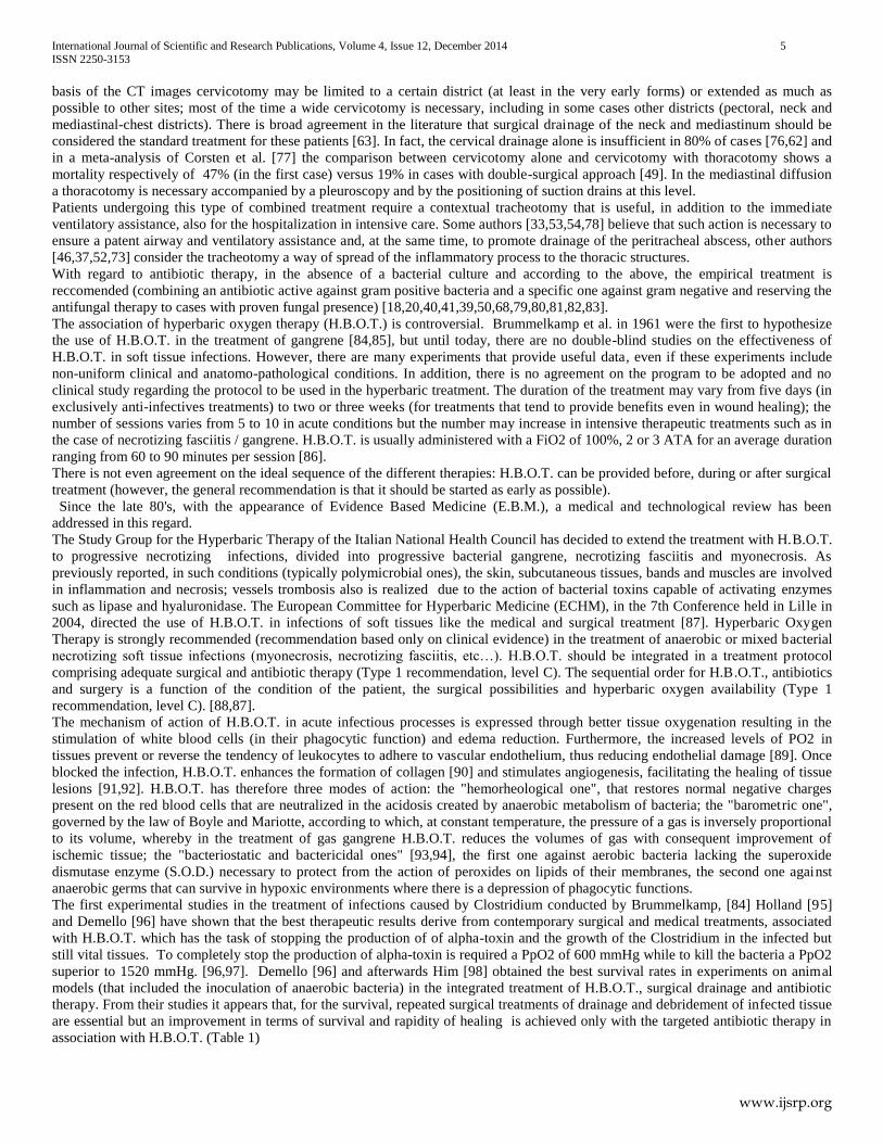
The following table (Table 2) reports the results of clinical trials as a function of therapy used (data from the literature).
Table. 2:Results of clinical trials as a function of therapy used (data from the literature).
Author N°. of Patients Hospital
admissions (%)
Deaths (%)
Surgery - Antibiotics - H.B.O.T.
Roding, 1972 130 101 (78) 29 (22)
Hitchcock, 1975 133 100 (75) 33 (25)
Hart, 1983 139 112 (81) 27 (19)
Darke, 1977 66 46 (70) 20 (30)
Holland, 1975 49 36 (73) 13 (27)
Unsworth, 1984 53 46 (87) 7 (13)
Hirm, 1988 32 23 (72) 9 (28)
Gibson, 1986 29 20 (70) 9 (30)
Werry,1986 28 21 (75) 7 (25)
Kofoed, 1983 23 20 (87) 3 (13)
Tonjum, 1980 14 12 (86) 2 (14)
Total 696 537 (78) 159 (22)
Surgery - Antibiotics
AIterneier, 1971 54 46 (85.2) 8 (14.8)
Hitchcock, 1975 44 24 (55) 20 (45)
Gibson, 1986 17 5 (29) 12 (71)
Freischiag, 1985 8 3 (37) 5 (63)
Total 123 78 (64) 45 (36)
Intensive Care Management of cervico-medistinal gangrene
Given the severity of the patients, especially those with overt disease stage, an assistance with mechanical ventilation or an intensive
care management is frequently necessary.
The monitoring and management of the airways prior to surgical and medical treatment of the injury, is a priority [102,103]; on the
other hand, the management of these patients in the critical area, even after surgery, is advisable due to the high risk of sudden
obstruction of the airways, which is one of the most frequent lethal complications [104]. The patients who reach the observation for
complications (in particular for sepsis and for acute respiratory distress syndrome-ARDS), require hospitalization in intensive care
before surgery because the management in critical care optimizes, as far as possible, the general conditions for the purpose of surgical
treatment (that in these cases becomes multidisciplinary).
International Journal of Scientific and Research Publications, Volume 4, Issue 12, December 2014 7
ISSN 2250-3153
www.ijsrp.org
Patients with continuous analgosedation, should undergo a lung protective ventilation at low tidal volume and high positive end-
expiratory pressure (P.E.E.P.), which has a positive impact on the survival of patients in intensive care unlike the traditional technical
ventilation, with high tidal volumes and low P.E.E.P. [105]. The invasive hemodynamic monitoring allows to optimize fluid therapy,
dose of vasopressors and inotropes by monitoring the ratio between peripheral oxygen availability and its use, with a drastic reduction
in mortality in patients who already have a septic state [106].
A so complex and expensive management has as objective the stabilization of vital functions more quickly and effectively as possible,
such as to optimize the recovery, try to improve the outcome and reduce the length of stay for patients with abscesses.
The management of cervico-mediastinal gangrene in a hyperbaric chamber requires an interdisciplinary approach between Hyperbaric
Medicine and Intensive Care as the treatment of critically ill patients in a hyperbaric chamber poses particular problems of care and
monitoring.
The alterations of consciousness, cardiovascular instability, respiratory failure, are the most frequently encountered problems but they
should not constitute an obstacle to the practice of H.B.O.T.; of course the resuscitation therapies must be carried out with the same
safety and efficacy in a hyperbaric chamber.
If a critically ill patient must undergo H.B.O.T., it is necessary to have a multiplace hyperbaric chamber with transfer lock to allow the
use of the equipment necessary for assistance and monitoring of the patient, but also the entrance in the chamber of the whole
resuscitation team.
The hyperbaric chamber must be equipped with the possibility of cardiorespiratory monitoring, respiratory care, oral and endotracheal
suction of secretions of the patient, etc.. in order to allow the assistance and the direct control of the patient throughout the treatment,
the continuation of resuscitation therapy and the ability to immediately perform all urgent therapeutic interventions that may become
necessary to ensure the main patient's vital functions: endotracheal intubation and artificial mechanical ventilation, circulatory
resuscitation, fluid infusion, administration of medication, etc..
Assisted ventilation requires an adaptation to the hyperbaric environment and a series of measures. In patients intubated with cuffed
tubes, as the pressure changes the volume of the cuff, this one must be filled with saline in order to avoid the continuous control of the
volume of the cuff during the phases of compression (air inlet) and decompression (removal of air).
It is best to choose ventilators that have been designed specifically for the use in a hyperbaric chamber.
The ideal ventilator should be a volume cycled ventilator (current volume remains acceptably constant with increases in the pressure
up to 6 ATA, the respiratory frequency is constant, there is an increase in the expiratory time during and above 2.8 ATA) that is not
influenced by variations of pressure that occur inside the chamber and not working on electrical current (danger of fire).
It must be remembered that the pressure antagonizes the effects of hypnotics and muscle relaxants. Prior myringotomy should be
performed in coma patients to be subjected to hyperbaric therapy. Before introducing patients with respiratory disease in the
hyperbaric chamber, you must be sure of the absence of pneumothorax. The presence of the transfer lock allows, in case of need, the
input of further personnel in the chamber. In addition, the medical lock allows at any time, the rapid introduction into the room of
everything that may be needed [107,108,109,110,111,112].
II. RESEARCH ELABORATIONS
Case Report
The case report regards a 22 year old man with no past medical history except for an allergy to Amoxicillin-Clavulanate, no history of
use of illegal drugs or alcohol.
The patient manifested sore throat, dysphagia and fever (39,5°C). The initial diagnosis was “severe oropharingeal infection”. After
two days of onset of symptoms, the patient began an empiric antibiotic therapy with Clarithromycin (500mg/die).
On 06 June, 4 days after the onset of symptoms and two days after the beginning of antibiotic therapy he was hospitalized in the
department of otolaryngology because of the worsening of symptoms.
A clinical examination showed the presence of purulent exudate on both tonsils, a left laterocervical swelling with bruised and
hyperemic skin. The swelling was greatly painful on palpation.
On 07 June an endoscopy of the upper airways showed edema of the left aryepiglottic fold.
The laboratory tests at the time of admission were as follows: white blood cell count 13.660/microliter with 90% of neutrophils,
erythrocyte sedimentation rate (E.S.R.) 49 mm/hour. Pulse rate was 120/min.
On 07 June the TC scan with contrast of the neck showed: "an abscess, in the context of the left deep laterocervical tissue, that
obliterates the left pyriform sinus and the ipsilateral laryngeal vestibule; marked and diffuse thickening and edema of the soft tissues
of the left lateral cervical region of the neck and upper mediastinum, high number of lateral cervical and submandibular lymph nodes
predominantly to the left. ".
Broad-spectrum intravenous antibiotics were administered (Teicoplanin 400mg/die , Levofloxacin 500mg x2/die and Metronidazole
500mg x4/die). The patient underwent urgent surgery (left cervicotomy and drainage of the pharingeal abscess).
On 08 June the patient presented worsening dyspnea, severe hypoxemia and respiratory acidosis at the blood gas analysis (pH 7.27,
PCO2 57 mmHg, PO2 45 mmHg, Sat 80% with oxygen mask), at the ECG there was an elevation of ST segment, the blood pressure
was 160/100 mmHg, procalcitonin (PCT) 15,66 µg/L, C-RP 53,90 mg/L.
On the same day, the patient was intubated, the femoral vein was cannulated, 1500 mL of crystalloid and colloid were administered. A
neck and chest CT scan was performed; it showed: "outcomes of cervicotomy with the presence of minute air bubbles and drainage,

International Journal of Scientific and Research Publications, Volume 4, Issue 12, December 2014 8
ISSN 2250-3153
www.ijsrp.org
marked edematous thickening of the walls of the oropharynx, hypopharynx, larynx, parapharyngeal spaces and subhyoid muscles;
massive phlegmon extended to all the mediastinal spaces until esophageal jato; pericardial and pleural effusion with imbibition of
pulmonary interstitium and alveolar commitment. "
He was transferred to the Intensive Care Unit (I.C.U.). A new antibiotic/antimycotic therapy was started (Penicillin G sodium
45.000.000 UIx2 continous infusion, Metronidazole 500mg x4/die , Teicoplanin 400mgx2/die, Meropenem 1,5g x 4/die, Caspofungin
70mg/die). The patient had obviously a bladder catheter.
The patient was transferred to the Thoracic Surgery Unit where he was subjected to right thoracotomy with intake of 600 ml of
purulent fluid,the opening of the anterior, medium and posterior mediastinum with leakage of purulent fluid, the washing of the
pleural cavity, double right pleural drainage (one at the top level and another one in the anterior subapical thoracic area), single left
pleural dranage and surgical tracheotomy.
The patient fulfilled the Estrera’s criteria for diagnosis of D.N.M.
The microbiological examination of the material from the abscess cavity and the pleural fluid was negative.
On 09 June he performed a CT scan with contrast that showed: "the presence of air bubbles in the bilateral parapharyngeal soft tissue,
reduced mediastinal collections, unchanged pericardial collections, reduced pleural effusion with better ventilation of the lung; air
bubbles in the context of the back muscles at the level of C6 extending in the subscapularis area, bubbles along the costal muscles of
the anterior and lateral chest wall extending along the muscles of the abdominal wall into the pelvis together with collections of fluid
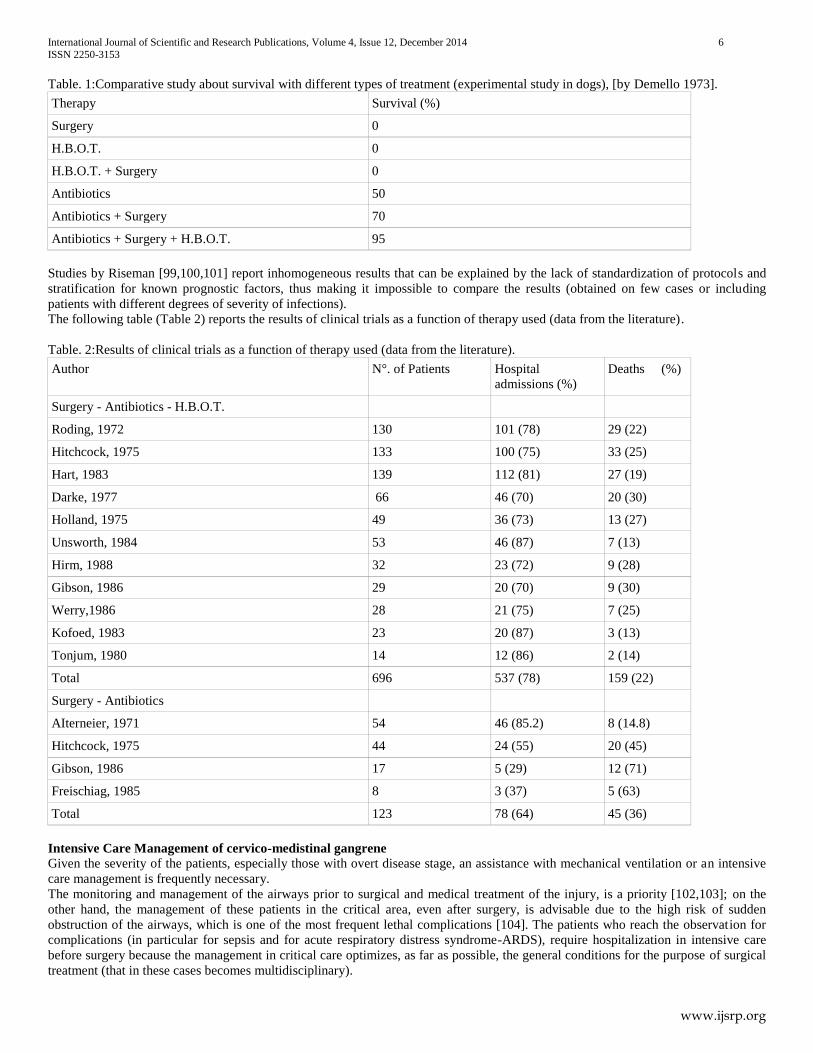
material, presence of fluid in the under-mesocolic area”.
Picture 1 (CT scan on 09 June): mediastinal collection, pericardial collection.
Picture 2 (CT scan on 09 June): air bubbles in the context of the back muscles at the level of C6 extending in the subscapularis area.

International Journal of Scientific and Research Publications, Volume 4, Issue 12, December 2014 9
ISSN 2250-3153
www.ijsrp.org
Picture 3 (CT scan on 09 June): bubbles extending along the muscles of the abdominal wall.
The patient underwent a surgical revision of the right lateral retropharyngeal region, a right cervicotomy with evidence of a diffuse
cellulitis of the adipose tissue.
The laboratory tests were as follows: white blood cell count 10.330/microliter with 94% of neutrophils, C-reactive protein (C.R.P.)
was 333 mg/L, Creatine Phosphokinase (C.P.K.) 670 UI/L, Myoglobin 1037 ng/ml.
Given the development of the CT scan and of the laboratory tests, on 09 June, after verification of fitness to the hyperbaric treatment,
the patient began H.B.O.T. at 2.8 ATA in accordance with a protocol that included three treatments in the first 24 hours, then one
treatments every twelve hours for the next two days, and daily in the following days. Each session contemplated the respiration of 60
minutes of oxygen (FiO2 of 1) at 2.8 ATA. The choice of each treatment, as far as the air breaks concern, was influenced by the
hemodynamic instability of the patient.
After the fourth session (11 June) the CT scan already revealed: "reduction of fluid collection in the lateral cervical structures and
reduction of air bubbles in the parapharyngeal soft tissues. In the dorsal right area, disappearance of air bubbles in the context of the
lower back muscles persisting along the muscles of the right abdominal wall and a small bubble in the pelvic region".
The laboratory tests showed an improvement: white blood cell count 8.360/microliter with 80% of neutrophils, C-reactive protein
(C.R.P.) was 83 mg/L, Creatine Phosphokinase (C.P.K.) 249 UI/L, Myoglobin 200 ng/ml.
On 12 June he performed only one session of H.B.O.T. at 2.8 ATA (the second session was aborted because of problems with the
hyperbaric ventilator).
After the 7th session (13 June) the TC scan highlighted: “riduced parapharyngeal bubbles, bubbles near the surgical breaches, no deep
lateral cervical fluid collections, reduced the bubble in the right anterior abdominal wall. Small left average apical pneumothorax
(P.N.X.)”. PNX was not confirmed at the following TC scan.
The laboratory tests were as follows: white blood cell count 11.000/microliter with 80% of neutrophils, C-reactive protein (C.R.P.)
was 42 mg/L.
Given the improvement, the patient began H.B.O.T. at 2.5 ATA.
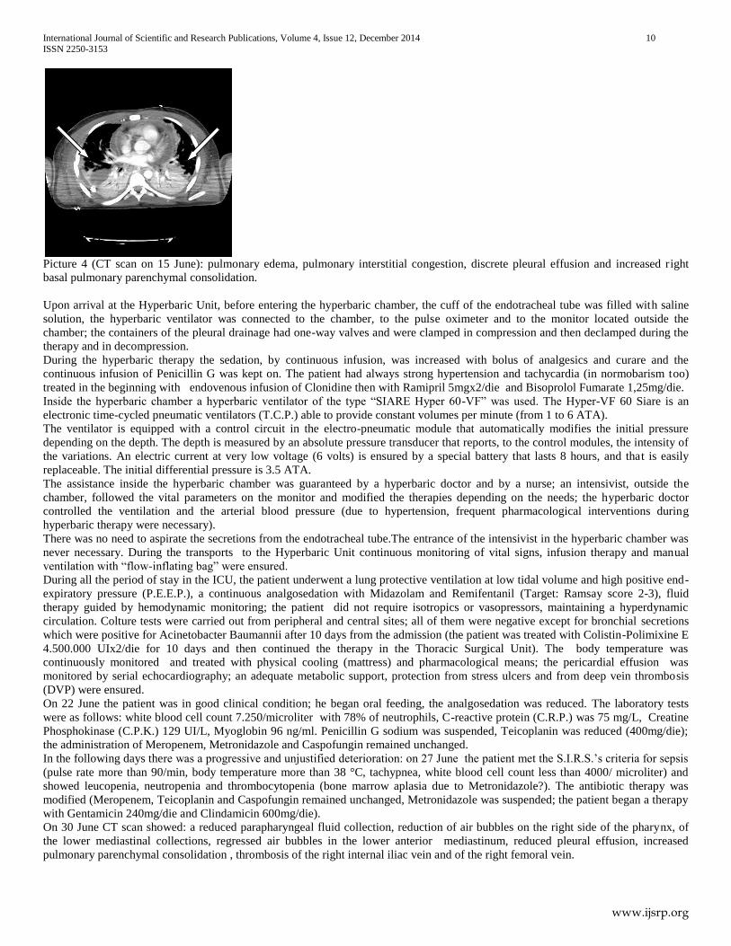
Given the continuous improvement of the infection in the TC scan (performed on 15 June after the ninth session of H.B.O.T.) (no
more obvious parapharyngeal bubbles, no more deep lateral cervical fluid collections, no more abdominal bubbles) and in the
laboratory tests and the occurrence of frontal, sphenoid and maxillary sinusophatic complications arisen with epistaxis and otorrhagia
during the last session of H.B.O.T. and, above all, pulmonary complications (a picture suggestive of pulmonary edema, pulmonary
interstitial congestion, discrete pleural effusion and increased right basal pulmonary parenchymal consolidation), we decided to
suspend H.B.O.T. after nine sessions in seven days.
International Journal of Scientific and Research Publications, Volume 4, Issue 12, December 2014 10
ISSN 2250-3153
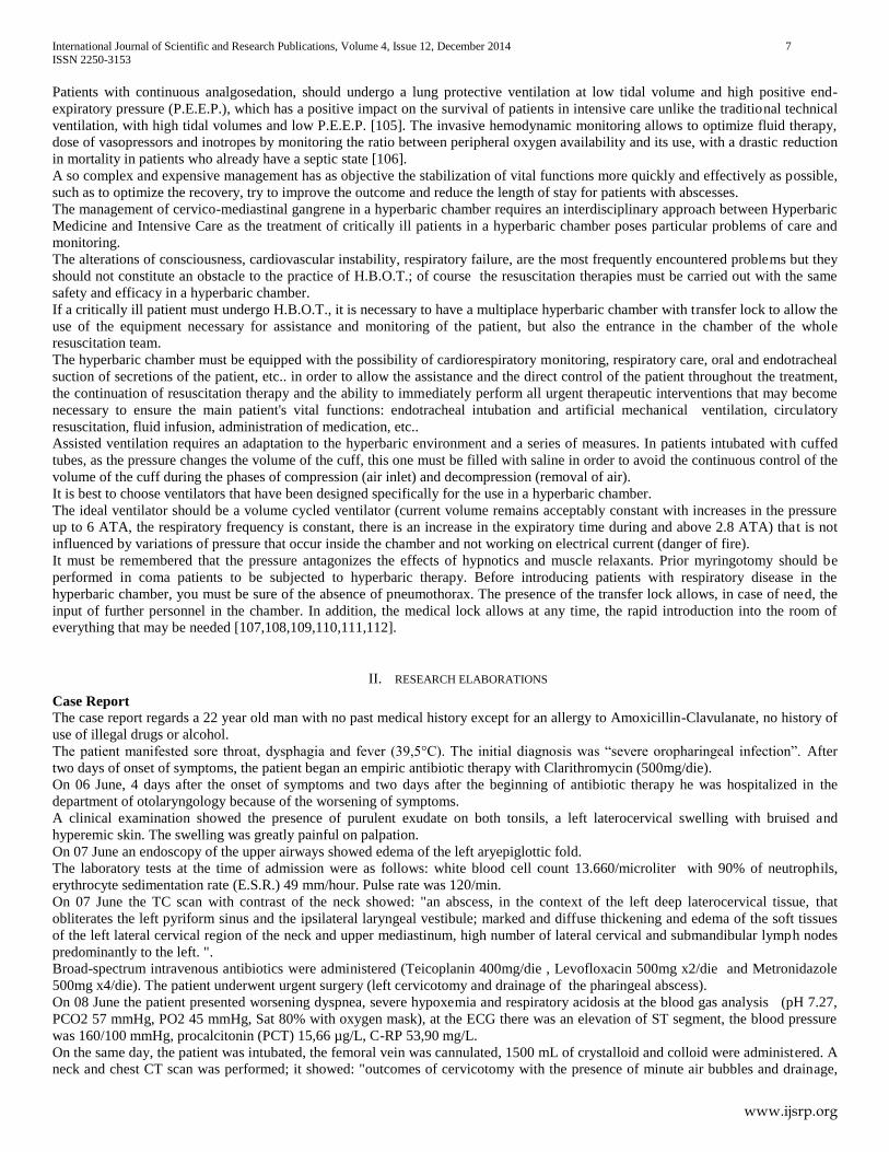
www.ijsrp.org
Picture 4 (CT scan on 15 June): pulmonary edema, pulmonary interstitial congestion, discrete pleural effusion and increased right
basal pulmonary parenchymal consolidation.
Upon arrival at the Hyperbaric Unit, before entering the hyperbaric chamber, the cuff of the endotracheal tube was filled with saline
solution, the hyperbaric ventilator was connected to the chamber, to the pulse oximeter and to the monitor located outside the
chamber; the containers of the pleural drainage had one-way valves and were clamped in compression and then declamped during the
therapy and in decompression.
During the hyperbaric therapy the sedation, by continuous infusion, was increased with bolus of analgesics and curare and the
continuous infusion of Penicillin G was kept on. The patient had always strong hypertension and tachycardia (in normobarism too)
treated in the beginning with endovenous infusion of Clonidine then with Ramipril 5mgx2/die and Bisoprolol Fumarate 1,25mg/die.
Inside the hyperbaric chamber a hyperbaric ventilator of the type “SIARE Hyper 60-VF” was used. The Hyper-VF 60 Siare is an
electronic time-cycled pneumatic ventilators (T.C.P.) able to provide constant volumes per minute (from 1 to 6 ATA).
The ventilator is equipped with a control circuit in the electro-pneumatic module that automatically modifies the initial pressure
depending on the depth. The depth is measured by an absolute pressure transducer that reports, to the control modules, the intensity of
the variations. An electric current at very low voltage (6 volts) is ensured by a special battery that lasts 8 hours, and that is easily
replaceable. The initial differential pressure is 3.5 ATA.
The assistance inside the hyperbaric chamber was guaranteed by a hyperbaric doctor and by a nurse; an intensivist, outside the
chamber, followed the vital parameters on the monitor and modified the therapies depending on the needs; the hyperbaric doctor
controlled the ventilation and the arterial blood pressure (due to hypertension, frequent pharmacological interventions during
hyperbaric therapy were necessary).
There was no need to aspirate the secretions from the endotracheal tube.The entrance of the intensivist in the hyperbaric chamber was
never necessary. During the transports to the Hyperbaric Unit continuous monitoring of vital signs, infusion therapy and manual
ventilation with “flow-inflating bag” were ensured.
During all the period of stay in the ICU, the patient underwent a lung protective ventilation at low tidal volume and high positive end-
expiratory pressure (P.E.E.P.), a continuous analgosedation with Midazolam and Remifentanil (Target: Ramsay score 2-3), fluid
therapy guided by hemodynamic monitoring; the patient did not require isotropics or vasopressors, maintaining a hyperdynamic
circulation. Colture tests were carried out from peripheral and central sites; all of them were negative except for bronchial secretions
which were positive for Acinetobacter Baumannii after 10 days from the admission (the patient was treated with Colistin-Polimixine E
4.500.000 UIx2/die for 10 days and then continued the therapy in the Thoracic Surgical Unit). The body temperature was
continuously monitored and treated with physical cooling (mattress) and pharmacological means; the pericardial effusion was
monitored by serial echocardiography; an adequate metabolic support, protection from stress ulcers and from deep vein thrombosis
(DVP) were ensured.
On 22 June the patient was in good clinical condition; he began oral feeding, the analgosedation was reduced. The laboratory tests
were as follows: white blood cell count 7.250/microliter with 78% of neutrophils, C-reactive protein (C.R.P.) was 75 mg/L, Creatine
Phosphokinase (C.P.K.) 129 UI/L, Myoglobin 96 ng/ml. Penicillin G sodium was suspended, Teicoplanin was reduced (400mg/die);
the administration of Meropenem, Metronidazole and Caspofungin remained unchanged.
In the following days there was a progressive and unjustified deterioration: on 27 June the patient met the S.I.R.S.’s criteria for sepsis
(pulse rate more than 90/min, body temperature more than 38 °C, tachypnea, white blood cell count less than 4000/ microliter) and
showed leucopenia, neutropenia and thrombocytopenia (bone marrow aplasia due to Metronidazole?). The antibiotic therapy was
modified (Meropenem, Teicoplanin and Caspofungin remained unchanged, Metronidazole was suspended; the patient began a therapy
with Gentamicin 240mg/die and Clindamicin 600mg/die).
On 30 June CT scan showed: a reduced parapharyngeal fluid collection, reduction of air bubbles on the right side of the pharynx, of
the lower mediastinal collections, regressed air bubbles in the lower anterior mediastinum, reduced pleural effusion, increased
pulmonary parenchymal consolidation , thrombosis of the right internal iliac vein and of the right femoral vein.

International Journal of Scientific and Research Publications, Volume 4, Issue 12, December 2014 11
ISSN 2250-3153
www.ijsrp.org
On the same day a worsening of the laboratory tests (as far as leucopenia, neutropenia and thrombocytopenia concerned) was
observed. Gentamicin, Teicoplanin and Meropenem were suspended, Clindamicin was increased to 600 mgx4/die. The patient was
treated also with Linesozid 600 mg/die and Levofloxacin 500 mgx2/die.
On the 25th
day of recovery in the Intensive Care Unit (on 02 July), for the peristent fever, the presence of signs of sepsis and high
value of endotoxin, the patient started a therapy with Toraymyxin-Polimixine B and in the same time he underwent a C.P.F.A.
(Coupled Plasma Filtration Adsorption) for five days.
On the 26th
day of recovery a haemocolture showed a candidemia (even if the patient was being treated with Caspofungin70 mg/die);
the patient started a therapy with Amphootericin B 3mg/kg/die that was continued for 15 days with favorable outcome.
III. RESULTS AND FINDING
The patient was discharged from Intensive Care Unit on 15 july, after 38 days of recovery and in good clinicl conditions, and was
transferred to the Thoracic Surgical Unit.
IV. DISCUSSION
P.N.I. is a severe infection of the cervical district; it is rarely observed in clinical practice, it is fast-spreading and with a multi regional
involvement and can affect any age of life. The odontogenic origin of the infection is the most frequent, but fasciitis can also arise as a
complication of inflammation of the oropharynx and any other infectious disease in the neck if neglected.
The first clinical signs and symptoms are attributable to the organ that is initially affected by the infection. The septic fever always
accompanies the infection, although often the discrepancy between the inflammatory process and the general state of the patient leads
to underestimate the severity of the infection.
The diagnosis as well as on clinical data must make use of CT scan, the only exam able to document the disease status and the
progression of the infection. The CT scan is also useful in monitoring the progression of the disease (other foci of necrosis frequently
appear after a first operation of drainage).
The surgical drainage should be performed as early as possible (no later than 24 hours from the diagnosis) when the presence of the
collections has been documented, as the disease evolves in the caudal direction and then quickly affects the mediastinum and the
organs contained in it.
The cervicotomy is the treatment to be carried out and may be limited to a single district if gangrene is caught at an early stage, but it
must involve all levels of the neck, and even beyond, when the inflammation is wide, or is spreading to the neck or pectoral region.
The tracheotomy must accompany the cervicotomy when there is a condition of shortness of breath or when the disease state suggests
that an appropriate ventilatory assistance will be necessary in a short time.
The surgical procedure should be accompanied by appropriate antibiotic therapy to be practiced, most of the times, in an empirical
way as the germs responsible for the infection are rarely identified and as the initial etiological factor often remains the only cause of
the gangrene. Therefore it is essential, more than the use of a single drug, the pharmacological association of drugs having elective
spectrum towards Gram-positive germs with those directed to treat infections by Gram-negative germs and also anaerobes. It is
important to consider the possibility of fungal associated infections sometimes with immediate onset, sometimes late-occurring
probably favored by the patient's stay in the intensive care environment. A treatment in this sense must always be considered in
advance.
In agreement with what was found in the literature, the bacterial culture has non been able to isolate any pathogen and thus was of no
help for the etiopathegenetic aspect. The diagnosis of cervico-mediastinal gangrene was placed according to clinic and to CT scan
with contrast. In this case report, many of the bubbles identified on CT scan were iatrogenic (drainage, surgery) and only few isolated
bubbles (with doubtful prognostic value) were highlighted. The CT scan was performed repeatedly during the therapy to monitor the
evolution of the disease. A multidisciplinary approach (surgery, antibiotics, H.B.O.T. and intensive care) has been applied. H.B.O.T.
has been suspended after the ninth session for the occurence of complications to the upper and low airways. A clinical improvement
was seen after the start of H.B.O.T., but its specific role in this improvement is difficult to be estimated.
Here comes the most crucial step for yourresearch publication. Ensure the drafted journal is critically reviewed by your peers or any
subject matter experts. Always try to get maximum review comments even if you are well confident about your paper.
V. CONCLUSION
The necrotizing mediastinitis has an insidious onset; indeed it has affected a patient without apparent risk factors who had already
begun an antibiotic therapy (this fact emphasizes the remarkable antibiotic resistance). The patient fulfilled Estrera's criteria for
clinical and radiological manifestation of mediastinitis. Diagnosis was possibile thanks to the TC scan. A multidisciplinary approach
was necessary. The cervicotomy was promptly performed, a broad-spectrum antibiotic therapy was begun and the hyperbaric therapy
was carried out together with an intensive care. This multidiscilinary approach led to a favorable outcome. Hyperbaric therapy, which
started according to the established protocol, continued with some changes: at the third day of treatment, for a technical problem of the
ventilator, the patient underwent only one treatment. The seventh and the eigth sessions were performed at 2.5 ATA and after the ninth

International Journal of Scientific and Research Publications, Volume 4, Issue 12, December 2014 12
ISSN 2250-3153
www.ijsrp.org
session H.B.O.T. was stopped because of the upper and lower airways complications and because of the improvement of the cervico-
mediastinal lesions documented with CT scan. It is not possible to estimate the exact role of H.B.O.T. in the outcome of the patient.

International Journal of Scientific and Research Publications, Volume 4, Issue 12, December 2014 13
ISSN 2250-3153
www.ijsrp.org
REFERENCES
[1] Arena F., Rossolini G.M., “Microbiologia delle raccolte ascessuali”, in: Complicanze ascessuali delle flogosi del distretto testa-collo (Petrone D. ed.),
Quaderni Monografici di Aggiornamento A.O.O.I. vol. mon.:53-61, 2011. [2] Herzon F., “Harris P. Mosher Award thesis. Peritonsillar abscess: incidence, current management practices, and a proposal for treatment guidelines”, in:
Laryngoscope 105(8 Pt 3 Suppl. 74):1-17, Aug. 1995.
[3] Barbara M., Cariti F., Grasso R., “Complicanze ascessuali peritonsillari”, in: Complicanze ascessuali delle flogosi del distretto testa-collo (Petrone D. ed.), Quaderni Monografici di Aggiornamento A.O.O.I. vol. mon.:127-135, 2011.
[4] Passy V., “Pathogenesis of peritonsillar abscess”, in: Laryngoscope 104(2):185–190, 1994.
[5] Johnson R.F., Stewart M.G., Wright C.G., “An evidence-based review of the treatment of peritonsillar abscess”, in: Otolaryngol Head Neck Surg. 128(3):332–343, 2003.
[6] Galioto N.J., “Peritonsillar abscess”. In: Am. Fam. Physician , 77(2):199-202, Broadlawns Medical Center, Des Moines, Iowa, 15 Jan. 2008.
[7] Repanos C., Mukherjee P., Alwahab Y., “Role of microbiological studies in management of peritonsillar abscess”, in: J. Laryngol. Otol. 123(8):877-9, Aug. 2009.
[8] Brook I., Frazier E.H., Thompson D.H., “Aerobic and anaerobic microbiology of peritonsillar abscess”, in: Laryngoscope,101(3):289-92, Mar. 1991.
[9] Brook I., “Microbiology of polymicrobial abscesses and implications for therapy”, in: J. Antimicrob. Chemother 50(6):805-10, Dec. 2002. [10] Brook I., “Non-odontogenic abscesses in the head and neck region”, in: Periodontol 2000 49:106-25, Feb. 2009.
[11] Kronenberg J., Wolf M., Leventon G., “Peritonsillar abscess: recurrence rate and the indication for tonsillectomy”, in: Am. J. Otolaryngol 8(2):82-4, Mar-
Apr. 1987. [12] Szuhay G., Tewfik T.L., “Peritonsillar abscess or cellulitis? A clinical comparative paediatric study”, in: J. Otolaryngol. 27(4):206-12, Aug. 1998.
[13] Garribba A.P., Ettorre G.C., “Diagnostica per immagini”, in: Complicanze ascessuali delle flogosi del distretto testa-collo (Petrone D. ed.), Quaderni
Monografici di Aggiornamento A.O.O.I. vol. mon.:85-100, 2011. [14] Irfan M., Baharudin A., “Management Of Peritonsillar Infection: Hospital Universiti Sains Malaysia Experience”, in: The Internet Journal of
Otorhinolaryngology 10(1)[html], 2008.
[15] Prior A., Montgomery P., Mitchelmore I., Tabaqchali S., “The microbiology and antibiotic treatment of peritonsillar abscesses”, in: Clin. Otolaryngol. Allied Sci. 20(3):219-23, Jun. 1995.
[16] Grassia R., Mosca F., Leone C.A., “Epidemiologia e clinica delle complicanze ascessuali del distretto cervico facciale”, in: Complicanze ascessuali delle
flogosi del distretto testa-collo (Petrone D. ed.), Quaderni Monografici di Aggiornamento A.O.O.I. vol. mon.:63-73, 2011. [17] Zurawski C.A. et al., “Invasive group A streptococcal disease in metropolitan Atlanta: a population-based assessment”, in: Clin. Infect. Dis. 27(1):150-7, Jul.
1998.
[18] Bahna M., Canalis R.F., “Necrotizing fasciitis. (Streptococcal gangrene) of the face. Report of a case and review of the literature”, in: Arch. Otolaryngol. 106(10):648-51, Oct. 1980.
[19] Rapoport Y., “Cervical necrotizing fasciitis of odontogenic origin”, in: Oral Surg. Oral Med. Oral Pathol. 72(1):15-8, Jul. 1991.
[20] Helmy A.S. et al., “Life-threatening cervical necrotizing fasciitis”, in: J. R. Coll. Surg. Edinb. 42(6):410-3, Dec. 1997. [21] Mathieu D., “Cervical necrotizing fasciitis: clinical manifestations and management”, in: Clin. Infect. Dis. 21(1):51-6, Jul. 1995.
[22] Wilson B., “Necrotizing fasciitis”, in: Am. Surg. 18(4): 416–431, 1952.
[23] Valko P.C., Barrett S.M., Campbell J.P., “Odontogenic cervical necrotizing fasciitis”, in: Ann. Emerg. Med. 19(5):568-71, May 1990. [24] Gillis A.R., Gillis TM “Necrotizing cervical fasciitis of unknown origin”, in: J. Otolaryngol. 21(3):171-3, Jun. 1992.
[25] Kantu S., Har-El G., “Cervical necrotizing fasciitis”, in: Ann. Otol. Rhinol. Laryngol. 106(11):965-70, Nov. 1997.
[26] Ohi K., Inamura N., Suzuki M., Suzuki N., Ishigaki M., Takasaka T., “Three patients with gas gangrene of the head and neck”, in: Nippon Jibiinkoka Gakkai Kaiho 96(7):1079-85, Jul. 1993.
[27] Skitarelić N., Mladina R., Morović M., Skitarelić N., “Cervical necrotizing fasciitis: sources and outcomes”, in: Infection 31(1):39-44, Jan. 2003.
[28] Banerjee A.R. et al., “Cervical Necrotizing fasciitis: a distinct clinicopathological entity?”, in: J. Laryngol. Otol. 110(1):81-86, 1996. [29] Mandell G.L., Bennet J.E., Dolin R., Mandell, Douglas, and Bennett’s Principles and Practice of Infectious Disease, 6th ed., Elsevier, 2005.
[30] Bottin R. et al., “Deep neck infection: a present-day complication. A retrospective review of 83 cases 1998-2001”, in: Eur. Arch. Otorhinolaryngol.
260(10):576-579, 2003. [31] Greinwald J.H. Jr., “Peritonsillar abscess: an unlikely cause of necrotizing fasciitis”, in: Ann. Otol. Rhinol. Laryngol. 104(2):133-7, Feb. 1995.
[32] Balcerak R.J. et al., “Cervicofacial necrotizing fasciitis: report of three cases and literature review”, in: J. Oral. Maxillofac. Surg. 46(6):450-9, Jun. 1988.
[33] Meisel R.H. et al., “Cervical necrotizing fasciitis”, in: Laryngoscope 104(7):795-8, Jul. 1994. [34] Freeman H.P., Oluwole SF, Ganepola GA, Dy E., “Necrotizing fasciitis”, in: Am. J. Surg. 142(3):377-83, Sept. 1981.
[35] De Backer T. et al., “Management of necrotizing fasciitis in the neck”, in: J. Craniomaxillofac. Surg. 24(6):366-71, Dec. 1996.
[36] Zbären P., Rothen H.U., Lang H., Becker M., “Necrotizing fasciitis of soft tissues of the face and neck”, in: HNO 43(10):619-23, Oct. 1995. [37] Haraden B.M., Zwemer F.L. Jr., “Descending necrotizing mediastinitis: complication of a simple dental infection”, in: Ann. Emerg. Med. 29(5):683-6, May
1997. [38] Chin R.S., Kaltman S.I., Colella J., “Fatal necrotizing fasciitis following a mandibular fracture”, in: J. Craniomaxillofac. Trauma. 1(3):22-9, Fall 1995.
[39] Beerens A.J., Bauwens L.J., Leemans C.R., “A fatal case of craniofacial necrotizing fasciitis”, in: Eur. Arch. Otorhinolaryngol. 256(10):506-9, 1999.
[40] Owada R., Kanamori A., Hirai N., Yajima Y., “ A case of NIDDM with non-clostridial gas-producing infection in the lower limb-the effects of hyperbaric
oxygen therapy”, in: Kansenshogaku Zasshi 68(2):263-7, Feb. 1994.
[41] Isaacs L.M., Kotton B., Peralta M.M. Jr., Shekar R., Meden G., Brown L.A., Raaf J.H., “Fatal mediastinal abscess from upper respiratory infection”, in: Ear
Nose Throat J. 72(9):620-2, 624-6, 631, Sept. 1993. [42] Chua H.K., Segar C.B., Krishnan R., Ho C.K., “Cervical necrotising fasciitis consequent to mastoid infection”, in: Med. J. Malaysia 57(1):104-7, Mar. 2002.
[43] Nduwke K.C., Fatusi O.A., Ugboko V.I., "Craniocervical necrotizing fasciitis in Ile-Ife”, in: Br. J. Oral Maxillofac. Surg. 40(1):64-7, Nigeria, Feb. 2002.
[44] Şafak M.A., Haberal I., Kilic D., Gocmen H., “Necrotizing fasciitis secondary to peritonsillar abscess: a new case and review of eight earlier cases”, in: Ear Nose Throat J. 80(11):824- 30,833, Nov. 2001.
[45] Marioni G., Bottin R., Tregnaghi A., Boninsegna M., Staffieri A., “Craniocervical Necrotizing Fasciitis Secondary to Parotid Gland Abscess”, in: Acta Oto-
Laryngologica 123(6):737-740, Aug. 2003. [46] Sethi D.S., Stanley R.E., “Deep neck abscesses-changing trends”, in: J. Laryngol. Otol. 108(2):138-43, Feb. 1994.
[47] Petrone P., Fiorella M.L., Fiorella R., Petrone D., Campanini A., Vicini C. Fasciti necrotizzanti del distretto cervico mediastinico: studio policentrico”, in:
Complicanze ascessuali delle flogosi del distretto testa-collo (Petrone D. ed.), Quaderni Monografici di Aggiornamento A.O.O.I. vol. mon.:161-183, 2011. [48] Nallathambi M.N., Ivatury R.R., Rohman M., Rao P.M., Stahl W.M., “Craniocervical necrotizing fasciitis: critical factor in management”, in: Can. J. Surg.
30(1): 61-3, Jan. 1987.
[49] Weaver E., Nguyen X., Brooks M.A., “Descending necrotising mediastinitis: two case reports and review af the literature”, in: Eur. Respir. Rev. 19(116):141-149, 2010.

International Journal of Scientific and Research Publications, Volume 4, Issue 12, December 2014 14
ISSN 2250-3153
www.ijsrp.org
[50] Gidley P.W., Ghorayeb B.Y., Stiernberg C.M., “Contemporary management of deep neck space infections”, in: Otolaryngol Head Neck Surg. 116(1):16-22,
Jan. 1997. [51] Krespi Y.P. et al., “Massive necrotizing infectionsof the neck”, in: Head Neck Surg. 3(6):475-81, Jul.-Aug. 1981.
[52] Satoh O., Miyabe M., Tsukamoto T., Seki S., Ohyama I., Namiki A., Iizuka K., “Deep neck infection following endotracheal intubation”, in: Masui
41(12):1981-5, Dec. 1992. [53] Infante Cossio P., Gonzalez Padilla J.D., Garcia Peria A., Salazar Fernandez C.I., Rollon Mayordomo A., “Acute mediastinitis with fatal outcome secondary
to odontogenic infection”, in: Rev. Actual Odontoestomatol. Esp. 51(400):51-4. Jan.-Feb. 1991.
[54] Umeda M., Minamikawa T., Komatsubara H., Shibuya Y., Yokoo S., Komori T., “Necrotizing fasciitis caused by dental infection: a retrospective analysis of 9 cases and a rewiev of the literature”, in: Oral Surg Oral Med Oral Pathol Oral Radiol Endod 95(3):283-90., Mar. 2003.
[55] Mohadjer C., Argat M., Maier H., Weidauer H., “Gas producing anaerobic infection of the neck”, in: Laryngorhinootologie 74(5):322-4, May 1995.
[56] Sakaguchi M., Sato S., Ishiyama T., Katsuno S., Taguchi K., “Characterization and management of deep neck infections”, in: Int. J. Oral Maxillofac. Surg. 26(2):131-4, Apr. 1997.
[57] Lin C., Yeh F.L., Lin JT, Ma H., Hwang C.H., Shen B.H., Fang R.H., “Necrotizing fasciitis of the head and neck: an analysis of 47 cases”, in: Plast.
Reconstr. Surg. 107(7):1684-93, Jun. 2001. [58] Chaplain A., Gouello J.P., Dubin J., “Acute cervical necrotizing fasciitis of pharingeal origin: possible role of steroidal and non-steroidal anti-inflammatory
agents. A propos of 5 cases”, in: Rev. Laryngol. Otol. Rhinol. 117(5):337-80, 1996.
[59] Bloching M., Gudziol S., Gajda M., Berghaus A., “Diagnosis and treatment of necrotizing fasciitis of the head and neck region”, in: Laryngorhinootologie 79(12):774-9, Dec. 2000.
[60] Wysoki M.G., Santora T.A., Shah R.M., Friedman A.C., “Necrotizing fasciitis: CT characteristics”, in: Radiology 203(3):859-63, Jun. 1997.
[61] Sardelli P., Quitadamo S., “Le mediastiniti da ascessi cervicali”, in: Complicanze ascessuali delle flogosi del distretto testa-collo (Petrone D. ed.), Quaderni Monografici di Aggiornamento A.O.O.I. vol. mon. 299-308, 2011.
[62] Kiernan P.D., Hernandez A., Byrne W.D., Bloom R., Dicicco B., Hetrick V., Graling P., Vaughan B., “Descending Cervical Mediastinitis”, in: Ann. Thorac.
Surg. 65:1483-1488, 1998. [63] Papalia E., Rena O., Oliario A., Cavallo A., Giobbe R., Casadio C., Maggi G., Mancuso M., “Descending necrotising mediastinitis: surgical management”,
in: Eur. J. Cardiothoracic Surg. 20(4):739-742, 2001.
[64] Estrera A.S., Landay J.M., Grisham J.M., Sinn D.P., Platt M.R., “Descending necrotizing mediastinitis”, in: Surg. Gynecol. Obstet. 157(6):545-52, 1983. [65] Kotrappa K.S., Bansal R.S., Amin N.M., “Necrotizing fasciitis”, in: Am. Fam. Physician. 1;55(2):448, Feb. 1997.
[66] Yasan H., Uygur K., Tüz M., Doğru H., “The adverse effect of gas formation on prognosis in a patient with deep neck infection”, in: Kulak Burun Boğaz İhtisas Dergisi 11(2):56-9, Aug. 2003.
[67] Becker M., et al., “Necrotizing fasciitis of the head and neck: role of CT in diagnosis and management”, in: Radiology 202(2):471-6, Feb. 1997.
[68] Kedzierski B., Calka K., Wilczyński K., Bojarski B., Jaźwiec P., Bogdal M.T., Stokrocki W., “Gas gangrene or inflammation of the neck-diagnostic difficulties”, in: Przegl Lek. 57(3):181-4, 2000.
[69] Pistolesi G.F., Pocacci C., Vademecum alla tomografia assiale computerizzata del torace, Cap. XVI, Piccin Ed., 1990.
[70] Moss R.M., Kunpittaya S., Sorasuchart A., "Cervical necrotizing fasciitis: an uncommon sequela to dental infection", in: Ann. Otol. Rhinol. Laryngol. 99(8):643-646, Aug. 1990.
[71] Nuber R., Müller W., “Cervical necrotizing fasciitis: clinicopathological aspects”, in: Schweiz Med. Wochenschr. (Suppl.) 125:38S-40S, 2000.
[72] Freischlag J.A., Ajalat G, Busuttil R.W., “Treatment of necrotizing soft tissue infections. The need for a new approach”, in: Am. J. Surg. 149(6):751-5, Jun. 1985.
[73] Hamza N.S., Farrel J., Strauss M., Bonomo R.A., "Deep fascial space infection of the neck: a continuing challenge", in: South Med. J. 96(9):928-32, Sep.
2003. [74] Durrani M.A., Mansfield J.F., "Anesthetic implications of cervicofacial necrotizing fasciitis", in: J. Clin. Anesth. 15(5):378-81, Aug. 2003.
[75] Clark P., Davidson D., Letts M., Lawton L., Jawadi A., "Necrotizing fasciitis secondary to chickenpox infection in children", in: Can. J. Surg. 46(1):9-14,
Feb. 2003. [76] Wheatley M.J., Stirling M.G., Kirsh M.M., Gago O., Orringer M.B., "Descending necrotizing. Mediastinitis: transcervical drainageis not enough", in: Ann.
Thorac. Surg. 49:780-784, 1990.
[77] Corsten M.J. et al., "Optimal treatment of descending necrotizing mediastinitis", in: Thorax 52:702-8, 1997. [78] Kiernan P.D., Hernandez A., Byrne W.D., Bloom R., Dicicco B., Hetrick V., Graling P., Vaughan B., "Descending cervical mediastinitis", in: Ann. Thorac.
Surg. 65(5):1483-1488, May 1998.
[79] Lang M.E., Vaudry W., Robinson J.L., "Case report and literature review of late-onset group B streptococcal disease manifesting as necrotizing fasciitis in preterm infants: is this a new syndrome?", in: Clin. Infect. Dis. 37(9):e132-5, Epub 2003 Oct 03, Nov 1 2003.
[80] Feinerman I.L., Tan H.K., Roberson D.W., Malley R., Kenna M.A., "Necrotizing fasciitis of the pharynx following adenotonsillectomy", in: Int. J. Pediatr.
Otorhinolaryngol., 48(1):1-7, Apr. 25 1999. [81] Skorina J., Kaufman D., "Necrotizing fasciitis originating from pinna perichondritis", in: Otolaryngol. Head Neck Surg., 113(4):467-73, Oct. 1995.
[82] Umbert I.J., Winkelmann R.K., Oliver G.F., Peters M.S., "Necrotizing fasciitis: a clinical, microbiologic, and histiopathologic study of 14 patients", in: J.
Am. Acad. Dermatol. 20(5Pt1):774-81, May 1989. [83] Ribeiro N.F., Cousin G.C., Wilson G.E., Butterworth D.M., Woodwards R.T., "Lethal invasive mucormycosis: case report and recommendations for
treatment", in: Int. J. Oral Maxillofac. Surg. 30(2):156-9, Apr. 2001.
[84] Brummelkamp W.H., Hoogendijk J.L., Boerema I., "Treatment of anaerobic infections (clostridial myositis) by drenching the tissues with oxygen under high atmospheric pressure", in: Surgery 49:299-302, 1961.
[85] Petrone D., Cortese M., Picca D., Laurendi V., “Il ruolo della terapia iperbarica” in: Complicanze ascessuali delle flogosi del distretto testa-collo (Petrone D.
ed.), Quaderni Monografici di Aggiornamento A.O.O.I. vol. mon.:353-368, 2011. [86] Sahni T., Singh P., John M.J., "Hyperbaric Oxygen Therapy: Current Trends And Applications", in: J Assoc. Physicians Iindia 51:280-284, 2003.
[87] Mathieu D., speak at 7th European Consensus Conference on hyperbaric medicine, Lille, December 3rd – 4th 2004, in: Europ. J. Und. Hyp. Med. 6(2):29-
38, 2005. [88] Jain K.K. ed., Textbook of hyperbaric medicine, 5th Ed., Hogefre-Huber Pub., 2009.
[89] Zamboni W.A., Roth A.C., Russell R.C., "The effect of hyperbaric oxygen in reperfusion of ischemic axial skin flaps: a laser Doppler analysis", in: Ann.
Plast. Surg. 28:339-341, 1992. [90] Hunt TK, "The physiology of wound healing", in: Ann. of Emerg. Med. 17(12):1265-1273, 1988.
[91] Babior B.M., "Oxygen dependent microbial killing by phagocytes" (first of two parts), in: N. Engl. J. Med. 298:659-668, 1978.
[92] Mader J.T., Brown G.L., "A mechanism for the amelioration of hyperbaric oxygen of experimental staphylococcal osteomyelitis in rabbits", in: J. Infect. Dis. 142(6):915-922, 1980.
[93] Longoni C., "Rational use of HBO in soft tissues infection in traumatology", in: Med. Sub. Iperb. 10, Dic. 1993.
[94] Park K.M., "Effects of hyperbaric oxygen in infectious disease: basic mechanism", in: Kindwall E.P., Whelan H.T. eds., Hyperbaric medicine practice, Best Publishing Company pp.205-44, 1999.

International Journal of Scientific and Research Publications, Volume 4, Issue 12, December 2014 15
ISSN 2250-3153
www.ijsrp.org
[95] Holland J.A., et al., "Experimental and clinical experience with hyperbaric oxygen in the treatment of clostridial myonecrosis", in: Surgery 77(1):75-85,
1975. [96] Demello F.J., Haglin J.J., Hitchcock C.R., "Comparative study of experimental Clostridium perfringens infection in dogs treated with antibiotics, surgery
and hyperbaric oxygen", in: Surgery 73(6):936-941, 1973.
[97] Hill G.B., Osterhout S., "Experimental effects of hyperbaric oxygen on selected clostridial species. In vitro studies.", in: J. Infect. Dis. 125:17-35, 1972. [98] Him M., et al., "Effects of hyperbaric oxygen and surgery on experimental gas gangrene" in: Eur. Surg. Res. 24:356-362, 1992.
[99] Riseman J.A., et al., "Hyperbaric Oxygen for necrotizing fascitis reduces mortality and need for debridements", in: Surgery 108(5):847-50, 1990.
[100] Brown D.R., et al., "A multicenter review of the treatment of major truncal necrotizing infections with and without hyperbaric oxygen therapy", in: Am. J. Surg. 167(5):485-89, 1994.
[101] Shupak A., et al., "Necrotizing fasciitis: an indication for HBO2?", in: Surgery 118(5):873-878, 1995.
[102] Stripoli T., Pagliarulo R., Masciandaro L., “La gestione anestesiologica”, in: Complicanze ascessuali delle flogosi del distretto testa-collo (Petrone D. ed.), Quaderni Monografici di Aggiornamento A.O.O.I. vol. mon.:369-375, 2011.
[103] Vieira F. et al., "Deep neck infection", in: Otolaryngol. Clin. North Am. 41(3): p. 459-83, 2008.
[104] Gidley P.W., B.Y. Ghorayeb, Stiernberg C.M., "Contemporary management of deep neck space infections", in: Otolaryngol. Head Neck Surg. 116(1):16-22, 1997.
[105] Brower R.G. et al., "Higher versus lower positive end-expiratory pressures in patients with the acute respiratory distress syndrome", in: N. Engl. J. Med.
351(4):327-336, 2004. [106] Rivers E. et al., "Early goal-directed therapy in the treatment of severe sepsis and septic shock", in: N. Engl. J. Med. 345(19):1368-77, 2001.
[107] Alaimo M. et al., "The realm of hyperbaric therapy", in: Atti Congresso Le Possibilità della Terapia Iperbarica, Salsomaggiore (Italy), pp. 38-45, 1992.
[108] Blanch P.B., Desautels D.A., Gallagher T.J., “Deviations in Function of Mechanical Ventilators during Hyperbaric Compression”, in: Resp. Care 36(8): 803-814, 1991.
[109] Camporesi E.M., “Management of critically ill patients in the hyperbaric environment”, in: J. Hyperbaric Med. 2(4):195-199, 1987.
[110] Pelaia P., Conti G., Rocco M., Volturo P., Sposato M., "Hyperbaric Chamber clinical support: mechanical ventilators", in: Proceedings of the XVh Meeting EUBS 89, Eilat Israel, p. 178, 1989.
[111] Youn B.A., Myers R.A., "Volume monitor for mechanical ventilation in the hyperbaric chamber", in: Crit. Care Med. 17(5):453-4, May 1989.
[112] Stahl W., Radermacher P., Calzia E., "Functioning of ICU ventilators under hyperbaric conditions comparison of volume - and pressure - controlled modes", in: Intensive Care Med. 26(4):442-8, Apr. 2000.
AUTHORS
First Author – Vernotico Laura, MD, U.O. Medicina Iperbarica, Ospedale San Paolo, Via Caposcardicchio 10, – 70100 Bari (Italy),
[email protected]– Chief Editor of JANUS (Journal of Aquanautic and Underwater Sciences)
Second Author – Picca Domenico, MD, Director U.O. Medicina Iperbarica, Ospedale San Paolo, Via Caposcardicchio 10, – 70100
Bari (Italy), [email protected]
Third Author – Calculli Luciano, MD, U.O. Medicina Iperbarica, Ospedale San Paolo, Via Caposcardicchio 10, – 70100 Bari (Italy),
Fourth Author – Valentini Nicolò, MD, U.O. Anestesia e Rianimazione, Ospedale San Paolo, Via Caposcardicchio 10, – 70100 Bari
(Italy), [email protected]
Fifth Author – Proto Franco, MD, Director U.O. Anestesia e Rianimazione, Ospedale San Paolo, Via Caposcardicchio 10, – 70100
Bari (Italy).
Sixth Author – Covelli Valentina, MD, U.O. Anestesia e Rianimazione, Ospedale San Paolo, Via Caposcardicchio 10, – 70100 Bari
(Italy), [email protected]
Correspondence Author – Vernotico Laura,[email protected], +393383097509.