Embed Size (px)
Citation preview
The Management of Monochorionic-Diamniotic Twins
Carla Ransom, MD
Vanderbilt University
November 30, 2012
The changing face of pregnancy

Learning objectives
• To understand the biology of twinning
• To understand the complications associated with monochorionic gestation
• To recognize the development of twin-twin transfusion syndrome (TTTS)
• To understand the therapeutic options for TTTS
• To understand the antenatal management of monochorionic gestation
Session 1 – Diagnosis of twin gestation
– Maternal & fetal morbidity
– Prevention of PTB
– Special situations: TTTS, death of one twin
Session 2 – Prenatal diagnosis in twins
– Antenatal testing
– Delivery timing

Disclosures
• None
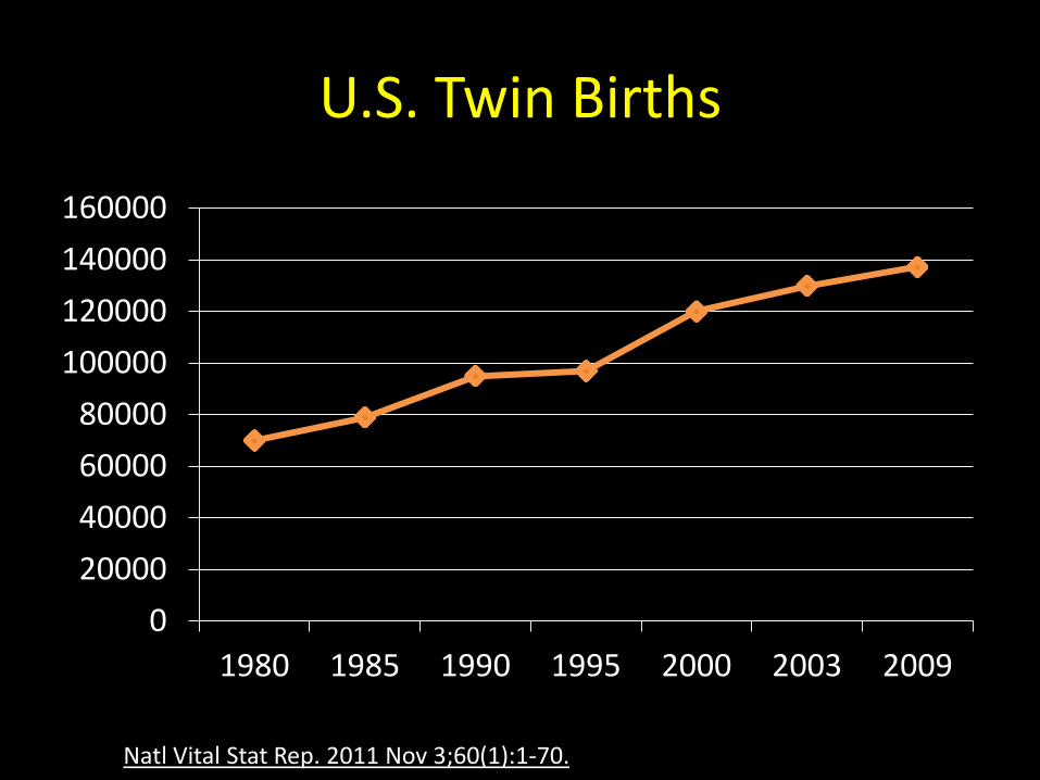
U.S. Twin Births
0
20000
40000
60000
80000
100000
120000
140000
160000
1980 1985 1990 1995 2000 2003 2009
Natl Vital Stat Rep. 2011 Nov 3;60(1):1-70.

With advancing age, FSH/LH rise, as does DZ twinning
Lotze, R. Twins. Introduction to the twin research. F. Rau, Oehirngen. 1937. Zwillinge: Einfiihrungin die Zwillingsforschun
With advancing age, FSH/LH rise, as does DZ twinning
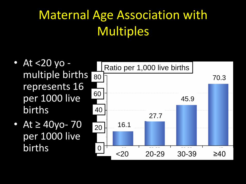
Maternal Age Association with Multiples
• At <20 yo - multiple births represents 16 per 1000 live births
• At ≥ 40yo- 70 per 1000 live births
Ratio per 1,000 live births
16.1
70.3
45.9
27.7
<20 20-29 30-39 ≥40
80
60
40
20
0
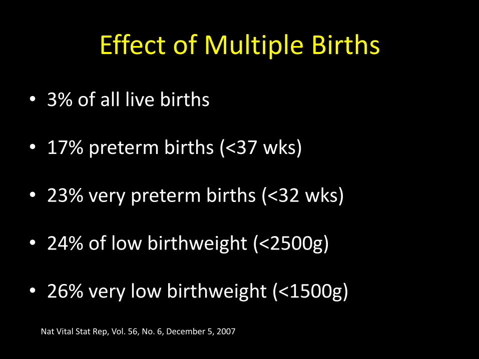
Effect of Multiple Births
• 3% of all live births
• 17% preterm births (<37 wks)
• 23% very preterm births (<32 wks)
• 24% of low birthweight (<2500g)
• 26% very low birthweight (<1500g)
Nat Vital Stat Rep, Vol. 56, No. 6, December 5, 2007
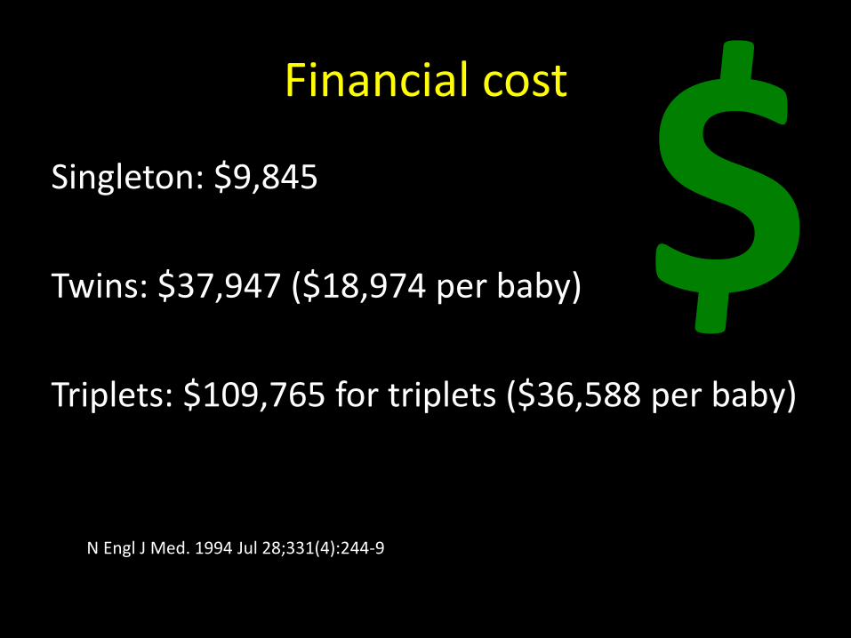
Financial cost
Singleton: $9,845
Twins: $37,947 ($18,974 per baby)
Triplets: $109,765 for triplets ($36,588 per baby)
N Engl J Med. 1994 Jul 28;331(4):244-9
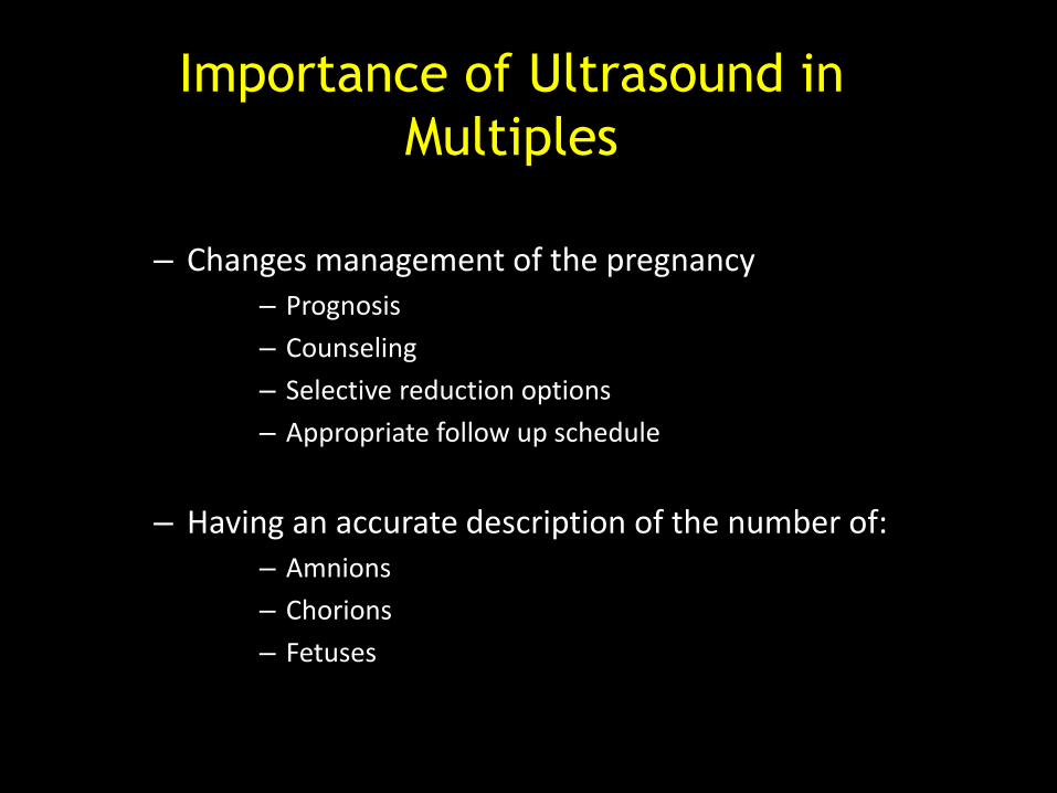
Importance of Ultrasound in
Multiples
– Changes management of the pregnancy – Prognosis
– Counseling
– Selective reduction options
– Appropriate follow up schedule
– Having an accurate description of the number of: – Amnions
– Chorions
– Fetuses
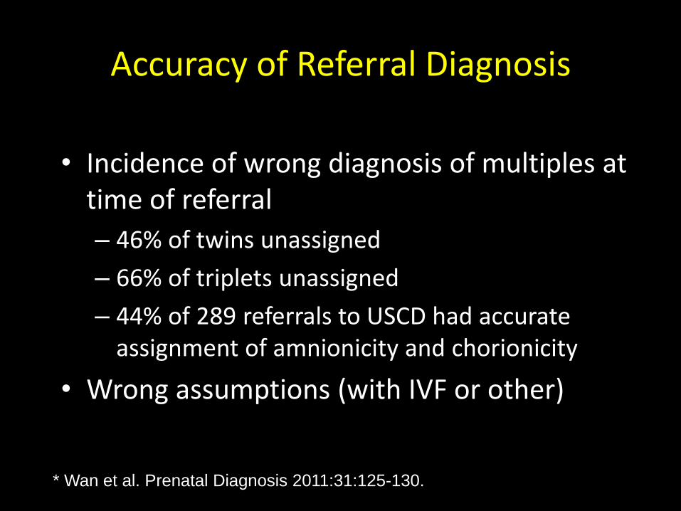
Accuracy of Referral Diagnosis
• Incidence of wrong diagnosis of multiples at time of referral
– 46% of twins unassigned
– 66% of triplets unassigned
– 44% of 289 referrals to USCD had accurate assignment of amnionicity and chorionicity
• Wrong assumptions (with IVF or other)
* Wan et al. Prenatal Diagnosis 2011:31:125-130.
The biology of twinning
Zygosity
Amnionicity & chorionicity
Placentation

Zygosity Chorionicity
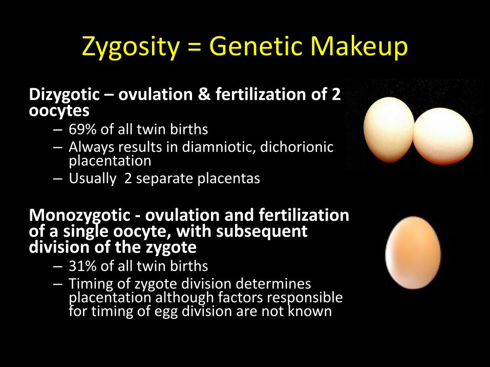
Zygosity = Genetic Makeup
Dizygotic – ovulation & fertilization of 2 oocytes
– 69% of all twin births – Always results in diamniotic, dichorionic
placentation – Usually 2 separate placentas
Monozygotic - ovulation and fertilization of a single oocyte, with subsequent division of the zygote
– 31% of all twin births – Timing of zygote division determines
placentation although factors responsible for timing of egg division are not known
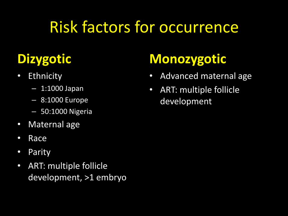
Risk factors for occurrence
Dizygotic • Ethnicity
– 1:1000 Japan
– 8:1000 Europe
– 50:1000 Nigeria
• Maternal age
• Race
• Parity
• ART: multiple follicle development, >1 embryo
Monozygotic • Advanced maternal age
• ART: multiple follicle development
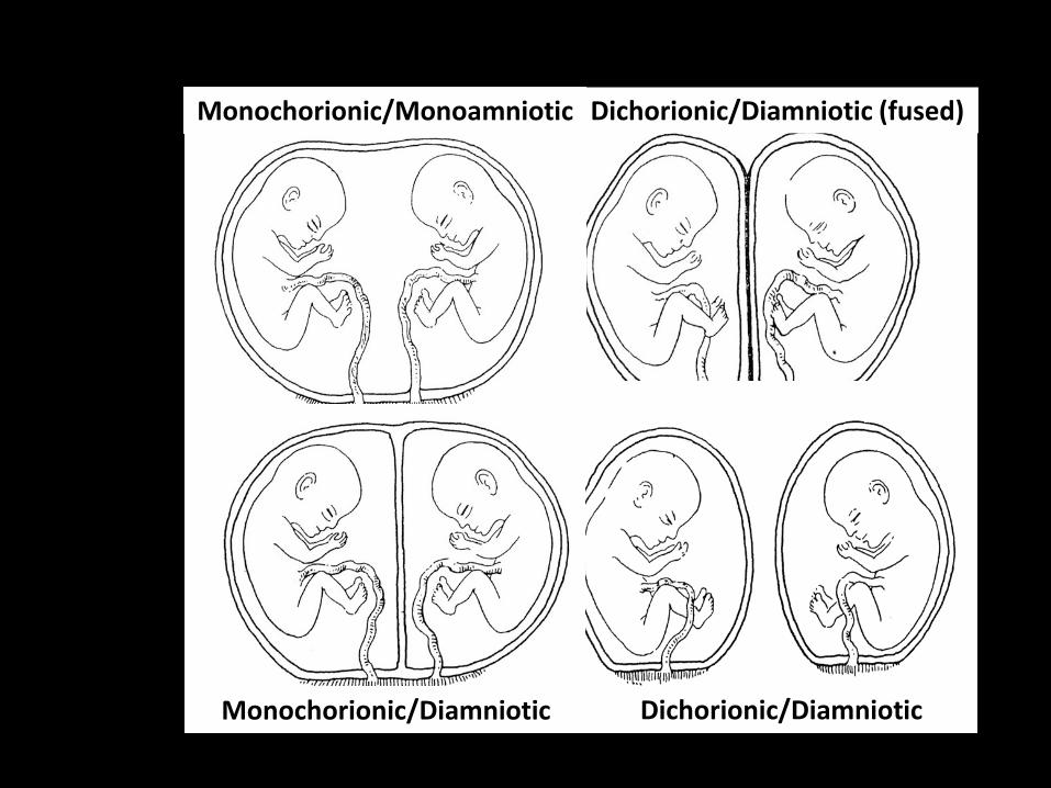
Monochorionic/Monoamniotic Dichorionic/Diamniotic (fused)
Monochorionic/Diamniotic Dichorionic/Diamniotic
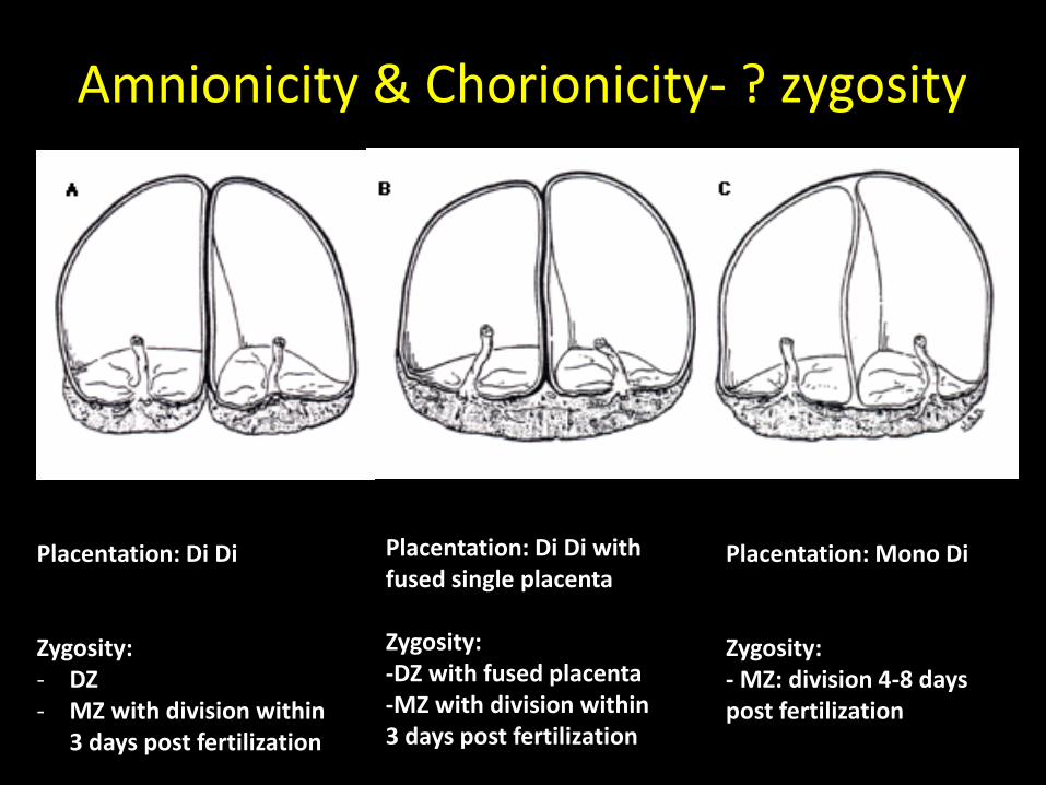
Amnionicity & Chorionicity- ? zygosity
Placentation: Di Di Zygosity: - DZ - MZ with division within
3 days post fertilization
Placentation: Di Di with fused single placenta Zygosity: -DZ with fused placenta -MZ with division within 3 days post fertilization
Placentation: Mono Di Zygosity: - MZ: division 4-8 days post fertilization
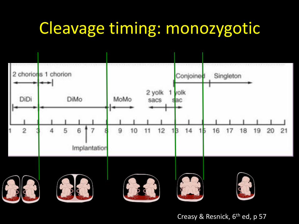
Cleavage timing: monozygotic
Creasy & Resnick, 6th ed, p 57
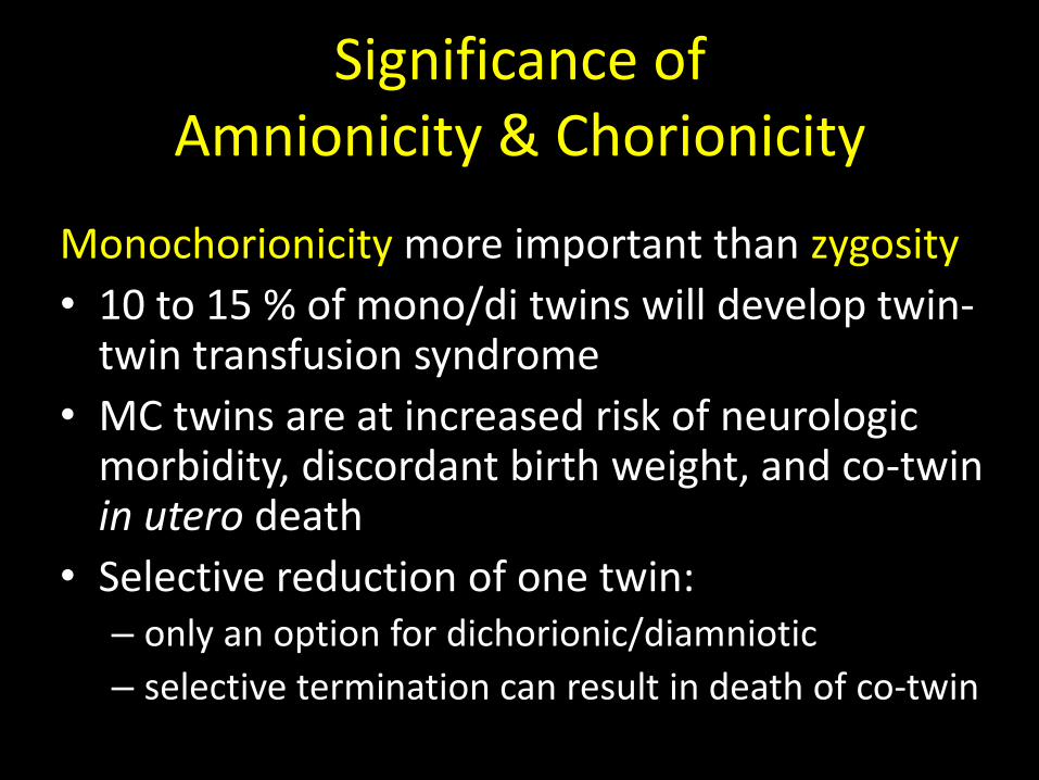
Significance of Amnionicity & Chorionicity
Monochorionicity more important than zygosity
• 10 to 15 % of mono/di twins will develop twin-twin transfusion syndrome
• MC twins are at increased risk of neurologic morbidity, discordant birth weight, and co-twin in utero death
• Selective reduction of one twin: – only an option for dichorionic/diamniotic
– selective termination can result in death of co-twin

SONOGRAPHIC SIGNS OF CHORIONICITY- DICHORIONIC PLACENTATION
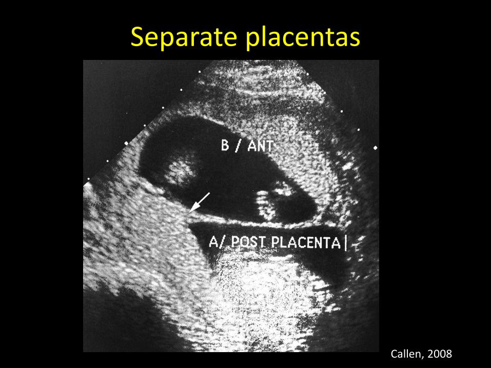
Separate placentas
Callen, 2008
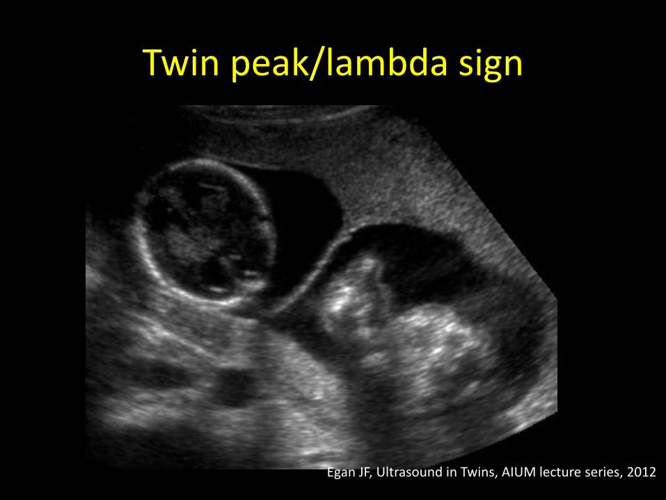
Twin peak/lambda sign
Egan JF, Ultrasound in Twins, AIUM lecture series, 2012
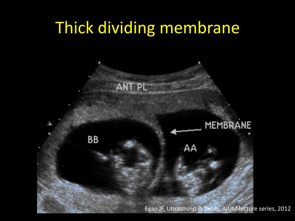
Thick dividing membrane
Egan JF, Ultrasound in Twins, AIUM lecture series, 2012
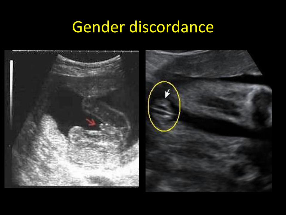
Gender discordance
SONOGRAPHIC SIGNS OF CHORIONICITY- MONOCHORIONIC PLACENTATION
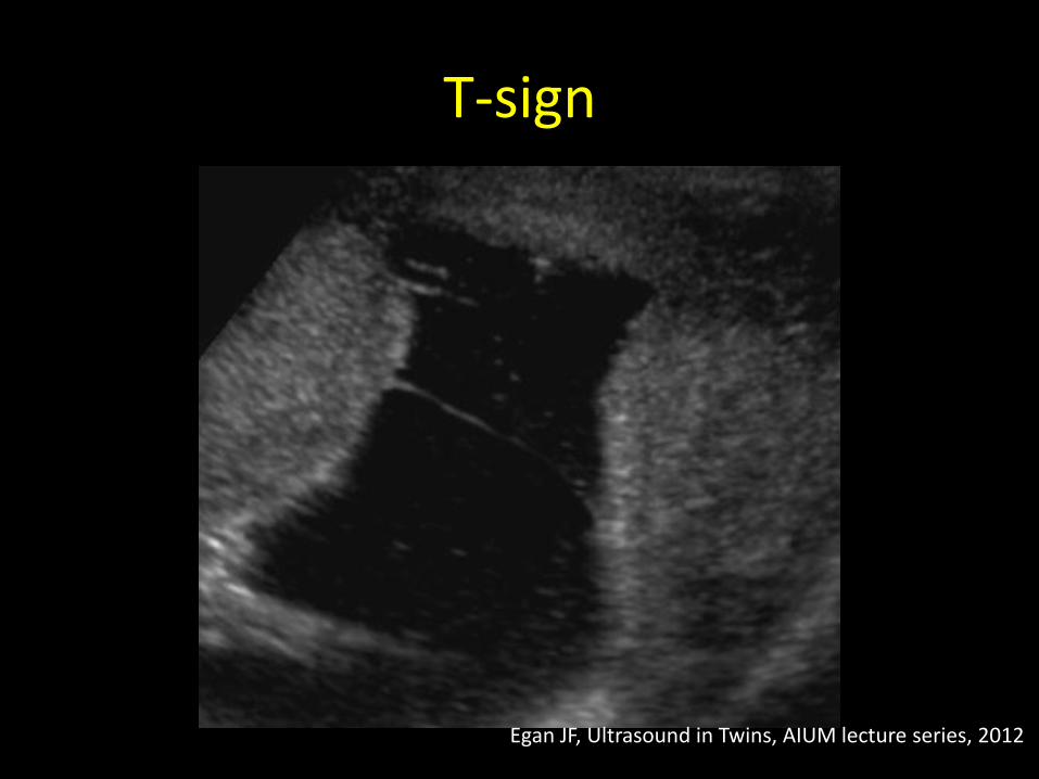
T-sign
Egan JF, Ultrasound in Twins, AIUM lecture series, 2012
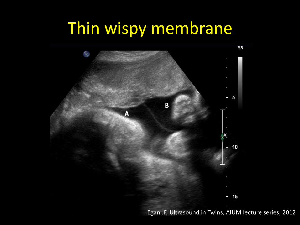
Thin wispy membrane
Egan JF, Ultrasound in Twins, AIUM lecture series, 2012
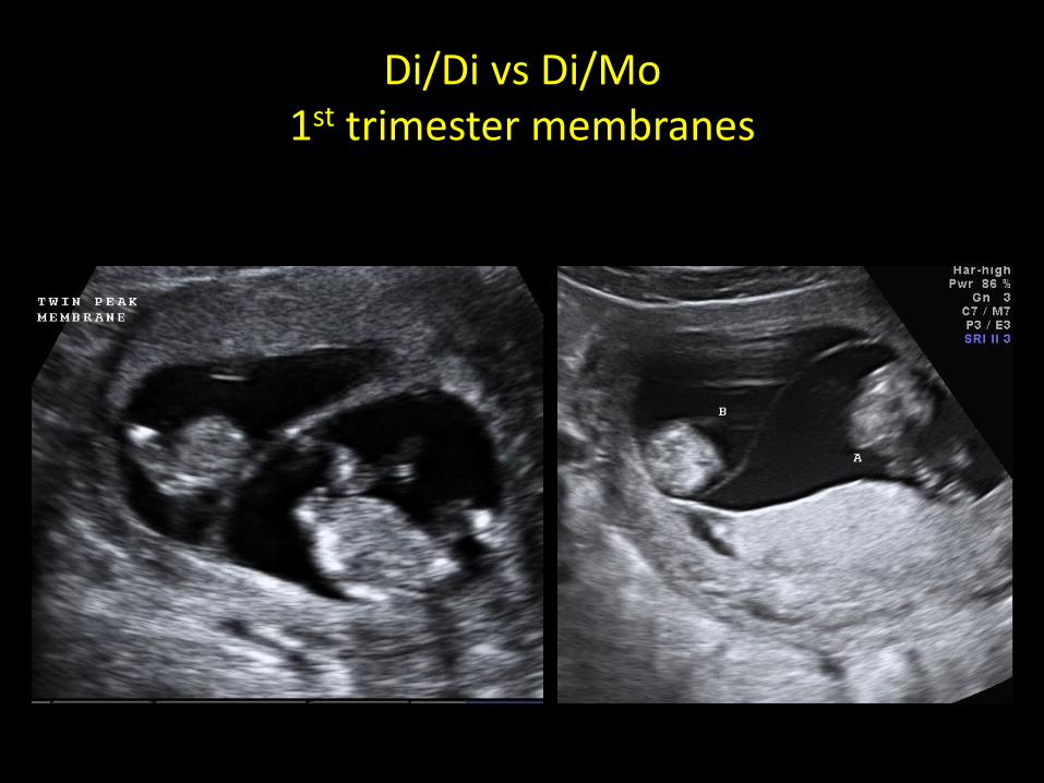
Di/Di vs Di/Mo 1st trimester membranes
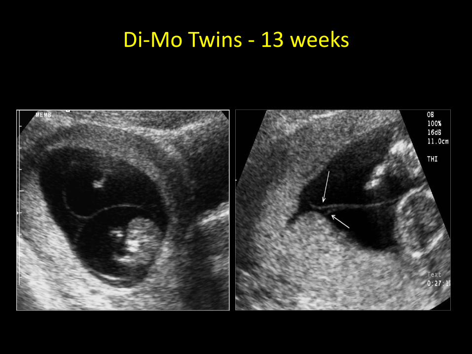
Di-Mo Twins - 13 weeks
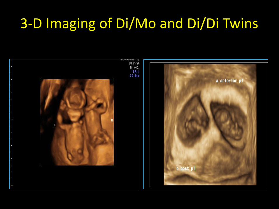
3-D Imaging of Di/Mo and Di/Di Twins
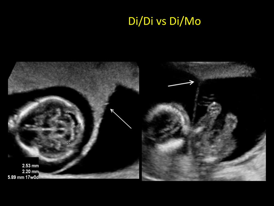
Di/Di vs Di/Mo
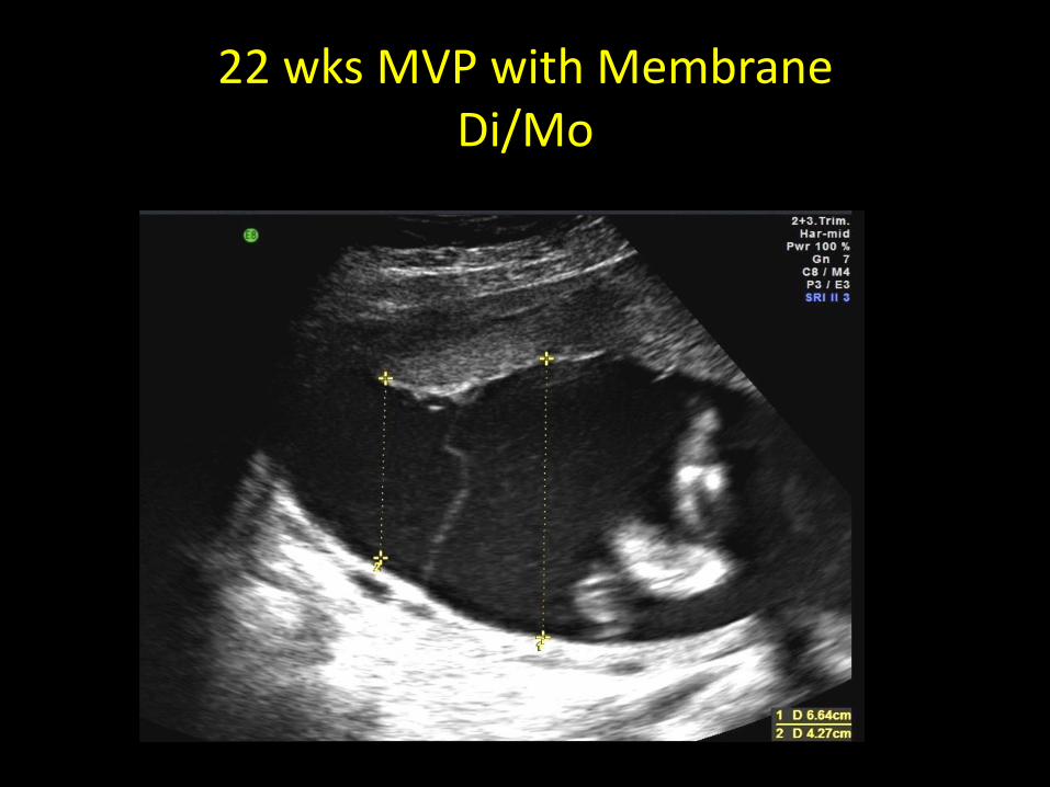
22 wks MVP with Membrane Di/Mo
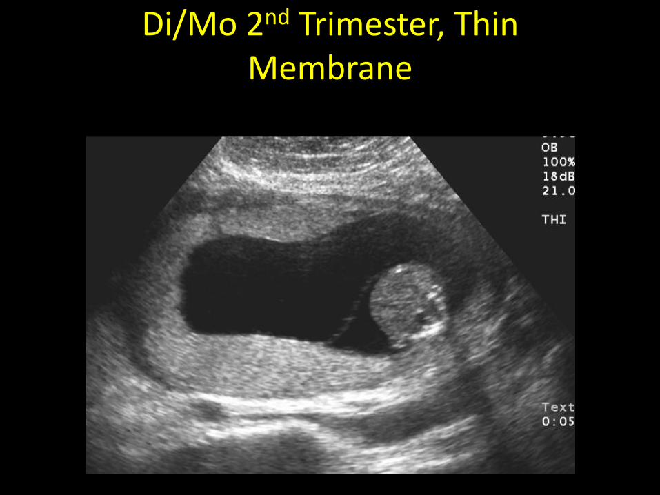
Di/Mo 2nd Trimester, Thin Membrane
Pitfalls of Membranes
+ Not appreciating the draping or cocooning of membranes.
+ Fetal movement (changing position)
+ Wispy vs. thick membranes, especially with advancing gestation.
+ Synechiae
+ Mistaking umbilical cord for a membrane
+ Vessels in the membranes
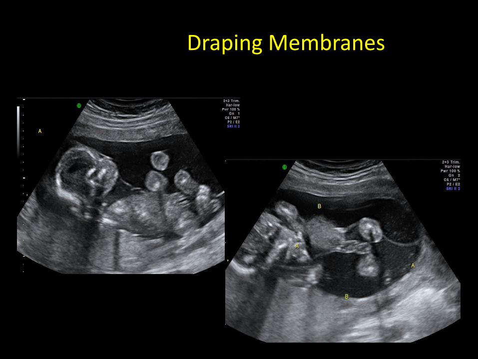
Draping Membranes
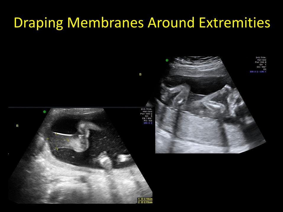
Draping Membranes Around Extremities
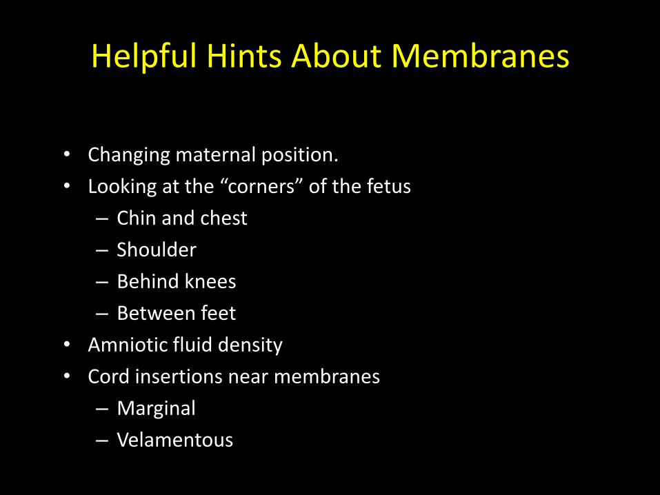
Helpful Hints About Membranes
• Changing maternal position.
• Looking at the “corners” of the fetus
– Chin and chest
– Shoulder
– Behind knees
– Between feet
• Amniotic fluid density
• Cord insertions near membranes
– Marginal
– Velamentous
MATERNAL COMPLICATIONS
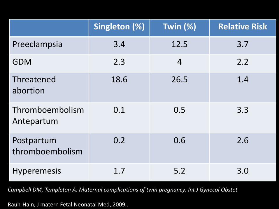
Singleton (%) Twin (%) Relative Risk
Preeclampsia 3.4 12.5 3.7
GDM 2.3 4 2.2
Threatened abortion
18.6 26.5 1.4
Thromboembolism Antepartum
0.1 0.5 3.3
Postpartum thromboembolism
0.2 0.6 2.6
Hyperemesis 1.7 5.2 3.0
Campbell DM, Templeton A: Maternal complications of twin pregnancy. Int J Gynecol Obstet Rauh-Hain, J matern Fetal Neonatal Med, 2009 .
FETAL COMPLICATIONS
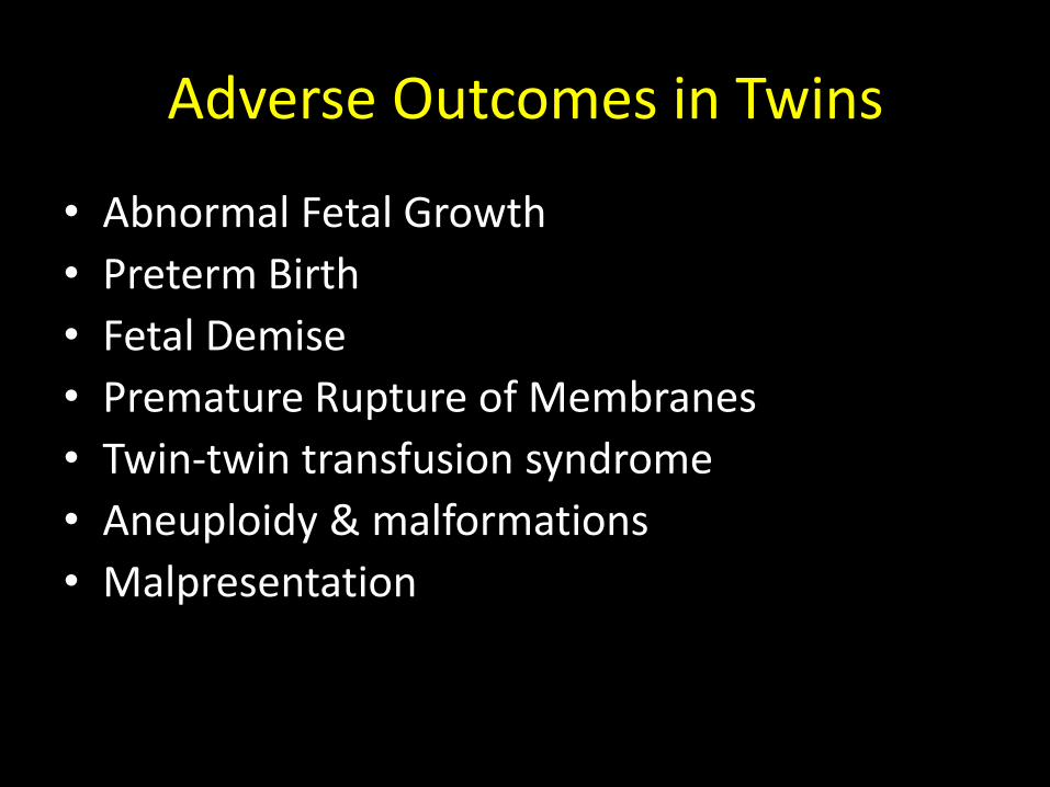
Adverse Outcomes in Twins
• Abnormal Fetal Growth
• Preterm Birth
• Fetal Demise
• Premature Rupture of Membranes
• Twin-twin transfusion syndrome
• Aneuploidy & malformations
• Malpresentation
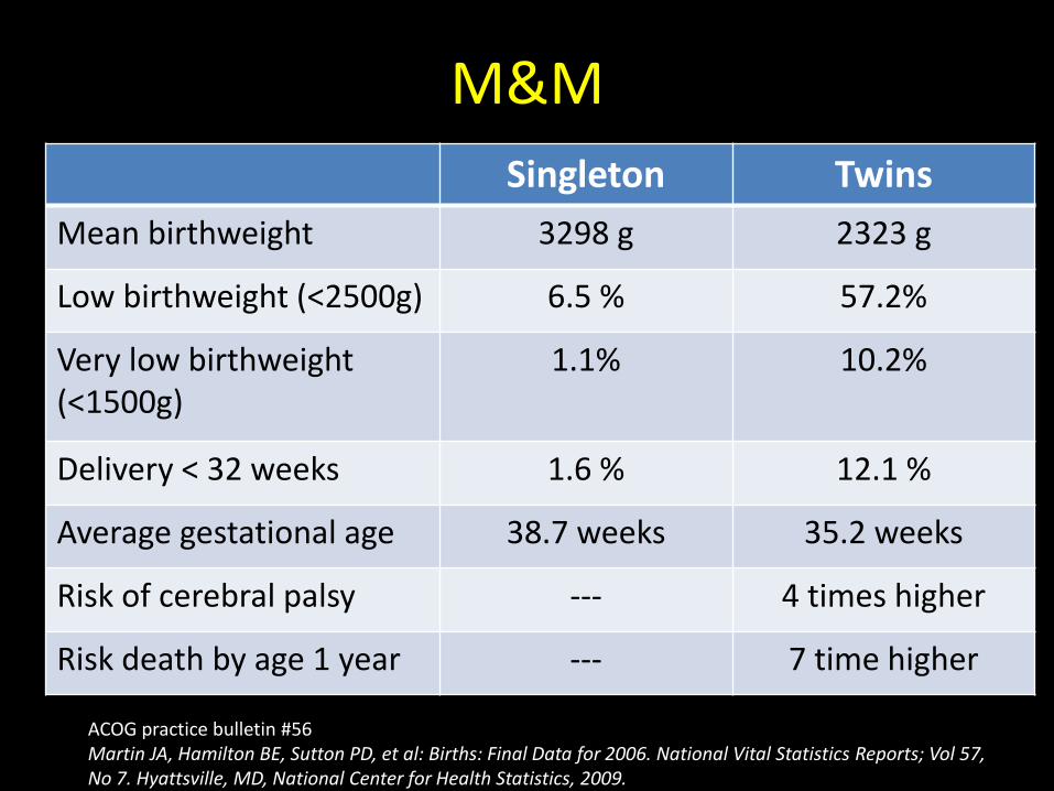
M&M
Singleton Twins
Mean birthweight 3298 g 2323 g
Low birthweight (<2500g) 6.5 % 57.2%
Very low birthweight (<1500g)
1.1% 10.2%
Delivery < 32 weeks 1.6 % 12.1 %
Average gestational age 38.7 weeks 35.2 weeks
Risk of cerebral palsy --- 4 times higher
Risk death by age 1 year --- 7 time higher
ACOG practice bulletin #56 Martin JA, Hamilton BE, Sutton PD, et al: Births: Final Data for 2006. National Vital Statistics Reports; Vol 57, No 7. Hyattsville, MD, National Center for Health Statistics, 2009.
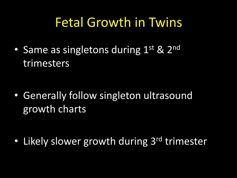
Fetal Growth in Twins
• Same as singletons during 1st & 2nd trimesters
• Generally follow singleton ultrasound growth charts
• Likely slower growth during 3rd trimester
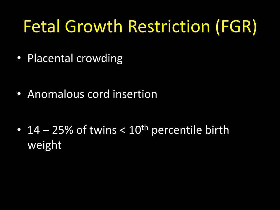
Fetal Growth Restriction (FGR)
• Placental crowding
• Anomalous cord insertion
• 14 – 25% of twins < 10th percentile birth weight
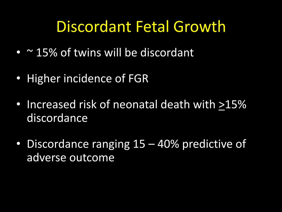
Discordant Fetal Growth
• ~ 15% of twins will be discordant
• Higher incidence of FGR
• Increased risk of neonatal death with >15% discordance
• Discordance ranging 15 – 40% predictive of adverse outcome
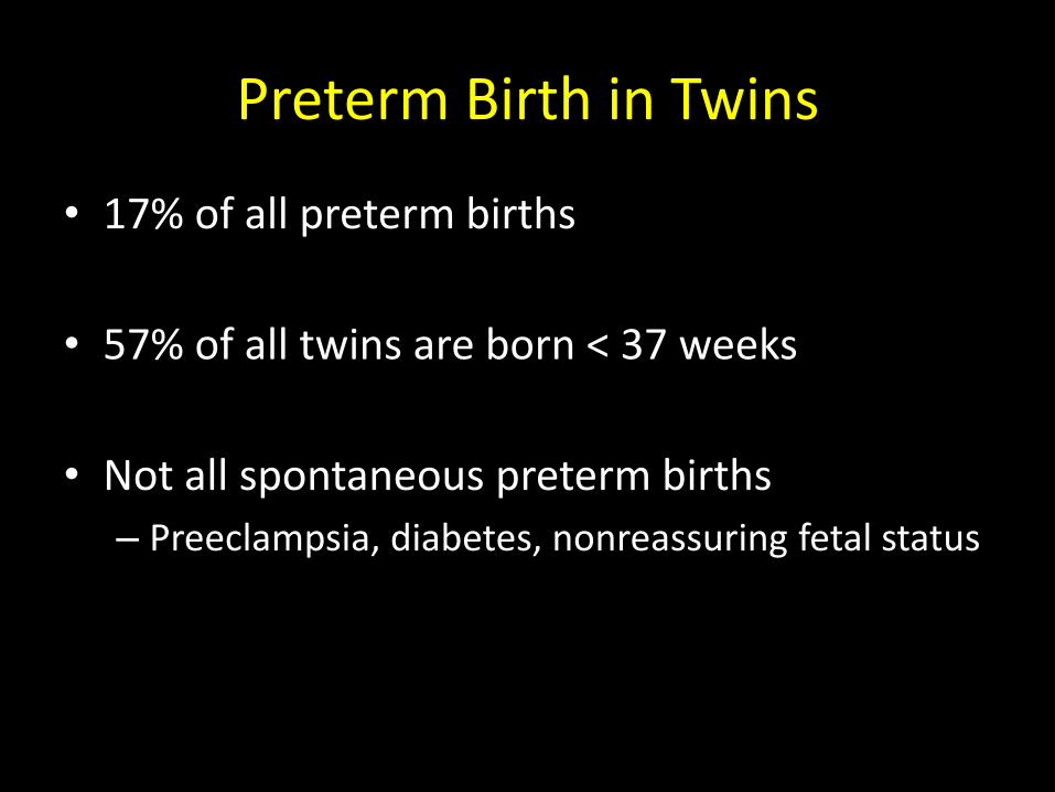
Preterm Birth in Twins
• 17% of all preterm births
• 57% of all twins are born < 37 weeks
• Not all spontaneous preterm births
– Preeclampsia, diabetes, nonreassuring fetal status
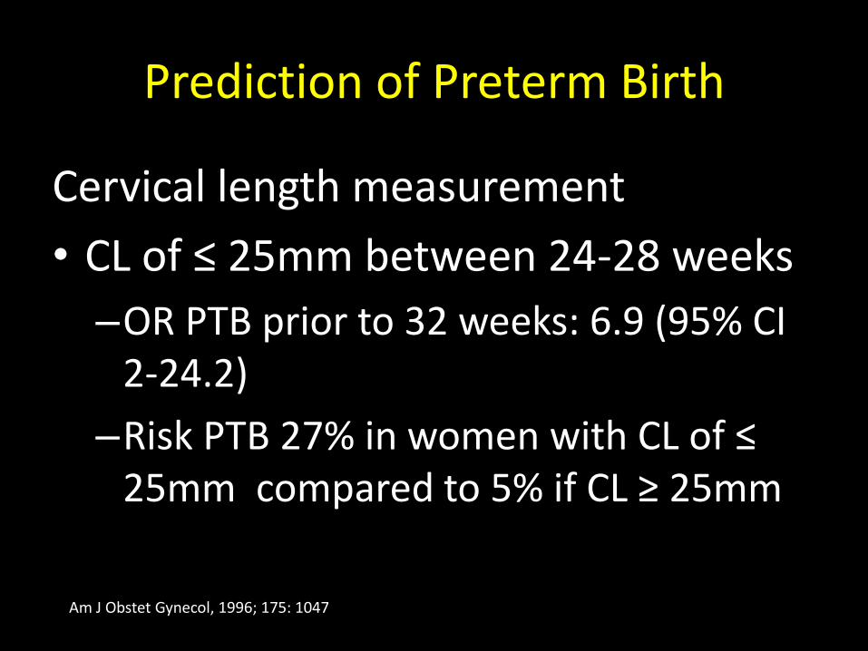
Prediction of Preterm Birth
Cervical length measurement
• CL of ≤ 25mm between 24-28 weeks
–OR PTB prior to 32 weeks: 6.9 (95% CI 2-24.2)
–Risk PTB 27% in women with CL of ≤ 25mm compared to 5% if CL ≥ 25mm
Am J Obstet Gynecol, 1996; 175: 1047
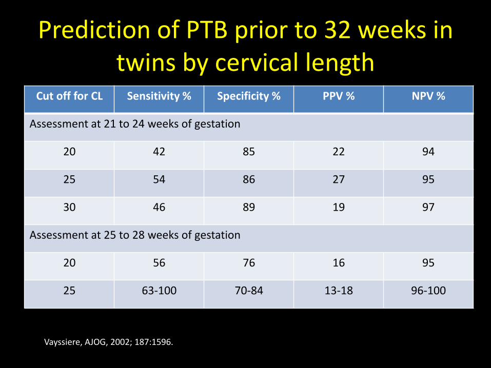
Prediction of PTB prior to 32 weeks in twins by cervical length
Cut off for CL Sensitivity % Specificity % PPV % NPV %
Assessment at 21 to 24 weeks of gestation
20 42 85 22 94
25 54 86 27 95
30 46 89 19 97
Assessment at 25 to 28 weeks of gestation
20 56 76 16 95
25 63-100 70-84 13-18 96-100
Vayssiere, AJOG, 2002; 187:1596.
Prediction of Preterm Birth
• Cervical length measurement
• Fetal fibronectin
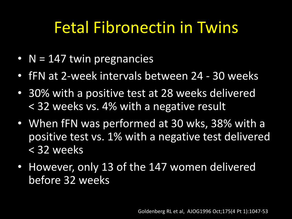
Fetal Fibronectin in Twins
• N = 147 twin pregnancies
• fFN at 2-week intervals between 24 - 30 weeks
• 30% with a positive test at 28 weeks delivered < 32 weeks vs. 4% with a negative result
• When fFN was performed at 30 wks, 38% with a positive test vs. 1% with a negative test delivered < 32 weeks
• However, only 13 of the 147 women delivered before 32 weeks
Goldenberg RL et al, AJOG1996 Oct;175(4 Pt 1):1047-53
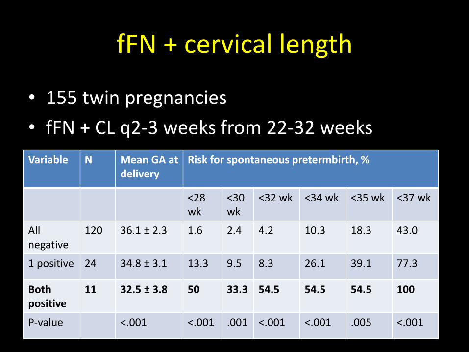
fFN + cervical length
• 155 twin pregnancies
• fFN + CL q2-3 weeks from 22-32 weeks
Variable N Mean GA at delivery
Risk for spontaneous pretermbirth, %
<28 wk
<30 wk
<32 wk <34 wk <35 wk <37 wk
All negative
120 36.1 ± 2.3 1.6 2.4 4.2 10.3 18.3 43.0
1 positive 24 34.8 ± 3.1 13.3 9.5 8.3 26.1 39.1 77.3
Both positive
11 32.5 ± 3.8 50 33.3 54.5 54.5 54.5 100
P-value <.001 <.001 .001 <.001 <.001 .005 <.001
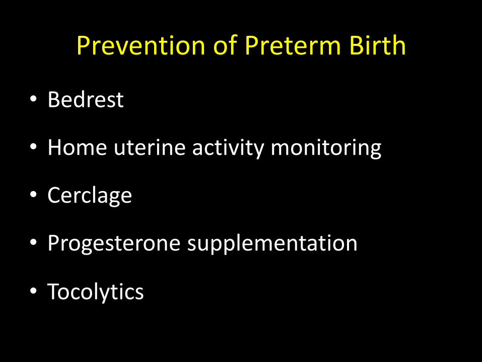
Prevention of Preterm Birth
• Bedrest
• Home uterine activity monitoring
• Cerclage
• Progesterone supplementation
• Tocolytics
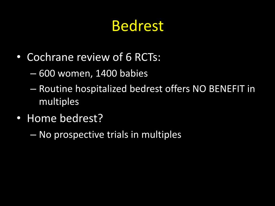
Bedrest
• Cochrane review of 6 RCTs:
– 600 women, 1400 babies
– Routine hospitalized bedrest offers NO BENEFIT in multiples
• Home bedrest?
– No prospective trials in multiples
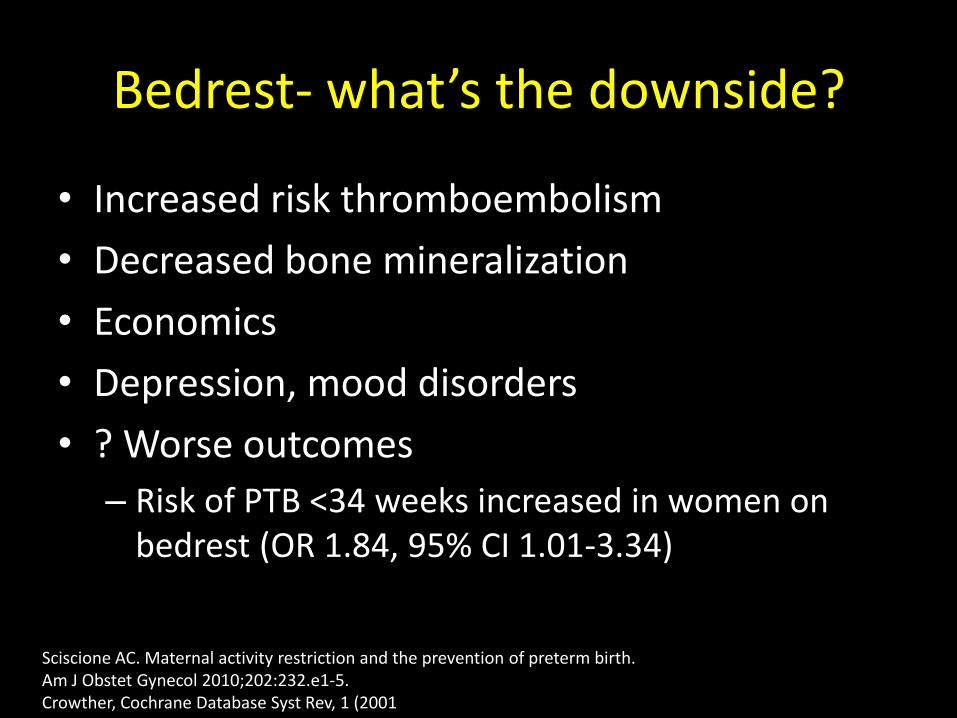
Bedrest- what’s the downside?
• Increased risk thromboembolism
• Decreased bone mineralization
• Economics
• Depression, mood disorders
• ? Worse outcomes
– Risk of PTB <34 weeks increased in women on bedrest (OR 1.84, 95% CI 1.01-3.34)
Sciscione AC. Maternal activity restriction and the prevention of preterm birth. Am J Obstet Gynecol 2010;202:232.e1-5. Crowther, Cochrane Database Syst Rev, 1 (2001
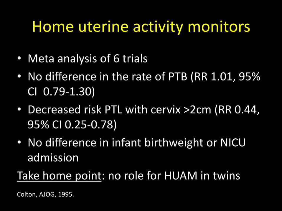
Home uterine activity monitors
• Meta analysis of 6 trials
• No difference in the rate of PTB (RR 1.01, 95% CI 0.79-1.30)
• Decreased risk PTL with cervix >2cm (RR 0.44, 95% CI 0.25-0.78)
• No difference in infant birthweight or NICU admission
Take home point: no role for HUAM in twins
Colton, AJOG, 1995.
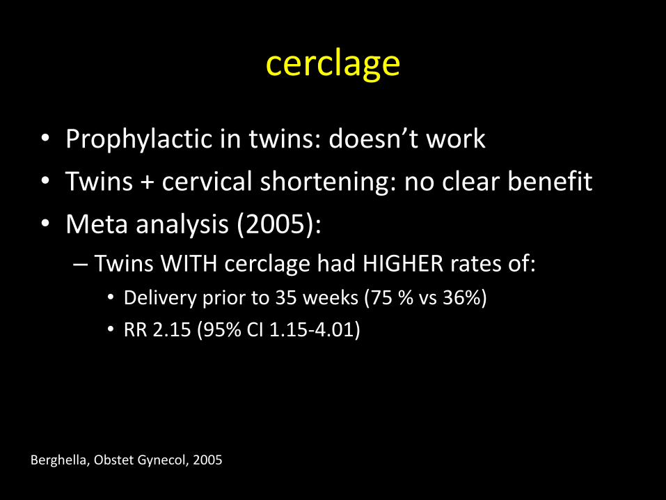
cerclage
• Prophylactic in twins: doesn’t work
• Twins + cervical shortening: no clear benefit
• Meta analysis (2005):
– Twins WITH cerclage had HIGHER rates of:
• Delivery prior to 35 weeks (75 % vs 36%)
• RR 2.15 (95% CI 1.15-4.01)
Berghella, Obstet Gynecol, 2005
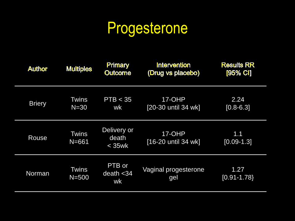
Progesterone
Briery Twins
N=30
PTB < 35
wk
17-OHP
[20-30 until 34 wk]
2.24
[0.8-6.3]
Rouse Twins
N=661
Delivery or
death
< 35wk
17-OHP
[16-20 until 34 wk]
1.1
[0.09-1.3]
Norman Twins
N=500
PTB or
death <34
wk
Vaginal progesterone
gel
1.27
[0.91-1.78}
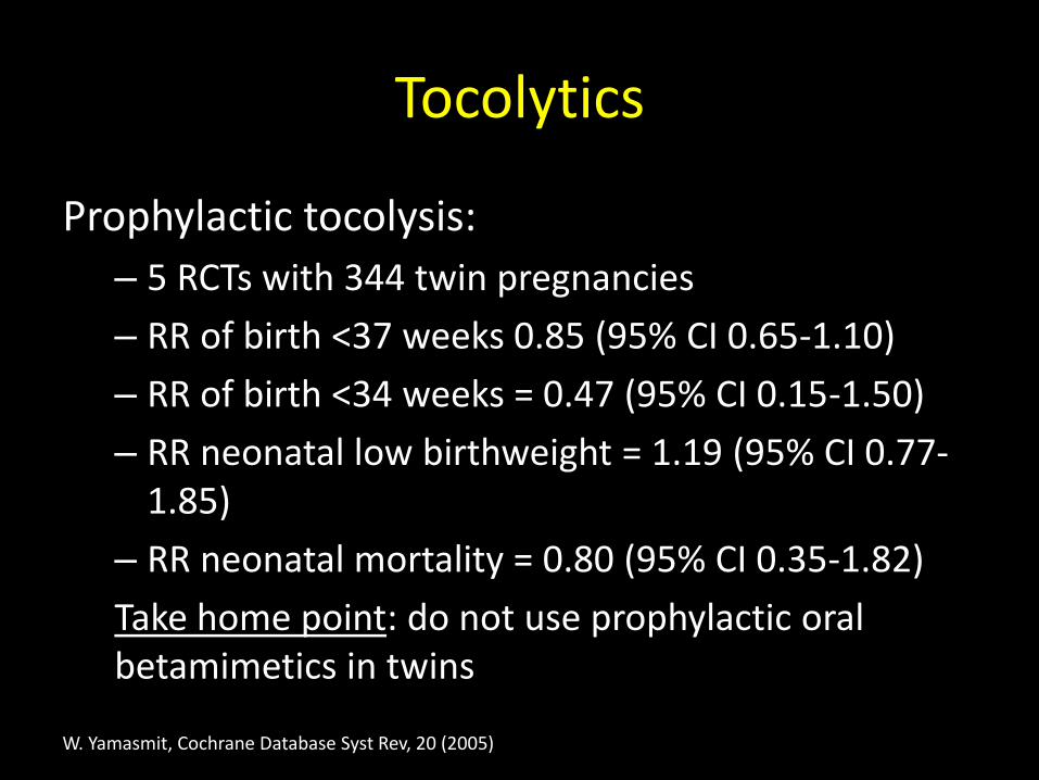
Tocolytics
Prophylactic tocolysis:
– 5 RCTs with 344 twin pregnancies
– RR of birth <37 weeks 0.85 (95% CI 0.65-1.10)
– RR of birth <34 weeks = 0.47 (95% CI 0.15-1.50)
– RR neonatal low birthweight = 1.19 (95% CI 0.77-1.85)
– RR neonatal mortality = 0.80 (95% CI 0.35-1.82)
Take home point: do not use prophylactic oral betamimetics in twins
W. Yamasmit, Cochrane Database Syst Rev, 20 (2005)
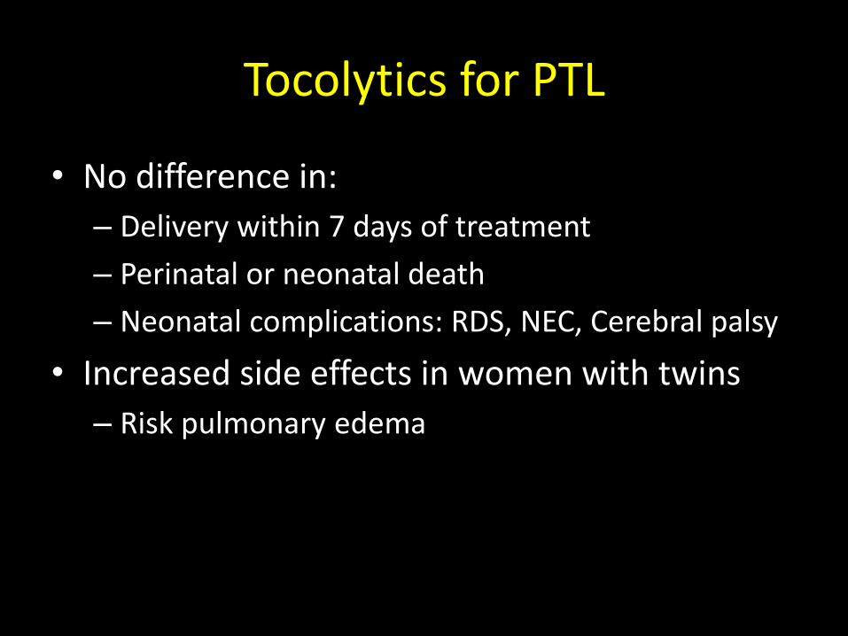
Tocolytics for PTL
• No difference in:
– Delivery within 7 days of treatment
– Perinatal or neonatal death
– Neonatal complications: RDS, NEC, Cerebral palsy
• Increased side effects in women with twins
– Risk pulmonary edema

TWINS: SPECIAL SITUATIONS
Management of the death of one fetus
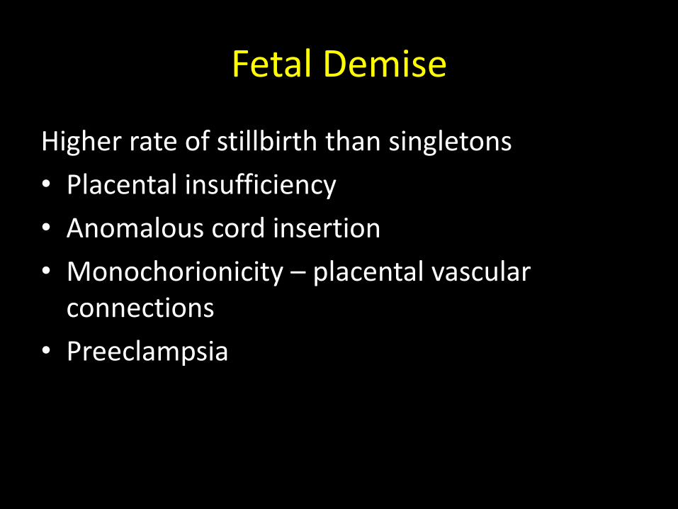
Fetal Demise
Higher rate of stillbirth than singletons
• Placental insufficiency
• Anomalous cord insertion
• Monochorionicity – placental vascular connections
• Preeclampsia
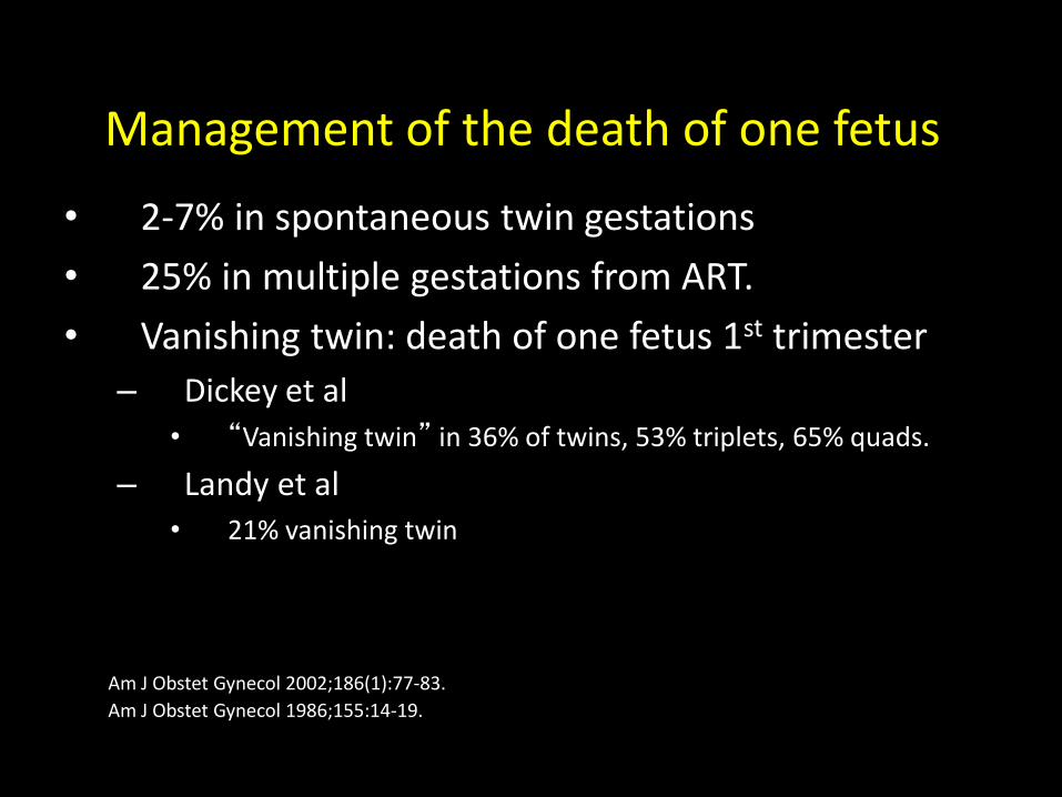
Management of the death of one fetus
• 2-7% in spontaneous twin gestations
• 25% in multiple gestations from ART.
• Vanishing twin: death of one fetus 1st trimester
– Dickey et al
• “Vanishing twin” in 36% of twins, 53% triplets, 65% quads.
– Landy et al • 21% vanishing twin
Am J Obstet Gynecol 2002;186(1):77-83.
Am J Obstet Gynecol 1986;155:14-19.
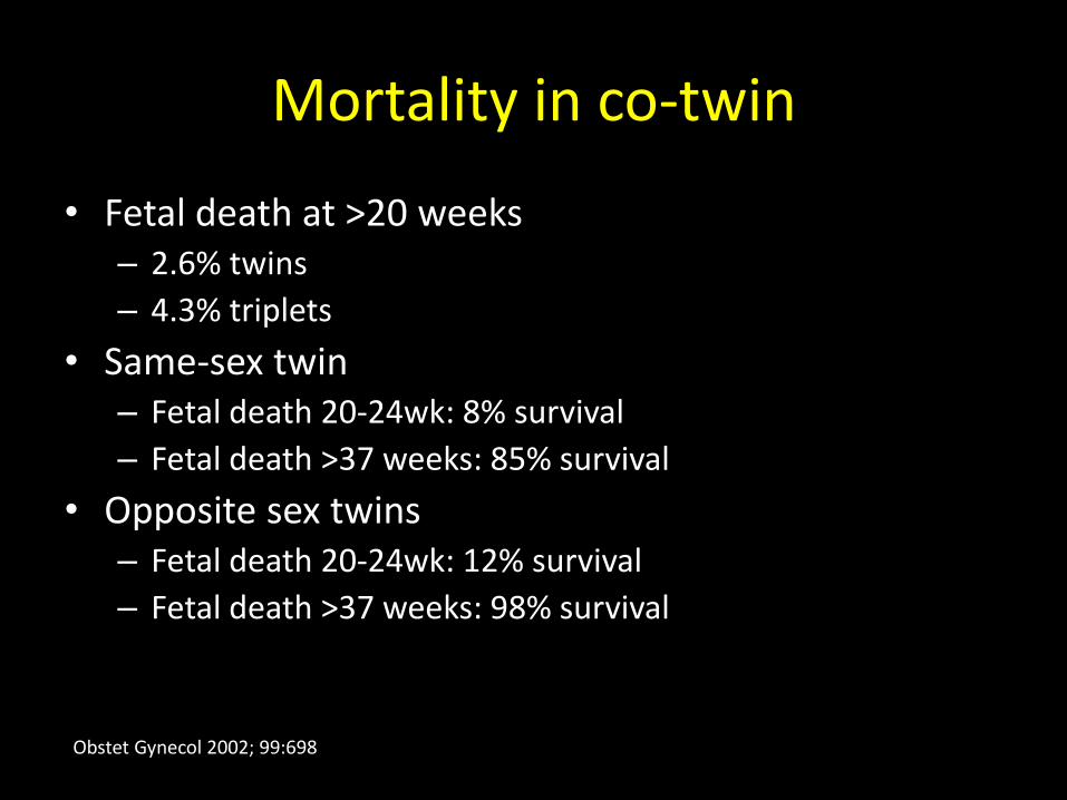
Mortality in co-twin
• Fetal death at >20 weeks – 2.6% twins
– 4.3% triplets
• Same-sex twin – Fetal death 20-24wk: 8% survival
– Fetal death >37 weeks: 85% survival
• Opposite sex twins – Fetal death 20-24wk: 12% survival
– Fetal death >37 weeks: 98% survival
Obstet Gynecol 2002; 99:698
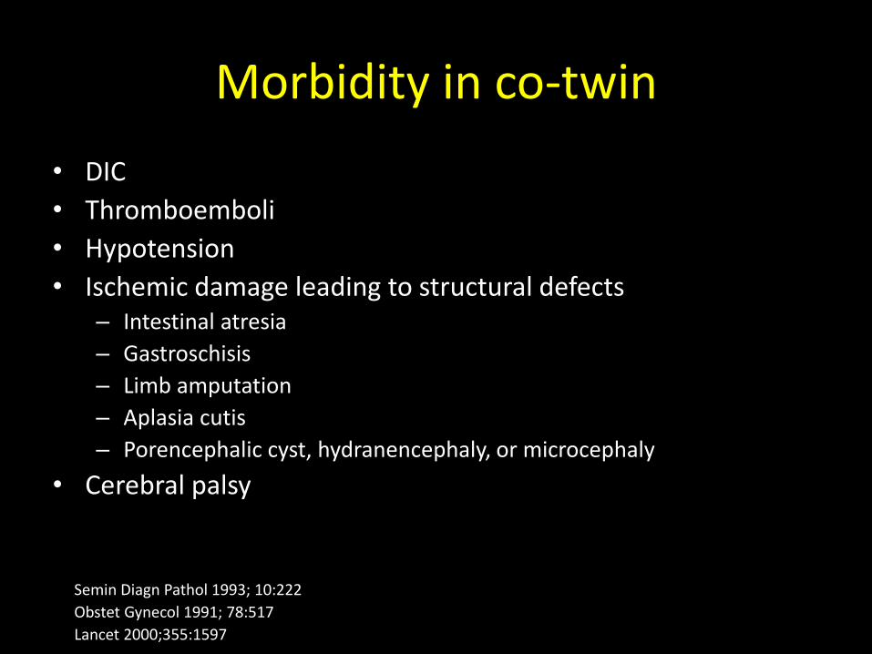
Morbidity in co-twin
• DIC
• Thromboemboli
• Hypotension
• Ischemic damage leading to structural defects – Intestinal atresia
– Gastroschisis
– Limb amputation
– Aplasia cutis
– Porencephalic cyst, hydranencephaly, or microcephaly
• Cerebral palsy
Semin Diagn Pathol 1993; 10:222
Obstet Gynecol 1991; 78:517
Lancet 2000;355:1597
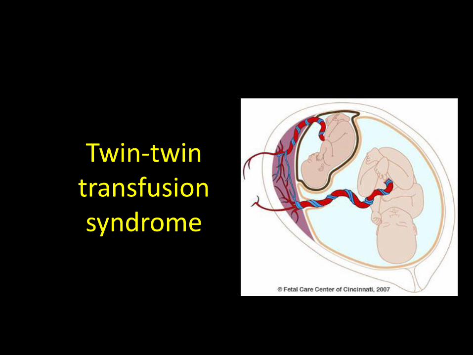
Twin-twin transfusion syndrome
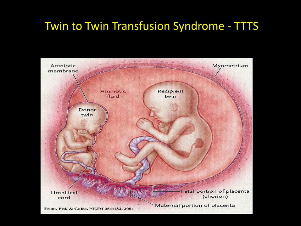
Twin to Twin Transfusion Syndrome - TTTS
Fisk NM: The scientific basis of feto-fetal transfusion syndrome and its treatment. In Ward RH, Whittle M
[eds]: Multiple Pregnancy, pp 235–250. London, RCOG Press, 1995)
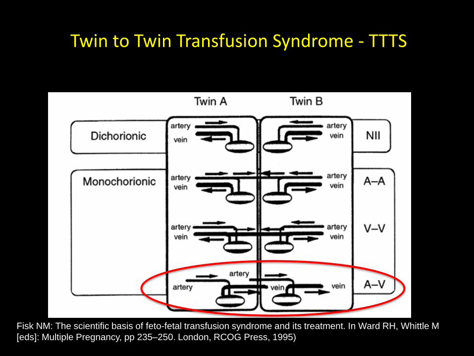
Twin to Twin Transfusion Syndrome - TTTS
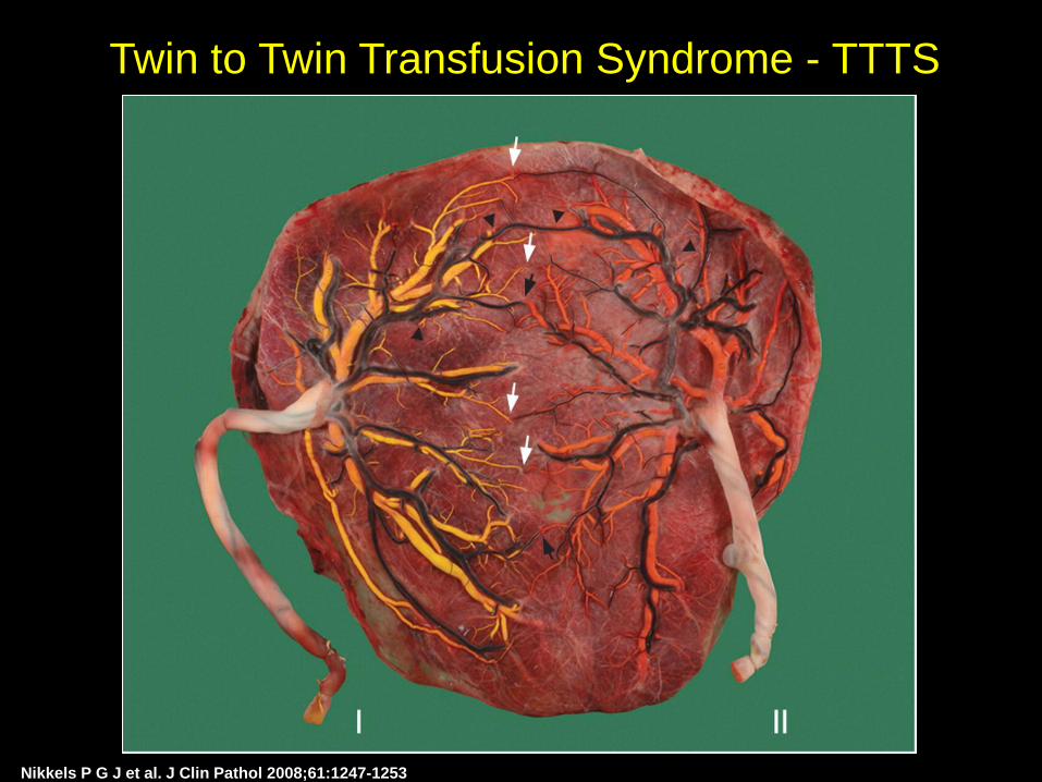
Nikkels P G J et al. J Clin Pathol 2008;61:1247-1253
Twin to Twin Transfusion Syndrome - TTTS
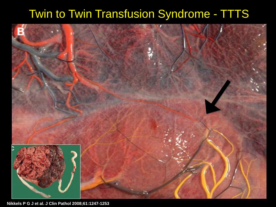
Nikkels P G J et al. J Clin Pathol 2008;61:1247-1253
Twin to Twin Transfusion Syndrome - TTTS
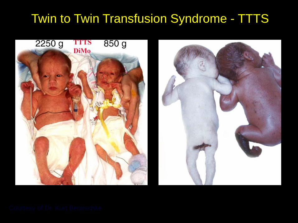
Courtesy of Dr. Kurt Benirschke
Twin to Twin Transfusion Syndrome - TTTS
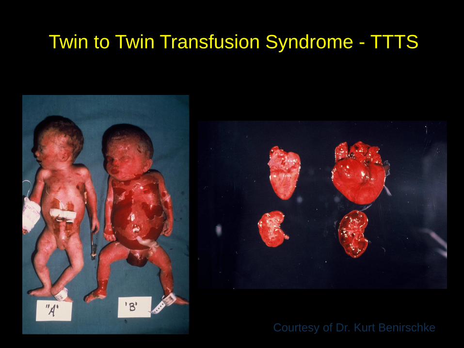
Courtesy of Dr. Kurt Benirschke
Twin to Twin Transfusion Syndrome - TTTS
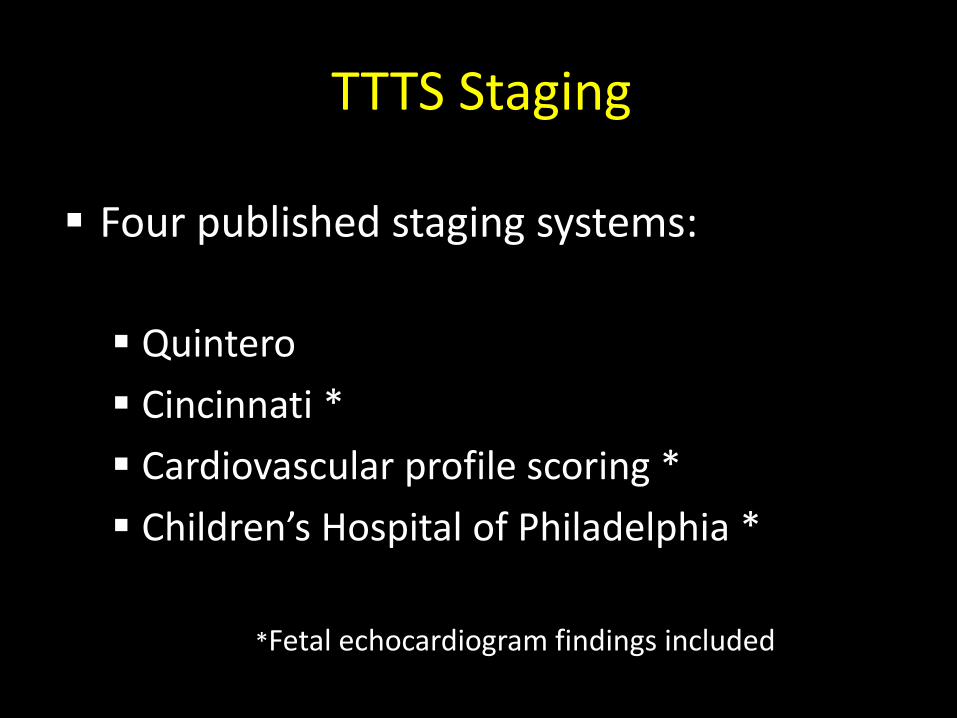
TTTS Staging
Four published staging systems:
Quintero
Cincinnati *
Cardiovascular profile scoring *
Children’s Hospital of Philadelphia *
*Fetal echocardiogram findings included
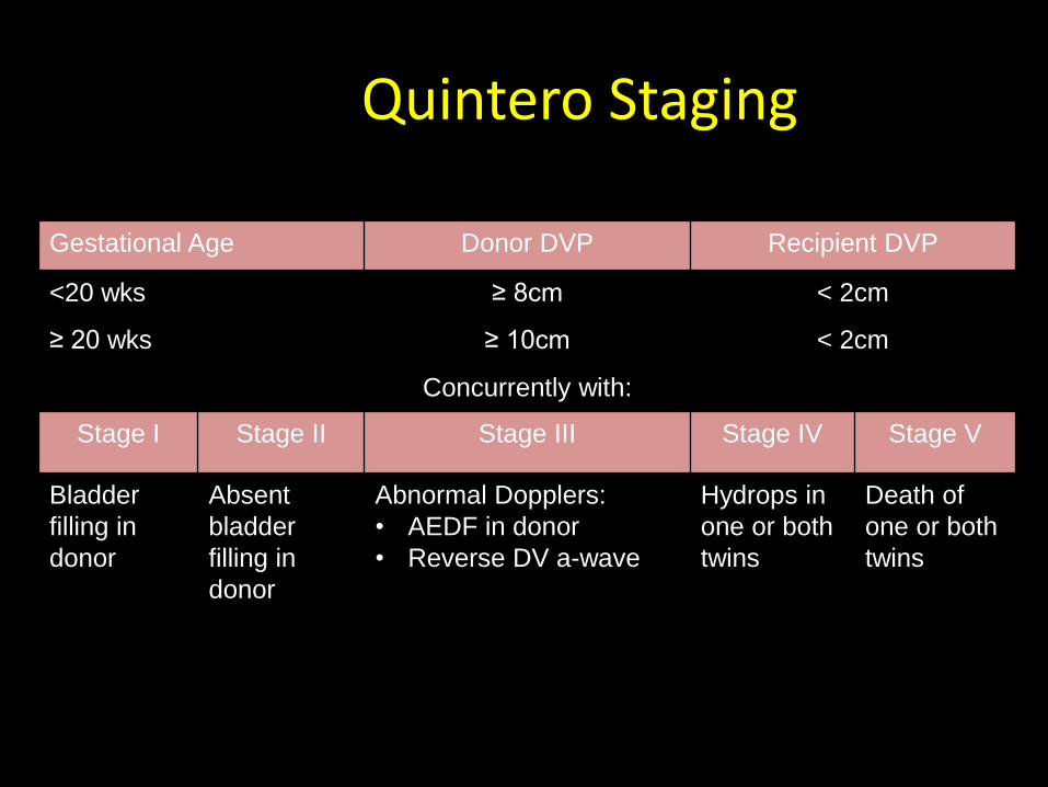
Quintero Staging
Gestational Age Donor DVP Recipient DVP
<20 wks ≥ 8cm < 2cm
≥ 20 wks ≥ 10cm < 2cm
Concurrently with:
Stage I Stage II Stage III Stage IV Stage V
Bladder
filling in
donor
Absent
bladder
filling in
donor
Abnormal Dopplers:
• AEDF in donor
• Reverse DV a-wave
Hydrops in
one or both
twins
Death of
one or both
twins
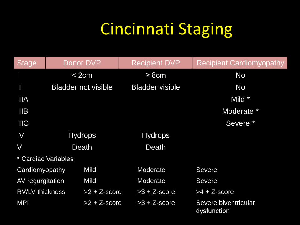
Cincinnati Staging
Stage Donor DVP Recipient DVP Recipient Cardiomyopathy
I < 2cm ≥ 8cm No
II Bladder not visible Bladder visible No
IIIA Mild *
IIIB Moderate *
IIIC Severe *
IV Hydrops Hydrops
V Death Death
* Cardiac Variables
Cardiomyopathy Mild Moderate Severe
AV regurgitation Mild Moderate Severe
RV/LV thickness >2 + Z-score >3 + Z-score >4 + Z-score
MPI >2 + Z-score >3 + Z-score Severe biventricular
dysfunction
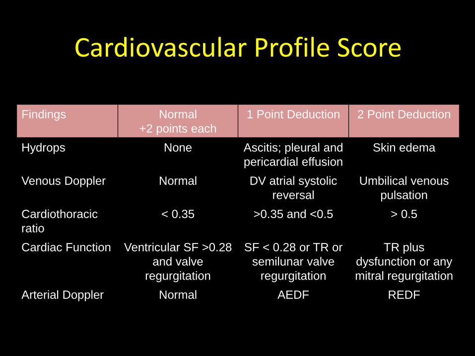
Cardiovascular Profile Score
Findings Normal
+2 points each
1 Point Deduction 2 Point Deduction
Hydrops None Ascitis; pleural and
pericardial effusion
Skin edema
Venous Doppler Normal DV atrial systolic
reversal
Umbilical venous
pulsation
Cardiothoracic
ratio
< 0.35 >0.35 and <0.5 > 0.5
Cardiac Function Ventricular SF >0.28
and valve
regurgitation
SF < 0.28 or TR or
semilunar valve
regurgitation
TR plus
dysfunction or any
mitral regurgitation
Arterial Doppler Normal AEDF REDF
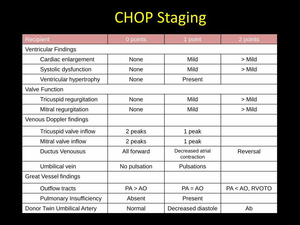
CHOP Staging Recipient 0 points 1 point 2 points
Ventricular Findings
Cardiac enlargement None Mild > Mild
Systolic dysfunction None Mild > Mild
Ventricular hypertrophy None Present
Valve Function
Tricuspid regurgitation None Mild > Mild
Mitral regurgitation None Mild > Mild
Venous Doppler findings
Tricuspid valve inflow 2 peaks 1 peak
Mitral valve inflow 2 peaks 1 peak
Ductus Venousus All forward Decreased atrial
contraction Reversal
Umbilical vein No pulsation Pulsations
Great Vessel findings
Outflow tracts PA > AO PA = AO PA < AO, RVOTO
Pulmonary Insufficiency Absent Present
Donor Twin Umbilical Artery Normal Decreased diastole Ab
Consensus Statement
• The Quintero staging system should be retained until a superior system has been appropriately validated.
• Cardiac indices and markers of systemic hemodynamic alterations may improve prediction of disease progression and/or perinatal outcomes…[but] should be assessed and validated individually and in combination within a clinical trial.
Stamilio DM, Fraser WD, Moore TR. Twin-twin transfusion syndrome: an ethics-based and evidence-
based argument for clinical research. Am J Obstet Gynecol. 2010; 203(1): 3-16
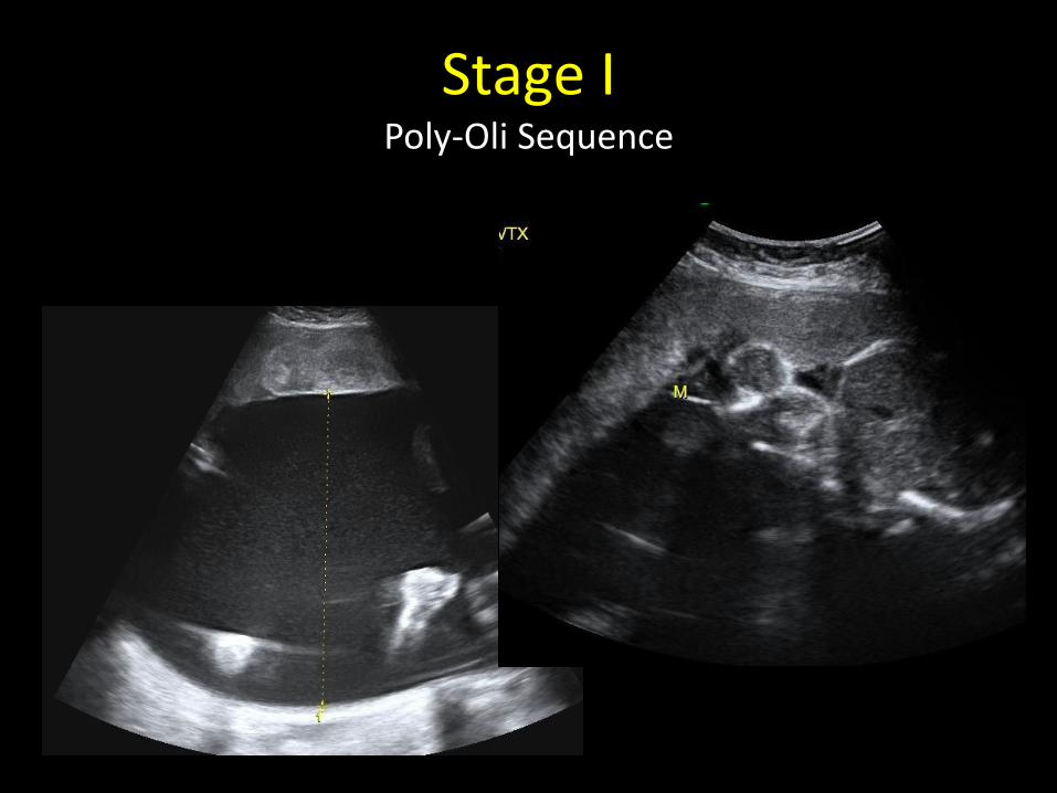
Stage I Poly-Oli Sequence
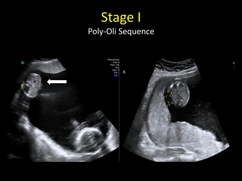
Stage I Poly-Oli Sequence
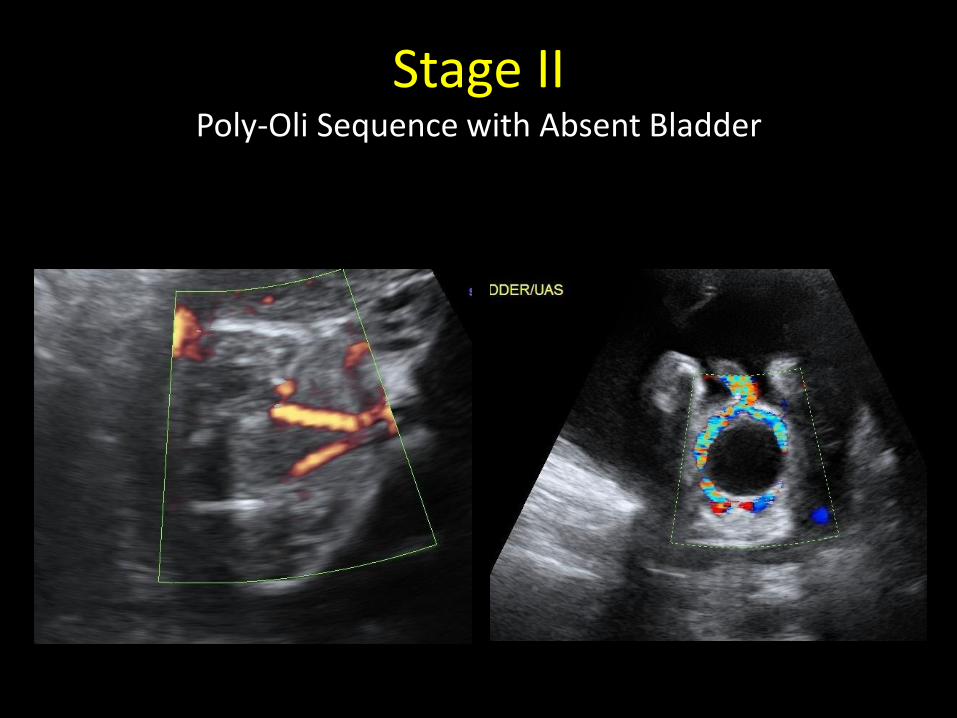
Stage II Poly-Oli Sequence with Absent Bladder
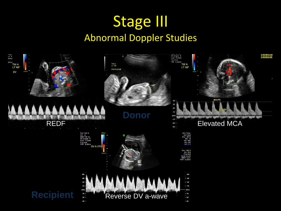
Stage III Abnormal Doppler Studies
Reverse DV a-wave
REDF Elevated MCA Donor
Recipient

Stage III Cardiac Changes
CT Ratio: 0.74
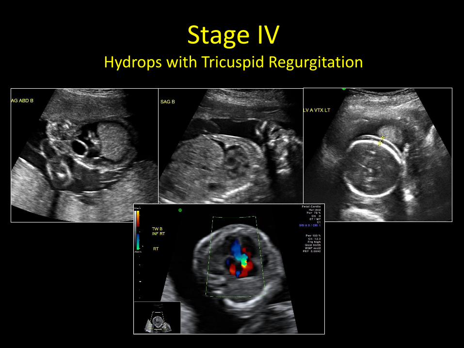
Stage IV Hydrops with Tricuspid Regurgitation
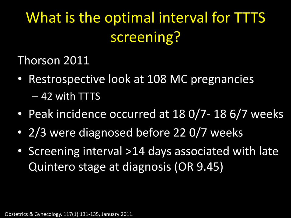
What is the optimal interval for TTTS screening?
Thorson 2011
• Restrospective look at 108 MC pregnancies
– 42 with TTTS
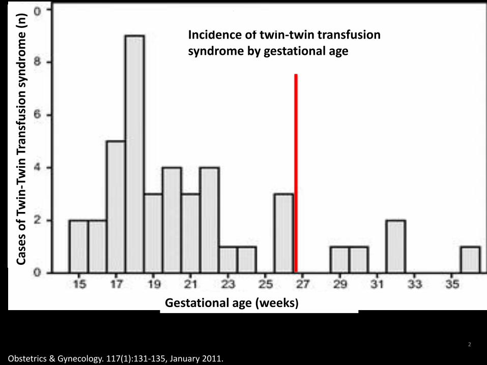
• Peak incidence occurred at 18 0/7- 18 6/7 weeks
• 2/3 were diagnosed before 22 0/7 weeks
• Screening interval >14 days associated with late Quintero stage at diagnosis (OR 9.45)
Obstetrics & Gynecology. 117(1):131-135, January 2011.
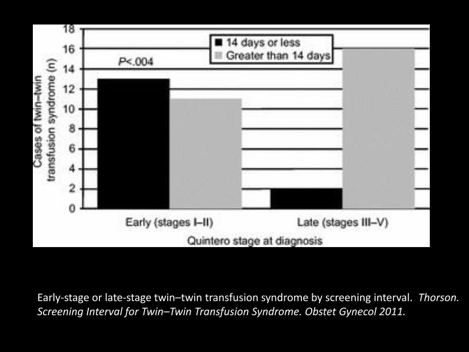
Early-stage or late-stage twin–twin transfusion syndrome by screening interval. Thorson. Screening Interval for Twin–Twin Transfusion Syndrome. Obstet Gynecol 2011.
2
Fig. 1.
Obstetrics & Gynecology. 117(1):131-135, January 2011.
Incidence of twin-twin transfusion syndrome by gestational age
Cas
es
of
Twin
-Tw
in T
ran
sfu
sio
n s
ynd
rom
e (
n)
Gestational age (weeks)
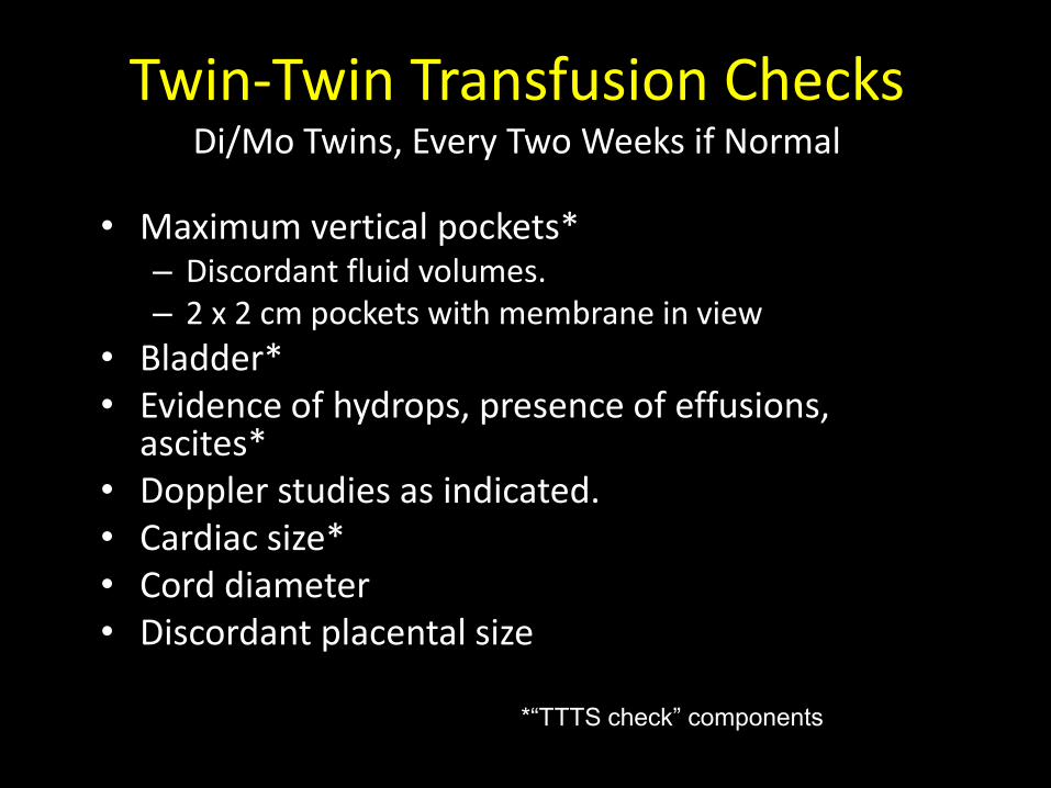
Twin-Twin Transfusion Checks Di/Mo Twins, Every Two Weeks if Normal
• Maximum vertical pockets* – Discordant fluid volumes. – 2 x 2 cm pockets with membrane in view
• Bladder* • Evidence of hydrops, presence of effusions,
ascites* • Doppler studies as indicated. • Cardiac size* • Cord diameter • Discordant placental size
*“TTTS check” components
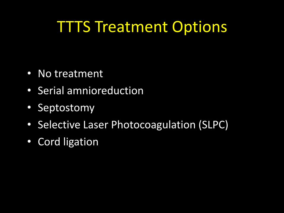
TTTS Treatment Options
• No treatment
• Serial amnioreduction
• Septostomy
• Selective Laser Photocoagulation (SLPC)
• Cord ligation
In summary
DIAGNOSIS- chorionicity matters!
NUTRITION- follow weight gain goals
REFERRAL TO MFM- for any high risk developments
TTTS- occurs in 10-15% . Important to screen for this every 1-2 weeks throughout.

Thank you!


![A monochorionic diamniotic twin pregnancy with selective ... · fetoscopy, a sequential Laser placental ablation was performed, identifying 6 anastomoses (5 arteriovenous [AV] and](https://img.pdfslide.net/doc/110x75/5f2d79ce00b49e3aa72f885c/a-monochorionic-diamniotic-twin-pregnancy-with-selective-fetoscopy-a-sequential.jpg)