Embed Size (px)
Citation preview

29
ORIGINAL ARTICLE
THE MAXILLARY ARCH AND ITS RELATIONSHIP TOCEPHALOMETRIC LANDMARKS OF SELECTED MALAY ETHNIC
GROUP
Khin Myo Thu, Than Winn1, Nizam Abdullah1, J.A.P. Jayasinghe1 and G.L.Chandima2.
Department of Anatomy, 1Department of Community Medicine,School of Medical Sciences, 2School of Dental Sciences
Universiti Sains Malaysia16150 Kubang Kerian, Kelantan, Malaysia
The objectives of this study were to estimate the maxillary arch measurements, toassess the validity of Pont’s & Korkhaus’ Indices; to determine the relationshipbetween maxillary arch form with head form; and to estimate the cephalic index(CI) of the study population. A cross-sectional study was conducted on 85 matureMalay students, 28 male students (32.98%), 57 females (67.02%) attendingTeachers’ Training College. Their mean age was 23.9 yr, and Cephalic Index (CI)86.4 (95% Confidence Interval 85.5-87.3). Arch and head dimensions weresignificantly larger in males than in females. CI was not significantly differentbetween males and females. Means of anterior arch width (AAW), posterior-arch-width (PAW) and arch-length (Lu) were 35.57mm, 47.3mm and 18.01mmrespectively. They were significantly different from their corresponding Indices.Correlation Coefficient between bizygomatic width and anterior-arch-width was0.18 and was not significant in both sexes of the present population.
Key words : Dental arch form, Pont’s Index, Korkhaus’ Index, Malay, Cephalic Index
Introduction
A sound knowledge of tooth size and dentalarch dimensions of a population is important forseveral dental treatment procedures. For examplein Restorative Dentistry, where teeth is beingrestored to its original morphology, knowledge ofthe tooth size will certainly implement in thetreatment administered. Differences in dental archand head dimensions of different populations canbe inherited and these inherited differences are usefulfor the practice of Aesthetic Dentistry and foreffective orthodontic treatment(1) It is thereforeimportant to have knowledge of certaincephalometric and dental arch parameters and theirrelationships for a given population. There areseveral indices derived from these measurements;indices of Pont(2) Linder(3), and Korkhaus(4), aremostly used in German-speaking countries(5). Theseindices predicts the ideal values (standard values)
of the arch width and length from the sum of upperfour incisors (SIu).A certain correlation existbetween the arch length, width, and mesiodistalwidth of the upper maxillary incisors. The standardvalues of these indices are then statisticallycorrelated and compared with the actual values ofthe individual case. Certain diagnostic andprognostic indications such as deviation in transversedevelopment of the arch widths and anteroposteriorposition of incisors can be gained by comparison ofthe actual and standard values .
In the 1840s the Swedish physician, AndersRetzius, developed one of the most influentialcraniometric techniques, the Cephalic Index whichmeasures the ratio between the width and length ofthe head. Generally he classified people as havingone of the three types of head shapes –brachycephalic, dolichocephalic or mesocephalic(6).
The objectives of this study were to establishthe dental arch indices, cephalometric measurements
Submitted-12.09.2004, Accepted-31.12.2004
Malaysian Journal of Medical Sciences, Vol. 12, No. 1, January 2005 (29-38)

30
and how these are correlated to one another, and tovalidate the dental arch indices in this studypopulation.
Materials and Methods
Bootstrap statistics based on 1000 simulatedmeans of sum of four maxillary incisors (SI u)estimated a standard deviation of 2.3mm. This SDwas used to calculate a sample of 85 subjects
required to estimate SIu with a precision of +/- 0.5mm at 95% confidence interval.
Preliminary screening procedures wereconducted among students from Teachers TrainingCollege, Kota Bharu. Subjects whose age rangebetween 20 and 35 yr, and has Malay parents andMalay grand parents from both the paternal andmaternal sides were selected for this study. Subjectswith maxillary dental arch irregularities and missingteeth, and whose first-degree relatives were selected
Khin Myo Thu, Than Winn et. al
Figure 1b : Diagram showing the measurements ofmaximum skull breadth (eu-eu), andmaximum face width (zy-zy).
Figure 1a : Diagram showing the measurement ofmaximum skull length (g-op)

31
for this study were excluded. Among those who wereeligible, 85 subjects were consecutively selected.After a brief self-administered questionnaire session,head measurements and maxillary casts were taken.The head measurements made on the subjects were(i) maximum skull length (g-op), distance fromopisthocranion (op) to glabella (g);(Fig-1a) (ii)maximum skull breadth or bieuryonic diameter (eu-eu), distance between the most lateral point of theskull(euryion), (iii) bizygomatic diameter (zy-zy),distance between two zygomatic prominences(zygion) ; (Fig-1b)
The following measurements were done onthe dental casts. (i) maximum mesiodistal distanceof each of the four maxillary incisors;(fig- 2a) (ii)anterior arch width (AAW), that is the distancebetween the lower-most points of the transversefissure of the upper first premolar teeth, the referencepoints for (AAW). (iii) posterior arch width (PAW),
the distance between the point of intersection of thetransverse fissure with the buccal fissure of the upperfirst permanent molar teeth (the reference points forPAW);(Fig-2b) (iv) anterior arch length Lu, whichis perpendicular from the most anterior labial surfaceof the central incisors to the connecting line of thereference points of AAW;(Fig-2c). All measurementswere in millimeter to the nearest 0.1 mm.
Data AnalysisSTATA 7.0 (7) was used to summarize the data
and validate the indices using correlations andregression statistics. From the measurements madeon dental casts, the sum of upper incisor mesiodistaldistances (SIu) was first computed and this measurewas used to estimate the values of arch widths, usingPont’s Index, for AAW and for PAW. Values of archlength (Lu) were computed using Korkhaus’ Index,.These index values(standard values) were calculated
THE MAXILLARY ARCH AND ITS RELATIONSHIP TO CEPHALOMETRIC LANDMARKS OF SELECTED MALAY ETHNIC GROUP
Figure 2b : Diagram showing the measurements ofanterior and posterior arch widths ofMaxilla (AAW and PAW)
Figure 2a : Diagram showing the measurment ofsum of maxillary incisors (SIu).

32
Figure 2c : Diagram showing the mesurement of archlength of maxilla (Lu)
Table 1: Dental arch measurements among Malay men and women.
Khin Myo Thu, Than Winn et. al
Dental Arch
Measurements
(DAM)
SIu
AAW
AAW*
PAW
PAW**
Lu
Lu***
SIu = sum of four upper incisordAAW = anterior arch width measured from the castAAW* = anterior arch width based on Pont’s index: (AAW =SIu*100/85)PAW = posterior arch width measured from the castPAW** = posterior arch width based on Pont’s index: (PAW=SIu*100/65)Lu*** = arch length measured from the castLu All measurements are not significantly different between males and females (independent t-tests p >.05)
Gender
Male n= 28
Mean
32.20
35.93
37.89
48.98
49.54
17.67
20.23
SD
2.32
2.32
2.73
2.46
3.57
2.48
1.45
Female n= 57
Mean
31.94
35.41
37.58
46.50
49.14
18.17
19.96
SD
2.29
3.57
2.70
6.58
3.53
4.14
1.43
Total
Both sexes n= 85
Mean
32.03
35.57
37.68
47.30
49.27
18.01
20.02
SD
2.29
3.22
2.69
5.69
3.52
3.67
1.43
Dental Arch
Measurements
(DAM)
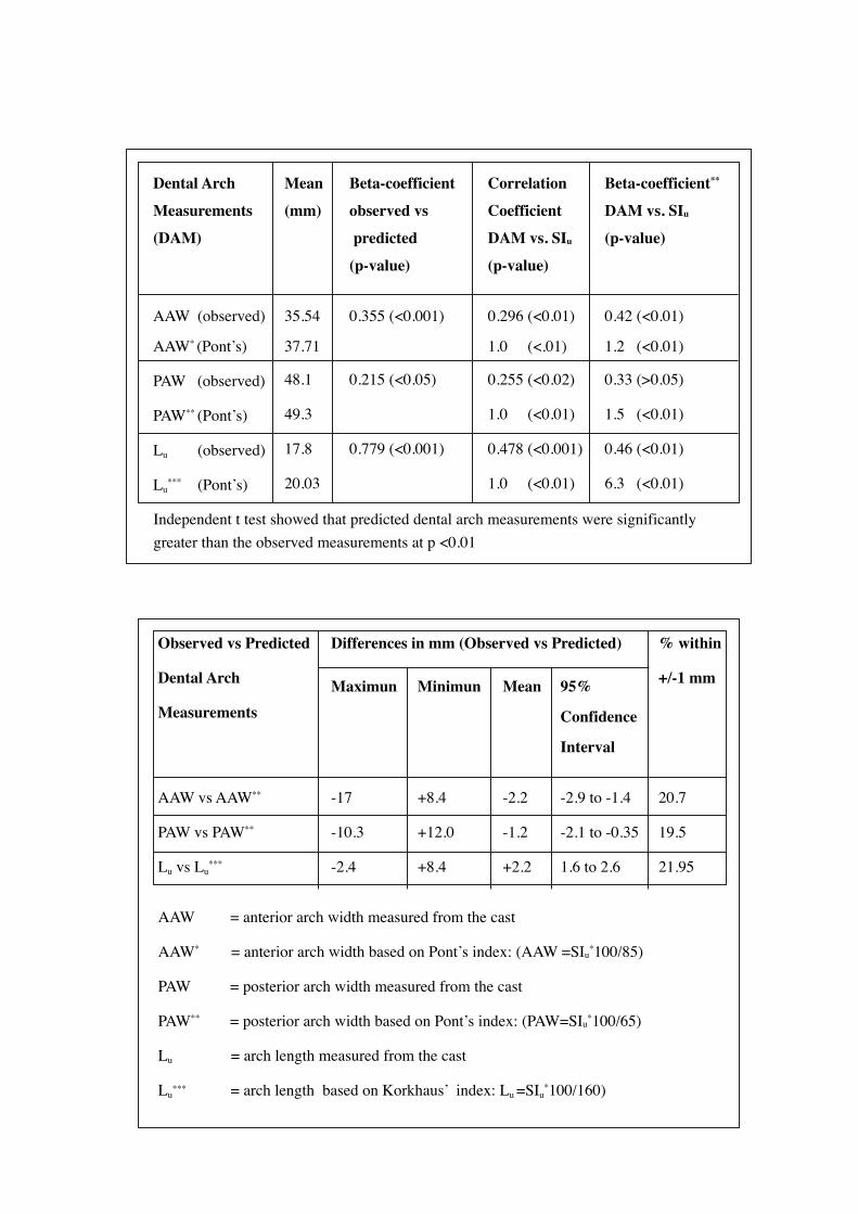
AAW (observed)
AAW* (Pont’s)
PAW (observed)
PAW** (Pont’s)
Lu (observed)
Lu*** (Pont’s)
Independent t test showed that predicted dental arch measurements were significantly greater than the observed measurements at p <0.01
Observed vs Predicted
Dental Arch
Measurements
AAW vs AAW**
PAW vs PAW**
Lu vs Lu***
AAW = anterior arch width measured from the cast
AAW* = anterior arch width based on Pont’s index: (AAW =SIu*100/85)
PAW = posterior arch width measured from the cast
PAW** = posterior arch width based on Pont’s index: (PAW=SIu*100/65)
Lu = arch length measured from the cast
Lu***
= arch length based on Korkhaus’ index: Lu =SIu*100/160)
Differences in mm (Observed vs Predicted)
Maximun
-17
-10.3
-2.4
Minimun
+8.4
+12.0
+8.4
Mean
-2.2
-1.2
+2.2
95%
Confidence
Interval
-2.9 to -1.4
-2.1 to -0.35
1.6 to 2.6
% within
+/-1 mm
20.7
19.5
21.95
Mean
(mm)
35.54
37.71
48.1
49.3
17.8
20.03
Beta-coefficient
observed vs
predicted
(p-value)
0.355 (<0.001)
0.215 (<0.05)
0.779 (<0.001)
Correlation
Coefficient
DAM vs. SIu
(p-value)
0.296 (<0.01)
1.0 (<.01)
0.255 (<0.02)
1.0 (<0.01)
0.478 (<0.001)
1.0 (<0.01)
Beta-coefficient**
DAM vs. SIu
(p-value)
0.42 (<0.01)
1.2 (<0.01)
0.33 (>0.05)
1.5 (<0.01)
0.46 (<0.01)
6.3 (<0.01)

34
Figure 3 : Correlations of observed and expected values of anterior arch width and sum ofincisor widths
Figure 4 : Correlations of observed and expected values of posterior arch width and sum ofincisor widths
Khin Myo Thu, Than Winn et. al

35
and then validated against the actual measurementsmade on the casts. Linear regression analysis wasdone between these values by fitting the regressionlines, further strengthened the validity testsperformed earlier.
Cephalic Index (CI) was calculated by takingthe ratio between maximum skull breadth andmaximum skull length. Finally, correlation betweendental arch and cephalic measurements were tested.
Results
The sample comprised of 28 (33%) males and57(67%) females of Malay ethnicity. Their meanages were 23.9 yr and 23.2 yr respectively, and werenot significantly different (p > 0.05). Mean SIu was32mm (+/- 2.3mm). Table 1 shows mean and SD ofthe dental arch measurements made directly fromthe casts and those derived from SIu values pluggedinto Pont’s and Korkhaus formulae as shownpreviously. Except for Lu, males had slightly largervalues than females for all the measurements, butneither of them was statistically significant. In Table2, the dental arch measurements, as predicted by theindices, were significantly greater that thosemeasured directly on the casts (p <0.01). Correlationcoefficients between the two measurements were
also very weak (correlation coefficient ranged from0.26 to 0.48). The indices, which formulae dependdirectly on the variation of SIu, produced perfectcorrelation, where as observation of AAW, PAW andLu, on the dental casts of subjects showed very weakcorrelation with SIu. Pont’s Index predicts that AAWincreases by 1.2 mm for every 1mm increase in SIu;but our study showed an increment less than 0.5mm.The discrepancy for PAW was 1.5mm vs. 0.3mm.This indicates that the increase in the mesiodistaldistances of the maxillary incisors in this study didnot necessarily increase the size of the dental archmeasurements proportionately. Table 3 depicts thedistribution of differences between observed andexpected dental arch measurements. Figures 3 to 5illustrate the comparisons of the regressioncoefficients between summation of incisors anddental arch measurements (observed vs expected).The regression lines predicted from the Pont’s andKorkhaus’ Indices were highly correlated with thesum of incisor widths whereas the observed dentalarch measurements was not in proportion to theincisor widths.
In Table 4, head measurements were seen tobe significantly larger among males by a differenceof 3mm to 6mm (p <.01). The mean cephalic index(CI) of the study subjects was found to be 86.4%,
Figure 5 : Correlations of observed and expected values of arch length and sum of incisorwidths
THE MAXILLARY ARCH AND ITS RELATIONSHIP TO CEPHALOMETRIC LANDMARKS OF SELECTED MALAY ETHNIC GROUP

36
and the mean CI of female subjects was slightlyhigher than the males (86.6 vs. 85.9), but werestatistically not significant. Correlation betweenbizygomatic diameter(zy-zy) or face width andAnterior Arch Width (AAW) was done to test therelationship of head form and arch form. They were0.01, 0.22, and 0.18 for male, female and in total,respectively. This showed a weak correlation andall r-values were not statistically significant.
Discussion
A similar study conducted on a group of ethnicChinese subjects reported mean anterior arch widthof 35.74 (+/- 2.17mm), and mean of SIu value of8.85 (+/- 0.59mm) (8). These findings indicate thatChinese people seemed to have bigger tooth sizethan the Malays as shown in this study. Very fewstudies have been done to measure the dental archand most of these studies focus on the effects ofcraniofacial anomalies and surgical procedures ondental arch measurements (9,10). Some studiessimply describe the racial and hereditary influences
on these measurements (1,11). Since our studyincluded adults of pure Malay ethnicity, maturedwith no dental abnormalities, the parametersobtained may represent ethnic Malays who sharethe same geographical environment as our sample.
The usefulness of Pont’s Index iscontroversial. In a study aimed to evaluate Pont’sIndex in the untreated, non-crowded samples ofAustralian Aborigines, Indonesians, and White, aconsiderable individual variability was noted in eachpopulation with regard to the difference betweenobserved values and Pont’s estimates, ranging from-5.9 mm to +6.2 mm (AAW) and -6.1 mm to +12.7mm (PAW) (12) which were comparable with ourresults shown in Table 3. None of the subjectsdisplayed ideal arch dimensions predicted by theIndex, but values were within +/- 1.0 mm for 17.5%of the Indonesian sample, 20.6% of the Aboriginalsample, 30.8% of the White sample (12), and 19.5to 20.7% in the Malays of this study. Dental archwidth was generally underestimated by the Index inIndonesians who tended to display relatively smalltooth size and large arch width. A more even
Table 4 : Gender distribution of head measurements (in mm)
Khin Myo Thu, Than Winn et. al
Head
Measure-
ments
g_op
eu_eu
zy_zy
CI (%)
g_op = Maximum skull length
eu_eu = Maximum skull breadth (Bieryuonic diameter)
zy_zy = Maximum face width (Bizygomatic diameter)
CI = Cephalic Index = (eu_eu/g_op)* 100
All measurements except CI are significantly different between males and females at
p< 0.005 level (independent t-test)
Gender
Male n = 28
Mean (95% Confidence
Interval)
18.18 (178.09, 182.69)
154.9 (153.02, 156.76)
138.2 (135.30, 139.77)
85.9 (84.55, 87.33)
Female n = 57
Mean (95% Confidence
Interval)
175.5 (173.59, 177.72)
151.6 (150.43, 153.29)
133.9 (132.30, 135.73)
86.6 (85.38, 87.84)
Total
Both sexes n =85
Mean (95% Confidence
Interval)
177.6 (175.57, 178.82)
152.8 (151.69, 154.01)
135.4 (133.77, 136.55)
86.4 (85.46, 87.32)

37
distribution of estimates was noted in AustralianAborigines and White subjects, with the Aboriginesshowing large tooth sizes and broad dental arches,and the White subjects displaying smaller tooth sizeand narrow arches (12). Correlation coefficientscomputed between observed and expected valueswere low in all three populations studied (range r =0.01 to r = 0.56).(12) These findings are comparablewith this study results as shown in Table 1 and 2,and Figures 3, 4 and 5. As seen in Table 3, the Pont’sindices consistently over-estimated the dental archwidths whereas Korkhaus’ Index under-estimatedthe arch lengths of our population. The existence ofnegative correlation between arch width and archlength was not supported by the results of our study.In this study, subjects’ arch widths did not increaseproportionately with the increasing size of incisors.In a similar study, maxillary arch dimensionsconducted on Chinese adult subjects revealed poorcorrelation between tooth size and arch width.
It is concluded that this variation could beattributed to differences in the genetic inheritanceof the different races. (13).
Regarding the Cephalic Index, our studysubjects were found to be brachycephalic ( 86.4%)with no significant gender difference. It wasconsistent with findings of Diament and Rodrigues,1976 (14). The reference values for cephalic indexwere < 76% dolichocephalic, 76 - 80.9%mesocephalic, 81 – 85.4% brachycephalic and>_85.5% hyperbrachycephalic. (15) Generally,Chinese, Japanese, Koreans and Filipinos werecharacterized by having longer lateral and smalleranteroposterior dimensions relative to theCaucasians(16). The information gathered about thetrend of CI over time by doing a cohort analysis ofCI data of a country by ages may be used forevidence of the effect of environment on theanthropometric dimensions of a population. This factwas observed in one study which showed that theCI among Jordanians changed with the economiccondition that prevailed when the person was born.(17).
There was a weak correlation between thebizygomatic width (face width) and the maxillaryanterior arch width. This finding was not consistentwith that reported by Sergl et al,1944 (18) wherethey found a strong correlation between thezygomatic width and the maxillary dental arch width.However, the analysis was based on the dataobtained from the models and anthropologicalmeasurements of 50 adult German subjects withfairly eugnathic dentition, and their dental arch
widths showed a perfect correlation with Pont’sIndices.
In conclusion, the results for the dental archmeasurements and its relationship to the head formobtained from this study should be further verifiedand compared with those of other ethnic groups inMalaysia..
Acknowledgement
This study was supported by the MalaysianGovernment through the short-term grant (No.304/PPSP/6131236). The authors would like to thankthe School of Medical Sciences, and the School ofDental Sciences, Universiti Sains Malaysia for thefacilites provided and finally to the staff and studentsfrom the Teachers’ Training School, Kota Bharu, forwithout their assistance and participation, this studywould not have been conducted.
Correspondence:
Dr Khin Myo Thu MBBS (YGN), M.Se. Anatomy(YGN)Department of AnatomySchool of Medical SciencesUniversiti Sains Malaysia16150 Kubang Kerian, Kelantan, MalaysiaTel: 6-09-7664164 (office); 6-09-7648690 (Home)Email: [email protected]
References
1. Cassidy KM, Harris EF, Tolley EA, Keim RG. Geneticinfluence on dental arch form in orthodontic patients.Angle.Orthod. 1998; 68(5): 445-54.
2. Pont A. Der zahnindex in der Orthodontie .Z.zahnartl.Orthop. 1909; 3: 306
3. Linder H. Biometrische Untersuchungen desNormalgebisses in verschiedenen Lebensaltern.Fortschr, Orthodont.1931; 1(22): 211-4.
4. Korkhaus G. G.GebiB, ed. Handbuch derZahnheilkunde Kiefer- und Gesichtsorthopadie. Bd. IV.Bergmann, Munchen; 1939.
5. Rakosi TJIGT. Orthdontic Diagnosis. First ed. ThiemeMedical Publisher, New York; 1993.
6. Anonymous. Racism and caste. Advocacy Internet3(20). 2002. (GENERIC)Ref Type: Electronic Citation.
7. Stata Corp. 2001. Stata Statistical Software: Release7.0. College Station, TX: Stata Corporation.
THE MAXILLARY ARCH AND ITS RELATIONSHIP TO CEPHALOMETRIC LANDMARKS OF SELECTED MALAY ETHNIC GROUP

38
8. Keng SB, Foong KW. Maxillary arch and centralincisor dimensions of an ethnic Chinese population inrelation to complete denture prosthodontics.Int.Dent.J. 1996; 46(2): 103-7.
9. Noroozi H, Saeeda R. Interrelationships between thewidth, depth, and perimeter of the dentalarch.Int.J.Adult.Orthodon.Orthognath.Surg.2000.Spring;15(1): 69-71.
10. Ong HB, Woods MG. An occlusal and cephalometricanalysis of maxillary first and second premolarextraction effects. Angle.Orthod.2001.Apr.; 71.(2.):90.-102.
11. Liu KL. Dental condition of two tribes of Taiwanaborigines—Ami and Atayal. J.Dent.Res. 1977; 56(2):117-27.
12. Dalidjan M, Sampson W, Townsend G. Prediction ofdental arch development: an assessment of Pont’s Indexin three human populations. Am.J.Orthod.Dentofacial.Orthop. 1995; 107(5): 465-75.
13. Lew K. Effects of variations in the msndibular planeangle on the Pont’s Index. Funct.Orthod. 1991; 8:24-7.
14. Diament AJ, Rodrigues FW. Cephalic measures innormal pre-school children 3 to 7-years of age.Arq.Neuropsiquiatr. 1976; 34(4): 325-30.
15. Hall JGF-IUGAJ. Handbook of Normal PhysicalMeasurements. Oxford Medical Publications; 1995.102p.
16. Chung CS, Runck DW, Bilben SE, Kau MC. Effectsof interracial crosses on cephalometric measurements.Am.J.Phys.Anthropol. 1986; 69(4): 465-72.
17. A.Abu Dalou. Head shape of adult males as a possibleindicator of economic changes in northern Jordan(1900-1978). Dept.of Anthropology, University ofMissouri-Columbia, Columbia, MO, 65211, USA.2000. (GENERIC) Ref Type: Electronic Citation.
18. Sergl HG, Reinhardt R, Zentner A, Schmidt J. [A dentalarch index related to tooth size and facial breadth]Ein zahngrossen- und gesichtsbreitenbezogenerZahnbogenindex. Fortschr. Kieferorthop. 1994; 55(3):119-25.
Khin Myo Thu, Than Winn et. al

![The difference of stress distribution of maxillary ... · larized by Korkhaus and Andrew Haas and becomes the treatment of choice for patients with constricted maxillary arch [1]](https://img.pdfslide.net/doc/110x75/5d66416488c99363078ba37b/the-difference-of-stress-distribution-of-maxillary-larized-by-korkhaus-and.jpg)