Embed Size (px)
Citation preview
A n n a l s o f C l i n i c a l L a b o r a t o r y S c i e n c e , Vol. 2 , No. 4C o p y r ig h t © 1 9 7 2 , I n s t i t u t e for C lin ic a l S c ie n c e
The Measurement o f Serum Total Phospholipids*
E U G E N E S. BAGINSKI, Ph .D ., E M A N U E L E PST E IN , Ph .D .,A N D B E N N IE ZAK, Ph .D .
From the Pathology Departments of Saint Joseph Mercy Hospital, Pontiac, MI 48053
William Beaumont Hospital, Royal Oak, MI 48072, and Wayne State University School of Medicine,
Detroit, MI 48201
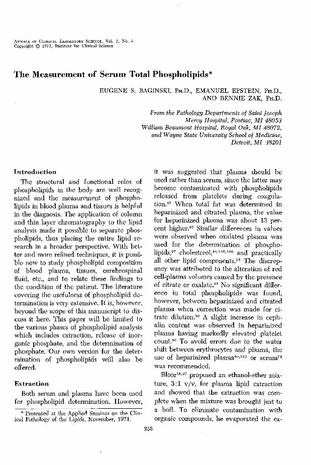
IntroductionThe structural and functional roles of
phospholipids in the body are well recognized and the measurement of phospholipids in blood plasma and tissues is helpful in the diagnosis. The application of column and thin layer chromatography to the lipid analysis made it possible to separate phospholipids, thus placing the entire lipid research in a broader perspective. W ith better and more refined techniques, it is possible now to study phospholipid composition of blood plasma, tissues, cerebrospinal fluid, etc., and to relate these findings to the condition of the patient. The literature covering the usefulness of phospholipid determination is very extensive. It is, however, beyond the scope of this manuscript to discuss it here. This paper will be limited to the various phases of phospholipid analysis which includes extraction, release of inorganic phosphate, and the determination of phosphate. Our own version for the determination of phospholipids will also be ofFered.
ExtractionBoth serum and plasma have been used
for phospholipid determination. However,
* Presented at the Applied Seminar on the Clinical Pathology of the Lipids, November, 1971.
255
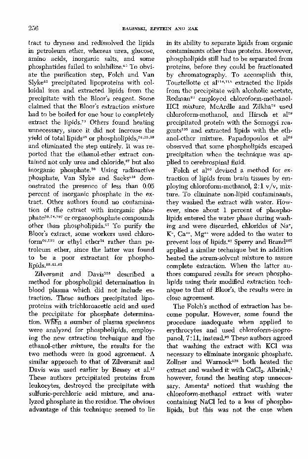
it was suggested that plasma should be used rather than serum, since the latter may become contaminated with phospholipids released from platelets dining coagulation.67 When total fat was determined in heparinized and citrated plasma, the value for heparinized plasma was about 13 percent higher.67 Similar differences in values were observed when oxalated plasma was used for the determination of phospholipids,97 cholesterol,48’103’106 and practically all other lipid components.25 The discrepancy was attributed to the alteration of red cell-plasma volumes caused by the presence of citrate or oxalate.97 No significant difference in total phospholipids was found, however, between heparinized and citrated plasma when correction was made for citrate dilution.50 A slight increase in ceph- alin content was observed in heparinized plasma having markedly elevated platelet count.50 To avoid errors due to the water shift between erythrocytes and plasma, the use of heparinized plasma50’123 or serum73 was recommended.
Bloor18’22 proposed an ethanol-ether mixture, 3:1 v/v, for plasma lipid extraction and showed that the extraction was complete when the mixture was brought just to a boil. To eliminate contamination with organic compounds, he evaporated the ex
256 BAGINSKI, EPSTEIN AND ZAK
tract to dryness and redissolved the lipids in petroleum ether, whereas urea, glucose, amino acids, inorganic salts, and some phosphatides failed to solubilize.45 To obviate the purification step, Folch and Van Slyke45 precipitated lipoproteins with colloidal iron and extracted lipids from the precipitate with the Bloor’s reagent. Some claimed that the Bloor’s extraction mixture had to be boiled for one hour to completely extract the lipids.73 Others found heating unnecessary, since it did not increase the yield of total lipids25 or phospholipids,24’25’39 and eliminated the step entirely. It was reported that the ethanol-ether extract contained not only urea and chloride,87 but also inorganic phosphate.26 Using radioactive phosphate, Van Slyke and Sacks118 demonstrated the presence of less than 0.05 percent of inorganic phosphate in the extract. Other authors found no contamination of the extract with inorganic phosphate39’74’107 or organophosphate compounds other than phospholipids.67 To purify the Bloor’s extract, some workers used chloroform66’122 or ethyl ether24 rather than petroleum ether, since the latter was found to be a poor extractant for phospholipids. 3̂ 9 ,«,45
Zilversmit and Davis125 described a method for phospholipid determination in blood plasma which did not include extraction. These authors precipitated lipoproteins with trichloroacetic acid and used the precipitate for phosphate determination. WEFn a number of plasma specimens were analyzed for phospholipids, employing the new extraction technique and the ethanol-ether mixture, the results for the two methods were in good agreement. A similar approach to that of Zilversmit and Davis was used earlier by Bessey et al.17 These authors precipitated proteins from leukocytes, destroyed the precipitate with sulfuric-perchloric acid mixture, and analyzed phosphate in the residue. The obvious advantage of this technique seemed to lie
in its ability to separate lipids from organic contaminants other than proteins. However, phospholipids still had to be separated from proteins, before they could be fractionated by chromatography. To accomplish this, Tourtellotte et al114’115 extracted the lipids from the precipitate with alcoholic acetate, Redman92 employed chloroform-methanol- HC1 mixture, McArdle and Zilkha78 used chloroform-methanol, and Hirsch et al59 precipitated protein with the Somogyi reagents105 and extracted lipids with the ethanol-ether mixture. Papadopoulos et al86 observed that some phospholipids escaped precipitation when the technique was applied to cerebrospinal fluid.
Folch et al46 devised a method for extraction of lipids from brain tissues by employing chloroform-methanol, 2:1 v/v, mixture. To eliminate non-lipid contaminants, they washed the extract with water. However, since about 1 percent of phospholipids entered the water phase during washing and were discarded, chlorides of Na+, K+, Ca++, Mg*+ were added to the water to prevent loss of lipids.47 Sperry and Brand107 applied a similar technique but in addition heated the serum-solvent mixture to assure complete extraction. When the latter authors compared results for serum phospholipids using their modified extraction technique to that of Bloor’s, the results were in close agreement.
The Folch’s method of extraction has become popular. However, some found the procedure inadequate when applied to erythrocytes and used chloroform-isopro- panol, 7:11, instead.95 These authors agreed that washing the extract with KC1 was necessary to eliminate inorganic phosphate. Zollner and Warnock128 both heated the extract and washed it with CaCl2. Albrink,1 however, found the heating step unnecessary. Amenta2 noticed that washing the chloroform-methanol extract with water containing NaCl led to a loss of phospholipids, but this was not the case when
THE MEASUREM ENT OF SERUM TOTAL PHOSPHOLIPIDS 257
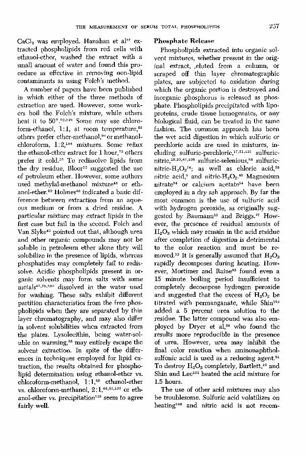
CaCl2 was employed. Hanahan et al57 extracted phospholipids from red cells with ethanol-ether, washed the extract with a small amount of water and found this procedure as effective in removing non-lipid contaminants as using Folch’s method.
A number of papers have been published in which either of the three methods of extraction are used. However, some workers boil the Folch’s mixture, while others heat it to 50 °.93’122 Some may use chloro- form-ethanol, 1:1, at room temperature,35 others prefer ether-methanol,96 or methanol- chloroform, 1:2,104 mixtures. Some reflux the ethanol-ether extract for 1 hour,73 others prefer it cold.25 To redissolve lipids from the dry residue, Bloor22 suggested the use of petroleum ether. However, some authors used methylal-methanol mixture43 or ethanol-ether.59 Holmes60 indicated a basic difference between extraction from an aqueous medium or from a dried residue. A particular mixture may extract lipids in the first case but fail in the second. Folch and Van Slyke45 pointed out that, although urea and other organic compounds may not be soluble in petroleum ether alone they will solubilize in the presence of lipids, whereas phosphatides may completely fail to redissolve. Acidic phospholipids present in organic solvents may form salts with some metals47’75’100 dissolved in the water used for washing. These salts exhibit different partition characteristics from the free phospholipids when they are separated by thin layer chromatography, and may also differ in solvent solubilities when extracted from the plates. Lysolecithin, being water-soluble on warming,88 may entirely escape the solvent extraction. In spite of the differences in techniques employed for lipid extraction, the results obtained for phospholipid determination using ethanol-ether vs. chloroform-methanol, 1: l ,89 ethanol-ether vs. chloroform-methanol, 2: l ,66’95’107 or eth- anol-ether vs. precipitation125 seem to agree fairly well.
Phosphate ReleasePhospholipids extracted into organic sol
vent mixtures, whether present in the original extract, eluted from a column, or scraped off thin layer chromatographic plates, are subjected to oxidation during which the organic portion is destroyed and inorganic phosphorus is released as phosphate. Phospholipids precipitated with lipoproteins, crude tissue homogenates, or any biological fluid, can be treated in the same fashion. The common approach has been the wet acid digestion in which sulfuric or perchloric acids are used in mixtures, including sulfuric-perchloric,17’71’115 sulfuric- nitric,19’29’67’108 sulfuric-selenious,86 sulfuric- nitric-H20 274; as well as chloric acid,53 nitric acid,5 and nitric-H2O2 .03 Magnesium nitrate94 or calcium acetate54 have been employed in a dry ash approach. By far the most common is the use of sulfuric acid with hydrogen peroxide, as originally suggested by Baumann12 and Briggs.27 However, the presence of residual amounts of H20 2 which may remain in the acid residue after completion of digestion is detrimental to the color reaction and must be removed.12 It is generally assumed that H2O2
rapidly decomposes during heating. However, Mortimer and Raine82 found even a 15 minute boiling period insufficient to completely decompose hydrogen peroxide and suggested that the excess of H20 2 be titrated with permanganate, while Shin102 added a 5 percent urea solution to the residue. The latter compound was also employed by Dryer et al,38 who found the results more reproducible in the presence of urea. However, urea may inhibit the final color reaction when aminonaphthol- sulfonic acid is used as a reducing agent.91 To destroy H20 2 completely, Bartlett,10 and Shin and Lee101 heated the acid mixture for1.5 hours.
The use of other acid mixtures may also be troublesome. Sulfuric acid volatilizes on heating108 and nitric acid is not recom
258 BAGINSKI, EPSTEIN AND ZAK
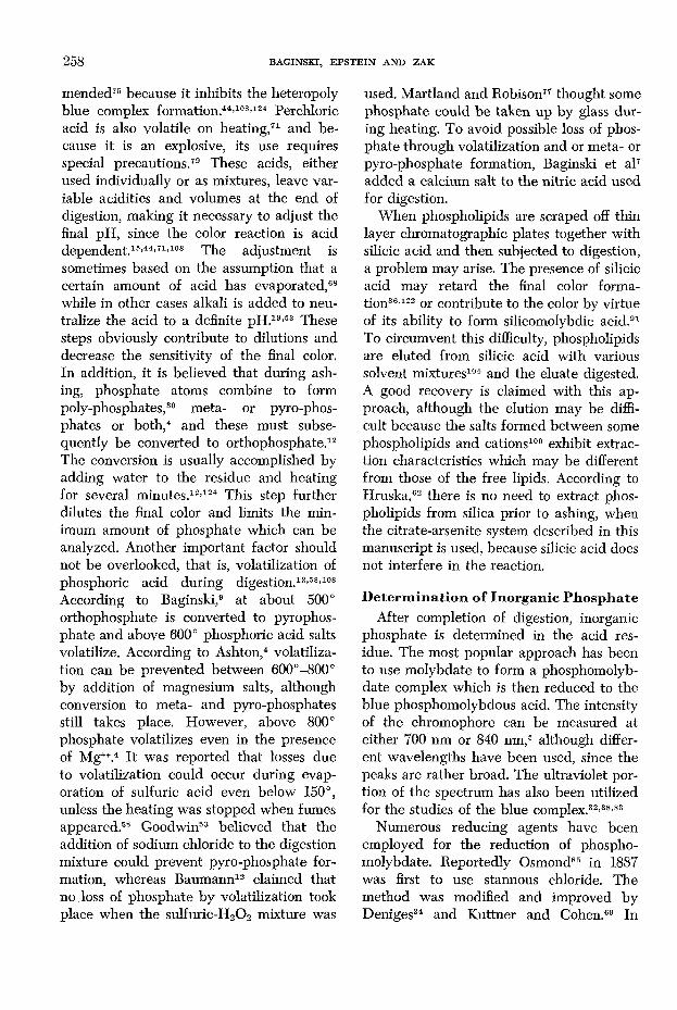
mended75 because it inhibits the heteropoly blue complex formation.44’108’124 Perchloric acid is also volatile on heating,71 and because it is an explosive, its use requires special precautions.79 These acids, either used individually or as mixtures, leave variable acidities and volumes at the end of digestion, making it necessary to adjust the final pH, since the color reaction is acid dependent.15’44’71’108 The adjustment is sometimes based on the assumption that a certain amount of acid has evaporated,68 while in other cases alkali is added to neutralize the acid to a definite pH.19’53 These steps obviously contribute to dilutions and decrease the sensitivity of the final color. In addition, it is believed that during ashing, phosphate atoms combine to form poly-phosphates,30 meta- or pyro-phos- phates or both,4 and these must subsequently be converted to orthophosphate.12 The conversion is usually accomplished by adding water to the residue and heating for several minutes.12’124 This step further dilutes the final color and limits the minimum amount of phosphate which can be analyzed. Another important factor should not be overlooked, that is, volatilization of phosphoric acid during digestion.12’58’108 According to Baginski,9 at about 500° orthophosphate is converted to pyrophosphate and above 600° phosphoric acid salts volatilize. According to Ashton,4 volatilization can be prevented between 600°-800° by addition of magnesium salts, although conversion to meta- and pyro-phosphates still takes place. However, above 800° phosphate volatilizes even in the presence of Mgw.4 It was reported that losses due to volatilization could occur during evaporation of sulfuric acid even below 150°, unless the heating was stopped when fumes appeared.58 Goodwin53 believed that the addition of sodium chloride to the digestion mixture could prevent pyro-phosphate formation, whereas Baumann12 claimed that no loss of phosphate by volatilization took place when the sulfuric-H20 2 mixture was
used. Martland and Robison77 thought some phosphate could be taken up by glass during heating. To avoid possible loss of phosphate through volatilization and or meta- or pyro-phosphate formation, Baginski et al7 added a calcium salt to the nitric acid used for digestion.
When phospholipids are scraped off thin layer chromatographic plates together with silicic acid and then subjected to digestion, a problem may arise. The presence of silicic acid may retard the final color formation36’122 or contribute to the color by virtue of its ability to form silicomolybdic acid.91 To circumvent this difficulty, phospholipids are eluted from silicic acid with various solvent mixtures104 and the eluate digested. A good recovery is claimed with this approach, although the elution may be difficult because the salts formed between some phospholipids and cations100 exhibit extraction characteristics which may be different from those of the free lipids. According to Hruska,62 there is no need to extract phospholipids from silica prior to ashing, when the citrate-arsenite system described in this manuscript is used, because silicic acid does not interfere in the reaction.
D eterm ination o f Inorganic PhosphateAfter completion of digestion, inorganic
phosphate is determined in the acid residue. The most popular approach has been to use molybdate to form a phosphomolyb- date complex which is then reduced to the blue phosphomolybdous acid. The intensity of the chromophore can be measured at either 700 nm or 840 nm,5 although different wavelengths have been used, since the peaks are rather broad. The ultraviolet portion of the spectrum has also been utilized for the studies of the blue complex.32’38’83
Numerous reducing agents have been employed for the reduction of phospho- molybdate. Reportedly Osmond85 in 1887 was first to use stannous chloride. The method was modified and improved by Deniges34 and Kuttner and Cohen.69 In
THE M EASUREM ENT OF SERUM TOTAL PHOSPHOLIPIDS 259
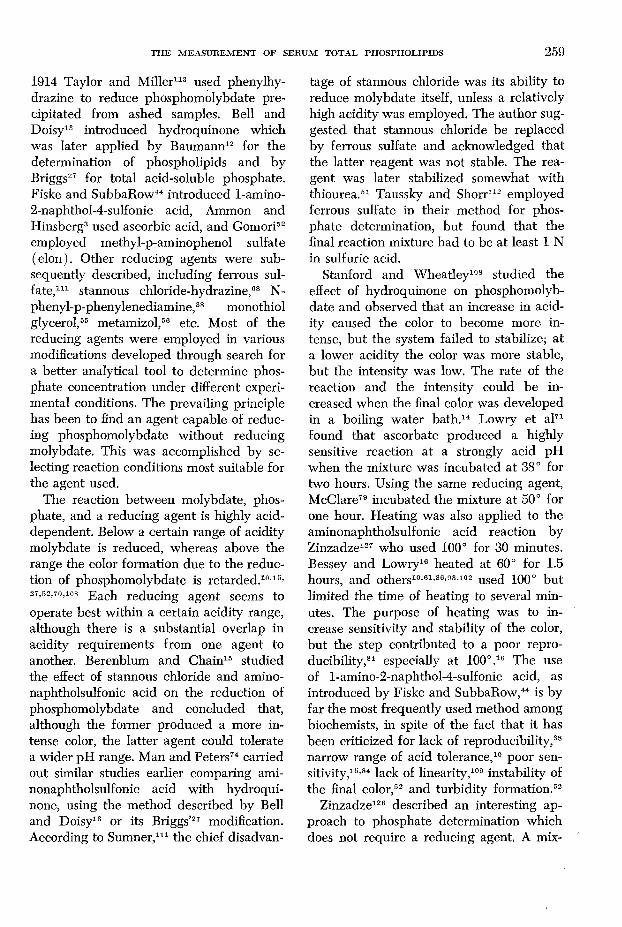
1914 Taylor and Miller113 used phenylhy- drazine to reduce phosphomolybdate precipitated from ashed samples. Bell and Doisy13 introduced hydroquinone which was later applied by Baumann12 for the determination of phospholipids and by Briggs27 for total acid-soluble phosphate. Fiske and SubbaRow14 introduced 1-amino- 2-naphthol-4-sulfonic acid, Ammon and Hinsberg3 used ascorbic acid, and Gomori52
employed methyl-p-aminophenol sulfate (elon). Other reducing agents were subsequently described, including ferrous sulfate, 1 1 1 stannous chloride-hydrazine, 68 N- phenyl-p-phenylenediamine, 38 monothiol glycerol, 55 metamizol, 56 etc. Most of the reducing agents were employed in various modifications developed through search for a better analytical tool to determine phosphate concentration under different experimental conditions. The prevailing principle has been to find an agent capable of reducing phosphomolybdate without reducing molybdate. This was accomplished by selecting reaction conditions most suitable for the agent used.
The reaction between molybdate, phosphate, and a reducing agent is highly acid- dependent. Below a certain range of acidity molybdate is reduced, whereas above the range the color formation due to the reduction of phosphomolybdate is retarded.10’15’ 27,52,70,108 Each reducing agent seems to operate best within a certain acidity range, although there is a substantial overlap in acidity requirements from one agent to another. Berenblum and Chain15 studied the effect of stannous chloride and amino- naphtholsulfonic acid on the reduction of phosphomolybdate and concluded that, although the former produced a more intense color, the latter agent could tolerate a wider pH range. Man and Peters74 carried out similar studies earlier comparing ami- nonaphtholsulfonic acid with hydroquinone, using the method described by Bell and Doisy13 or its Briggs’ 27 modification. According to Sumner, 1 1 1 the chief disadvan
tage of stannous chloride was its ability to reduce molybdate itself, unless a relatively high acidity was employed. The author suggested that stannous chloride be replaced by ferrous sulfate and acknowledged that the latter reagent was not stable. The reagent was later stabilized somewhat with thiourea . 51 Taussky and Shorr112 employed ferrous sulfate in their method for phosphate determination, but found that the final reaction mixture had to be at least 1 N in sulfuric acid.
Stanford and Wheatley108 studied the effect of hydroquinone on phosphomolybdate and observed that an increase in acidity caused the color to become more intense, but the system failed to stabilize; at a lower acidity the color was more stable, but the intensity was low. The rate of the reaction and the intensity could be increased when the final color was developed in a boiling water bath .14 Lowry et al71
found that ascorbate produced a highly sensitive reaction at a strongly acid pH when the mixture was incubated at 38° for two hours. Using the same reducing agent, McClare79 incubated the mixture at 50° for one hour. Heating was also applied to the aminonaphtholsulfonic acid reaction by Zinzadze127 who used 100° for 30 minutes. Bessey and Lowry16 heated at 60° for 1.5 hours, and others10’6 1’86’93’102 used 1 0 0 ° but limited the time of heating to several minutes. The purpose of heating was to increase sensitivity and stability of the color, but the step contributed to a poor reproducibility, 84 especially at 100° . 16 The use of l-amino-2-naphthol-4-sulfonic acid, as introduced by Fiske and SubbaRow, 44 is by far the most frequently used method among biochemists, in spite of the fact that it has been criticized for lack of reproducibility , 38
narrow range of acid tolerance, 10 poor sensitivity,15 ’84 lack of linearity , 109 instability of the final color, 52 and turbidity formation. 52
Zinzadze126 described an interesting approach to phosphate determination which does not require a reducing agent. A mix
260 BAGINSKI, EPSTEIN AND ZAK
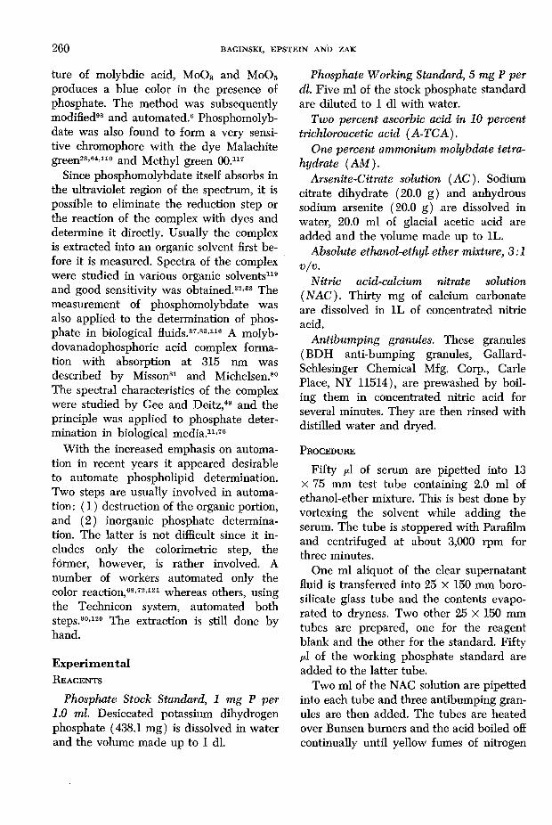
ture of molybdic acid, M0 O3 and MoOg produces a blue color in the presence of phosphate. The method was subsequently modified98 and automated . 6 Phosphomolyb- date was also found to form a very sensitive chromophore with the dye Malachite green28’64*110 and Methyl green 00.117
Since phosphomolybdate itself absorbs in the ultraviolet region of the spectrum, it is possible to eliminate the reduction step or the reaction of the complex with dyes and determine it directly. Usually the complex is extracted into an organic solvent first before it is measured. Spectra of the complex were studied in various organic solvents119
and good sensitivity was obtained . 32’33 The measurement of phosphomolybdate was also applied to the determination of phosphate in biological fluids. 37’83’116 A molyb- dovanadophosphoric acid complex formation with absorption at 315 nm was described by Misson81 and Michelsen.80
The spectral characteristics of the complex were studied by Gee and Deitz,49 and the principle was applied to phosphate determination in biological media .1 1 ’76
With the increased emphasis on automation in recent years it appeared desirable to automate phospholipid determination. Two steps are usually involved in automation: ( 1 ) destruction of the organic portion, and (2 ) inorganic phosphate determination. The latter is not difficult since it includes only the colorimetric step, the former, however, is rather involved. A number of workers automated only the color reaction, 68’72’12 1 whereas others, using the Technicon system, automated both steps. 90’120 The extraction is still done by hand.
ExperimentalR eagents
Phosphate Stock Standard, 1 mg P per1.0 ml. Desiccated potassium dihydrogen phosphate (438.1 mg) is dissolved in water and the volume made up to 1 dl.
Phosphate Working Standard, 5 mg P per dl. Five ml of the stock phosphate standard are diluted to 1 dl with water.
Two percent ascorbic acid in 10 percent trichloroacetic acid (A-TCA).
One percent ammonium molybdate tetra- hydrate (AM).
Arsenite-Citrate solution (AC). Sodium citrate dihydrate ( 2 0 . 0 g) and anhydrous sodium arsenite (2 0 . 0 g) are dissolved in water, 2 0 . 0 ml of glacial acetic acid are added and the volume made up to 1L.
Absolute ethanol-ethyl ether mixture, 3:1 v/v.
Nitric acid-calcium nitrate solution (NAC). Thirty mg of calcium carbonate are dissolved in 1L of concentrated nitric acid.
Antibumping granules. These granules (BDH anti-bumping granules, Gallard- Schlesinger Chemical Mfg. Corp., Carle Place, NY 11514), are prewashed by boiling them in concentrated nitric acid for several minutes. They are then rinsed with distilled water and dryed.
P rocedure
Fifty /j.I of serum are pipetted into 13 X 75 mm test tube containing 2.0 ml of ethanol-ether mixture. This is best done by vortexing the solvent while adding the serum. The tube is stoppered with Parafilm and centrifuged at about 3,000 rpm for three minutes.
One ml aliquot of the clear supernatant fluid is transferred into 25 X 150 mm boro- silicate glass tube and the contents evaporated to dryness. Two other 25 X 150 mm tubes are prepared, one for the reagent blank and the other for the standard. Fifty /¿I of the working phosphate standard are added to the latter tube.
Two ml of the NAC solution are pipetted into each tube and three antibumping granules are then added. The tubes are heated over Bunsen burners and the acid boiled off continually until yellow fumes of nitrogen
THE M EASUREM ENT OF SERUM TOTAL PHOSPHOLIPIDS 261
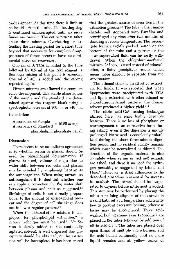
oxides appear. At this time there is little or no liquid left in the tube. The heating step is continued uninterrupted until no more fumes are present. The entire process takes about three minutes. Overheating or extending the heating period for a short time beyond that necessary for complete disappearance of fumes seems to have no detrimental effect on recoveries.
One ml of A-TCA is added to the tube followed by 0.5 ml of the AM reagent; a thorough mixing at this point is essential. One ml of AC is added and the mixing repeated again.
Fifteen minutes are allowed for complete color development. The stable absorbances of the sample and the standard are determined against the reagent blank using a spectrophotometer set at 700 nm or 840 nm.
Calculations
Absorbance of Sample „e _ mo.Absorbance of Standard
phospholipid phosphate per dl
DiscussionThere seems to be no uniform agreement
as to whether serum or plasma should be used for phospholipid determination. If plasma is used, volume changes due to water shift between red cells and plasma can be avoided by employing heparin as the anticoagulant. When using tartrate as anticoagulant it is doubtful whether one can apply a correction for the water shift between plasma and cells as suggested. 50
Shrinkage of cells is not directly proportional to the amount of anticoagulant present and the degree of cell shrinkage does not follow a regular pattern .40
When the ethanol-ether mixture is employed for phospholipid extraction, 18 a proper technique must be used .2 1 ’22’23 Serum is slowly added to the continually agitated solvent. A well dispersed fine precipitate should be obtained, or the extraction will be incomplete. It has been stated
that the greatest source of error lies in the extraction process. 12 The tube is then immediately well stoppered with Parafilm and centrifuged any time after two minutes of standing at room temperature. The precipitate forms a tightly packed button on the bottom of the tube and a portion of the clear supernatant fluid can be easily withdrawn. When the chloroform-methanol mixture, 2 : 1 v/v, is used instead of ethanol- ether, a fluffy precipitate results which seems more difficult to separate from the supernatant.
The ethanol-ether is an effective extractant for lipids. It was reported that when lipoproteins were precipitated with TCA and lipids extracted with ether-ethanol or chloroform-methanol mixture, the former solvent produced a higher yield. 114
The nitric acid-Ca++ ashing technique utilized here has some highly desirable features. There is no loss of phosphate or rearrangement to an unreactive form during ashing, even if the digestion is unduly prolonged. Nitric acid is completely volatilized during the short three-minute digestion period and no residual acidity remains which must be neutralized or diluted. Destruction of the organic material is also complete when serum or red cell extracts are ashed, and there is no need for hydrogen peroxide, as suggested by Ichida and Hine . 63 However, a strict adherence to the described procedure is essential for successful analysis. The extract should be evaporated to dryness before nitric acid is added. This step may be performed by placing the tubes containing aliquots of the extract in a sand bath set at a temperature sufficiently low to prevent excessive boiling, otherwise losses may be encountered. Three acid- washed boiling stones (see Procedure) are placed in the tubes followed by addition of nitric acid-Ca++. The tubes are placed over open flames of multiple micro-bumers and the acid boiled continually until no more liquid remains and all yellow fumes of
262 BAGINSKI, EPSTEIN AND ZAK
nitrogen oxides disappear. When raw serum or tissue homogenates are digested one may occasionally find a few black particles left on the side of the tube. In such a case the side of the tube can be exposed to the flame for a few seconds until all black particles disappear.
After the TCA-ascorbic acid and ammonium molybdate reagents have been added to the dry residue, it is essential to mix the content very well before the citrate-arsenite addition follows. During digestion nitric acid refluxes and creeps up the walls of the tube carrying phosphate with it. Therefore, after the addition of TCA-ascorbate and molybdate reagents, it is important to vortex the contents up and down to wash phosphate off the walls. The 1.5 ml liquid volume is sufficient for thorough washing. After the citrate-arsenite reagent has been added, phosphate, washed off the walls at this point, will not react. After the analysis is complete the tubes should be rinsed with distilled water, since phosphate tends to adhere to glass when the tubes are left to dry, and rinsing alone may not be helpful. Boiling in concentrated nitric acid followed by a thorough rinsing with distilled water is then necessary before the tubes can be used again. New tubes should be treated in the same fashion before they can be used. When the absorbance of the reagent blank is determined against water at 700 nm in a 1 cm cuvet, it is usually 0.03 to0.04. It may vary slightly according to impurities present in reagents, but should be constant for each set of reagents. An increase in the blank absorbance may indicate that some contamination has occurred.
The described procedure has an additional feature which makes the method more flexible and can expand its range of applicability. The reaction can tolerate a wide range of acidity in the final mixture. This indicates that when the use of a different acid is more desirable for digestion, the residual acid should not interfere in the reaction. However, there seems no need to
employ any other acid since nitric acid is capable of destroying serum extracts or small amounts of raw serum or tissue homogenates. Since the final color is sensitive, the procedure requires a very small sample which is easily destroyed. A high acidity leads to precipitation of citric acid and the color is not as stable as it is at a lower acid strength. Lowry and Lopez70 using ascorbic acid studied the effect of acidity on the reaction and concluded that the reaction could proceed between pH 0.4 to 0.9 and 2.8 to 4.6 but not at intermediate pH levels because a large blank resulted. They also found that an increase in ascorbate led to a continuous color development. The final pH in the citrate-arsenite reaction is about1.5 and the blank is negligible. In our procedure, a 1 0 -fold increase in ascorbic acid concentration has no effect. Chen et al29
also analyzed the effect of acidity, ascorbic acid concentration, time necessary for color development, temperature, etc. They found the final acid concentration optimum to be0.5 to 1.0 N. Above 1.0 N no reduction took place and below 0.1 N the blank itself developed color. These authors also stated that the color did not develop fully even after three hours of standing at room temperature, but at 37° it developed completely within one hour.
Inorganic phosphate determination following digestion does not require precautions concerning the presence of phosphate esters which may hydrolyze and release inorganic phosphate. For this reason rigid control of acidity seems unnecessary. However, since the final reaction is highly acid- dependent in various methods used the acidity must generally be adjusted before the reaction is carried out. Furthermore, any pyro- or meta-phosphate must be converted to orthophosphate. Although the ash does not contain organic compounds which may interfere in the color reaction, inorganic contaminants such as arsenate or silicate may be present and they are known to interfere10 ’29’38,99 by forming arseno- and
THE M EASUREM ENT OF SEBUM TOTAL PHOSPHOLIPIDS 263
silico-molybdate complexes. However, these do not interfere in the citrate-arsenite system, nor is there a need to adjust acidity.
The final acid concentration employed in the citrate-arsenite system was chosen purposely to enable one to analyze inorganic phosphate in the presence of acid-labile phosphate-containing organic compounds known to be present in plasma.20’42’129 Furthermore, the method was adapted to the determination of Glucose-6-phosphatase in tissue homogenates8 which contain appreciable amount of acid-labile phosphate esters. Under the relatively mild acid conditions employed here, one would expect molyb- date to undergo reduction in the absence of phosphate. Indeed, when a mixture of TCA, ascorbic acid, and molybdate is allowed to stand at room temperature, the solution becomes blue and turbidity develops,5 indicating some reduction of molybdate itself. However, this phenomenon does not occur when arsenite-citrate is present. The purpose of the citrate addition is to complex molybdate and prevent the latter from reduction by ascorbic acid. Oxalic acid was employed for the same purpose by James and Allen65 in their method for inorganic phosphate determination. Marsh76 also used citrate to bind molybdate after phosphomolybdate was extracted into butanol. The ability of citrate and other car- boxylic acids to bind molybdate was reported by Davies and Davies.31 In the citrate-arsenite system described by the present authors, the presence of citrate contributes to an increase in sensitivity, prevents reduction of molybdate, and stabilizes the system. The role of arsenite is not clear, although it also contributes somewhat to the overall increase in sensitivity.
Sum m aryThe various aspects of phospholipid de
termination in blood plasma are discussed, including the extraction of phospholipids, release of phosphate from the organic portion, and the colorimetric determination of
inorganic phosphate. A method for phospholipid determination is proposed which is simple, rapid, and relatively free of the various shortcomings encountered in other methods.
References1. A l b r i n k , M. J.: The microtitration of total
fatty acids of serum, w ith notes on the estimation of triglycerides. J. L ipid Res. 2:53-59, 1959.
2. A m e n t a , J. S.: A rapid extraction and quantification of total lipids and lipid fractions in blood and feces. Clin. Chem. 26:339-346, 1970.
3. A m m o n , R. a n d H i n s b e r g , K.: Colorimetri- sche Phosphor- und Arsensaure-bestimmung m it Ascorbinsäure. Physiol. Chem. 239:207- 216, 1936.
4. A s h t o n , F. L.: Influence of the tem perature of ashing on the accuracy of the determination of phosphorus in grass. J. Soc. Chem. Ind. 55:106-108, 1936.
5. B a g i n s k i , E. S. a n d Z a k , B . : Microdetermination of serum phosphate and phospholipids. Clin. Chem. Acta 5:834—838, 1960.
6. B a g i n s k i , E . S., E p s t e i n , E . , a n d Z a k , B . : Automation of methods for the determination of serum phosphorus. Clin. Chim. Acta 20: 76-80, 1964.
7. B a g i n s k i , E. S., W e i n e r , L. M., a n d Z a k , B .: The simple determination of nucleotide phosphorus. Clin. Chim. Acta 20:378—379, 1964.
8. B a g i n s k i , E. S., F o a , P. P., a n d Z a k , B . : Glucose-6-Phosphatase. Methoden der E nzymatischen Analyse. Verlag Chemie W ein- heim/Bergstr. vol. 2:837-843, 1970.
9. B a g i n s k i , S.: Mikroveraschung. Einige praktische Hinweise. Z. Wiss. Mikr. 55:241-248, 1938.
10. B a r t l e t t , G. R.: Phosphorus assay in column chromatography. J. Biol. Chem. 234:466-468, 1959.
11. B a r t l e t t , E. M. a n d L e w i s , D. H.: Spectro- photometric determination of phosphate esters in the presence and absence of orthophosphate. Anal. Biochem. 36:159-167, 1970.
12. B a u m a n n , E. J.: On the estimation of organic phosphorus. J. Biol. Chem. 59:667-674, 1924.
13. B e l l , R. D . a n d D o i s y , E. A.: Rapid colorimetric methods for the determination of phosphorus in urine and blood. J. Biol. Chem. 44:55-67, 1920.
14. B e n e d i c t , S. R. a n d T h e i s , R. C.: A modification of the molybdic method for the determination of inorganic phosphorus in serum. J. Biol. Chem. 62:63-66, 1924.
15. B e r e n b l u m , I . a n d C h a i n , E .: Studies on the colorimetric determination of phosphate. Biochem. J. 32:286-294, 1938.
264 BAGINSKI, EPSTEIN AND ZAK
16. B e s s e y , O. A. a n d L o w r y , O. H.: Factors influencing the riboflavin content of the cornea. J. Biol. Chem. 155:535-643, 1944.
17. B e s s e y , O. A., L o w r y , O. H., a n d B r o c k , M. J.: The quantitative determination of ascorbic acid in small amounts of white blood cells and platelets. J . Biol. Chem. 264:197- 205, 1946.
18. B l o o r , W. R.: A method for the determination of fat in small amounts of blood. J. Biol. Chem. 27:377-384, 1914.
19. B l o o r , W. R.: Studies on blood fat. II. Fat absorption and the blood lipoids. J. Biol. Chem. 23:317-326, 1915.
20. B l o o r , W. R.: The distribution of phosphoric acid in normal human blood. J. Biol. Chem. 36:49-57, 1918.
21. B l o o r , W. R.: Methode nephelometrique pour la determination de l’acide phospho- rique et de ses composes contenus dans de petites quantités de sang. Bull. Soc. Chim. Biol. 3:451-475, 1921.
22. B l o o r , W. R.: The determination of small amounts of lipid in blood plasma. J. Biol. Chem. 77:53-73, 1928.
23. B l o o r , W. R.: The oxidative determination of phospholipid (lecithin and cephalin) in blood and tissues. J. Biol. Chem. 82:273-286, 1929.
24. B o y d , E. M.: L o w phospholipid values in dog plasma. J. Biol. Chem. 91:1-12, 1931.
25. Boyd, E. M.: Extraction of blood lipids. J. Biol. Chem. 114:223-234, 1936.
26. B r a n t e , G.: Studies on lipids in the nervous system with special reference to quantitative chemical determination and typical distribution. Acta Physiol. Scand. 28:Suppl. 63, 1 - 189, 1949.
27. B r ig g s , A. P.: Some applications of the colorimetric phosphate method. J. Biol. Chem. 59:252-264, 1924.
28. C h a l v a r d j i a n , A. a n d R u d n ic k i , E.: Determination of lipid phosphorus in the nanomolar range. Anal. Biochem. 36:225-227,1970.
29. C h e n , P. S., T o r ib a r a , T . Y., a n d W a r n e r ,H. : Microdetermination of phosphorus. Anal. Chem. 28:1756-1758, 1956.
30. C o h e n , L . E. a n d C h e c h , F. W.: The determination of phosphorus in organic compounds via flash combustion. Chemist-Analyst 47:86-87, 1958.
31. D a v ie s , D . R. a n d D a v ie s , W. C.: The colorimetric determination of phosphorus in the presence of interfering substances. Biochem. J. 26:2046-2055, 1932.
3 2 . D e l s a l , J . L . a n d M a n h o u r i , H.: Etude comparative des dosages colorimetriques du phosphore. II. Spectrophotometrie dans l’ul- tra-violet. Bull. Soc. Chim. Biol. 40:1169- 1177, 1958.
33. D e l s a l , J. L . a n d M a n h o u r i , H.: Etude comparative des dosages colorimetriques du
phosphore. III. Dosage de l’orthophosphate en presence d’esters phosphoriques. Bull. Soc. Chim. Biol. 40:1179-1187, 1958.
34. D e n i g e s , G.: Reaction de coloration extre- m ent sensible des phosphates et de arsenates. Compt. Rend. Acad, de Sciences. 171:802, 1920.
35. D o d g e , J. a n d P h i l l i p s , G. B.: Composition of phospholipid fatty acids and aldehydes in human red cells. J. Lipid Res. 8:667-675, 1967.
36. D o i z a k i , W. M. a n d Z i e v e , L . : Quantitative estimation of some phosphatides and their hydrolysis products by th in layer chromatography. Proc. Soc. Exp. Biol. Med. 223:91-94, 1963.
37. D r e i s b a c h , R. H.: Submicrogram determination of inorganic phosphate in biological material. Anal. Biochem. 20:169—171, 1965.
38. D r y e r , R . L., T a m m e s , A. R ., a n d R o u t h ,I . : The determination of phosphorus and phosphatase w ith N-phenyl-p-phenylenedia- mine. J. Biol. Chem. 225:177-183, 1957.
39. E g s g a a r d , J.: On the determination of the phosphatide content of serum. Acta Physiol. Scand. 26:171-178, 1948.
40. E i s e n m a n , A. J.: The effect of potassium oxalate on electrolytes of blood and plasma. J. Biol. Chem. 71:587-605, 1926-27.
41. E l l i s , G. a n d M a y n a r d , L. A.: The determination of phospholipids in bovine blood. J. Biol. Chem. 228:701-709, 1937.
42. F e i g l , J.: U ber das Vorkommen von Phos- phaten im menschlichen Blutserum. II. Saur- eloslicher (G esam t-) Phosphor, vorgebildetes Orthophosphat und “Restphosphor” beim Gesunden. Biochem. Z. 83:82—95, 1917.
43. F i l l e r u p , D. L. a n d M e a d , J. F . : Chromatographic separation of the plasma lipids. Proc. Soc. Exp. Biol. Med. 83:574-577, 1953.
44. F i s k e , C. H. a n d S u b b a R o w , Y.: The colorimetric determination of phosphorus. 66:375-400, 1925.
45. F o l c h , J. a n d V a n S l y k e , D. D .: Preparation of blood lipid extracts free from nonlipid extractives. Proc. Soc. Exp. Biol. Med. 41:514-515, 1939.
46. F o l c h , J., A s c o l i , I., M e a t h , J. A ., a n d L e B a r o n , F . N.: Preparation of lipid extracts from brain tissue. J. Biol. Chem. 292:833-841, 1951.
47. F o l c h , J., L e e s , M . , a n d S l o a n e S t a n l e y ,G. H.: A simple method for the isolation and purification of total lipids from animal tissues. J. Biol. Chem. 226:497-509, 1957.
48. G a r d n e r , J. A., G a i n s b o r o u g h , H., a n d M u r r a y , R .: CXCI. Studies on the cholesterol content of normal hum an plasma. VIII. A note on the effect of anticoagulants. Biochem. J. 32:1457-1459, 1938.
49. G e e , A. a n d D e i t z , V. R.: Determ ination of phosphate by differential spectrophotometry. Anal. Chem. 25:1320-1324, 1953.
THE M EASUREM ENT OF SERUM TOTAL PHOSPHOLIPIDS 265
5 0 . G j o n e , E. a n d O r n in g , O . M .: Plasma phospholipids in patients with liver disease. A quantitative thin layer chromatographic study. J. Clin. Lab. Invest. 2 8 :2 0 9 - 2 1 6 , 1 9 6 6 .
51. G o l d e n b e r g , H . a n d F e r n a n d e z , A.: Simplified method for the estimation of inorganic phosphorus in body fluids. Clin. Chem. 22:871-882, 1966.
52. G o m o r i , G . : A modification of the colorimetric phosphorus determination for use with the photelectric colorimeter. J. Lab. Clin. Med. 27:955-960, 1942.
53. G o o d w i n , J. F.: Total, phospholipid and labile phosphorus in serum and tissue employing chloric acid and N-phenyl-p-phenyl- enediamine. Proc. Soc. Exp. Biol. Med. 100: 217-219, 1959.
54. G r e e n , H . H . : Studies in mineral metabolism. IV. Determination o f phosphorus compounds in blood by dry combustion. J. Agr. Sci. 28: 372-375, 1928.
55. G r i n d e y , G . B. a n d N i c h o l , C. A.: Micro procedure for determination of pyrophosphate and orthophosphate. Anal. Biochem. 33:114-119, 1970.
56. G u i r g i s , F. K. a n d H a b i b , Y. A.: Use of a new reducing agent, Metamizol, in determining inorganic phosphorus in blood and urine. Clin. Chem. 17:78-81, 1971.
5 7 . H a n a h a n , D. J., W a t t s , R. M ., a n d P a p p a - j o h n , D.: Some chemical characteristics of the lipids of human and bovine erythrocytes and plasma. J. Lipid Res. 2:421-432, 1960.
58. H il l e b r a n d , W. F. a n d L u n d e l l , G. E. F.: Volatilization losses of phosphorus during evaporations of phosphates with sulfuric acid or fusions with pyrosulfate. J. Amer. Chem. Soc. 42:2609-2615, 1920.
59. H i r s c h , J. a n d A h r e n s , H . : The separation of complex lipid mixture by the use of silicic acid chromatography. J. Biol. Chem. 233:311-320, 1958.
60. H o l m e s , F. E.: A turbulent drier-extractor and a wide-interface extractor. A method for determining total lipid in serum, feces, and urine. Clin. Chem. 10:1007-1024, 1964.
61. H o r e c k e r , B. L., M a , T. S., a n d H a s s , E.: Note on the determination of microquantities of organic phosphorus. J. Biol. Chem. 136: 775-776, 1940.
62. H r u s k a , K. J.: Interference of pyrophosphate in the determination of orthophosphate. Clin. Chim. Acta 22:454^155, 1968.
63. I c h id a , T. a n d H i n e , N.: Improvement of Baginski’s method for phosphorus determination. Clin. Chim. Acta 23:378—379, 1969.
64. I t a y a , K. a n d Ui, M.: A new micromethod for the colorimetric determination of inorganic phosphate. Clin. Chim. Acta 24:361- 366, 1966.
65. J a m e s , R. a n d A l l e n , J . L.: The estimation
of phosphorus. Biochem. J. 34:858-865, 1940.
6 6 . J e s t i n g , E. a n d B a n g , H. O.: Methods of lipid extraction from blood plasma. Scand. J . Clin. Lab. Invest. 25:654-656, 1963.
67. K i r k , E. K ., P a g e , I. H., a n d V a n S l y k e ,D. D.: Gasometric microdetermination of lipids in plasma, blood cells and tissues. J . Biol. Chem. 206:203-234, 1934.
6 8 . K r a m l , M.: A semi-automated determination of phospholipids. Clin. Chim. Acta 23: 442-448, 1966.
69. K u t t n e r , T. a n d C o h e n , H. R . : Micro colorimetric studies. I. A molybdic acid, stannous chloride reagent. The micro estimation of phosphate and calcium in pus, plasma, and spinal fluid. J . Biol. Chem. 75: 517-531, 1927.
70. L o w r y , O. H. a n d L o p e z , J. A.: The determination of inorganic phosphate in the presence of labile phosphate esters. J. Biol. Chem. 162:421-428, 1946.
71. L o w r y , O. H., R o b e r t s , N. R . , L e i n e r , K . Y., Wu, M„ a n d F a r r , A. L . : The quantitative histochemistry o f brain. I. Chemical Methods. J. Biol. Chem. 207:1-17, 1954.
72. M a l l e i n , R . , P i n a t e l , H., a n d R i v i e r e , J . F . : Determination semi-atomatique des phospholipides seriques. Ann. Biol. Clin. 28: 257-264, 1970.
73. M a n , E. B. a n d G i l d e a , E. F . : A modification of the Stoddard and Drury titrimetric method for the determination of the fatty acids in blood serum. J. Biol. Chem. 99:43- 60, 1932.
74. M a n , E. B. a n d P e t e r s , J . P . : Gravimetric determination of serum cholesterol adapted to the Man and Gildea fatty acid method, with a note on the estimation of lipoid phosphorus. J . Biol. Chem. 101:685-695, 1933.
75. M a r i n e t t i , B. V .: Chromatographic separation, identification, and analysis of phos- phatides. J . Lipid Res. 3:1-20, 1962.
76. M a r s h , B. B.: The estimation of inorganic phosphate in the presence of adenosine triphosphate. Biochim. Biophys. Acta 32: 357-361, 1959.
77. M a r t l a n d , M . a n d R o b i s o n , R . : XCVII. Note on the estimation of phosphorus in blood. Biochem. J. 28:765-768, 1924.
78. M c A r d l e , B. a n d Z i l k h a , K . J . : The phospholipid composition of cerebrospinal fluid in neurologic disorders. Brain 85:389-402, 1962.
79. M c C l a r e , C . W. F . : An accurate and convenient organic phosphorus assay. Anal. Biochem. 39:527-530, 1971.
80. M i c h e l s e n , O. B.: Photometric determination of phosphorus as molybdovanadophos- phoric acid. Anal. Chem. 29:60-62, 1957.
266 BAGINSKI, EPSTEIN AND ZAK
81. M i s s o n , G.: Colorimetrische Phosphorbestim- mung in Stahl. Chem.-Ztg. 32:633-634, 1908.
82. M o r t i m e r , J. G. a n d R a i n e , D. N.: An improved estimation of phosphorus following digestion with hydrogen peroxide. Anal. Biochem. 9:492-495, 1964.
83. M o z e r s k y , S. M ., P e t t i n a t i , J. D., a n d
K o l m a n , S. D.: An improved method for the determination of orthophosphate suitable for assay of adenosine triphosphatase activity. Anal. Chem. 38:1182-1187, 1966.
84. N o r b e r g , B.: The working out of a method for histo- and cyto-chemical determination of phosphorus. Acta Physiol. Scand. 5:1-99, 1942.
85. O s m o n d , F.: Sur une reaction pouvantservir au dosage colorimetrique du phosphore dans les fontes, les aciers, etc. Bull. Soc.Chim. 47:745, 1887.
8 6 . P a p a d o p o u l o s , N. M., C e v a l l o s , W., a n d H e s s , W. C .: Determination of phospholipids in spinal fluid and brain. Arch. Neurol. 3: 677-682, 1960.
87. P e r n o k i s , E . W., F r e e l a n d , M. R., a n d
K r a u s , I.: The determination of blood lipids in blood dyscrasias. J. Lab. Clin. Med. 26: 1978-1980, 1941.
8 8 . P h i l l i p s , B. M. a n d R o b i n s o n , N.: Quantitative thin layer chromatography of cerebrospinal fluid phospholipids. Clin. Chim. Acta 8:832-842, 1963.
89. P h i l l i p s , G. B.: The isolation and quantitation of the principle phospholipid components of human serum using chromatography on silicic acid. Biochim. Biophys. Acta 29: 594-602, 1958.
90. P o l l a r d , F. H., N ic k l e s s , G., R o g e r s , D. E., a n d R o t h w e l l , M. T.: Quantitative inorganic chromatography. Part XII. Automatic analysis of phosphorusanion mixtures by anion exchange chromatography. J. Chro- matog. 17:157-167, 1965.
91. R a e , J. J. a n d E a s t c o t t , E . V.: The effect of urea and sodium chloride on the colorimetric determination of organic phosphate by King’s method. J. Biol. Chem. 129:255- 262, 1939.
92. R e d m a n , C. M.: Phospholipid metabolism in intact and modified erythrocyte membranes. J. Cell Biol. 49:35-49, 1971.
93. R o b i n s o n , N . a n d P h i l l i p s , B. M.: Quantitative thin layer chromatography of serum phospholipids. Clin. Chim. Acta. 8:385-392, 1963.
94. Roepke, R. R.: Determination of phosphorus fractions in blood serum. Ind. & Eng. Chem. Anal. Ed. 7:78, 1935.
95. R o s e , H. G. a n d O k l a n d e r , M.: Improved procedure for the extraction of lipids from human erythrocytes. J. Lipid Res. 6:428-431,1965.
96. R o s e n t h a l , A. F. a n d C h i n g - H s i e n , S.: A simple nonchromatographic determination of
glycerophosphatide in serum. Anal. Chem. 15:197-203, 1969.
97. S c h m i d t , L . H.: The nature of the difference in phospholipid content of oxalated and heparinized plasma. J. Biol. Chem. 109:448- 453, 1935.
98. S c h r i c k e r , J. A. a n d D a w s o n , P. R.: Improved molybdenum blue reagent for determination of phosphorus and arsenic. J. Assoc. Off. Agr. Chem. 22: 167-176, 1939.
99. S h e n , C. Y. a n d D y r o f f , D. R.: Determination of phosphate in presence of silicates by molydenum bine method. Anal. Chem. 34: 1367-1370, 1962.
100. S h i m o j o , T., K a n o h , H., a n d O h n o , K .: The elution behaviors of acidic phospholipids on column chromatography. J. Biochem. 69: 255-263, 1971.
101. S h i n , Y. S . a n d L e e , J. C.: Microdetermination of cholesterol and phospholipid in cerebrospinal fluid and serum by silicic acid column chromatography. Clin. Chem. 8:598- 605, 1962.
102. S h i n , Y. S .: Spectrophotometric ultramicrodetermination of inorganic phosphorus and lipid phosphorus in serum. Anal. Chem. 34: 1164-1166, 1962.
103. S h o p e , R. E.: Differences in serum and plasma content of cholesterol ester. J. Biol. Chem. 80:125-126, 1928.
104. S k i p s k i , V. P . , P e t e r s o n , R. F., a n d B a r c l a y , M.: Quantitative analysis of phospholipids by thin layer chromatography. Biochem. J. 90:374-378, 1964.
105. S o m o g y i , M.: Method for preparation of blood filtrates for determination of sugar. J. Biol. Chem. 86:655-663, 1930.
106. S p e r r y , W . M . a n d S c h o e n h e i m e r , R.: A comparison of serum, heparinized plasma, and oxalated plasma in regard to cholesterol content. J. Biol. Chem. 110:655-648, 1935.
107. S p e r r y , W . M. a n d B r a n d , F. C.: The determination of total lipids in blood serum. J. Biol. Chem. 213:69-76, 1955.
108. S t a n f o r d , R. V. a n d W h e a t l e y , A. H. M .: The estimation of phosphorus compounds in blood. Biochem. J. 19:697-705, 1925.
109. S t a n t o n , M. G .: Colorimetric determination of inorganic phosphate in the presence of biological material and adenosine triphosphate. Anal. Biochem. 22:27-34, 1968.
110. S t e p a n o v a , I.: Bestimmung des anorgan- ischen Phosphors in einer Mikromenge von Serum. Clin. Chim. Acta. 16:330-332, 1967.
111. S u m m e r , J . B.: A method for the colorimetric determination of phosphorus. Science 100:413-414, 1944.
112. T a u s s k y , H. H. a n d S h o r r , E.: A micro- colorimetric method for the determination of inorganic phosphorus. J. Biol. Chem. 202: 675-685, 1953.
113. T a y l o r , A. E. and M i l l e r , C. W . : On the estimation of phosphorus in biological material. J. Biol. Chem. 18:215-224, 1914.
THE M EASUBEM ENT OF SERUM TOTAL PHOSPHOLIPIDS 267
114. T o u r t e l l o t t e , W. W., V a n d e r , A. J . , S k r e n t n y , B. A., a n d D e J o n g , R. N.: A study of lipids in the cerebrospinal fluid. II. The determination of total lipids. J . Lab. Clin. Med. 52:481-490, 1958.
115. T o u r t e l l o t t e , W. W., P a r k e r , F. M., a n d D e J o n g , R . N .: A study of lipids in the cerebrospinal fluid. III. The determination of total phospholipids. J. Lab. Clin. Med. 52:491-495, 1958.
116. T u r a k u l o w , J. C., K u r g u l e v a , L. I., a n d G a g e l g a n c , A. I.: Determination of inorganic phosphate in biological materials. Biokhim. 32:106-110, 1967.
117. V a n B e l l e , N.: New and sensitive reaction for automatic determination of inorganic phosphate and its application to serum. Anal. Biochem. 33:132-142, 1970.
118. V a n S l y k e , D. D. a n d S a c k s , J.: Preparation of serum lipid extracts free of inorganic phosphate. J. Biol. Chem. 200:525-528, 1953.
119. W a d e l i n , C. a n d M e l l o n , M . G .: Extraction of heteropoly acids. Application to determination of phosphorus. Anal. Chem. 25:1668- 1673, 1953.
120. W e i n s t e i n , L. H., B o z a r t h , R. F., P o r t e r , C. A., M a n d l , R. H., a n d T w e e d y , B . G.: Automated analysis of phosphorus containing compounds in biological materials. Contrib. Boyce Thompson Inst. 22:389-397, 1964.
121. W h i t l e y , R. W . a n d A l b u r n , H. E.: A semi-automated method for the determination of serum phospholipids. Technicon International Symposium, New York, 1946.
122. W i l l i a m s , J. H., K u c h m a k , M., a n d W i t t e r , R. F.: Quantitative determination of phospholipid classes in human serum by combined thin-layer chromatography and phosphorus analysis. Clin. Chim. Acta 25: 447-452, 1969.
123. W i n t r o b e , M. W . : The size and hemoglobin content of the erythrocyte. J. Lab. Clin. Med. 17:899-912, 1932.
124. Y o u n g b u r g , G . E. a n d Y o u n g b u r g , M. V .: Phosphorus metabolism. I. A system for blood phosphorus analysis. J. Lab. Clin. Med. 16:158-166, 1930.
125. Z i l v e r s m i t , D . B. a n d D a v i s , A. K.: Microdetermination of plasma phospholipids by trichloroacetic acid precipitation. J. Lab. Clin. Med. 35:155-160, 1950.
126. Z i n z a d z e , S. R.: Neue Methoden zurkolorimetrischen Bestimmung der Phosphor- und Arsensaure. Z . Pflanzenernahr. 16:129- 180, 1930.
127. Z i n z a d z e , S. R.: Colorimetric methods for the determination of phosphorus. Ind. Eng. Chem., Anal. Ed. 7:227-230, 1935.
128. Z ö l l n e r , N. a n d W a r n o c k , S. A.: The enzymic determination of glycerophosphates and its application to the determination of phosphoglycerides, particularly those of serum phospholipids. Clin. Chim. Acta 7: 607-613, 1962.
129. Z u c k e r , T. F. a n d G u t m a n , M.: Distribution of phosphorus in blood. Proc. Soc. Exp. Biol. Med. 20:133-136, 1922.