Embed Size (px)
Citation preview
Submitted 16 August 2016, Accepted 20 February 2017, Published online 27 March 2017
Corresponding Author: Dr. V. Ramesh – e-mail – [email protected] 26
The molecular phylogeny and taxonomy of endophytic fungal
species from the leaves of Vitex negundo L.
Ramesh V1*, Arivudainambi USE
2 and Rajendran A
3
1Department of Botany, Vivekananda College, Tiruvedakam West - 625 234, Madurai, Tamilnadu, India 2Department of Chemistry, VHNSN College, Virudhunagar – 626 00, Tamilnadu, India 3Department of Botany, VHNSN College, Virudhunagar – 626 00, Tamilnadu, India
Ramesh V, Arivudainambi USE, Rajendran A
2017 – The molecular phylogeny and
taxonomy of endophytic fungal species from the leaves of Vitex negundo L. Studies in Fungi
2(1), 26–38, Doi 10.5943/sif/2/1/4
Abstract
Enormous fungal species live within the healthy plant tissues, some of which
presumably occur in a symbiotic association with host. Some fungal endophytes are
widespread and can be found in many different plant species, whereas others are highly
specific to single hosts. In this study, we isolated three endophytic fungi from the medicinal
plant Vitex negundo. They were identified based on morphological characteristics such as
size, shape, and colour of the spore and it was reinforced by 18s rRNA gene sequence
analysis. The phylogenetic tree showed that C. gloeosporioides VN1 and Pestalotiopsis
virgatula VN2 were closely relationship between. But they were not closely relationship
between the other endophytic fungal species that were obtained from geographically different
part of the world. This aspect can be further explored to understand the relationships between
plant hosts and their fungal endophyte.
Key words – Endophytic fungi – MEGA 6.0 – phylogenetic relationship – rRNA
Introduction
Endophytes are to be found in virtually every plant on earth. They reside in the living
tissues of the host plant and do so in a variety of relationships ranging from symbiotic to
pathogenic (Strobel et al. 2004). Endophytes receive nutrition and protection from the host
plant, while the host plant may benefit from enhanced competitive abilities and increased
resistance to herbivores, pathogens, and various abiotic stresses by attaining the metabolic
substances of endophytes (Saikkonen et al. 1998, Tan & Zou 2001, Zhang et al. 2006).
Endophytic fungi have been found in all plant families so far investigated, which represent
many species in different climatic regions of the world (Spurr & Welty 1975, Petrini &
carroll 1981, Petrini et al. 1992). Endophytes have been reported from all major groups of
plants including algae (Zuccaro et al. 2008, Suryanarayanan et al. 2010), lichens
(Suryanarayanan et al. 2005), mosses (Schulz et al. 1993), ferns (Petrini et al. 1992), conifers
(Giordano et al. 2009) and angiosperms (Saikkonen 2007), and may persist even in
aseptically cultured plants (Lucero et al. 2008). Endophytic fungi are reported from plants
that grow in various environments including tropic (Mohali et al. 2005), temperate (Ganley et
Studies in Fungi 2 (1): 26–37 (2017) www.studiesinfungi.org ISSN 2465-4973
Article Copyright © Mushroom Research Foundation 2017
Doi 10.5943/stif/ 2/1/4
27
al. 2004), xerophytic (Suryanarayanan et al. 2005) coastal mangroves (Kumaresan &
Suryanarayanan 2001, Okane et al. 1998) and aquatic environment (Sati & Belwal 2005).
Environment plays an important role on endophyte biodiversity, while the species diversity is
dependent upon the nature of the host plant and their ecological location.
Medicinal plants have been recognized as a repository of fungal endophytes with
novel metabolites of pharmaceutical importance (Strobel et al. 2004, Wiyakrutta et al. 2004,
Kumar et al. 2005, Tejesvi et al. 2007). Plant with pharmaceutical importance is being
exploited because of their healing properties. However, large scale harvesting of medicinal
plants has already become a major threat to biodiversity. As an alternative, microbe which
lives inside the plants (endophytes) may often become a tremendous potential source of
therapeutic compounds.
Traditional classification and identification of endophytic fungi depends upon
microscopic features, colony characteristics on artificial media and biochemical reactions
(Sutton & Cundell 2004). This kind of methods have served in the past but they have major
drawbacks as they cannot be applied to non cultivatable organisms and occasionally
biochemical characteristic of some organisms do not fit into the patterns of any known genus
and species. Amplification and sequencing of target regions within the ribosomal DNA gene
complex has emerged as a useful adjunctive tool for the identification of endophytic fungi
and does not depend on fungus sporulation for identification (Buzina et al. 2001, Iwen et al.
2002, Rakeman et al. 2005, Schwarz et al. 2006).
The key elements for the evolution of the endophytes are quite complex, involving
various types of interactions between the host plant, numerous levels of happenstance, and
multidirectional flows, they are also influenced by random events, such as living and non
living factors, which guide the process of co-evolution between endophytic fungi and their
hosts (Saikkonen et al. 2004).
Eventhough knowledge regarding the ecology, life cycle and phylogeny of endophytic
fungi has quickly increased and accumulated over the last three decades, questions
concerning their evolutionary origin, species and ecological role are not yet completely
understood (Saikkonen et al. 2004). There is good reason to believe that partnership co-
evolution was essential for the survival of both, and in this case, the symbiosis was
mutualistic (Read et al. 2000).
The ribosomal DNA (rDNA) is present in all organisms and its evolution is rapid, so it
is used to discriminate related species or even varieties of the same species. The ITS regions
are flanked by preserved segments (18S, 5.8S and 28S genes). These preserved regions
provide the information about the phylogeny and the taxonomic level, since their evolution is
slow and they are highly similar within different taxa
Considering the importance of the Vitex negundo L. as a medicinal plant, the aim of
the present work was to determine the phylogenetic relationship of three endophytic fungi
comparison with other endophytes from different geographic region that were deposited in
NCBI database.
Materials & Methods
Plant material and study area
Healthy leaves of medicinal plant Vitex negundo L. were collected from various
seasons in Botanical garden, Department of Botany, VHNSN College, Virudhunagar, Tamil
Nadu, India (Fig.1) Leaves were cut from the plants and placed in plastic bags after removal
of excess moisture. The leaf samples were stored at 4°C.
28
Isolation of endophytic fungi
The leaf samples were washed thoroughly under running tap water and air dried
before they were processed. An endophytic fungus was isolated according to the reported
protocol (Petrini 1986), which was modified slightly based on preliminary testing. All the
leaf samples were washed twice in distilled water and then surface sterilized by immersion
for 1 min in 70% v/v ethanol, 4 min in sodium hypochlorite (3% v/v available chlorine) and
30 s in 70% v/v ethanol, and further washed three times in sterilized distilled water for 1 min
each time. After surface sterilization, the samples were cut into 5-7 mm pieces and aseptically
transferred to Petri plates containing potato dextrose agar (PDA) with 50 µg/mL of
streptomycin to suppress bacterial growth. The Petri plates were incubated at 30°C with
normal daily light and dark periods. The plates were examined daily for up to 1 month for the
development of fungal colonies growing on the leaf segments. The fungi growing on the leaf
tissue were subsequently transferred onto fresh PDA plates without antibiotics.
Microscopic analysis
The endophytic fungi were grown on PDA at 30 oC for 7 - 9 d, and the formation of
conidia was examined under a microscope. Moreover, slide culture technique was also used
to observe the morphology of the fungi. For spore dimensions determinations we were used
50 spores. Lacto phenol cotton blue and distilled water were used as mounting media for
microscopic analysis. Photography was carried out with the assistance of light microscope
and binocular microscope (COSLAP) with computer attached. The isolated endophytic fungi
were identified at Centre for Advanced Studies in Botany, University of Madras, Tamil Nadu,
India.
DNA extraction, amplification and sequencing Fungal isolates were incubated a week at 30 ˚C on PDA. The mycelia were harvested
and transferred into 2 ml plastic tubes using a sterile spatula and lyophilized for DNA
isolation. Genomic DNA was isolated by using the method of Doyle & Doyle (1987).
Further, the ribosomal DNA amplification, ITS1-5.8S-ITS2 region, was carried out and
primers ITS1 and ITS4 were used as described by White et al. (1990). Isolates of 18s rRNA
fungal sequences obtained were submitted to GenBank (NCBI, USA) (accession numbers:
HQ191217, JF795287 and JF795288). All the studies of DNA isolation and sequencing were
done by Synergy Scientific Services, Chennai.
Phylogenetic analysis
Phylogenetic analysis was conducted in MEGA 6 software (Tamura et al. 2007).
Sequenced ITS1-5.8S-ITS2 regions were aligned initially using the alignment algorithm
Clustal W (Thompson et al. 1997) with the gap open penalty 7.0 and gap extension penalty
4.0. Due to some variation in areas of ITS1 and ITS2 regions, an alignment was then
improved manually. The evolutionary history was inferred using the neighbor joining method
(Saitou & Nei 1987). All positions containing gaps with missing data were eliminated from
the dataset. Strengths of internal branches of resulting trees were statistically tested by the
bootstrap analysis of 1000 replications (Felsenstein 1985). Additional sequences were
retrieved from GenBank (Table 1).
Results
Taxonomy
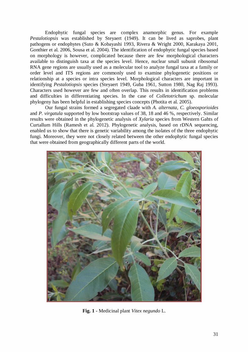
Fungal isolate VN1
The morphological characteristics of the endophytic fungal isolate VN1 was observed
29
on PDA after 7 days of growth at 30 oC. Colonies on PDA was circular, raised, at first
orange-white, sometimes grey and becoming pale orange with age, aerial mycelia white
dense, cottony without visible conidial masses, reverse bright orange but sometimes
yellowish-brown to olive-brown and very slow-growing. Acervuli and Setae were absent in
culture. Conidia were hyaline, unicellular and cylindrical with obtuse apices and tapering
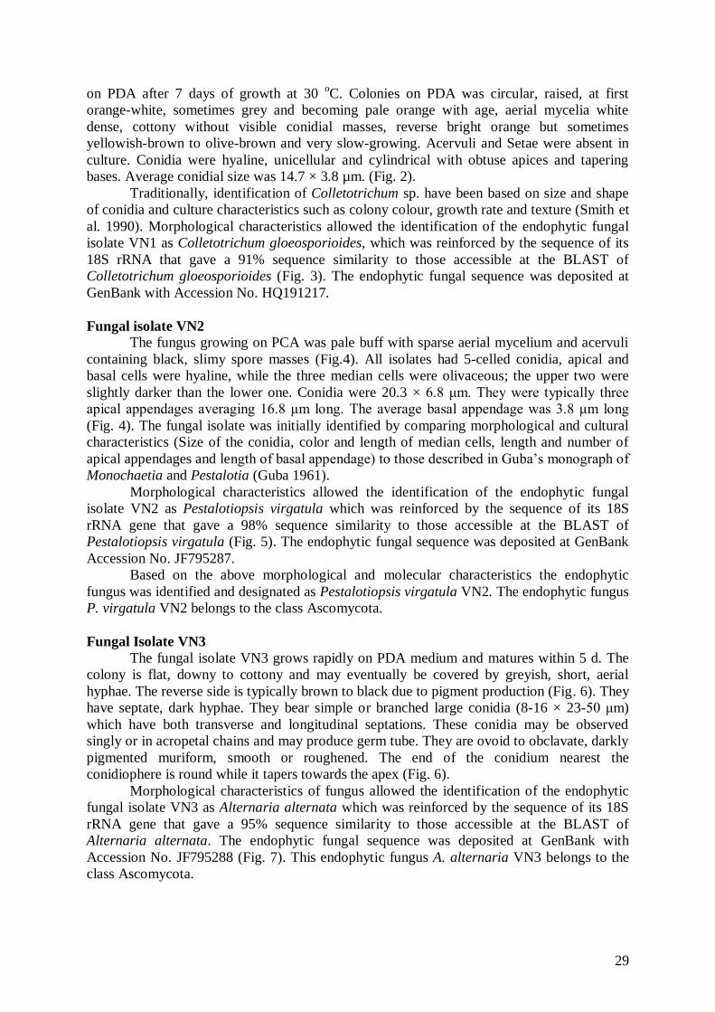
bases. Average conidial size was 14.7 × 3.8 µm. (Fig. 2).
Traditionally, identification of Colletotrichum sp. have been based on size and shape
of conidia and culture characteristics such as colony colour, growth rate and texture (Smith et
al. 1990). Morphological characteristics allowed the identification of the endophytic fungal
isolate VN1 as Colletotrichum gloeosporioides, which was reinforced by the sequence of its
18S rRNA that gave a 91% sequence similarity to those accessible at the BLAST of
Colletotrichum gloeosporioides (Fig. 3). The endophytic fungal sequence was deposited at
GenBank with Accession No. HQ191217.
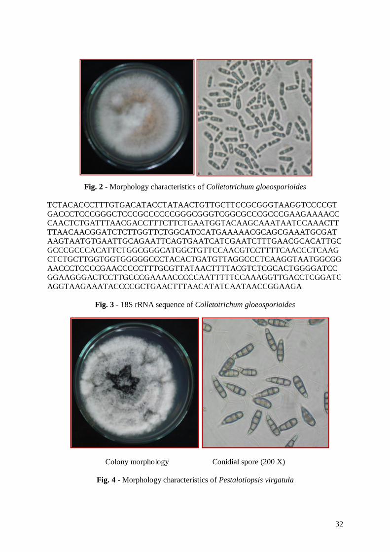
Fungal isolate VN2
The fungus growing on PCA was pale buff with sparse aerial mycelium and acervuli
containing black, slimy spore masses (Fig.4). All isolates had 5-celled conidia, apical and
basal cells were hyaline, while the three median cells were olivaceous; the upper two were
slightly darker than the lower one. Conidia were 20.3 × 6.8 μm. They were typically three
apical appendages averaging 16.8 μm long. The average basal appendage was 3.8 μm long
(Fig. 4). The fungal isolate was initially identified by comparing morphological and cultural
characteristics (Size of the conidia, color and length of median cells, length and number of
apical appendages and length of basal appendage) to those described in Guba’s monograph of
Monochaetia and Pestalotia (Guba 1961).
Morphological characteristics allowed the identification of the endophytic fungal
isolate VN2 as Pestalotiopsis virgatula which was reinforced by the sequence of its 18S
rRNA gene that gave a 98% sequence similarity to those accessible at the BLAST of
Pestalotiopsis virgatula (Fig. 5). The endophytic fungal sequence was deposited at GenBank
Accession No. JF795287.
Based on the above morphological and molecular characteristics the endophytic
fungus was identified and designated as Pestalotiopsis virgatula VN2. The endophytic fungus
P. virgatula VN2 belongs to the class Ascomycota.
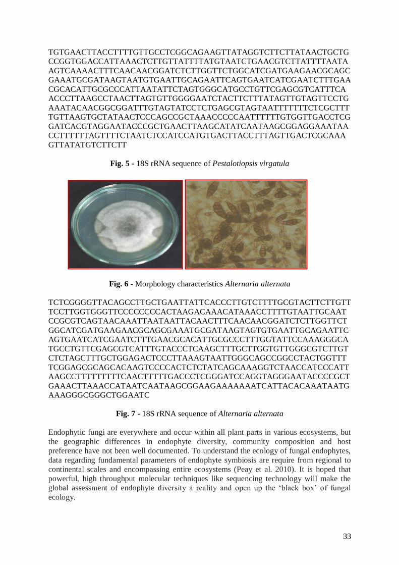
Fungal Isolate VN3
The fungal isolate VN3 grows rapidly on PDA medium and matures within 5 d. The
colony is flat, downy to cottony and may eventually be covered by greyish, short, aerial
hyphae. The reverse side is typically brown to black due to pigment production (Fig. 6). They
have septate, dark hyphae. They bear simple or branched large conidia (8-16 × 23-50 μm)
which have both transverse and longitudinal septations. These conidia may be observed
singly or in acropetal chains and may produce germ tube. They are ovoid to obclavate, darkly
pigmented muriform, smooth or roughened. The end of the conidium nearest the
conidiophere is round while it tapers towards the apex (Fig. 6).
Morphological characteristics of fungus allowed the identification of the endophytic
fungal isolate VN3 as Alternaria alternata which was reinforced by the sequence of its 18S
rRNA gene that gave a 95% sequence similarity to those accessible at the BLAST of
Alternaria alternata. The endophytic fungal sequence was deposited at GenBank with
Accession No. JF795288 (Fig. 7). This endophytic fungus A. alternaria VN3 belongs to the
class Ascomycota.
30
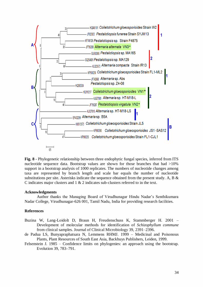
Phylogenetic analysis
Phylogenetic relationships inferred from ITS1-5.8S-ITS2 region sequences of three
species are shown in Figure 8. The tree is divided into three main clusters (A, B and C) and
further each one divided into two sub-clusters like A1, A2, B1, B2, C1 & C2. Based on the
evolution, among the present three fungal endophytes Colletotrichum gloesporoides VN1 and
Pestalotiopsis virgatula VN2 were grouped into the single sub cluster B2. Another present
endophytic fungal species of Alternaria alternata VN3 was located in the subclaade A1. In
sub-cluster A1 Colletotrichum gloeosporoides, Pestalotiopsis funerea strain SYJM13,
Pestalotiopsis sp. Strain F4875 and Alternaria alterna VN3 were grouped together.
Pestalotiopsis sp. MA165, Pestalotiopsis sp. MA129 and A. compacta strain IR13 were
grouped together in sub-cluster A2. In sub-cluster B1 C. gloeosporiodes strain FL1-ML2,
Alternaria sp. Abs and Pestalotiopsis sp. Z4-08 were grouped together. The phylogenetic tree
results showed C. gloeosporioides VN1 and Pestalotiopsis virgatula VN2 were closely
relationship between. But they were not closely relationship between the other endophytic
fungal species that were obtained from geographically different parts of the world.
Discussion
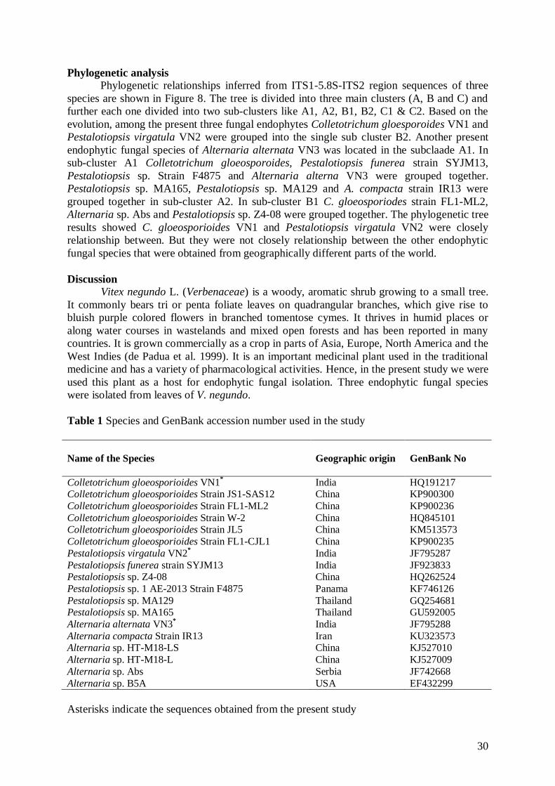
Vitex negundo L. (Verbenaceae) is a woody, aromatic shrub growing to a small tree.
It commonly bears tri or penta foliate leaves on quadrangular branches, which give rise to
bluish purple colored flowers in branched tomentose cymes. It thrives in humid places or
along water courses in wastelands and mixed open forests and has been reported in many
countries. It is grown commercially as a crop in parts of Asia, Europe, North America and the
West Indies (de Padua et al. 1999). It is an important medicinal plant used in the traditional
medicine and has a variety of pharmacological activities. Hence, in the present study we were
used this plant as a host for endophytic fungal isolation. Three endophytic fungal species
were isolated from leaves of V. negundo.
Table 1 Species and GenBank accession number used in the study
Name of the Species Geographic origin
GenBank No
Colletotrichum gloeosporioides VN1* India HQ191217
Colletotrichum gloeosporioides Strain JS1-SAS12 China KP900300
Colletotrichum gloeosporioides Strain FL1-ML2 China KP900236
Colletotrichum gloeosporioides Strain W-2 China HQ845101 Colletotrichum gloeosporioides Strain JL5 China KM513573
Colletotrichum gloeosporioides Strain FL1-CJL1 China KP900235
Pestalotiopsis virgatula VN2* India JF795287
Pestalotiopsis funerea strain SYJM13 India JF923833 Pestalotiopsis sp. Z4-08 China HQ262524
Pestalotiopsis sp. 1 AE-2013 Strain F4875 Panama KF746126
Pestalotiopsis sp. MA129 Thailand GQ254681 Pestalotiopsis sp. MA165 Thailand GU592005
Alternaria alternata VN3* India JF795288
Alternaria compacta Strain IR13 Iran KU323573 Alternaria sp. HT-M18-LS China KJ527010
Alternaria sp. HT-M18-L China KJ527009
Alternaria sp. Abs Serbia JF742668
Alternaria sp. B5A USA EF432299
Asterisks indicate the sequences obtained from the present study
31
Endophytic fungal species are complex anamorphic genus. For example
Pestalotiopsis was established by Steyaert (1949). It can be lived as saprobes, plant
pathogens or endophytes (Suto & Kobayashi 1993, Rivera & Wright 2000, Karakaya 2001,
Gonthier et al. 2006, Sousa et al. 2004). The identification of endophytic fungal species based
on morphology is however, complicated because there are few morphological characters
available to distinguish taxa at the species level. Hence, nuclear small subunit ribosomal
RNA gene regions are usually used as a molecular tool to analyze fungal taxa at a family or
order level and ITS regions are commonly used to examine phylogenetic positions or
relationship at a species or intra species level. Morphological characters are important in
identifying Pestalotiopsis species (Steyaert 1949, Guba 1961, Sutton 1980, Nag Raj 1993).
Characters used however are few and often overlap. This results in identification problems
and difficulties in differentiating species. In the case of Colletotrichum sp. molecular
phylogeny has been helpful in establishing species concepts (Photita et al. 2005).
Our fungal strains formed a segregated claade with A. alternata, C. gloeosporioides
and P. virgatula supported by low bootstrap values of 38, 18 and 46 %, respectively. Similar
results were obtained in the phylogenetic analysis of Xylaria species from Western Gahts of
Curtallum Hills (Ramesh et al. 2012). Phylogenetic analysis, based on rDNA sequencing,
enabled us to show that there is genetic variability among the isolates of the three endophytic
fungi. Moreover, they were not closely related between the other endophytic fungal species
that were obtained from geographically different parts of the world.
Fig. 1 - Medicinal plant Vitex negundo L.
32
Colony morphology Conidial spore (200 X)
Fig. 2 - Morphology characteristics of Colletotrichum gloeosporioides
TCTACACCCTTTGTGACATACCTATAACTGTTGCTTCCGCGGGTAAGGTCCCCGT
GACCCTCCCGGGCTCCCGCCCCCCGGGCGGGTCGGCGCCCGCCCGAAGAAAACC
CAACTCTGATTTAACGACCTTTCTTCTGAATGGTACAAGCAAATAATCCAAACTT
TTAACAACGGATCTCTTGGTTCTGGCATCCATGAAAAACGCAGCGAAATGCGAT
AAGTAATGTGAATTGCAGAATTCAGTGAATCATCGAATCTTTGAACGCACATTGC
GCCCGCCCACATTCTGGCGGGCATGGCTGTTCCAACGTCCTTTTCAACCCTCAAG
CTCTGCTTGGTGGTGGGGGCCCTACACTGATGTTAGGCCCTCAAGGTAATGGCGG
AACCCTCCCCGAACCCCCTTTGCGTTATAACTTTTACGTCTCGCACTGGGGATCC
GGAAGGGACTCCTTGCCCGAAAACCCCCAATTTTTCCAAAGGTTGACCTCGGATC
AGGTAAGAAATACCCCGCTGAACTTTAACATATCAATAACCGGAAGA
Fig. 3 - 18S rRNA sequence of Colletotrichum gloeosporioides
Colony morphology Conidial spore (200 X)
Fig. 4 - Morphology characteristics of Pestalotiopsis virgatula
33
TGTGAACTTACCTTTTGTTGCCTCGGCAGAAGTTATAGGTCTTCTTATAACTGCTG
CCGGTGGACCATTAAACTCTTGTTATTTTATGTAATCTGAACGTCTTATTTTAATA
AGTCAAAACTTTCAACAACGGATCTCTTGGTTCTGGCATCGATGAAGAACGCAGC
GAAATGCGATAAGTAATGTGAATTGCAGAATTCAGTGAATCATCGAATCTTTGAA
CGCACATTGCGCCCATTAATATTCTAGTGGGCATGCCTGTTCGAGCGTCATTTCA
ACCCTTAAGCCTAACTTAGTGTTGGGGAATCTACTTCTTTATAGTTGTAGTTCCTG
AAATACAACGGCGGATTTGTAGTATCCTCTGAGCGTAGTAATTTTTTTCTCGCTTT
TGTTAAGTGCTATAACTCCCAGCCGCTAAACCCCCAATTTTTTGTGGTTGACCTCG
GATCACGTAGGAATACCCGCTGAACTTAAGCATATCAATAAGCGGAGGAAATAA
CCTTTTTTAGTTTTCTAATCTCCATCCATGTGACTTACCTTTAGTTGACTCGCAAA
GTTATATGTCTTCTT
Fig. 5 - 18S rRNA sequence of Pestalotiopsis virgatula
Colongy morphology Conidial spore (200 X)
Fig. 6 - Morphology characteristics Alternaria alternata
TCTCGGGGTTACAGCCTTGCTGAATTATTCACCCTTGTCTTTTGCGTACTTCTTGTT
TCCTTGGTGGGTTCCCCCCCCACTAAGACAAACATAAACCTTTTGTAATTGCAAT
CCGCGTCAGTAACAAATTAATAATTACAACTTTCAACAACGGATCTCTTGGTTCT
GGCATCGATGAAGAACGCAGCGAAATGCGATAAGTAGTGTGAATTGCAGAATTC
AGTGAATCATCGAATCTTTGAACGCACATTGCGCCCTTTGGTATTCCAAAGGGCA
TGCCTGTTCGAGCGTCATTTGTACCCTCAAGCTTTGCTTGGTGTTGGGCGTCTTGT
CTCTAGCTTTGCTGGAGACTCCCTTAAAGTAATTGGGCAGCCGGCCTACTGGTTT
TCGGAGCGCAGCACAAGTCCCCACTCTCTATCAGCAAAGGTCTAACCATCCCATT
AAGCCTTTTTTTTTCAACTTTTTGACCCTCGGGATCCAGGTAGGGAATACCCCGCT
GAAACTTAAACCATAATCAATAAGCGGAAGAAAAAAATCATTACACAAATAATG
AAAGGGCGGGCTGGAATC
Fig. 7 - 18S rRNA sequence of Alternaria alternata
Endophytic fungi are everywhere and occur within all plant parts in various ecosystems, but
the geographic differences in endophyte diversity, community composition and host
preference have not been well documented. To understand the ecology of fungal endophytes,
data regarding fundamental parameters of endophyte symbiosis are require from regional to
continental scales and encompassing entire ecosystems (Peay et al. 2010). It is hoped that
powerful, high throughput molecular techniques like sequencing technology will make the
global assessment of endophyte diversity a reality and open up the ‘black box’ of fungal
ecology.
34
Fig. 8 - Phylogenetic relationship between three endophytic fungal species, inferred from ITS
nucleotide sequence data. Bootstrap values are shown for those branches that had >10%
support in a bootstrap analysis of 1000 replicates. The numbers of nucleotide changes among
taxa are represented by branch length and scale bar equals the number of nucleotide
substitutions per site. Asterisks indicate the sequence obtained from the present study. A, B &
C indicates major clusters and 1 & 2 indicates sub-clusters referred to in the text.
Acknowledgments
Author thanks the Managing Board of Virudhunagar Hindu Nadar’s Senthikumara
Nadar College, Virudhunagar-626 001, Tamil Nadu, India for providing research facilities.
References
Buzina W, Lang-Loidolt D, Braun H, Freudenschuss K, Stammberger H. 2001 –
Development of molecular methods for identification of Schizophyllum commune
from clinical samples. Journal of Clinical Microbiology 39, 2391–2396.
de Padua LS, Bunyapraphatsara N, Lemmens RHMJ. 1999 – Medicinal and Poisonous
Plants, Plant Resources of South East Asia, Backhuys Publishers, Leiden, 1999.
Felsenstein J. 1985 – Confidence limits on phylogenies: an approach using the bootstrap.
Evolution 39, 783–791.
35
Ganley RJ, Brunsfeld SJ, Newcombe G. 2004 – A community of unknown, endophytic fungi
in western white pine. Proceedings of the National Academy of Sciences USA 101,
10107–10112.
Giordano L, Gonthier P, Varese GC, Miserere L, Nicolotti G. 2009 – Mycobiota inhabiting
sapwood of healthy and declining Scots pine (Pinus sylvestris L.) trees in the Alps.
Fungal Diversity 38, 69–83.
Gonthier P, Massimo G Nicolotti G. 2006 – Effects of water stress on the endophytic mycota
of Quercus robur. Fungal Diversity 21, 69–80.
Guba EF. 1961 – Monograph of Pestalotioa and Monochaetia. Harvard University Press,
Cambridge, MA.
Iwen PC, Hinrichs SH, Rupp ME. 2002 – Utilization of the internal transcribed spacer
regions as molecular targets to detect and identify human fungal pathogens. Medical
Mycology 40, 87–109.
Karakaya A. 2001 – First report of infection of kiwifruit by Pestalotiopsis sp. in Turkey.
Plant Disease 85, 1028.
Kumar DSS, Lau CS, Wan JMF, Yang D, Hyde KD. 2005 – Immunomodulatory compounds
from Pestalotiopsis leucothёs (HKUCC 10197), an endophytic fungus of
Tripterygium wilfordii. Life Sciences 78, 147–156
Kumaresan V, Suryanarayanan TS. 2001 – Occurrence and distribution of endophytic fungi
in a mangrove community. Mycological Research 105, 1388–1391.
Lucero ME, Barrow JR, Osuna P, Reyes I, Duke SE. 2008 – Enhancing native grass
productivity by co-cultivating with endophyte Laden calli. Rangeland Ecology
Management 61, 124–130.
Mohali SR, Burgess TI, Wingfield MJ. 2005 – Diversity and host association of the tropical
tree endophyte Lasiodiplodia theobromae revealed using simple sequence repeat
markers. Forest Pathology 35, 385–396.
Nag Raj TR. 1993 – Coelomycetous anamorphs with appendage bearing conidia. Mycologue
Publications, Waterloo, Ontario, Canada.
Okane I, Nagagiri A, Ito T. 1998 – Endophytic fungi in leaves of ericaceous plants. Canadian
Journal of Botany 76, 657–663
Petrini O. 1986 – Taxonomy of endophytic fungi of aerial plant tissues. Microbiology of the
Phyllosphere (Fokkema NJ & van den Heuvel J, eds), pp. 175–187. Cambridge
University Press, Cambridge.
Petrini O, Carroll GC. 1981 – Endophytic fungi in foliage of some Cupressaceae in Oregon.
Canadian Journal of Botany 59, 629–636.
Petrini O, Fisher PJ, Petrini LE. 1992 – Fungal endophytes of bracken (Pteridium aquilinum)
with some reflections on their use in biological control. Sydowia 44, 282–293.
Photita W, Taylor PWJ, Ford R, Hyde KD, Lumyong S. 2005 – Morphological and molecular
characterization of Colletotrichum species from herbaceous plants in Thailand. Fungal
Diversity 18, 117–133.
Rakeman JL, Bui U, La Fe K, Chen YC, Honeycutt RJ, Cookson BT. 2005 – Multilocus
DNA sequence comparisons rapidly identify pathogenic molds. Journal of Clinical
Microbiology 43, 3324–3333.
Ramesh V, Thalavaipandian A, Karunakaran C, Rajendran A 2012 – Identification and
Comparison of Xylaria curta and Xylaria sp. from Western Ghats-Courtallum Hills,
India. Mycosphere 3, 607–615.
Read DJ, Duckett JG, Francis R, Ligrone ARU. 2000 – Symbiotic fungal associations in
‘lower’ land plants. Philosophical Transactions of the royal society B 355, 815–831
Rivera MC, Wright ER. 2000 – First report of azalea petal blight caused by Pestalotiopsis
guepinii in Argentina. Plant Disease 84, 100.
36
Saikkonen K. 2007 – Forest structure and fungal endophytes. Fungal Biology Reviews 21,
67–74.
Saikkonen K, Faeth SH, Helander M, Sullivan TJ. 1998 – Fungal endophytes: a continuum of
interaction with host plants. Annual Review of Ecology, Evolution and Systematics
29, 319–343.
Saikkonen K, Wali P, Helander M, Faeth SH. 2004 – Evolution of endophyte-plant
symbioses. Trends Plant Science 9, 275–280.
Saitou N, Nei M. 1987 – The neighbor-joining method: a new method for reconstructing
phylogenetic trees. Molecular Biology and Evolution 4, 406–425.
Sati SG, Belwal M. 2005 – Aquatic hyphomycetes as endophytes of riparian plant roots.
Mycologia 97, 45–49
Schulz U, Wanke S, Draeger HJ. 1993 – Endophytes from herbaceous plants and shrubs:
effectiveness of surface sterilization methods. Mycological Research 97, 1447–1450.
Schwarz P, Bretagne S, Gantier JC, Garcia-Hermoso D, Lortholary O, Dromer F, Dannaoui
E. 2006 – Molecular identification of Zygomycetes from culture and experimentally
infected tissues. Journal of Clinical Microbiology 44, 340–349.
Simth BJ, Black LL. 1990 – Morphological, cultural and pathogenic variation among
Colletotrichum species isolated from strawberry. Plant Diseases 74, 69–67.
Sousa MF, Tavares RM, Geros H, Lino-Neto T. 2004 – First report of Hakea sericea leaf
infection caused by Pestalotiopsis funerea in Portugal. Plant Pathology 53, 535.
Spurr HW, Welty RE. 1975 – Characterization of endophytic fungi in healthy leaves of
Nicotiana spp. Phytopathology 65, 417–422.
Steyaert RL. 1949 – Contribution a l'etude monographique de Pestalotia de NOT et
Monochaetia SACC. (Truncatella gen. nov. et Pestalotiopsis gen. nov.). Bull Jard Bot
EEtat Bruxelles 19, 285–354.
Strobel G, Daisy B, Castillo U, Harper J. 2004 – Natural products from endophytic
microorganisms. Journal of Natural Products 67, 257–268.
Suryanarayanan TS, Thirunavukkarasu N, Hariharan GN, Balaji P. 2005 – Occurrence of non
obligate microfungi inside lichen thalli. Sydowia 57, 120–130
Suryanarayanan TS, Venkatachalam A, Thirunavukkarasu N, Ravishankar JP, Doble M,
Geetha V. 2010 – Internal mycobiota of marine macroalgae from the Tamilnadu
coast: distribution, diversity and biotechnological potential. Botanica Marina 53, 457–
468.
Suto K, Kobayashi T. 1993 – Taxonomic studies on the species of Pestalotiopsis, parasitic on
conifers in Japan. Transactions of the Mycological Society of Japan 34, 323–344.
Sutton. 1980 – The Coelomycetes. Commonwealth Mycological Institute, UK.
Sutton SVW, Cundell AM. 2004 – Microbial identification in the pharmaceutical industry.
Pharmacopeial Forum 30, 1884–1894.
Tamura K, Dudley J, Nei M, Kumar S. 2007 – MEGA4: Molecular Evolutionary Genetics
Analysis (MEGA) software version 4. Molecular Biology and Evolution 24, 1596–
1599.
Tan RX, Zou WX. 2001 – Endophyts: A rich source of functional metabolites. Natural
Product Reports 18, 448–459.
Tejesvi MV, Kini KR, Prakash HS, Ven Subbiah, Shetty HS. 2007 – Genetic diversity and
antifungal activity of species of Pestalotiopsis isolated as endophytes from medicinal
plants. Fungal Diversity 24, 37–54.
Thompson JD, Gibson TJ, Plewniak F, Jeanmougin F, Higgins DG. 1997 – The CLUSTAL X
windows interface: flexible strategies for multiple sequence alignment aided by
quality analysis tools.
37
White TJ, Bruns TD, Lee S, Taylor JW. 1990 – Amplification and direct sequencing of
fungal ribosomal RNA genes for phylogenetics. In: PCR Protocols: a Guide to
Methods and Applications (eds Innis MA, Gelfand D, Sninsky JS, White TJ)
Academic, San Diego pp, 315–322.
Wiyakrutta S, Sriubolmas N, Panphut W, Thongon N, Danwisetkanjana K, Ruangrungsi N, et
al. 2004 –Endophytic fungi with anti-microbial, anti-cancer and anti-malarial
activities isolated from Thai medicinal plants. World Journal of Microbiology and
Biotechnology 20, 265–272.
Zhang HW, Song YC, Tan RX. 2006 – Biology and chemistry of endophytes. Natural
Product Report 23, 753–771.
Zuccaro A, Schoch CL, Spatafora JW, Kohlmeyer J, Draeger S, Mitchell J. 2008 – Detection
and identification of fungi associated with the brown seaweed Fucus serratus.
Applied Environmental Microbiology 74, 931–941.