Embed Size (px)
Citation preview
The Multiplication of Vaccinia Virus in Tissue Culturesof Adult Rabbit Skin
By G. N. C. CRAWFORD AND F. K. SANDERS
(From the Departments of Human Anatomy, and Zoology and Comparative Anatomy, Oxford)
SUMMARY
1. The optimum conditions for vaccinia virus multiplication in shavings of adultrabbit skin maintained in rocker flasks in a fluid medium have been investigated.Satisfactory virus multiplication occurred in 5 ml. of medium containing 40 per cent,rabbit serum, 40 per cent, phosphate-buffered saline, and 20 per cent, mouse embryoextract; an air atmosphere was used.
2. A growth curve for vaccinia virus grown in vitro under these conditions wasobtained by estimating the virus content of cultured skin at known intervals afterinoculation. The curve was essentially similar to that obtained by growth of vacciniain vivo, and could be divided into three phases: (a) during the first few hours infec-tivity fell, (b) from 10 to 40 hours there was a rapid increase of infectivity, and(c) flattening of the curve started after 40 hours, although some increase of virus con-tinued, until, at 80 hours, the concentration of virus present was at a maximum.
3. An attempt is made to correlate the multiplication of virus with the cytologicalchanges known to occur in infected cells.
INTRODUCTION
AT the present time most of our knowledge of virus multiplication comesi l from a study of bacterial viruses, since they can easily be freed fromcontaminating material and used to infect standardized suspensions of sus-ceptible bacteria maintained in simple media. Consequently more is knownof the actual process of virus synthesis with respect to these agents than withthose infecting animal tissues. Growth of a bacterial virus within its host cellcan roughly be divided into four phases: (1) a period of 'adsorption' andpenetration into the cell; (2) a constant period, during which new virusmaterial may be synthesized; (3) a period of rapid resumption of infectivity,followed by (4) a period during which virus is released, often by sudden lysisof the infected cells (Luria, 1950). It would be of interest to discover whethera similar growth cycle characterizes the multiplication of viruses in general,in particular such viruses as vaccinia and psittacosis, which approximate tosmall bacteria in size and chemical complexity (Smadel and Hoagland, 1942).
In the case of most animal viruses it is difficult even to approach thestandardized conditions which are essential for a study of this sort. Thesource of virus is usually a crude suspension of ground-up infected tissuewhich contains much apart from active virus particles. Moreover, the tissueinoculated contains many kinds of cells differently situated as regards sus-ceptibility and availability for infection; so the number of susceptible cells[Quarterly Journal of Microscopical Science, Vol. 93, part 2, pp. 119-32, June 1952.]
2421.2 K
120 Crawford and Sanders—The Multiplication of
reached by the inoculum is almost always unknown. And, when experimentsare conducted in vivo, the whole result may be complicated by nutritionaland genetic factors, or by an immune response on the part of the host organ-ism. However, in the case of vaccinia virus there is an opportunity of makingan approximation to the standardized conditions used with bacterial viruses.The extra-cellular infective phase of this virus consists of brick-shapedparticles roughly 236 X 200 m/x, which can be obtained fairly easily in purifiedform from a variety of infected tissues (Smadel and Hoagland, 1942). More-over, virus multiplication can occur in association with fragments of variousanimal tissues suspended in serum or other nutrient fluids. Virus growth hasthus been obtained in rabbit tissue with minced testis, kidney, spleen, cornea,or suspensions of Kupffer or mononuclear cells; minced whole chick embryoshave also been used (see van Rooyen and Rhodes, 1948, for bibliography).Therefore, with purified virus and tissue cultivated in vitro, it might bepossible to study virus growth under approximately standardized con-ditions.
The experiments described in this paper were undertaken with the objectof exploring the possibilities of such a system, the first step being to obtaina picture of virus growth under tissue culture conditions and to compare itwith that obtained with the same tissue in vivo. Rabbit skin was selected forinvestigation since virus multiplies readily to high titre in this tissue, causingthe production of definite lesions. Also Medawar (1948) has shown that smallfragments of rabbit skin can be maintained easily up to 8 days in vitro.
VIRUS
The virus used was the WR strain of vaccinia virus obtained through thecourtesy of the Army Medical Department Research and Graduate School,Washington, D.C., U.S.A. This virus had had at least nine passages throughthe brains of mice before the present experiments were undertaken. A 10 percent, suspension in saline of the infected brains from the ninth of these pas-sages was inoculated into the shaved skin of the back and flank of a normaladult rabbit. On the third day a confluent vaccinial lesion had developed.The animal was then killed, and a suspension of purified elementary bodiesof vaccinia virus was obtained from the skin by the technique of Craigie(1932). Briefly, this consisted of lightly scraping the surface of the infectedskin into a pool of Mcllvaine's buffered saline (pH 7-2), so that a suspensionconsisting of virus and cell debris was obtained. By centrifuging the latterdifferentially, an almost pure saline suspension of vaccinia elementary bodiescould be obtained. Such a suspension formed the virus pool that was usedthroughout these experiments. It was kept stored at — 700 C. in a carbondioxide ice cabinet, samples being thawed out for use when required, andunder these conditions its virus content did not alter significantly over longperiods.
Estimation of virus content. The amount of virus present in suspensionswas estimated by the inoculation of serial dilutions into the skin of adult
Vaccinia Virus in Tissue Cultures of Adult Rabbit Skin 121
castrated male albino rabbits obtained from the Agricultural Research CouncilField Station, Compton, Berks. They proved to be very susceptible to thisstrain of virus, sharp and consistent end-points being obtained. Tenfolddilutions of all material to be titrated were made up in Simms and Sanders(1942) saline X. 6 (a phosphate bicarbonate buffered saline of pH 7-6, whichwas also used as a constituent of the culture medium), o-i ml. quantities ofeach dilution were injected intracutaneously into the shaved back and flanksof the rabbit, five injections of each dilution being made. The rabbit wasexamined for lesions on the second to fifth days after inoculation. That dilu-tion of the suspension which, when injected in these circumstances, causeda lesion in 50 per cent, of cases (that is contained one 'infective dose' (IDS0)per o-i ml.) was calculated by the method of Reed and Muench (1938). Suchtitrations are accurate to within 0-2 of the interval between successive dilu-tions. In the case of tenfold dilutions the limit of accuracy is thus abouti io 0 " 2 . For example, it is probably not significant that in one experimentthe virus content of skin grown in 10 ml. of ultrafiltrate-containing mediumis about double that grown in 5 ml. of the serum-containing medium (seeTable 2).
The virus pool described above and used throughout these experimentscontained io6'6 ID50 per ml.
GROWTH OF VACCINIA VIRUS IN VIVO
In order to have a standard by which to judge the growth of virus in tissuecultures of skin, an attempt was made to obtain a curve showing the growthof virus in the skin of an adult rabbit.
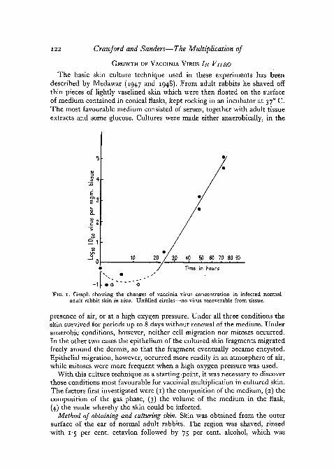
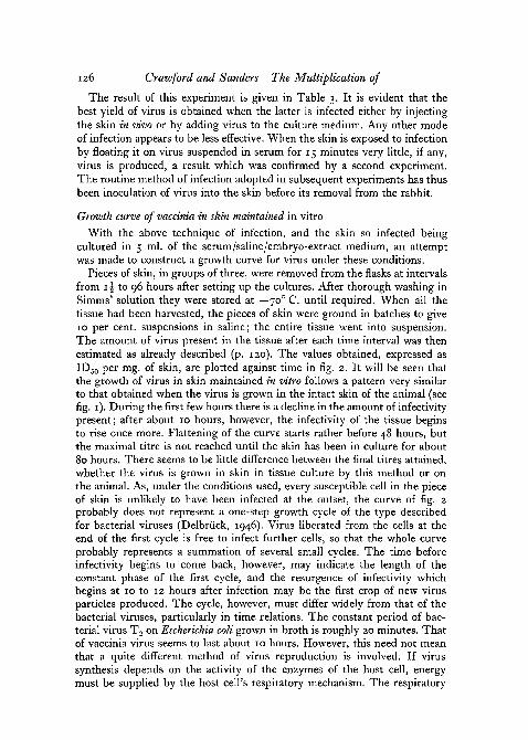
The skin of the back and flanks of a normal adult albino male rabbit wasshaved and twenty-four separate symmetrically placed o-i ml. intradermalinoculations, each containing approximately 10 ID50 of virus, were made. At0, i\, 3, 6, 12, 24, 48, and 72 hours after injection, three pieces of infectedskin were shaved off with a safety razor blade, as little connective tissue aspossible being removed with the epidermis. The three pieces of skin removedat each time interval were stored together at —70° C. until all had been soharvested. Each set of three pieces of skin was then weighed on a torsionbalance and ground in sufficient saline to make a 10 per cent, suspension.It was found that over 80 per cent, of the tissue went into suspension, theresidue being presumably mainly collagen. The virus content of the tissuewas then estimated, and expressed as ID50 per mg. of tissue. The valuesobtained at each time chosen are plotted against time in fig. 1. The methodof assay adopted does not allow of a particularly accurate estimation of thesmall amount of virus present in the skin during the first few hours followinginfection. However, it appears as though there is a phase of increase ininfectivity extending over the period from 24 to 72 hours after infection.The maximum virus titre is first reached after about 80 hours. This comparesclosely with the result obtained by Widelock (1938).
122 Crawford and Sanders—The Multiplication of
GROWTH OF VACCINIA VIRUS IN VITRO
The basic skin culture technique used in these experiments has beendescribed by Medawar (1947 and 1948). From adult rabbits he shaved offthin pieces of lightly vaselined skin which were then floated on the surfaceof medium contained in conical flasks, kept rocking in an incubator at 370 C.The most favourable medium consisted of serum, together with adult tissueextracts and some glucose. Cultures were made either anaerobically, in the
<3
« 4t»
mg
m.
s.
enoJ0
10
>
2P /y
/
/ 3,0 40
Time
•
50
in
•/
/•
/
60 70 80 90
hours
- i f mo ^ " o
FIG. 1. Graph showing the changes of vaccinia virus concentration in infected normaladult rabbit skin in vivo. Unfilled circles—no virus recoverable from tissue.
presence of air, or at a high oxygen pressure. Under all three conditions theskin survived for periods up to 8 days without renewal of the medium. Underanaerobic conditions, however, neither cell migration nor mitoses occurred.In the other two cases the epithelium of the cultured skin fragments migratedfreely around the dermis, so that the fragment eventually became encysted.Epithelial migration, however, occurred more readily in an atmosphere of air,while mitoses were more frequent when a high oxygen pressure was used.
With this culture technique as a starting-point, it was necessary to discoverthose conditions most favourable for vaccinial multiplication in cultured skin.The factors first investigated were (1) the composition of the medium, (2) thecomposition of the gas phase, (3) the volume of the medium in the flask,(4) the mode whereby the skin could be infected.
Method of obtaining and culturing skin. Skin was obtained from the outersurface of the ear of normal adult rabbits. The region was shaved, rinsed-with 1 s per cent, cetavlon followed by 75 per cent, alcohol, which was
Vaccinia Virus in Tissue Cultures of Adult Rabbit Skin 123
allowed to dry. o-i ml. intradermal inoculations were then made of saline,which, if necessary, contained vaccinia virus. Each injection caused theformation of a raised bleb of skin about 7 mm. in diameter, which was thenshaved off with a safety razor blade. Care was taken to make the shavings asthin as possible; each one weighed approximately 20 mg. After washing ina large volume of solution X. 6, four skin shavings were transferred to eachof a number of sterile 500 ml. conical flasks fitted with flask heads for gasperfusion (Medawar, 1948). In later experiments each flask contained 5 ml.of tissue culture medium, which consisted of normal adult rabbit serum,Simms' solution (X. 6), and mouse embryo extract. The latter was preparedby mincing 14-18-day old mouse foetuses with scissors in an equal volumeof Simms' solution and incubating the resulting suspension at 3 7°C. for 1 hour.The suspension was then centrifuged at 2,750 r.p.m. for 10 minutes and thesupernatant stored at — 70° C. over carbon dioxide ice until required. Strepto-mycin was added to the culture medium in all the experiments to give a finalconcentration of 2 mg. per ml. and was in every case successful in preventingbacterial contamination. The flasks were incubated at 37° C. on a con-tinuously rocking platform, an angle of about 15 degrees on each side of thehorizontal being achieved with each excursion. The speed of rocking wasapproximately 4 times per minute. In most experiments each flask contained5 ml. of medium, and this did not cover the whole of the bottom of the flaskwhen the rocking platform was fully tilted. The pieces of tissue thus tendedto be washed intermittently by the medium as in roller-tube cultures.
In some experiments it was necessary to culture the infected skin at a highoxygen pressure. This was attained by perfusing the flask with a 95 per cent.oxygen/5 per cent, carbon dioxide mixture for 10 minutes daily, and thensealing off the flask head.
Effect of medium on vaccinial growth
(1) Experiments with another strain of virus had previously shown thatvaccinia grew well in the presence of serum diluted with Simms' solution (X. 6).The following experiment tested the effect of adding embryo extract to themedium, and of high oxygen tension.
Skin shavings were obtained from the rabbit's ear as described above(p. 122). In this case, however, each injection of saline into the ear skincontained about 40 ID50 of vaccinia virus—that is, about 2 ID50 per mg. ofskin shaved off. After washing in solution X. 6 for 20 minutes, four shavingswere transferred to each of two pairs of conical flasks containing 5 ml. ofmedium. In one pair of flasks the medium consisted of equal parts of serumand saline only, while to the other pair embryo extract had also be'en addedat a final concentration of 20 per cent. One flask of each pair was gassedintermittently (see above) with 95 per cent, oxygen and 5 per cent, carbondioxide (see Table 1).
All flasks were incubated at 370 C. for 72 hours, at the end of which timethe skin was removed, washed, and the quantity of virus present in each case
124 Crawford and Sanders—The Multiplication of
estimated by the method already described. The concentration of virusattained in each of the four sets of skin fragments is shown in Table i.
TABLE I . Effect of composition of (i) culture medium, and (2) flask atmosphereon the multiplication of vaccinia virus in skin cultures. Figures = ID50 of virus
present per mg. tissue harvested
Composition of medium
Serum-saline (1:1)no embryo-extract
Serum-saline (1:1)+ 20 per cent, embryo-extract
Composition of flask atmosphere
Air
400
1,580
95 per cent. O2;5 per cent. CO3
20
16
Culture of skin in an atmosphere of air resulted in the production of 25-100times as much virus as when a high oxygen pressure was used. Addition ofembryo extract to the basic medium did not greatly affect the final virus titrein the presence of a high oxygen pressure. However, in an atmosphere ofair, embryo-extract increased the virus yield approximately fourfold.
(2) Experiments were also made to test whether virus yield was affectedby the volume of medium present, or by replacing its serum content by oxserum ultra-filtrate (Simms and Sanders, 1942).
Infected skin shavings were obtained exactly as described in the previousexperiment, the dosage of virus per mg. of skin being the same. Three pairsof flasks were set up, each containing four pieces of skin. One pair containedi\ ml., one 5 ml., and one 10 ml. of medium per flask. Within each pair, themedium in one flask consisted of 40 per cent, serum, 40 per cent, saline, and20 per cent, mouse embryo extract; the medium in the other flask consistedof 20 per cent, ox serum ultrafiltrate, 60 per cent, saline, and 20 per cent,embryo extract. All flasks were incubated at 37° C. for 72 hours, at the endof which time the skin was removed, washed, and its virus content deter-mined. The result of this experiment is shown in Table 2.
TABLE 2. Effect of (1) volume, and (2) presence of serum ultrafiltrate in culturemedium on the multiplication of vaccinia virus in skin cultures. Figures = ID 5 0
of virus present per mg. of tissue harvested
Composition of medium
Serum (40 per cent.)Saline (40 per cent.)Embryo-extract (20 per cent.)
Serum ultrafiltrate (20 per cent.)Saline (60 per cent.)Embryo-extract (20 per cent.)
Volume of medium in ml.
2-5
25
320
5
3,200
3
10
32
6,300
Vaccinia Virus in Tissue Cultures of Adult Rabbit Skin 125
When the medium contained serum, the concentration of virus in the skingrown in the flask containing 5 ml. was about 100 times as high as when theflask contained either 10 or z\ ml. The maximum concentration of virus inthe skin was not much increased by substituting ultrafiltrate for serum. Onthe other hand, the yield of virus was higher when 10 ml. of the ultrafiltratecontaining medium was used as compared with 5 or z\ ml. Undue signifi-cance should not be attached to the low virus titre attained when the skinwas grown in 5 ml. of the ultrafiltrate containing medium. This ratheranomalous result was based on a single experiment, which, in view of thefact that the use of ultrafiltrate rather than serum did not greatly increasethe growth of vaccinia in the skin, seemed scarcely worth repeating. Five ml.of a similarly constituted serum medium were used routinely in subsequentexperiments.
(3) Finally, an experiment was performed in order to determine thatmethod of infecting the skin with virus, which resulted in the highest virustitre.
o-i ml. injections of solution X. 6 alone were made into the skin of a rabbit'sear, and the blebs of skin were shaved off as previously described. Four ofthe skin shavings so obtained were then infected by floating them for 15minutes in 2 ml. of solution X. 6 containing virus at a concentration of400 ID50 per ml.; four more were infected by floating them for 15 minutesin 2 ml. of normal rabbit serum, containing virus at the same concentration.Another four skin shavings were washed in saline for 30 minutes, after whicheach was injected in vitro with 40 ID50 of virus. In the case of four otherpieces of skin, virus was added to the medium in the flask in which theywere subsequently cultured at a concentration of 400 ID50 per ml. Finally,as a control, four o-i ml. intradermal injections of 40 ID50 were then madein vivo into the skin of the ear and the infected skin blebs shaved off.
The batches of skin infected by these different methods were cultured inseparate flasks in 5 ml. of medium consisting of 40 per cent, normal rabbitserum, 40 per cent, saline, and 20 per cent, embryo extract; all flasks wereincubated for 72 hours, when the pieces of skin were removed, washed, andtheir virus content determined.
TABLE 3. Effect of method of inoculation on multiplication of vaccinia virus inskin cultures
Method of inoculation
Skin injected in vivo
Skin cultured in medium containing virus
Skin floated on saline + virus
Skin injected in vitro
Skin floated on serum + virus
Virus present after 72 hoursculture (L,Di0/mg.)
320,000320,000
4,000
< 630
< 3
126 Crawford and Sanders—The Multiplication of
The result of this experiment is given in Table 3. It is evident that thebest yield of virus is obtained when the latter is infected either by injectingthe skin in vivo or by adding virus to the culture medium. Any other modeof infection appears to be less effective. When the skin is exposed to infectionby floating it on virus suspended in serum for 15 minutes very little, if any,virus is produced, a result which was confirmed by a second experiment.The routine method of infection adopted in subsequent experiments has thusbeen inoculation of virus into the skin before its removal from the rabbit.
Growth curve of vaccinia in skin maintained in vitro
With the above technique of infection, and the skin so infected beingcultured in 5 ml. of the serum/saline/embryo-extract medium, an attemptwas made to construct a growth curve for virus under these conditions.
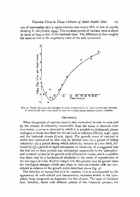
Pieces of skin, in groups of three, were removed from the flasks at intervalsfrom 1 \ to 96 hours after setting up the cultures. After thorough washing inSimms' solution they were stored at — 700 C. until required. When all thetissue had been harvested, the pieces of skin were ground in batches to give10 per cent, suspensions in saline; the entire tissue went into suspension.The amount of virus present in the tissue after each time interval was thenestimated as already described (p. 120). The values obtained, expressed asID50 per mg. of skin, are plotted against time in fig. 2. It will be seen thatthe growth of virus in skin maintained in vitro follows a pattern very similarto that obtained when the virus is grown in the intact skin of the animal (seefig. 1). During the first few hours there is a decline in the amount of infectivitypresent; after about 10 hours, however, the infectivity of the tissue beginsto rise once more. Flattening of the curve starts rather before 48 hours, butthe maximal titre is not reached until the skin has been in culture for about80 hours. There seems to be little difference between the final titres attained,whether the virus is grown in skin in tissue culture by this method or onthe animal. As, under the conditions used, every susceptible cell in the pieceof skin is unlikely to have been infected at the outset, the curve of fig. 2probably does not represent a one-step growth cycle of the type describedfor bacterial viruses (Delbruck, 1946). Virus liberated from the cells at theend of the first cycle is free to infect further cells, so that the whole curveprobably represents a summation of several small cycles. The time beforeinfectivity begins to come back, however, may indicate the length of theconstant phase of the first cycle, and the resurgence of infectivity whichbegins at 10 to 12 hours after infection may be the first crop of new virusparticles produced. The cycle, however, must differ widely from that of thebacterial viruses, particularly in time relations. The constant period of bac-terial virus T2 on Escherichia coli grown in broth is roughly 20 minutes. Thatof vaccinia virus seems to last about 10 hours. However, this need not meanthat a quite different method of virus reproduction is involved. If virussynthesis depends on the activity of the enzymes of the host cell, energymust be supplied by the host cell's respiratory mechanism. The respiratory
Vaccinia Virus in Tissue Cultures of Adult Rabbit Skin izy
rate of mammalian skin is approximately one-twenty-fifth of that of rapidlydividing E. coli (Krebs, 1934). The constant period of vaccinia virus is about30 times as long as that of the bacterial virus. The difference is thus roughlythe same as that in the respiratory rates of the cells concerned.
I 2
20 30 40 50 60 70 80 90Time in Sours
FIG. 2. Graph showing the changes of virus concentration in vaccinia-infected shavingsof adult rabbit skin maintained in vitro in a saline serum embryo-extract medium.
DISCUSSION
When the growth of vaccinia virus in skin maintained in vitro is measuredby the amount of infectivity recoverable from the tissue at intervals afterinoculation, a curve is obtained in which it is possible to distinguish phasesanalogous to those described for viruses such as influenza (Hoyle, 1948, 1950)and the bacterial viruses (Luria, 1950). The growth curve of vaccinia inrabbit skin maintained in vitro may be divided into: (1) a period of fallinginfectivity; (2) a period during which infectivity remains at a low level, fol-lowed by (3) a period of rapid resumption of infectivity. It is suggested thatthe first two of these periods may correspond respectively to the 'adsorptive'and 'constant' phases in the growth cycle of bacterial viruses, and it is possiblethat there may be a fundamental similarity in the mode of reproduction ofthe two types of virus. Further insight into this process may be gained whenthe cytological changes which take place in vaccinia-infected cells are con-sidered in relation to the growth curve described above (fig. 2).
The infection of susceptible cells by vaccinia virus is accompanied by theappearance of well-defined and characteristic inclusion bodies in the cyto-plasm, long recognized as diagnostic for this disease. The type of inclusionseen, however, varies with different phases of the infectious process; for
128 Crawford and Sanders—The Multiplication of
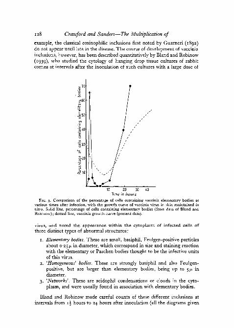
example, the classical eosinophilic inclusions first noted by Guarneri (1892)do not appear until late in the disease. The course of development of vacciniainclusions, however, has been described quantitatively by Bland and Robinow(1939), who studied the cytology of hanging drop tissue cultures of rabbitcornea at intervals after the inoculation of such cultures with a large dose of
tain
10 20 30Time in hours
40
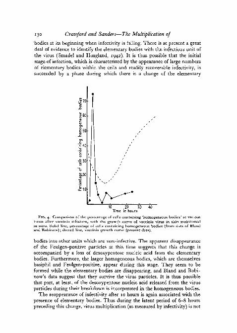
FIG. 3. Comparison of the percentage of cells containing vaccinia elementary bodies atvarious times after infection, with the growth curve of vaccinia virus in skin maintained invitro. Solid line, percentage of cells containing elementary bodies (from data of Bland andRobinow); dotted line, vaccinia growth curve (present data).
virus, and noted the appearance within the cytoplasm of infected cells ofthree distinct types of abnormal structures:
1. Elementary bodies. These are small, basiphil, Feulgen-positive particlesabout 0-25 )JL in diameter, which correspond in size and staining reactionwith the elementary or Paschen bodies thought to be the infective unitsof this virus.
2. 'Homogeneous' bodies. These are strongly basiphil and also Feulgen-positive, but are larger than elementary bodies, being up to 5/i indiameter.
3. 'Networks'. These are acidophil condensations or clouds in the cyto-plasm, and were usually found in association with elementary bodies.
Bland and Robinow made careful counts of these different inclusions atintervals from i£ hours to 24 hours after inoculation (all the diagrams given
Vaccinia Virus in Tissue Cultures of Adult Rabbit Skin 129
here of types of inclusions present are based upon their data). Fig. 3, forinstance, shows the percentage of cells containing elementary bodies in theirtissue cultures, plotted against time since inoculation. It will be seen that atthe outset there is a marked rise in the proportion of cells containing ele-mentary bodies, a maximum being reached after about i\ hours. It may beconjectured that this phase represents the initial penetration of virus into thecells. From i\ hours onwards, however, there is an equally spectacular fallin the proportion of cells containing elementary bodies, so that after 3 to 6hours virtually no cells can be found which contain them. Starting at about10 hours, however, there is a second increase in the proportion of cells con-taining elementary bodies. The increase this time is maintained, so that 24hours after inoculation they are to be found in over 70 per cent, of theinfected cells. The dotted line in fig. 3 shows the growth curve of vacciniavirus in tissue cultures of rabbit skin obtained during the present investiga-tion. While the results obtained with our method cannot be directly relatedto those obtained by Bland and Robinow using a different tissue-culturesystem, a comparison of the data obtained in the two cases gives rise to aninteresting hypothesis with regard to the possible mode of multiplication ofvaccinia virus. Fig. 3 shows that the initial rise in the proportion of cellscontaining elementary bodies occurs at a time when the infectivity of theculture may actually be falling. Only in the later stage is a rise in the numberof elementary bodies accompanied by a corresponding rise in infectivity.
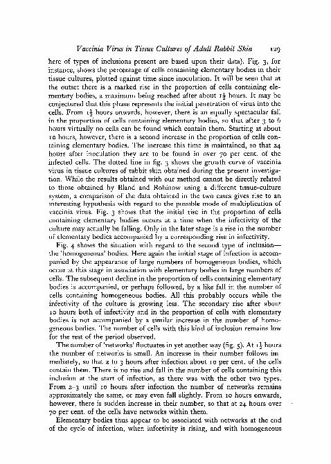
Fig. 4 shows the situation with regard to the second type of inclusion—the 'homogeneous' bodies. Here again the initial stage of infection is accom-panied by the appearance of large numbers of homogeneous bodies, whichoccur at this stage in association with elementary bodies in large numbers ofcells. The subsequent decline in the proportion of cells containing elementarybodies is accompanied, or perhaps followed, by a like fall in the number ofcells containing homogeneous bodies. All this probably occurs while theinfectivity of the culture is growing less. The secondary rise after about10 hours both of infectivity and in the proportion of cells with elementarybodies is not accompanied by a similar increase in the number of homo-geneous bodies. The number of cells with this kind of inclusion remains lowfor the rest of the period observed.
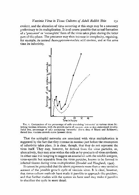
The number of 'networks' fluctuates in yet another way (fig. 5). At 1 \ hoursthe number of networks is small. An increase in their number follows im-mediately, so that 2 to 3 hours after infection about 10 per cent, of the cellscontain them. There is no rise and fall in the number of cells containing thisinclusion at the start of infection, as there was with the other two types.From 2-3 until 10 hours after infection the number of networks remainsapproximately the same, or may even fall slightly. From 10 hours onwards,however, there is sudden increase in their number, so that at 24 hours over70 per cent, of the cells have networks within them.
Elementary bodies thus appear to be associated with networks at the endof the cycle of infection, when infectivity is rising, and with homogeneous
130 Crawford and Sanders—The Multiplication of
'bodies at its beginning when infectivity is falling. There is at present a greatdeal of evidence to identify the elementary bodies with the infectious unit ofthe virus (Smadel and Hoagland, 1942). It is thus possible that the initialstage of infection, which is characterized by the appearance of large numbersof elementary bodies within the cells and readily recoverable infectivity, issucceeded by a phase during which there is a change of the elementary
.2 70
E50
;i40
= 30
£10
10 ,20 30TTme in hours
40
FIG. 4. Comparison of the percentage of cells containing 'homogeneous bodies' at varioustimes after vaccinia infection, with the growth curve of vaccinia virus in skin maintainedin vitro. Solid line, percentage of cells containing homogeneous bodies (from data of Blandand Robinow); dotted line, vaccinia growth curve (present data).
bodies into other units which are non-infective. The apparent disappearanceof the Feulgen-positive particles at this time suggests that this change isaccompanied by a loss of desoxypentose nucleic acid from the elementarybodies. Furthermore, the larger homogeneous bodies, which are themselvesbasiphil and Feulgen-positive, appear during this stage. They seem to beformed while the elementary bodies are disappearing, and Bland and Robi-now's data suggest that they survive the virus particles. It is thus possiblethat part, at least, of the desoxypentose nucleic acid released from the virusparticles during their breakdown is incorporated in the homogeneous bodies.
The reappearance of infectivity after 10 hours is again associated with thepresence of elementary bodies. Thus during the latent period of 6-8 hourspreceding this change, virus multiplication (as measured by infectivity) is not
Vaccinia Virus in Tissue Cultures of Adult Rabbit Skin 131
evident, and the alteration of virus occurring at this stage may be a necessarypreliminary to its multiplication. It is of course possible that actual formationof a 'precursor' or 'incomplete' form of the virus takes place during the latterpart of this phase. The precursor may then increase in complexity, regaining,for example, its normal desoxypentose-nucleic acid content, and at the sametime its infectivity.
SZ2Q
10 20Time in hours
30 40
FIG. 5. Comparison of the percentage of cells containing 'networks' at various times fol-lowing vaccinia infection, with the growth curve of vaccinia virus in skin maintained in vitro.Solid line, percentage of cells containing 'networks' (from data of Bland and Robinow);dotted line, vaccinia growth curve (present data).
That the acidophil networks are associated with virus multiplication issuggested by the fact that they increase in number just before the resumptionof infectivity takes place. It is clear, though, that they do not represent thevirus itself. They may, however, be derived from the virus particles, or,alternatively, they may arise within the cells as by-products of virus synthesis.In either case it is tempting to suggest an association with the soluble antigens,virus-specific but separable from the virus particles, known to be formed ininfected tissues during virus multiplication (Smadel and Hoagland, 1942).
It cannot be pretended that the above represents more than a very tentativeaccount of the possible growth cycle of vaccinia virus. It is clear, however,that tissue-culture methods have made it possible to approach this problem,and that further studies with the system we have used may make it possibleto elucidate the cycle in more detail.

132 Crawford and Sanders
REFERENCESBLAND, J. O. W., and ROBINOW, C. F., 1939. J. Path. Bact., 48, 381.CRAIGIE, J., 1932. Brit. J. exp. Path., 13, 259.DELBEOCK, M., 1946. Biol. Rev., 21, 30.GUARNIERI, G., 1892. Arch. Sci. med., 16, 403.HOYLE, L., J948. Brit. J. exp. Path., 29, 390.
1950. J. Hyg., 48, 277.KREBS, H. A., 1934. Tab. Biol., 9, 209.LURIA, S. E., 1950. Chapter in Viruses, edited by M. Delbriick. Calif. Inst. Techn.MEDAWAR, P. B., 1947. Quart. Journ. micr. Sci., 88, 27.
1948. Ibid., 89, 187.REED, L. J., and MUENCH, H., 1938. Amer. J. Hyg., 27, 493.VAN ROOYEN, C. E., and RHODES, A. J., 1948. Virus diseases of man. New York (Nelson).SIMMS, H. S., and SANDERS, M., 1942. Arch. Path., 33, 619.SMADEL, J. E., and HOAGLAND, C. L., 1942. Bact. Rev., 6, 79.WIDELOCK, D., 1938. J. infect. Dis., 62, 27.