Embed Size (px)
Citation preview
REPUBLIC OF TURKEY
HACETTEPE UNIVERSITY
INSTITUTE OF HEALTH SCIENCES
THE IN VITRO AND IN VIVO EFFECTS OF
TELOMERASE SUBSTRATE
6-THIO-2’-DEOXYGUANOSINE
Ilgen MENDER
Biochemistry Program
PhD THESIS
MENTOR
Assoc. Prof. Dr. Zeliha Günnur Dikmen
CO-MENTOR
Prof. Dr. Jerry William Shay
ANKARA
2014
iv
ACKNOWLEDGEMENT
First and foremost, I would like to thank my mentors Prof. Dr. Jerry W. Shay,
Prof. Dr. Woodring E. Wright and Assoc. Prof. Dr. Z. Günnur Dikmen for their
personal and academic guidance throughout my study for doctorate. Especially, I
would like to express my gratitude to Jerry W. Shay for his limitless support and
trust in me. He treated me like a father when I was thousands miles away from my
home. It was a privilege for me to work with him. And I would like to give a special
thank to Z. Günnur Dikmen. I could not have been at this point as a young scientist
without her help and encouragement.
I would like to thank my committee members, Prof. Dr. Pakize Doğan, Prof. Dr. Ediz
Demirpençe, Prof. Dr. Gülberk Uçar and Prof. Dr. Can Akçalı, for their comments
and support throughout the years. In addition, I would like to thank all professors at
Hacettepe University, Faculty of Medicine, Department of Biochemistry, for their
guidance principles, which helped me to find my direction in my academic career.
Thank to all the current and former Shay/Wright lab and Hacettepe University
members that have made my time more enjoyable and valuable in the lab. In
particular, I would like to thank Sang Bum Kim, Abhijit Budge, Summer Barron,
Kimberly Batten, Natasha Buxton, and Raj Pandita for their help. Likewise thanks to
Bahram Sarkarati, Oliver Delgado, Jennifer-Peters Hall, Crystal Cornelius, Mandy
Wong, Lu Zhang, and Elijah Huang for their kind friendship. Special thanks to Kevin
Kennon for his administrative support.
I would like to dedicate this dissertation to my parents Pelin Gülay-Emin Mender and
my brother İlker Mender. Their endless, unconditional love, unlimited support, and
patience was always with me. I could not have achieved this without them. Thank
you for being with me in every decision I make throughout my life.
I also would like to thank and acknowledge TÜBİTAK and UTSouthwestern
Medical Center’s support. I was personally supported by TÜBİTAK, through BIDEB
2211 and 2214 scholarships and UTSouthwestern fellowship.

v
ABSTRACT
Mender, I. The In vitro and In vivo Effects of Telomerase Substrate
6-Thio-2’-Deoxyguanosine. Hacettepe University Institute of Health Sciences,
Ph.D. Thesis in Biochemistry, Ankara, 2014. Telomerase mediated telomere
targeted therapy represents a new approach in cancer therapy. In this study, I report
that the nucleoside analogue 6-thio-2’-deoxyguanosine (6-thio-dG) is recognized by
the telomerase holoenzyme and incorporated into de novo synthesized telomeres to
alter the structure and function of telomeres. This results in structurally and
functionally modified telomeres, loss of telomeric end protective complexes, leading
to telomere dysfunction. Additionally, 6-thio-dG causes progressive telomere
shortening, which is independent from inhibition of telomerase activity in vitro.
6-thio-dG induced telomere dysfunction is observed in hTERT expressing normal
human BJ fibroblast cells and cancer cells, but not in telomerase-negative BJ cells.
Moreover, one week treatment with 6-thio-dG results in 80-90% cell death for the
majority of the cancer cells (H2882, HCC2429, HCC827, HCC15, H2087,
HCC4017, HCC515, H2009, H2073), whereas normal BJ fibroblast and human
colonic epithelial (HCEC1) cells were largely unaffected. In A549 lung cancer cell
based xenograft model studies, intraperitoneally 6-thio-dG treatment (2 mg/kg)
caused dramatic tumor reduction as well as telomere dysfunction, superior to that
observed for 6-thioguanine (2 mg/kg) treatment. These results indicate that 6-thio-dG
may provide a new telomere-addressed telomerase-dependent anti-cancer approach,
targeting both genomic and telomeric DNA. It was observed that some of the cancer
cell lines (H1819, H1993, H1693) were resistant to 6-thio-dG compared with the
sensitive cell lines. The methylation analysis showed that several genes were highly
hypermethylated in resistant cell lines. In addition, in gene expression data, there
were 3 different genes (FSCN1, TLE2, ALDH1A2) that were differentially
expressed between resistant and sensitive cell lines. The results of this study will
help in the future directions focusing on 6-thio-dG resistance.
Key Words: Telomerase, 6-thio-2’-deoxyguanosine, telomere dysfunction.
vi
ÖZET
Mender, I. Telomeraz Substratı 6-Tiyo-2’-Deoksiguanozin’in in vitro ve in vivo
etkileri. Hacettepe Üniversitesi Sağlık Bilimleri Enstitüsü Biyokimya Programı
Doktora Tezi, Ankara, 2014. Telomeraz aracılığı ile telomerleri hedefleyen
terapiler, kanserde yeni bir tedavi yaklaşımı sunmaktadır. Bu çalışmada, nükleozid
analogu olan 6-tiyo-2’-deoksiguanozinin (6-tiyo-dG) telomeraz enzimi tarafından
tanınıp, yeni sentezlenen telomerik yapılara eklenerek telomerlerde yapısal ve
fonksiyonel bozukluğa yol açması incelenmiştir. Bu durum, telomerlerin yapısal ve
fonksiyonel modifikasyonuna, telomerik uçlardaki koruyucu proteinlerin kaybına ve
sonuç olarak telomerik disfonksiyona yol açmaktadır. In vitro şartlarda 6-tiyo-dG,
telomeraz inhibisyonundan bağımsız olarak progresif telomerik kısalığa neden
olmaktadır. 6-tiyo-dG, bu etkilerini telomeraz eksprese eden BJhTERT ve kanser
hücreleri üzerinde gösterirken, telomeraz negatif normal BJ fibroblast hücrelerinde
göstermemektedir. Akciğer kanseri hücre hatları (H2882, HCC2429, HCC827,
HCC15, H2087, HCC4017, HCC515, H2009, H2073) 6-tiyo-dG ile 1 hafta inkübe
edildiğinde %80-90 oranında hücre ölümü gözlenmiş, ancak normal BJ fibroblast ve
kolon epitel hücrelerinin (HCEC1) etkilenmediği saptanmıştır. A549 akciğer kanseri
hücrelerinin enjekte edilmesiyle oluşturulan ksenograft modellerde intraperitoneal
6-tiyo-dG uygulaması (2 mg/kg), 6-tiyoguanine (2 mg/kg) göre belirgin telomer
fonksiyon bozukluğu ile birlikte etkin bir tümör küçülmesi sağlamıştır. Bu sonuçlar,
6-tiyo-dG’nin kanser tedavisinde telomerlere yönelik, telomeraz aracılı, hem
genomik hem telomerik DNA’yı hedefleyen yeni bir yaklaşım olabileceğini
göstermektedir. Çalışmada kullanılan bazı kanser hücre hatlarının (H1819, H1993,
H1693) 6-tiyo-dG’ye dirençli olduğu saptanmıştır. Yapılan metilasyon analizinde,
6-tiyo-dG’ye direnç gösteren hücrelerde bazı genlerin yüksek seviyede metile olduğu
gözlenmiştir. Bu hücrelerin gen ekspresyon profilleri karşılaştırıldığında, tedaviye
dirençli ve duyarlı olan kanser hücrelerinde 3 gende (FSCN1, TLE2, ALDH1A2)
farklılık olduğu tespit edilmiştir. Bu çalışmadan elde edilen veriler, hücrelerin
6-tiyo-dG’ye direnç mekanizmalarının incelenmesine yönelik yeni araştırmalarda yol
gösterici olacaktır.
Anahtar Kelimeler: Telomeraz, 6-tiyo-2’-deoksiguanozin, telomer disfonksiyonu.
vii
TABLE OF CONTENTS
Page
Approval page iii
Acknowledgement iv
Abstract v
Özet vi
Table of Contents vii
Abbreviations xi
List of Figures xix
List of Tables xxii
1. INTRODUCTION 1
2. GENERAL INFORMATION 3
2.1. Brief History on Telomere Biology 3
2.2. DNA Damage Response to Endogenous and Exogenous Sources 5
2.3. Telomeres and Shelterin Complex 6
2.4. Telomerase and Its Regulation 9
2.5. Telomere and Telomerase Targeted Therapies 12
2.6. The Role of Telomeres on Cellular and Organismal Aging and Cancer 18
2.7. The Thiopurines 19
2.8. The Importance of Liver and Kidney Function on Drug Toxicity 22
2.9. Tumor Resistance to Therapy 23
viii
2.10. Purpose and Significance 26
3. METHODS 28
3.1. Cell lines 28
3.2. Drug preparation 28
3.3. Cell Viability Assay 28
3.4. Long-term cell culture studies 29
3.5. Telomerase activity assay (TRAP, Telomere Repeat Amplification Protocol) 29
3.6. Telomere length assay (TRF, Terminal Restriction Fragment) 31
3.7. 6-thio-dG and GRN163L Treatment for Cell Killing Effect and 32
Telomere Restriction Fragment Analysis
3.8. Telomere dysfunction Induced Foci (TIF) assay 32
3.9. Quantitative PCR (qPCR) 33
3.10. Colony formation assay 33
3.11. Isolation and Culture of Primary Human Lymphocyte 34
3.12. Drug toxicity animal experiments 34
3.13. Histology and serum analysis 34
3.14. Establishment of xenograft models 38
3.15. Ki67 proliferation assay 38
3.16. ImmunoFISH 39
3.17. Immunohistochemical analysis 40
3.18. Statistical analysis 40
ix
4. RESULTS 41
4.1. Induction of Telomere Dysfunction Mediated by the 41
Telomerase Substrate Precursor 6-Thio-2’-Deoxyguanosine
4.1.1. The Effects of 6-thio-dG and 6-thioguanine on Cell Viability 44
4.1.2. Telomere Dysfunction Induced Foci (TIF) Induced by 6-thio-dG, 48
but not 6-thioguanine
4.1.3. 6-thio-dG, but not 6-thioguanine, Induces Progressive Telomere 51
Shortening via Independent Mechanism from Telomerase Inhibition
in Cancer Cells
4.1.4. 6-thio-dG does not Lead to Signficant Weight Loss or 53
Hematological Toxicities in Wild Type Mice
4.1.5. 6-thio-dG does not Cause Significant Liver, Kidney and 57
Gastrointestinal Tract Side Effects
4.1.6. 6-thio-dG Reduces Tumor Growth Rate In Vivo 59
4.1.7. 6-thio-dG Induces Telomere Dysfunction as well as Tumor 62
Growth Rate Reduction on A549 NSCLC Derived Tumor
4.2. Possible Resistance Mechanisms of 6-thio-dG Treatment 65
4.2.1 HCT116 Cells are not Sensitive to 6-thio-dG with 68
GRN163L Pretreatment
4.2.2. H1819, H1993 and H1693 NSCLCs are resistant to 6-thio-dG 71

x
4.2.3. Telomerase positive peripheral blood mononuclear cells (PBMC) 74
are partially sensitive to 6-thio-dG
5. DISCUSSION 76
REFERENCES 84
xi
ABBREVIATIONS
Ab Antibody
ABC ATP binding cassette
ABCB1 ATP binding cassette, sub-family B (MDR/TAP), member 1
ABCC1 ATP binding cassette, sub-family C (CFTR/MRP), member 1
ALDH1A1 Aldehyde dehydrogenase 1 family, member A1
ALDH1A2 Aldehyde dehydrogenase 1 family, member A2
ALT Alternative lengthening of telomeres
ALT Alanine aminotransferase
Alt-NHEJ Alternative form of NHEJ
ASCL1 Achaete-scute homolog 1
AST Aspartate aminotransferase
ATM Ataxia telangiectasia mutated kinase
ATP Adenosine triphosphate
ATR Ataxia telangiectasia and Rad3-related
BCRP Breast cancer resistance protein (ABCG2)
BIBR1532 2-[(E)-3-naphthalene-2-yl-but-2-enoylamino]-benzoic acid
BRCA1/2 Breast cancer 1/2
BSA Bovine serum albumin
BUN Blood urea nitrogen
CBC Complete blood count

xii
CDC25A Cell division cycle 25A
Cdk1/2 Cyclin dependent kinase 1/2
Cdk4/6 Cyclin dependent kinase 4/6
cDNA Complementary DNA
Chk1/2 Checkpoint kinase 1/2
CO2 Carbon dioxide
CSC Cancer stem cell
CtIP Mammalian ortholog of ctp1
DAPI 4’, 6-diamidino-2-phenylindole
DLK1 Delta like homolog
D-loop Displacement loop
DMEM Dulbecco's modified eagle medium
DMSO Dimethyl sulfoxide
DNA Deoxyribonucleic acid
DNA-PK DNA dependent protein kinase
DSB Double strand break
EDTA Ethylenediaminetetraacetic acid
FDA Food and drug administration
FISH Fluorescence in situ hybridization
FITC Fluorescein isothiocyanate
FSCN1 Fascin actin-bundling protein 1
xiii
GABA Gamma-aminobutyric acid
GAPDH Glyceraldehyde 3-phosphate dehydrogenase
GI Gastrointestinal
GMPS Guanine monophosphate synthetase
HASH1 Human achaete-scute homologue
HAT Histone acetyltransferase
HCEC Human colonic epithelial cell
HCT Hematocrit
HDAC Histone deacetylases
HGB Hemoglobin
HPRT1 Hypoxanthine guanine phosphoribosyl transferase 1
HR Homologous recombination
hTR/hTERC Telomerase RNA template component
H2AX Variant of the H2A protein family
H&E Hematoxylin and eosin
h or hr hour
IC50 Half maximal inhibitory concentration
IMPDH Inosine monophosphate dehydrogenase
IP Intraperitoneal
ITAS Internal telomerase assay standard
kb Kilobase
xiv
KCl Potassium chloride
kg Kilogram
MDC1 Mediator of DNA-damage checkpoint 1
MDR1 Multidrug resistance gene
meTIMP S-methyl-thioinosine 5’-monophosphate
meTGMP S-methyl-thioguanosine 5’-monophosphate
mg Miligram
MgCl2 Magnesium chloride
min minute
miRNA MicroRNA
MM Milimeter
mm3
a cubic milimetre
MRE11 Meiotic recombination 11 homolog A
MRN MRE11-RAD50-NBS1
MRP1 Multidrug resistance protein 1
μg Microgram
μl Microliter
μM Micromolar
Na-citrate Sodium citrate
NaCl Sodium chloride
Na2HPO4 Disodium phosphate
xv
Na2H2P2O7 Disodium pyrophosphate
NaOH Sodium hydroxide
NBS1 Nibrin
NHEJ Non-homologous end joining
NP-40 Nonidet-P40
ns Non significant
NSCLC Non small cell lung cancer
Nu nude
O2 Oxygen
PARP1 Poly (ADP-ribose) polymerase 1
PBMC Peripheral blood mononuclear cell
PBS Phosphate buffered saline
PBST Phosphate buffered saline with tween 20
PCR Polymerase chain reaction
PDX Patient derived xenograft
PFS Prolonged progression free survival
P-gp P-glycoprotein
PHA Phytohemagglutinin
PIT1 Pituitary transcription factor 1
PLT Platelet
PNA Peptide nucleic acid

xvi
POT1 Protection of telomeres 1
pRB Retinoblastoma protein
pre-mRNA Precursor mRNA
qPCR Quantitative PCR
RAD50 DNA repair protein RAD50
Rap1 the human ortholog of the yeast repressor/activator protein 1
RBC Red blood count
RNA Ribonucleic acid
ROS Reactive oxygen species
RPA Replication protein A
SA-βGal Senescence associated betagalactosidase
SCLC Small cell lung cancer
SCr Serum creatinin
SD Standard deviation
SDS Sodium dodecyl sulfate
sec second
SP Side population
SSB Single strand break
SSC Saline-sodium citrate
Telomerase Telomere terminal transferase
TERT Telomerase reverse transcriptase
xvii
TGMP Thioguanosine monophosphate
TIF Telomere dysfunction induced foci
TIMP Thioinosine monophosphate
TIN2 TRF2 and TRF1 interacting nuclear protein 2
TLE2 Transducin-like enhancer of split 2
T-loop Telomere loop
TPMT Thiopurine S-methyltransferase
TPP1 Adrenocortical dysplasia homolog
TRAP Telomere repeat amplification protocol
TRF Terminal restriction fragment
TRF1/2 Telomeric repeat binding factor 1/2
Tric-HCl Tris hydrochloride
V Volt
VOD Veno-occlusive disease
WBC White blood count
WHO World health organization
Wnt Wingless
XLF XRCC4-like factor
XO Xanthine oxidase
XRCC1/4 X-ray repair cross complementing protein 1/4
xviii
5-aza 5-aza-2’-deoxycytidine
6-MP 6-mercaptopurine
6-TG 6-thioguanine
6-thio-dG 6-thio-2’-deoxyguanosine
6-thio-dGTP 6-thio-2’-deoxyguanosine triphosphate
6-thio-GTP 6-thio-guanosine triphosphate
53BP1 p53 binding protein 1
xix
FIGURES
Pages
1.1. Telomerase as a universal target 2
2.1. Diagram of hypothetical telomere demonstrating t-loop 8
structure with shelterin proteins
2.2. Telomere Elongation by Telomerase 9
2.3. Targeting telomerase with GRN163L (Imetelstat) 13
2.4. Disadvantages of different telomerase based therapy strategies 17
2.5. Shematic illustration of thiopurine metabolism 21
2.6. The biochemical conversion reaction of 6-thio-dG conversion to 27
6-thio-dGTP to be incorporated into telomeres by telomerase
3.1. The elongation and amplification steps of Telomere 30
Repeat Amplification Protocol (TRAP)
3.2. Reactions of transaminases 35
4.1. The growth inhibition kinetics of normal human BJ fibroblasts, 42
colonic epithelial cells (HCEC1), VA13 (ALT-telomerase negative),
HCT116 colon, A549 and HCC15 non small cell lung cancer cells
4.2. IC50 values of BJ, HCEC1, HCT116, A549, HCC15, H2882, 45
H2009, H2087, HCC515, HCC827, HCC2429 and HCC4017 cells
following one week treatment with 6-thio-dG and 6-thioguanine
xx
4.3. Colocalization of gamma-H2AX and TRF2 exerts DNA damage 49
on telomeres
4.4. Binding of gamma-H2AX on damaged telomeres 50
4.5. Telomere shortening analysis by TRF (Terminal Restriction 52
Fragment) assay
4.6. Telomerase activity analysis by TRAP (Telomeric Repeat 53
Amplification Protocol) assay
4.7. Wild type mice weight during 6-thio-dG and 6-thioguanine 54
treatment with different doses
4.8. Complete blood count following 2 mg/kg 6-thio-dG and 56
6-thioguanine treatment in wild type mice
4.9. Liver, kidney function tests and histopatological evaluation 58
of liver, kidney, spleen and colon
4.10. Intraperitoneally and intratumoral injection with 6-thio-dG 60
and 6-thioguanine
4.11. Correlation of tumor growth reduction and telomere dysfunction 62
in A549-derived tumors
4.12. The development of 6-thio-dG resistance in HCT116 cells 65
4.13. The mRNA levels of ALDH1A1 and ABCG2 in HCT116 66
cells following 6-thio-dG treatment
4.14. The colony formation efficiency of HCT116 cells following 67
10 μM 6-thio-dG treatment with different time points
xxi
4.15. Two different experimental designs of combination therapy 69
with GRN163L and 6-thio-dG
4.16. Telomere Restriction Fragment analysis in HCT116 cells 70
treated with only GRN163L, only 6-thio-dG, or combination
of 6-thio-dG and GRN163L
4.17. Clustering analysis of DNA methylation profiles for 13 72
NSCLCs
4.18. Hierarchical clustering of the gene expression profile in 74
sensitive and resistance NSCLCs to 6-thio-dG treatment
4.19. The cell counts of peripheral blood mononuclear cells 75
(PBMC) following 6-thio-dG treatment in the presence/absence
phytohemagglutinin (PHA), which is a telomerase stimulator
5.1. Two different approaches for telomerase based therapies 77
xxii
TABLES
Pages
2.1. Telomerase based therapeutic approaches 12
2.2. Clinical trials of GRN163L 15
4.1. Average telomere lengths of normal and cancer cell lines 47
and IC50 values of these cell lines following one week treatment
with 6-thio-dG and 6-thioguanine
4.2. The average telomere lenghts and IC50 values of 73
H1819/H1693 and H1993/H2073 base pairs with 6-thio-dG treatment
with/without 5-Aza pretreatment
1
1. INTRODUCTION
Cancer is one of the leading causes of disease in the world. According to
World Health Organization (WHO), 8.2 million people died due to lung, stomach,
liver, colorectal and female breast cancers in 2012 (1).
In both developed and developing countries, colorectal and lung cancers are
the most common cancers, 136.830 and 224.210 people in the US will be diagnosed
with colorectal and lung cancer in 2014, respectively (2). Lung cancer is divided into
two different histological categories, which are small cell lung cancer (SCLC) and
non small cell lung cancer (NSCLC). SCLC presents ~25% of all bronchogenic
malignancies and shows highly aggresive clinical outcomes. Metastasis are generally
found in SCLC patients and combination therapy is usually chosen for treatment.
While less than 5% SCLC patients survive 5 years past the initial diagnosis, 15%
NSCLC patients have 5 year survival rate. However, NSCLC is less sensitive to
chemotherapy than SCLC. Surgical resection is the best treatment choice for NSCLC
patients (3). Therefore, prevention and early detection in addition to finding new
therapeutic strategies are vitally important to prevent the high incidence and
mortality risk of cancer.
Since the side effects with standard chemotherapy are highly challenging for
cancer patients, targeted therapy with fewer side effects is a major focus for cancer
research due to less damage to normal cells. Therefore, indefinite cell proliferation,
one of the hallmarks of cancer, via activation of telomerase is a highly attractive
target for cancer therapy because telomerase activity is detected in ~90% of primary
human cancers (Figure 1.1), but not normal cells except proliferating progenitor and
transit amplifying cells (e.g., male germline spermatocytes, activated lymphocytes,
some proliferating gastrointestinal cells and skin epidermal) and embryonic stem
cells (4, 5).
Although telomerase is a unique and universal target, there have been no US
Food and Drug Administration (FDA) approved telomerase targeted drugs as even
though this target has been studies for well over a decade. Therefore, it is important
to develop new therapeutic strategies that target telomerase for anticancer therapy.

2
For this reason, we generated a new telomerase mediated telomere targeted agent,
6-thio-2'-deoxyguanosine (6-thio-dG), and tested its in vitro and in vivo effects on
normal and cancer cells.
Figure 1.1. Telomerase as a universal target. While most normal cells do not have
telomerase activity, ~90% of cancer cells can express telomerase. This difference
between normal and cancer cells make telomerase unique and universal target for
cancer therapy (Submitted to Cancer Medicine, book chapter, Mender et al.).
3
2. GENERAL KNOWLEDGE
2.1. Brief History on Telomere Biology
The appreciation to chromosomal ends started in the 1930's through two
independent researchers. Herman Muller noted that there are unique structures at the
ends of chromosomes. Muller named these ends telomeres, based on their positions
on chromosomes (Telo:end, mere:part in Greek). Later, he generated various
mutations by X-rays when he was working with fruit flies and realized that X-rays
caused chromosome breakages and fusions (6). About the same time, Barbara
McClintock observed dicentric chromosomes, chromosomes with two centromes, in
maize. When dicentric chromosomes broke at mitosis, she noticed that these ends
fused with any other broken ends. However, natural chromosomal ends were not
involved in this process such as in embryonic cells (7).
In 1961, Leonard Hayflick discovered that cultured normal cells have limited
capacity to divide, which is related to rounds of DNA replication, then they become
senescent (also known as the Hayflick limit). Hayflick used phase I, II and III
terminology for the growth phase of cultured cells. Phase I is the primary culture as
cells from tissues adapt to the in vitro conditions. Phase II is the growth phase and
phase III is the period when cell replication diminishes and finally stops. These
observations showed that in contrary to cancer cells that have limitless capacity to
divide in culture, normal cells had a limited dividing capacity (8, 9).
In the early 1970s, James Watson realized that there was a problem with
replicating the ends of linear DNA because of the nature of lagging strand synthesis,
not the leading strand. Since DNA polymerase can not completely replicate the 3'
end of linear chromosomes, Watson called this the end replication problem (10).
Olovnikov, a Russian theoretical scientist, proposed that chromosomes get shorter
with each replication cycle and eventually run into essential genes, and speculated
this end replication problems might be the reason for the replicative senescence,
which was described by Hayflick (11, 12). When early molecular biology
technologies became available, Elizabeth Blackburn was the first scientist to report
tandem repeats of 6 mers (TTGGGG) at the end of chromosomes in Tetrahymena
4
thermophila (13). Human cells also consist of thousands of repeats, but this sequence
is TTAGGG in mammals (14). The presence of telomeres (tandem repeats) at the
ends of chromosomes was first speculated in 1938 by Hermann Muller (15) and
Barbara McClintock (16). Once the sequence of telomeres was known, telomere
length could be measured. Then, it was found that telomere lengths in different
tissues were variable (17). These studies showed that as normal human fibroblasts
divided in culture, telomeres got progressively shorter with each cell division (18).
If normal cells have telomere attrition and their growth rate is being limited as
they are cultured, there had to be a different mechanism to counteract this attrition
and cause unlimited growth for cancer cells. Greider and Blackburn discovered
enzyme activity called telomerase (telomere terminal transferase) that elongates
telomeres by adding six hexameric sequences (TTGGGG) to the ends of
chromosomes in Tetrahymena thermophila (19). Greider also found that telomerase
activity was sensitive to RNA and she co-purified RNA with telomerase and found
that the RNA sequence contains 5'CAACCCCAA3' which was complementary to the
Tetrahymena telomere repeat TTGGGG. She blocked this putative enzyme activity
by oligonucleotides, showing that the RNA template is required for enzyme activity
(20). It wasn’t until the telomerase reverse transcriptase (TERT) was cloned in 1997
(21, 22), that it was shown that introduction of a single gene into telomerase silent
human cells was sufficient to detect telomerase activity and this resulted in telomere
elongation and greatly extended lifespan without undergoing senescence (23, 24).
In 1990s, Jerry Shay and Geron scientists showed that telomerase is present in
all cancer-derived cell lines and ~90% of primary human cancers (25). Woodring
Wright and Geron Scientists found that ectopic expression of telomerase in normal
fibroblasts and epithelial cells bypassed the Hayflick limit, indicating that telomeres
are the cellular replicometer (23). And that time, it was realized that normal cells do
not have telomerase activity, while most cancer cells express telomerase activity,
which is required to prevent telomere attrition and consequent replicative senescence,
results in enabling proliferation and immortality of cells (25). While ~90% of cancer
cells have telomerase activity, the other relatively small group of telomerase negative
cancers use a non telomerase based recombination mechanism to extend their
5
telomere length, which is called Alternative Lengthening of Telomeres (ALT). ALT
mechanism involves intra-telomeric recombination, leading to telomere length
maintenance in mostly rare cancers (26).
Following these major discoveries, pharmaceutical companies initiated large
compound screen to identify compounds to inhibit telomerase in cancer cells (27).
2.2. DNA Damage Response to Endogenous and Exogenous Sources
When genomic DNA is exposed to genotoxic stresses, different types of DNA
damage occurs such as base modifications, intrastrand crosslinks, interstrand
crosslinks, DNA-protein crosslinks, single strand breaks (SSBs) and double strand
breaks (DSBs). Double strand breaks are the most deleterious ones (28) and can be
generated by reactive oxygen species produced by cellular metabolic processes and
replication associated errors, which are derived from endogenous sources, as well as
exogenous sources such as ionizing radiation and chemotherapeutic agents. If DSBs
are not repaired, they can result in cell death. If they are accurately repaired, cells can
survive with no adverse effects. If the repair occurs inaccurately, then surviving cells
may gain genomic alterations that may contribute to tumor development (29). To
maintain genomic integrity is important for the fate of cell. Therefore, cells that have
a well coordinated network of so called DNA damage responses, can transmit signals
upon damage to effector proteins leading to the induction of cellular responses to
arrest the cell cycle, activate DNA repair pathways, and or cell death (28).
In response to DSBs, the MRE11-RAD50-NBS1 (MRN) complex binds to
DSB sites and activates the ataxia telangiectasia mutated (ATM) kinase through
autophosphorylation (30, 31). ATM along with DNA-PK phosphorylates the histone
variant H2AX at serine 139, resulting in gamma-H2AX. H2AX is also
phosphorylated by ATR protein kinase (ataxia telangiectasia and Rad3-related)
during DNA replication associated DSB induction (32, 33). Gamma-H2AX modified
chromatin is found at the DSB sites until the damage is repaired (34). There are other
proteins that are retained at DSBs such as MDC1, 53BP1. The focal accumulation of
gammaH2AX, 53BP1 and other factors are the markers for DSB responses (35).
6
DSB response involves a significant cell cycle arrest/delay checkpoints in
terms of cellular proliferation, which are the early G1, the G1/S, the intra-S and the
G2/M (36). Prevention of progression in G1/S phase is the most rapid cell cycle
arrest and has two major pathways. These pathways start with phosphorylation of the
downstream proteins by ATM (37). First, when Chk1 and Chk2 are phosphorylated,
they then induce phosphorylation of the protein phosphatase CDC25A and
subsequent degradation, leading to Cdk1/Cdk2 inhibition and cell cycle arrest.
activation of p53 initiates transcription of the cyclin dependent kinase inhibitor p21
and triggers either cell death/apoptosis or cell cycle arrest/cellular senescence (38,
39). Physiological stresses (i.e., culture conditions) cause the upregulation of p16
(cyclin dependent kinase inhibitor), which binds specifically to CDK4 and CDK6 to
inhibit the interaction with D-type cyclins, and then p16 causes pRB protein to
remain in hypophosphorylated (inactive form), resulting in cell cycle arrest and
eventually cellular senescence. pRB involves in heterochromatin formation, which
induces cellular senescence (40).
When the DNA damage response mediator 53BP1 is activated, it contributes
to DSB repair pathways via non-homologous end joining (NHEJ) (41). Double
strand breaks are repaired by either NHEJ or HR (homologous recombination).
NHEJ is an error-prone pathway and Ku70/Ku80 complex, DNA PK catalytic
subunit (DNA-PKcs), the Artemis nuclease, XLF, XRCC4, and DNA ligase IV are
involved in this pathway. In contrast, HR is an error-free repair pathway that requires
non-damaged sister chromatid for recombination (42). MRN complex, CtIP,
replication protein A (RPA), BRCA1, PALB2, BRCA2, and RAD51 are involved in
HR. Alt-NHEJ is the alternative form of NHEJ and also involved in DSB repair that
includes PARP1, XRCC1, DNAligase IIIα, polynucleotide kinase, and Flap
endonuclease I (43).
2.3. Telomeres and Shelterin Complex
Telomeric DNA in eukaryotes (double stranded) are maintained by
telomerase during early developments, in some proliferative stem like cells and in
almost all cancer cells. Generally, the strand that constitutes the 3' end is rich in
guanosine. Therefore, telomeric DNA has two strands which are called G- and
7
C- rich strands. Telomere length is variable in different organisms such as humans at
birth (10-15 kb of the TTAGGG repeats) and mice (up to 100 kb) (44). Studies
showed that even 400 bp of telomeric repeats are enough for telomeres to function
properly (45-47), and in general telomere shortening to less than 1 kb by inhibiting
telomerase is the needed length reduction to induce senescence in tumor cells (48).
The 3' single G-stranded telomeres terminate with single stranded G-rich overhang
and this overhang varies between 50-500 nt in mammalian cells. However, it is still
not clear how this overhang is generated (49, 50). More recently it was shown that
the single strand overhang, along with the terminal portion of the duplex TTAGGG
repeats make a large duplex lariat structure that is called telomere loop (T-loop). It is
believed that the single strand G-rich overhang strand invades back into the duplex
telomere repeats to form a triplex structure resulting in the the overhang base pairs
with displaced C-rich strand, which is called displacement loop (D-loop). These
loops are the protective structures that hide telomere ends from being recognized by
the DNA damage repair machinery (51).
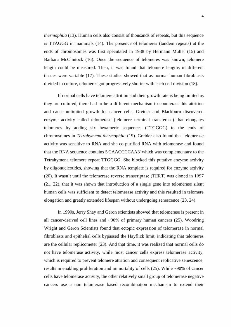
Telomeric repeats at the end of chromosomes are associated with a 6 protein
complex (TRF1, TRF2, Rap1, TIN2, TPP1, and POT1), which is called shelterin
(52). They are abundant on telomeres throughout the cell cyle and they do not appear
to function elsewhere in the nucleus (44). TRF1 and TRF2 (Telomeric Repeat
Binding Factor 1 and 2) can bind to the duplex DNA and POT1 (Protection of
Telomeres 1) can bind to the single strand DNA present at the 3' overhang. TRF1 and
TRF2 can recruit with TIN2 (TRF2 and TRF1 Interacting Nuclear Protein 2), Rap1
(the human ortholog of the yeast Repressor/Activator Protein 1), TPP1 and POT1
(53, 54) (Figure 2.1).
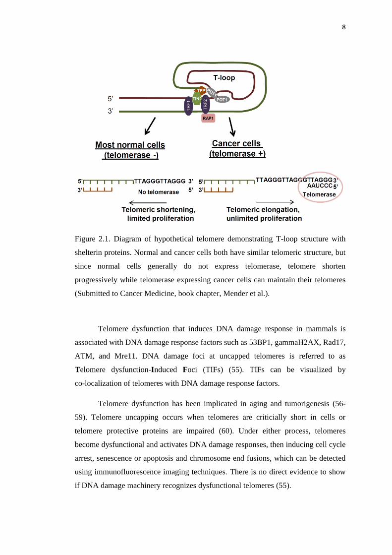
8
Figure 2.1. Diagram of hypothetical telomere demonstrating T-loop structure with
shelterin proteins. Normal and cancer cells both have similar telomeric structure, but
since normal cells generally do not express telomerase, telomere shorten
progressively while telomerase expressing cancer cells can maintain their telomeres
(Submitted to Cancer Medicine, book chapter, Mender et al.).
Telomere dysfunction that induces DNA damage response in mammals is
associated with DNA damage response factors such as 53BP1, gammaH2AX, Rad17,
ATM, and Mre11. DNA damage foci at uncapped telomeres is referred to as
Telomere dysfunction-Induced Foci (TIFs) (55). TIFs can be visualized by
co-localization of telomeres with DNA damage response factors.
Telomere dysfunction has been implicated in aging and tumorigenesis (56-
59). Telomere uncapping occurs when telomeres are criticially short in cells or
telomere protective proteins are impaired (60). Under either process, telomeres
become dysfunctional and activates DNA damage responses, then inducing cell cycle
arrest, senescence or apoptosis and chromosome end fusions, which can be detected
using immunofluorescence imaging techniques. There is no direct evidence to show
if DNA damage machinery recognizes dysfunctional telomeres (55).
9
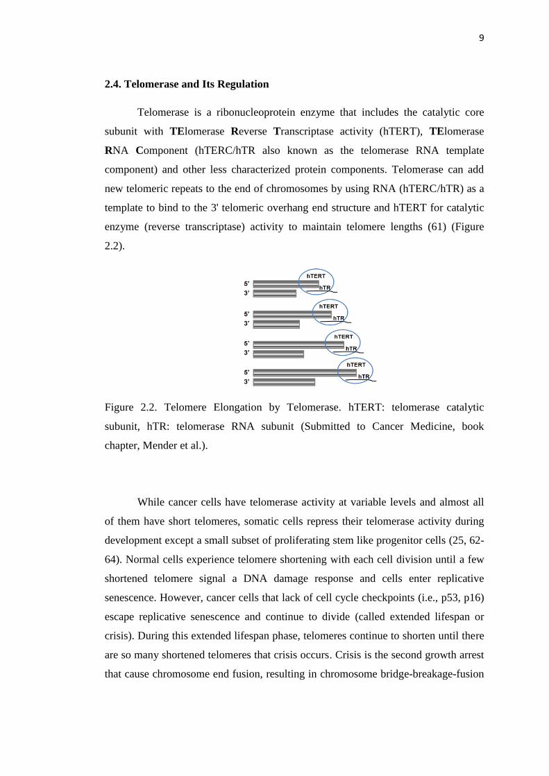
2.4. Telomerase and Its Regulation
Telomerase is a ribonucleoprotein enzyme that includes the catalytic core
subunit with TElomerase Reverse Transcriptase activity (hTERT), TElomerase
RNA Component (hTERC/hTR also known as the telomerase RNA template
component) and other less characterized protein components. Telomerase can add
new telomeric repeats to the end of chromosomes by using RNA (hTERC/hTR) as a
template to bind to the 3' telomeric overhang end structure and hTERT for catalytic
enzyme (reverse transcriptase) activity to maintain telomere lengths (61) (Figure
2.2).
Figure 2.2. Telomere Elongation by Telomerase. hTERT: telomerase catalytic
subunit, hTR: telomerase RNA subunit (Submitted to Cancer Medicine, book
chapter, Mender et al.).
While cancer cells have telomerase activity at variable levels and almost all
of them have short telomeres, somatic cells repress their telomerase activity during
development except a small subset of proliferating stem like progenitor cells (25, 62-
64). Normal cells experience telomere shortening with each cell division until a few
shortened telomere signal a DNA damage response and cells enter replicative
senescence. However, cancer cells that lack of cell cycle checkpoints (i.e., p53, p16)
escape replicative senescence and continue to divide (called extended lifespan or
crisis). During this extended lifespan phase, telomeres continue to shorten until there
are so many shortened telomeres that crisis occurs. Crisis is the second growth arrest
that cause chromosome end fusion, resulting in chromosome bridge-breakage-fusion
10
cycles. The cells that continue to divide eventually enter to the crisis, which
generally results in apoptosis. Senescence and crisis are the two independent
mechanisms that are important in protecting most large, long-lived species from the
early occurence of cancer. Rarely, cells can acquire telomerase activity, which leads
to an escape from crisis, and a hallmark of this event is that cells no longer
progressively shorten their telomeres but maintain generally very short telomere
lengths and become immortal (65). Importantly, immortalization is one of the
hallmarks of cancer. Therefore, strict regulation of telomerase is highly important for
fate of cell.
While we still know very little about the regulation of telomerase, there is
mounting evidence that enzyme activity can be controlled at the transcriptional,
posttranscriptional (alternative splicing) and epigenetic levels. While almost all cells
have high levels of the template RNA (hTR) component, irrespective of having
telomerase activity, hTERT can be detected at very low levels in telomerase positive
cells with longer telomeres such as stem cells, progenitor cells and even in cancer
cells (66). However, both components are necessary for telomerase to be activated
(67).
Alternative splicing is one mechanism that is proposed to activate telomerase
in cancer or repress telomerase in normal cells. Precursor mRNA (pre-mRNA) is
generated by transcription of TERT DNA, then the generated pre-mRNA can be
processed by excluding introns (noncoding sequences) and joining exons (coding
sequences) in order to code protein sequence. During RNA splicing, exons might be
included or excluded from the final mRNA to create multifunctional proteins, a
process called alternative splicing (67). Misregulation of alternative splicing of
telomerase and many other genes is a hallmark of almost all cancers. Therefore,
understanding hTERT alternative splicing can also help to develop new strategies for
anticancer therapies.
A recent study speculated that low abundance transcripts that are regulated by
alternative splicing need more specific mechanisms for fine-tuning regulation such as
hTERT. While hTERT coding sequence is conserved among species, some of the
intronic elements that regulate hTERT splicing are only conserved among Old World
11
primates (68), suggesting that more specialized regulatory mechanisms for low
abundance transcripts may be necessary for proper splicing (67).
Full length of hTERT that has 16 exons is the only transcript has reverse
transcribed activity. Other identified alternative spliced forms do not have telomerase
activity (69, 70). The major alternative spliced forms of hTERT are minus alpha,
minus beta or minus alpha beta. The minus alpha form is a dominant-negative form,
which does not have reverse transcriptase activity (70, 71) but overexpression of this
spliced form causes inhibition of telomerase activity in telomerase positive cells,
which can lead to senescence or apoptosis (71). The minus beta splicing creates a
frame shift by skipping TERT exon 7 and 8 leading to a stop codon in exon 10, and
is often the major hTERT spliced form in cancer cells (68). Although this form has a
stop codon and does not have telomerase activity, it was shown that it can be
translated to protein and overexpression of this form can be advantage for growth of
breast cancer cells (72). A recent study showed that hTERT alternative splicing is
regulated by variable number tandem repeats (VNTR), far from exon/intron
junctions, that may use RNA:RNA pairing to regulate hTERT splicing (73).
Telomerase activity is detected in early fetal development but is lost during
human gestations (between 12-18 weeks) due to a dramatic shift from full length
hTERT to other isoforms (mostly minus beta) (74). These studies showed that
alternative splicing of hTERT can be an important mechanism for telomerase
regulation.
There have been different approaches to better understand telomerase
regulation. Breakpoints of DNA rearrangement in TERT promoter region causes
increased TERT expression in some cancers (75), and also mutations upstream of the
TERT promoter can lead to telomerase activation by changing transcriptional
regulatory sites (76). Different studies showed that the mutations in TERT promoter
(77) and gene amplification (78, 79) is also related with transcriptional activation of
hTERT.
12
2.5. Telomere and Telomerase Targeted Therapies
Considering the link of telomerase activity in tumor development and
progression, researchers have focused on developing telomerase targeted strategies
for anticancer therapy. Ideally, telomerase based anticancer strategies could have
toxic effect on cancer cells with no or less toxicity on normal cells (25, 80, 81). Most
tumor cells that express telomerase activity can accelerate their proliferation rate
upon oncogenic stimuli. This may partially explain why tumor cells have shorter
telomeres than healthy tissues. Because as their proliferation rate increases, they will
continue to divide and eventually their telomere length will decrease (81-85). Normal
progenitor cells and stem cells have relatively long telomeres, undergoing less
divisions. This should make them more resistance to the therapy compared to cancer
cells (65). Therefore, the expression of telomerase in cancer cells versus normal cells
is the fundamental rationale for telomerase based cancer therapy. The most
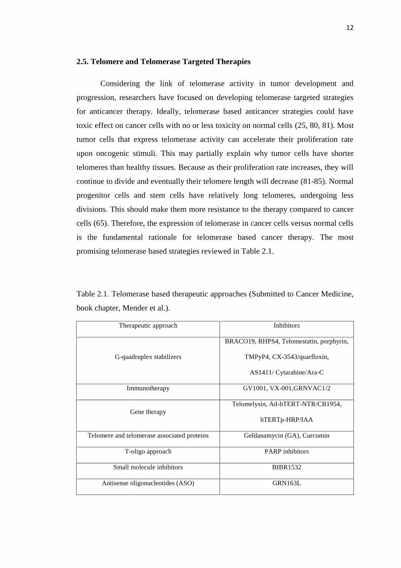
promising telomerase based strategies reviewed in Table 2.1.
Table 2.1. Telomerase based therapeutic approaches (Submitted to Cancer Medicine,
book chapter, Mender et al.).
Therapeutic approach Inhibitors
G-quadruplex stabilizers
BRACO19, RHPS4, Telomestatin, porphyrin,
TMPyP4, CX-3543/quarfloxin,
AS1411/ Cytarabine/Ara-C
Immunotherapy GV1001, VX-001,GRNVAC1/2
Gene therapy
Telomelysin, Ad-hTERT-NTR/CB1954,
hTERTp-HRP/IAA
Telomere and telomerase associated proteins Geldanamycin (GA), Curcumin
T-oligo approach PARP inhibitors
Small molecule inhibitors BIBR1532
Antisense oligonucleotides (ASO) GRN163L
13
Very few small molecule inhibitors have been found to inhibit telomerase
activity in cancer cells. BIBR1532 (2-((E)-3-naphthalene-2-yl-but-2-enoylamino)-
benzoic acid) is the one canditate small molecule that is non-competitive inhibitor of
both hTERT and hTERC (86). Although in vitro and in vivo effects of BIBR1532 has
been shown (i.e.; telomere shortening, inhibition of cell proliferation, cellular
senescence, delayed tumor growth with pretreatment) (48, 87), it was not effective
enough to enter into clinical trials (88, 89). After many years of screening new drugs
a promising oligonucleotide, GRN163L (Imetelstat, Geron Corporation) was tested,
GRN163 was the first generation of this oligonucleotide that is a 13 mer that is
complementary to the hTR template region and thus is a competitive telomerase
inhibitors (not a typical antisense approach). Although GRN163 showed good
telomerase inhibition in vitro, its potential was reduced due to rapid uptake in cells
(90, 91). Then researchers modified GRN163 by adding a lipid carrier molecule
(covalently bound lipophilic palmitoyl (C16) group to the 5'-thio-phosphate) to
increase its uptake into cells (91). GRN163L, competitive enzyme inhibitor, is a 13
mer N3'-P5'-thio-phosphoramidate oligonucleotide and shows highly potent
telomerase inhibition as a direct hTR template antagonist (91, 92) (Figure 2.3).
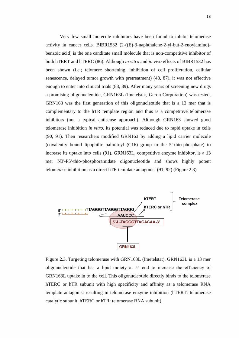
Figure 2.3. Targeting telomerase with GRN163L (Imetelstat). GRN163L is a 13 mer
oligonucleotide that has a lipid moiety at 5’ end to increase the efficiency of
GRN163L uptake in to the cell. This oligonucleotide directly binds to the telomerase
hTERC or hTR subunit with high specificity and affinity as a telomerase RNA
template antagonist resulting in telomerase enzyme inhibition (hTERT: telomerase
catalytic subunit, hTERC or hTR: telomerase RNA subunit).
14
After showing promising results in preclinical studies of different cancer cell
lines by inhibiting telomerase activity and resulting progressive telomere shortening
(92-101), GRN163L progressed into 17 different clinical trials (10 phase 1, and 6
phase 2 trials) (Table 2.2). However, it has not progressed in most clinical trials due
to hematological side effects and liver function abnormalities besides other adverse
events. When GRN163L causes hematological toxicity in patients, these patients
have to go off trial until their platelet numbers come back to normal levels, resulting
transient telomerase reactivation and telomere re-elongation during the drug holiday
period. The other potential challenge for telomerase inhibitors, such as GRN163L, is
the ''lag period''. The lag period is the time between the beginning of therapy and the
therapeutic response. Telomere attrition is required to induce senescence or apoptosis
with telomerase inhibitors, but the lag period of cancer cells is variable. Therefore, if
a tumor has heterogeneous telomere length, there could be a long treatment period
and this might result in increased side effects and costs. Yet, if they are used
following conventional therapy to kill the surviving cells, it might reduce the chance
of cancer recurrence (102). A clinical trial for this approach was completed for
advanced non-small cell lung cancer (NSCLC) (clinicaltrials.gov: NCT01137968).
Eligible stage IV or recurrent locally advanced NSCLC patients were randomly
divided to different groups that are GRN163L with/without bevacizumab for day 1 of
each 21 day cycle or observation versus standard of care alone (patients who had
received bevacizumab before randomization were continued with bevacizumab
maintenance or had no followup maintenance treatment). This study showed a trend
in prolonged progression free survival (PFS) in the patients with the shortest quartile
of telomere at the initiation of the trial in the GRN163L arm (103). Experience from
these type of trials can facilitate our understanding how we can treat cancer patients
to take a better advantage of anti-telomerase therapies. GRN163L clinical trials are
reviewed in Table 2.2.
15
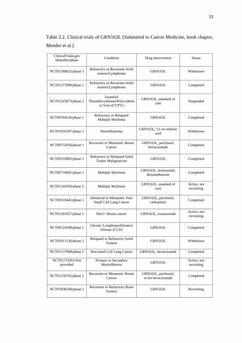
Table 2.2. Clinical trials of GRN163L (Submitted to Cancer Medicine, book chapter,
Mender et al.).
ClinicalTrials.gov
Identifier/phase Condition Drug Intervention Status
NCT01568632/phase 1 Refractory or Recurrent Solid
tumors/Lymphoma GRN163L Withdrawn
NCT01273090/phase 1 Refractory or Recurrent Solid
tumors/Lymphoma GRN163L Completed
NCT01243073/phase 2
Essential
Thrombocythemia/Polycythem
ia Vera (ET/PV)
GRN163L, standard of
care Suspended
NCT00594126/phase 1 Refractory or Relapsed
Multiple Myeloma GRN163L Completed
NCT01916187/phase 1 Neuroblastoma GRN163L, 13-cis retinoic
acid Withdrawn
NCT00732056/phase 1 Recurrent or Metastatic Breast
Cancer
GRN163L, paclitaxel,
bevacizumab Completed
NCT00310895/phase 1 Refractory or Relapsed Solid
Tumor Malignancies GRN163L Completed
NCT00718601/phase 1 Multiple Myeloma GRN163L, bortezomib,
dexamethasone Completed
NCT01242930/phase 2 Multiple Myeloma GRN163L, standard of
care
Active, not
recruiting
NCT00510445/phase 1 Advanced or Metastatic Non-
Small Cell Lung Cancer
GRN163L, paclitaxel,
carboplatin Completed
NCT01265927/phase 1 Her2+ Breast cancer GRN163L, trastuzumab Active, not
recruiting
NCT00124189/phase 1 Chronic Lymphoproliferative
Disease (CLD) GRN163L Completed
NCT02011126/phase 2 Relapsed or Refractory Solid
Tumors GRN163L Withdrawn
NCT01137968/phase 2 Non-small Cell Lung Cancer GRN163L, bevacizumab Completed
NCT01731951/Not
provided
Primary or Secondary
Myelofibrosis GRN163L
Active, not
recruiting
NCT01256762/phase 2 Recurrent or Metastatic Breast
Cancer
GRN163L, paclitaxel,
w/wo bevacizumab Completed
NCT01836549/phase 2 Recurrent or Refractory Brain
Tumors GRN163L Recruiting
16
Guanosines can assemble by themselves in the presence of guanine tandem
repeats and generate G-quadruplex structures by monovalent cations (i.e.;
potassium). G-quadruplexes can be found in telomeres, oncogene promoter
sequences and other regions of genome (104). G-quadruplex structures are the
another target to design small molecule ligands for telomerase-based therapies.
G-quadruplex ligands stabilize G-quadruplex structure by preventing G-quadruplex
from unwinding and opening T-loops, which makes telomerase indirectly a target
and can result in telomerase inhibition. Also, G-quadruplex ligands may cause
telomere uncapping by leading to dissociation of telomeric proteins (80, 105).
BRACO-19, RHPS4 and telomestatin have been the most commonly studied
G-quadruplex ligands to date. They inhibit telomerase activity by activating DNA
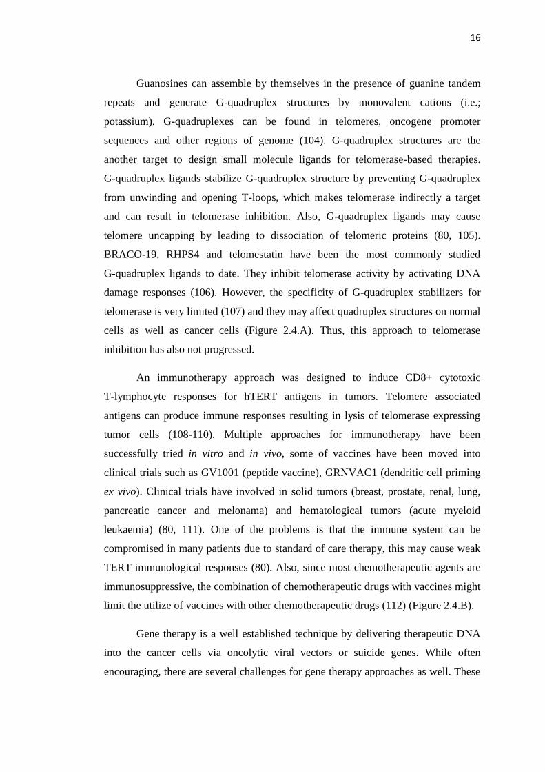
damage responses (106). However, the specificity of G-quadruplex stabilizers for
telomerase is very limited (107) and they may affect quadruplex structures on normal
cells as well as cancer cells (Figure 2.4.A). Thus, this approach to telomerase
inhibition has also not progressed.
An immunotherapy approach was designed to induce CD8+ cytotoxic
T-lymphocyte responses for hTERT antigens in tumors. Telomere associated
antigens can produce immune responses resulting in lysis of telomerase expressing
tumor cells (108-110). Multiple approaches for immunotherapy have been
successfully tried in vitro and in vivo, some of vaccines have been moved into
clinical trials such as GV1001 (peptide vaccine), GRNVAC1 (dendritic cell priming
ex vivo). Clinical trials have involved in solid tumors (breast, prostate, renal, lung,
pancreatic cancer and melonama) and hematological tumors (acute myeloid
leukaemia) (80, 111). One of the problems is that the immune system can be
compromised in many patients due to standard of care therapy, this may cause weak
TERT immunological responses (80). Also, since most chemotherapeutic agents are
immunosuppressive, the combination of chemotherapeutic drugs with vaccines might
limit the utilize of vaccines with other chemotherapeutic drugs (112) (Figure 2.4.B).
Gene therapy is a well established technique by delivering therapeutic DNA
into the cancer cells via oncolytic viral vectors or suicide genes. While often
encouraging, there are several challenges for gene therapy approaches as well. These
17
concerns are about whether delivery of gene therapy to cancer cells throughout the
body can be achieved efficiently or not and also immunological responses to vector
systems can limit dosing schedules (80) (Figure 2.4.C). Mutant hTERC (dominant-
negative approach) is the other gene therapy approach in preclinical studies. Wild
type telomeres contain DNA sequences for binding of telomere associated proteins
(i.e.; POT1, TRF1, TRF2) (113, 114). Mutant DNA that is synthesized from mutant
hTERC disrupts the binding of telomere associated proteins (115-117) and this
results in telomere uncapping. Although in vitro and in vivo effects of mutant hTERC
therapy approach via lentiviral vectors has been succesfully reported (116, 117), it is
still a gene therapy approach that needs to be more efficient to target the vast
majority of cancer cells.
Figure 2.4. Disadvantages of different telomerase based therapy strategies.
A) G-quadruplex structures can be found at the other region of chromosomes besides
telomeres, thus they can be toxic normal cells as well as cancer cells. B) One of the
important disadvantages of immunotherapy is the immunosuppresive effects of
chemotherapeutic agents when they are used together as an adjuvant therapy.
C) Delivery of gene therapy into the cancer cells throughout the body is challenging
for cancer patients and oral therapy is not generally available for efficient delivery
(Submitted to Cancer Medicine, book chapter, Mender et al.).
18
2.6. The Role of Telomeres on Cellular and Organismal Aging and Cancer
Studies showed that having short telomeres in mice (with knockout of
mTERT) can eventually lead to some aspect of aging observed in humans. When
telomerase activity and expression of p53 pathway were increased at the same time,
they found that these mice extended their life span by ~40% (118). A different study
showed that the mice with critical short telomeres had many hallmark of advance age
immunological problems, wound healing problems, and stem cell regenerative
problems such as errosive dermatitis and failure of liver regeneration. However,
when telomerase was reactivated in these mice with experimentally shortened
telomeres, animals became fertile (119), more directly showing that telomeres play a
role in aging process. In addition, individual critically short telomeres trigger cellular
responses to the telomere dysfunction (120). Each human cell has 92 telomeres, and
each telomere has its own unique length and the lowest percentiles of telomere length
have a higher risk for aging related disease. Therefore, it is important to understand
aging and cancer process so that we can increase the years of healthy life by
diminishing age associated diseases and controlling unlimited proliferation for
prevention of cancer (121).
Cellular senescence is one of the tumor suppressor mechanisms to counteract
the development of cancer. Cellular senecence is the proliferative arrest that forces
cells to exit the cell cycle permanently (122). There are several nuclear biomarkers
for senescence such as telomere dysfunction induced DNA damage foci, TIFs (123,
124), altered gene expression (upregulation of p16, p21Waf1
/Cip1 and p53) (125-129),
evidence of chromosomal instability but only in combination with other alterations in
important cell cycle check point genes (130), an increased DNA damaged response
(123), and changes in chromatin structure [the development of senescence-associated
heterochromatin foci (SAHF)] (131-133). Different studies showed in mouse models
that high levels of oncogene expression can cause benign tumors in which cells
display cellular senescence features such as high levels of certain heterochromatin
proteins and senescence-associated-beta galactosidase (SA-βGal) activity. Disruption
of senescence pathways, for example by inactivating p53 tumor suppresor, promotes
19
malignant cancer progression, indicating that cellular senescence supresses cancer
development in humans and mice (61).
When double strand breaks (DSBs) are generated by genotoxic stresses such
as DNA replication stress, ionizing radiation, endonucleases, or ROS, most DBSs are
repaired. In contrast, breaks that occur on or near telomeres (in the absence of
telomerase activity) can not be repaired for months, maybe for years, both in vitro
and in vivo (134, 135). Persistent telomeric damage foci are also generated by
ionizing radiation in the presence of endogenous and ectopically expressed
telomerase, suggesting that even telomerase is unable to prevent formation of
telomeric damage under some ionizing radiation conditions (136). However,
telomerase can suppress the formation of telomeric damage under the DNA damage
stress in somatic human cells (136). Dysfunctional telomeres that are generated by
progressive telomere erosion, DNA replication stress, telomeric DSB formation, or
other genotoxic events induce cellular senescence in normal cells, which are called
Telomere Dysfunction-Induced Senescence (TDIS). This telomere dysfunction
induced senescence is a tumor suppressing mechanism. Telomeres or telomere
associated proteins can inhibit the DNA damage repair machinery under some
conditions (61). It might seem that permanency of damage on telomeres is
disadvantageous, however, as DSB machinery tries to repair damage, it will cause
mutation at the breakage site. Therefore, it provides that dysfunctional telomeres
induce senescence prematurely, accumulate in our tissues as we age. This is
potentially a beneficial mechanism to the organism to prevent accumulation of
mutations and protect organism from uncontrolled proliferation and invasive tumors
early in life (61).
2.7. The Thiopurines
Azathioprine, 6-thioguanine and 6-mercaptopurine are the thiopurines that are
used as an anti-inflammatory, anticancer and immunosuppresive drugs. Although
they have been in clinical trials for well over a half century, only 6-mercaptopurine
and azathioprine were approved by FDA (137). However, it has been known that
prolonged treatment with thiopurines is associated with high risk of various cancers

20
such as non-Hodgkin lymphoma, skin squamous cell carcinoma. Therefore, it is
advisable to follow up with patients for any signs of therapy related cancers (138).
Thiopurines are prodrugs that need to be converted to metabolically active
compounds (139). 6-thioguanine is the final active metabolite of all the thiopurines.
The first step of this metabolic pathway is the removal of the nitroimidazole group
off azathioprine in a non-enzymatic reaction involving glutathione. This step occurs
in erythrocytes which release the active metabolite 6-mercaptopurine.
6-mercaptopurine and 6-thioguanine then enters the purine salvage pathway in cells.
Hypoxanthine-guanine phosphoribosyl transferase 1 (HPRT1) is one of the
important enzymes in purine biosynthesis, which catalyzes the addition of
ribose-5-phosphate to 6-mercaptopurine and 6-thioguanine to generate thioinosine
monophosphate (TIMP) and thioguanosine monophosphate (TGMP), respectively.
TIMP and TGMP are the precursors for incorporation of 6-thioguanine into RNA or
DNA. Additional reactions with deoxynucleoside kinases and reductases are required
to convert thioguanine nucleotides to 6-thio-GTP and 6-thio-dGTP, which are
substrates for incorporation of 6-thioguanine into RNA and DNA (138).
The degradation of 6-mercaptopurine is catalysed by xanthine oxidase, also
both 6-mercaptopurine and 6-thioguanine are inactivated by thiopurine
S-methyltransferase (TPMT) (140) (Figure 2.5).
21
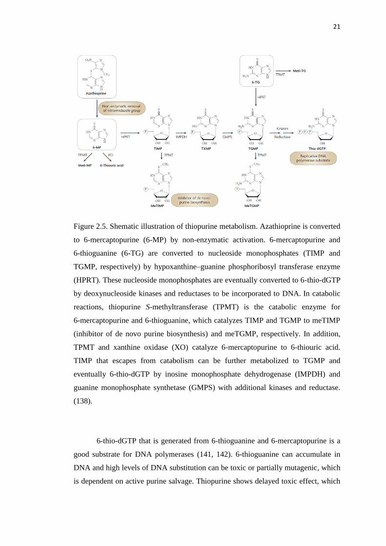
Figure 2.5. Shematic illustration of thiopurine metabolism. Azathioprine is converted
to 6-mercaptopurine (6-MP) by non-enzymatic activation. 6-mercaptopurine and
6-thioguanine (6-TG) are converted to nucleoside monophosphates (TIMP and
TGMP, respectively) by hypoxanthine–guanine phosphoribosyl transferase enzyme
(HPRT). These nucleoside monophosphates are eventually converted to 6-thio-dGTP
by deoxynucleoside kinases and reductases to be incorporated to DNA. In catabolic
reactions, thiopurine S-methyltransferase (TPMT) is the catabolic enzyme for
6-mercaptopurine and 6-thioguanine, which catalyzes TIMP and TGMP to meTIMP
(inhibitor of de novo purine biosynthesis) and meTGMP, respectively. In addition,
TPMT and xanthine oxidase (XO) catalyze 6-mercaptopurine to 6-thiouric acid.
TIMP that escapes from catabolism can be further metabolized to TGMP and
eventually 6-thio-dGTP by inosine monophosphate dehydrogenase (IMPDH) and
guanine monophosphate synthetase (GMPS) with additional kinases and reductase.
(138).
6-thio-dGTP that is generated from 6-thioguanine and 6-mercaptopurine is a
good substrate for DNA polymerases (141, 142). 6-thioguanine can accumulate in
DNA and high levels of DNA substitution can be toxic or partially mutagenic, which
is dependent on active purine salvage. Thiopurine shows delayed toxic effect, which
22
can partially be explained with the requirement of passage through S-phase of the
cell cycle (138). 6-thioguanine (more reactive than canonical DNA bases) is
incorporated into the template DNA, rather than the daughter DNA, therefore DNA
polymerases can not find the precise daughter-strand partner for the aberrant base.
This results in potentially lethal DNA lesions (143). Increased levels of spontaneous
mutations by 6-thioguanine related DNA damage is due to uncorrected replication
errors. It is also reported that DNA that has 6-thioguanine substitution are
photochemically active and generate reactive oxygen species (ROS), which can
eventually cause damage to DNA, protein and other macromolecules (138).
2.8. The Importance of Liver and Kidney Function on Drug Toxicity
Assessment of organ function (i.e.; liver, kidney) is an important aspect in
selecting the appropriate drug and its doses for cancer patient after assessment of
tumor histology. The possibility of organ abnormalities following treatment can be
due to drug regime rather than progressive disease (144). An appropriate strategy for
effective and safe chemotherapy dosing is poorly defined in cancer. Cancer patients
have different pharmacokinetics/pharmacodynamics, and many agents have narrow
therapeutic windows (e.g. differences between toxicity to normal versus cancer
cells). Chemotherapy dosing decisions are often made base on limiting toxicity in
cancer (144). Therefore, selecting an appropriate chemotherapy and its dosing can be
a challenge for clinicians and their patients.
The liver is a vital organ that has functions in multiprocesses such as
metabolism, biosynthesis, excretion, secretion and detoxification. The liver is an
oxygen dependent tissue since the processes in liver are energy required. The liver is
a regenerative organ that can regrow from even massive cellular losses. However,
cellular loss above certain threshold will prevent tissue regeneration and lead to
hepatic failure and death (145). Several chemotherapy agents cause liver injury,
which can lead to necrosis, steatosis, fibrosis, cholestasis, and vascular injury (146).
This can be determined by serum liver biochemistry or histology. Serum liver
biochemical tests are generally an indicator of liver inflammation or damage, rather
than liver function (147). Several studies showed the relation between abnormal
serum biochemical tests and toxic effects of chemotherapeutic drugs. For instance,
23
the toxicity of antimetabolites such as 6-mercaptopurine, 6-thioguanine have been
shown in preclinical and clinical trials. When 6-mercaptopurine dose exceed 2 mg/kg
daily dose in adults, it causes either hepatocellular or cholestatic liver disease and a
hepatocellular injury (148, 149). It has been shown that 14 of 40 patients treated with
6-mercaptopurine developed aspartate aminotransferase (AST) or alanine
aminotransferase (ALT) values above 150 U/L (150). 6-thioguanine, which is
another thiopurine metabolite causes the production of hepatic veno-occlusive
disease (VOD) and in a single case of peliosis hepatitis as well as jaundice (144, 151-
155).
The kidney is the essential organ that functions in many important processes
such as excretion of waste products of metabolism, reabsorption of vital nutrients,
acid-base homeostasis, osmolality regulation, blood pressure regulation, hormone
secretion. Also the importance of kidney on drug toxicity is due to most drugs are
eliminated by the kidneys and therefore dose adjustments are needed in patients with
renal insufficiency (156). The amount of damage in the kidneys is dependent on the
type of chemotherapy, patient age, and underlying disease. Most potent
chemotherapeutics have been shown to have nephrotoxic effects. These effects can
lead to acute (frequently reversible) or chronic kidney disease. Acute toxicity can
progress to chronic toxicity, which develops chronic tubulointerstitial nephritis,
papillary necrosis, or prolonged proteinuria with some chemotherapeutics (157).
Impaired renal function such as excretion and metabolism might cause systemic toxic
effect such as bone marrow suppression (158, 159). Therefore, it is important to
know commonly used chemotherapeutics that cause renal dysfunction and also
available preventive strategies to decrease the risk of renal disease in cancer patients
under treatment (160).
2.9. Tumor Resistance to Therapy
There have been different mechanisms to explain tumor resistance to
chemotherapy or radiotherapy. Tumor that arises from one single clonogenic cells
accumulates multiple mutations and genetically diverse clones are found in the same
tumor (161-166). This diversity in intratumor cells exerts distinct treatment
sensitivity and different clones in the same tumor may have different resistance
24
mechanisms (167, 168). In addition, the cells in the same clone have different
fundamental and phenotypic differences that might be explained by the stem cell
model of cancer development. Recent studies showed that some cancer cells have a
hierarchical organization, which is similar to normal tissue. For instance, tumorigenic
cancer stem cells (CSCs) differentiate into non-tumorigenic progenies (168). Cancer
stem cells represent a different subpopulation that can be identified and isolated from
the tumor tissues by using putative cancer stem cell markers. Cancer stem cells,
which are different than bulk tumor cells, have self renewal capacity and long term
repopulation potential. These features of cancer stem cells can facilitate the initiation
and maintainence of tumorigenesis (169, 170). Cancer stem cells can be either stable
or transient cell populations in human malignant neoplasms (170). Studies have
shown that although CSC and their non tumorigenic progenies in the same clone
have same genotype, they display different epigenetic profiles, which cause
alteration of the signaling pathways (171-174). In addition, different studies have
shown that the number of molecular mechanisms contribute to resistance of certain
CSCs to conventional therapy (168, 169), but this property can not be generalized
since some CSCs are sensitive to conventional therapy and some are not. However,
cancer can potentially arise from a single CSC, therefore to target all CSCs might be
required for efficient therapy and durable responses in patients (175, 176).
Cancer stem cells can be detected in human tumors and cell cultures by
specific cell surface proteins such as CD133, CD44, CD24, α2β1 integrin and others
(169). In addition, cancer stem cells can be determined by biochemical activity of the
marker proteins. For instance, the aldefluor assay is based on enzymatic activity of
aldehyde dehydrogenase (ALDH) in putative tumor initiating populations. Also
identification of CSCs is possible by the capability to efflux lipophilic fuorescent dye
Hoeschst 33342 efflux. The cells that are negative for the dye staining have a tail like
shape called side population (SP) (177). This phenotype is correlated with
upregulation of ATP-Binding Cassette (ABC) transporter superfamily proteins. ABC
transporters are known to participate in multidrug resistance functions in various
human cancers (178-180). The ABC transporter family has 49 proteins;
P-glycoprotein (P-gp, MDR1, ABCB1), multidrug resistance protein 1 (MRP1,
ABCC1), and breast cancer resistance protein (BCRP, ABCG2) have been
25
commonly investigated for drug resistance mechanisms (181, 182). Increased
expression of these proteins in some type of cancers results in ATP-dependent efflux
of cytotoxic drugs from cells. This leads to the inability to maintain within the cancer
cells a toxic drug concentration (183). Population of cancer stem cells in different
tumors have high expression of ABC transporters, leading to drug resistance (184,
185).
Aldehyde dehydrogenase activity is one of the common features of cancer
stem cells. The proteins of aldehyde dehydrogenase family are enzymes that catalyze
oxidation of intracellular aldehydes to carboxylic acids and also contribute the
synthesis of retinoic acid and neurotransmitter gamma-amino butyric acid (GABA),
which are also important for normal and cancer stem cells maintenance and
differentiation (186, 187). ALDH1A1 is the most studied isoform of ALDH protein
family (188). Studies showed that increased level of ALDH1A1 is the marker for
poor clinical outcome in breast and prostate cancer patients (189-191). It is also
known that ALDH1A2 is involved in cell proliferation, colony formation and drug
resistance (192) .
Epigenetic changes are the heritable changes in gene expression that occur
without any alterations in the DNA sequence including DNA methylation, histone
modification, and posttranslational gene regulation by microRNAs (miRNAs) (193,
194). DNA methylation is a major epigenetic factor that influences gene activities
and plays an important role in cancer and chemotherapy drug resistance. DNA
methylation is catalyzed by DNA methyltransferases that transfer methyl group from
S-adenosyl methionine (SAM) to the fifth carbon of a cytosine residue to generate
methylated cytosine (195). Cytosines are followed immediately by a guanine, it is so-
called CpG dinucleotides. Most genome are lack of CpG dinucleotides, and those
that are present in the genome are generally methylated. In contrast, CpG islands that
are comparatively rich in CpG nucleotides are almost methylation free. These CpG
islands are frequently located in the promoters of human genes and methylated
cytosines in the CpG islands are generally associated with transcriptional inactivation
of gene such as tumor supressor gene like p53. Aberration in DNA methylation is
correlated with cancer development and it has been shown that methylation of CpG
26
islands plays an important role on transcriptional repression of tumor suppresor
genes (196). The second epigenetic mechanism is the histone modification. Histones
are the major components of chromatin and can undergo multiple post-translational
modifications such as acetylation by histone acetyltransferases (HATs) and
deacetylation by histone deacetylases (HDACs). Acetylated form of histone proteins
causes less-condensed packaging of genes in chromatin that might lead to the
re-expression of silenced tumor suppressor genes. It is known that micro-RNAs
(miRNAs) are endogenous small non-coding RNAs that negatively regulate gene
expression. Previous studies showed the crucial role of miRNAs in regulating gene
expression and the relation between differential expression of miRNAs with drug
resistance in cancer (197).
Genetic alterations in signaling pathways have also effect on drug response.
In most cases, mutated oncogenes start the signals in pathways leading to
upregulation of survival, drug resistance or downregulation of cell death responses.
For example, Wnt/β-catenin and Notch pathways are one of the signaling pathways
that are important for cancer progression. Transducin-like enhancer of split 2 (TLE2)
is a mammalian homologue of the Drosophila transcriptional repressor groucho,
which plays a role in Wnt/β-catenin and Notch pathway. It was shown that increased
Wnt signals and nuclear β-catenin results in elevated Cyclin D1 and cellular
proliferation in Wnt/β-catenin pathway, additionally increased TLE2 affected tumor
progression. In Notch pathway, Notch3 repressed HASH1/ASCL1, resulting in
reduced DLK1 and inhibited cellular differentiation, PIT1 and growth hormone
expression. However, how increased TLE2 levels affect tumor progression requires
further investigation (198). It is also known that Wnt and Notch signaling pathways
participate in the regulation of drug resistance in several cancers (199, 200).
2.10. Purpose and Significance
Telomerase is an highly attractive and almost universal target for cancer
therapy, yet there are few telomerase based therapies in clinical trials. Since
telomerase inhibition based therapies depend on initial telomere length of cancer
cells, long treatment period is necessary to induce senescence or apoptosis by
progressive telomere shortening in the presence of the inhibitors. Therefore, this long
27
treatment period (lag phase) may lead to increased toxicities (side effects) and
require high costs. Standard telomerase inhibitors have not been effective in
long-term clinical trials due to the increased toxicities and the long lag period. In this
study, we first aimed to generate a nucleoside analog to reduce this long lag period
treatment period and hopefully with minimal side effects. Secondly, we aimed that
this nucleoside analog should be recognized by telomerase, then incorporated into
the telomeres, upcapping them immediately and resulting in telomere induced
dysfunctional foci and cell death. Importantly, we sought to identify a nucleoside that
was more specifically toxic to telomerase expressing cancer cells compared to
normal cells. We hypothesized that this nucleoside analogue would alter the
telomeric structure and function, cause telomeric DNA damage due to telomere
uncapping and reduce the treatment phase, leading massive and fast cancer cell death
by being converted to nucleoside triphosphate. The biochemical conversion of this
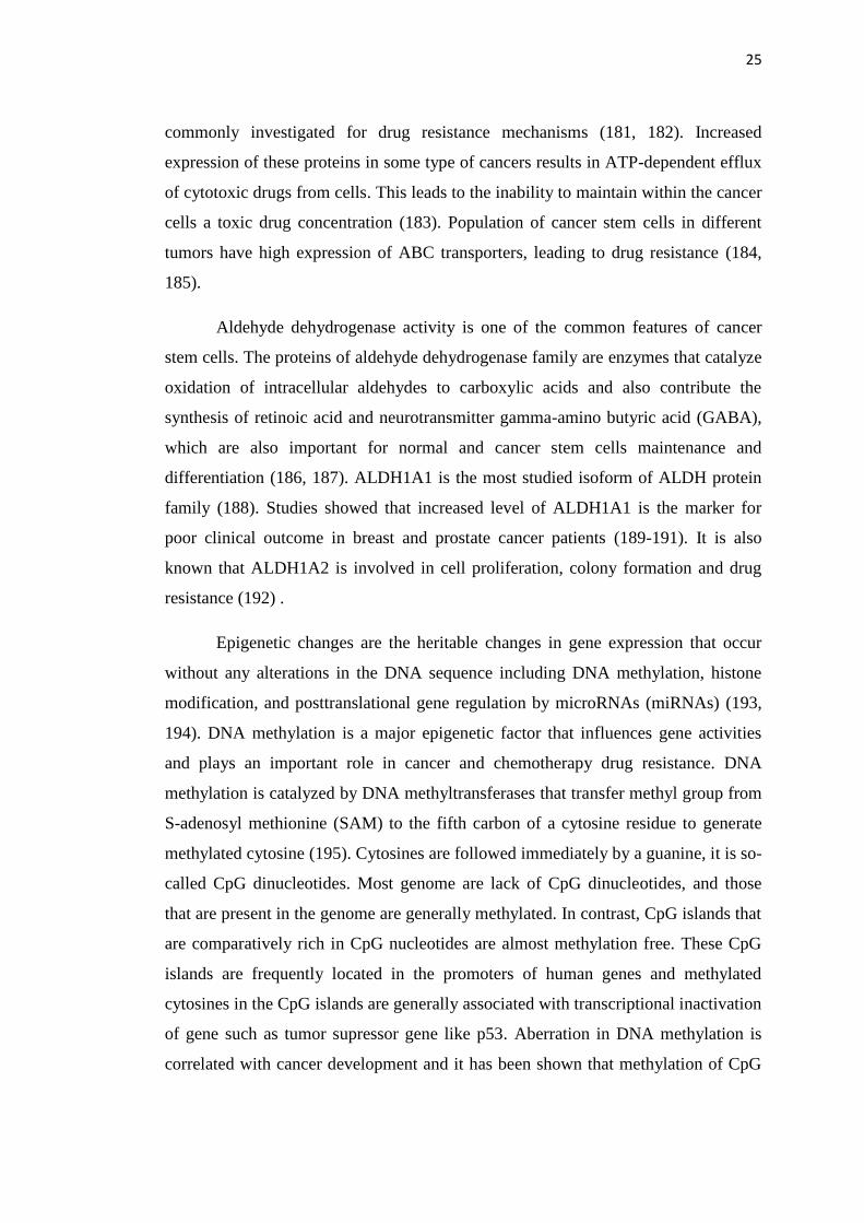
nucleoside analogue (6-thio-dG) to 6-thio-dGTP is shown in Figure 2.6. The
overarching focus of this study was to identify such a nucleoside, to test it both in
vitro and in xenograft mouse experiments and finally to determine the side effects in
wild type mice.
Figure 2.6. The biochemical conversion reaction of 6-thio-dG to 6-thio-dGTP to be
incorporated into telomeres by telomerase.

28
3. METHODS
3.1. Cell lines
The panel of non-small lung cancer cell lines (H2882, HCC2429, HCC827,
HCC15, H2087, HCC4017, HCC515, H2009) were provided by John D. Minna at
the University of Texas Southwestern Medical Center. HCT116 human colon, A549
human non-small cell lung cancer (NSCLC), the panel of additional non-small cell
lung cancer cell lines (H2882, HCC2429, HCC827, HCC15, H2087, HCC4017,
HCC515, H2009) and BJ human fibroblasts were grown in a Medium X
(DMEM:199, 4:1, Hyclone, Logan, UT) supplemented with 10% cosmic calf serum
(Hyclone) without antibiotic. HCEC1 cells were cultured in medium consisting of
mediumX (DMEM:199, 4:1), 2% cosmic calf serum, insulin, and gentamycin. BJ
and HCEC1 cells were cultured at 37◦C in low oxygen (2-5%) to prevent damaging
cells in culture (three gas mixture; 2% oxygen, 5% CO2 and 93% nitrogen) (201). BJ
cells were immortalized by transfection of a retroviral hTERT-TK-hygromycin
cassette. Successful hTERT-hygromycin expression was confirmed in clones by
testing for hygromycin resistance and the presence of telomerase activity.
3.2. Drug preparation
6-thio-dG (Metkinen Oy, Kuopio, Finland) and 6-thioguanine (Sigma, St
Louis, MO) was dissolved in DMSO/water (1:2) to prepare 50 mM or 10 mM stock
solutions, which were kept frozen at –20˚C. Once in vitro experiments were
conducted, a 1 mM final concentration was prepared in serum free medium and
added at varying amounts for cell treatments. For mouse in vivo studies, drugs were
prepared in 5% DMSO solution.
3.3. Cell Viability Assay
HCT116 (0.5 x 10³ cells/well), A549 (0.6 x 10
3 cells/well), and H2882,
HCC2429, HCC827, HCC15, H2087, HCC4017, HCC515, H2009 (1.5 x 10³
cells/well), BJ and HCEC1 cells (2 x 10³
cells/well) were plated in growth media on
96 well plate. Cells were incubated for 1 week and treated with varying
concentrations of 6-thio-dG and 6-thioguanine or DMSO every three days. The 96
29
well plates were analyzed according manufacturer’s directions for the CellTiterGlo
Luminescent Cell Viability Assay (Promega, Madison, WI) to obtain IC50 values,
The IC50 is defined as the concentration of drug at which 50% of the cells are
inhibited by the drug. Sigmoidal dose-response curves (GraphPad Prism, La Jolla,
CA) were used to calculate IC50 values.
3.4. Long-term cell culture studies
For long-term cellular experiments, HCT116 (1,000 cells/cm2) and BJ
(10,000 cells/cm2) cells were treated with 6-thio-dG (1, 3, 10 μM) containing
medium every three days. The cells were counted and replated every week for 10-12
weeks. Additionally, HCT116 cells (1,000 cells/cm2) were fed with 6-thioguanine (1,
3, 10 μM) every three days, the cells were counted each week, collected 1x10⁶ cells
for TRF (Telomere Restriction Fragment) analysis and replated.
3.5. Telomerase activity assay (TRAP, Telomere Repeat Amplification Protocol)
General Information and Principle
TRAP is a standard method to determine telomerase activity. It is developed
and referred to as the TRAP assay by Kim et al. (25). TRAP assay includes three
steps that are extension, amplification, and detection of telomerase products (Figure
3.1). In the extension step, telomeric repeats are added to the telomeric substrate,
which is a non telomeric oligonucleotide, by telomerase. In amplification step, the
extension products are amplified by the polymerase chain reaction (PCR) by using
specific primers and in detection step, mostly the presence or absence of telomerase
is analyzed by electrophoresis. The extraction of telomerase from cell or tissue is
very important step since the amount of telomerase affects the quantitative
determination of telomerase activity (202). However, the standard TRAP assay has
the ability to detect telomerase activity even in a single cell. But it is worth to note
that the accuracy of detection of telomerase activity depends on amplification step
and the primers used (202). Although this is an advantageous assay, there are some
disadvantages due to the annealing problem of primers. The extended product by
PCR utilizes TS specific primers that can produce primer-primer interactions and
these dimers might generate false positive results. Therefore, there have been
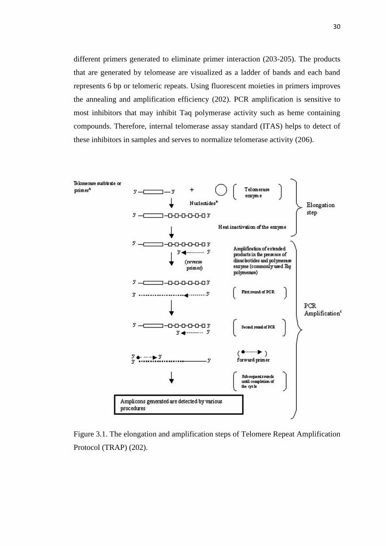
30
different primers generated to eliminate primer interaction (203-205). The products
that are generated by telomease are visualized as a ladder of bands and each band
represents 6 bp or telomeric repeats. Using fluorescent moieties in primers improves
the annealing and amplification efficiency (202). PCR amplification is sensitive to
most inhibitors that may inhibit Taq polymerase activity such as heme containing
compounds. Therefore, internal telomerase assay standard (ITAS) helps to detect of
these inhibitors in samples and serves to normalize telomerase activity (206).
Figure 3.1. The elongation and amplification steps of Telomere Repeat Amplification
Protocol (TRAP) (202).
31
Procedure
Telomerase activity was measured by the TRAP (Telomeric Repeat
Amplification Protocol) assay as mentioned above (62). Briefly, HCT116 cells were
treated with 1 or 10 μM 6-thio-dG for 1 week. 1x10⁵ cells were collected and lysed
with ice-cold NP-40 lysis buffer (10 mM Tris–HCl pH 8.0, 1.0 mM MgCl2, 1 mM
EDTA, 1% NP-40, 0.25 mM sodium deoxycholate, 10% glycerol, 150 mM NaCl,
5 mM β-mercaptoethanol) for 30 min. One microliter cellular lysate was used for
each reaction. Hela cells were used as a positive control and lysis buffer was used as
a negative control. Samples were prepared and then the telomerase extension
products were amplified using PCR (95˚C for 5 min to inactivate telomerase, then
95˚C for 30 sec, 52˚C for 30 sec, 72˚C for 30 sec; 24 cycles). Samples were run on a
10% nondenaturating acrylamide gel and visualized using a Typhoon
PhosphorImager scanner system (Molecular Dynamics, GE Healthcare, Piscataway,
NJ) that is capable of reading Cy5 fluorescence.
3.6. Telomere length assay (TRF, Terminal Restriction Fragment)
General Information and Principle
Telomere restriction fragment analysis, which is based on southern blot
technique, can be used to obtain average telomere length of cell population. This
method measures canonical and noncanonical telomere components and also
provides telomere length distribution in each DNA. The principle of this analysis lies
in the relative lack of restriction sites in telomeric (TTAGGG repeats) and
subtelomeric (adjacent to telomeric repeats) regions. Genomic DNA digestion by
restriction endonucleases cut within chromosomal DNA, not telomeric repeats. HinfI
and RsaI is typically used in TRF analysis rather than HphI and MnlI because while
HphI/MnlI cuts DNA within noncanonical subtelomeric region, HinfI/RsaI leaves
this region intact, resulting in ~1 kb longer than mean telomere length (207).
Procedure
1x10⁶ cells were collected and washed with PBS. DNA was isolated using the
manufacturer’s instructions (Qiagen, Valencia, CA). 2.5 μg DNA was digested with
32
six different restriction enzymes (HhaI, HinfI, MspI, HaeIII, RsaI, AluI) (New
England Bio, Ipswich, MA) and incubated at 37◦C overnight. Digested DNA was
separated on a 0.7% agarose gel overnight at 70 V. The terminal restriction fragment
(TRF) gel was placed in denaturating solution for 20 min (0.5 M NaOH, 1.5 M NaCl,
pH 13.2) and dried on Whatman 3MM paper under vacuum for 3 hr at 56◦C. The gel
was then placed for 15 min in neutralization buffer (1.5 M NaCl, 0.5 M Tris-HCl, pH
8.0) and then probed with a radiolabeled telomeric probe (C-rich) for 16 hr at 42◦C in
5x SSC buffer, 5xDenhardt’s solution, 10 mmol/L Na2HPO4, and 1 mmol/L
Na2H2P207. The gel was washed once with 2x SSC, 0.1% SDS, twice with 0.5x SSC,
0.1% SDS and then twice with 0.5x SSC, 1% SDS at room temperature for 15 min.
Gels were exposed to a PhosphorImager screen overnight and analyzed using a
Typhoon PhosphorImager scanner system (Molecular Dynamics). ImageQuant and
graphpad prism were used to determine telomere length of cells.
3.7. 6-thio-dG and GRN163L Treatment for Cell Killing Effect and Telomere
Restriction Fragment Analysis
HCT116 cells were grown without any treatment, with only 3 μM GRN163L
treatment, 3 days pretreatment with 3 μM GRN163L, and 3 μM GRN163L+1 μM
6-thio-dG treatment at the same time. Drugs were added every three days for 1 week.
At the end of treatment, cells were countedand compared with each treatment design.
HCT116 cells (5x10⁴ per 10 cm dish) were treated with 10 μM 6-thio-dG
and/or 3 μM GRN163L every three days for 11-14 weeks. Also, HCT116 cells
treated with 10 μM 6-thio-dG for 12 weeks were allowed to grow without any
treatment for additional 2-4 weeks or combination of 10 μM 6-thio-dG and 3 μM
GRN163L for additional 2-4 weeks. Each week cells were counted, collected for
TRF (Telomere restriction fragment) analysis and replated.
3.8. Telomere dysfunction Induced Foci (TIF) assay
The TIF assay is based on the co-localization detection of DNA damage by an
antibody against DNA damage markers, such as gamma-H2AX, and telomeres using
an antibody against the telomeric protein TRF2. Briefly, HCT116 cells were plated
in 4-well chamber slides and after the cells attached to the surface, either 3 μM
33
6-thio-dG or 3 μM 6-thioguanine was added to the medium at different time points
(0, 30 min, 12 hr, 24 hr, 48 hr, 72 hr). BJ and BJ hTERT cells were plated in 4-well
chamber slides and after the cells attached to the surface, either 10 Μm 6-thio-dG or
10 μM 6-thioguanine was added to the medium for 48 hr. Slides were rinsed once
with PBS and fixed in 4% paraformaldehyde in PBS for 10 min. Then, cells were
washed twice with PBS and permeabilized in 0.5% Nonidet-P40 in PBS, blocked
with 0.5% Bovine Serum Albumin (BSA) and 0.2% fish gelatin in PBS for 30 min.
Gamma-H2AX (mouse) (Millipore, Billerica, MA) was diluted 1:1000 and TRF2
(rabbit) (Abcam, Cambridge, MA) was diluted 1:200 in blocking solution and this
primary Ab mixture was incubated on cells for 2 hr. Following three washes with
PBST (1x PBS in 0.1 % Triton) and 3 washes with PBS, cells were incubated with
Alexaflour 488 conjugated goat anti rabbit (1:500) (Invitrogen, Grand Island, NY)
and Alexaflour 568 conjugated goat anti mouse (1:500) (Invitrogen) for 40 min, then
washed six times with PBS. After drying, the slides were mounted with Vectashield
mounting medium with DAPI (Vector Laboratories, Burlingame, CA). Images were
captured with Deltavision wide-field microscope using the 60X objective, then
deconvoluted using Autoquant X3. TIFs were quantified using Imaris software.
3.9. Quantitative PCR (qPCR)
HCT116 cells were pelleted and total RNA was isolated according to
manufacturer’s instructions (Qiagen). cDNA was done by reverse transcryptase and
qPCR was performed using SsoFast EvaGreen Supermix (Bio-Rad) with the
following cycling parameters: 95°C for 30 sec and 40 cycles of 95°C for 10 sec,
60°C for 24 sec, and 60°C for 1 sec. Samples were run in triplicate, and data were
normalized to untreated control. GAPDH was used as a housekeeping gene.
3.10. Colony formation assay
HCT116 cells were treated with 10 μM 6-thio-dG for 1 week and 10 weeks.
Then treated cells were plated at low density (100-300 cells per 10 cm2 dish) and
incubated for 10 days without giving any treatment. Cells were then washed with
PBS, fixed and stained with 6% glutaraldeyde plus 0.5% crystal violet, incubated at
room temperature for 30 min, washed and the number of colonies was counted.
34
3.11. Isolation and Culture of Primary Human Lymphocyte
Antecubital vein blood was drawn into a 10 ml EDTA Vaccutainer. One part
blood was mixed with three parts 1xPBS and layered over Ficoll (Ficoll-Paque,
Amersham-Pharmacia) and centrifuged at 300xg for 30 min at room temperature.
Mononuclear cells were collected, washed with 1xPBS and centrifuged at 300xg for
10 min at room temperature. Cells were resuspended in 10 ml of 20% FBS in 1640
RPMI Glutamax media (Life Technologies, Invitrogen, Carlsbad, CA, USA)
supplemented with penicilin and streptomycin, then counted. 6-thio-dG (1, 3 and
10 μM) was added ~5 hr after cells were plated into two different 6 well plates and
one of them was stimulated with PHA (phytohemagglutinin, 500 ng/ml, Sigma-
Aldrich) for the indicated period of time prior to cell counts.
3.12. Drug toxicity animal experiments
All procedures and experiments involving mice were approved by The
University of Texas Southwestern Institutional Animal Care and Use Committee and
conducted as per institutional guidelines. Wildtype female 129S2 mice were used to
determine a 6-thio-dG drug toxicity curve. Mice were randomly divided to control,
6-thio-dG and 6-thioguanine treatment groups. Animals were injected
intraperitoneally every day at a dose 5 mg/kg or 1.67 mg/kg in 100 μL DMSO/PBS
mixture per mouse. Animals were weighed and observed every day for 25 days
(n=3).
3.13. Histology and serum analysis
General Information About Liver Function Tests
Conventional serum liver biochemical tests show current status of liver
functions; Aspartate aminotransferase (AST), alanine aminotransferase (ALT),
alkaline phosphatase (ALP), and gamma-glutamyltransferase (GGT) are the enzymes
that are commonly used for liver tests.
AST catalyses a reversible reaction, which participates in transfering an
amino group from aspartic acid to α-ketoglutaric acid resulting in formation of

35
oxaloacetic acid and glutamic acid. Likewise, ALT transfers an amino group from
alanine to α-ketoglutaric acid resulting in formation of pyruvic acid and glutamic
acid (Figure 3.2). All transaminases require the coenzyme pyridoxal phosphate
(PLP), which is converted into pyridoxamine in the first reaction, when an aminoacid
is converted to α-ketoacid. ALT and AST are intracellular enzymes that are found in
the cytoplasm including mitochondria (147, 208). However, when hepatocytes are
damaged, ALT and/or AST are released from cells into the serum. While AST is
found in other tissues such as skeletal muscle, heart muscle, kidney, ALT is more
specific to hepatocytes (147).
Figure 3.2. Reactions of transaminases. Alanine aminotransferase catalyses the
transfer of amino group from L-alanine to α-ketoglutarate. This irreversible reaction
produces pyruvate and L-glutamate. Likewise, aspartate aminotransferase catalyses
the transfer of amino group from L-aspartate to α-ketoglutarate, then produces
oxaloacetate and L-glutamate.
36
General Information About Kidney Function Tests
There are two serum biomarkers commonly used to detect kidney toxicity in
preclinical and clinial studies as well as in routine clinical care. These are serum
creatinine and blood urea nitrogen (BUN) (209). Creatinine is excreted by the
kidneys. Creatinine is produced by creatine, phosphocreatine and adenosine
triphosphate (ATP). Creatine is synthesized from the methylation of glycocyamine
by S-adenosyl methionine in liver, then phosphorylated while it is being transported
through the blood to the other organs by creatine kinase. While creatine kinase
produces phosphocreatinine, creatinine can spontaneously occur (210). Creatinine
reacts with picric acid in an alkaine environment and produces a colored compound.
The color change that occured in 520/800 nm is directly proportional to the
creatinine concentration (211).
Urea is produced by liver in the urea cycle following protein digestion. Urea
cycle consists of five reactions: two mitochondrial and three cytosolic. Urea cycle
converts two amino groups (one from ammonium, the other from aspartate) and one
carbon atom (from bicarbonate) to the excretion product urea. Ornithine is the carrier
of these nitrogen and carbon atoms. Urea nitrogen is an other indication marker for
kidney function as well as creatinine in blood. Urea is hydrolized to ammonia and
carbondioxide by urease. Ammonia then reacts with 2-oxoglutarate and NADH by
glutamate dehydrogenase. This reaction produces glutamate and NAD+. The decrease
of NADH absorbance is correlated with the urea concentration (211).
Additionally, histological examination is another way to detect the kidney
injury in preclinical animal studies (212, 213).
37
Procedure
Wildtype female 129S2 mice were used for histology and serum analysis.
Mice were randomly divided to control, 6-thio-dG and 6-thioguanine treatment
groups. Animals were injected intraperitoneally every two days at a dose 2 mg/kg in
100 μL DMSO/PBS mixture per mouse for 14 days and sacrificed two days after last
injection. After euthanasia by CO2, some organs were examined and fixed in 10%
neutral buffered formalin. Fixed tissues were embedded in paraffin, sectioned at
5 μM thickness, stained with hematoxylin and eosin. The following tissues were
examined: livers, kidneys, spleen and colon.
For hematology analysis, whole blood was collected via cardiac drawl in
Sarstedt 100 µl K3E EDTA tubes and immediately analyzed for red blood cells
(RBC), hemoglobin (HGB), hematocrit (HCT), white blood cell count (WBC),
platelets (PLT), lymphocytes, monocytes, neutrophils, basophils using automated
IDEXX ProCyte DX Hematology analyzer (IDEXX Laboratories, Inc., Westbrook,
ME, USA) in which laser flow cytometry is used to perform the analysis of white
blood cell types and laminar flow impedance for the count of red blood cells.
The liver function tests (AST, ALT) and the kidney function tests (creatinine,
BUN) were performed at the same time on a Vitros 250 Analyzer (Ortho Clinical
Diagnostics, Johnson & Johnson Co, Rochester, NY) which uses dry slide
technology based on reflectance spectrophotometry. The colorimetric/rate slides used
in the analyzer are composed of spreading layer, reagent layer, indicator layer and
support layer. As the drop of the sample (plasma or serum) moves down these layers,
a series of chemical reactions occur. The spreading layer uniformly distributes the
sample over the slide area and filters out large molecules such as proteins, lipids and
hemoglobin that interfere with the assay. The reagent layer contains enzymes, buffers
and catalysts necessary for the chemical reactions. The indicator layer contains a dye
or indicator to produce a colored complex. The support layer is made of clear plastic
which allows light to pass through it, so the colored complex can be measured. The
color development in the slide is proportional to the analyte concentration.
38
3.14. Establishment of xenograft models
A subcutaneous xenograft mouse model of the human A549 NSCLC was
used to evaluate the effects of 6-thio-dG and 6-thioguanine treatment in vivo.
Athymic NCR nu/nu female mice (~6 weeks old) were used (National Cancer
Institute, Bethesda, MD). A total of 2.5 x 10⁶ A549 cells were inoculated
subcutaneously into the left and right dorsal flanks of the nude mice in 100 μL
phosphate buffered saline (PBS). When tumors reached ~40mm3
average volume,
mice were randomly divided as control, 6-thio-dG and 6-thioguanine treatment
groups (3 animals in each group). Animals were injected intraperitoneally every two
days for 17 days at a dose of 2 mg/kg in 100 μL DMSO/PBS mixture per mouse. In
addition, different animals were injected intratumorally every day for 16 days at a
dose of 2.5 mg/kg in 50 μL DMSO/PBS mixture per mouse. Tumor size was
measured by calipers and recorded either every day or every two days. Tumor
volumes were calculated by taking length to be the longest diameter across the tumor
and width to be the corresponding perpendicular diameter, using the following
formula: (length x width2) mm
2 x 0.5. No animal died during the experimental
period. The animals were sacrificed by CO2 inhalation 24 hr following the last dose
of treatment. The tumors were resected, fixed with 10% neutral buffered formalin
and paraffin-embedded for sectioning and immunohistochemistry staining.
3.15. Ki67 proliferation assay
Tissues were processed, paraffin-embedded, and cut in to 5-μm sections.
Sections were deparaffinized, rehydrated, and then antigens retrieved with citrate
buffer (10 mmol/L sodium citrate, pH 6.0; 0.05% Tween 20) in microwave medium
power (20 min). Endogenous peroxidase, biotin, and proteins were sequentially
blocked with solutions of 3% hydrogen peroxide (Sigma-Adrich), the Avidin/Biotin
Blocking Kit (Vector Laboratories), and 10% bovine serum albumin (Vector
Laboratories). Ki67 primary antibody (1:1000) were diluted in 1× PBS and then
sections were incubated 30 min at room temperature. After washing with PBS,
secondary antibody and ABC reagent were applied using the VECTASTAIN ABC
Kit (Vector Laboratories) following the manufacturer's protocol. Tissue sections
were then incubated with ImmPACT DAB peroxidase substrate (Vector
39
Laboratories), counterstained with hematoxylin, and then dried overnight before
mounting coverslip. Images were captured with Axiovision software v4.6.3 on
Axioskop 2 plus microscope mounted with AxioCamHR color camera (Carl Zeiss
Microscopy) using Plan-APOCHROM 20x, 40x and 63x objectives.
3.16. ImmunoFISH
Tissue sections were processed for TIF analysis by fluorescence in situ
hybridization (FISH) as described previously (136). Briefly, to deparaffinize 5 μM
tissue sections, xylene (2x5 min), 100% ethanol (2x2 min), 95% ethanol (1x2 min),
75% ethanol (1x2 min), 50% ethanol (1x2 min) were sequentially used and then
washed with tap water (2x3 min). Deparaffinized tissue sections were incubated in
sodium citrate buffer (10 mM Na-citrate, 0.05 % Tween 20, pH 6.0) at microwave
(medium power) for 20 min to retrieve antigens. After tissue sections cooled down,
they were rinsed with 1 x PBS for 5 min and then dehydrated in 95% ethanol for
3 min. After air-drying, denaturation was conducted with hybridization buffer
containing FITC-conjugated telomere sequence (TTAGGG)3-specific peptide nucleic
acid (PNA) probe (PNA Bio, Thousand Oaks, CA), 70% formamide, 12 mM
Tris-HCl pH=8.0, 0.5 mM KCl, 1mM MgCl2, 0.08% Triton X-100 and 0.25% BSA
for 5 min at 80◦C, followed by 2 hr incubation in the same buffer at 37
◦C. Slides were
washed sequentially with 70% formamide (Ambion, Life Technologies, Grand
Island, NY) / 0.6 x SSC (Invitrogen) (3 x 15 min), 2 x SSC (1 x 15 min), PBS (1 x 5
min), PBST (PBS + 0.1% Tween 20; 1 x 5 min) and incubated with blocking buffer
(4% BSA in PBST) for 30 min. Sections were incubated with primary polyclonal
anti-53BP1 antibody (1:500) (Novus Bio, Littleton, CO) in blocking buffer at room
temperature for 1 hr. Following 2 x 5 min washes with PBST, tissue sections
incubated with Alexaflour 568 conjugated goat anti Rabbit (1:500) (Invitrogen) in
blocking buffer at room temperature for 1 hr. Sections were washed sequentially with
PBST (3 x 5 min) and PBS (1 x 5 min). The slides were mounted with Vectashield
mounting medium with DAPI. Images were captured with a Deltavision wide-field
microscope using a 100X objective, then deconvoluted using Autoquant X3. TIFs
were quantified using Imaris software.
40
3.17. Immunohistochemical analysis
Formalin-fixed, paraffin-embedded tissue was used for immunohistochemical
analyses. Sections were cut to 5 μm, deparaffinized and stained with haematoxylin I
and eosin Y (Fisher Scientific, Pittsburgh, PA).
3.18. Statistical analysis
The observed datas were presented as mean values ± Standard Deviation
(SD). SDs were showed at above to make graphs more clear. Comparisons of
different groups for statistical significance were analyzed using a two-tailed,
unpaired Student t test. P value of 0.05 or less was considered significant.
Statistically analyses were performed using GraphPad Prism software version 6.01.
41
4. RESULTS
4.1. Induction of Telomere Dysfunction Mediated by the Telomerase Substrate
Precursor 6-Thio-2’-Deoxyguanosine
(Ilgen Mender, Sergei Gryaznov, Zeliha G. Dikmen, Woodring E. Wright, Jerry
W. Shay) (Cancer Discovery, 2014, in press)
Human BJ normal fibroblasts, normal diploid immortalized human colonic
epithelial cells (HCEC1), colon cancer (HCT116), VA13 (ALT-telomerase negative)
and the panel of non small cell lung cancer (H2882, HCC2429, HCC827, HCC15,
H2087, HCC4017, HCC515, H2009) cell lines were tested for in vitro effects of
6-thio-dG and 6-thioguanine (0.5-10 μM) every three days for a one week period. At
the end of one week treatment period, 6-thio-dG and 6-thioguanine treatment
resulted in the death of the vast majority of HCT116, A549, H2882, HCC2429,
HCC827, HCC15, H2087, HCC4017, HCC515 and H2009 cancer cell lines, whereas
human BJ fibroblasts, HCEC1 and VA13 ALT cells were not significantly sensitive.
The growth inhibition results of BJ fibroblasts, HCEC1, VA13, HCT116, A549 and
HCC15 cells following one week 6-thio-dG treatment and the morphology of BJ,
HCEC1, HCT116, A549 and HCC15 cells following one week 6-thio-dG and
6-thioguanine treatment are shown in Figure 4.1A, and B, respectively.
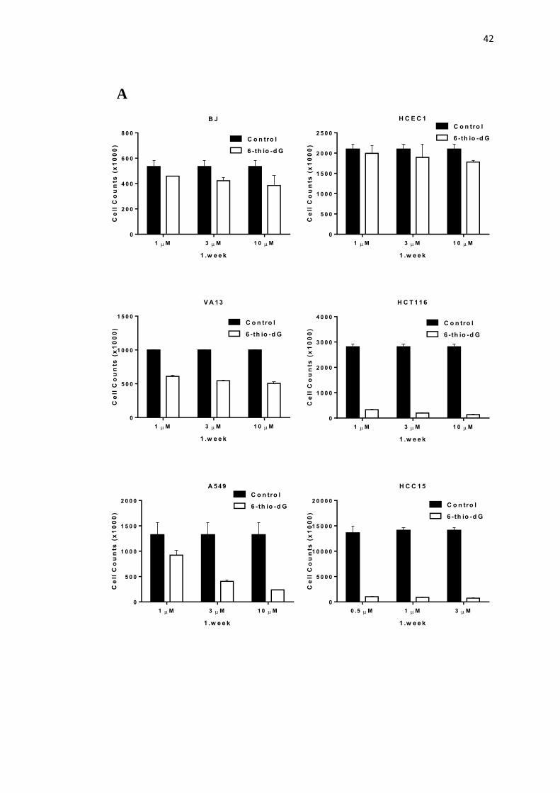
42
1 M 3 M 1 0 M
0
5 0 0
1 0 0 0
1 5 0 0
2 0 0 0
2 5 0 0
H C E C 1
1 .w e e k
Ce
ll C
ou
nts
(x
10
00
)
C o n tro l
6 -th io -d G
0 .5 M 1 M 3 M
0
5 0 0 0
1 0 0 0 0
1 5 0 0 0
2 0 0 0 0
H C C 1 5
1 .w e e k
Ce
ll C
ou
nts
(x
10
00
)
C o n tro l
6 -th io -d G
1 M 3 M 1 0 M
0
2 0 0
4 0 0
6 0 0
8 0 0
B J
1 .w e e k
Ce
ll C
ou
nts
(x
10
00
)
C o n tro l
6 -th io -d G
1 M 3 M 1 0 M
0
5 0 0
1 0 0 0
1 5 0 0
2 0 0 0
A 549
1 .w e e k
Ce
ll C
ou
nts
(x
10
00
)
C o n tro l
6 -th io -d G
A
1 M 3 M 1 0 M
0
1 0 0 0
2 0 0 0
3 0 0 0
4 0 0 0
H C T 1 1 6
1 .w e e k
Ce
ll C
ou
nts
(x
10
00
)
C o n tro l
6 -th io -d G
1 M 3 M 1 0 M
0
5 0 0
1 0 0 0
1 5 0 0
V A 13
1 .w e e k
Ce
ll C
ou
nts
(x
10
00
)
C o n tro l
6 -th io -d G
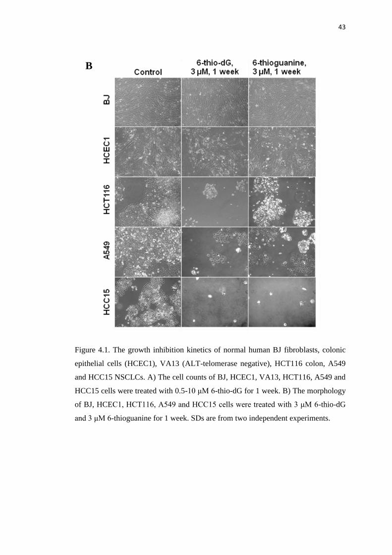
43
Figure 4.1. The growth inhibition kinetics of normal human BJ fibroblasts, colonic
epithelial cells (HCEC1), VA13 (ALT-telomerase negative), HCT116 colon, A549
and HCC15 NSCLCs. A) The cell counts of BJ, HCEC1, VA13, HCT116, A549 and
HCC15 cells were treated with 0.5-10 μM 6-thio-dG for 1 week. B) The morphology
of BJ, HCEC1, HCT116, A549 and HCC15 cells were treated with 3 μM 6-thio-dG
and 3 μM 6-thioguanine for 1 week. SDs are from two independent experiments.
B
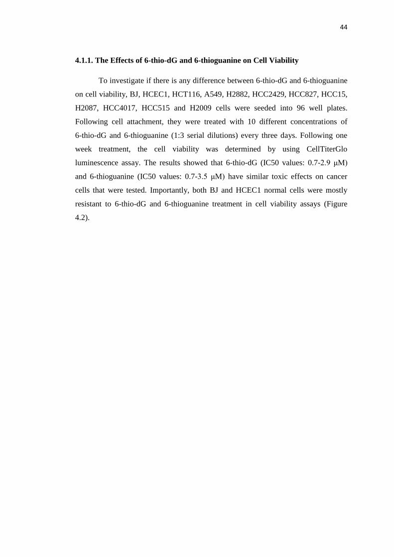
44
4.1.1. The Effects of 6-thio-dG and 6-thioguanine on Cell Viability
To investigate if there is any difference between 6-thio-dG and 6-thioguanine
on cell viability, BJ, HCEC1, HCT116, A549, H2882, HCC2429, HCC827, HCC15,
H2087, HCC4017, HCC515 and H2009 cells were seeded into 96 well plates.
Following cell attachment, they were treated with 10 different concentrations of
6-thio-dG and 6-thioguanine (1:3 serial dilutions) every three days. Following one
week treatment, the cell viability was determined by using CellTiterGlo
luminescence assay. The results showed that 6-thio-dG (IC50 values: 0.7-2.9 μM)
and 6-thioguanine (IC50 values: 0.7-3.5 μM) have similar toxic effects on cancer
cells that were tested. Importantly, both BJ and HCEC1 normal cells were mostly
resistant to 6-thio-dG and 6-thioguanine treatment in cell viability assays (Figure
4.2).
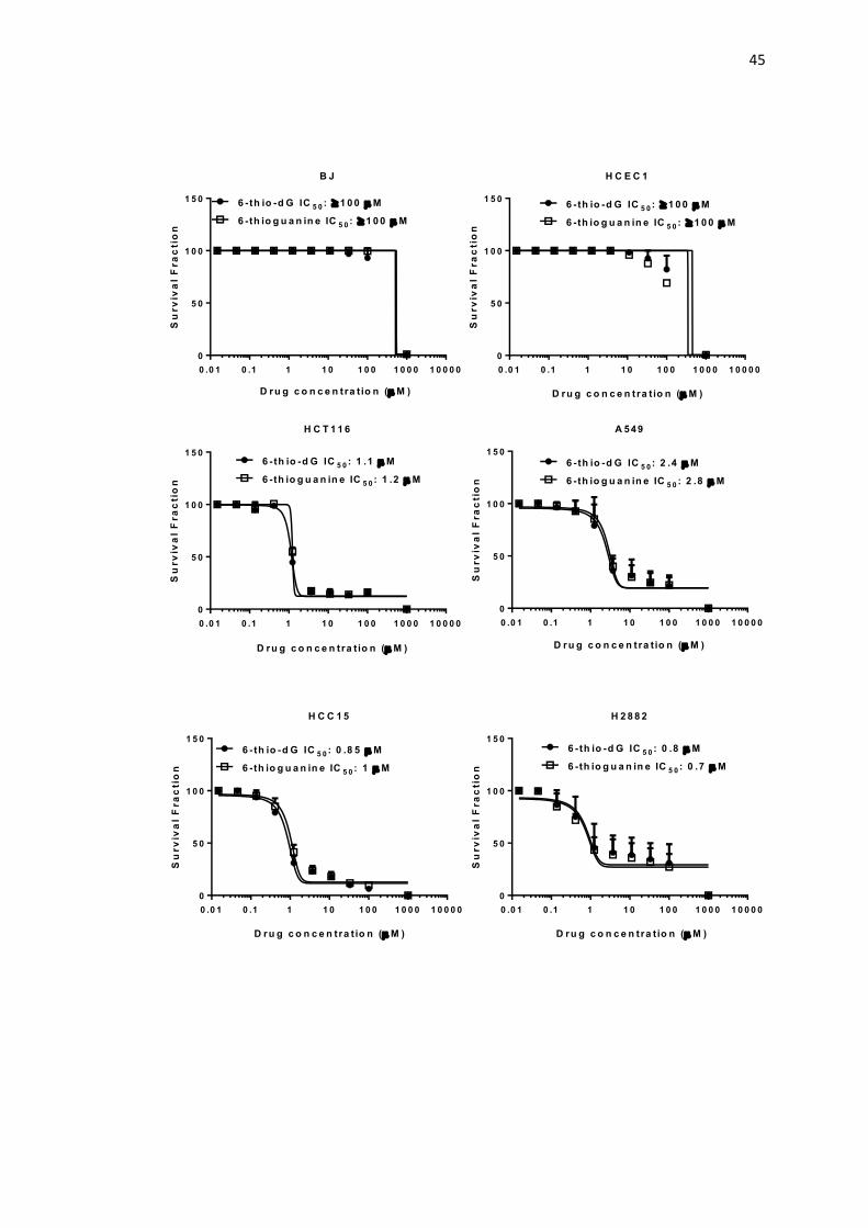
45
0 .0 1 0 .1 1 1 0 1 0 0 1 0 0 0 1 0 0 0 0
0
5 0
1 0 0
1 5 0
H C E C 1
Su
rv
iva
l F
ra
cti
on
6 - th io -d G IC 5 0 : 1 0 0 M
6 -th io g u a n in e IC 5 0 : 1 0 0 M
D ru g c o n c e n tra t io n ( M )
0 .0 1 0 .1 1 1 0 1 0 0 1 0 0 0 1 0 0 0 0
0
5 0
1 0 0
1 5 0
B JS
urv
iva
l F
ra
cti
on
6 - th io -d G IC 5 0 : 1 0 0 M
6 -th io g u a n in e IC 5 0 : 1 0 0 M
D ru g c o n c e n tra t io n ( M )
0 .0 1 0 .1 1 1 0 1 0 0 1 0 0 0 1 0 0 0 0
0
5 0
1 0 0
1 5 0
H C T 1 1 6
Su
rv
iva
l F
ra
cti
on
6 - th io -d G IC 5 0 : 1 .1 M
6 -th io g u a n in e IC 5 0 : 1 .2 M
D ru g c o n c e n tra t io n ( M )
0 .0 1 0 .1 1 1 0 1 0 0 1 0 0 0 1 0 0 0 0
0
5 0
1 0 0
1 5 0
A 549
Su
rv
iva
l F
ra
cti
on
6 - th io -d G IC 5 0 : 2 .4 M
6 -th io g u a n in e IC 5 0 : 2 .8 M
D ru g c o n c e n tra t io n ( M )
0 .0 1 0 .1 1 1 0 1 0 0 1 0 0 0 1 0 0 0 0
0
5 0
1 0 0
1 5 0
H C C 1 5
Su
rv
iva
l F
ra
cti
on
6 - th io -d G IC 5 0 : 0 .8 5 M
6 -th io g u a n in e IC 5 0 : 1 M
D ru g c o n c e n tra t io n ( M )
0 .0 1 0 .1 1 1 0 1 0 0 1 0 0 0 1 0 0 0 0
0
5 0
1 0 0
1 5 0
H 2 8 8 2
Su
rv
iva
l F
ra
cti
on
6 - th io -d G IC 5 0 : 0 .8 M
6 -th io g u a n in e IC 5 0 : 0 .7 M
D ru g c o n c e n tra t io n ( M )
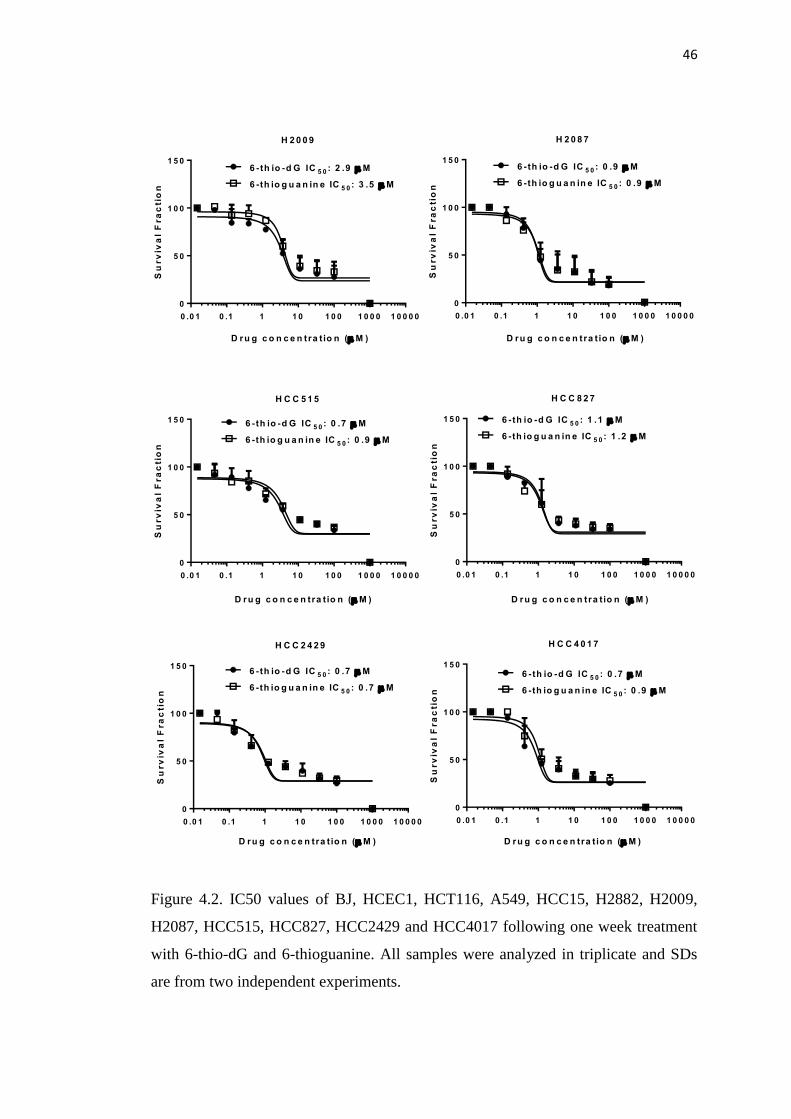
46
Figure 4.2. IC50 values of BJ, HCEC1, HCT116, A549, HCC15, H2882, H2009,
H2087, HCC515, HCC827, HCC2429 and HCC4017 following one week treatment
with 6-thio-dG and 6-thioguanine. All samples were analyzed in triplicate and SDs
are from two independent experiments.
0 .0 1 0 .1 1 1 0 1 0 0 1 0 0 0 1 0 0 0 0
0
5 0
1 0 0
1 5 0
H 2 0 0 9
Su
rv
iva
l F
ra
cti
on
6 - th io -d G IC 5 0 : 2 .9 M
6 -th io g u a n in e IC 5 0 : 3 .5 M
D ru g c o n c e n tra t io n ( M )
0 .0 1 0 .1 1 1 0 1 0 0 1 0 0 0 1 0 0 0 0
0
5 0
1 0 0
1 5 0
H 2 0 8 7
Su
rv
iva
l F
ra
cti
on
6 - th io -d G IC 5 0 : 0 .9 M
6 -th io g u a n in e IC 5 0 : 0 .9 M
D ru g c o n c e n tra t io n ( M )
0 .0 1 0 .1 1 1 0 1 0 0 1 0 0 0 1 0 0 0 0
0
5 0
1 0 0
1 5 0
H C C 2 4 2 9
Su
rv
iva
l F
ra
cti
on
6 - th io -d G IC 5 0 : 0 .7 M
6 -th io g u a n in e IC 5 0 : 0 .7 M
D ru g c o n c e n tra t io n ( M )
0 .0 1 0 .1 1 1 0 1 0 0 1 0 0 0 1 0 0 0 0
0
5 0
1 0 0
1 5 0
H C C 5 1 5
Su
rv
iva
l F
ra
cti
on
6 - th io -d G IC 5 0 : 0 .7 M
6 -th io g u a n in e IC 5 0 : 0 .9 M
D ru g c o n c e n tra t io n ( M )
0 .0 1 0 .1 1 1 0 1 0 0 1 0 0 0 1 0 0 0 0
0
5 0
1 0 0
1 5 0
H C C 4 0 1 7
Su
rv
iva
l F
ra
cti
on
6 - th io -d G IC 5 0 : 0 .7 M
6 -th io g u a n in e IC 5 0 : 0 .9 M
D ru g c o n c e n tra t io n ( M )
0 .0 1 0 .1 1 1 0 1 0 0 1 0 0 0 1 0 0 0 0
0
5 0
1 0 0
1 5 0
H C C 8 2 7
Su
rv
iva
l F
ra
cti
on
6 - th io -d G IC 5 0 : 1 .1 M
6 -th io g u a n in e IC 5 0 : 1 .2 M
D ru g c o n c e n tra t io n ( M )
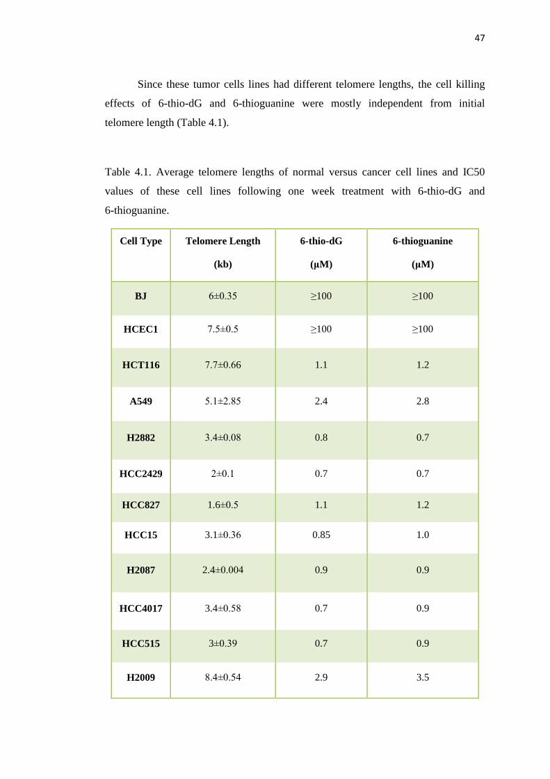
47
Since these tumor cells lines had different telomere lengths, the cell killing
effects of 6-thio-dG and 6-thioguanine were mostly independent from initial
telomere length (Table 4.1).
Table 4.1. Average telomere lengths of normal versus cancer cell lines and IC50
values of these cell lines following one week treatment with 6-thio-dG and
6-thioguanine.
Cell Type Telomere Length
(kb)
6-thio-dG
(μM)
6-thioguanine
(μM)
BJ 6±0.35 ≥100 ≥100
HCEC1 7.5±0.5 ≥100 ≥100
HCT116 7.7±0.66 1.1 1.2
A549 5.1±2.85 2.4 2.8
H2882 3.4±0.08 0.8 0.7
HCC2429 2±0.1 0.7 0.7
HCC827 1.6±0.5 1.1 1.2
HCC15 3.1±0.36 0.85 1.0
H2087 2.4±0.004 0.9 0.9
HCC4017 3.4±0.58 0.7 0.9
HCC515 3±0.39 0.7 0.9
H2009 8.4±0.54 2.9 3.5
48
4.1.2. Telomere Dysfunction Induced Foci (TIF) Induced by 6-thio-dG, but not
6-thioguanine
To understand whether 6-thio-dG or 6-thioguanine causes telomeric damage
on cancer cell, HCT116 cells were seeded on chamber slides and treated with 3 μM
6-thio-dG and 3 μM 6-thioguanine following cell attachment at variable time points
(30 min, 3 hr, 12 hr, 24 hr, 48 hr and 72 hr). After gamma-H2AX and TRF2
co-localization staining was conducted, images were captured by fluorescence
microscopy and analyzed by Imaris Software. Cells that had 4 or more than 4 TIFs
were accepted as damaged telomeres in interphase. The results showed that
6-thio-dG treatment induced TIFs starting at 12 hr post drug treatment. At 72 hr post
6-thio-dG treatment resulted in a 7.8 fold increase in telomeric DNA damage
compared to 6-thioguanine (Figure 4.3A and B). Since telomeres constitute only
~1/6000th of total genomic DNA, any TIFs above a background threshold are likely
to be significant. When general DNA damage was analyzed, it was found that
6-thio-dG causes modest general DNA damage as well as telomeric damage
compared to 6-thioguanine which only caused general DNA damage (Figure 4.3C).
To understand better if these results are dependent on telomerase, we generated
normal BJ cells overexpressing hTERT that have detectable telomerase activity. BJ
and BJhTERT cells were treated with 10 μM 6-thio-dG or 10 μm 6-thioguanine for
48 hr, stained with gamma-H2AX and TRF2 and co-localization analysis was
conducted (Figure 4.4A). We observed that 6-thio-dG and 6-thioguanine did not
induce telomere dysfunction (TIFs) in telomerase silent BJ cells following 48 hr
treatment (Figure 4.4A and B), whereas 6-thio-dG, but not 6-thioguanine, caused
significant increased of telomere dysfunction in hTERT telomerase expressing BJ
cells (BJ hTERT+) compared to controls (Figure 4.4A and C). This increase in
co-localization was 3.3 % (TIF≥5), 4.2 % (TIF≥6) or 5.7% (TIF≥7) of cells treated
with 6-thio-dG. These results indicate that 6-thio-dG is rapidly converted to
6-thio-dGTP, recognized by telomerase and incorporated into telomeres in
telomerase expressing cells, while 6-thioguanine is not.
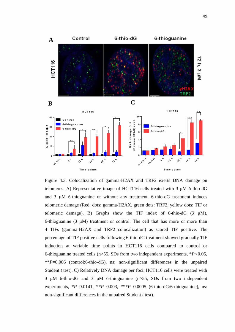
49
Figure 4.3. Colocalization of gamma-H2AX and TRF2 exerts DNA damage on
telomeres. A) Representative image of HCT116 cells treated with 3 μM 6-thio-dG
and 3 μM 6-thioguanine or without any treatment. 6-thio-dG treatment induces
telomeric damage (Red: dots: gamma-H2AX, green dots: TRF2, yellow dots: TIF or
telomeric damage). B) Graphs show the TIF index of 6-thio-dG (3 μM),
6-thioguanine (3 μM) treatment or control. The cell that has more or more than
4 TIFs (gamma-H2AX and TRF2 colocalization) as scored TIF positive. The
percentage of TIF positive cells following 6-thio-dG treatment showed gradually TIF
induction at variable time points in HCT116 cells compared to control or
6-thioguanine treated cells (n>55, SDs from two independent experiments, *P<0.05,
**P=0.006 (control:6-thio-dG), ns: non-significant differences in the unpaired
Student t test). C) Relatively DNA damage per foci. HCT116 cells were treated with
3 μM 6-thio-dG and 3 μM 6-thioguanine (n>55, SDs from two independent
experiments, *P=0.0141, **P=0.003, ***P=0.0005 (6-thio-dG:6-thioguanine), ns:
non-significant differences in the unpaired Student t test).
H C T 1 1 6
T im e p o in ts
% c
ell
s T
IFs
4
30 m
in3 h
12 h
24 h
48 h
72 h
0
1 0
2 0
3 0
4 0
C o n tro l
6 -th io g u a n in e
6 -th io -d G
* *
** **
n s
H C T 1 1 6
T im e p o in ts
D
NA
da
ma
ge
fo
ci
(G
am
ma
H2
AX
) /
ce
ll
Co
ntr
ol
30 m
in3 h
12 h
24 h
48 h
72 h
0
2
4
6
8
1 0
6 -th io g u a n in e
6 -th io -d G
* *
* * *
*
A
B C
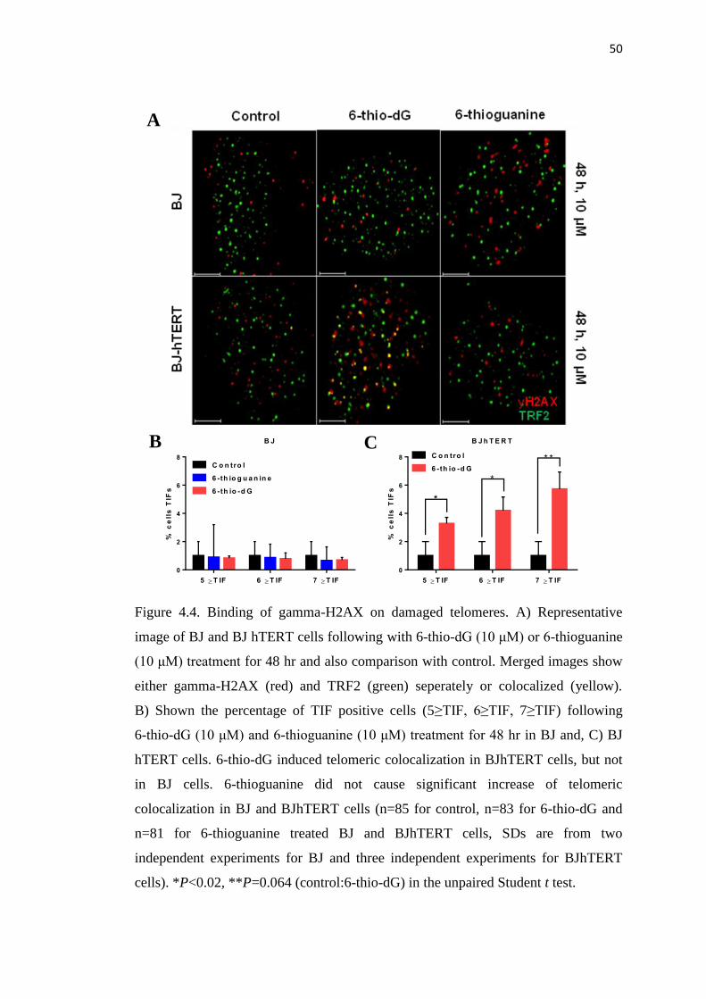
50
Figure 4.4. Binding of gamma-H2AX on damaged telomeres. A) Representative
image of BJ and BJ hTERT cells following with 6-thio-dG (10 μM) or 6-thioguanine
(10 μM) treatment for 48 hr and also comparison with control. Merged images show
either gamma-H2AX (red) and TRF2 (green) seperately or colocalized (yellow).
B) Shown the percentage of TIF positive cells (5≥TIF, 6≥TIF, 7≥TIF) following
6-thio-dG (10 μM) and 6-thioguanine (10 μM) treatment for 48 hr in BJ and, C) BJ
hTERT cells. 6-thio-dG induced telomeric colocalization in BJhTERT cells, but not
in BJ cells. 6-thioguanine did not cause significant increase of telomeric
colocalization in BJ and BJhTERT cells (n=85 for control, n=83 for 6-thio-dG and
n=81 for 6-thioguanine treated BJ and BJhTERT cells, SDs are from two
independent experiments for BJ and three independent experiments for BJhTERT
cells). *P<0.02, **P=0.064 (control:6-thio-dG) in the unpaired Student t test.
B J
% c
ell
s T
IFs
5 T IF 6 T IF 7 T IF
0
2
4
6
8
C o n tro l
6 -th io g u a n in e
6 -th io -d G
B J h T E R T
% c
ell
s T
IFs
5 T IF 6 T IF 7 T IF
0
2
4
6
8 C o n tro l
6 -th io -d G
*
*
* *
A
B C
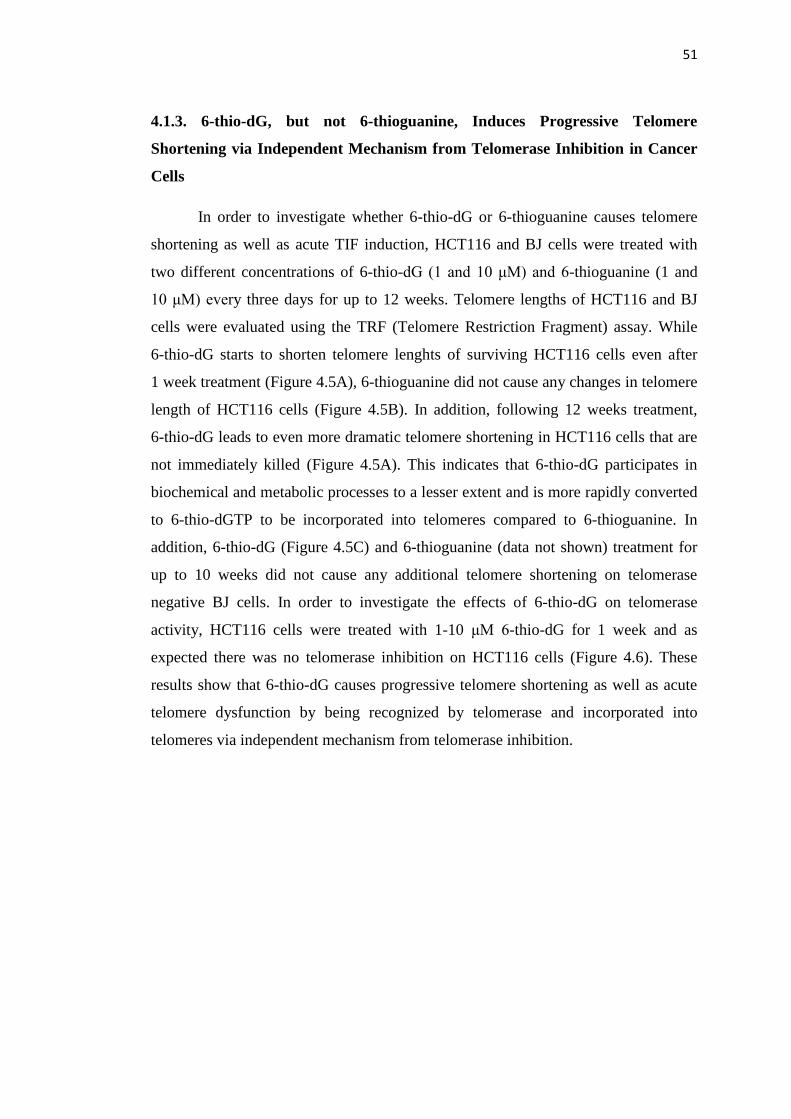
51
4.1.3. 6-thio-dG, but not 6-thioguanine, Induces Progressive Telomere
Shortening via Independent Mechanism from Telomerase Inhibition in Cancer
Cells
In order to investigate whether 6-thio-dG or 6-thioguanine causes telomere
shortening as well as acute TIF induction, HCT116 and BJ cells were treated with
two different concentrations of 6-thio-dG (1 and 10 μM) and 6-thioguanine (1 and
10 μM) every three days for up to 12 weeks. Telomere lengths of HCT116 and BJ
cells were evaluated using the TRF (Telomere Restriction Fragment) assay. While
6-thio-dG starts to shorten telomere lenghts of surviving HCT116 cells even after
1 week treatment (Figure 4.5A), 6-thioguanine did not cause any changes in telomere
length of HCT116 cells (Figure 4.5B). In addition, following 12 weeks treatment,
6-thio-dG leads to even more dramatic telomere shortening in HCT116 cells that are
not immediately killed (Figure 4.5A). This indicates that 6-thio-dG participates in
biochemical and metabolic processes to a lesser extent and is more rapidly converted
to 6-thio-dGTP to be incorporated into telomeres compared to 6-thioguanine. In
addition, 6-thio-dG (Figure 4.5C) and 6-thioguanine (data not shown) treatment for
up to 10 weeks did not cause any additional telomere shortening on telomerase
negative BJ cells. In order to investigate the effects of 6-thio-dG on telomerase
activity, HCT116 cells were treated with 1-10 μM 6-thio-dG for 1 week and as
expected there was no telomerase inhibition on HCT116 cells (Figure 4.6). These
results show that 6-thio-dG causes progressive telomere shortening as well as acute
telomere dysfunction by being recognized by telomerase and incorporated into
telomeres via independent mechanism from telomerase inhibition.
A

52
Figure 4.5. Telomere shortening analysis by TRF (Terminal Restriction Fragment)
assay A) HCT116 cells treated with 6-thio-dG B) HCT116 cells treated with
6-thioguanine C) BJ cells were treated with 6-thio-dG (1-10 μM) every three days for
1-12 weeks. Cells were collected in indicated numbers of weeks for TRF. TRF
lenghts are expressed as kilobase pairs (kb) (Control: untreated).

53
Figure 4.6. Telomerase activity analysis by TRAP (Telomeric Repeat Amplification
Protocol). HCT116 cells were treated with 1-10 μM 6-thio-dG every three days for
1-10 weeks. Cells were collected following one week treatment for TRAP assay.
Serial dilutions of Hela cells and lysis buffer were used as a positive and negative
control, respectively (Control: untreated).
4.1.4. 6-thio-dG does not Lead to Signficant Weight Loss or Hematological
Toxicities in Wild Type Mice
To determine effective and less toxic drug concentration for in vivo
experiments, we used 129S9 wild type mice. We randomly separated the mice into
different groups as controls, 6-thio-dG and 6-thioguanine at different concentrations
were injected intraperitonealy daily. We initiated the experiments with 1.67 and
5 mg/kg drug concentrations because it was reported that higher than 3 mg/kg
6-thioguanine has toxic effects on mice (214). 6-thio-dG, 6-thioguanine and DMSO
controls were injected intraperitoneally every day for 25 days. Three mice treated
with 5 mg/kg 6-thioguanine died following 17 days of treatment (Figure 4.7A).
Three mice treated with 1.67 mg/kg 6-thioguanine did not gain weight and started to
lose weight during the 25 days treatment period (Figure 4.7B). In contrast to
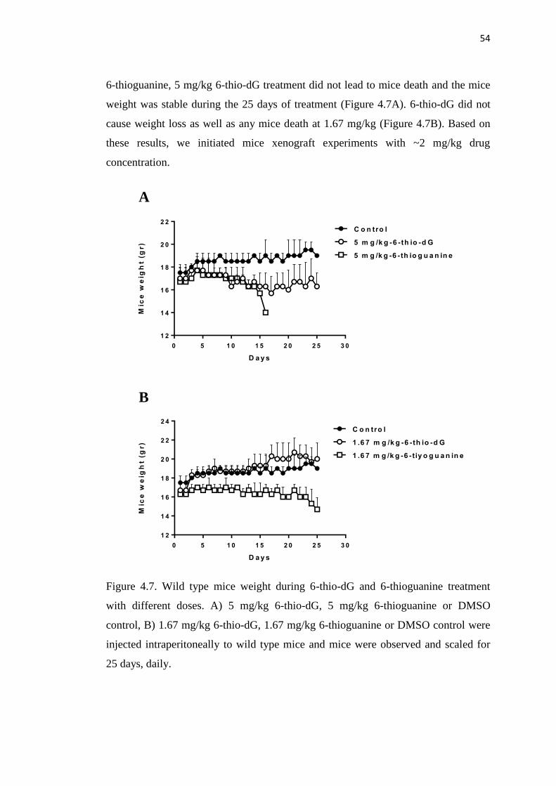
54
6-thioguanine, 5 mg/kg 6-thio-dG treatment did not lead to mice death and the mice
weight was stable during the 25 days of treatment (Figure 4.7A). 6-thio-dG did not
cause weight loss as well as any mice death at 1.67 mg/kg (Figure 4.7B). Based on
these results, we initiated mice xenograft experiments with ~2 mg/kg drug
concentration.
Figure 4.7. Wild type mice weight during 6-thio-dG and 6-thioguanine treatment
with different doses. A) 5 mg/kg 6-thio-dG, 5 mg/kg 6-thioguanine or DMSO
control, B) 1.67 mg/kg 6-thio-dG, 1.67 mg/kg 6-thioguanine or DMSO control were
injected intraperitoneally to wild type mice and mice were observed and scaled for
25 days, daily.
0 5 1 0 1 5 2 0 2 5 3 0
1 2
1 4
1 6
1 8
2 0
2 2
D a y s
Mic
e w
eig
ht
(gr)
C o n tro l
5 m g /k g -6 -th io -d G
5 m g /k g -6 -th io g u a n in e
0 5 1 0 1 5 2 0 2 5 3 0
1 2
1 4
1 6
1 8
2 0
2 2
2 4
D a y s
Mic
e w
eig
ht
(gr)
C o n tro l
1 .6 7 m g /k g -6 -th io -d G
1 .6 7 m g /k g -6 -tiy o g u a n in e
A
B
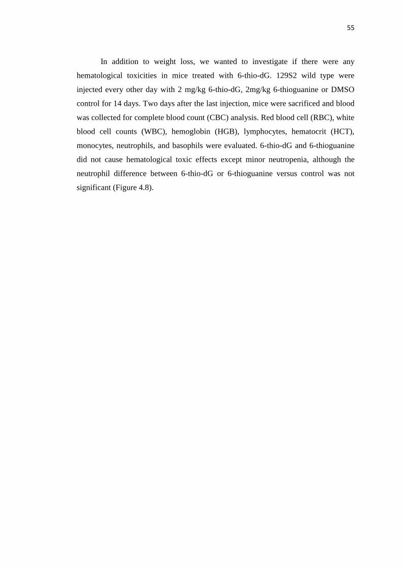
55
In addition to weight loss, we wanted to investigate if there were any
hematological toxicities in mice treated with 6-thio-dG. 129S2 wild type were
injected every other day with 2 mg/kg 6-thio-dG, 2mg/kg 6-thioguanine or DMSO
control for 14 days. Two days after the last injection, mice were sacrificed and blood
was collected for complete blood count (CBC) analysis. Red blood cell (RBC), white
blood cell counts (WBC), hemoglobin (HGB), lymphocytes, hematocrit (HCT),
monocytes, neutrophils, and basophils were evaluated. 6-thio-dG and 6-thioguanine
did not cause hematological toxic effects except minor neutropenia, although the
neutrophil difference between 6-thio-dG or 6-thioguanine versus control was not
significant (Figure 4.8).
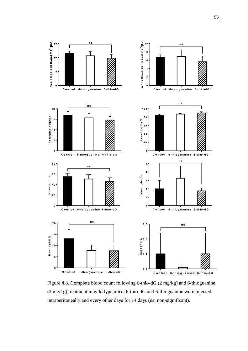
56
Figure 4.8. Complete blood count following 6-thio-dG (2 mg/kg) and 6-thioguanine
(2 mg/kg) treatment in wild type mice. 6-thio-dG and 6-thioguanine were injected
intraperitoneally and every other days for 14 days (ns: non-significant).
Ne
utr
op
hil
%
C o n tro l 6 -th io g u a n in e 6 -th io -d G
0
5
1 0
1 5
2 0n s
C o n tro l 6 -th io g u a n in e 6 -th io -d G
0 .0
0 .1
0 .2
0 .3
Ba
so
ph
il %
n s
Ly
mp
ho
cy
tes
%
C o n tro l 6 -th io g u a n in e 6 -th io -d G
0
2 0
4 0
6 0
8 0
1 0 0
n s
Mo
no
cy
tes
%
C o n tro l 6 -th io g u a n in e 6 -th io -d G
0
1
2
3
4
5
n s
Wh
ite
Blo
od
Ce
ll C
ou
nt
(10
3/
L)
C o n tro l 6 -th io g u a n in e 6 -th io -d G
0
2
4
6
8
1 0 n s
Re
d B
loo
d C
ell
Co
un
t (1
06/
L)
C o n tro l 6 -th io g u a n in e 6 -th io -d G
0
5
1 0
1 5 n s
He
mo
glo
bin
(g
/dL
)
C o n tro l 6 -th io g u a n in e 6 -th io -d G
0
5
1 0
1 5
2 0
n s
C o n tro l 6 -th io g u a n in e 6 -th io -d G
0
2 0
4 0
6 0
8 0
He
ma
toc
rit
%
n s
Re
d B
loo
d C
ell
Co
un
t (1
06/
L)
C o n tro l 6 -th io g u a n in e 6 -th io -d G
0
5
1 0
1 5 n s
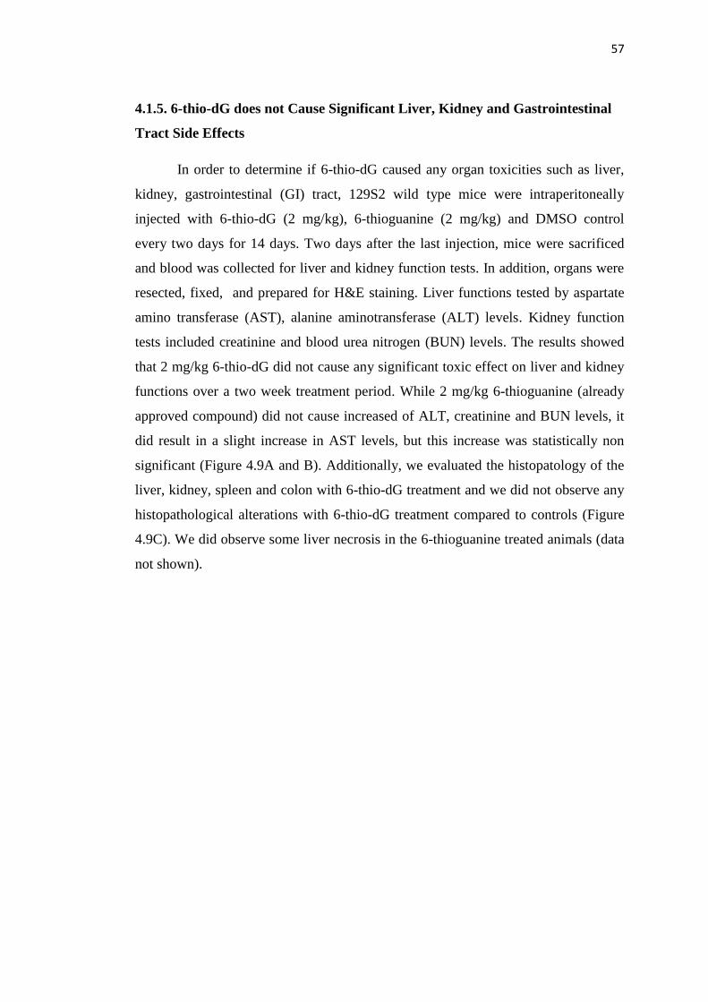
57
4.1.5. 6-thio-dG does not Cause Significant Liver, Kidney and Gastrointestinal
Tract Side Effects
In order to determine if 6-thio-dG caused any organ toxicities such as liver,
kidney, gastrointestinal (GI) tract, 129S2 wild type mice were intraperitoneally
injected with 6-thio-dG (2 mg/kg), 6-thioguanine (2 mg/kg) and DMSO control
every two days for 14 days. Two days after the last injection, mice were sacrificed
and blood was collected for liver and kidney function tests. In addition, organs were
resected, fixed, and prepared for H&E staining. Liver functions tested by aspartate
amino transferase (AST), alanine aminotransferase (ALT) levels. Kidney function
tests included creatinine and blood urea nitrogen (BUN) levels. The results showed
that 2 mg/kg 6-thio-dG did not cause any significant toxic effect on liver and kidney
functions over a two week treatment period. While 2 mg/kg 6-thioguanine (already
approved compound) did not cause increased of ALT, creatinine and BUN levels, it
did result in a slight increase in AST levels, but this increase was statistically non
significant (Figure 4.9A and B). Additionally, we evaluated the histopatology of the
liver, kidney, spleen and colon with 6-thio-dG treatment and we did not observe any
histopathological alterations with 6-thio-dG treatment compared to controls (Figure
4.9C). We did observe some liver necrosis in the 6-thioguanine treated animals (data
not shown).
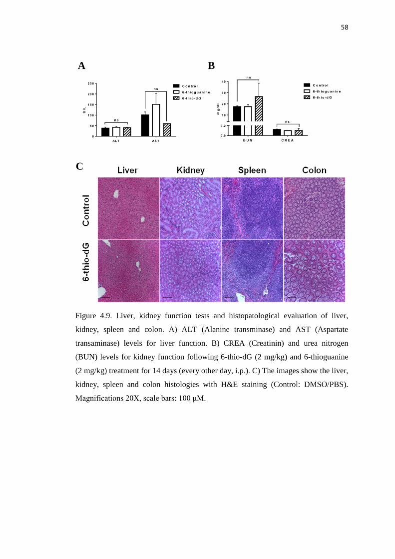
58
Figure 4.9. Liver, kidney function tests and histopatological evaluation of liver,
kidney, spleen and colon. A) ALT (Alanine transminase) and AST (Aspartate
transaminase) levels for liver function. B) CREA (Creatinin) and urea nitrogen
(BUN) levels for kidney function following 6-thio-dG (2 mg/kg) and 6-thioguanine
(2 mg/kg) treatment for 14 days (every other day, i.p.). C) The images show the liver,
kidney, spleen and colon histologies with H&E staining (Control: DMSO/PBS).
Magnifications 20X, scale bars: 100 μM.
A B
C
U/L
AL T AS T
0
5 0
1 0 0
1 5 0
2 0 0
2 5 0C o n tro l
6 -th io g u a n in e
6 -th io -d G
n s
n s
mg
/dL
B U N C R E A
0 .0
0 .2
1 0
2 0
3 0
4 0
C o n tro l
6 -th io g u a n in e
6 -th io -d G
n s
n s

59
4.1.6. 6-thio-dG Reduces Tumor Growth Rate In Vivo
In order to investigate the anticancer effects of 6-thio-dG on tumor growth
compared to 6-thioguanine and control, we used A549 NSCLC derived xenograft
murine models. 6-thiodG and 6-thioguanine injections were initiated when the
xenografted tumor size reached 50-100 mm3. 6-thio-dG and 6-thioguanine at a dose
of 2 mg/kg were intraperitoneally injected into athymic nude mice every other day
for 17 days. At the end of treatment, 6-thio-dG caused a decreased of tumor growth
rate compared to 6-thioguanine and control (Figure 4.10A). In addition, Ki67
staining, which is a proliferation marker, showed that 6-thio-dG treatment reduced
Ki67 levels in A549-derived tumor xenografts compared to 6-thioguanine treatment
and controls (Figure 4.10B). These results document in vivo differences between
6-thio-dG and 6-thioguanine on tumor growth rates and suggests 6-thio-dG is more
effective compared to 6-thioguanine. To test whether intratumoral injection caused
any additional therapeutic improvement on tumor growth compared to intraperitoneal
injection, 6-thio-dG and 6-thioguanine were intratumorally injected every day at a
dose of 2.5 mg/kg into the nude mice after xenograft establishment. 6-thio-dG
treatment with intratumoral injection showed even more dramatic reduction on
growth rate compare to 6-thioguanine and control (Figure 4.10C). In addition,
6-thio-dG treatment exerted highly vascularized tumors with infiltrating
inflammatory cells, which were not observed with 6-thioguanine injections. Thus, the
residual small tumors following 6-thio-dG treatment underrepresent the true
effectiveness in vivo (Figure 4.10D). It is certain that additional, and longer
treatments will be required to determine if tumors can be completely eliminated.
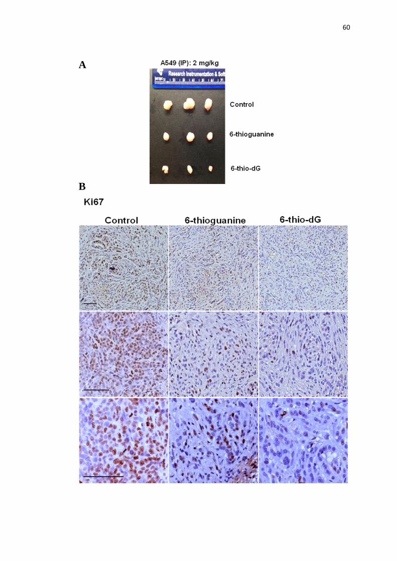
60
A
B
61
Figure 4.10. Intraperitoneally and intratumoral injection with 6-thio-dG and
6-thioguanine. A) Representative images form isolated tumors. 2 mg/kg 6-thio-dG
and 2 mg/kg 6-thioguanine intraperitoneally (IP) injected to nude mice every other
day for 17 days. B) Representative images show Ki67 proliferation level in 6-thio-dG
(2 mg/kg) treated A549 derived tumor compared to 6-thioguanine (2 mg/kg) and
control. Magnifications 20X, 40X, 60X, scale bars: 100 μM. C) The tumor volumes
(mm3) are shown following 6-thio-dG treatment (2.5 mg/kg) compared to
6-thoguanine (2.5 mg/kg) and control. Treatments were done as intratumoral
injections, every day for 17 days. Tumor volumes were calculated using the
following formula: (length x width2) mm
2 x 0.5. D) The images show the histology
of A549-derived tumors with hematoxylin and eosin staining following 6-thio-dG,
6-thioguanine and DMSO control treatment with intratumoral injection at a dose of
2.5 mg/kg, every day. Magnification 10X, scale bar: 100 μM.
0 5 1 0 1 5 2 0
0
5 0
1 0 0
1 5 0
2 0 0
A 549
D a y s
Tu
mo
r v
olu
me
(m
m3
)
C o n tro l
6 -th io g u a n in e
6 -th io -d G
C
D
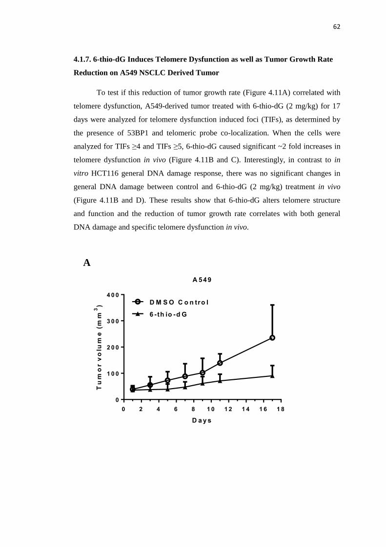
62
4.1.7. 6-thio-dG Induces Telomere Dysfunction as well as Tumor Growth Rate
Reduction on A549 NSCLC Derived Tumor
To test if this reduction of tumor growth rate (Figure 4.11A) correlated with
telomere dysfunction, A549-derived tumor treated with 6-thio-dG (2 mg/kg) for 17
days were analyzed for telomere dysfunction induced foci (TIFs), as determined by
the presence of 53BP1 and telomeric probe co-localization. When the cells were
analyzed for TIFs ≥4 and TIFs ≥5, 6-thio-dG caused significant ~2 fold increases in
telomere dysfunction in vivo (Figure 4.11B and C). Interestingly, in contrast to in
vitro HCT116 general DNA damage response, there was no significant changes in
general DNA damage between control and 6-thio-dG (2 mg/kg) treatment in vivo
(Figure 4.11B and D). These results show that 6-thio-dG alters telomere structure
and function and the reduction of tumor growth rate correlates with both general
DNA damage and specific telomere dysfunction in vivo.
A 549
D a y s
Tu
mo
r v
olu
me
(m
m3
)
0 2 4 6 8 1 0 1 2 1 4 1 6 1 8
0
1 0 0
2 0 0
3 0 0
4 0 0
D M S O C o n tro l
6 -th io -d G
A

63
B
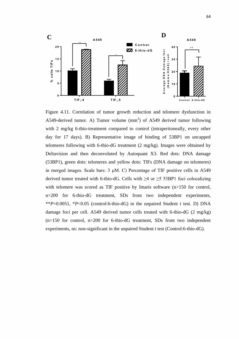
64
Figure 4.11. Correlation of tumor growth reduction and telomere dysfunction in
A549-derived tumor. A) Tumor volume (mm3) of A549 derived tumor following
with 2 mg/kg 6-thio-treatment compared to control (intraperitoneally, every other
day for 17 days). B) Representative image of binding of 53BP1 on uncapped
telomeres following with 6-thio-dG treatment (2 mg/kg). Images were obtained by
Deltavision and then deconvoluted by Autoquant X3. Red dots: DNA damage
(53BP1), green dots: telomeres and yellow dots: TIFs (DNA damage on telomeres)
in merged images. Scale bars: 3 μM. C) Percentage of TIF positive cells in A549
derived tumor treated with 6-thio-dG. Cells with ≥4 or ≥5 53BP1 foci colocalizing
with telomere was scored as TIF positive by Imaris software (n>150 for control,
n>200 for 6-thio-dG treatment, SDs from two independent experiments,
**P=0.0051, *P˂0.05 (control:6-thio-dG) in the unpaired Student t test. D) DNA
damage foci per cell. A549 derived tumor cells treated with 6-thio-dG (2 mg/kg)
(n>150 for control, n>200 for 6-thio-dG treatment, SDs from two independent
experiments, ns: non-significant in the unpaired Student t test (Control:6-thio-dG).
T IF 4 T IF 5
0
5
1 0
1 5
2 0
A 549
% c
ell
s T
IFs
C o n tro l
6 -th io -d G
* *
*
C o n tr o l 6 - th io -d G
0
1 0
2 0
3 0
4 0
A 549
Av
er
ag
e D
NA
Da
ma
ge
fo
ci
(G
am
ma
H2
AX
) /
ce
ll
n s
C D

65
4.2. Possible Resistance Mechanisms of 6-thio-dG Treatment
While it is promising that while 6-thio-dG kills the vast majority of cancer
cell lines and it does not have major toxic effect on normal cells (BJ and HCEC1),
the question of drug resistance must be addressed. In this study when HCT116 cells
were treated with 6-thio-dG for long-term treatment, they eventually developed
resistance to 6-thio-dG treatment (Figure 4.12A and B). This indicates that following
the death of vast majority of HCT116 cells, there are still some cells that survive,
gain the benefits of drug resistance and proliferate.
Figure 4.12. The development of 6-thio-dG resistance in HCT116 cells. A) The
morhology of HCT116 cells treated with 10 μM 6-thio-dG for 1 and 10 weeks.
B) The cell counts of HCT116 cells with 1, 3 and 10 μM 6-thio-dG treated every
three days for 1-10 weeks. While HCT116 cells are highly sensitive to 6-thio-dG
treatment for 1-2 weeks, they start to develop resistance to 6-thio-dG with over 3
weeks treatments.
Ce
ll C
ou
nts
(x
10
00
)
1
M
3
M
10
M
1
M
3
M
10
M
1
M
3
M
10
M
1
M
3
M
10
M
1
M
3
M
10
M
1
M
3
M
10
M
1
M
3
M
10
M
1
M
3
M
10
M
1
M
3
M
10
M
1
M
3
M
10
M
0
1 0 0 0
2 0 0 0
3 0 0 0
4 0 0 0C o n tro l
6 -th io -d G
H C T 1 1 6
1 .w e e k 2 .w e e k 3 .w e e k 4 .w e e k 5 .w e e k 6 .w e e k 1 0 .w e e k9 .w e e k8 .w e e k7 .w e e k
A
B

66
We next sought to determine if 6-thio-dG resistance in HCT116 cells can be
explained by cancer stem cell properties and/or increased levels of ATP-Binding
Cassette (ABC) transporter proteins, that are known to be important for multidrug
resistance functions in various human cancers. ALDH1A1 (aldehyde dehydrogenase
1A1) and ABCG2 are the most commonly studied markers for cancer stem cell and
ABC transporter functions, respectively. Therefore, we compared the ALDH1A1 and
ABCG2 mRNA levels of the populations that were treated with 10 μM 6-thio-dG for
1 and 8 weeks by using qPCR. We found that while the ALDH1A1 and ABCG2
mRNA levels of HCT116 cells treated with 6-thio-dG for 1 week are decreased
compared to control, 6-thio-dG treated HCT116 cells for 8 weeks had similar or
more ALDH1A1 (Figure 4.13A) and ABCG2 (Figure 4.13B) mRNA expression
levels compared to controls and 1 week 6-thio-dG treatment. These results indicate
that the surviving cells in the population following 6-thio-dG treatment shows
increases cancer stem cell biomarkers and also 6-thio-dG drug levels are insufficient
to be toxic due to development of increased drug efflux capability of cells.
Figure 4.13. The mRNA levels of ALDH1A1 and ABCG2 in HCT116 cells
following 6-thio-dG treatment. A, B) HCT116 cells treated with 10 μM 6-thio-dG
every three days for 1 and 8 weeks. A) ALDH1A1 and B) ABCG2 mRNA levels
were significantly decreased with 1 week and increased with 8 weeks 6-thio-dG
compared to control (*P<0.05, ****P<0.0001 in the unpaired student t test).
Re
lati
ve
ly f
old
ch
an
ge
of
AL
DH
1A
1 m
RN
A l
ev
el
0 .0
0 .5
1 .0
1 .5
H C T 1 1 6
1 . w e e k
6 -th io -d G
8 . w e e k
6 -th io -d G
C o n tro l
* * * *
*
Re
lati
ve
ly f
old
ch
an
ge
of
AB
CG
2 m
RN
A l
ev
el
0
1
2
3
4
H C T 1 1 6
1 . w e e k
6 -th io -d G
8 . w e e k
6 -th io -d G
C o n tro l
* * * *
* * * *
B A
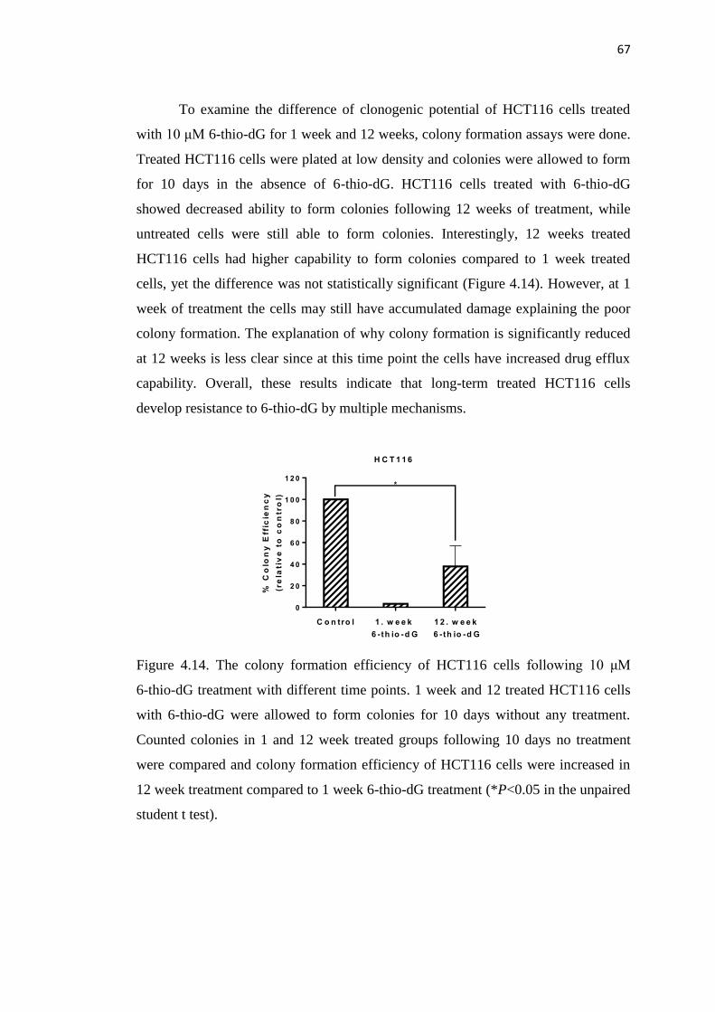
67
To examine the difference of clonogenic potential of HCT116 cells treated
with 10 μM 6-thio-dG for 1 week and 12 weeks, colony formation assays were done.
Treated HCT116 cells were plated at low density and colonies were allowed to form
for 10 days in the absence of 6-thio-dG. HCT116 cells treated with 6-thio-dG
showed decreased ability to form colonies following 12 weeks of treatment, while
untreated cells were still able to form colonies. Interestingly, 12 weeks treated
HCT116 cells had higher capability to form colonies compared to 1 week treated
cells, yet the difference was not statistically significant (Figure 4.14). However, at 1
week of treatment the cells may still have accumulated damage explaining the poor
colony formation. The explanation of why colony formation is significantly reduced
at 12 weeks is less clear since at this time point the cells have increased drug efflux
capability. Overall, these results indicate that long-term treated HCT116 cells
develop resistance to 6-thio-dG by multiple mechanisms.
Figure 4.14. The colony formation efficiency of HCT116 cells following 10 μM
6-thio-dG treatment with different time points. 1 week and 12 treated HCT116 cells
with 6-thio-dG were allowed to form colonies for 10 days without any treatment.
Counted colonies in 1 and 12 week treated groups following 10 days no treatment
were compared and colony formation efficiency of HCT116 cells were increased in
12 week treatment compared to 1 week 6-thio-dG treatment (*P<0.05 in the unpaired
student t test).
% C
olo
ny
Eff
icie
nc
y
(re
lati
ve
to
co
ntr
ol)
0
2 0
4 0
6 0
8 0
1 0 0
1 2 0
H C T 1 1 6
1 . w e e k
6 -th io -d G
1 2 . w e e k
6 -th io -d G
C o n tro l
*
68
4.2.1 HCT116 Cells are not Sensitive to 6-thio-dG with GRN163L Pretreatment
In order to test the effects of combined therapy with GRN163L and
6-thio-dG, we initiated experiments with two different designs. First, HCT116 cells
were treated with 3 μM GRN163L and 1 μM 6-thio-dG at the same time every three
days for 1 week. We observed that HCT116 cells were partially sensitive to
combined therapy compared to control and only GRN163L treatment. It is important
to note that HCT116 cells are more sensitive to 6-thio-dG treatment compared to
combined therapy of GRN163L and 6-thio-dG that were added at the same time. In
the second series of experiments, HCT116 cells were pretreated with 3 μM
GRN163L for 3 days and then GRN163L (3 μM) and 6-thio-dG (1 μM) were added
together to the HCT116 cells. Pretreatment with GRN163L inhibited the 6-thio-dG
toxic effect on HCT116 cells following 1 week treatment. The partial response in the
first experimental design can be explained by the earlier effect of 6-thio-dG on
HCT116 cells. Thus, 6-thio-dG induced initial telomere dysfunction by 12 hr
treatment and initiated cell death in some of the HCT116 cells in the population by
72 hr until telomerase activity is inhibited by GRN163L. After GRN163L inhibits
telomerase activity, the toxic effect of 6-thio-dG on HCT116 was partially lost. This
would be totally expected since the mode of action of 6-thio-dG for induction of
telomere dysfunction depends on active telomerase activity. In second series of
experiments since telomerase activity is inhibited with pretreatment of GRN163L,
6-thio-dG can not be incorporated into telomeres without active telomerase. It was
not expected to see any toxicity with only GRN163L following 1 week treatment
because inducing senescence or apoptosis with GRN163L on cancer cells would
require many weeks of progressive telomere shortening, which can be achieved with
long-term treatment (Figure 4.15A and B). Overall, these experiments provide a
further proof of principal of the mode of action of 6-thio-dG.
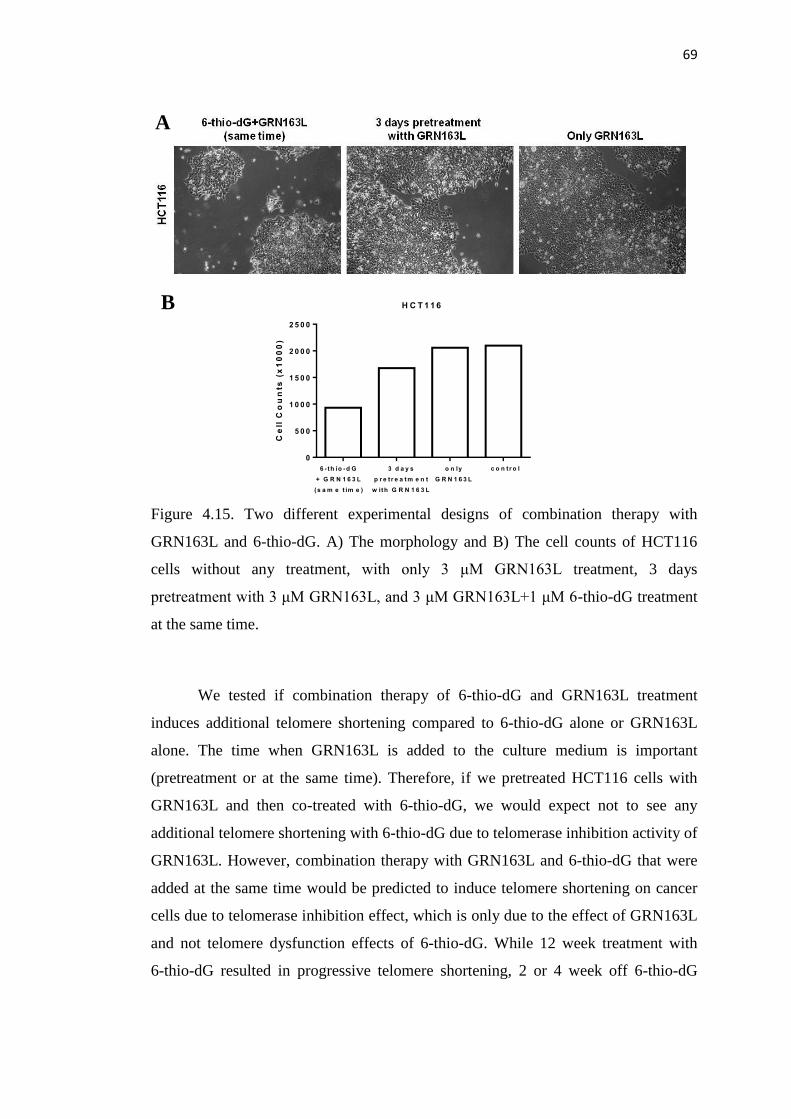
69
Figure 4.15. Two different experimental designs of combination therapy with
GRN163L and 6-thio-dG. A) The morphology and B) The cell counts of HCT116
cells without any treatment, with only 3 μM GRN163L treatment, 3 days
pretreatment with 3 μM GRN163L, and 3 μM GRN163L+1 μM 6-thio-dG treatment
at the same time.
We tested if combination therapy of 6-thio-dG and GRN163L treatment
induces additional telomere shortening compared to 6-thio-dG alone or GRN163L
alone. The time when GRN163L is added to the culture medium is important
(pretreatment or at the same time). Therefore, if we pretreated HCT116 cells with
GRN163L and then co-treated with 6-thio-dG, we would expect not to see any
additional telomere shortening with 6-thio-dG due to telomerase inhibition activity of
GRN163L. However, combination therapy with GRN163L and 6-thio-dG that were
added at the same time would be predicted to induce telomere shortening on cancer
cells due to telomerase inhibition effect, which is only due to the effect of GRN163L
and not telomere dysfunction effects of 6-thio-dG. While 12 week treatment with
6-thio-dG resulted in progressive telomere shortening, 2 or 4 week off 6-thio-dG
Ce
ll C
ou
nts
(x
10
00
)
0
5 0 0
1 0 0 0
1 5 0 0
2 0 0 0
2 5 0 0
H C T 1 1 6
6 -th io -d G
+ G R N 1 6 3 L
(s a m e t im e )
3 d a y s
p r e t r e a t m e n t
w ith G R N 1 6 3 L
o n ly
G R N 1 6 3 L
c o n t r o l
A
B
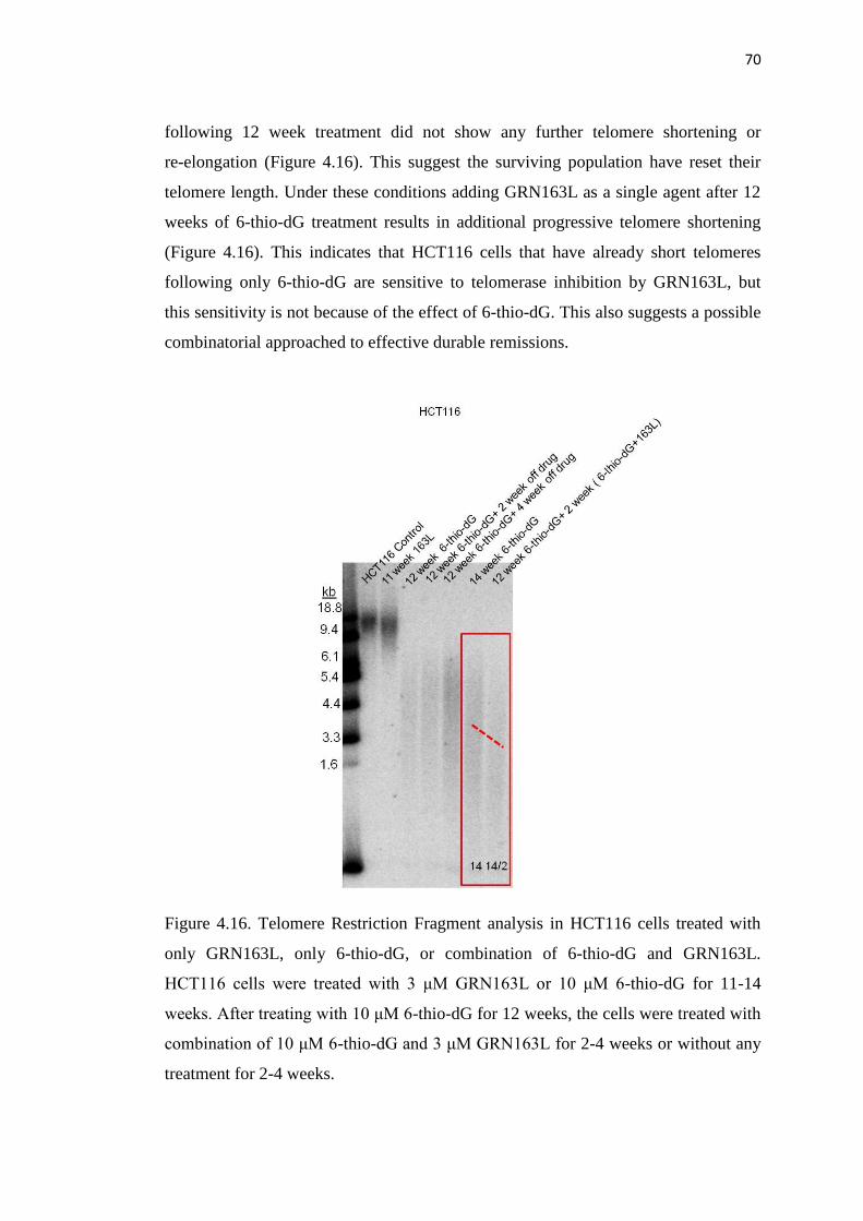
70
following 12 week treatment did not show any further telomere shortening or
re-elongation (Figure 4.16). This suggest the surviving population have reset their
telomere length. Under these conditions adding GRN163L as a single agent after 12
weeks of 6-thio-dG treatment results in additional progressive telomere shortening
(Figure 4.16). This indicates that HCT116 cells that have already short telomeres
following only 6-thio-dG are sensitive to telomerase inhibition by GRN163L, but
this sensitivity is not because of the effect of 6-thio-dG. This also suggests a possible
combinatorial approached to effective durable remissions.
Figure 4.16. Telomere Restriction Fragment analysis in HCT116 cells treated with
only GRN163L, only 6-thio-dG, or combination of 6-thio-dG and GRN163L.
HCT116 cells were treated with 3 μM GRN163L or 10 μM 6-thio-dG for 11-14
weeks. After treating with 10 μM 6-thio-dG for 12 weeks, the cells were treated with
combination of 10 μM 6-thio-dG and 3 μM GRN163L for 2-4 weeks or without any
treatment for 2-4 weeks.
71
4.2.2. H1819, H1993 and H1693 NSCLCs are resistant to 6-thio-dG
6-thio-dG was toxic for 10 NSCLCs (A549, H2882, HCC2429, HCC827,
HCC15, H2087, HCC4017, HCC515, H2009, H2073), whereas H1819, H1993 and
H1693 NSCLCs were not sensitive to 6-thio-dG. H1819/H1693 and H2073/H1993
are the isogenous cell lines. Each cell line of one pair are from same patient and have
different telomere lengths (Table 4.2). H1693 cell line was established prior to
chemotherapy from a patient with IIIB adenocarcinoma lung cancer. Residual
primary lung tumor was treated with cisplatin, etoposide, radiotherapy and then
H1819 was established. Likewise, another patient who had stage IIIA
adenocarcinoma lung cancer was biopsied at a regional lymph node. H1993 was
established prior to chemotherapy. H2073 was established from residual primary
lung tumor after cisplatin and etoposide treatment. H2073 is the chemo-resistant
primary lung tumor with shorter telomeres compared to H1819, H1993 and H1693
cells with long telomeres. Since the methylation patterns and gene expression
profiles of these NSCLCs are known, we compared and contrasted the aberrant
methylation and gene expression profiles of sensitive and resistance NCSLCs. Figure
4.17 shows complete data for 13 NSCLCs sorted using a clustering algorithm. Since
cell lines have heterogenous population, there is hypomethylated or hypermethylated
forms of all genes. When we compared the sensitive and resistant cell lines, some
genes were hypermethlated in both resistant cell lines, but not sensitive cell lines.
And interestingly, H2073/H1993 matched pair of NSCLCs that is both sensitive and
resistant to 6-thio-dG treatment had different methylation pattern (Figure 4.17). This
cluster demonstrates that methylation profiles of some genes can be important for the
cell lines that are resistant to 6-thio-dG.
In order to investigate the effect of 6-thio-dG following
5-aza-2’-deoxycytidine pretreatment (500 nM), which is an inhibitor of DNA
methylation, we seeded resistant NSCLCs in 96-well plates, and treated the cells
with different concentrations of 6-thio-dG every three days. Following one week of
treatment (using different doses with serial dilutions, 1000-0.03 μM), the cell
viability was determined by the celltiterglo luminescent cell viability assay. We
observed that 6-thio-dG inhibited cell viability in a dose dependent manner after
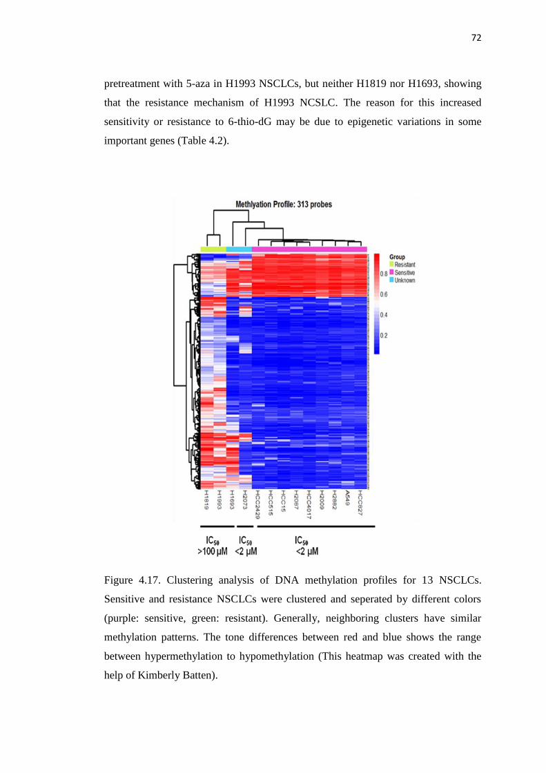
72
pretreatment with 5-aza in H1993 NSCLCs, but neither H1819 nor H1693, showing
that the resistance mechanism of H1993 NCSLC. The reason for this increased
sensitivity or resistance to 6-thio-dG may be due to epigenetic variations in some
important genes (Table 4.2).
Figure 4.17. Clustering analysis of DNA methylation profiles for 13 NSCLCs.
Sensitive and resistance NSCLCs were clustered and seperated by different colors
(purple: sensitive, green: resistant). Generally, neighboring clusters have similar
methylation patterns. The tone differences between red and blue shows the range
between hypermethylation to hypomethylation (This heatmap was created with the
help of Kimberly Batten).
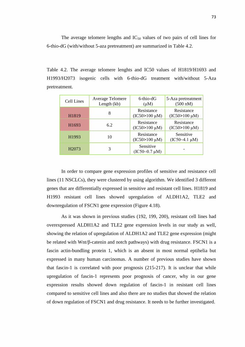
73
The average telomere lengths and IC50 values of two pairs of cell lines for
6-thio-dG (with/without 5-aza pretreatment) are summarized in Table 4.2.
Table 4.2. The average telomere lenghts and IC50 values of H1819/H1693 and
H1993/H2073 isogenic cells with 6-thio-dG treatment with/without 5-Aza
pretreatment.
Cell Lines Average Telomere
Length (kb)
6-thio-dG
(μM)
5-Aza pretreatment
(500 nM)
H1819 8 Resistance
(IC50>100 μM) Resistance
(IC50>100 μM)
H1693 6.2 Resistance
(IC50>100 μM)
Resistance
(IC50>100 μM)
H1993 10 Resistance
(IC50>100 μM) Sensitive
(IC50~4.1 μM)
H2073 3 Sensitive
(IC50~0.7 μM) -
In order to compare gene expression profiles of sensitive and resistance cell
lines (11 NSCLCs), they were clustered by using algorithm. We identified 3 different
genes that are differentially expressed in sensitive and resistant cell lines. H1819 and
H1993 resistant cell lines showed upregulation of ALDH1A2, TLE2 and
downregulation of FSCN1 gene expression (Figure 4.18).
As it was shown in previous studies (192, 199, 200), resistant cell lines had
overexpressed ALDH1A2 and TLE2 gene expression levels in our study as well,
showing the relation of upregulation of ALDH1A2 and TLE2 gene expression (might
be related with Wnt/β-catenin and notch pathways) with drug resistance. FSCN1 is a
fascin actin-bundling protein 1, which is an absent in most normal eptihelia but
expressed in many human carcinomas. A number of previous studies have shown
that fascin-1 is correlated with poor prognosis (215-217). It is unclear that while
upregulation of fascin-1 represents poor prognosis of cancer, why in our gene
expression results showed down regulation of fascin-1 in resistant cell lines
compared to sensitive cell lines and also there are no studies that showed the relation
of down regulation of FSCN1 and drug resistance. It needs to be further investigated.
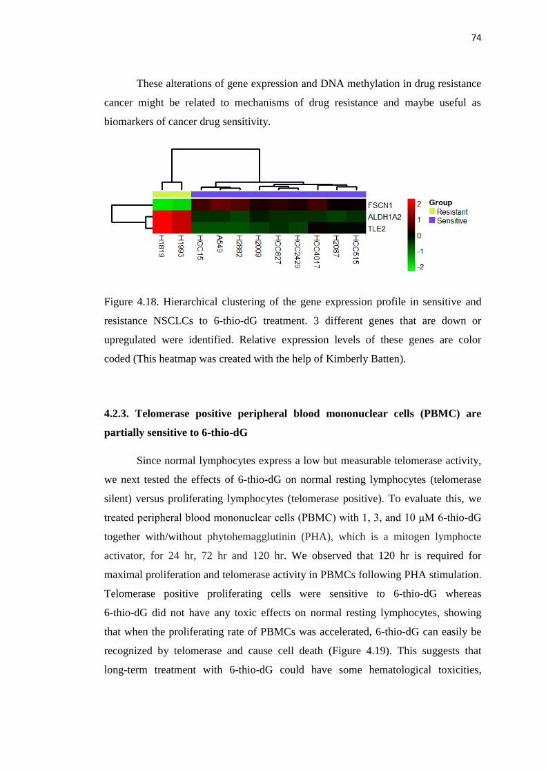
74
These alterations of gene expression and DNA methylation in drug resistance
cancer might be related to mechanisms of drug resistance and maybe useful as
biomarkers of cancer drug sensitivity.
Figure 4.18. Hierarchical clustering of the gene expression profile in sensitive and
resistance NSCLCs to 6-thio-dG treatment. 3 different genes that are down or
upregulated were identified. Relative expression levels of these genes are color
coded (This heatmap was created with the help of Kimberly Batten).
4.2.3. Telomerase positive peripheral blood mononuclear cells (PBMC) are
partially sensitive to 6-thio-dG
Since normal lymphocytes express a low but measurable telomerase activity,
we next tested the effects of 6-thio-dG on normal resting lymphocytes (telomerase
silent) versus proliferating lymphocytes (telomerase positive). To evaluate this, we
treated peripheral blood mononuclear cells (PBMC) with 1, 3, and 10 μM 6-thio-dG
together with/without phytohemagglutinin (PHA), which is a mitogen lymphocte
activator, for 24 hr, 72 hr and 120 hr. We observed that 120 hr is required for
maximal proliferation and telomerase activity in PBMCs following PHA stimulation.
Telomerase positive proliferating cells were sensitive to 6-thio-dG whereas
6-thio-dG did not have any toxic effects on normal resting lymphocytes, showing
that when the proliferating rate of PBMCs was accelerated, 6-thio-dG can easily be
recognized by telomerase and cause cell death (Figure 4.19). This suggests that
long-term treatment with 6-thio-dG could have some hematological toxicities,
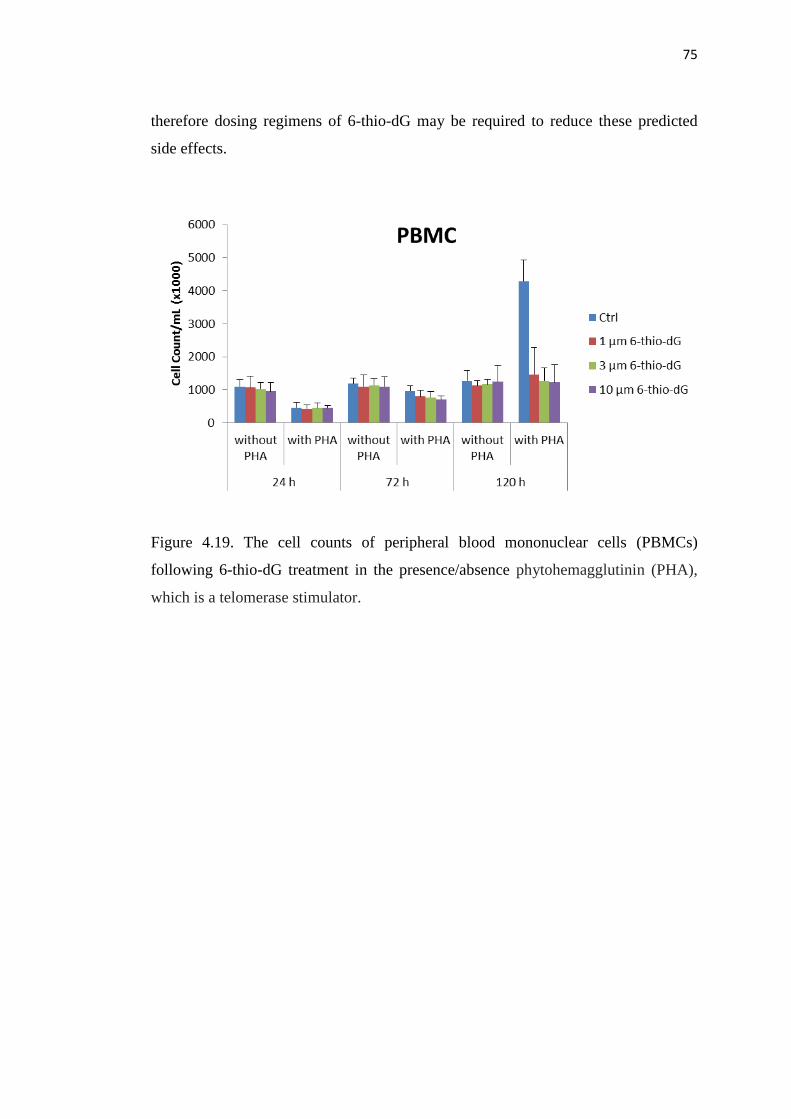
75
therefore dosing regimens of 6-thio-dG may be required to reduce these predicted
side effects.
Figure 4.19. The cell counts of peripheral blood mononuclear cells (PBMCs)
following 6-thio-dG treatment in the presence/absence phytohemagglutinin (PHA),
which is a telomerase stimulator.
76
5. DISCUSSION
Telomerase remains a potent and attractive target for cancer therapy.
However, while most previous telomerase targeted therapies showed some promising
results in preclinical and early clinical studies, there are still concerns about side
effects and drug resistance that leads to cancer recurrence and metastasis. GRN163L
(Imetelstat) is the potent telomerase inhibitor that entered into clinical trials over the
last several years. One of the main limitations of GRN163L is the variable telomere
lengths of tumor cells that patients have. Therefore, initial telomere length variability
and a potential long ''lag period'' (the time between the initiation of treatment and
therapeutic effect) are the main limitations of this and other telomerase inhibition
based therapies (Figure 5.1) in additional to hematologic and hepatic toxicities. A
combination therapy with GRN163L and standard of care or traditional
chemotherapy following surgical resection might have been the most effective way
to target telomerase positive tumors. Unfortunately, combining GRN163L with other
chemotherapeutics in human clinical trials have mostly been terminated primarily
due to toxicities in patients (218, 219). In addition, because of the drug
administration cycle, GRN163L was administered on days 1 and 8 of 21 day cycle, it
is likely that telomerase can not be inhibited sufficiently, resulting telomere
re-elongation or maintenance of telomeres. Due to the hematologic toxicities most
patients had to be taken on protocol and this likely results in re-elongation of
telomeres. Therefore, it is important to find appropriate drug dose, administration
cycle with combination therapy to increase therapeutic efficacy with fewer side
effects.
In this study, we aimed to identify a nucleoside analogue that is toxic for
cancer cells, but less so far for normal cells, and one that is recognized and then
incorporated into telomeres by telomerase. We predicted that the heterocyclic
guanine base modified nucleoside analogue 6-thio-2’-deoxyguanosine would make
an immediate change in the telomere structure and function, causing immediate
telomeric damage due to telomere uncapping and thus lead to a significantly reduced
lag phase. Our goal was to identify a telomerase mediated telomere uncapping small
molecule that would lead to massive and fast cancer cell death independent from the
initial tumor telomere length (Figure 5.1). In contrast to other telomerase inhibitors,
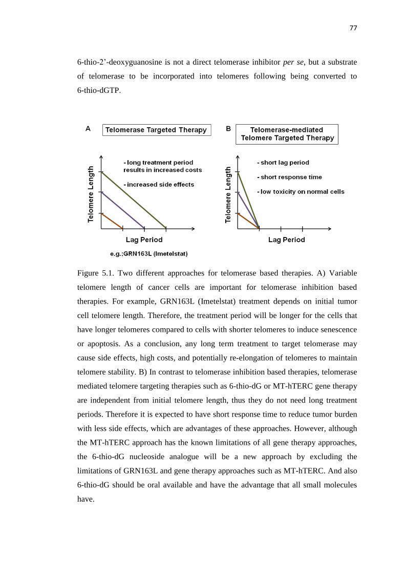
77
6-thio-2’-deoxyguanosine is not a direct telomerase inhibitor per se, but a substrate
of telomerase to be incorporated into telomeres following being converted to
6-thio-dGTP.
Figure 5.1. Two different approaches for telomerase based therapies. A) Variable
telomere length of cancer cells are important for telomerase inhibition based
therapies. For example, GRN163L (Imetelstat) treatment depends on initial tumor
cell telomere length. Therefore, the treatment period will be longer for the cells that
have longer telomeres compared to cells with shorter telomeres to induce senescence
or apoptosis. As a conclusion, any long term treatment to target telomerase may
cause side effects, high costs, and potentially re-elongation of telomeres to maintain
telomere stability. B) In contrast to telomerase inhibition based therapies, telomerase
mediated telomere targeting therapies such as 6-thio-dG or MT-hTERC gene therapy
are independent from initial telomere length, thus they do not need long treatment
periods. Therefore it is expected to have short response time to reduce tumor burden
with less side effects, which are advantages of these approaches. However, although
the MT-hTERC approach has the known limitations of all gene therapy approaches,
the 6-thio-dG nucleoside analogue will be a new approach by excluding the
limitations of GRN163L and gene therapy approaches such as MT-hTERC. And also
6-thio-dG should be oral available and have the advantage that all small molecules
have.
78
Purine analogues such as 6-thioguanine and 6-mercaptopurine are
chemotherapeutic agents that can be converted to thioguanine nucleotides.
6-thioguanine is one of the thiopurine drugs that are used in the treatment of acute
leukaemia. 6-thioguanine is synthesized by substitution of oxygen by sulphur at
carbon 6 of guanine. It was shown that 6-thioguanine inhibits purine utilisation and
was initially shown to be effective against rodent tumors and then in children with
acute lymphoblastic leukaemia. 6-thioguanine is the prodrug that is metabolized and
incorporated into DNA and RNA as thioguanine nucleotides to exert its cytotoxic
effect (220). 6-thioguanine has various bioavailability characteristics ranging from
14 to 46% (221) and a plasma half life is 90 min (222). 6-mercaptopurine is the other
thiopurine which is also metabolised to 6-thio-dGTP, but 6-mercaptopurine
participates in more biochemical processes to be converted to 6-thio-dGTP compared
to 6-thioguanine. It was suggested that since 6-thioguanine is metabolised to
thioguanine nucleotide more directly than 6-mercaptopurine, 6-thioguanine might be
the drug to choice instead of 6-mercaptopurine in children with acute lymphoblastic
leukaemia who had failed 6-mercaptopurine treatment (220). The idea was since
6-thioguanine participated in fewer biochemical and metabolic processes, the
efficiency and perhaps toxicity in patients would be different from 6-mercaptopurine.
In previous studies, 6-thioguanine and 6-mercaptopurine treatment was compared in
patients and thioguanine nucleotide concentration was higher in patients on
6-thioguanine compared to 6-mercaptopurine. There was no difference in anaemia or
neutropenia in 6-thioguanine and 6-mercaptopurine treated group except increased
thrombocytopenia was seen in 6-thioguanine treated group (223-225). It was also
shown that 6-thioguanine was associated with sinusiodal obstructive syndrome,
hypertension and veno-occlusive disease (VOD). Since toxicities of 6-thioguanine
was significant in trials, the use of 6-thioguanine has largely been stopped (220).
79
In this study, the nucleoside analogue, 6-thio-2’deoxyguanosine shows
Both general DNA and telomere specific damage but only in cells that
express telomerase in vitro and in vivo.
Progressive telomere shortening in cancer cells that are not immediately
killed by 6-thio-dG.
Less toxicity compared to approved 6-thioguanine.
Reduction of tumor growth rate.
We found that 6-thio-dG that is rapidly converted to 6-thio-dGTP by kinases
and is recognized by telomerase and incorporated into telomeres. This guanine-based
modified nucleoside incorporation causes increased telomere dyfunction induced foci
in HCT116 and telomerase expressing BJ cells (BJhTERT), but not in telomerase
negative BJ cells, indicating that the effect of 6-thio-dG is mostly telomerase
dependent (which was not observed with 6-thioguanine in HCT116, BJhTERT and
BJ cells). In addition, 6-thio-dG also caused increased general DNA damage in
HCT116 cells. We also found that while 6-thio-dG treatment causes progressive
telomere shortening in rare surviving telomerase positive cancer cells, 6-thioguanine
did not show telomere shortening on cancer cells, showing that 6-thio-dG has a
bifunctional role that targets both genomic and telomeric DNA in vitro as well as
antimetabolite functions. Interestingly, the effect of 6-thio-dG on telomeres was not
dependent on inhibition of telomerase activity, indicating that 6-thio-dG is not a
direct telomerase inhibitor, but telomerase is a mediator for its action. Importantly,
while 6-thio-dG kills the majority of cancer cells, it does not appear to have major
toxic effects on normal telomerase silent cells, which provide a therapeutic and
selective window of opportunity for cancer treatment. 6-thioguanine also showed
similar toxic effect on cancer cells besides no short-term major toxic effect on
normal cells. In contrast to the in vitro studies, in vivo studies showed more
efficiency and less toxic effects of 6-thio-dG (lack of weight loss, no changes
hematological except minor neutropenia, liver or kidney functions) compared to
6-thioguanine. We believe that these toxicity differences might be because of the
80
differences in structure of 6-thio-dG (a nucleoside) and 6-thioguanine (a heterocyclic
base). And also while 6-thioguanine participates in multiple biochemical processes to
be converted to thiopurine nucletiode including purine salvage, 6-thio-dG
participated in biochemical processes to a lesser extent, which was also shown in
comparison between 6-thioguanine and 6-mercaptopurine treatment. The differences
in biodistribution, relative solubility (6-thio-dG is more hydrophilic than
6-thioguanine), plasma protein binding and major organ accumulation may also be
related with observed toxicity differences in vivo.
It has been demonstrated that telomere dysfunction induces senescence or
apoptosis and can limit tumor growth rate in animal models (226, 227). Our results
also showed that 6-thio-dG significantly induced telomere dysfunction in
A549-derived tumor cells compared to control, suggesting that 6-thio-dG can be
incorporated into telomeres (~1/6000 of the genome) during replication and causes
tumor growth reduction.
Telomerase positive stem cells are a potential concern going forward with
6-thio-dG, but stem cells often grow slower compared to cancer cells, and also
cancer cells have relatively shorter telomeres (thus cancer cells may be more
susceptible to anticancer agents compared to normal cells). However, we showed that
increased telomerase activity caused sensitivity to 6-thio-dG in transient telomerase
positive lymphocytes compared to resting lymphocytes (telomerase silent), showing
that while there is likely to be some effects on transient amplifying telomerase
positive normal transit amplfying cells, quiescent stem cells (which as telomerase
negative) are unlikely to be affected by 6-thio-dG. Going forward, other potential
side effects will be tested in broader toxicology studies. As a conclusion, these novel
findings in this dissertation research provide encouraging support to further develop
new telomerase-based telomere targeted therapy that primarily target telomerase
positive cancer cells.
Cancer relapse is often a predicted outcome with single (monotherapy) anti-
cancer therapy. In our study, we observed that when we started to treat HCT116
colon cancer cells with 6-thio-dG, there was massive cell death. However, there were
still a small percentage of cells that appeared to survive and become resistant to
81
6-thio-dG. Eventually, after several weeks of treatment these cells started to grow
and became a resistant population of cells. There are several drug resistance
mechanisms to explain why these cells gained the ability to grow when continuously
treated with 6-thio-dG. ABC transporter proteins and cancer stem cell markers as
well as epigenetic biomarkers are commonly investigated for drug resistance. We
showed that resistant populations had higher levels of ABCG2 (ABC transporter
family member) and ALDH1A2 (aldehyde dehydrogenase, cancer stem cell marker)
compared to sensitive populations. In addititon, colony formation was higher in the
resistant cell population compared to sensitive cells. However, when compared to
control cells, the resistant cell population had less colony formation, a finding
suggesting that additional factors are reducing the ability of these resistent cells to
form colonies. Almost all lung cancer cell lines tested were sensitive to 6-thio-dG
except H1819, H1993 and H1693 cells. H1819/H1693 and H2073/H1993 were
isogenic cell lines from the same patient and each had different telomere lengths.
H1693 was established prior to chemotherapy from patient with lung
adenocarcinoma at regional lymph node. H1819 was established from residual
primary lung tumor following treatment with cisplatin, etoposide, and radiotherapy.
Likewise, H1993 was established prior to chemotherapy from a patient with
adenocarcinoma lung cancer. H2073 was established from a residual primary lung
tumor following cisplatin and etoposide treatment. Interestingly, while 6-thio-dG
causes less toxic effects on H1819, H1693 and H1993 cells, H2073 were more
sensitive.
To attempt to dissect the mechanism for the differential sensitivity to
6-thio-dG, methylation patterns were examined. A differential methlylation pattern
between sensitive and resistant cell lines was observed. While resistant cell lines
were hypermethylated in some genes, sensitive cell lines were hypomethylated in the
same genes. In addition, 5-aza (demethylation agent) treatment induced H1993
sensitivity to 6-thio-dG, but not H1819 or H1693 cells, indicating that the resistance
mechanism of H1993 can be correlated with hypermethylation status but not in the
other two cell lines. Gene expression profiles showed 3 different genes between
resistant (H1819, H1993) and sensitive cell lines, which are ALDH1A2, TLE2 and
FSCN1. ALDH1A2 is a cancer stem cell marker, which is upregulated in drug
82
resistance. Therefore, it is possible that resistant cells also have cancer stem cell
properties such as self renewal that play a role in the drug resistance mechanism.
TLE2 is correlated with Wnt/β-catenin and notch signaling pathways, which have
important roles in cell proliferation and also drug resistance. Thus, these methylation
and gene expression results show that resistant cells have several properties to
become resistant compared to sensitive cells. There are no published studies that
show a correlation between FSCN1 (Fascin-actin bundling protein 1) and drug
resistance, therefore it is unclear why FSCN1 is downregulated in resistant cancer
cells. Thus, FSCN1 should be further investigated.
Since GRN163L inhibits telomerase activity, it is expected that adding
GRN163L together with 6-thio-dG treatment would inhibit the effect of 6-thio-dG on
telomeres due to inability of using telomerase as a mediator of telomere uncapping.
We showed that GRN163L treatment that was added before 6-thio-dG did not cause
any toxic effect on HCT116 cells, predicting that by inhibiting telomerase activity
with GRN163L would not lead to telomere upcapping with 6-thio-dG treatment. In
one series of experiment we showed additional telomere shortening and partially
toxic effect on HCT116 cells following with GRN163L after initial effects of
6-thio-dG. This indicated that additional telomere shortening effect with GRN163L
in cancer cells that have already short telomeres following only 6-thio-dG treatment
may cause increased sensitivity to GRN163L. However, this sensitivity would not be
because of the effect of 6-thio-dG. This also suggests a possible combinatorial
approach to sensitize the surviving cancer cells following short term treatment with
6-thio-dG.
In future experiments, additional toxicity studies with long term 6-thio-dG
treatment are needed. In addition, chosing appropriate double therapy (carboplatin,
paclitaxel or cisplatin) and dose scheduling with combination therapy in vivo is
needed. We have established patient derived xenografts (PDX) by implanting of
primary tumor directly from patients into immunodeficient mouse. PDXs provide
partial solutions to the challenge that researchers face in cancer drug discovery such
as the problems with translating tumor responses to humans. Therefore, in progress
are experiment testing the effect of 6-thio-dG with/without combination therapy in
PDXs and also oral gavage of 6-thio-dG with proper drug concentration will be
83
tested to mimic the therapeutic efficiency of oral 6-thio-dG and minimize the risk of
adverse effects. In addition, the effect of oral 6-thio-dG on telomere dysfunction will
be investigated in PDX. Another direction to be pursued is treating with 6-thio-dG
and then in the maintenance setting examining the sensititivity of surviving cells to
radiation therapy. It may be that cancer cells with somewhat dysfunctional telomeres
may be more sensitive to radiation therapy.
We are also planning to further investigate the resistance mechanism of
cancer cells. HCT116 colon and/or A549 lung cancer cells will further be tested for
colony formation capability after 6-thio-dG treatment in vitro to understand if this
resistance of cancer cells are rare or common events and also what causes the
resistance? In addition, to show more efficiency (maybe tumor shrinkage) and
eliminate drug resistance with single agent, we are going to use combination therapy
with 6-thio-dG and cisplatin/paclittaxel (conventional therapies for lung cancer) in
H1819, H1993 or H1693 resistant cell lines in vitro and in vivo.
Telomerase negative ALT (Alternative Lengthening of Telomeres) cell lines
will be tested for telomere dysfunction following 6-thio-dG treatment to see if
6-thio-dG causes damage on telomeres in ALT cell lines as well as telomerase
positive cancer cells. This is somewhat unexpected since ALT cells do not express
telomerase. However, during telomere recombination in ALT cells it is possible that
6-thio-dGTP could be incorporated into ALT telomeres.
Further understanding of resistance mechanisms to eliminate and find an
appropriate dose and dose scheduling with multimodal (chemotherapy, radiation
therapy, and possibly other telomerse directed therapies) will provide a path forward
to treat patients with less side effects and perhaps more therapeutic and durable
responses.
84
REFERENCES
1. International Agency for Research on Cancer and Cancer Research (2014).
World Cancer Factsheet. London: Cancer Research UK.
2. American Cancer Society (2011). Global Cancer Facts & Figures. Atlanta:
American Cancer Society.
3. Toyooka, S., Toyooka, K.O., Maruyama, R., Virmani, A.K., Girard, L.,
Miyajima, K. and et al. (2001). DNA methylation profiles of lung tumors. Molecular
cancer therapeutics, 1 (1), 61-7.
4. Shay, J.W. and Wright, W.E. (2011). Role of telomeres and telomerase in
cancer. Seminars in cancer biology, 21 (6), 349-53.
5. Shay, J.W. and Wright, W.E. (2000). Implications of mapping the human
telomerase gene (hTERT) as the most distal gene on chromosome 5p. Neoplasia, 2
(3), 195-6.
6. Muller, H. (1938). The remaking of chromosomes. Collecting Net, 8, 182-95.
7. McClintock, B. (1939). The Behavior in Successive Nuclear Divisions of a
Chromosome Broken at Meiosis. Proceedings of the National Academy of Sciences
of the United States of America, 25 (8), 405-16.
8. Hayflick, L. and Moorhead, P.S. (1961). The serial cultivation of human
diploid cell strains. Experimental cell research, 25, 585-621.
9. Hayflick, L. (1965). The Limited in Vitro Lifetime of Human Diploid Cell
Strains. Experimental cell research, 37, 614-36.
10. Watson, J.D. (1972). Origin of concatemeric T7 DNA. Nature: New biology,
239 (94), 197-201.
11. Olovnikov, A.M. (1971). Principle of marginotomy in template synthesis of
polynucleotides. Doklady Akademii nauk SSSR, 201 (6), 1496-9.
12. Olovnikov, A.M. (1973). A theory of marginotomy. The incomplete copying
of template margin in enzymic synthesis of polynucleotides and biological
significance of the phenomenon. Journal of theoretical biology, 41 (1), 181-90.
13. Blackburn, E.H. and Gall, J.G. (1978). A tandemly repeated sequence at the
termini of the extrachromosomal ribosomal RNA genes in Tetrahymena. Journal of
molecular biology, 120 (1), 33-53.

85
14. Moyzis, R.K., Buckingham, J.M., Cram, L.S., Dani, M., Deaven, L.L., Jones,
M.D. and et al. (1988). A highly conserved repetitive DNA sequence, (TTAGGG)n,
present at the telomeres of human chromosomes. Proceedings of the National
Academy of Sciences of the United States of America, 85 (18), 6622-6.
15. Muller, H. (1962). In Studies of Genetics: The Selected Papers of H.J. Muller.
Indiana Univ Press, Bloomington, 384-408.
16. McClintock, B. (1941). The Stability of Broken Ends of Chromosomes in Zea
Mays. Genetics, 26 (2), 234-82.
17. Cooke, H.J. and Smith, B.A. (1986). Variability at the telomeres of the human
X/Y pseudoautosomal region. Cold Spring Harbor symposia on quantitative biology,
51 Pt 1, 213-9.
18. Harley, C.B., Futcher, A.B. and Greider, C.W. (1990). Telomeres shorten
during ageing of human fibroblasts. Nature, 345 (6274), 458-60.
19. Greider, C.W. and Blackburn, E.H. (1985). Identification of a specific
telomere terminal transferase activity in Tetrahymena extracts. Cell, 43 (2 Pt 1), 405-
13.
20. Greider, C.W. and Blackburn, E.H. (1989). A telomeric sequence in the RNA
of Tetrahymena telomerase required for telomere repeat synthesis. Nature, 337
(6205), 331-7.
21. Kilian, A., Bowtell, D.D., Abud, H.E., Hime, G.R., Venter, D.J., Keese, P.K.
and et al. (1997). Isolation of a candidate human telomerase catalytic subunit gene,
which reveals complex splicing patterns in different cell types. Human molecular
genetics, 6 (12), 2011-9.
22. Meyerson, M., Counter, C.M., Eaton, E.N., Ellisen, L.W., Steiner, P., Caddle,
S.D. and et al. (1997). hEST2, the putative human telomerase catalytic subunit gene,
is up-regulated in tumor cells and during immortalization. Cell, 90 (4), 785-95.
23. Bodnar, A.G., Ouellette, M., Frolkis, M., Holt, S.E., Chiu, C.P., Morin, G.B.
and et al. (1998). Extension of life-span by introduction of telomerase into normal
human cells. Science, 279 (5349), 349-52.
24. Mason, P.J. and Perdigones, N. (2013). Telomere biology and translational
research. Translational research : the journal of laboratory and clinical medicine,
162 (6), 333-42.
86
25. Kim, N.W., Piatyszek, M.A., Prowse, K.R., Harley, C.B., West, M.D., Ho,
P.L. and et al. (1994). Specific association of human telomerase activity with
immortal cells and cancer. Science, 266 (5193), 2011-5.
26. Bryan, T.M., Englezou, A., Dalla-Pozza, L., Dunham, M.A. and Reddel, R.R.
(1997). Evidence for an alternative mechanism for maintaining telomere length in
human tumors and tumor-derived cell lines. Nature medicine, 3 (11), 1271-4.
27. Shay, J.W. and Wright, W.E. (2000). Hayflick, his limit, and cellular ageing.
Nature reviews Molecular cell biology, 1 (1), 72-6.
28. Ciccia, A. and Elledge, S.J. (2010). The DNA damage response: making it
safe to play with knives. Molecular cell, 40 (2), 179-204.
29. Hoeijmakers, J.H. (2009). DNA damage, aging, and cancer. The New
England journal of medicine, 361 (15), 1475-85.
30. Lee, J.H. and Paull, T.T. (2005). ATM activation by DNA double-strand
breaks through the Mre11-Rad50-Nbs1 complex. Science, 308 (5721), 551-4.
31. Bakkenist, C.J. and Kastan, M.B. (2003). DNA damage activates ATM
through intermolecular autophosphorylation and dimer dissociation. Nature, 421
(6922), 499-506.
32. Downs, J.A., Lowndes, N.F. and Jackson, S.P. (2000). A role for
Saccharomyces cerevisiae histone H2A in DNA repair. Nature, 408 (6815), 1001-4.
33. Ward, I.M. and Chen, J. (2001). Histone H2AX is phosphorylated in an ATR-
dependent manner in response to replicational stress. The Journal of biological
chemistry, 276 (51), 47759-62.
34. Rogakou, E.P., Pilch, D.R., Orr, A.H., Ivanova, V.S. and Bonner, W.M.
(1998). DNA double-stranded breaks induce histone H2AX phosphorylation on
serine 139. The Journal of biological chemistry, 273 (10), 5858-68.
35. Lobrich, M., Shibata, A., Beucher, A., Fisher, A., Ensminger, M., Goodarzi,
A.A. and et al. (2010). gammaH2AX foci analysis for monitoring DNA double-
strand break repair: strengths, limitations and optimization. Cell cycle, 9 (4), 662-9.
36. Goodarzi, A.A. and Jeggo, P.A. (2013). The repair and signaling responses to
DNA double-strand breaks. Advances in genetics, 82, 1-45.
87
37. Matsuoka, S., Ballif, B.A., Smogorzewska, A., McDonald, E.R., 3rd, Hurov,
K.E., Luo, J. and et al. (2007). ATM and ATR substrate analysis reveals extensive
protein networks responsive to DNA damage. Science, 316 (5828), 1160-6.
38. Zhao, H. and Piwnica-Worms, H. (2001). ATR-mediated checkpoint
pathways regulate phosphorylation and activation of human Chk1. Molecular and
cellular biology, 21 (13), 4129-39.
39. Deckbar, D., Stiff, T., Koch, B., Reis, C., Lobrich, M. and Jeggo, P.A.
(2010). The limitations of the G1-S checkpoint. Cancer research, 70 (11), 4412-21.
40. Klement, K. and Goodarzi, A.A. (2014). DNA double strand break responses
and chromatin alterations within the aging cell. Experimental cell research.
41. Zimmermann, M. and de Lange, T. (2014). 53BP1: pro choice in DNA repair.
Trends in cell biology, 24 (2), 108-17.
42. Hartlerode, A.J. and Scully, R. (2009). Mechanisms of double-strand break
repair in somatic mammalian cells. The Biochemical journal, 423 (2), 157-68.
43. Hosoya, N. and Miyagawa, K. (2014). Targeting DNA damage response in
cancer therapy. Cancer science, 105 (4), 370-88.
44. Palm, W. and de Lange, T. (2008). How shelterin protects mammalian
telomeres. Annual review of genetics, 42, 301-34.
45. Barnett, M.A., Buckle, V.J., Evans, E.P., Porter, A.C., Rout, D., Smith, A.G.
and et al. (1993). Telomere directed fragmentation of mammalian chromosomes.
Nucleic acids research, 21 (1), 27-36.
46. Farr, C., Fantes, J., Goodfellow, P. and Cooke, H. (1991). Functional
reintroduction of human telomeres into mammalian cells. Proceedings of the
National Academy of Sciences of the United States of America, 88 (16), 7006-10.
47. Hanish, J.P., Yanowitz, J.L. and de Lange, T. (1994). Stringent sequence
requirements for the formation of human telomeres. Proceedings of the National
Academy of Sciences of the United States of America, 91 (19), 8861-5.
48. Damm, K., Hemmann, U., Garin-Chesa, P., Hauel, N., Kauffmann, I.,
Priepke, H. and et al. (2001). A highly selective telomerase inhibitor limiting human
cancer cell proliferation. The EMBO journal, 20 (24), 6958-68.
88
49. Makarov, V.L., Hirose, Y. and Langmore, J.P. (1997). Long G tails at both
ends of human chromosomes suggest a C strand degradation mechanism for telomere
shortening. Cell, 88 (5), 657-66.
50. McElligott, R. and Wellinger, R.J. (1997). The terminal DNA structure of
mammalian chromosomes. The EMBO journal, 16 (12), 3705-14.
51. Griffith, J.D., Comeau, L., Rosenfield, S., Stansel, R.M., Bianchi, A., Moss,
H. and et al. (1999). Mammalian telomeres end in a large duplex loop. Cell, 97 (4),
503-14.
52. de Lange, T. (2005). Shelterin: the protein complex that shapes and
safeguards human telomeres. Genes & development, 19 (18), 2100-10.
53. Liu, D., O'Connor, M.S., Qin, J. and Songyang, Z. (2004). Telosome, a
mammalian telomere-associated complex formed by multiple telomeric proteins. The
Journal of biological chemistry, 279 (49), 51338-42.
54. Ye, J.Z., Donigian, J.R., van Overbeek, M., Loayza, D., Luo, Y.,
Krutchinsky, A.N. and et al. (2004). TIN2 binds TRF1 and TRF2 simultaneously and
stabilizes the TRF2 complex on telomeres. The Journal of biological chemistry, 279
(45), 47264-71.
55. Takai, H., Smogorzewska, A. and de Lange, T. (2003). DNA damage foci at
dysfunctional telomeres. Current biology : CB, 13 (17), 1549-56.
56. Marciniak, R.A., Johnson, F.B. and Guarente, L. (2000). Dyskeratosis
congenita, telomeres and human ageing. Trends in genetics : TIG, 16 (5), 193-5.
57. Maser, R.S. and DePinho, R.A. (2002). Connecting chromosomes, crisis, and
cancer. Science, 297 (5581), 565-9.
58. Campisi, J. (2001). Cellular senescence as a tumor-suppressor mechanism.
Trends in cell biology, 11 (11), S27-31.
59. Campisi, J., Kim, S.H., Lim, C.S. and Rubio, M. (2001). Cellular senescence,
cancer and aging: the telomere connection. Experimental gerontology, 36 (10), 1619-
37.
60. de Lange, T. (2002). Protection of mammalian telomeres. Oncogene, 21 (4),
532-40.
89
61. Suram, A. and Herbig, U. (2014). The replicometer is broken: telomeres
activate cellular senescence in response to genotoxic stresses. Aging cell, 13 (5), 780-
6.
62. Shay, J.W. and Bacchetti, S. (1997). A survey of telomerase activity in
human cancer. European journal of cancer, 33 (5), 787-91.
63. Shay, J.W., Zou, Y., Hiyama, E. and Wright, W.E. (2001). Telomerase and
cancer. Human molecular genetics, 10 (7), 677-85.
64. Wright, W.E., Piatyszek, M.A., Rainey, W.E., Byrd, W. and Shay, J.W.
(1996). Telomerase activity in human germline and embryonic tissues and cells.
Developmental genetics, 18 (2), 173-9.
65. Mocellin, S., Pooley, K.A. and Nitti, D. (2013). Telomerase and the search
for the end of cancer. Trends in molecular medicine, 19 (2), 125-33.
66. Yi, X., Shay, J.W. and Wright, W.E. (2001). Quantitation of telomerase
components and hTERT mRNA splicing patterns in immortal human cells. Nucleic
acids research, 29 (23), 4818-25.
67. Wong, M.S., Wright, W.E. and Shay, J.W. (2014). Alternative splicing
regulation of telomerase: a new paradigm? Trends in genetics : TIG.
68. Wong, M.S., Chen, L., Foster, C., Kainthla, R., Shay, J.W. and Wright, W.E.
(2013). Regulation of telomerase alternative splicing: a target for chemotherapy. Cell
reports, 3 (4), 1028-35.
69. Saeboe-Larssen, S., Fossberg, E. and Gaudernack, G. (2006).
Characterization of novel alternative splicing sites in human telomerase reverse
transcriptase (hTERT): analysis of expression and mutual correlation in mRNA
isoforms from normal and tumour tissues. BMC molecular biology, 7, 26.
70. Yi, X., White, D.M., Aisner, D.L., Baur, J.A., Wright, W.E. and Shay, J.W.
(2000). An alternate splicing variant of the human telomerase catalytic subunit
inhibits telomerase activity. Neoplasia, 2 (5), 433-40.
71. Colgin, L.M., Wilkinson, C., Englezou, A., Kilian, A., Robinson, M.O. and
Reddel, R.R. (2000). The hTERTalpha splice variant is a dominant negative inhibitor
of telomerase activity. Neoplasia, 2 (5), 426-32.
72. Listerman, I., Sun, J., Gazzaniga, F.S., Lukas, J.L. and Blackburn, E.H.
(2013). The major reverse transcriptase-incompetent splice variant of the human
90
telomerase protein inhibits telomerase activity but protects from apoptosis. Cancer
research, 73 (9), 2817-28.
73. Wong, M.S., Shay, J.W. and Wright, W.E. (2014). Regulation of human
telomerase splicing by RNA:RNA pairing. Nature communications, 5, 3306.
74. Ulaner, G.A., Hu, J.F., Vu, T.H., Giudice, L.C. and Hoffman, A.R. (2001).
Tissue-specific alternate splicing of human telomerase reverse transcriptase (hTERT)
influences telomere lengths during human development. International journal of
cancer Journal international du cancer, 91 (5), 644-9.
75. Davis, C.F., Ricketts, C.J., Wang, M., Yang, L., Cherniack, A.D., Shen, H.
and et al. (2014). The somatic genomic landscape of chromophobe renal cell
carcinoma. Cancer cell, 26 (3), 319-30.
76. Zhao, Y., Wang, S., Popova, E.Y., Grigoryev, S.A. and Zhu, J. (2009).
Rearrangement of upstream sequences of the hTERT gene during cellular
immortalization. Genes, chromosomes & cancer, 48 (11), 963-74.
77. Huang, F.W., Hodis, E., Xu, M.J., Kryukov, G.V., Chin, L. and Garraway,
L.A. (2013). Highly recurrent TERT promoter mutations in human melanoma.
Science, 339 (6122), 957-9.
78. Weir, B.A., Woo, M.S., Getz, G., Perner, S., Ding, L., Beroukhim, R. and et
al. (2007). Characterizing the cancer genome in lung adenocarcinoma. Nature, 450
(7171), 893-8.
79. Chudnovsky, Y., Adams, A.E., Robbins, P.B., Lin, Q. and Khavari, P.A.
(2005). Use of human tissue to assess the oncogenic activity of melanoma-associated
mutations. Nature genetics, 37 (7), 745-9.
80. Harley, C.B. (2008). Telomerase and cancer therapeutics. Nature reviews
Cancer, 8 (3), 167-79.
81. Shay, J.W. and Wright, W.E. (2006). Telomerase therapeutics for cancer:
challenges and new directions. Nature reviews Drug discovery, 5 (7), 577-84.
82. Gertler, R., Rosenberg, R., Stricker, D., Friederichs, J., Hoos, A., Werner, M.
and et al. (2004). Telomere length and human telomerase reverse transcriptase
expression as markers for progression and prognosis of colorectal carcinoma.
Journal of clinical oncology : official journal of the American Society of Clinical
Oncology, 22 (10), 1807-14.
91
83. Kobitsu, K., Tsutsumi, M., Tsujiuchi, T., Suzuki, F., Kido, A., Okajima, E.
and et al. (1997). Shortened telomere length and increased telomerase activity in
hamster pancreatic duct adenocarcinomas and cell lines. Molecular carcinogenesis,
18 (3), 153-9.
84. Raynaud, C.M., Hernandez, J., Llorca, F.P., Nuciforo, P., Mathieu, M.C.,
Commo, F. and et al. (2010). DNA damage repair and telomere length in normal
breast, preneoplastic lesions, and invasive cancer. American journal of clinical
oncology, 33 (4), 341-5.
85. Engelhardt, M., Albanell, J., Drullinsky, P., Han, W., Guillem, J., Scher, H.I.
and et al. (1997). Relative contribution of normal and neoplastic cells determines
telomerase activity and telomere length in primary cancers of the prostate, colon, and
sarcoma. Clinical cancer research : an official journal of the American Association
for Cancer Research, 3 (10), 1849-57.
86. Pascolo, E., Wenz, C., Lingner, J., Hauel, N., Priepke, H., Kauffmann, I. and
et al. (2002). Mechanism of human telomerase inhibition by BIBR1532, a synthetic,
non-nucleosidic drug candidate. The Journal of biological chemistry, 277 (18),
15566-72.
87. El-Daly, H., Kull, M., Zimmermann, S., Pantic, M., Waller, C.F. and
Martens, U.M. (2005). Selective cytotoxicity and telomere damage in leukemia cells
using the telomerase inhibitor BIBR1532. Blood, 105 (4), 1742-9.
88. Barma, D.K., Elayadi, A., Falck, J.R. and Corey, D.R. (2003). Inhibition of
telomerase by BIBR 1532 and related analogues. Bioorganic & medicinal chemistry
letters, 13 (7), 1333-6.
89. Piotrowska, K., Kleideiter, E., Murdter, T.E., Taetz, S., Baldes, C., Schaefer,
U. and et al. (2005). Optimization of the TRAP assay to evaluate specificity of
telomerase inhibitors. Laboratory investigation; a journal of technical methods and
pathology, 85 (12), 1565-9.
90. Rankin, A.M., Faller, D.V. and Spanjaard, R.A. (2008). Telomerase inhibitors
and 'T-oligo' as cancer therapeutics: contrasting molecular mechanisms of
cytotoxicity. Anti-cancer drugs, 19 (4), 329-38.
92
91. Roth, A., Harley, C.B. and Baerlocher, G.M. (2010). Imetelstat (GRN163L)--
telomerase-based cancer therapy. Recent results in cancer research Fortschritte der
Krebsforschung Progres dans les recherches sur le cancer, 184, 221-34.
92. Dikmen, Z.G., Wright, W.E., Shay, J.W. and Gryaznov, S.M. (2008).
Telomerase targeted oligonucleotide thio-phosphoramidates in T24-luc bladder
cancer cells. Journal of cellular biochemistry, 104 (2), 444-52.
93. Djojosubroto, M.W., Chin, A.C., Go, N., Schaetzlein, S., Manns, M.P.,
Gryaznov, S. and et al. (2005). Telomerase antagonists GRN163 and GRN163L
inhibit tumor growth and increase chemosensitivity of human hepatoma. Hepatology,
42 (5), 1127-36.
94. Dikmen, Z.G., Gellert, G.C., Jackson, S., Gryaznov, S., Tressler, R., Dogan,
P. and et al. (2005). In vivo inhibition of lung cancer by GRN163L: a novel human
telomerase inhibitor. Cancer research, 65 (17), 7866-73.
95. Gellert, G.C., Dikmen, Z.G., Wright, W.E., Gryaznov, S. and Shay, J.W.
(2006). Effects of a novel telomerase inhibitor, GRN163L, in human breast cancer.
Breast cancer research and treatment, 96 (1), 73-81.
96. Hochreiter, A.E., Xiao, H., Goldblatt, E.M., Gryaznov, S.M., Miller, K.D.,
Badve, S. and et al. (2006). Telomerase template antagonist GRN163L disrupts
telomere maintenance, tumor growth, and metastasis of breast cancer. Clinical
cancer research : an official journal of the American Association for Cancer
Research, 12 (10), 3184-92.
97. Shammas, M.A., Koley, H., Bertheau, R.C., Neri, P., Fulciniti, M., Tassone,
P. and et al. (2008). Telomerase inhibitor GRN163L inhibits myeloma cell growth in
vitro and in vivo. Leukemia, 22 (7), 1410-8.
98. Burchett, K.M., Yan, Y. and Ouellette, M.M. (2014). Telomerase inhibitor
Imetelstat (GRN163L) limits the lifespan of human pancreatic cancer cells. PloS one,
9 (1), e85155.
99. Tamakawa, R.A., Fleisig, H.B. and Wong, J.M. (2010). Telomerase inhibition
potentiates the effects of genotoxic agents in breast and colorectal cancer cells in a
cell cycle-specific manner. Cancer research, 70 (21), 8684-94.
100. Marian, C.O., Cho, S.K., McEllin, B.M., Maher, E.A., Hatanpaa, K.J.,
Madden, C.J. and et al. (2010). The telomerase antagonist, imetelstat, efficiently
93
targets glioblastoma tumor-initiating cells leading to decreased proliferation and
tumor growth. Clinical cancer research : an official journal of the American
Association for Cancer Research, 16 (1), 154-63.
101. Shammas, M.A., Qazi, A., Batchu, R.B., Bertheau, R.C., Wong, J.Y., Rao,
M.Y. and et al. (2008). Telomere maintenance in laser capture microdissection-
purified Barrett's adenocarcinoma cells and effect of telomerase inhibition in vivo.
Clinical cancer research : an official journal of the American Association for Cancer
Research, 14 (15), 4971-80.
102. Ouellette, M.M., Wright, W.E. and Shay, J.W. (2011). Targeting telomerase-
expressing cancer cells. Journal of cellular and molecular medicine, 15 (7), 1433-42.
103. Chiappori A, Bassett K, Burington B, Kolevska T, Spigel DR, Hager S and et
al (April 2013). Improved progression-free survival (PFS) in patients with short
tumor telomere length: subgroup analysis from a randomized phase II study of the
telomerase inhibitor imetelstat as maintenance therapy for advanced NSCLC
(Poster). American Association for Cancer Research (AACR) Annual Meeting 2013.
Walter E. Washington Convention Center, Washington, DC.
104. Bates, P., Mergny, J.L. and Yang, D. (2007). Quartets in G-major. The First
International Meeting on Quadruplex DNA. EMBO reports, 8 (11), 1003-10.
105. Xu, Y. (2011). Chemistry in human telomere biology: structure, function and
targeting of telomere DNA/RNA. Chemical Society reviews, 40 (5), 2719-40.
106. Neidle, S. (2010). Human telomeric G-quadruplex: the current status of
telomeric G-quadruplexes as therapeutic targets in human cancer. The FEBS journal,
277 (5), 1118-25.
107. Gowan, S.M., Harrison, J.R., Patterson, L., Valenti, M., Read, M.A., Neidle,
S. and et al. (2002). A G-quadruplex-interactive potent small-molecule inhibitor of
telomerase exhibiting in vitro and in vivo antitumor activity. Molecular
pharmacology, 61 (5), 1154-62.
108. Shay, J.W. and Keith, W.N. (2008). Targeting telomerase for cancer
therapeutics. British journal of cancer, 98 (4), 677-83.
109. Beatty, G.L. and Vonderheide, R.H. (2008). Telomerase as a universal tumor
antigen for cancer vaccines. Expert review of vaccines, 7 (7), 881-7.
94
110. Vonderheide, R.H. (2008). Prospects and challenges of building a cancer
vaccine targeting telomerase. Biochimie, 90 (1), 173-80.
111. Cortez-Gonzalez, X. and Zanetti, M. (2007). Telomerase immunity from
bench to bedside: round one. Journal of translational medicine, 5, 12.
112. Kyte, J.A., Gaudernack, G., Dueland, S., Trachsel, S., Julsrud, L. and
Aamdal, S. (2011). Telomerase peptide vaccination combined with temozolomide: a
clinical trial in stage IV melanoma patients. Clinical cancer research : an official
journal of the American Association for Cancer Research, 17 (13), 4568-80.
113. Baumann, P. and Cech, T.R. (2001). Pot1, the putative telomere end-binding
protein in fission yeast and humans. Science, 292 (5519), 1171-5.
114. Fairall, L., Chapman, L., Moss, H., de Lange, T. and Rhodes, D. (2001).
Structure of the TRFH dimerization domain of the human telomeric proteins TRF1
and TRF2. Molecular cell, 8 (2), 351-61.
115. Guiducci, C., Cerone, M.A. and Bacchetti, S. (2001). Expression of mutant
telomerase in immortal telomerase-negative human cells results in cell cycle
deregulation, nuclear and chromosomal abnormalities and rapid loss of viability.
Oncogene, 20 (6), 714-25.
116. Kim, M.M., Rivera, M.A., Botchkina, I.L., Shalaby, R., Thor, A.D. and
Blackburn, E.H. (2001). A low threshold level of expression of mutant-template
telomerase RNA inhibits human tumor cell proliferation. Proceedings of the National
Academy of Sciences of the United States of America, 98 (14), 7982-7.
117. Marusic, L., Anton, M., Tidy, A., Wang, P., Villeponteau, B. and Bacchetti,
S. (1997). Reprogramming of telomerase by expression of mutant telomerase RNA
template in human cells leads to altered telomeres that correlate with reduced cell
viability. Molecular and cellular biology, 17 (11), 6394-401.
118. Tomas-Loba, A., Flores, I., Fernandez-Marcos, P.J., Cayuela, M.L., Maraver,
A., Tejera, A. and et al. (2008). Telomerase reverse transcriptase delays aging in
cancer-resistant mice. Cell, 135 (4), 609-22.
119. Jaskelioff, M., Muller, F.L., Paik, J.H., Thomas, E., Jiang, S., Adams, A.C.
and et al. (2011). Telomerase reactivation reverses tissue degeneration in aged
telomerase-deficient mice. Nature, 469 (7328), 102-6.

95
120. Hemann, M.T., Strong, M.A., Hao, L.Y. and Greider, C.W. (2001). The
shortest telomere, not average telomere length, is critical for cell viability and
chromosome stability. Cell, 107 (1), 67-77.
121. Eisenstein, M. (2011). Telomeres: All's well that ends well. Nature, 478
(7368), S13-5.
122. Campisi, J. and d'Adda di Fagagna, F. (2007). Cellular senescence: when bad
things happen to good cells. Nature reviews Molecular cell biology, 8 (9), 729-40.
123. Herbig, U., Jobling, W.A., Chen, B.P., Chen, D.J. and Sedivy, J.M. (2004).
Telomere shortening triggers senescence of human cells through a pathway involving
ATM, p53, and p21(CIP1), but not p16(INK4a). Molecular cell, 14 (4), 501-13.
124. Jeyapalan, J.C., Ferreira, M., Sedivy, J.M. and Herbig, U. (2007).
Accumulation of senescent cells in mitotic tissue of aging primates. Mechanisms of
ageing and development, 128 (1), 36-44.
125. Ressler, S., Bartkova, J., Niederegger, H., Bartek, J., Scharffetter-Kochanek,
K., Jansen-Durr, P. and et al. (2006). p16INK4A is a robust in vivo biomarker of
cellular aging in human skin. Aging cell, 5 (5), 379-89.
126. Robles, S.J. and Adami, G.R. (1998). Agents that cause DNA double strand
breaks lead to p16INK4a enrichment and the premature senescence of normal
fibroblasts. Oncogene, 16 (9), 1113-23.
127. Zindy, F., Soares, H., Herzog, K.H., Morgan, J., Sherr, C.J. and Roussel,
M.F. (1997). Expression of INK4 inhibitors of cyclin D-dependent kinases during
mouse brain development. Cell growth & differentiation : the molecular biology
journal of the American Association for Cancer Research, 8 (11), 1139-50.
128. Krishnamurthy, J., Torrice, C., Ramsey, M.R., Kovalev, G.I., Al-Regaiey, K.,
Su, L. and et al. (2004). Ink4a/Arf expression is a biomarker of aging. The Journal of
clinical investigation, 114 (9), 1299-307.
129. Herbig, U., Wei, W., Dutriaux, A., Jobling, W.A. and Sedivy, J.M. (2003).
Real-time imaging of transcriptional activation in live cells reveals rapid up-
regulation of the cyclin-dependent kinase inhibitor gene CDKN1A in replicative
cellular senescence. Aging cell, 2 (6), 295-304.
130. Sikora, E., Arendt, T., Bennett, M. and Narita, M. (2011). Impact of cellular
senescence signature on ageing research. Ageing research reviews, 10 (1), 146-52.
96
131. Narita, M., Nunez, S., Heard, E., Narita, M., Lin, A.W., Hearn, S.A. and et al.
(2003). Rb-mediated heterochromatin formation and silencing of E2F target genes
during cellular senescence. Cell, 113 (6), 703-16.
132. Narita, M. (2007). Cellular senescence and chromatin organisation. British
journal of cancer, 96 (5), 686-91.
133. Funayama, R. and Ishikawa, F. (2007). Cellular senescence and chromatin
structure. Chromosoma, 116 (5), 431-40.
134. Fumagalli, M., Rossiello, F., Clerici, M., Barozzi, S., Cittaro, D., Kaplunov,
J.M. and et al. (2012). Telomeric DNA damage is irreparable and causes persistent
DNA-damage-response activation. Nature cell biology, 14 (4), 355-65.
135. Hewitt, G., Jurk, D., Marques, F.D., Correia-Melo, C., Hardy, T., Gackowska,
A. and et al. (2012). Telomeres are favoured targets of a persistent DNA damage
response in ageing and stress-induced senescence. Nature communications, 3, 708.
136. Suram, A., Kaplunov, J., Patel, P.L., Ruan, H., Cerutti, A., Boccardi, V. and
et al. (2012). Oncogene-induced telomere dysfunction enforces cellular senescence in
human cancer precursor lesions. The EMBO journal, 31 (13), 2839-51.
137. Flowers, C.R. and Melmon, K.L. (1997). Clinical investigators as critical
determinants in pharmaceutical innovation. Nature medicine, 3 (2), 136-43.
138. Karran, P. and Attard, N. (2008). Thiopurines in current medical practice:
molecular mechanisms and contributions to therapy-related cancer. Nature reviews
Cancer, 8 (1), 24-36.
139. Aarbakke, J., Janka-Schaub, G. and Elion, G.B. (1997). Thiopurine biology
and pharmacology. Trends in pharmacological sciences, 18 (1), 3-7.
140. Wang, L. and Weinshilboum, R. (2006). Thiopurine S-methyltransferase
pharmacogenetics: insights, challenges and future directions. Oncogene, 25 (11),
1629-38.
141. Yoshida, S., Yamada, M., Masaki, S. and Saneyoshi, M. (1979). Utilization
of 2'-deoxy-6-thioguanosine 5'-triphosphate in DNA synthesis in vitro by DNA
polymerase alpha from calf thymus. Cancer research, 39 (10), 3955-8.
142. Ling, Y.H., Nelson, J.A., Cheng, Y.C., Anderson, R.S. and Beattie, K.L.
(1991). 2'-Deoxy-6-thioguanosine 5'-triphosphate as a substrate for purified human
97
DNA polymerases and calf thymus terminal deoxynucleotidyltransferase in vitro.
Molecular pharmacology, 40 (4), 508-14.
143. Karran, P. and Bignami, M. (1994). DNA damage tolerance, mismatch repair
and genome instability. BioEssays : news and reviews in molecular, cellular and
developmental biology, 16 (11), 833-9.
144. King, P.D. and Perry, M.C. (2001). Hepatotoxicity of chemotherapy. The
oncologist, 6 (2), 162-76.
145. Malhi, H., Gores, G.J. and Lemasters, J.J. (2006). Apoptosis and necrosis in
the liver: a tale of two deaths? Hepatology, 43 (2 Suppl 1), S31-44.
146. Ishak, K.G. and Zimmerman, H.J. (1995). Morphologic spectrum of drug-
induced hepatic disease. Gastroenterology clinics of North America, 24 (4), 759-86.
147. Field, K.M., Dow, C. and Michael, M. (2008). Part I: Liver function in
oncology: biochemistry and beyond. The Lancet Oncology, 9 (11), 1092-101.
148. Einhorn, M. and Davidsohn, I. (1964). Hepatotoxicity of Mercaptopurine.
Jama, 188, 802-6.
149. Shorey, J., Schenker, S., Suki, W.N. and Combes, B. (1968). Hepatotoxicity
of mercaptopurine. Archives of internal medicine, 122 (1), 54-8.
150. Adamson, P.C., Zimm, S., Ragab, A.H., Steinberg, S.M., Balis, F., Kamen,
B.A. and et al. (1990). A phase II trial of continuous-infusion 6-mercaptopurine for
childhood solid tumors. Cancer chemotherapy and pharmacology, 26 (5), 343-4.
151. Griner, P.F., Elbadawi, A. and Packman, C.H. (1976). Veno-occlusive disease
of the liver after chemotherapy of acute leukemia. Report of two cases. Annals of
internal medicine, 85 (5), 578-82.
152. Satti, M.B., Weinbren, K. and Gordon-Smith, E.C. (1982). 6-thioguanine as a
cause of toxic veno-occlusive disease of the liver. Journal of clinical pathology, 35
(10), 1086-91.
153. Gill, R.A., Onstad, G.R., Cardamone, J.M., Maneval, D.C. and Sumner, H.W.
(1982). Hepatic veno-occlusive disease caused by 6-thioguanine. Annals of internal
medicine, 96 (1), 58-60.
154. Krivoy, N., Raz, R., Carter, A. and Alroy, G. (1982). Reversible hepatic
veno-occlusive disease and 6-thioguanine. Annals of internal medicine, 96 (6 Pt 1),
788.
98
155. Larrey, D., Freneaux, E., Berson, A., Babany, G., Degott, C., Valla, D. and et
al. (1988). Peliosis hepatis induced by 6-thioguanine administration. Gut, 29 (9),
1265-9.
156. Brater, D.C. (2009). Drug dosing in patients with impaired renal function.
Clinical pharmacology and therapeutics, 86 (5), 483-9.
157. Choudhury, D. and Ahmed, Z. (2006). Drug-associated renal dysfunction and
injury. Nature clinical practice Nephrology, 2 (2), 80-91.
158. Glezerman, I., Kris, M.G., Miller, V., Seshan, S. and Flombaum, C.D. (2009).
Gemcitabine nephrotoxicity and hemolytic uremic syndrome: report of 29 cases from
a single institution. Clinical nephrology, 71 (2), 130-9.
159. Duffull, S.B. and Robinson, B.A. (1997). Clinical pharmacokinetics and dose
optimisation of carboplatin. Clinical pharmacokinetics, 33 (3), 161-83.
160. Sahni, V., Choudhury, D. and Ahmed, Z. (2009). Chemotherapy-associated
renal dysfunction. Nature reviews Nephrology, 5 (8), 450-62.
161. Bea, S., Valdes-Mas, R., Navarro, A., Salaverria, I., Martin-Garcia, D., Jares,
P. and et al. (2013). Landscape of somatic mutations and clonal evolution in mantle
cell lymphoma. Proceedings of the National Academy of Sciences of the United
States of America, 110 (45), 18250-5.
162. Gerlinger, M., Rowan, A.J., Horswell, S., Larkin, J., Endesfelder, D.,
Gronroos, E. and et al. (2012). Intratumor heterogeneity and branched evolution
revealed by multiregion sequencing. The New England journal of medicine, 366 (10),
883-92.
163. Hwang-Verslues, W.W., Kuo, W.H., Chang, P.H., Pan, C.C., Wang, H.H.,
Tsai, S.T. and et al. (2009). Multiple lineages of human breast cancer
stem/progenitor cells identified by profiling with stem cell markers. PloS one, 4 (12),
e8377.
164. Navin, N. and Hicks, J. (2011). Future medical applications of single-cell
sequencing in cancer. Genome medicine, 3 (5), 31.
165. Notta, F., Mullighan, C.G., Wang, J.C., Poeppl, A., Doulatov, S., Phillips,
L.A. and et al. (2011). Evolution of human BCR-ABL1 lymphoblastic leukaemia-
initiating cells. Nature, 469 (7330), 362-7.
99
166. Sottoriva, A., Spiteri, I., Piccirillo, S.G., Touloumis, A., Collins, V.P.,
Marioni, J.C. and et al. (2013). Intratumor heterogeneity in human glioblastoma
reflects cancer evolutionary dynamics. Proceedings of the National Academy of
Sciences of the United States of America, 110 (10), 4009-14.
167. Kreso, A., O'Brien, C.A., van Galen, P., Gan, O.I., Notta, F., Brown, A.M.
and et al. (2013). Variable clonal repopulation dynamics influence chemotherapy
response in colorectal cancer. Science, 339 (6119), 543-8.
168. Meacham, C.E. and Morrison, S.J. (2013). Tumour heterogeneity and cancer
cell plasticity. Nature, 501 (7467), 328-37.
169. Peitzsch, C., Kurth, I., Kunz-Schughart, L., Baumann, M. and Dubrovska, A.
(2013). Discovery of the cancer stem cell related determinants of radioresistance.
Radiotherapy and oncology : journal of the European Society for Therapeutic
Radiology and Oncology, 108 (3), 378-87.
170. Kreso, A. and Dick, J.E. (2014). Evolution of the cancer stem cell model. Cell
stem cell, 14 (3), 275-91.
171. Chen, T. and Dent, S.Y. (2014). Chromatin modifiers and remodellers:
regulators of cellular differentiation. Nature reviews Genetics, 15 (2), 93-106.
172. Sun, L., Mathews, L.A., Cabarcas, S.M., Zhang, X., Yang, A., Zhang, Y. and
et al. (2013). Epigenetic regulation of SOX9 by the NF-kappaB signaling pathway in
pancreatic cancer stem cells. Stem cells, 31 (8), 1454-66.
173. Suva, M.L., Riggi, N. and Bernstein, B.E. (2013). Epigenetic reprogramming
in cancer. Science, 339 (6127), 1567-70.
174. Wend, P., Fang, L., Zhu, Q., Schipper, J.H., Loddenkemper, C., Kosel, F. and
et al. (2013). Wnt/beta-catenin signalling induces MLL to create epigenetic changes
in salivary gland tumours. The EMBO journal, 32 (14), 1977-89.
175. Armstrong, A.B., Wang, M., Eble, J.N., MacLennan, G.T., Montironi, R.,
Tan, P.H. and et al. (2009). TP53 mutational analysis supports monoclonal origin of
biphasic sarcomatoid urothelial carcinoma (carcinosarcoma) of the urinary bladder.
Modern pathology : an official journal of the United States and Canadian Academy
of Pathology, Inc, 22 (1), 113-8.
100
176. Liu, W., Laitinen, S., Khan, S., Vihinen, M., Kowalski, J., Yu, G. and et al.
(2009). Copy number analysis indicates monoclonal origin of lethal metastatic
prostate cancer. Nature medicine, 15 (5), 559-65.
177. Golebiewska, A., Brons, N.H., Bjerkvig, R. and Niclou, S.P. (2011). Critical
appraisal of the side population assay in stem cell and cancer stem cell research. Cell
stem cell, 8 (2), 136-47.
178. DeGorter, M.K., Xia, C.Q., Yang, J.J. and Kim, R.B. (2012). Drug
transporters in drug efficacy and toxicity. Annual review of pharmacology and
toxicology, 52, 249-73.
179. Mo, W. and Zhang, J.T. (2012). Human ABCG2: structure, function, and its
role in multidrug resistance. International journal of biochemistry and molecular
biology, 3 (1), 1-27.
180. Gottesman, M.M., Fojo, T. and Bates, S.E. (2002). Multidrug resistance in
cancer: role of ATP-dependent transporters. Nature reviews Cancer, 2 (1), 48-58.
181. An, Y. and Ongkeko, W.M. (2009). ABCG2: the key to chemoresistance in
cancer stem cells? Expert opinion on drug metabolism & toxicology, 5 (12), 1529-42.
182. Vasiliou, V., Vasiliou, K. and Nebert, D.W. (2009). Human ATP-binding
cassette (ABC) transporter family. Human genomics, 3 (3), 281-90.
183. Leonard, G.D., Fojo, T. and Bates, S.E. (2003). The role of ABC transporters
in clinical practice. The oncologist, 8 (5), 411-24.
184. Bleau, A.M., Hambardzumyan, D., Ozawa, T., Fomchenko, E.I., Huse, J.T.,
Brennan, C.W. and et al. (2009). PTEN/PI3K/Akt pathway regulates the side
population phenotype and ABCG2 activity in glioma tumor stem-like cells. Cell stem
cell, 4 (3), 226-35.
185. Patrawala, L., Calhoun, T., Schneider-Broussard, R., Zhou, J., Claypool, K.
and Tang, D.G. (2005). Side population is enriched in tumorigenic, stem-like cancer
cells, whereas ABCG2+ and ABCG2- cancer cells are similarly tumorigenic. Cancer
research, 65 (14), 6207-19.
186. Ma, I. and Allan, A.L. (2011). The role of human aldehyde dehydrogenase in
normal and cancer stem cells. Stem cell reviews, 7 (2), 292-306.
187. Young, S.Z. and Bordey, A. (2009). GABA's control of stem and cancer cell
proliferation in adult neural and peripheral niches. Physiology, 24, 171-85.
101
188. Marcato, P., Dean, C.A., Giacomantonio, C.A. and Lee, P.W. (2011).
Aldehyde dehydrogenase: its role as a cancer stem cell marker comes down to the
specific isoform. Cell cycle, 10 (9), 1378-84.
189. Ginestier, C., Hur, M.H., Charafe-Jauffret, E., Monville, F., Dutcher, J.,
Brown, M. and et al. (2007). ALDH1 is a marker of normal and malignant human
mammary stem cells and a predictor of poor clinical outcome. Cell stem cell, 1 (5),
555-67.
190. Li, T., Su, Y., Mei, Y., Leng, Q., Leng, B., Liu, Z. and et al. (2010).
ALDH1A1 is a marker for malignant prostate stem cells and predictor of prostate
cancer patients' outcome. Laboratory investigation; a journal of technical methods
and pathology, 90 (2), 234-44.
191. Le Magnen, C., Bubendorf, L., Rentsch, C.A., Mengus, C., Gsponer, J.,
Zellweger, T. and et al. (2013). Characterization and clinical relevance of
ALDHbright populations in prostate cancer. Clinical cancer research : an official
journal of the American Association for Cancer Research, 19 (19), 5361-71.
192. Moreb, J.S., Ucar, D., Han, S., Amory, J.K., Goldstein, A.S., Ostmark, B. and
et al. (2012). The enzymatic activity of human aldehyde dehydrogenases 1A2 and 2
(ALDH1A2 and ALDH2) is detected by Aldefluor, inhibited by
diethylaminobenzaldehyde and has significant effects on cell proliferation and drug
resistance. Chemico-biological interactions, 195 (1), 52-60.
193. Despierre, E., Lambrechts, D., Neven, P., Amant, F., Lambrechts, S. and
Vergote, I. (2010). The molecular genetic basis of ovarian cancer and its roadmap
towards a better treatment. Gynecologic oncology, 117 (2), 358-65.
194. Matei, D.E. and Nephew, K.P. (2010). Epigenetic therapies for
chemoresensitization of epithelial ovarian cancer. Gynecologic oncology, 116 (2),
195-201.
195. Moore, L.D., Le, T. and Fan, G. (2013). DNA methylation and its basic
function. Neuropsychopharmacology : official publication of the American College
of Neuropsychopharmacology, 38 (1), 23-38.
196. Strathdee, G. and Brown, R. (2002). Aberrant DNA methylation in cancer:
potential clinical interventions. Expert reviews in molecular medicine, 4 (4), 1-17.
102
197. Suh, D.H., Kim, M.K., Kim, H.S., Chung, H.H. and Song, Y.S. (2013).
Epigenetic therapies as a promising strategy for overcoming chemoresistance in
epithelial ovarian cancer. Journal of cancer prevention, 18 (3), 227-34.
198. Moreno, C.S., Evans, C.O., Zhan, X., Okor, M., Desiderio, D.M. and
Oyesiku, N.M. (2005). Novel molecular signaling and classification of human
clinically nonfunctional pituitary adenomas identified by gene expression profiling
and proteomic analyses. Cancer research, 65 (22), 10214-22.
199. Li, Y., Ma, C., Shi, X., Wen, Z., Li, D., Sun, M. and et al. (2014). Effect of
nitric oxide synthase on multiple drug resistance is related to Wnt signaling in non-
small cell lung cancer. Oncology reports, 32 (4), 1703-8.
200. Abdi, J., Chen, G. and Chang, H. (2013). Drug resistance in multiple
myeloma: latest findings and new concepts on molecular mechanisms. Oncotarget, 4
(12), 2186-207.
201. Wright, W.E. and Shay, J.W. (2006). Inexpensive low-oxygen incubators.
Nature protocols, 1 (4), 2088-90.
202. Saldanha, S.N., Andrews, L.G. and Tollefsbol, T.O. (2003). Analysis of
telomerase activity and detection of its catalytic subunit, hTERT. Analytical
biochemistry, 315 (1), 1-21.
203. Krupp, G., Kuhne, K., Tamm, S., Klapper, W., Heidorn, K., Rott, A. and et
al. (1997). Molecular basis of artifacts in the detection of telomerase activity and a
modified primer for a more robust 'TRAP' assay. Nucleic acids research, 25 (4), 919-
21.
204. Kim, N.W. and Wu, F. (1997). Advances in quantification and
characterization of telomerase activity by the telomeric repeat amplification protocol
(TRAP). Nucleic acids research, 25 (13), 2595-7.
205. Falchetti, M.L., Levi, A., Molinari, P., Verna, R. and D'Ambrosio, E. (1998).
Increased sensitivity and reproducibility of TRAP assay by avoiding direct primers
interaction. Nucleic acids research, 26 (3), 862-3.
206. Wright, W.E., Shay, J.W. and Piatyszek, M.A. (1995). Modifications of a
telomeric repeat amplification protocol (TRAP) result in increased reliability,
linearity and sensitivity. Nucleic acids research, 23 (18), 3794-5.

103
207. Kimura, M., Stone, R.C., Hunt, S.C., Skurnick, J., Lu, X., Cao, X. and et al.
(2010). Measurement of telomere length by the Southern blot analysis of terminal
restriction fragment lengths. Nature protocols, 5 (9), 1596-607.
208. Swick, R.W., Barnstein, P.L. and Stange, J.L. (1965). The Metabolism of
Mitochondrial Proteins. I. Distribution and Characterization of the Isozymes of
Alanine Aminotransferase in Rat Liver. The Journal of biological chemistry, 240,
3334-40.
209. Bonventre, J.V., Vaidya, V.S., Schmouder, R., Feig, P. and Dieterle, F.
(2010). Next-generation biomarkers for detecting kidney toxicity. Nature
biotechnology, 28 (5), 436-40.
210. Allen, P.J. (2012). Creatine metabolism and psychiatric disorders: Does
creatine supplementation have therapeutic value? Neuroscience and biobehavioral
reviews, 36 (5), 1442-62.
211. Burtis C., Bruns.D. (2014). Tietz Fundamentals of Clinical Chemistry and
Molecular Diagnostics. United States of America: Elsevier.
212. Ferguson, M.A., Vaidya, V.S. and Bonventre, J.V. (2008). Biomarkers of
nephrotoxic acute kidney injury. Toxicology, 245 (3), 182-93.
213. Parikh, C.R. and Devarajan, P. (2008). New biomarkers of acute kidney
injury. Critical care medicine, 36 (4 Suppl), S159-65.
214. Issaeva, N., Thomas, H.D., Djureinovic, T., Jaspers, J.E., Stoimenov, I., Kyle,
S. and et al. (2010). 6-thioguanine selectively kills BRCA2-defective tumors and
overcomes PARP inhibitor resistance. Cancer research, 70 (15), 6268-76.
215. Hashimoto, Y., Kim, D.J. and Adams, J.C. (2011). The roles of fascins in
health and disease. The Journal of pathology, 224 (3), 289-300.
216. Hashimoto, Y., Skacel, M. and Adams, J.C. (2005). Roles of fascin in human
carcinoma motility and signaling: prospects for a novel biomarker? The international
journal of biochemistry & cell biology, 37 (9), 1787-804.
217. Hashimoto, Y., Skacel, M., Lavery, I.C., Mukherjee, A.L., Casey, G. and
Adams, J.C. (2006). Prognostic significance of fascin expression in advanced
colorectal cancer: an immunohistochemical study of colorectal adenomas and
adenocarcinomas. BMC cancer, 6, 241.
104
218. Williams, S.C. (2013). No end in sight for telomerase-targeted cancer drugs.
Nature medicine, 19 (1), 6.
219. Thompson, P.A., Drissi, R., Muscal, J.A., Panditharatna, E., Fouladi, M.,
Ingle, A.M. and et al. (2013). A phase I trial of imetelstat in children with refractory
or recurrent solid tumors: a Children's Oncology Group Phase I Consortium Study
(ADVL1112). Clinical cancer research : an official journal of the American
Association for Cancer Research, 19 (23), 6578-84.
220. Coulthard, S. and Hogarth, L. (2005). The thiopurines: an update.
Investigational new drugs, 23 (6), 523-32.
221. LePage, G.A. and Whitecar, J.P., Jr. (1971). Pharmacology of 6-thioguanine
in man. Cancer research, 31 (11), 1627-31.
222. Konits, P.H., Egorin, M.J., Van Echo, D.A., Aisner, J., Andrews, P.A., May,
M.E. and et al. (1982). Phase II evaluation and plasma pharmacokinetics of high-
dose intravenous 6-thioguanine in patients with colorectal carcinoma. Cancer
chemotherapy and pharmacology, 8 (2), 199-203.
223. Lennard, L., Van Loon, J.A., Lilleyman, J.S. and Weinshilboum, R.M.
(1987). Thiopurine pharmacogenetics in leukemia: correlation of erythrocyte
thiopurine methyltransferase activity and 6-thioguanine nucleotide concentrations.
Clinical pharmacology and therapeutics, 41 (1), 18-25.
224. Lancaster, D.L., Lennard, L., Rowland, K., Vora, A.J. and Lilleyman, J.S.
(1998). Thioguanine versus mercaptopurine for therapy of childhood lymphoblastic
leukaemia: a comparison of haematological toxicity and drug metabolite
concentrations. British journal of haematology, 102 (2), 439-43.
225. Erb, N., Harms, D.O. and Janka-Schaub, G. (1998). Pharmacokinetics and
metabolism of thiopurines in children with acute lymphoblastic leukemia receiving
6-thioguanine versus 6-mercaptopurine. Cancer chemotherapy and pharmacology,
42 (4), 266-72.
226. Cosme-Blanco, W., Shen, M.F., Lazar, A.J., Pathak, S., Lozano, G., Multani,
A.S. and et al. (2007). Telomere dysfunction suppresses spontaneous tumorigenesis
in vivo by initiating p53-dependent cellular senescence. EMBO reports, 8 (5), 497-
503.

105
227. Feldser, D.M. and Greider, C.W. (2007). Short telomeres limit tumor
progression in vivo by inducing senescence. Cancer cell, 11 (5), 461-9.