Embed Size (px)
Citation preview
The Neglected Clubfoot
John Norgrove Penny, M.D., F.R.C.S.(C)
Summary: The neglected clubfoot deformity is a major disabler of children and adultsin developing nations. The bones and joints of the foot deform into fixed equinus,adductus, cavus, and supination as patients walk on the side or dorsum of the foot.There is severe obliquity of the calcaneocuboid joint, which must be corrected in mostcases. An algorithmic surgical approach, using peritalar soft tissue release and selectedmidfoot osteotomies, corrects most deformities. In severe cases, a specific form ofmodified Lambrinudi triple arthrodesis is required, excising large bone wedges fromthe anterior process of the calcaneus. The Ponseti method of serial casting is provingapplicable to developing countries in reducing the burden of disability. In developingnations, combining surgical outreach with existing community-based rehabilitationprograms will improve outcomes. Key Words: Clubfoot—Triple arthrodesis—Pon-seti—Calcaneo-cuboid joint.
The neglected clubfoot deformity is a problem ofpoorer developing countries. It is the most commoncongenital problem leading to locomotor disability. Ap-proximately 80% of children born with a clubfoot defor-mity are born in the developing world, and the largemajority of these do not have access to appropriatemedical care. The obstacles of poverty, lack of aware-ness, and lack of appropriate medical resources in acces-sible locations mean that treatment is either not initiatedor incompletely performed.
The orthopaedic literature on clubfoot deformity fo-cuses on early intervention in a resource-rich environ-ment, with numerous surgical options outlined for bothprimary treatment and treatment of the relapsed club-foot.7,9 There is very little literature available on treat-ment of the neglected clubfoot with major texts provid-ing little more than anecdotal reference to triplearthrodesis as a salvage.
Orthopaedic surgeons visiting countries in the devel-oping world for volunteer or teaching assignments can-not help but be struck by the large numbers of childrenseen, the product of high birth rates in most developing
countries, and the large numbers of children with ne-glected clubfoot deformities presenting to outreach clin-ics. Deciding how to manage and treat these children,therefore, becomes a significant challenge. The purposeof this article is to provide a practical algorithmic ap-proach developed by the author over a 6-year span offull-time work in Uganda, East Africa, while developinga Children’s Orthopaedic Rehabilitation Project. Duringthis time, more than 500 surgical procedures were per-formed for neglected clubfoot deformities. Most of thesurgical procedures can be accomplished with basicsurgical instruments in the low-technology environmentof the developing world.
RELAPSED VERSUS NEGLECTED CLUBFEET
The Western literature focuses its attention on thetreatment of relapsed clubfeet.9 These are feet that havehad early intervention, usually just after birth, with serialcasting or surgery. Relapses may then occur as a result ofincomplete initial correction or inadequate attention to long-term splinting. Relapses may also occur during growthspurts. In all relapsed clubfeet, a large degree of initialcorrection is achieved and subsequent deformity tends notto be as severe as in neglected clubfeet. Usually, the child isstill walking on the sole of the foot and is able to wearshoes, although deforming them out of shape. There may beassociated iatrogenic deformities.
From Paediatric Orthopaedic Surgery, Vancouver Island HealthAuthority, Vancouver, British Columbia, Canada, and Uganda Chil-dren’s Orthopaedic Rehabilitation Project, 1996–2002.
Address correspondence and reprint requests to John NorgrovePenny, MD, FRCS(C) P.O. Box 5007, Victoria, BC, V8R 6N2, Canada.E-mail: [email protected]
Techniques in Orthopaedics®
20(2):153–166 © 2005 Lippincott Williams & Wilkins, Inc., Philadelphia
153
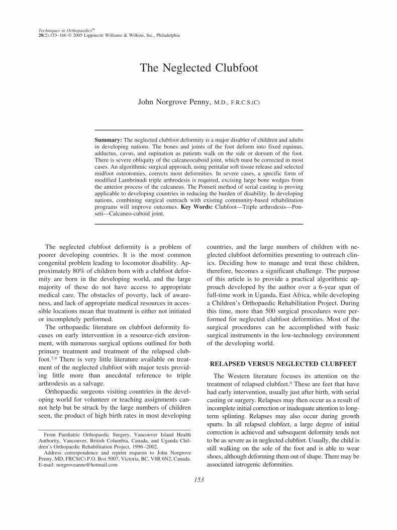
The neglected clubfoot, however, is one in which therehas been no initial treatment or perhaps very inadequateand incomplete initial treatment. The deformity is madeworse at the time the child starts to walk because weight-bearing takes place on the side or dorsum of the foot,exaggerating the abnormal shape and causing furtherdeformation. The contracted soft tissues on the medialside of the foot are encouraged to contract further. Thebones are compressed unnaturally at a time when theyare plastic and deform into abnormal shapes. Bones thatnormally support the arch of the midfoot now bear axialload, which they were never intended to do. The sole ofthe foot never experiences proper weightbearing, and it isimpossible to wear normal shoes. A thickened callousand large bursa develop over the prominent weightbear-ing head of the talus on the dorsolateral side of the foot,often associated with deep fissures, which are vulnerableto breakdown and infection (Fig. 1).
PATHOLOGIC ANATOMY
The primary contractile forces of the soft tissues inclubfoot deformity result in progressive bony deformity.These primary soft tissue and bony deformities havebeen well described in the dissections of Ponseti 28 and inthe magnetic resonance imaging studies of Pirani.27 Theneck of the talus develops a medial angulation and thehead of the talus becomes conical in shape. The navicularbone articulates with the medial aspect of the head of thetalus and becomes wedge-shaped. The calcaneocuboidjoint becomes oblique with medial subluxation of thecuboid on the calcaneus. The subtalar joint is held in itsmaximal position of supination (plantar flexion, inver-sion, and adduction). Early manipulation and cast treat-ment using the Ponseti technique shows cartilage remod-
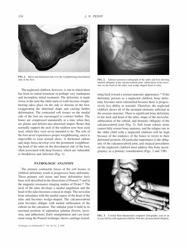
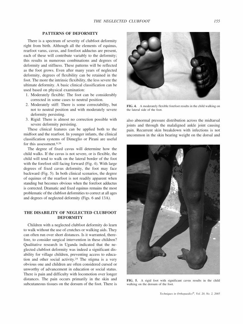
eling back toward a normal anatomic appearance.27 If thedeformity persists as a neglected clubfoot, bony defor-mity becomes more entrenched because there is progres-sively less ability to remodel. Therefore, the neglectedclubfoot shows all of the neonatal elements reflected inthe osseous structure. There is significant bony deformityin the neck and head of the talus, shape of the navicular,subluxation of the cuboid, and dramatic obliquity of thecalcaneocuboid joint (Fig. 2). Soft tissue release alonecannot fully restore bony anatomy, and the relapse rate inthe older child with a neglected clubfoot will be highbecause of the tendency of the bones to revert to theirdeformed position. Of particular importance is the obliq-uity of the calcaneocuboid joint, and surgical procedureson the neglected clubfoot must address this bony incon-gruency as a primary consideration (Figs. 3 and 13B)
FIG. 1. Bursa and thickened skin over the weightbearing dorsolateralside of the foot. FIG. 2. Anterior–posterior radiograph of the ankle and foot showing
marked obliquity at the calcaneocuboid joint, subluxation of the navic-ular on the head of the talus, and wedge-shaped head of talus.
FIG. 3. Coronal three-dimensional computed tomography scan in an8-year-old boy with neglected clubfoot. Note the calcaneocuboid obliquity.
154 J. N. PENNY
Techniques in Orthopaedics®, Vol. 20, No. 2, 2005
PATTERNS OF DEFORMITY
There is a spectrum of severity of clubfoot deformityright from birth. Although all the elements of equinus,rearfoot varus, cavus, and forefoot adductus are present,each of these will contribute variably to the deformity;this results in numerous combinations and degrees ofdeformity and stiffness. These patterns will be reflectedas the foot grows. Even after many years of neglecteddeformity, degrees of flexibility can be retained in thefoot. The more the intrinsic flexibility, the less severe theultimate deformity. A basic clinical classification can beused based on physical examination:1. Moderately flexible: The foot can be considerably
corrected in some cases to neutral position.2. Moderately stiff: There is some correctability, but
not to neutral position and with moderately severedeformity persisting.
3. Rigid: There is almost no correction possible withsevere deformity persisting.
These clinical features can be applied both to themidfoot and the rearfoot. In younger infants, the clinicalclassification systems of Dimeglio or Pirani are usefulfor this assessment.8,26
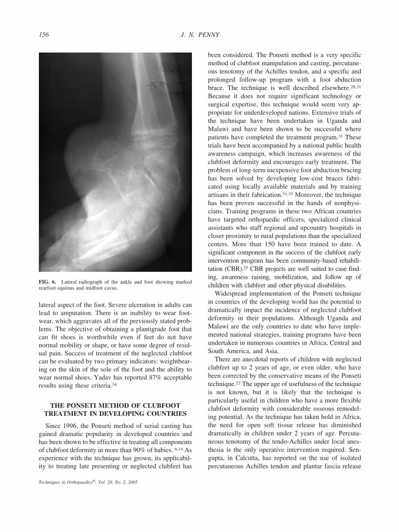
The degree of fixed cavus will determine how thechild walks. If the cavus is not severe, or is flexible, thechild will tend to walk on the lateral border of the footwith the forefoot still facing forward (Fig. 4). With largedegrees of fixed cavus deformity, the foot may facebackward (Fig. 5). In both clinical scenarios, the degreeof equinus of the rearfoot is not readily apparent whenstanding but becomes obvious when the forefoot adductusis corrected. Dramatic and fixed equinus remains the mostproblematic of the clubfoot deformities to correct at all agesand degrees of neglected deformity (Figs. 6 and 13A).
THE DISABILITY OF NEGLECTED CLUBFOOTDEFORMITY
Children with a neglected clubfoot deformity do learnto walk without the use of crutches or walking aids. Theycan often run over short distances. Is it warranted, there-fore, to consider surgical intervention in these children?Qualitative research in Uganda indicated that the ne-glected clubfoot deformity was indeed a significant dis-ability for village children, preventing access to educa-tion and other social activity.24 The stigma is a veryobvious one and children are often considered cursed orunworthy of advancement in education or social status.There is pain and difficulty with locomotion over longerdistances. The pain occurs primarily in the skin andsubcutaneous tissues on the dorsum of the foot. There is
also abnormal pressure distribution across the midtarsaljoints and through the malaligned ankle joint causingpain. Recurrent skin breakdown with infections is notuncommon in the skin bearing weight on the dorsal and
FIG. 4. A moderately flexible forefoot results in the child walking onthe lateral side of the foot.
FIG. 5. A rigid foot with significant cavus results in the childwalking on the dorsum of the foot.
155THE NEGLECTED CLUBFOOT
Techniques in Orthopaedics®, Vol. 20, No. 2, 2005
lateral aspect of the foot. Severe ulceration in adults canlead to amputation. There is an inability to wear foot-wear, which aggravates all of the previously stated prob-lems. The objective of obtaining a plantigrade foot thatcan fit shoes is worthwhile even if feet do not havenormal mobility or shape, or have some degree of resid-ual pain. Success of treatment of the neglected clubfootcan be evaluated by two primary indicators: weightbear-ing on the skin of the sole of the foot and the ability towear normal shoes. Yadav has reported 87% acceptableresults using these criteria.34
THE PONSETI METHOD OF CLUBFOOTTREATMENT IN DEVELOPING COUNTRIES
Since 1996, the Ponseti method of serial casting hasgained dramatic popularity in developed countries andhas been shown to be effective in treating all componentsof clubfoot deformity in more than 90% of babies. 6,14 Asexperience with the technique has grown, its applicabil-ity to treating late presenting or neglected clubfeet has
been considered. The Ponseti method is a very specificmethod of clubfoot manipulation and casting, percutane-ous tenotomy of the Achilles tendon, and a specific andprolonged follow-up program with a foot abductionbrace. The technique is well described elsewhere.28,31
Because it does not require significant technology orsurgical expertise, this technique would seem very ap-propriate for underdeveloped nations. Extensive trials ofthe technique have been undertaken in Uganda andMalawi and have been shown to be successful wherepatients have completed the treatment program.31 Thesetrials have been accompanied by a national public healthawareness campaign, which increases awareness of theclubfoot deformity and encourages early treatment. Theproblem of long-term inexpensive foot abduction bracinghas been solved by developing low-cost braces fabri-cated using locally available materials and by trainingartisans in their fabrication.31,32 Moreover, the techniquehas been proven successful in the hands of nonphysi-cians. Training programs in these two African countrieshave targeted orthopaedic officers, specialized clinicalassistants who staff regional and upcountry hospitals incloser proximity to rural populations than the specializedcenters. More than 150 have been trained to date. Asignificant component in the success of the clubfoot earlyintervention program has been community-based rehabili-tation (CBR).25 CBR projects are well suited to case find-ing, awareness raising, mobilization, and follow up ofchildren with clubfeet and other physical disabilities.
Widespread implementation of the Ponseti techniquein countries of the developing world has the potential todramatically impact the incidence of neglected clubfootdeformity in their populations. Although Uganda andMalawi are the only countries to date who have imple-mented national strategies, training programs have beenundertaken in numerous countries in Africa, Central andSouth America, and Asia.
There are anecdotal reports of children with neglectedclubfeet up to 2 years of age, or even older, who havebeen corrected by the conservative means of the Ponsetitechnique.22 The upper age of usefulness of the techniqueis not known, but it is likely that the technique isparticularly useful in children who have a more flexibleclubfoot deformity with considerable osseous remodel-ing potential. As the technique has taken hold in Africa,the need for open soft tissue release has diminisheddramatically in children under 2 years of age. Percuta-neous tenotomy of the tendo-Achilles under local anes-thesia is the only operative intervention required. Sen-gupta, in Calcutta, has reported on the use of isolatedpercutaneous Achilles tendon and plantar fascia release
FIG. 6. Lateral radiograph of the ankle and foot showing markedrearfoot equinus and midfoot cavus.
156 J. N. PENNY
Techniques in Orthopaedics®, Vol. 20, No. 2, 2005
followed by serial casting in large numbers of childrenup to walking age having neglected clubfeet.30
There is obviously a significant role for correctivecasting in newborns and young infants with clubfeet. InUganda, it was found useful to apply serial casts beforesurgical correction in all children up to age 12 withclubfeet. Casting allowed for stretching of the contractedtissues and skin on the medial side of the foot, reducingthe risk of postoperative skin necrosis. Some correctionof the bony deformity was achieved, minimizing surgicalresections. Protecting the skin in casts allowed for heal-ing of ulcers and fissures in the callus on the dorsolateralside of the foot. Preliminary casting, however, requiressupervision in rehabilitation facilities. This is often notpossible in rural upcountry outreach surgical situations.
GENERAL CONSIDERATIONS
The treatment of the neglected clubfoot is largelysurgical. The dilemma in the developing world is thelarge number of cases presenting with an extreme short-age of skilled surgeons. These surgeries are difficult andtime-consuming; postoperative care is prolonged andrequires access to bracing. There are difficulties withlimited anesthesia technology. It is preferable to delaysurgery until a child is at least 9 months of age becauseanesthesia is safer. It is also better to avoid pronepositioning on the operating table to make anestheticmonitoring easier. Malnutrition, anemia, and chronicdiseases such as malaria are common. Children live inunhygienic circumstances and the skin of the foot isoften ulcerated or infested with parasites. A preliminaryadmission to hospital or a rehabilitation unit is useful toallow for nutrition priming and treatment of skin lesions.Children are kept bedbound for 24 hours before surgeryand washed frequently to ensure cleanliness of the skin.In bilateral cases, it is usually best to do both feet at onesitting as a result of transportation and follow up diffi-culties in rural environments. This might be the child’sonly opportunity for correction. The use of pins andinternal fixation devices in upcountry outreach situationsmay be compromised because of concerns for infectionand pin care after the surgeons have left. CBR projects offerthe best support in postoperative care and follow up.
ALGORITHMIC APPROACH TO SURGICALTREATMENT OF THE NEGLECTED
CLUBFOOT
There is no single surgical procedure that can resolveall clubfoot scenarios. A full armamentarium of proce-dures must be at the ready. What is presented here is an
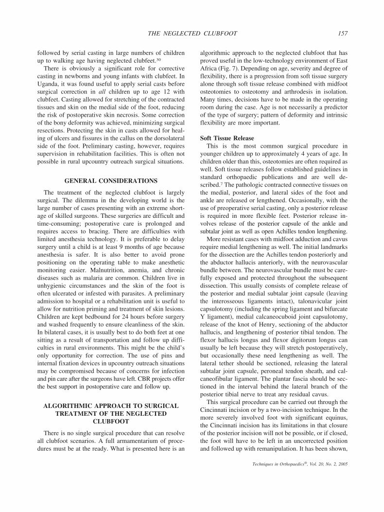
algorithmic approach to the neglected clubfoot that hasproved useful in the low-technology environment of EastAfrica (Fig. 7). Depending on age, severity and degree offlexibility, there is a progression from soft tissue surgeryalone through soft tissue release combined with midfootosteotomies to osteotomy and arthrodesis in isolation.Many times, decisions have to be made in the operatingroom during the case. Age is not necessarily a predictorof the type of surgery; pattern of deformity and intrinsicflexibility are more important.
Soft Tissue ReleaseThis is the most common surgical procedure in
younger children up to approximately 4 years of age. Inchildren older than this, osteotomies are often required aswell. Soft tissue releases follow established guidelines instandard orthopaedic publications and are well de-scribed.7 The pathologic contracted connective tissues onthe medial, posterior, and lateral sides of the foot andankle are released or lengthened. Occasionally, with theuse of preoperative serial casting, only a posterior releaseis required in more flexible feet. Posterior release in-volves release of the posterior capsule of the ankle andsubtalar joint as well as open Achilles tendon lengthening.
More resistant cases with midfoot adduction and cavusrequire medial lengthening as well. The initial landmarksfor the dissection are the Achilles tendon posteriorly andthe abductor hallucis anteriorly, with the neurovascularbundle between. The neurovascular bundle must be care-fully exposed and protected throughout the subsequentdissection. This usually consists of complete release ofthe posterior and medial subtalar joint capsule (leavingthe interosseous ligaments intact), talonavicular jointcapsulotomy (including the spring ligament and bifurcateY ligament), medial calcaneocuboid joint capsulotomy,release of the knot of Henry, sectioning of the abductorhallucis, and lengthening of posterior tibial tendon. Theflexor hallucis longus and flexor digitorum longus canusually be left because they will stretch postoperatively,but occasionally these need lengthening as well. Thelateral tether should be sectioned, releasing the lateralsubtalar joint capsule, peroneal tendon sheath, and cal-caneofibular ligament. The plantar fascia should be sec-tioned in the interval behind the lateral branch of theposterior tibial nerve to treat any residual cavus.
This surgical procedure can be carried out through theCincinnati incision or by a two-incision technique. In themore severely involved foot with significant equinus,the Cincinnati incision has its limitations in that closureof the posterior incision will not be possible, or if closed,the foot will have to be left in an uncorrected positionand followed up with remanipulation. It has been shown,
157THE NEGLECTED CLUBFOOT
Techniques in Orthopaedics®, Vol. 20, No. 2, 2005
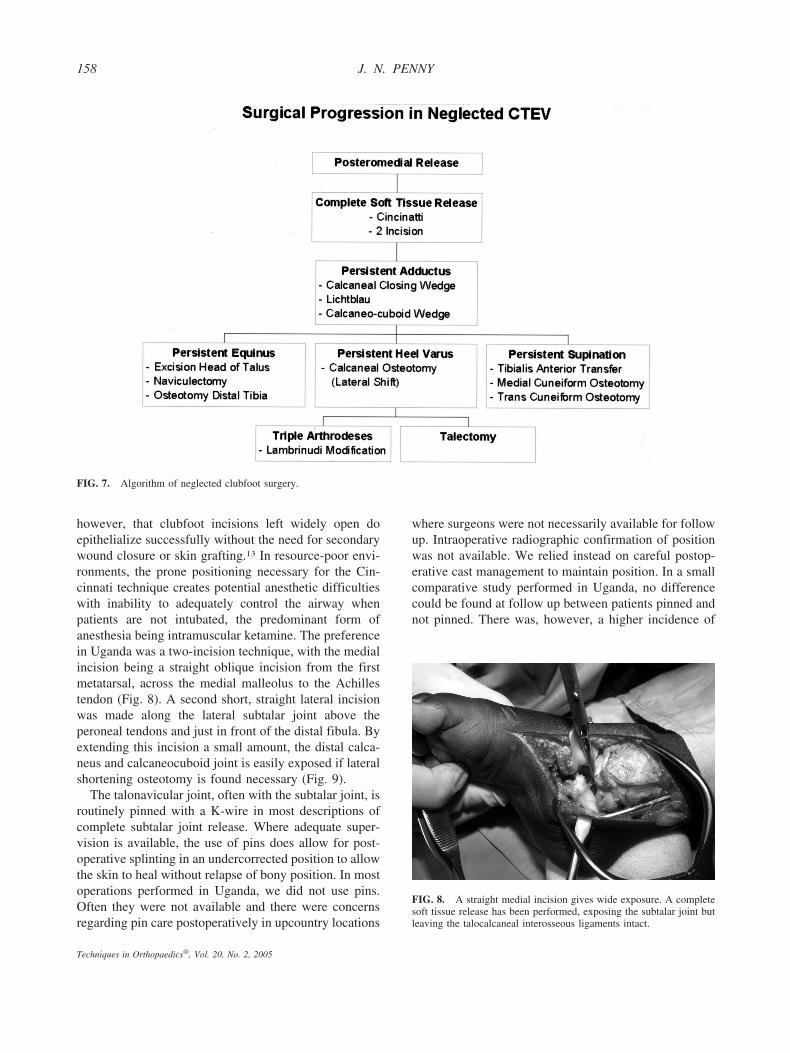
however, that clubfoot incisions left widely open doepithelialize successfully without the need for secondarywound closure or skin grafting.13 In resource-poor envi-ronments, the prone positioning necessary for the Cin-cinnati technique creates potential anesthetic difficultieswith inability to adequately control the airway whenpatients are not intubated, the predominant form ofanesthesia being intramuscular ketamine. The preferencein Uganda was a two-incision technique, with the medialincision being a straight oblique incision from the firstmetatarsal, across the medial malleolus to the Achillestendon (Fig. 8). A second short, straight lateral incisionwas made along the lateral subtalar joint above theperoneal tendons and just in front of the distal fibula. Byextending this incision a small amount, the distal calca-neus and calcaneocuboid joint is easily exposed if lateralshortening osteotomy is found necessary (Fig. 9).
The talonavicular joint, often with the subtalar joint, isroutinely pinned with a K-wire in most descriptions ofcomplete subtalar joint release. Where adequate super-vision is available, the use of pins does allow for post-operative splinting in an undercorrected position to allowthe skin to heal without relapse of bony position. In mostoperations performed in Uganda, we did not use pins.Often they were not available and there were concernsregarding pin care postoperatively in upcountry locations
where surgeons were not necessarily available for followup. Intraoperative radiographic confirmation of positionwas not available. We relied instead on careful postop-erative cast management to maintain position. In a smallcomparative study performed in Uganda, no differencecould be found at follow up between patients pinned andnot pinned. There was, however, a higher incidence of
FIG. 7. Algorithm of neglected clubfoot surgery.
FIG. 8. A straight medial incision gives wide exposure. A completesoft tissue release has been performed, exposing the subtalar joint butleaving the talocalcaneal interosseous ligaments intact.
158 J. N. PENNY
Techniques in Orthopaedics®, Vol. 20, No. 2, 2005
wound complications, specifically medial wound break-down, in the unpinned group indicating excessivestretching of skin in corrective plasters.3
Lateral transfer of the tibialis anterior tendon is usu-ally reserved for relapse after corrective cast manage-ment, usually after age 2 years once the lateral cuneiformhas begun to ossify.31 Because many children in poorerenvironments present for soft tissue release at an olderage, consideration should be given to tibialis anteriortransfer at the index procedure. This helps control anyresidual forefoot supination. It might also reduce the riskof relapse and dependency on lengthy postoperativeabduction bracing protocols. The tibialis anterior tendonattachment to the first metatarsal and medial cuneiformis easily accessible through the standard medial incisionused for soft tissue release. A proximal incision is notnecessary. A separate small incision is necessary over thelateral (third) cuneiform. The tendon is implanted into adrill hole with sutures tied over a button on the sole of thefoot.
Correcting Residual AdductusSoft tissue release alone may not fully correct the
deformity because of secondary bony deformity. If this isnot addressed at surgery, the foot will not only be leftincompletely corrected, but will relapse. The combina-tion of this soft tissue release with midfoot osteotomy isusually required in children between approximately 4and 12 years of age with neglected clubfeet. The bonylateral column is longer than the medial column, result-ing in midfoot adductus. Shortening osteotomy throughthe lateral column is required and should occur throughthe calcaneus or calcaneocuboid joint. The primary bony
pathology is obliquity of the calcaneocuboid joint andrelative lengthening of the lateral portion of the anteriorprocess of the calcaneus. Shortening should thereforeoccur through the distal calcaneus with an attempt tomake the calcaneocuboid joint transverse. Alternativesinclude closing wedge osteotomy through the anteriorprocess of the calcaneus leaving the articular surfaceintact, excision of the anterior process of calcaneus(Lichtblau procedure), or calcaneocuboid wedge resec-tion (Dilwynn-Evans procedure).12,21 Correction throughthe cuboid is not indicated because this does not addressthe primary pathology. In younger children, the Licht-blau procedure adequately shortens the lateral columnand allows for a pseudoarthrosis to develop after remod-eling. This potentially allows for more subtalar joint andmidfoot motion than a calcaneocuboid arthrodesis. Inolder children, more correction can be achieved by doinga calcaneocuboid wedge excision, and the arthrodesis soperformed may act as an epiphysiodesis to improve footposition with growth. Arthrodesis of this joint also po-tentially reduces the risk of relapse by providing apermanent solution but is associated with more potentialstiffness of the subtalar and midfoot articulations. Wherecalcaneocuboid wedge excision is done, internal fixationmay or may not be performed, depending on availabilityand surgeon preference.
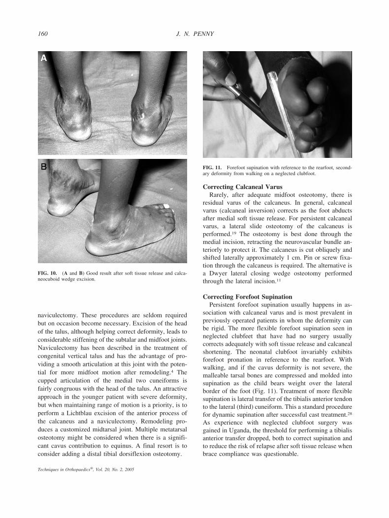
The combination of radical soft tissue release andcalcaneal shortening osteotomy is the most commonoperative procedure required in children between ages 4and 11, giving satisfactory results in the majority of cases(Fig. 10).
Correcting Residual EquinusIt is not uncommon to perform a full soft tissue release
in younger children only to find that the equinus defor-mity is not fully corrected. In this circumstance, the firstthing to check is adequacy of release of the lateral tether,consisting of the posterolateral subtalar joint capsule,peroneal tendon sheath, and the calcaneofibular liga-ment. These structures have been shown to be critical toobtaining dorsiflexion.29 If equinus deformity persists,the next step is lateral column shortening. Calcanealshortening is usually recommended to correct midfootadductus and the bean-shaped foot. Occasionally, how-ever, it is required to correct equinus. Lateral columnshortening allows a little more dorsiflexion through themidtarsal joint, although it does so by creating a mildrocker-bottom foot. In very severe cases, even thesesteps may not allow adequate dorsiflexion. This is usu-ally the result of impingement of the navicular againstthe head of the talus. Consideration must then be given toexcision of a portion of the head of the talus or to
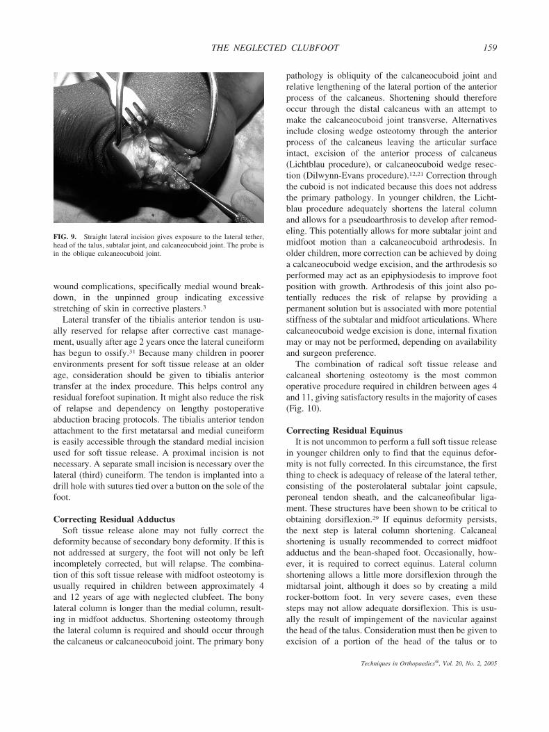
FIG. 9. Straight lateral incision gives exposure to the lateral tether,head of the talus, subtalar joint, and calcaneocuboid joint. The probe isin the oblique calcaneocuboid joint.
159THE NEGLECTED CLUBFOOT
Techniques in Orthopaedics®, Vol. 20, No. 2, 2005
naviculectomy. These procedures are seldom requiredbut on occasion become necessary. Excision of the headof the talus, although helping correct deformity, leads toconsiderable stiffening of the subtalar and midfoot joints.Naviculectomy has been described in the treatment ofcongenital vertical talus and has the advantage of pro-viding a smooth articulation at this joint with the poten-tial for more midfoot motion after remodeling.4 Thecupped articulation of the medial two cuneiforms isfairly congruous with the head of the talus. An attractiveapproach in the younger patient with severe deformity,but when maintaining range of motion is a priority, is toperform a Lichtblau excision of the anterior process ofthe calcaneus and a naviculectomy. Remodeling pro-duces a customized midtarsal joint. Multiple metatarsalosteotomy might be considered when there is a signifi-cant cavus contribution to equinus. A final resort is toconsider adding a distal tibial dorsiflexion osteotomy.
Correcting Calcaneal VarusRarely, after adequate midfoot osteotomy, there is
residual varus of the calcaneus. In general, calcanealvarus (calcaneal inversion) corrects as the foot abductsafter medial soft tissue release. For persistent calcanealvarus, a lateral slide osteotomy of the calcaneus isperformed.19 The osteotomy is best done through themedial incision, retracting the neurovascular bundle an-teriorly to protect it. The calcaneus is cut obliquely andshifted laterally approximately 1 cm. Pin or screw fixa-tion through the calcaneus is required. The alternative isa Dwyer lateral closing wedge osteotomy performedthrough the lateral incision.11
Correcting Forefoot SupinationPersistent forefoot supination usually happens in as-
sociation with calcaneal varus and is most prevalent inpreviously operated patients in whom the deformity canbe rigid. The more flexible forefoot supination seen inneglected clubfeet that have had no surgery usuallycorrects adequately with soft tissue release and calcanealshortening. The neonatal clubfoot invariably exhibitsforefoot pronation in reference to the rearfoot. Withwalking, and if the cavus deformity is not severe, themalleable tarsal bones are compressed and molded intosupination as the child bears weight over the lateralborder of the foot (Fig. 11). Treatment of more flexiblesupination is lateral transfer of the tibialis anterior tendonto the lateral (third) cuneiform. This a standard procedurefor dynamic supination after successful cast treatment.28
As experience with neglected clubfoot surgery wasgained in Uganda, the threshold for performing a tibialisanterior transfer dropped, both to correct supination andto reduce the risk of relapse after soft tissue release whenbrace compliance was questionable.
FIG. 10. (A and B) Good result after soft tissue release and calca-neocuboid wedge excision.
FIG. 11. Forefoot supination with reference to the rearfoot, second-ary deformity from walking on a neglected clubfoot.
160 J. N. PENNY
Techniques in Orthopaedics®, Vol. 20, No. 2, 2005
For rigid supination deformity, a complete midfootosteotomy is performed after the lateral shortening pro-cedure is completed. When the deformity is not severeand where the bones are of sufficient size, a plantarclosing wedge osteotomy of the medial cuneiform isdone. For more severe degrees of deformity, the osteot-omy is carried transversely through all the cuneiformsfreeing up the forefoot from the rearfoot. The forefootcan then be pronated on the rearfoot and fixed with pins.This is a difficult correction to achieve. These osteoto-mies may also be combined with transfer of the tibialisanterior tendon.
Triple ArthrodesisTriple arthrodesis is a versatile procedure used to
correct major deformities in children. In the developedworld, triple arthrodesis is used primarily as a salvageprocedure for pain after previous surgical correction.9 Inthe developing world context, with reference to ne-glected clubfeet, the procedure has its versatility incorrection of large degrees of deformity. In severe de-formity, particularly with marked cavus when the foot isfacing backward, soft tissue release and osteotomy isunlikely to achieve full correction (Fig. 5). In this case,triple arthrodesis can be very useful. Conventional or-thopaedic wisdom recommends triple arthrodesis not beperformed before advanced skeletal maturity, at age 10to 12, to avoid growth retardation by removal of cartilageinvolved in enchondral ossification. However, I haveused triple arthrodesis in children as young as age 6 andhave not seen adverse growth characteristics. Moreover,discrepancy in the size of the feet is seldom more than acosmetic nuisance and in bilateral cases not relevant.Although triple arthrodesis has been recommended as atreatment for the neglected clubfoot in standard texts,insufficient detail is given as to the precise method ofperforming the arthrodesis. In neglected clubfoot, a spe-cific method of arthrodesis is required with particularreference to correction of equinus. A modification of theclassic Lambrinudi triple arthrodeses, originally de-scribed for polio equinus deformity, is necessary.20 Ingeneral, resection through the talus should be minimizedbecause of its tenuous blood supply and most of thecorrection made through the calcaneus. The calcaneus isrich in blood supply with ample bone allowing for largewedge excisions for correction.
The Technique of Triple ArthrodesisA standard oblique Ollier-type incision is made on the
lateral side of the foot and the extensor digitorum brevisshelled out sharply from the sinus tarsi. The calcane-ocuboid, subtalar, and talonavicular joints are thus ex-
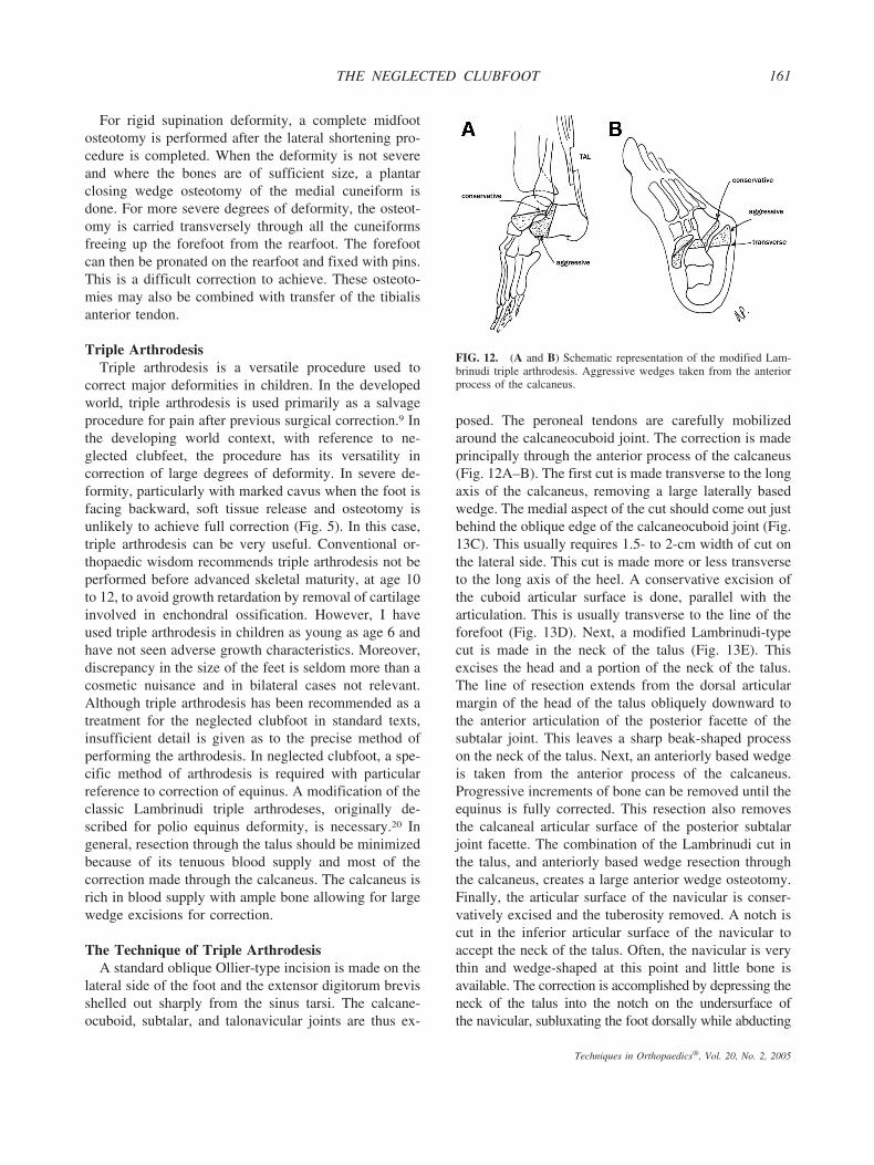
posed. The peroneal tendons are carefully mobilizedaround the calcaneocuboid joint. The correction is madeprincipally through the anterior process of the calcaneus(Fig. 12A–B). The first cut is made transverse to the longaxis of the calcaneus, removing a large laterally basedwedge. The medial aspect of the cut should come out justbehind the oblique edge of the calcaneocuboid joint (Fig.13C). This usually requires 1.5- to 2-cm width of cut onthe lateral side. This cut is made more or less transverseto the long axis of the heel. A conservative excision ofthe cuboid articular surface is done, parallel with thearticulation. This is usually transverse to the line of theforefoot (Fig. 13D). Next, a modified Lambrinudi-typecut is made in the neck of the talus (Fig. 13E). Thisexcises the head and a portion of the neck of the talus.The line of resection extends from the dorsal articularmargin of the head of the talus obliquely downward tothe anterior articulation of the posterior facette of thesubtalar joint. This leaves a sharp beak-shaped processon the neck of the talus. Next, an anteriorly based wedgeis taken from the anterior process of the calcaneus.Progressive increments of bone can be removed until theequinus is fully corrected. This resection also removesthe calcaneal articular surface of the posterior subtalarjoint facette. The combination of the Lambrinudi cut inthe talus, and anteriorly based wedge resection throughthe calcaneus, creates a large anterior wedge osteotomy.Finally, the articular surface of the navicular is conser-vatively excised and the tuberosity removed. A notch iscut in the inferior articular surface of the navicular toaccept the neck of the talus. Often, the navicular is verythin and wedge-shaped at this point and little bone isavailable. The correction is accomplished by depressing theneck of the talus into the notch on the undersurface ofthe navicular, subluxating the foot dorsally while abducting
FIG. 12. (A and B) Schematic representation of the modified Lam-brinudi triple arthrodesis. Aggressive wedges taken from the anteriorprocess of the calcaneus.
161THE NEGLECTED CLUBFOOT
Techniques in Orthopaedics®, Vol. 20, No. 2, 2005
the forefoot (Fig. 13F–H). With abduction, the heel varusspontaneously corrects. It is not usually necessary to per-form a laterally based wedge excision through the subtalarjoint to correct heel varus. An additional small amount ofequinus correction can be achieved by percutaneous or openlengthening of the Achilles tendon. Plantar fasciotomy canalso be added for cavus, performed through a short longi-tudinal incision in the midfoot.
The decision for triple arthrodesis should be madepreoperatively. An extensive medial release combinedwith triple arthrodesis potentially devascularizes the ta-lus. There is a natural desire to be more conservative inyounger children, but for severe deformity, if there is anyquestion, it is preferable to proceed directly to triplearthrodesis. Our experience in Uganda with severe de-
formities was that patients underdoing triple arthrodesisachieved better corrections than attempts at soft tissuerelease and osteotomy. Moreover, triple arthrodesis is astraightforward procedure that can be accomplished in amuch shorter timeframe than soft tissue dissection andrelease combined with osteotomy. Operative time con-straints are often a serious consideration in outreachsurgery, where anesthesia is questionable. The concernfor triple arthrodesis is the degree of stiffness it producesand the potential for growth arrest by excising enchon-dral growth centers. Experience has shown, however,that the degree of stiffness in the foot between triplearthrodesis and soft tissue release with midfoot osteot-omy is commensurate, and growth and size characteris-tics of feet seem similar as well.
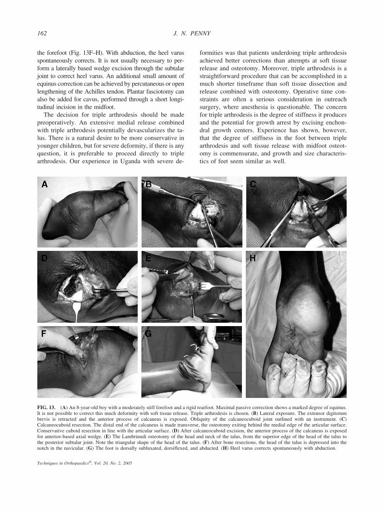
FIG. 13. (A) An 8-year-old boy with a moderately stiff forefoot and a rigid rearfoot. Maximal passive correction shows a marked degree of equinus.It is not possible to correct this much deformity with soft tissue release. Triple arthrodesis is chosen. (B) Lateral exposure. The extensor digitorumbrevis is retracted and the anterior process of calcaneus is exposed. Obliquity of the calcaneocuboid joint outlined with an instrument. (C)Calcaneocuboid resection. The distal end of the calcaneus is made transverse, the osteotomy exiting behind the medial edge of the articular surface.Conservative cuboid resection in line with the articular surface. (D) After calcaneocuboid excision, the anterior process of the calcaneus is exposedfor anterior-based axial wedge. (E) The Lambrinudi osteotomy of the head and neck of the talus, from the superior edge of the head of the talus tothe posterior subtalar joint. Note the triangular shape of the head of the talus. (F) After bone resections, the head of the talus is depressed into thenotch in the navicular. (G) The foot is dorsally subluxated, dorsiflexed, and abducted. (H) Heel varus corrects spontaneously with abduction.
162 J. N. PENNY
Techniques in Orthopaedics®, Vol. 20, No. 2, 2005
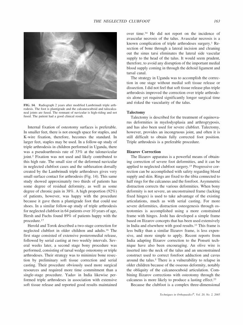
Internal fixation of osteotomy surfaces is preferable.In smaller feet, there is not enough space for staples, andK-wire fixation, therefore, becomes the standard. Inlarger feet, staples may be used. In a follow-up study oftriple arthrodesis in children performed in Uganda, therewas a pseudoarthrosis rate of 33% at the talonavicularjoint.2 Fixation was not used and likely contributed tothis high rate. The small size of the deformed navicularin neglected clubfoot cases and the subluxation dorsallycreated by the Lambrinudi triple arthrodeses gives verysmall surface contact for arthrodesis (Fig. 14). This samestudy showed approximately two thirds of patients hassome degree of residual deformity, as well as somedegree of chronic pain in 38%. A high proportion (92%)of patients, however, was happy with the procedurebecause it gave them a plantigrade foot that could useshoes. In a similar follow-up study of triple arthrodesisfor neglected clubfoot in 64 patients over 10 years of age,Hersh and Fuchs found 89% of patients happy with theprocedure.17
Herold and Torok described a two-stage correction forneglected clubfeet in older children and adults.16 Thefirst stage consisted of extensive posteromedial release,followed by serial casting at two weekly intervals. Sev-eral weeks later, a second stage bony procedure wasperformed, consisting of tarsal wedge osteotomy or triplearthrodeses. Their strategy was to minimize bone resec-tion by preliminary soft tissue correction and serialcasting. Their procedure obviously used more surgicalresources and required more time commitment than asingle-stage procedure. Yadav in India likewise per-formed triple arthrodeses in association with extensivesoft tissue release and reported good results maintained
over time.34 He did not report on the incidence ofavascular necrosis of the talus. Avascular necrosis is aknown complication of triple arthrodeses surgery.1 Re-section of bone through a lateral incision and cleaningout the sinus tarsi eliminates the lateral side vascularsupply to the head of the talus. It would seem prudent,therefore, to avoid any disruption of the important medialblood supply coming in through the deltoid ligament andtarsal canal.
The strategy in Uganda was to accomplish the correc-tion in one stage without medial soft tissue release ordissection. I did not feel that soft tissue release plus triplearthrodesis improved the correction over triple arthrode-sis alone yet required significantly longer surgical timeand risked the vascularity of the talus.
TalectomyTalectomy is described for the treatment of equinova-
rus deformities in myelodysplasia and arthrogryposis,and has also been used for severe clubfeet. Talectomy,however, provides an incongruous joint, and often it isstill difficult to obtain fully corrected foot position.Triple arthrodesis is a preferable procedure.
Ilizarov CorrectionThe Ilizarov apparatus is a powerful means of obtain-
ing correction of severe foot deformities, and it can beapplied to neglected clubfoot surgery.15 Progressive cor-rection can be accomplished with safety regarding bloodsupply and skin. Rings are fixed to the tibia connected tohalf rings for the calcaneus and the forefoot. Asymmetricdistraction corrects the various deformities. When bonydeformity is not severe, an unconstrained frame (lackingfixed hinges) is used to take advantage of the existingarticulations, much as with serial casting. For moresevere deformities, distraction osteogenesis through os-teotomies is accomplished using a more constrainedframe with hinges. Joshi has developed a simple framebased on Ilizarov concepts that has been used extensivelyin India and elsewhere with good results.33 This frame isless bulky than a similar Ilizarov frame, is less expen-sive, and more simple to apply. Recent reports fromIndia adapting Ilizarov correction to the Ponseti tech-nique have also been encouraging. An olive wire isinserted into the neck of the talus and an unconstrainedconstruct used to correct forefoot adduction and cavusaround the talus.5 There is a vulnerability to relapse inolder children because of the osseous deformity, notablythe obliquity of the calcaneocuboid articulation. Com-bining Ilizarov corrections with osteotomy through thecalcaneus is more likely to produce a lasting effect.23
Because the clubfoot is a complex three-dimensional
FIG. 14. Radiograph 2 years after modified Lambrinudi triple arth-rodesis. The foot is plantigrade and the calcaneocuboid and talocalca-neal joints are fused. The remnant of navicular is high-riding and notfused. The patient had a good clinical result.
163THE NEGLECTED CLUBFOOT
Techniques in Orthopaedics®, Vol. 20, No. 2, 2005
deformity, these are complex and difficult correctionsrequiring considerable expertise. Use of Ilizarov instru-mentation is limited in the developing world by the largenumber of cases requiring treatment, the lengthy operat-ing time involved, the availability of the instrumentationand intraoperative imaging, and the logistics of adequatepostoperative care. Although promising, this technique isnot appropriate for the majority of practitioners in thedeveloping world or for volunteer surgeons on short-term assignment. The foot after Ilizarov correction isvery stiff, as stiff as after osteotomy procedures. There isa functional advantage in a shorter stiff foot after triplearthrodesis as compared with a longer stiff foot afterIlizarov correction, and so triple arthrodesis remains theprocedure of choice in resource-poor environments.
SKIN COMPLICATIONS
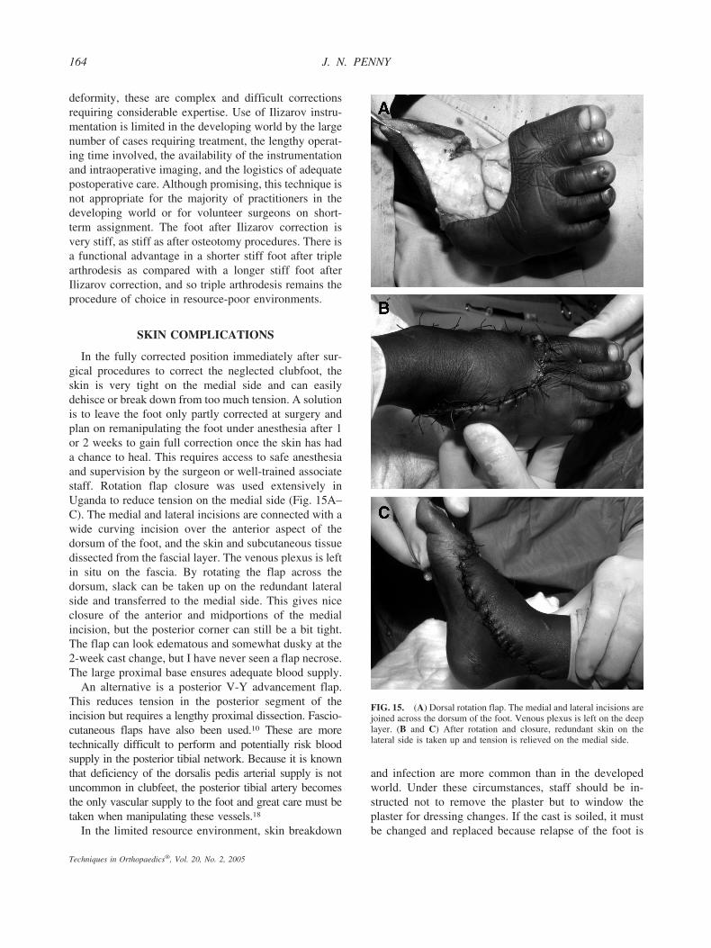
In the fully corrected position immediately after sur-gical procedures to correct the neglected clubfoot, theskin is very tight on the medial side and can easilydehisce or break down from too much tension. A solutionis to leave the foot only partly corrected at surgery andplan on remanipulating the foot under anesthesia after 1or 2 weeks to gain full correction once the skin has hada chance to heal. This requires access to safe anesthesiaand supervision by the surgeon or well-trained associatestaff. Rotation flap closure was used extensively inUganda to reduce tension on the medial side (Fig. 15A–C). The medial and lateral incisions are connected with awide curving incision over the anterior aspect of thedorsum of the foot, and the skin and subcutaneous tissuedissected from the fascial layer. The venous plexus is leftin situ on the fascia. By rotating the flap across thedorsum, slack can be taken up on the redundant lateralside and transferred to the medial side. This gives niceclosure of the anterior and midportions of the medialincision, but the posterior corner can still be a bit tight.The flap can look edematous and somewhat dusky at the2-week cast change, but I have never seen a flap necrose.The large proximal base ensures adequate blood supply.
An alternative is a posterior V-Y advancement flap.This reduces tension in the posterior segment of theincision but requires a lengthy proximal dissection. Fascio-cutaneous flaps have also been used.10 These are moretechnically difficult to perform and potentially risk bloodsupply in the posterior tibial network. Because it is knownthat deficiency of the dorsalis pedis arterial supply is notuncommon in clubfeet, the posterior tibial artery becomesthe only vascular supply to the foot and great care must betaken when manipulating these vessels.18
In the limited resource environment, skin breakdown
and infection are more common than in the developedworld. Under these circumstances, staff should be in-structed not to remove the plaster but to window theplaster for dressing changes. If the cast is soiled, it mustbe changed and replaced because relapse of the foot is
FIG. 15. (A) Dorsal rotation flap. The medial and lateral incisions arejoined across the dorsum of the foot. Venous plexus is left on the deeplayer. (B and C) After rotation and closure, redundant skin on thelateral side is taken up and tension is relieved on the medial side.
164 J. N. PENNY
Techniques in Orthopaedics®, Vol. 20, No. 2, 2005
dramatic when it is left unprotected. Satisfactory resultscan be salvaged in most cases either by skin grafting overthe granulating base or allowing epithelialization.
Superficial infection in the lateral wound is a commoncomplication in older children. This is the result of thedeep fissures in the skin over the lateral callus and skinredundancy with underlying dead space hematoma. Itusually resolves with local dressing changes and antibi-otics. The redundant callus on the lateral side of the footspontaneously disappears once there is no weightbearingover it. It can be ignored. Elliptic excision of redundantskin can be considered, but in my experience resulted inmore wound healing problems.
POSTOPERATIVE CARE
The results of treatment are only as good as thepostoperative care. There is no purpose in undertakingclubfoot surgery if postoperative plaster care and bracingsupport is inadequate because relapses will be invariableand will result in more serious disability than initially.Volunteer surgeons visiting the developing world on ashort-term assignment must ensure appropriate fol-low-up services before undertaking clubfoot surgery.
In the immediate postoperative period, vascular supplyto the foot is compromised and marked swelling is to beexpected. Feet should be kept elevated, particularly dur-ing the first 48 hours, and circulation to the toes checkedfrequently. Casts should be split if there is any concernabout undue swelling or sluggish circulation. Elevatingthe foot of the bed on blocks is particularly helpful.
Plaster changes need to be done expertly, maintainingfull correction and in some cases manipulating the footfurther. In the case of upcountry outreach surgery, localstaff needs to be trained adequately. For most clubfootsurgery, at least 3 months of immobilization is necessaryand sometimes longer if postoperative splinting is notavailable. Discharging children back to the village envi-ronment during this time, particularly if plaster of Paris isused, can be problematic with destruction of the plasterand subsequent loss of position. Construction of rehabil-itation hostels in outreach areas to house and supervisechildren has proved beneficial, particularly if attached toa community-based rehabilitation project. Wherever pos-sible, community-based rehabilitation principles shouldbe used.25
For younger children who have undergone soft tissuerelease, casting should be followed up with a foot ab-duction brace (FAB). Nighttime foot abduction bracesfor 2 or more years will reduce the risk of relapse. TheSteenbeek foot abduction brace has been developed inAfrica and is a low-cost appliance made of locally
available materials.31 Plastic ankle–foot orthoses (AFO)technology is not often available in remote areas but isparticularly useful in older children to help preventrelapse after soft tissue release and osteotomy.
It is impossible to obtain perfection in the treatment ofthe neglected clubfoot. The object should be to obtain afoot that is more or less plantigrade, with weightbearingon the plantar skin, and able to fit shoes. Childrenobtaining this result will be happy the majority of thetime.
REFERENCES
1. Angus PD, Cowell HR. Triple arthrodesis. A critical long termreview. J Bone Joint Surg [Br] 1986;68:260–265.
2. Bitariho D, Penny JN. Triple arthrodesis in children for severeneglected clubfoot deformity. Presented at the Association ofSurgeons of East Africa; December 3–5, 2003; Kampala.
3. Bitariho D. Short term outcome in one stage pantalar soft tissuerelease with transfixation of talonavicular joint in resistant idio-pathic congenital clubfoot [MMed (Orth) Thesis Dissertation].Makarere University; Uganda; April 2001.
4. Clark MW, D’Ambrosia RD, Ferguson AB. Congenital verticaltalus: treatment by open reduction and navicular excision. J BoneJoint Surg [Am] 1977;59:816–824.
5. Chaudhary M. Using Ponseti principles for external fixation treat-ment of older children with residual and recurrent clubfoot. Pre-sented at the Baltimore Limb Deformities Pre-course; September2003; Baltimore.
6. Cooper DM, Dietz FR. Treatment of idiopathic clubfoot: a thirtyyear follow-up note. J Bone Joint Surg [Am] 1995;77:1477–1489.
7. Cummings RJ, Davidson RS, Armstrong P, et al. Congenitalclubfoot. AAOS instructional course lecture. J Bone Joint Surg[Am] 2002;84:290–308.
8. Dimeglio A, Bensahel H, Souchet P, et al. Classification ofclubfoot. J Pediatr Orthop [lsbq]B] 1996;4:129–136.
9. Davidson RS. Clubfoot salvage: a review of the past decade’scontributions. J Pediatr Orthop 2003;23:410–418.
10. D’Souza MS, Aroojis MS, Yagnik MS. Rotation fasciocutaneousflap for neglected clubfoot: a new technique. J Pediatr Orthop1998;18:319–322.
11. Dwyer FC. Osteotomy of the calcaneum for pes cavus. J BoneJoint Surg [Br] 1959;41:80–86.
12. Evans D. Relapsed clubfoot. J Bone Joint Surg [Br] 1961;43:722–733.
13. Ferlic RJ, Breed AL, Mann DC, et al. Partial wound closure aftersurgical correction of equinovarus foot deformity. J Pediatr Or-thop 1997;17:486–489.
14. Herzenberg JE, Radler C, Bor N. Ponseti versus traditional meth-ods of casting for idiopathic clubfoot. J Pediatr Orthop 2002;22:517–521.
15. Heurta F. Correction of the neglected clubfoot by the Ilizarovtechnique. Clin Orthop 1994;301:89–93.
16. Herold HV, Torok G. Surgical correction of neglected club foot inthe older child and adult. J Bone Joint Surg [Am] 1973;55:1385–1395.
17. Hersh A, Fuchs LA. Treatment of the uncorrected clubfoot bytriple arthrodeses. Orthop Clin North Am 1973;4:103–115.
18. Hootnik DR, Levinsohn EM, Crider RJ, et al. Congenital arterialmalformations associated with clubfoot. A report of two cases.Clin Orthop 1982;167:160–163.
19. Koutsogiannis E. Treatment of mobile flat foot by displacementosteotomy of the calcaneus. J Bone Joint Surg [Br] 1971;53:96–100.
165THE NEGLECTED CLUBFOOT
Techniques in Orthopaedics®, Vol. 20, No. 2, 2005
20. Lambrinudi C. New operation on drop-foot. Br J Surg 1927;15:193.
21. Lichtblau S. A medial and lateral release operation for clubfoot. JBone Joint Surg [Am] 1973;55:1377–1384.
22. Lourenco AF, Prata SD, Sodre H, et al. Conservative treatment ofclubfoot after walking age. Presented at the Third InternationalClubfoot Congress; August 27–28, 2002; San Diego
23. Paley D. The correction of complex foot deformities using Il-izarov’s distraction osteotomies. Clin Orthop 1993;293:97–111.
24. Penny A. School access: children with motor disabilities in ruralUganda [MA Thesis Dissertation]. University of Victoria; 2001.
25. Penny JN, Steenbeek M. Community based rehabilitation (CBR) inthe management of clubfoot deformity in Africa. Presented at theThird International Clubfoot Congress; August 27–28, 2002; SanDiego.
26. Pirani S. A reliable and valid method of assessing the amount ofdeformity in the congenital clubfoot. Presented at the PediatricOrthopaedic Society of North America; May 2004; St. Louis.
27. Pirani S, Zeznik L, Hodges D. Magnetic resonance imaging studyof the congenital clubfoot treated with the Ponseti method. J Pe-diatr Orthop 2001;21:719–726.
28. Ponseti IV. Congenital Clubfoot: Fundamentals for Treatment.Oxford: Oxford University Press; 1996.
29. Scott WA, Hosking SW, Catterall A. Club foot. Observations onthe surgical anatomy of dorsiflexion. J Bone Joint Surg [Br]1984;66:71–76.
30. Sengupta A. The management of congenital talipes equinovarus indeveloping countries. Int Orthop 1987;11:183–187.
31. Staheli L, ed. Clubfoot: Ponseti Management. Seattle: Global-HELP Publications, 2003. Available at: http://www.global-help.org/publications/ponseti-cf.html.
32. Steenbeek M. The Steenbeek foot abduction brace (SFAB): pre-venting recurrence of deformity in developing nations duringtreatment of congenital clubfoot by the Ponseti method. Presentedat the Third International Clubfoot Congress; August 27–28, 2002;San Diego.
33. Suresh S, Ahmed A, Sharma VK. Role of Joshi’s external stabili-zation system fixator in the management of idiopathic clubfoot.J Orthop Surg (Hong Kong) 2003;11:194–201.
34. Yadav SS. Observations on operative management of neglectedclubfoot. Int Orthop 1981;5:189–192.
166 J. N. PENNY
Techniques in Orthopaedics®, Vol. 20, No. 2, 2005


![Clubfoot: Ponseti Management [Italian]](https://img.pdfslide.net/doc/110x75/613d460c736caf36b75b61e2/clubfoot-ponseti-management-italian.jpg)


![The Neglected Clubfoot [Indonesian] - Global HELP](https://img.pdfslide.net/doc/110x75/586360f81a28ab0e3090549d/the-neglected-clubfoot-indonesian-global-help.jpg)