Embed Size (px)
Citation preview
© 2012 Pearson Education, Inc.
17 The Nervous System:
Autonomic Nervous
System
PowerPoint® Lecture Presentations prepared by
Steven Bassett
Southeast Community College
Lincoln, Nebraska

© 2012 Pearson Education, Inc.
Introduction
• The autonomic nervous system functions
outside of our conscious awareness
• The autonomic nervous system makes
routine adjustments in our body’s systems
• The autonomic nervous system:
• Regulates body temperature
• Coordinates cardiovascular, respiratory,
digestive, excretory, and reproductive
functions

© 2012 Pearson Education, Inc.
A Comparison of the Somatic and Autonomic
Nervous Systems
• Autonomic nervous system
• Axons innervate the visceral organs
• Has afferent and efferent neurons
• Afferent pathways originate in the visceral receptors
• Somatic nervous system
• Axons innervate the skeletal muscles
• Has afferent and efferent neurons
• Afferent pathways originate in the skeletal muscles
ANIMATION The Organization of the Somatic and
Autonomic Nervous Systems

© 2012 Pearson Education, Inc.
Subdivisions of the ANS
• The autonomic nervous system consists of
two major subdivisions
• Sympathetic division
• Also called the thoracolumbar division
• Known as the “fight or flight” system
• Parasympathetic division
• Also called the craniosacral division
• Known as the “rest and repose” system

© 2012 Pearson Education, Inc.
Figure 17.1b Components and Anatomic Subdivisions of the ANS (Part 1 of 2)
Anatomical subdivisions. At the thoracic and lumbar
levels, the visceral efferent fibers that emerge form the
sympathetic division, detailed in Figure 17.4. At the
cranial and sacral levels, the visceral efferent fibers
from the CNS form the parasympathetic division,
detailed in Figure 17.8.
Thoracic
nerves
T1
T2
T3
T4
T5
T6
T7
T8
AUTONOMIC NERVOUS SYSTEM
THORACOLUMBAR DIVISION
(sympathetic
division of ANS)
CRANIOSACRAL DIVISION
(parasympathetic
division of ANS)
Cranial nerves
(N III, N VII, N IX,
and N X)

© 2012 Pearson Education, Inc.
Figure 17.1b Components and Anatomic Subdivisions of the ANS (Part 2 of 2)
Anatomical subdivisions. At the thoracic and lumbar
levels, the visceral efferent fibers that emerge form the
sympathetic division, detailed in Figure 17.4. At the
cranial and sacral levels, the visceral efferent fibers
from the CNS form the parasympathetic division,
detailed in Figure 17.8.
Sacral
nerves
(S2, S3, S4 only)
Lumbar
nerves (L1, L2 only)
Thoracic
nerves
T10
T11
T12
L1
L2
L3
L4
L5
S1
S2
S3
S4
S5
T9

© 2012 Pearson Education, Inc.
Subdivisions of the ANS
• Sympathetic division
• Thoracic and upper lumbar nerves synapse in
ganglia near the spinal cord
• Sympathetic activation results in:
• Increased metabolism and alertness
• Parasympathetic division
• Synapses are located near the target organ
• Parasympathetic activation results in:
• Energy conservation
© 2012 Pearson Education, Inc.
Subdivisions of the ANS
• Sympathetic division
• All preganglionic fibers release acetylcholine.
The effects are stimulatory.
• Most postganglionic fibers release
norepinephrine.The effects are stimulatory.
• Parasympathetic division
• All preganglionic fibers release acetylcholine.
The effects are stimulatory.
• Postganglionic fibers release acetylcholine but
the effects can be inhibitory.

© 2012 Pearson Education, Inc.
Figure 17.1a Components and Anatomic Subdivisions of the ANS
AUTONOMIC NERVOUS SYSTEM
Consists of 2 divisions
SYMPATHETIC
(thoracolumbar)
DIVISION
PARASYMPATHETIC
(craniosacral)
DIVISION
Preganglionic
neurons in
lateral gray horns of
spinal segments T1–L2
Preganglionic neurons in brain
stem and in lateral portion of anterior
gray horns of S2–S4
Send preganglionic
fibers to
Which send postganglionic
fibers to
Ganglia near
spinal cord
Ganglia in or
near target organs
Preganglionic fibers release ACh (excitatory),
stimulating ganglionic neurons
Preganglionic fibers release ACh (excitatory),
stimulating ganglionic neurons
Target organs Target organs
Most postganglionic fibers release NE at
neuroeffector junctions
All postganglionic fibers release ACh at
neuroeffector junctions
“Fight or flight”
response “Rest and repose”
response
Functional components of the ANS
© 2012 Pearson Education, Inc.
The Sympathetic Division
• Sympathetic division consists of:
• Preganglionic neurons between T1 and L2
• Two types of ganglionic neurons near the
vertebral columns: sympathetic chain
ganglia (lateral to the vertebral column) and
collateral ganglia (anterior to the vertebral
column)
• Specialized neurons in the interior of the
suprarenal gland

© 2012 Pearson Education, Inc.
The Sympathetic Division
• Sympathetic division
• Preganglionic neurons
• Cell bodies are in the lateral gray horns
• Axons enter the ventral roots
• Sympathetic chain ganglia (paravertebral
ganglia)
• Control effectors in the body wall, head, neck,
limbs, and thoracic cavity

© 2012 Pearson Education, Inc.
The Sympathetic Division
•Sympathetic division
• Collateral ganglia (prevertebral ganglia)
• Neurons innervate effectors in the abdominopelvic
cavity
• Specialized neurons
• Modified sympathetic ganglion in the suprarenal
gland
• Neurons release neurotransmitters that act like
hormones

© 2012 Pearson Education, Inc.
Figure 17.2 Organization of the Sympathetic Division of the ANS
Preganglionic fibers
Postganglionic fibers
Hormones released
into circulation
KEY
Lateral gray
horns of spinal
segments
T1–L2
Sympathetic
chain ganglia
(paired)
Collateral
ganglia
(unpaired)
Suprarenal
medullae
(paired)
Preganglionic Neurons
Ganglionic Neurons
Innervation by
postganglionic
fibers
Through release of
hormones into
the circulation
Target Organs
Visceral effectors
in thoracic cavity,
head, body wall,
and limbs
Visceral effectors
in abdominopelvic
cavity
Organs and systems
throughout body
Sympathetic Division of ANS

© 2012 Pearson Education, Inc.
The Sympathetic Division
• Sympathetic Chain Ganglia
• The ventral root joins a dorsal root
• Forms a spinal nerve
• Passes through an intervertebral foramen
• White ramus branches off the spinal nerve
• Goes to a nearby sympathetic chain ganglion

© 2012 Pearson Education, Inc.
The Sympathetic Division
• Functions of Sympathetic Chain Ganglia
• Reduction of circulation to the skin
• More circulation to skeletal muscles
• Stimulates more energy production by skeletal
muscles
• Releases stored adipose
• Stimulation of arrector pili muscles
• Dilation of pupils
• Increased heart rate
• Dilation of respiratory tubes
© 2012 Pearson Education, Inc.
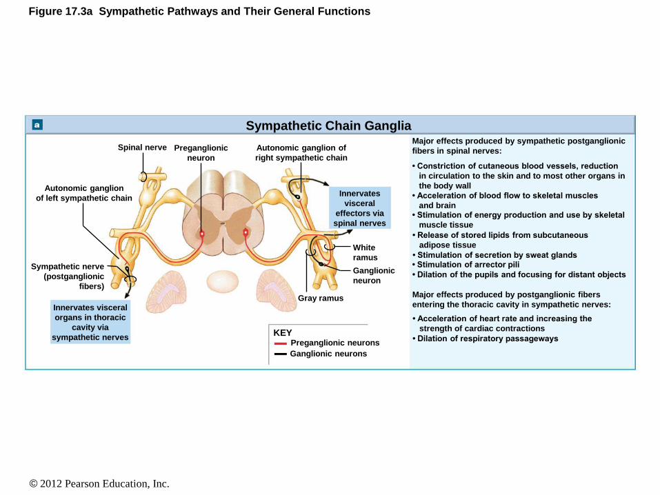
Figure 17.3a Sympathetic Pathways and Their General Functions
Sympathetic Chain Ganglia
KEY Preganglionic neurons
Ganglionic neurons
Innervates visceral
organs in thoracic
cavity via
sympathetic nerves
Sympathetic nerve
(postganglionic
fibers)
Autonomic ganglion
of left sympathetic chain
Spinal nerve Preganglionic
neuron
Autonomic ganglion of
right sympathetic chain
Innervates
visceral
effectors via
spinal nerves
White
ramus
Ganglionic
neuron
Gray ramus
Major effects produced by sympathetic postganglionic
fibers in spinal nerves:
Major effects produced by postganglionic fibers
entering the thoracic cavity in sympathetic nerves:
• Constriction of cutaneous blood vessels, reduction
in circulation to the skin and to most other organs in
the body wall
• Acceleration of blood flow to skeletal muscles
and brain • Stimulation of energy production and use by skeletal
muscle tissue
• Release of stored lipids from subcutaneous
adipose tissue
• Stimulation of secretion by sweat glands • Stimulation of arrector pili
• Dilation of the pupils and focusing for distant objects
• Acceleration of heart rate and increasing the
strength of cardiac contractions
• Dilation of respiratory passageways
© 2012 Pearson Education, Inc.
The Sympathetic Division
• Anatomy of the Sympathetic Chain Ganglia
• Each spinal nerve consists of:
• Preganglionic and postganglionic fibers
• There are:
• cervical sympathetic chain ganglia
• thoracic sympathetic chain ganglia
• lumbar sympathetic chain ganglia
• sacral sympathetic chain ganglia
• coccygeal sympathetic chain ganglia

© 2012 Pearson Education, Inc.
Figure 17.4 Anatomical Distribution of Sympathetic Postganglionic Fibers
Superior
Middle
Inferior
PONS
Cervical
sympathetic
ganglia
Gray rami to
spinal nerves
Greater
splanchnic
nerve
Superior
mesenteric
ganglion
Cardiac and
pulmonary
plexuses
Celiac ganglion
Sympathetic nerves
T1 T1
T2 T2
T3 T3
T4 T4
T5 T5
T6 T6
T7 T7
T8 T8
T9 T9
T10 T10
T11 T11
T12 T12
L1 L1
L2 L2
L3 L3
L4 L4
L5 L5 S1 S1 S2 S2
S3 S3 S4 S4
S5 S5
Postganglionic fibers
to spinal nerves
(innervating skin, blood
vessels, sweat glands,
arrector pili muscles,
adipose tissue)
Sympathetic
chain ganglia
Spinal cord
Coccygeal
ganglia (Co1)
fused together
(ganglion impar)
Preganglionic neurons
Ganglionic neurons
KEY
Uterus Ovary
Sacral
splanchnic
nerves
Lesser
splanchnic
nerve
Lumbar
splanchnic nerves Inferior
mesenteric
ganglion
Penis Scrotum Urinary bladder
Kidney
Suprarenal
medulla
Small intestine
Large intestine
Pancreas
Spleen
Stomach
Liver and
gallbladder
Lung
Heart
Eye
Salivary
glands
© 2012 Pearson Education, Inc.
The Sympathetic Division
• Collateral Ganglia
• Preganglionic neurons originate in the inferior
thoracic and superior lumbar areas of the spinal
cord
• Fibers pass through the sympathetic chain
ganglia without synapsing
• Converge to form the greater, lesser, and
lumbar splanchnic nerves
• Splanchnic nerves converge on the collateral
ganglia

© 2012 Pearson Education, Inc.
The Sympathetic Division
• Functions of the Collateral Ganglia
• Reduction of flow of blood to the visceral organs
• Decrease in activity of the digestive organs
• Stimulation of the release of glucose from glycogen in the liver
• Stimulates adipose cells to release energy reserves
• Relaxation of smooth muscles in the urinary bladder
• Cause ejaculation in males

© 2012 Pearson Education, Inc.
Figure 17.3b Sympathetic Pathways and Their General Functions
Collateral Ganglia
Lateral gray horn
White
ramus
Collateral
ganglion Postganglionic
fibers
Splanchnic nerve
(preganglionic
fibers)
Innervates
visceral organs in
abdominopelvic
cavity
Major effects produced by preganglionic fibers
innervating the collateral ganglia:
• Constriction of small arteries and reduction in the
flow of blood to visceral organs
• Decrease in the activity of digestive glands and
organs
• Stimulation of the release of glucose from glycogen
reserves in the liver • Stimulation of the release of lipids from adipose
tissue • Relaxation of the smooth muscle in the wall of the
urinary bladder
• Reduction of the rate of urine formation at the
kidneys
• Control of some aspects of sexual function, such as
ejaculation in males
© 2012 Pearson Education, Inc.
The Sympathetic Division
• Anatomy of the Collateral Ganglia
• Splanchnic nerves innervate:
• Celiac ganglion: fibers innervate the stomach,
duodenum, liver, gallbladder, pancreas, spleen, and
kidney
• Superior mesenteric ganglion: fibers innervate the
small intestine and the first parts of the large
intestine
• Inferior mesenteric ganglion: fibers innervate the
kidney, urinary bladder, sex organs, and terminal
ends of the large intestine
© 2012 Pearson Education, Inc.
The Sympathetic Division
• Suprarenal Medullae
• Fibers pass through sympathetic chain and the
celiac ganglion without synapsing
• Proceed to the suprarenal medulla
• Fibers then synapse on modified neurons that
when stimulated will release neurotransmitters
that act as hormones:
• Epinephrine and norepinephrine

© 2012 Pearson Education, Inc.
The Sympathetic Division
• Functions of the suprarenal medullae
• Increase alertness by activating the reticular
activating system
• Increase cardiovascular and respiratory activity
• Increase muscle tone
• Increase the mobilization of energy reserves
• Increased release of lipids from adipose cells
• Increased breakdown of glycogen in liver cells

© 2012 Pearson Education, Inc.
Figure 17.3c Sympathetic Pathways and Their General Functions
The Suprarenal Medullae
Secretes
neurotransmitters
into general
circulation
Suprarenal
medullae
Preganglionic fibers
Endocrine cells
(specialized ganglionic
neurons)
• Release of epinephrine and norepinephrine into
the general circulation
Major effect produced by preganglionic fibers
innervating the suprarenal medullae:

© 2012 Pearson Education, Inc.
Figure 17.5a Suprarenal Medulla
Relationship of a suprarenal gland to a kidney
Cortex
Medulla
Suprarenal
gland
Right
kidney

© 2012 Pearson Education, Inc.
Figure 17.5ab Suprarenal Medulla
Relationship of a suprarenal gland to a kidney Histology of the suprarenal medulla,
a modified sympathetic ganglion
Suprarenal medulla LM 426
Capillaries Modified neurons
(sympathetic ganglion cells)
of suprarenal medulla
Nucleolus
in nucleus
Cortex
Medulla

© 2012 Pearson Education, Inc.
The Sympathetic Division
• Sympathetic activation and neurotransmitter release
• Sympathetic ganglion fibers release acetylcholine at
the synapse with ganglionic neurons
• These are cholinergic synapses
• The stimulation of ganglionic neurons causes the
release of norepinephrine at the neuroeffector
junction
• These terminals are adrenergic
• Some ganglionic neurons also release acetylcholine
• Especially at the neuroeffector junctions of skeletal
muscles

© 2012 Pearson Education, Inc.
Figure 17.6 Sympathetic Postganglionic Nerve Endings Preganglionic fiber
(myelinated) Ganglionic
neuron
Ganglion
Postganglionic fiber
(unmyelinated)
Varicosities
Mitochondrion
5 m
Vesicles containing
norepinephrine
Schwann cell
cytoplasm
Smooth muscle cells Varicosities

© 2012 Pearson Education, Inc.
The Sympathetic Division
• Summary of the Sympathetic Division
• Consists of parallel chains on either side of the spinal
cord
• Preganglionic fibers are short and extend from the
spinal cord to the sympathetic chain
• Postganglionic fibers are long and extend from the
spinal cord to the body organs
• The sympathetic division shows considerable
divergence
• All preganglionic neurons release ACh / most
postganglionic neurons release norepinephrine

© 2012 Pearson Education, Inc.
Figure 17.4 Anatomical Distribution of Sympathetic Postganglionic Fibers
Superior
Middle
Inferior
PONS
Cervical
sympathetic
ganglia
Gray rami to
spinal nerves
Greater
splanchnic
nerve
Superior
mesenteric
ganglion
Cardiac and
pulmonary
plexuses
Celiac ganglion
Sympathetic nerves
T1 T1
T2 T2
T3 T3
T4 T4
T5 T5
T6 T6
T7 T7
T8 T8
T9 T9
T10 T10
T11 T11
T12 T12
L1 L1
L2 L2
L3 L3
L4 L4
L5 L5 S1 S1 S2 S2
S3 S3 S4 S4
S5 S5
Postganglionic fibers
to spinal nerves
(innervating skin, blood
vessels, sweat glands,
arrector pili muscles,
adipose tissue)
Sympathetic
chain ganglia
Spinal cord
Coccygeal
ganglia (Co1)
fused together
(ganglion impar)
Preganglionic neurons
Ganglionic neurons
KEY
Uterus Ovary
Sacral
splanchnic
nerves
Lesser
splanchnic
nerve
Lumbar
splanchnic nerves Inferior
mesenteric
ganglion
Penis Scrotum Urinary bladder
Kidney
Suprarenal
medulla
Small intestine
Large intestine
Pancreas
Spleen
Stomach
Liver and
gallbladder
Lung
Heart
Eye
Salivary
glands

© 2012 Pearson Education, Inc.
The Sympathetic Division
ANIMATION The Distribution of Sympathetic Innervation

© 2012 Pearson Education, Inc.
The Parasympathetic Division
• Parasympathetic Division
• Preganglionic neurons are in the brain stem and
sacral segments
• Preganglionic neurons do not diverge as much
as the sympathetic division
• Therefore, the parasympathetic division is more
localized and specific as compared to the
sympathetic division
• Postganglionic neurons are near (terminal) the
target organ or within (intramural) the target
organ
© 2012 Pearson Education, Inc.
The Parasympathetic Division
• Organization and Anatomy of the
Parasympathetic Division
• Preganglionic fibers leave the brain via:
• CN III (to the intrinsic eye muscles, pupil, and lens)
• CN VII (to the tear glands and salivary glands)
• CN IX (to the parotid salivary glands)
• CN X (to the visceral organs of the thoracic cavity
and abdominal cavity)
• Preganglionic fibers leave the sacral region via:
• Pelvic nerves (to the visceral organs in the inferior
portion of the abdominopelvic cavity

© 2012 Pearson Education, Inc.
Figure 17.7 Organization of the Parasympathetic Division of the ANS
Parasympathetic Division of ANS
Preganglionic Neurons Ganglionic Neurons Target Organs
Ciliary ganglion
Pterygopalatine
and submandibular
ganglia
Otic ganglion
Intramural ganglia
Intrinsic eye muscles (pupil and lens shape)
Nasal glands, tear glands, and salivary
glands
Visceral organs of neck,
thoracic cavity, and most of
abdominal cavity
N III
N VII
N IX
N X
Nuclei in brain stem
Nuclei in spinal cord segments
S2–S4
Pelvic nerves Intramural
ganglia
Visceral organs in inferior portion of
abdominopelvic cavity
KEY
Preganglionic fibers
Postganglionic fibers
Parotid salivary gland

© 2012 Pearson Education, Inc.
Figure 17.8 Autonomic Distribution of the Parasympathetic Output Pterygopalatine ganglion
PONS
N III
N VII
N IX
N X (Vagus)
Ciliary ganglion
Submandibular
ganglion
Otic ganglion
Autonomic plexuses
(see Figure 17.9)
Pelvic
nerves
Lacrimal gland
Eye
Salivary glands
Heart
Lungs
Liver and
gallbladder
Stomach
Spleen
Pancreas
Large intestine
Small intestine
Rectum
Kidney
Urinary bladder Scrotum Penis Ovary Uterus
S2
S3
S4
Spinal
cord
Preganglionic neurons
Ganglionic neurons
KEY

© 2012 Pearson Education, Inc.
The Parasympathetic Division
• Functions of the Parasympathetic Division
• Pupil constriction
• Secretion of digestive enzymes from digestive glands
• Increased smooth muscle activity of the digestive system
• Stimulation and coordination of defecation
• Contraction of the urinary bladder
• Constriction of respiratory passages
• Reduced heart rate
• Sexual arousal

© 2012 Pearson Education, Inc.
The Parasympathetic Division
• Parasympathetic Activation and
Neurotransmitter Release
• All preganglionic and postganglionic fibers
release ACh at their synapses and
neuroeffector junctions
• Most stimulations are short lived due to the
immediate breakdown of ACh by
acetylcholinesterase

© 2012 Pearson Education, Inc.
The Parasympathetic Division
• Plasmalemma Receptors and Responses
• Two types of ACh receptors are found on the postsynaptic plasmalemmae: • Nicotinic receptors: respond to nicotine
• Found on surfaces of parasympathetic and sympathetic ganglionic neurons
• Muscarinic receptors: respond to muscarine
• Found on surfaces of parasympathetic cholinergic neuroeffector junctions

© 2012 Pearson Education, Inc.
The Parasympathetic Division
• Summary of the Parasympathetic Division
• Involves CN III, CN VII, CN IX, and CN X
• Involves sacral segments S2 to S4
• All parasympathetic neurons are cholinergic
• Release of ACh stimulates nicotinic receptors
on ganglionic neurons
• Release of ACh on neuroeffector junctions
stimulates muscarinic receptors

© 2012 Pearson Education, Inc.
The Parasympathetic Division
ANIMATION The Distribution of Parasympathetic Innervation

© 2012 Pearson Education, Inc.
Relationships between the Sympathetic and
Parasympathetic Divisions
• Sympathetic
• Widespread effect on visceral organs
• Parasympathetic
• Modifies the activity of structures innervated by
specific cranial nerves and pelvic nerves
• Most vital organs are innervated by both the
sympathetic and parasympathetic nerves
• The two often oppose (antagonistic) each other

© 2012 Pearson Education, Inc.
Figure 17.10 A Comparison of the Sympathetic and Parasympathetic Divisions
Sympathetic Parasympathetic
CNS
PNS
KEY
Sympathetic
ganglion
Circulatory
system
Preganglionic
neuron
Preganglionic
fiber
Ganglionic
neurons
Postganglionic
fiber
TARGET
Parasympathetic
ganglion
Neurotransmitters
Acetylcholine
Norepinephrine
Epinephrine or

© 2012 Pearson Education, Inc.
• Anatomy of Dual Innervation
• Head region
• The parasympathetic fibers accompany the
sympathetic fibers to the target organ
• Thoracic and abdominopelvic regions
• The parasympathetic and sympathetic fibers mingle
together forming plexuses
• Cardiac plexus
• Pulmonary plexus
• Esophageal plexus
• Celiac plexus
• Inferior mesenteric plexus
• Hypogastric plexus
Relationships between the Sympathetic and
Parasympathetic Divisions
© 2012 Pearson Education, Inc.
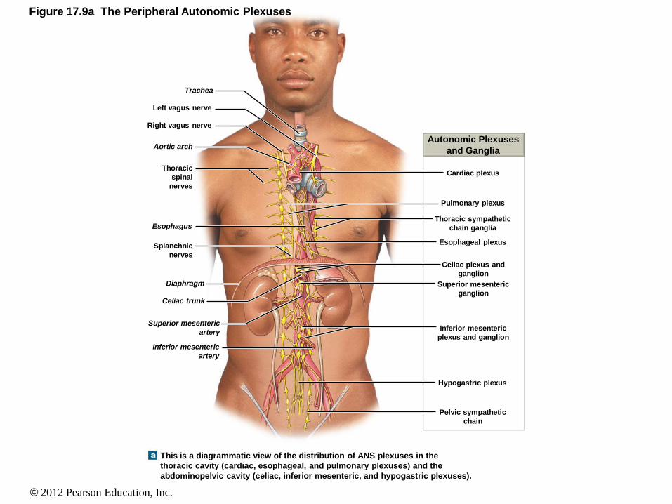
Figure 17.9a The Peripheral Autonomic Plexuses
This is a diagrammatic view of the distribution of ANS plexuses in the
thoracic cavity (cardiac, esophageal, and pulmonary plexuses) and the
abdominopelvic cavity (celiac, inferior mesenteric, and hypogastric plexuses).
Splanchnic
nerves
Inferior mesenteric
artery
Superior mesenteric
artery
Celiac trunk
Diaphragm
Esophagus
Thoracic
spinal
nerves
Left vagus nerve
Right vagus nerve
Aortic arch
Trachea
Cardiac plexus
Autonomic Plexuses
and Ganglia
Pulmonary plexus
Thoracic sympathetic
chain ganglia
Esophageal plexus
Celiac plexus and
ganglion
Superior mesenteric
ganglion
Inferior mesenteric
plexus and ganglion
Pelvic sympathetic
chain
Hypogastric plexus

© 2012 Pearson Education, Inc.
Figure 17.9b The Peripheral Autonomic Plexuses
A sectional view of the autonomic plexuses
Cardiac plexus
Autonomic Plexuses
and Ganglia
Thoracic sympathetic
chain ganglia
Esophageal plexus
Celiac plexus and
ganglion
Superior mesenteric
ganglion
Inferior mesenteric
plexus and ganglion
Pelvic sympathetic
chain
Hypogastric plexus
Urinary
bladder
Colon
Stomach
Diaphragm
Heart
Esophagus
Trachea
Vagus nerve
(N X)
Cranial nerve III
Cranial nerve VII
Cranial nerve IX

© 2012 Pearson Education, Inc.
Visceral Reflexes
• Provide autonomic motor responses to:
• Modify or facilitate higher centers
• All are polysynaptic
• Reflexes can be:
• Long reflexes
• Short reflexes
© 2012 Pearson Education, Inc.
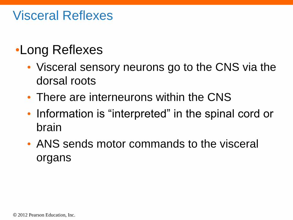
Visceral Reflexes
•Long Reflexes
• Visceral sensory neurons go to the CNS via the
dorsal roots
• There are interneurons within the CNS
• Information is “interpreted” in the spinal cord or
brain
• ANS sends motor commands to the visceral
organs

© 2012 Pearson Education, Inc.
Visceral Reflexes
• Short Reflexes
• Sensory nerve impulses go to the ganglionic
neurons
• Motor commands are distributed by the
postganglionic fibers
• Impulses bypass the CNS
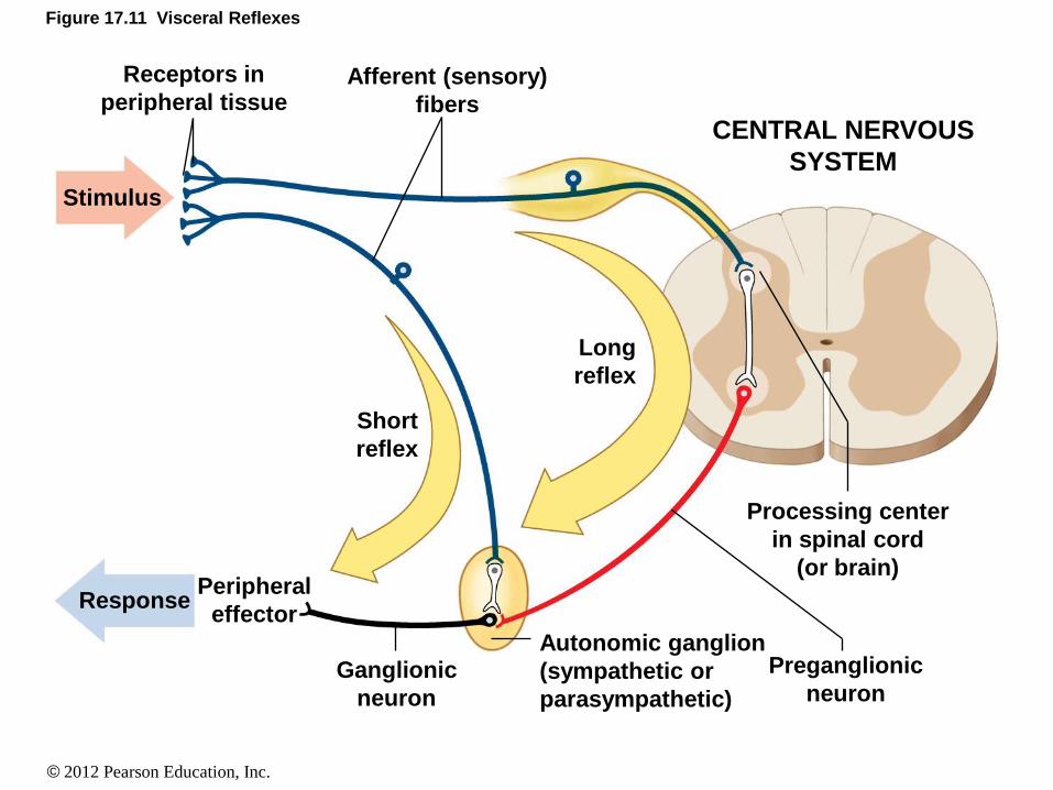
© 2012 Pearson Education, Inc.
Figure 17.11 Visceral Reflexes
Stimulus
Response
Receptors in
peripheral tissue Afferent (sensory)
fibers
Short
reflex
Long
reflex
Peripheral
effector
Ganglionic
neuron
Preganglionic
neuron
Processing center
in spinal cord
(or brain)
Autonomic ganglion
(sympathetic or
parasympathetic)
CENTRAL NERVOUS
SYSTEM