Embed Size (px)
Citation preview

This work is protected by copyright and other intellectual property rights and duplication or sale of all or part is not permitted, except that material may be duplicated by you for research, private study, criticism/review or educational
purposes. Electronic or print copies are for your own personal, non-commercial use and shall not be passed to any other individual. No quotation may be published without proper acknowledgement. For any other use, or to
quote extensively from the work, permission must be obtained from the copyright holder/s.

The neuroprotective role of polyunsaturated fatty
acids in a palmitic acid model of diabetes mellitus
type 2
Emily Elizabeth Benn
Submitted for the Degree of Master of Philosophy
Keele University
June 2020

Emily Benn Thesis
1
The neuroprotective role of polyunsaturated fatty
acids in a palmitic acid model of diabetes mellitus
type 2
Contents The neuroprotective role of polyunsaturated fatty acids in a palmitic acid model of diabetes mellitus type 2 ................. 1
Abstract ..................................................................................................................................................................................... 2
Acknowledgements.................................................................................................................................................................. 2
Introduction .............................................................................................................................................................................. 3
The Effect of Diabetes on the Brain ................................................................................................................................... 3
Diabetes and Insulin ....................................................................................................................................................... 3
Insulin Signalling in the Brain ....................................................................................................................................... 6
Diabetes in the Brain .................................................................................................................................................... 11
Modelling Insulin Signalling Impairment in Cells ......................................................................................................... 17
Palmitic Acid ................................................................................................................................................................ 17
Role of Polyunsaturated Fatty Acids in Neuroprotection ............................................................................................... 19
Polyunsaturated Fatty Acids ........................................................................................................................................ 19
Role of Polyunsaturated Fatty Acids in Neuronal Cells ............................................................................................ 21
Aims and Hypotheses ....................................................................................................................................................... 33
Materials and Methods .......................................................................................................................................................... 35
Methods ............................................................................................................................................................................. 35
Cell Culture ................................................................................................................................................................... 35
Cell Plating.................................................................................................................................................................... 35
Palmitic Acid Treatment .............................................................................................................................................. 35
MTT Assay ................................................................................................................................................................... 36
Pharmacology ............................................................................................................................................................... 36
Statistics ........................................................................................................................................................................ 36
Results ..................................................................................................................................................................................... 37
Discussion ............................................................................................................................................................................... 42
References .............................................................................................................................................................................. 47

Emily Benn Thesis
2
Abstract
The negative ways in which diabetes affects the brain, and the role of neural insulin and glucose is an interesting research
topic as research has shown a correlation between neurodegenerative diseases and diabetes. This thesis focuses on how
polyunsaturated fatty acids (PUFAs) induce neuroprotective effects in a Palmitic Acid (PA) model of Diabetes Mellitus
Type 2, commonly known as Type 2 Diabetes. Palmitic acid was administered to SHSY-5Y cells with the intention of
modelling type 2 diabetes by means of insulin impairment, and an autoimmune response. The role of fatty acids was
explored by administering 3 concentrations of docosahexaenoic acid (DHA), eicosapentaenoic acid (EPA), or alpha-
lipoic acid (ALA) either before or after a dose of PA which was administered at different concentrations. Additionally,
pharmacological inhibitors of DHA signalling pathways were utilised to characterise the potential DHA signalling
pathways involved in the DHA-mediated neuroprotection. The results showed that DHA (but not EPA or ALA) was
able to both prevent, and reverse damage caused by a palmitic acid model of neurodegeneration. Additionally,
antagonists of the DHA pathway successfully prevented damage caused by palmitic acid, providing a greater insight
into the DHA pathway.
Acknowledgements
I would like to thank Keele University for this opportunity to carry out my MPhil. Being in the first year of students
who were able to apply for funding from Student Finance England, it has been a difficult, but rewarding journey. I thank
my supervisor Dr. David Mazzocchi-Jones for his support throughout the last 2 years with my research, offering
academic advice, and support during difficult personal issues. I would also like to thank Professor David Furness for
making my viva experience less stressful and providing me with support after my examination.
In addition, I would like to thank my fiancé Dean Jarvis who has also been studying an MPhil at Keele University, in
Computer Science. With an interest in each other’s work, and a caring relationship, we have supported each other
throughout this journey.
I would also like to thank my sister Kirsten Benn who took her time to read my thesis and offer a plethora of extremely
helpful comments about my writing style. Because of her advice, I have been able to adapt my own writing style and
improve my thesis.

Emily Benn Thesis
3
Introduction
The Effect of Diabetes on the Brain
Diabetes and Insulin
Diabetes is a common disease which affects the ability to produce and use insulin effectively. There are two categories:
type 1 diabetes, and type 2. Type 1 diabetes is an autoimmune disease causing the immune system to attack the cells in
the pancreas that produce insulin. Type 1 has a genetic link and is not associated with age, diet, or obesity unlike type
2. It is usually diagnosed in childhood and insulin injections must be taken regularly in order to keep the levels of glucose
in the body balanced. Type 2 is slightly different; in the case of type 2 diabetes, the body can produce insulin, but it does
not produce enough and the cell signalling pathways for insulin are non functioning. Although insulin injections are
regularly used to treat diabetes type 2, some believe that eating low-carbohydrate diets after being diagnosed can also
be effective – although many studies have shown inconsistent results (Meng et al., 2017). Diabetes mellitus type 2
(DMT2) causes impairment of insulin release via pancreatic cells, resulting in an inability to maintain normal blood
sugar levels. Whilst patients with DMT2 can live a healthy life style, through monitoring of blood sugar levels,
stimulation of endogenous insulin release, and/or application of exogenous insulin (Olczuk and Priefer, 2017); there are
significant associated pathologies that remain currently untreated. Although much is known about DMT2, how it affects
the pancreas, and how to treat and manage diabetes, little is known about whether diabetes affects the brain. It is well
known that all parts of the body require glucose to produce energy; however, due to the presence of the blood brain
barrier, insulin and glucose cannot enter the brain through the blood stream passively. This has raised many questions
about whether the brain uses insulin and glucose, and if it does, how they get into the brain. Studies have found that
there is insulin and glucose inside of the brain (first discovered by Havrankova, Roth and Brownstein, 1978).
Consequently, leading researchers to investigate the role of neural insulin and whether signalling impairment could lead
to a ‘brain related diabetes,’ that could potentially have harmful effects on brain cells. Although significant studies have

Emily Benn Thesis
4
focussed on the role of insulin in the brain, little insight remains as to whether insulin impairment can lead to disease
and neurodegeneration.
Insulin is a hormone that is vital in metabolism in animal cells. It works by controlling the amount of glucose transported
into cells, which is then used to release energy; it is essential that this process is not disrupted (Lee et al., 2016). Insulin
is secreted by β cells which together with α, γ, δ, and ϵ cells make up the Islets of Langerhans, located in the pancreas.
It is the β cells that secrete insulin, along with amylin, a hormone important for slowing down the digestive process. As
glucose is secreted in the body, the role of insulin is to ensure that the amount that is transported to cells is highly
controlled as it is vital for respiration (Khan et al., 2018; Nirmalan and Nirmalan, 2017). As blood-glucose concentration
increases, signalling encourages insulin to be secreted by β cells to bring blood-glucose levels into the optimum range,
and as glucose concentrations then begin to fall, the role of insulin is inhibited by glucagon, a hormone released from α
cells in the Islets of Langerhans which is also involved in the homeostasis of glucose (Wild et al., 2016). Insulin and
glucagon are also important for regulating the amount of glucose released by the liver, where glucose is stored (Banks,
Owen and Erickson, 2012). Other hormones involved in glucose metabolism are: somatostatin, secreted by δ cells that
inhibit the release of insulin and glucagon, ghrelin which is released by ϵ cells and regulates appetite, and pancreatic
polypeptide secreted by γ cells which regulate exocrine and endocrine function (Khan et al., 2018; Nirmalan and
Nirmalan, 2017). Many of the roles and functions of insulin signalling pathways can be seen in Fig. 1.
To enable glucose to enter a cell, it must pass through a protein within the cell membrane known as glucose transporters
which allows for facilitated diffusion of glucose (Nirmalan and Nirmalan, 2017; Wild et al., 2016). There are 13 glucose
Figure 1 The cascadic events of insulin signalling pathways as explained by Boucher, Kleinridders and
Kahn, 2014. This image clearly displays the many functions and roles of insulin.

Emily Benn Thesis
5
transporters which allow for diffusion of glucose inside of the cell, most common of which being the primary class
which contains GLUT1-4. GLUT-1 is found on the surface of mainly endothelial cells and is activated when the level
of glucose outside of the cell is lower than that inside of the cell. Found in the liver and pancreatic cells, GLUT-2 allows
glucose to diffuse both in and out of the cells. GLUT-3 however, is found inside of the brain and allows glucose to get
into neurones. GLUT-4 is found in fat cells (Bell et al., 1990). The other classes and types of glucose transporters are
mainly found in specific organs and some are activated under certain events, such as when glycogen is being broken
down. When insulin is needed, levels of calcium ions (Ca2+) are increased, which is detected by synaptotagmins, which
complex with synaptosomal-associated receptor proteins (SNAREs). This causes vesicles containing insulin to fuse
with the cell plasma membrane causing the release of insulin (Miyazaki et al., 2009; Nirmalan and Nirmalan, 2017).
During this event, the precursor protein of insulin, proinsulin, is cleaved to form insulin and C-peptide. Although the
role of insulin is well documented, the role of C-peptide remains uncertain (Nirmalan and Nirmalan, 2017). Insulin
carries out its functions by signalling through various receptors and associated pathways (Posner, 2017). One main
pathway is the insulin receptor substrate (IRS) phosphatidylinositol 3-OH (PI3K)-Akt pathway which involves a large
cascade of signalling molecules. The purpose of this pathway is to decrease glucose production, inhibit glycogenolysis,
and stimulate gluconeogenesis, all of which is important for metabolism (Copps and White, 2012; Yan et al., 2017). As
Figure 2 The ‘Minardo Plot’ produced by Ma et al., 2015 which shows the complex cascade of events that happens after digestion of food in
order for metabolism to be carried out.

Emily Benn Thesis
6
insulin binds to an insulin receptor, this causes phosphorylation of proteins inside of the cell. This phosphorylation
signals to the cell which functions to turn on and off.
As insulin binds to receptors, it stimulates GLUT-4 receptors which causes upregulation and high activity of GLU-4
along the plasma membrane. These GLUT-4 receptors are responsible for allowing glucose to move from outside of
the cell to the inside of the cell. Following the cascade of phosphorylation events, glucose begins to be turned into fat
storage through processes called glycogenesis and lipogenesis. While this is happening, protein synthesis and gene
modification also occurs. An indepth description of this can be seen in Fig. 2.
Insulin is an extremely important hormone for metabolism. However, there is a large gap in research about the functions
of insulin in the brain and whether disruption of this can lead to disease. In recent years, not only has there has been a
significant increase in the number of diabetes diagnoses, but also dementia. The increase in dementia has been thought
to have accrued as life expectancy has increased. However, it is possible that the two correlations could be associated
as recent research has suggested a link between DMT2 and neurodegenerative diseases. This is a novel area of research
that is currently being investigated (Verdile, Fuller and Martins, 2015).
Insulin Signalling in the Brain
Insulin in the Brain and its Receptors
It was once believed that insulin was not present in the central nervous system (CNS) due to the presence of the blood
brain barrier (BBB) which stops many types of molecules from entering the brain. However, recent research has
discovered the presence of insulin and insulin receptors inside of the brain and their role is being explored (Banks, Owen
and Erickson, 2012; Griffith et al., 2018; Havrankova, Roth and Brownstein, 1978). This was originally discussed when
GLUT-1 receptors were present on the membranes of endothelial cells which line the blood-brain barrier. As more
research in this area has accumulated, more is known about the role of neural insulin. Neural insulin has been found to
be involved in many aspects of cognition and impairment may contribute towards symptoms of neurodegeneration,
such as memory loss. Research by Fine et al., (2017) found that intranasal deferoxamine, which has been shown to
reduce oxidative stress and up-regulates insulin and glucose signalling, improves memory in a rat model of Alzheimer’s
Disease (AD) by signalling through insulin receptors in the brain (Ito et al., 2016). The results showed that the treated
rats had significantly shorter latency times in a Morris water maze. Furthermore, analysis of the brain tissue showed
that the treated rats also had a reduction in oxidation of the cells. Thus showing that symptoms related to
neurodegeneration, such as memory loss and oxidation of neural cells were reduced by using a drug that acts through

Emily Benn Thesis
7
insulin receptors, further suggesting that insulin impairment may be involved in the onset of neurodegenerative diseases
and insulin acting and signalling drugs may be a new target for therapy.
The hippocampus, hypothalamus, and the cerebellum of the brain have all been shown to contain GLUT-4 receptors,
which are insulin sensitive, suggesting that GLUT-4 receptors are involved in metabolism in brain cells (Fernandez et
al., 2017). They have been shown to be involved in transporting glucose to neuronal cells via insulin and are found high
in concentration in areas associated with cognition (Jurcovicova, 2014). Insulin in the brain has also been shown to be
involved in learning and memory, as well as the formation of cells, and their development; its role in areas important
for cognition suggests a potential link with plasticity (Zhao and Alkon, 2001). Although much of the literature shows
that insulin is involved in many of these mechanisms, there is little known about potential mechanisms of action and
signalling pathways and the consequences of long-term insulin disruption (Banks et al, 2012). In addition to GLUT-4
receptors, another insulin associated receptor also found in the brain is insulin receptor kinases (IRK) which are involved
in both long-term and short-term effects of growth and development, such as growth of cells, and long-term potentiation
(LTP) (Gralle, 2016; Zhao et al., 2017). Specifically, these types of receptors have been found in large quantities in the
hippocampus, suggesting that they have a role in learning and memory; aiding LTP (Zhao and Alkon, 2001). While
some studies have shown that insulin impairment in the brain leads to cognitive deficits, some studies have revealed
that the IRK are not involved in this (Ferrario and Reagan, 2017). This suggests that insulin resistance may be involved
in other mechanisms when cognitive deficits are displayed. Alternatively, the deficits seen in insulin resistance may be
due to impairments not involving the IRK receptors. A study by Schubert et al., (2004) found that neurone specific
insulin receptor knockout mice displayed a deficit of insulin mediated activation of phosphatidylinositol 3-kinase as
well as inhibition of apoptosis. However, there were no cognitive deficits in these mice which was assessed through
various tasks such as the Morris water maze, vertical pole test, upside-down grid, and placing response tests. Other
studies have found cognitive deficits when the mice also exhibit tau phosphorylation, suggesting that insulin resistance
through IRK receptors may interact with other pathways in the brain (Schubert et al., 2004).
Is Insulin Important in the Brain?
The role of glucose regulation in the brain is not clearly understood, with contrasting evidence supporting the regulatory
role of insulin. Systemically, insulin is vital for metabolism in every cell, and although researchers have shown that
insulin and insulin receptors are high in concentration in areas associated with learning and memory, their role in glucose
metabolism in brain cells remains unclear. A study conducted by Hwang et al (2017) investigated the effect of DMT2
on brain glucose levels. In the study, 9 healthy participants, 10 obese patients, and 6 poorly controlled DMT2 patients
had their intracerebellar glucose concentrations measured. They found that those with poorly controlled DMT2
Emily Benn Thesis
8
exhibited lower levels of neural glucose, consequently suggesting that glucose entry into the brain may be dependent
on insulin levels as those with DMT2 showed lower levels of glucose. However, this does not mean that glucose entry
is completely dependent on insulin, as there may be an insulin signalling impairment, meaning that other receptors could
be involved in this process. Furthermore, the study also has implications for research in DMT2 in the brain and the
potential consequences thus giving more insight into the potential roles of insulin (Hwang et al., 2017)
Insulin however, has also been shown to be important for neuronal survival. Signalling through the IRK receptor has
been shown to stimulate cell survival in the presence of glucose and oxygen deprivation which is shown by a study by
Mielke, Taghibiglou and Wang, (2006). In this study, cells were exposed to oxygen-glucose deprivation (OGD) and
were then investigated for changes in insulin binding. They had found that during OGD, insulin failed to stimulate the
phosphorylation of the IR β subunit, suggesting that insulin does play a role in glucose signalling in the brain. However,
this is opposed by an earlier study by Clarke and Raizada, (1986). In this study, the researchers attempted to investigate
the role of insulin, and its possible binding sites using 1-day old rat brain primary cell cultures. They had found that the
addition of insulin had no effect of glucose uptake in the cells. This however, was not found in their previous study,
where they had found that insulin increased glucose uptake twofold in astrocytes (Clarke et al., 1984). This study
suggests that the role of insulin in glucose uptake may only be present in glial cells. Overall, the presence of
contradictory research shows the complexity of the brain, thus making it difficult to obtain a definitive answer regarding
the role of neural glucose and insulin.
In addition, insulin has also shown to prevent apoptosis in a range of cells. For example, research by Qian et al., (2001)
reported that high concentrations of insulin prevented apoptosis in cardiomyocytes through blocking TNFα (tumour
necrosis factor). Increased levels of TNFα returned the normal level of apoptosis in the cells (Qian et al., 2001). This
suggests that insulin may be involved in blocking the role of TNFα during apoptosis. However, it is uncertain that this
will be the same for neuronal cells. If insulin is involved in glucose uptake by neuronal cells, then it could be that
reduction of insulin would lead to cell death. Supporting evidence provided in a study conducted by Song et al., (2015)
showed that impairment of selected insulin receptors leads to apoptosis of neuronal cells. Additionally, this impairment
also leads to inhibition of neurite outgrowth. The authors suggest that these findings conclude that insulin receptor
impairment contributes towards neurodegeneration and cognitive impairment. This research could therefore have many
implications for the understanding of DMT2 in the brain, and neurodegeneration.

Emily Benn Thesis
9
Learning and Memory
A range of studies have demonstrated the effect of insulin on both long, and short-term memory. One of the symptoms
of low blood sugar in DMT2 is memory loss, which suggests that DMT2 consequently effects cognition in the brain.
Studies have found that insulin signalling plays a vital role in cognition and memory. An earlier study by Marks et al.,
(2009) investigated how insulin affects cognition in mice. They discovered that after 1 week of intranasal insulin
administration, there was an increase in the expression of the potassium ion channel Kv1.3 in the olfactory bulb. This
is significant because the olfactory bulb has been shown to have the highest level of insulin receptors than any other
area of the brain (Baskin et al., 1983), suggesting that intranasal insulin may have had an effect in insulin signalling,
and consequently caused an increase in the Kv1.3 potassium ion channels. Furthermore, the potassium ion channel
Kv1.3 is involved in the inhibition of action potentials through glucose binding to insulin receptors. The study also
found that mice treated with insulin had better short- and long-term memory in object memory recognition, and
increased odour discrimination. This suggests that insulin may play a signalling role between different areas of the brain
as insulin receptors are found to be in high quantity in the olfactory bulb, and these mice also had learned to discriminate
between a larger variety of odours. However, in pre-diabetic mice, insulin was not effective. This further suggests that
insulin is important in plasticity, as it has shown to positively effect learning and memory. It also suggests that DMT2
may impair insulin in learning and memory, however the study used pre-diabetic mice which have higher than
considered healthy blood sugar levels but are not classed as diabetic.
Evidence suggests that insulin has a role in the hippocampus; the primary site in the brain for learning and memory. A
study by Dou, (2005) found that during spatial learning (using the Morris maze), IRK expression increased in the CA1
area of the hippocampus, but not the CA3 area. They also found that in rats exposed to DMT2 had only minor cognitive
impairment. This suggests that although the IRK receptors play a role in spatial learning, insulin itself may not be
important as there was no significant cognitive impairment. However, there could also be other pathways in the
hippocampus that protect the cells from insulin impairment, although more studies would have to be carried out targeting
the hippocampus to find out why this happens (Dou, 2005).
There are also high concentrations of the GLUT-4 receptor in the hippocampus, further suggesting a role of glucose and
insulin in learning and memory. A study by Pearson-Leary et al., (2017) had found that when insulin was delivered to
the hippocampus, inhibiting the GLUT-4 receptor prevented cognitive enhancement during spatial working memory
tasks. This again suggests that insulin has a role in learning and memory, and if this were impaired, possibly by DMT2,
cognition is also impaired.

Emily Benn Thesis
10
Other studies have found that insulin works as a neurotransmitter and influences the activity of NMDA, AMPA, and
GABA-A receptors which are all involved in LTP, a vital mechanism of learning and memory. Neural insulin levels
are also affected by synaptic activity which was found in a study by Zhao et al., (1999) who established that there was
an increase in insulin receptors (IR) protein and mRNA in the CA1 area of the hippocampus measured by synaptic
membrane fraction following water maze training; suggesting a role in plasticity. Insulin signalling has shown
importance for regulating AMPA receptors; specifically, the GluR2 subunit used in long term depression (LTD)
plasticity (Ahmadian et al., 2004; Schmitz et al., 2018). This suggests that insulin is important for LTD, a type of
plasticity which allows for downregulation of unnecessary connections in the brain. More evidence for this comes from
a study by Zhou, Xiao and Nicola (2001) who showed that insulin is important for endocytosis in the synapse. As insulin
concentration increased, so did endocytosis. This is displayed in Fig. 1. which was taken from Zhou, Xiao and Nicola
(2001). However, endocytosis was not enhanced by the addition of glutamate (which is what would have been expected
due to the role of glutamate in AMPA receptors at the synapse), further suggesting that insulin has vital roles in the
brain. Further research should aim to investigate what happens when the levels of insulin are disrupted. This could lead
to additional knowledge about brain disease, specifically in neurodegenerative diseases in which research has already
shown a relationship with insulin.
Evidence has also shown that insulin is important for producing GluR1 when they are needed (Adzovic and Domenici,
2014). This proposes that insulin plays an important role in synaptic activity as GluR1 are an important component of
AMPA receptors as they are a site of phosphorylation during synaptic plasticity (Boehm et al., 2006). Research by
Adzovic and Domenici, (2014) has revealed that insulin stimulates phosphorylation of the GluR1 subunit of AMPA
receptors. The results of this study are particularly interesting as they reveal insight into insulin signalling pathways;
Figure 3 A graph taken from Zhou, Xiao and Nicola (2001) that shows an increase of FM1-43 uptake in the control vs the presence of insulin. Application of insulin was able to enhance endocytosis and internalization
of AMPA receptors in neurones.

Emily Benn Thesis
11
insulin induces the over-expression of protein kinase M zeta (PKMζ) and phosphorylation of MAPK Erk1/2 (signalling
proteins). This leads to the phosphorylation of AMPA receptors, an important step in synaptic plasticity. Another very
interesting result of this study was that amyloid beta 42 (Aβ42) significantly decreases the effects of insulin in this
specific signaling pathway (Adzovic and Domenici, 2014). This has implications in neurodegenerative research as Aβ42
is a biomarker for AD and this research suggests that this has an effect on insulin signalling.
A significant downfall to a lot of the research that has been carried out assumes that the connection between insulin and
glucose in the brain is not mutually exclusive. Many of the researchers have found deficits when insulin is impaired but
this does not mean that glucose also plays a role, as it does in the rest of the body. Glucose administration is required in
order to definitively conclude that insulin and glucose are both involved in memory and cognition and disruption of one
leads to the disruption of the other.
Diabetes in the Brain
Diabetes and Alzheimer’s Disease
Alzheimer’s disease (AD) is one of the most prevalent neurodegenerative diseases, and the most common cause of
dementia (Snyder et al., 2018). As the length of human life has dramatically increased over the last hundred years due
to improvements in not only the quality of human life, but also health care, the rate of neurodegenerative diseases has
increased; specifically, AD (Shao, Peng and Wang, 2017; Snyder et al., 2018). AD can be familial (having a genetic
cause; known genes involved are APOE e3, PSEN-1, PSEN-2, and APP) which can result in early on-set, or sporadic
(where the on-set usually begins at a later stage of the patient’s life) (Ułamek-Kozioł et al., 2016). Apolipoprotein E
(APOE) is coded for a gene found on chromosome 19 and it is involved in fat metabolism and is important for the
breakdown of Aβ. The varients of this gene do not break down Aβ as efficiently, leading to plaque formation. The E3
and E4 variants of APOE carry the largest risk for onset of AD (Jiang et al., 2008). Some of the consequences of AD
are cognitive impairment and memory loss as well as changes in personality and behaviour (Sevinçer et al., 2017). There
is a great deal of research which has aimed to and continues to find the causes of AD and what can be done to prevent
and treat this. Some of the biomarkers include amyloid beta (specifically Aβ42 which is a fibrillogenic isoform) plaques,
and neurofibrillary tangles, and even biomarkers of DMT2 have shown a strong correlation with AD (Chen, Sawa and
Mobley, 2018; Li et al., 2018; Shao, Peng and Wang, 2017).

Emily Benn Thesis
12
Aβ plaques begin with amyloid precursor protein
(APP) which is a protein found in the cell
membrane that is involved in growth and repair
of neurons. APP is usually cleaved using α
secretase as well as γ secretase to produce Aβ
(Crane et al., 2018). However, sometimes β
secretase is used with α secretase to cleave the
protein. This produces a more dangerous isoform
of Aβ known as Aβ42. Aβ42 monomers stick to
each other and form harmful plaques which can
impair signaling between neurons and produce an
immune response which results in inflammation
(Chen, Sawa and Mobley, 2018; Shao, Peng and
Wang, 2017).
Neurofibrillary tangles are caused by hyperphosphorylation of tau. Tau is a protein that is responsible for holding
together microtubules inside of neurons. Aβ plaques outside of the cell can activate kinases inside of the cell (Theofilas
et al., 2018). These kinases are responsible for transporting phosphate groups to the tau proteins. When the kinases are
over stimulated, hyperphosphorylation of the tau proteins occur thus causing them to change shape so that they can no
longer support the microtubules. Additionally, the newly shaped tau proteins clump together and form tangles. This
leads to apoptosis of the cell (Hu et al., 2016). The formation of Aβ plaques and neurofibrillary tangles and how they
lead to cell death can be seen in Fig. 4.
In addition to Aβ plaques and tau aggregation, another factor which has been implicated in the onset of AD is excessive
caspase activation. Caspases are enzymes which are involved in programmed cell death. However, overactivity of these
enzymes can lead to unnecessary cell death and neuronal inflammation, resulting in mass loss of neurons. Research
such as that carried out by Flores et al., (2018) discovered that inhibition of capsase-1 leads to a decrease in cognitive
impairment in a mouse model of AD. This is also discussed in an article by Rohn, (2010).
Although there has been significant research into the different biomarkers found in AD, recent research has begun to
find a link between the presence of DMT2 and AD as a consequence of insulin signaling impairment in the brain
(Shinohara and Sato, 2017). For example, insulin impairment has consequences for metabolism and signaling systems
Figure 4 An illustration showing the aetiology of Alzheimer’s Disease. This includes
the formation of amyloid beta 42 plaques, and neurofibrillary tangles and how these
events lead to damaged neurones through damage to the mitochondria (Panza et al.,
2019)

Emily Benn Thesis
13
in the brain. This can eventually lead to cell death that is seen in neurodegenerative diseases. Furthermore, insulin has
also been seen to play a role in LTP which is a component of memory. Loss of memory is one of the most prevalent
symptoms in many neurodegenerative diseases.
Previously, research has focused on investigating the role of insulin in the brain, but now, research has started to examine
what happens when insulin function is impaired and whether this is considered as a brain specific type of diabetes;
consequently, resulting in neurodegeneration. DMT2 has been shown to cause neurodegeneration by causing changes
to insulin signalling in the brain, as well as glucose metabolism, which leads to impairments in memory and cognition
(Shinohara and Sato, 2017). Recent research has focused both on discovering the mechanisms in which insulin
impairment may be associated with neurodegeneration, and whether application of insulin into an AD patient may
alleviate the symptoms, reverse the pathology, or even prevent the progression of this disease.
In 2016, Hoscheidt et al., investigated the relationship between insulin resistance, cerebrospinal fluid biomarkers of AD,
and memory in healthy adults with genetic predispositions to AD due to the APOE ε4 allele, which has been shown to
increase the risk of late-onset AD (Lyall et al., 2016). The patients were tested for amyloid precursor protein β (sAPP-
β), β amyloid42 (Aβ42), and phosphorylated tau (P-tau181) in cerebrospinal fluid (CSF) through a lumbar puncture.
Neuropsychological testing was used to assess memory function and homeostatic model assessment (HOMA) was used
to assess insulin resistance. Interestingly, higher HOMA-IR was associated with higher levels of sAPP-β, and Aβ42,
suggesting that insulin resistance has a relationship with AD biomarkers. Further research should aim to find out the
mechanics of what causes this relationship.
A further study by, Li et al., (2018) also showed similar results; in this study, biomarkers of AD such as Aβ42 in CSF
were found to be correlated with DMT2. In their study, it was found that DMT2 was positively correlated with Aβ42.
Although the link between DMT2 and AD has already been established, the mechanisms of the link are still being
investigated. While some studies have found that insulin resistance has an effect on cognition, recent studies have shown
that the presence of diabetes is correlated with biomarkers of AD. This suggests that insulin resistance may cause some
of the pathological properties of AD and that insulin impairment in cells has a negative pathological effect (Li et al.,
2018).
An alternative way in which insulin may be a part of AD pathology is through the mammalian target of rapamycin
(mTOR) pathway. Norambuena et al., (2017) investigated how impaired insulin in the brain might affect mTOR and
neuronal cell cycle reentry. One of the causes of neuron death in AD is cell cycle reentry which is mediated by Aβ
oligomers and tau hyperphosphorylation (Kuhla et al., 2015). Cell cycle reentry (CCR) is a mechanism found in AD

Emily Benn Thesis
14
pathology which causes DNA damage and apoptosis (Folch et al., 2011). Aβ oligomers cause this by activation of
mTORC1, a protein kinase involved in the growth and survival of cells which forms complexes (mTORC1 and
mTORC2) (Lipton and Sahin, 2014). Not only did the Aβ oligomers activate mTORC1, they also reduced insulin
signaling. Furthermore, insulin stimulation was found to prevent CCR. This research has clinical implications as it
shows how insulin application prevents one of the pathologies that causes AD (Norambuena et al., 2017). There have
been studies that show that insulin can prevent, and even reverse the onset of AD in animal models, but very few have
shown the mechanisms of how this happens; however this study suggests that insulin could be involved in cell cycle
reentry as this resulted in insulin signaling impairment; the cause and effect of this could be debated.
Due to its discovered link with the pathology of AD, the administration of insulin into animal models, and humans, has
been tested. The results have been positive, showing that insulin not only restores memory and cognition, but also
reduces the biomarkers of AD. One example of this is by Guo et al., (2017) who explored the role of insulin in the brain
in an AD model by investigating whether intranasal insulin would alleviate the symptoms of AD, specifically cognitive
impairment, hyperphosphorylation of tau, and microglial activation. Streptozotocin was used to model diabetes induced
AD in rats due to its toxic affect to beta cells in the pancreas and thus preventing the secretion of insulin. If this is able
to model AD in the brain, then it suggests that insulin is involved in the pathology. When insulin was given intranasally
for 6 weeks, cognition was improved in the rats. Hyperphosphorylation of tau, and microglial activation were also
diminished; a reduction in AD symptoms and biomarkers (Gou et al., 2017). However, interestingly, the levels of
glucose in the rats was not affected which suggests that diabetes in the brain may not specifically be directly associated
with glucose levels and that insulin is involved with different functions. This questions what diabetes of the brain may
actually mean.
A similar study by Chen et al., (2014) found that insulin application restored insulin signaling in the brain, increased the
concentration of synaptic proteins, and reduced Aβ40 (another isoform of amyloid beta that is less fibrillogenic than Aβ42
and not associated with AD) (Yin et al., 2007). In this study, AD was modelled using 3xTg-AD mice which were treated
with intranasal insulin for seven days. Application of insulin intranasally would mean that it is delivered straight to the
brain. After the application period, the mice were euthanized and biochemical and immunohistochemical analysis was
performed using western blot analysis, immune-dot-blot assays, and ELISA kits which were specific for Aβ antibodies.
They had found that insulin was capable of restoring insulin signaling, reducing Aβ40, restoring the concentration of
synaptic proteins, and reducing microglia activation. (Chen et al., 2014). This study further suggests that the delivery of
insulin into the brain reduces the biomarkers of AD and could potentially aid in regeneration. This also suggests that

Emily Benn Thesis
15
one of the causes of the biomarkers found in AD is an impairment in insulin signaling, possibly caused by the lack of
insulin, or inability for insulin to bind to specific regions, such as in diabetes.
Diabetes and Parkinson’s Disease
Parkinson’s disease (PD) is a debilitating neurodegenerative disorder that affects the dopaminergic neurones in the
substantia nigra pars compacta (SNpc) of the basal ganglia. This results in both motor and cognitive symptoms. PD is
a progressive disease which is usually in a sporadic form, affecting elderly individuals. However, it can also be familial,
causing onset at an earlier age. The genes involved in familial PD are mutations in the PINK1, PARKIN, or ALPHA
SYNUCLEIN gene (Bentley et al., 2018; Deng, Wang and Jankovic, 2017). One of the features of PD are tremors
which are most commonly found in the hand; referred to as a pill rolling tremor. It is a resting tremor as it is present
during rest and diminishes when the individual moves their hand. Another clinical feature is rigidity which causes
stiffness when moving the arms or legs (Kim et al., 2017). There are three types of movement impairments which are
bradykinesia (slow movement), hypokinesia (lessened movement), or akinesia (absence of movement). It is thought
that many of these physical features of PD are caused by a breakdown in signalling in the SNpc, resulting in impairments
in movement and posture. Other symptoms related to PD are depression (which has been shown to correlate with posture
problems), dementia, sleep disturbances, and difficulty with olfaction (Kim et al., 2017). These types of symptoms are
brought about by impaired dopamine signalling, but the exact locations of this within the brain are unclear. Researchers
have suggested that impairments with other neurotransmitters, such as acetylcholine, may be involved (Rizzi and Tan,
2017). Following post-mortem of a PD brain, Lewy bodies can be seen to aggregate in the SNpc, an area of the brain
responsible for sending signals to the striatum through dopaminergic neurones; this forms the nigrostriatal pathway
which is involved in movement (Dickson, 2018). Lewy bodies are inclusions made up of alpha-synuclein; the function
of which is currently uncertain, but it is believed to be involved in the secretion of dopamine (Castillo-Carranza et al.,
2018).
Recent research has targeted the association between diabetes and neurodegenerative disease due to the complex roles
of insulin and insulin signalling in the brain. There has been a lot of research to show that insulin resistance has a role
in PD, but has not yet shown the mechanisms of how this happens. This is what very recent studies have been aiming
to find out. A paper by Braatz and Coleman, (2015) suggests that insulin resistance is involved in the pathology of
Parkinson’s Disease (PD) and this is caused by inflammation and oxidative stress. This paper proposes a mathematical
model using biochemical systems theory (BST) that can be used to investigate the changes brought about by PD and
identify ways to effectively treat the disease. In this model, a cell with PD and insulin resistance is modelled in a healthy
state (pre PD), a diseased state, and a recovery state. Thus conveying that insulin signalling is impaired by inflammation
Emily Benn Thesis
16
present in PD. The results also show that insulin signalling was recovered during a treatment model. Furthermore,
insulin signalling partially recovered following late treatment (Braatz and Coleman, 2015). While the research made
significant contributions to the understanding of the involvements of insulin resistance in PD, it did not discuss what
the treatment was, or how it was modelled.
The paper failed to describe exactly how their model was made. They had suggested that insulin impairment is present
following biomarkers of PD such as inflammation but did not describe in great detail how the inflammation happens,
and how this is modelled. The model includes ROS (reactive oxygen species) and RNS (reactive nitrogen species)
production, p38 phosphorylation, tau hyperphosphorylation, inflammation, dopamine synthesis and dopamine
degradation, and protein transport (Braatz and Coleman, 2015). However, it does not describe how all of this was
modelled, other than the use of a program called CellDesigner. The study primarily focuses on the result of insulin
signalling impairment due to the PD model. If the paper were to describe how CellDesigner accounts for every
biochemical reaction that results in PD, this could greatly benefit the use of using mathematical models for diseases. A
disadvantage is that mathematical models cannot be used to describe a real-life situation. For example, although much
is known about the pathology of PD, there are many theories as to how the disease initiates. Each theory, with lots of
evidence, cannot describe every single case as there will be biological variability in every PD patient. Although the
model does not accurately depict the pathological growth of PD due to the many possible variables, it does aid in
predicting certain stages of the disease. This can then be used in other types of research, such as cell culture and animal
models, to predict what might happen. Although there are many disadvantages of using mathematical models of diseases,
they can be useful to describe an outline of the different stages of the disease, and what might be expected to happen.
This is also helpful as if different types of research show different results that do not lie within the model, these can be
investigated, providing more research into many of the different variables that affect the onset of PD.
Current research is aiming to find out how an impairment in insulin signalling contributes towards neurodegenerative
diseases such as PD as this is currently unclear. A study that addresses this issue was carried out by Sekar and
Taghibiglou, (2018). This paper explains that it is estimated that 50-80% of patients with PD have shown to have
abnormal glucose tolerance; which suggests that diabetes may play a role in the pathology. While there is a lot of
variation in the figures presented, there is still cause for concern over this association. The research presented in this
paper investigates how insulin signalling and insulin signalling impairment is involved in the SNpc, the area of the brain
most affected by PD. By using post-mortem cadavers of PD patients (and healthy brains as controls), the authors used
nuclear fraction (NF) and tissue homogenate (TH) to investigate the role of insulin. They had found that there were

Emily Benn Thesis
17
raised levels of PTEN and GSK3β, and decreased levels of PI3K p85, Akt1/2/3 and PIP3; all of which are involved in
insulin signalling pathways. Therefore, these results suggest that insulin signalling impairment may play a role in the
onset of PD (Sekar and Taghibiglou, 2018).
A negative outcome of the results is that cause and effect cannot be inferred. These results cannot tell us whether insulin
signalling impairment is a cause of, or a result of PD. Another disadvantage of the methods used encompasses sampling,
where only 4 PD patients were used, thus introducing generalisation issues due to the limited sample size. Although it
can be extremely difficult to obtain samples from human post-mortem brains with PD, 4 samples cannot represent the
whole population of individuals with PD. Furthermore, there is a lot of variability in the pathogenesis of PD, such as
familial and sporadic differences. Therefore, 4 samples do not represent the huge variability between each patient.
Additionally, the paper does not state whether the 4 samples came from familial or sporadic cases, therefore,differences
such as the genes that play a role of familial cases cannot be accounted for.
This research is a start in investigating the roles of insulin signalling impairment in PD (Sekar and Taghibiglou, 2018).
The next stages of research would be to find out whether insulin signalling impairment in the disease is a cause or effect
biomarker. This would make a very large difference in the way in which PD is treated. Furthermore, similar studies
should be carried out to verify the findings that were only found in 4 samples. If more studies find very similar results,
this would suggest that the results are reliable, alongside increase samples to further the representability of the disease
itself.
If insulin signalling impairment were a cause of PD, this could mean that diabetic treatments could be used to treat the
symptoms of the disease. This would also greatly expand the knowledge of the pathogenesis of the disease. However,
if insulin signalling impairment was found to be an effect of PD, more would be known about the importance of insulin
signalling in the brain, and the many roles it plays.
Modelling Insulin Signalling Impairment in Cells
Palmitic Acid
A new method in modelling insulin signalling impairment is through the use of palmitic acid (PA). PA is a naturally
occurring saturated fatty acid found in animals, as well as olive oil, and palm oil, the structure of which can be seen in
Fig. 2. Current interest in the use of PA models in research is due to its ability to alter insulin secretion and suppress
leptin and insulin which was shown in a study by Benoit et al., (2010) which illustrated this using a mouse model. In
this study, PA was delivered directly to the CNS which resulted in an increase of protein kinase C theta (PKC-θ). This

Emily Benn Thesis
18
is significant as PKC-θ is a major signalling enzyme involved in many immune responses, the disruption of which has
been known to cause autoimmune attacks (Madouri et al., 2017). In the study by Benoit et al (2010), this increase in
PKC-θ was also associated with impairment of insulin and leptin signalling in the hypothalamus. As research has
highlighted diabetes and insulin resistance as a risk factor for neurodegeneration, new models for this are currently being
produced. One of which is the use of PA.
The research conducted by Calvo-Ochoa et al., (2017) investigates how different concentrations of PA affect
undifferentiated human neuroblastoma cells from a cell line, and cortical cells from embryonic rats and whether this is
a good model for inhibiting insulin signalling. Using cell counts and western blotting, the authors discovered many
interesting features of the use of PA. 24-hour exposure to 500µM of PA reduced the viability of the undifferentiated
human neuroblastoma cells to 29.72%. Interestingly, 1-hour exposure to 500µM of PA increased the metabolic activity,
suggesting that short term application may aid insulin signalling. Additionally, exposure to PA impaired insulin
dependent mitochondrial activity. Exposure to glucose did not alter cell viability in any condition suggesting that insulin
is not responsible for the metabolism of glucose in undifferentiated human neuroblastoma cells (Calvo-Ochoa et al.,
2017). This paper provides evidence for PA reducing insulin signalling in undifferentiated human neuroblastoma cells
and therefore would be able to model diabetes in brain cells. The research also suggests that short term exposure to PA
increased cell viability which is a very interesting result. However, further research is required in order to investigate
why short-term exposure increases cell viability, whereas long-term exposure reduces cell viability. As there is an
increase in interest and research around the idea that diabetes could be responsible for cell death, new models for
diabetes in the brain are very important for current research as it will help to understand and possibly prevent
neurodegeneration. Oxidative stress caused by an increase in ROS is a common complication in diabetes that causes
insulin resistance. If it is not treated, it and can have many negative long-term effects (Giacco and Brownlee, 2010; Dos
Santos, Tewari and Mendes, 2019; Yuan et al., 2019). This pathway is a common target for diabetes treatment, and also
research into the effects of oxidative stress on other cells, such as brain cells.
Figure 5 The structure of palmitic acid taken from Moreno et al., (2012).
Emily Benn Thesis
19
Role of Polyunsaturated Fatty Acids in Neuroprotection
Polyunsaturated Fatty Acids
Polyunsaturated fatty acids (PUFAs) are characterised by long chains, with double bonds, which derived from
triglycerides and phospholipids (de Oliveira et al., 2017). PUFAs are part of the phospholipid by-layer that forms the
cell membrane that surrounds cells. Additionally, research suggests that they also play important roles in the brain and
retina by aiding in signal transduction, neurotransmission, and neurogenesis (Kerdiles, Layé and Calon, 2017).
PUFAs are categorised into two groups, methylene-interrupted polyenes, and conjugated fatty acids. Research has
recently paid attention to methylene-interrupted polyenes which are categorised into omega-3, omega-6, and omega-9
(de Oliveira et al., 2017). PUFAs are vital for growth, the development of the brain, and the development of vision.
Numerous studies have shown that the concentrations of PUFAs are increased in prenatal animals, and their off spring.
Studies have also shown that offspring with very low birth weights also have low docosahexaenoic acid (DHA; a type
of omega-3) concentrations, due to the lack of fat storage. This suggests that PUFAs are important for development (de
Oliveira et al., 2017).
Humans are capable of synthesising the monomers of fatty acids. However, we do not possess the enzymes that are
needed for the production of omega-3. Therefore, it is vital that these are obtained through diet (Harris and Baack, 2014).
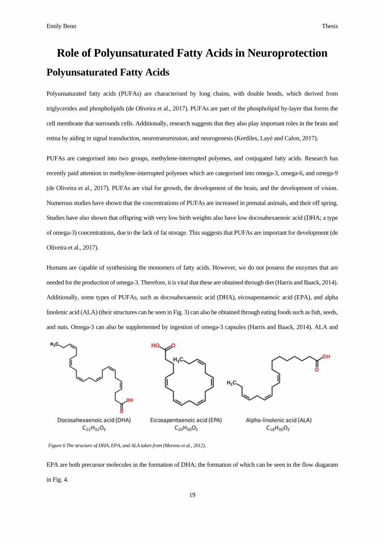
Additionally, some types of PUFAs, such as docosahexaenoic acid (DHA), eicosapentaenoic acid (EPA), and alpha
linolenic acid (ALA) (their structures can be seen in Fig. 3) can also be obtained through eating foods such as fish, seeds,
and nuts. Omega-3 can also be supplemented by ingestion of omega-3 capsules (Harris and Baack, 2014). ALA and
EPA are both precursor molecules in the formation of DHA; the formation of which can be seen in the flow diagaram
in Fig. 4.
Figure 6 The structure of DHA, EPA, and ALA taken from (Moreno et al., 2012).

Emily Benn Thesis
20
Figure 4 A flowchart showing how DHA is formed from its very initial precursor, oleic acid, and its important precursors ALA and EPA. Made using
Microsoft word with information taken from (Banerjee and Roychoudhury, 2014).
Docosahexaenoic Acid (DHA)
Docosapentaenoic AcidΔ4-desaturase
Eicosapentaenoic Acid (EPA)Δ5-elongase
Eicosatetraenoic AcidΔ5-desaturase
Stearidonic AcidΔ6-elongase
α-Linoleic Acid (ALA)Δ6-desaturase
Linoleic AcidΔ15-desaturase
Oleic AcidΔ-12desaturase

Emily Benn Thesis
21
PUFAs have a large range of roles in the body and research has focused on the positive effects of dietary
supplementation of PUFAs (specifically DHA) in our brains; many of which can be seen in Fig. 5 (Tanaka et al., 2012;
Kerdiles, Layé and Calon, 2017; Tang et al., 2018).
Role of Polyunsaturated Fatty Acids in Neuronal Cells
Role of Polyunsaturated Fatty Acids in the Brain
Studies have recently shown that omega-3 has positive effects on the brain, such as restoring LTP, improving memory
and cognition, increasing neurite growth in neurones, and many other protective benefits. However, there are very few
researchers who have investigated this by using omega-3 in clinical trials involving patients with neurodegenerative
diseases. Some research has found that omega-3, in high concentrations, can even decrease cell viability. This is
particularly true for cancer cells. Research by Mandal et al (2010) has even shown that DHA has an ‘anticancer’ effect
on cancerous breast cells which has led to research into what other types of cancerous cells it may be able to eliminate.
One of which is glioblastoma cells in which a study by Ruan et al., (2019) shows that, through transcriptome sequencing
of DHA, DHA caused an anticancer effect as it is cytotoxic to glioblastoma cells and stimulates apoptosis and shows
characteristics similar to other antitumour agents. It is clear that science is on the edge of understanding the positive
effects of DHA and how it may be used in treatment of several diseases in the future.
Roles of DHA in Neural
Membranes
Modulation of Gene
Expression
Modulation of
Nociception
Modulation of LTP and
Memory Formation
Modulation of
Neuroinflammation
Inhibition of Apoptosis
Inhibition of Chemokine
and Cytokine Gene
Expression
Modulation of Enzymes
and Receptors
Figure 5 A diagram showing the many roles of DHA inside of the brain. This was adapted from an image in a paper by Tanaka et al., 2012
which describes these roles in more detail and how research has helped to enhance our understanding of them.

Emily Benn Thesis
22
The positive effects DHA can have on health are particularly true for the brain and research is now targeting DHA as a
potential way to prevent and treat neurodegeneration. Research by Taghizadeh et al., (2017) investigated whether
supplementation of vitamin E with omega-3 would have an effect on symptoms of PD. Vitamin E was used in the study
as research has shown that patients with PD have significantly lower levels of it. The researchers conducted a double-
blind clinical trial in which 60 patients with PD were either given omega-3 fatty acids (at 1000mg) and vitamin E co-
supplementation or conversely, placebo drugs. After 12 weeks of daily supplementation, the authors reported a
significant improvement of the unified Parkinson’s disease rating stage (UPDRS) (−3.3 ± 10.0 vs. +4.4 ± 14.9, P = 0.02)
(Taghizadeh et al., 2017). The researchers tested for differences in the concentration of many different components.
Interestingly, insulin levels, and beta cell function decreased in the test group. Decreases in insulin in the brain have
been shown to lead to impairments in insulin signalling which has many detrimental consequences such as: apoptosis
and impairments in learning, memory, and cognition. This suggests that the omega-3 and vitamin E supplementation
may have an effect on insulin concentration; providing evidence for the role of insulin impairment in neurodegenerative
diseases, and the effect of omega-3. However, this study suffers from a cause and effect issue; where it is questionable
whether the treatment lowered levels of insulin as it was insulin causing the symptoms, or whether other factors were
perhaps involved
The increase in the UPDRS, although significant, is not very consistent due to the large variability between the scores,
meaning that there is a large overlap between both the control and test group. An additional downfall to the research is
that the patients were given standard omega-3 supplementation, but the paper failed to state specifically what types of
omega-3 the final supplement consisted of (Taghizadeh et al., 2017). This poses as a disadvantage as it means that future
research that builds on from the previous research will not be able to effectively replicate the results without knowing
the exact composition of the supplementation. For example, if the supplementation contains more than 1 type of omega-
3, it may only be 1 type that is having a beneficial effect, or both having an equal effect, but this cannot be investigated.
A final negative feature of this research is that the stage of PD was not considered, posing a problem for the study as
early and late stage PD have different symptoms, biomarkers, and brain environments. This means that the
supplementation may have had a different effect on early and late stage. More studies need to be conducted to find out
why omega-3 decreased insulin concentration in the study by Taghizadeh et al., (2017) and whether this reduction has
negative consequences. Furthermore, future trials need to be giving specific types of omega-3 as supplements so that
the exact mechanisms of the benefits can be investigated. Additionally, future trials should investigate how
supplementation affects early, and late stage PD to see if there are any differences. An interesting investigation would
be to find out whether supplementation could work as a preventative treatment for individuals who are at risk for PD

Emily Benn Thesis
23
(such as those with genetic defects), or early stages of PD. This research is important as it shows that omega-3 may have
a protective role in the brain as the UPDRS scores were seen to decrease in patients with PD, allowing for the potential
of more research to be carried out in order to explore how different types of omega-3 are capable of protecting the brain
from neurodegeneration.
Another area where there is little research is the effects of maternal intake of omega-3 fatty acids in brain development
and cognition of offspring. In a study by Kavraal et al., (2012) the effect of maternal omega-3 supplementation was
studied in rats. Spatial learning and memory were tested using the Morris water maze and field potentials were recorded
from the dentate gyrus. They found that omega-3 supplemented rats had a shorter latency during the Morris water maze
over a 4-day testing period. The supplemented rats also travelled less distance in the maze, indicating that they had
learned the Maze while additionally learning the correct route to the platform. They found no difference between
supplemented rats and the control rats when tested 24 hours after the final trial, suggesting that both groups were able
to retain spatial accuracy. Overall, they found that LTP was improved in the dentate gyrus of rats who came from
maternal omega-3 supplemented rats. However, there was no significant difference between rats with omega-3
supplementation and the controls in the field potential slopes which measured basal synaptic efficacy (Kavraal et al.,
2012).
This study shows the benefits of omega-3 supplementation during gestation of rats and how it might affect the
development of the brain. Rats who had originated from omega-3 supplemented mothers had shown to learn an
environment at a faster rate, suggesting that there was enhanced LTP in the dentate gyrus of the hippocampus. However,
the molecular mechanisms of how omega-3 might have enhanced learning are not investigated in this study. The authors
suggest that due to the enhancement in LTP both in early, and late phases, DHA and EPA may work as transcription
factors and bind to retinoic acid which would promote the synthesis of new proteins needed in late phases of LTP. This
would be another interesting point to explore in further research as many neurodegenerative diseases display cognitive
defects due to impaired LTP.
Further research should aim to measure LTP in different areas of the brain to investigate whether omega-3 also enhances
LTP during different tasks. Additionally, improved methods for measuring LTP could be utilized. Measurement of LTP
in the hippocampus has been an interesting topic in neuroscience research due to its effects on learning and memory
(Patten et al., 2013). However, there is currently little research about LTP in other areas of the brain and the importance
of plasticity and the degradation of this seen in neurodegeneration. Additional research should investigate the role

Emily Benn Thesis
24
omega-3 has on synaptic plasticity, not just in the hippocampus, but in other areas of the brain, as other cell types are
affected by neurodegeneration.
Research into the roles of polyunsaturated fatty acids in the brain has taken a giant leap over the past decade; it has
shown the positives of omega-3 supplementation on learning, memory, and cognition. Research has even shown that
omega-3 may even aid in the development of neurones. A study by Robson et al., (2010) showed that omega-3 increased
the neurite outgrowth of sensory neurones of both developing rats, and in older rats. In this study, primary neurones
from rats of different ages were prepared in culture and incubated with both omega-6 (arachidonic acid) and omega-3
(DHA and EPA). They had found that the effect of PUFAs on neurite growth was present in all ages of tissue.
Specifically, DHA showed to have the most benefits. EPA was shown to have complex results as it decreased the
number of cells without neurites, but increased the amount of cells with growth cones. DHA showed to increase the
maximum and total length of neurite growth and this affect was higher than any other PUFA (Robson et al., 2010). An
overview of the results can be seen in Fig. 6.
Figure 6 Neurite growth following the application of DHA (A) EPA (B) and AA (C) on different aged tissue (P3 – 3 days, P9 – 9 days,
2-4 moths – adult, 18-20 months – old. Taken from the work by Robson et al., (2010).

Emily Benn Thesis
25
The next steps in research should aim to investigate the mechanisms of how PUFAs aid in neurite outgrowth. Doing so
will determine exactly what types of PUFAs do this, and why. This would also show how PUFAs may be safely used
as treatments in the future.
This research has large implications for the lives of those suffering from neurodegenerative diseases as it shows that
neurite growth can be enhanced even in older, non-developing animals, thus suggesting that there could be possible
treatment properties of PUFAs for neurodegenerative disorders as they promote growth. This could aid in repairing
neurones, replacing lost neurones, or even preventing damage. But this requires further exploration to investigate the
mechanisms of how this works before using it to treat disease.
Effect of DHA in the Brain
Many positive benefits of PUFAs have been identified in recent years but there is a gap in current knowledge that shows
how PUFAs work to protect neurones from neurodegeneration. This has been shown both in cellular, and animal studies,
but the mechanisms of why PUFAs have positive effects remains unclear. Some scientists believe that they may work
as transcription factors which bind to retinoic acid. This would encourage synthesis of proteins needed in LTP which is
described in the paper by Kavraal et al., (2012). However, this has not been investigated in depth in any studies.
Interestingly, decreased levels of DHA are observed in patients with neurodegenerative diseases suggesting that lack of
PUFAs may play a role in the pathogenesis (Sun et al., 2018). However, inversely, it is possible that the disease may be
causing the decrease. Much of the research investigating the roles of PUFAs in the brain have mainly focused on omega-
3 supplementation. However, this can cause issues as the breakdown of what type of PUFAs are in the supplementation
are not considered and therefore the mechanisms cannot be investigated in depth. Therefore, it is not clear whether the
effects have been caused by one fatty acid or the other. The two PUFAs that are commonly found in fish oil (or omega-
3) supplementation are DHA and EPA, in which the roles of both need to be investigated equally.

Emily Benn Thesis
26
A recent study by Ozkan et al., (2016) found that DHA
has a protective role in a mouse model of PD. Overall,
they had found that DHA treatment improved motor
coordination, balance, and locomotor activity. Three
month old male C57BL/6 were used in this study. They
were divided into 4 groups: a control group, a DHA
treated group, an MPTP (toxin which models PD) treated
group, and an MPTP plus DHA treated group. Motor
performance was assessed through a pole test, locomotor
activity open-field test, and a rotarod test. Tissues from the
brains of the mice were collected at the end of the study. They had found that for all of the motor performance tests in
those given MPTP, performance was increased in those with MPTP+DHA treatment compared to those with just MPTP
treatment showing that the administration of DHA into a chemical model of PD had a neuroprotective effect. Tyrosine
hydrolase was used in immuohistochemical staining and visibly shows that the MPTP+DHA group have a higher
number of viable neurones compared to the group treated with MPTP only. This can be seen in Fig. 7. (Ozkan et al.,
2016). Interestingly, the DHA group shows to have slightly less viable neurones compared to the control group (both
of these groups were not given MPTP). This shows the complexity of DHA and its effects.
The study uses C57BL/6 mice and have noted that these mice are more susceptible to MPTP toxicity (Ozkan et al.,
2016). Although this may be seen as beneficial as it aids in modelling PD, however, it is not representable of the disease.
There are still many unknown risk factors for PD in humans and the exact pathogenesis is still yet to be clearly outlined.
Therefore, using toxins to model a neurodegenerative disease will always have disadvantages. In the conclusion, the
authors discuss how a decrease in fatty acids leads to an increase in free radicals which are thought to play a role in the
pathogenesis of PD, however, this was not investigated or measured in their study.
A decrease in PUFAs has shown to increase the concentration of free radicals and impair the antioxidant system.
Therefore, research should aim to find out how the production of free radicals may play a role in neurodegeneration
caused by oxidative stress. Some research suggests that this might be because of mitochondrial impairment which
produces many reactive oxygen species (ROS) and nitric oxide synthase (NOS) (Hung et al., 2018). Reactive species
have free electrons which are able to quickly react, proving to be a danger for the cells as they could react with vital
components of the cell, damaging them and impairing metabolism (Hung et al., 2018). Furthermore, research into how
Figure 7 Immunohistochemical staining of dopaminergic neurones under a light microscope at 40x magnification. This image shows how there is a
loss of neurones in the MPTP group, but the administration of DHA after
toxin appears to protect neurones from cell death. This image was taken
from the research by Ozkan et al., (2016).

Emily Benn Thesis
27
PUFAs may protect against this would also be very interesting and have huge implications for additional research, and
even treatment for neurodegenerative diseases. This research is beneficial for PD research as it suggests that DHA may
be used to protect neurones from the disease. The animals used in this research were not only seen to have a decrease
in PD symptoms, but also showed to have an improved number of viable neurones at the end of testing, compared to
the group that were not given DHA.
Effect of EPA in the Brain
As a DHA precursor, EPA has also been shown to have benefits in the brain. The effects of DHA have clearly been
shown to be beneficial for the brain, however, the effects of EPA are not so clear as little attention has focused on this
particular omega-3 and whether DHA selective signalling pathways are also effective for EPA. Previous work has used
omega-3 in their methods but have not specified which types of omega-3 are being used. Therefore, they do not know
what specifically has caused the effects. Studies have begun to do this by using specifically DHA in their research, but
this has not been done as extensively with other fatty acids such as EPA. There is little known about the effects of EPA
on neurodegenerative diseases. However, there has been a lot of research that suggests that PUFAs have beneficial
effects on the brain.
A clinical trial carried out by Sarbolouki et al., (2013) investigated the effects of EPA on patients with T2DM. Altogether,
67 patients with T2DM were subject to the trial, 32 of which received 2g of EPA every day, while the remaining 35
patients received a placebo drug. The fasting plasma glucose (FPG) levels of the patients, serum insulin levels, and
insulin sensitivity were all recorded. 3 months following daily supplementation of EPA, patients showed a significant
decrease in the FPG score and also showed a decrease in insulin resistance. Supplementation of EPA also showed to
decrease serum insulin levels (Sarbolouki et al., 2013).
This study suggests that daily supplementation of EPA may have positive effects on individuals with T2DM as it showed
to decrease the plasma glucose concentration, as well as decrease insulin resistance (Sarbolouki et al., 2013). This
suggests that EPA may be causing insulin to work correctly. Although this research shows positive effects of EPA, it
does not show exactly how it does this. The next stages of research should aim to look at the mechanisms that cause
EPA to have positive effects and whether it is linked to insulin signalling in the brain.
This research is beneficial as it shows that there is a lot to find out about diseases and how they are treated. It has recently
discovered that insulin signalling impairment may be involved in the onset of neurodegenerative diseases due to the
effects it has on plasticity. If EPA is capable of restoring insulin, it may also help to restore insulin levels in the brain.
Therefore, it is possible that EPA may be able to treat and prevent neurodegeneration.

Emily Benn Thesis
28
Effect of ALA in the Brain
ALA is one of the earlier precursors of DHA, and although its role in the formation of DHA is well documented, its
potential roles as its own fatty acid in the brain have been studied very little. Many receptors involved in the signalling
of DHA have been shown to be selective for DHA only; but there has been limited research to suggest whether EPA or
ALA could have an effect on these pathways as well. In addition, there is very little research that investigates the role
of ALA alone for potential protective effects similar to that of DHA. It is unknown whether DHA obtained through diet
is enough to promote positive effects seen in brain cells, and therefore scientists argue that the precursors of DHA, such
as EPA and ALA, may also have effects in the brain to combat the lack of enough DHA in our diet. A paper by Barceló-
Coblijn and Murphy, (2009) suggests that dietary supplementation of ALA is critical for maintaining long chain omega-
3 levels as it is turned into DHA via the liver. However, the researched discussed in this paper is outdated and clearly
highlights the need for more research in this area.
One study by Choi, Kim and Kim, (2013) discovered
that instantaneous and long-term administration of
ALA in adult rats following an ischemic injury caused
restorative and protective effects on the cells through
insulin receptor activation. This is shown in Fig. 8
where ALA is clearly shown to have an effect on
glucose activity following 1 week of ALA
administration after ischemic injury. This suggests
that ALA could be used to help prevent cell loss
following strokes if given immidiately after damage
has occurred, and up to 1 week (or potentially longer).
Research must not investigate whether ALA could be
used to prevent cells from dying, or restore cell loss following neurodegeneration.
Figure 8 PET scan images of coronal sections of the adult rat brains showing
metabolic glucose activity which demonstrate how ALA has a positive
regenerative effect on cells both immediately after ischemic injury, and
following long term administration of ALA (Choi, Kim and Kim, 2013).

Emily Benn Thesis
29
DHA Signalling Pathways
When studying the roles of PUFAs and their effects on the brain, it is very important to consider their pathways, and
how they enter the cell. This is especially important when exploring the pharmacology of these molecules. There are
three features of the PUFA (specifically DHA) pathway that will be investigated in this thesis. These consist of the:
calcium-independent phospholipase A2 receptor
(iPLA2), G-coupled protein receptor 40
(GPR40/free fatty acid 1), and the retinoid x
receptor (RXR).
iPLA2
Phospholipases are enzymes found in
mammalian cells, pancreatic juices, as well as bee
and snake venoms. Its role is to cleave the tail of
fatty acid molecules by hydrolysis. This is vital
for a number of important functions in
metabolism which includes: signal transduction,
phospholipid digestion, and the production of
fatty acid precursors (Dennis, 1994). The calcium independent variant of these groups of enzymes was studied by Hazen,
Stuppy and Gross, (1990) who discovered that these enzymes prefer arachidonyl containing phospholipids and are
activated through ATP independently of Ca2+. iPLA2 reacts with DHA and is converted to form signalling oxylipin by-
products such as resolvins, seen in Fig. 9 (Sun et al., 2018). It would therefore be assumed that blocking this pathway
would reduce the effects seen by DHA and ALA and EPA would not be turned into DHA. Some positive effects may
be expected as EPA has also been shown to have slight effects on cell survivability (discussed earlier) however, these
would not be as significant as the effects of DHA administration.
iPLA2 comes from a large family of enzymes (phospholipase A2) PLA2 involved in breaking fatty acids from the
membrane in order to allow fatty acids into the cell (Scott et al., 1990). Additionally, it has been shown that PLA2
enzymes are involved in LTP, as shown in the study by Chabot et al., (1998) in which AMPA receptor activity increases
following high concentration application of PLA2 receptors. iPLA2 enzymes are known as calcium-independent and are
involved mainly in intracellular signalling. Interestingly, iPLA2 seem to be selective for DHA (cleaving precursors of
DHA to produce DHA) (Kudo and Murakami, 2002).
Figure 9 The signalling pathway of DHA through iPLA2 enzymes and their by-
products (Sun et al., 2018).

Emily Benn Thesis
30
Research is now beginning to focus on the link between the iPLA2 and its selectivity for DHA. A study by Mazzocchi-
Jones (2015) found that when blocking LTP through the iPLA2 receptor with bromenol lactone (BEL), LTP was restored
following application of DHA. This strongly suggests that the iPLA2 receptor is associated with DHA. There is still
room for research that help to reinforce this result and improve our understanding of how the iPLA2 enzyme is selective
for DHA. Using methods to block the iPLA2 enzyme may give us a clearer understanding of the DHA pathways, which
is important when trying to solve why DHA has protective effects.
Arachidonyl trifluoromethyl ketone (ATK/AACOCF3) is another molecule known to block the iPLA2 enzyme. ATK
is a molecule structurally similar to arachidonic acid. A study by Khan et al., (2015) investigated the role PLA2 enzymes
in cauda equina compression (CEC) and found that treatment of ATK reduced phosphorylation of PLA2. This was
shown using a Western Blot, by elevated locomotor function of the rats in the rotarod task, and a reduced pain threshold.
The levels of free fatty acids were also lowered suggesting that the enzyme had been blocked from cleaving molecules
into free fatty acids. This experiment focused on the specific role of PLA2 enzymes in tissue injury, but this can also be
extended to other diseases in which fatty acids play a role.
Palmityl trifluoromethyl ketone (PTK/PACOCF3) has also been shown to block the activity of the iPLA2 enzyme.
Interestingly, PTK is structurally similar to palmitic acid (PA) which will be used in the experiments in this thesis to
model diabetes and cause degeneration (this is discussed later). It has been shown to block the cleaving effect of iPLA2
with a more potent effect compared with ATK. Ackermann, Conde-Frieboes and Dennis, (1995) found that BEL, ATK,
and PTK all block the iPLA2 enzyme. PTK was shown to be approximately 4 times more potent than ATK.
It has been demonstrated by the research discussed above that the enzyme iPLA2 is clearly indicated in the DHA
pathway. This supports the idea that DHA is produced and used inside of the body. Research has already begun to show
that DHA shows promising effects for protecting cells from damage which leads to the potential use of DHA for
preventing damage to cells by diseases. The next stages in research are to use PUFAs in cell models in order to
investigate whether the toxic effects caused by diseases can be prevented and how the PUFAs do this on a molecular
level.
GPR40
Another receptor that is found in the DHA pathway is the GPR40/FFAR1 (G-coupled protein receptor 40/free fatty acid
receptor 1) receptor. This receptor is found in both the nervous system, and the pancreas and been shown to be involved
in pain (Brown et al., 2012; Nakamoto et al., 2013; Nakamoto and Tokuyama, 2018). Interestingly, GPR40 is expressed
in not only the central nervous system (CNS), but also pancreatic β-cells and are involved in insulin secretion where

Emily Benn Thesis
31
studies show that GPR40 knock-out mice display a 50% reduction in insulin secretion, displaying its importance in
insulin regulation and prevention of diabetes (Nagasumi et al., 2009; Briscoe et al., 2002; Stein et al., 1997). This is
interesting as the role of insulin in the brain and how type 2 diabetes may be involved in neurodegeneration is
investigated in this thesis. These receptors are activated by medium and long chain fatty acids (both saturated and
unsaturated) where longer, more saturated fatty acids are more potent (Ichimura et al., 2009). Because of its abundance
in the pancreatic β-cells, the GPR40 is widely used by pharmaceutical companies as a target for drug therapies that aim
to control type 2 diabetes; this includes agonists such as GW9508, and antagonists such as GW1100 (Ichimura et al.,
2009). In addition to its expression in the pancreatic β-cells, the GPR40 receptor has also been found in high
concentration in the human brain. Specifically, the highest concentrations are found in the substantia nigra, and the
medulla oblongata (both of which areas have also been found to be high in DHA concentration) (Svennerholm, 1968;
Briscoe et al., 2002). It is suggested that lower levels of DHA can be found in diseases such as Alzheimer’s disease, and
Parkinson’s disease and higher levels of DHA, or administration of DHA may cause an increase in phosphatidylserine
(a phospholipid involved in myelination and protecting brain cells) synthesis and gene expression. In order to investigate
the role of the GPR40 receptor and its involvement in the DHA pathway, ways of blocking this receptor must be
investigated and the effects observed. This may lead to a greater understanding of the importance of DHA in specific
areas of the brain and why a disruption of this may lead to neurodegeneration; and potentially how administration of
DHA may prevent neurodegeneration.
One of the inhibitors known to antagonize the GPR40 receptor is DC260126, an antagonist molecule which means it
opposes the effects of agonists; effectively blocking the receptor and stopping it from working. A study by Zhang et al.,
(2010) investigated the role of the GPR40 receptor by using DC260126 to antagonize the effects. In their study, db/db
mice were treated for 21 days with DC260126. Interestingly, they found that the administration of DC260126 has no
effect on the levels of blood glucose in the mice, but it significantly inhibited blood glucose stimulated secretion of
insulin, and reduced the blood insulin levels while also improved insulin sensitivity. These results display a complicated
signaling pathway of the DC260126 as the results shown suggest that this inhibitor causes a reduction of insulin but has
no effect on the blood glucose concentration and therefore the authors suggest that the GPR40 antagonist DC260126
may not be an effective treatment for DMT2. More research is needed to discover the pharmacological mechanisms
and how this inhibitor may demonstrate that GPR40 is involved in diabetes. A later study by Sun et al., (2013) followed
up on these results by repeating these experiments and found similar results; that DC260126 would not improve DMT2,
however, they did find that it could protect against pancreatic β-cell dysfunction. Little research is found on the effects
of the DC260126 antagonist on the effects of DHA and DHA signaling. Therefore, it would be interesting to fill the gap

Emily Benn Thesis
32
in this knowledge as DHA has been linked to the GPR40 receptor due to its abundance in the brain. Research must
target the GPR40 receptor with DC260126 to investigate whether this has any effect on DHA uptake in brain cells,
DHA signaling, and DHA synthesis.
Another antagonist of the GPR40 receptor is GW1100. The GPR40 receptor and its agonist DHA have both been shown
to be involved in the pain pathway as seen in research by Nakamoto et al., (2012) and Nakamoto, (2017). These studies
have suggested that the use of GW1100 as an antagonist for the GPR40 receptor blocks these effects and consequently
causes pain. This is shown in the study by Nakamoto et al., (2017) who found that administration of GW1100
exacerbated incision induced mechanical allodynia, and that DHA levels were increased in the hypothalamus two days
after incisions were performed. This is significant as the results of this study suggest that DHA is involved in pain
management, and that the GPR40 receptor is involved in pain; and can be blocked using GW1100. Additionally, a study
by Briscoe et al., (2006) investigated the effects of GPR40 agonists and antagonists on the regulation of insulin and
glucose secretion. They found that while the GW9507 (GPR40 agonist) facilitated the secretion of insulin, the antagonist
GW1100 was found to block these effects. More research is required as a means of investigate these results further, and
in addition, discover whether the same effects can be seen in insulin secretion in the brain.
RXR
The retinoid x receptor (RXR) is a steroid hormone receptor that acts as a transcription factor, controlling the binding
of homodimers and heterodimers (Dawson and Xia, 2012). RXRs are split into 3 different subcategories, these are α, β,
and γ, all of which have slightly different physiologies and functions (Mangelsdorf and Evans, 1995). Due to its ability
to form many heterodimers, RXRs can have pleiotropic effects on many different signaling pathways. One role in which
RXRs are shown to be involved in is forming heterodimers with thyroid hormone receptor (TR), initiating binding of
thyroid hormone response elements as part of the T3 pathway (Hsu, 1995). Interestingly, the T3 pathway is primarily
involved in metabolism. Additionally, acute phase response, caused by inflammation and injury, has shown to cause
oxidation of fatty acids, and endotoxin LPS (an inducer of acute phase response) has shown to downregulate RXRs,
suggesting other important roles (Szanto et al., 2004). Overall, RXR is a complicated transcription factor that has shown
to have multiple roles, and its disfunction has been implicated in some diseases (Kolsch et al., 2009).
An antagonist of the RXR is HX531. DHA has been shown numerous times to have protective effects on cells and in
research by German et al., (2013), it was discovered that activation of the RXRs was important for the protection of
photoreceptors in the retina by DHA against oxidative stress in a rat model. In further studies, the researchers
administered RXR antagonists to the cultures to test whether DHA was still having positive effects. These antagonists

Emily Benn Thesis
33
were HX531 and PA452 and administration of these showed a dampened effect of the DHA’s protective effects against
oxidative stress on the photoreceptors. This showed that the DHA was having an effect through the RXR pathway.
Researchers also used BEL to test whether DHA was having an effect through the iPLA2 pathway; interestingly,
inhibition of the iPLA2 receptors through the inhibitor BEL also blocked the effects of DHA (German et al., 2013). The
results of this study suggest that HX531 and PA452 are effective antagonists of the RXR and will block positive
protective effects seen by DHA. Additionally, this study further suggests that BEL is an effective antagonist for iPLA2
and this will likewise block effects from DHA. Similar results were shown in a study by Ayala-Pena et al., (2016) in
which the RXR antagonist HX531 was used to block the positive effects of, this time, HX630, from apoptosis in retinal
epithelium cells. This again suggests that HX531 is an antagonist for the RXR.
Interestingly, it has been suggested in a paper by Morishita and Kakuta, (2017) that RXRs may be a candidate for DMT2
treatment due to its involvement in the thyroid hormone control of metabolism. Peroxisome proliferator-activated
receptor gamma (PPARγ) are transcription factors that are dependent in the regulation of glucose. Agonists of these
receptors are targeted for DMT2 treatments as they have anti-hyperglycemic effects. Interestingly, it is not just agonists
that can have these effects on PPARγ, but also RXRs. This paper reviews the potential of RXR as a treatment for
diabetes and what current research is showing. This is interesting as it could also play a role in glucose metabolism in
the brain and the role of DHA should be investigated following research showing its role in diabetes.
In conclusion, there are many different pathways of the fatty acid DHA that can be seen through numerous studies, and
the effects of DHA can also be reversed by using antagonists to block certain DHA signaling pathways. This can be
used to study the effects DHA has on cells and whether this can be reversed by using inhibitors. Many of the receptors
are selective for DHA and therefore research into whether the precursors of DHA, EPA and ALA, also have an effect.
Additionally, different diseases could be modelled to investigate whether DHA has a protective effect and whether this
can be blocked, thus providing research into how different pathways can be manipulated to prevent cell damage and
apoptosis.
Aims and Hypotheses
The aim of this thesis is to investigate whether DHA, EPA, and ALA have any effects on neurodegeneration caused by
PA to model DMT2 in SHSY-5Y cells and whether application before or after the toxin makes a difference to this effect.
In addition, to explore whether the signalling routes in which DHA does this by, by blocking the iPLA2, GPR40, and
RXR receptors using various antagonists. DHA and EPA will both protect SHSY-5Y cells from a palmitic acid model
of neurodegeneration. It is also predicted that DHA will show increased protective effects on the cells, compared to

Emily Benn Thesis
34
EPA, and ALA. Application of the fatty acids before the toxin will show protective effects and suggest that DHA rich,
or supplemented diets, are beneficial for preventing type 2 diabetes and type 2 diabetes related neurodegeneration. In
addition, treating cells after the toxin with DHA will also show positive effects on cells, suggesting that those with type
2 diabetes could benefit from a DHA rich diet. Finally, it is predicted that blocking the iPLA2, GPR40, and RXR
receptors will diminish the effects of DHA, with the iPLA2 receptor antagonists being more efficacious than the GRP40
or the RXR receptor antagonists.

Emily Benn Thesis
35
Materials and Methods
Methods
Cell Culture
The SHSY-5Y human neuroblastoma clonal cell line was used for each experiment of this study (ATCC, USA). Cells
were cultured in T25/T75 flasks (Starstedt, UK) with maintenance media containing 87% F12:DMEM (Gibco, UK),
0.01% GlutaMAX-1 (Life Technologies, UK), 0.01% non-essential amino acids (NEAA; Life Technologies, UK)),
0.01% Penicillin/Streptomyocin (Sigma-Aldritch, UK), and 10% foetal calf-serum (FCS; Lonza, UK); at 37℃ and 5%
CO2.
Cell Plating
When cells were 80% confluent, they were lifted from the flask using 5ml of 0.01% trypsin/EDTA (Sigma-Aldritch,
UK). The resulting cell suspension was centrifuged at 5000RPM for 5 minutes and resuspended with fresh media. To
determine cell suspension density a trypan exclusion cell count was utilised. Briefly, a 100µl sample of the cell
suspension was mixed with an equal volume of Trypan blue (Life Technologies, UK). 50µl of the mixed Tryan blue
cell suspension was seeded into a haemocytometer, visualised under light microscopy and subjected to cell counting of
non-blue cells, to calculate cell density. The final cell suspension was diluted to 3x10⁵ cells/ml and seeded into 96-well
plates (Starstedt, UK) for MTT analysis. Cell were subjected to 4 days of retinoic acid (RA) differentiation treatment,
using differention media containing, in addition to the maintenance media, 10µM RA.
Palmitic Acid Treatment
Palmitic acid (PA; Tocris, UK) was used a toxin model of DMT2. 0.1282g of PA was dissolved into 10ml of absolute
ethanol in a water bath at 70℃. 1.1g of bovine serum albumin (BSA) was dissolved into 9.9ml of deionized water in a
water bath at 37℃. Both solutions were then complexed together at a ratio of 9:1 (BSA:PA) and applied for 72 hours
prior to viability measurements. Initially, dose-response curves were generated using PA at concentrations of 0, 20, 40,
60, 80, 100, 200, 400, 600, 800, and 1000 (µM); with the lethal-dose 50% (LD50) calculated. Subsequent experiments
involving fatty acid treatment used the LD50 dose.

Emily Benn Thesis
36
MTT Assay
Cells were plated in triplicate for each condition tissue culture treated 96-well plates (Starstedt, UK). 3-(4,5-Dimethyl-
thiazol-2-yl)-2,5-diphenyl-tetrazolium bromide (MTT: Tocris, UK) was dissolved in sterile phosphate buffered saline
(PBS) at a concentration of 5mg/ml. 20ul of MTT stock solution was added to each well and incubated at 37oC for 3
hours. Media was then removed from each well and 100μl of DMSO was added to every well. Plates were transferred
to the orbital shaker for 15mins before being processed on a plate reader at 540nm.
Pharmacology
DHA (Tocris, UK), EPA (Tocris, UK) and ALA (Sigma-Aldritch, UK) was dissolved in 100% ethanol at a stock
solution of 10mM, bubbled with nitrogen, and stored at -20oC. Working concentrations of all PUFA’s were made fresh
on each day of experimentation.
Bromoenol lactone (BEL: Sigma-Aldrich, UK), GW1100 (Cambridge Bioscience, UK), Arachidonyl trifluoromethyl
ketone (AACOCF3: Tocris, UK), Palmityl trifluoromethyl ketone (PACOCF3: Tocris, UK), DC260126 (Tocris, UK),
PA456 (Tocris, UK), HX531 (Tocris, UK), were dissolved in DMSO at a stock solution of 10mM and stored at -20oC.
Statistics
All data were normalised as viability measured as a percentage of control or vehicle treatments. In all cases, statistical
significance was assessed via Two-way ANOVA and post-hoc Tukey tests in Graphpad Prism 8.

Emily Benn Thesis
37
Results
Palmitic Acid Model
Palmitic acid induces a significant dose-dependant reduction in SH-SY5Y cell viability (P = <0.0001), with an LD50 of
848.4 when administered to cells for 72 hours and counted via MTT. The graph shown in Fig. 10A displays a significant
dose-dependent reduction in cell viability which was analysed using an ANOVA.
A t-test was performed to show that there is a statistical significance between cell viability of the control group
and the highest concentration of PA (1000μM). This showed that there was a significant difference (P = 0.0001). This
allowed more studies to be carried out using 1000μM of PA. This difference can be seen in Fig 10B.
Figure 10 A graph showing the dose response curve of difference concentrations of PA (0, 20, 40, 60, 80, 100, 200, 400, 600, 800, 1000 μM) on SHSY-
5Y cells where n=6 (A). A graph showing the difference between cell viability of the control group (0μM of PA) and the cell viability when highest
concentration of PA (1000μM) was administered with a sigmoidal curve plotted (B).

Emily Benn Thesis
38
Difference Between Fatty Acid Concentrations Administered Before Palmitic Acid
To assess the consequence of PUFA pre-treatment on PA induced toxicity, cells were pre-treated with PUFA’s for
24Hrs prior to PA treatment.
24Hrs pre-treatment with DHA demonstrated a significant increase in cell viability at 0.1µm (76.20%), 1µM (94.97%),
and 10µm (100.75%), compared to cells pre-treated with vehicle. Conversely, there was no significant difference in
viability following 24Hrs pre-treatment with EPA at 0.1µm (53.23%), 1µM (58.13%), and 10µm (50.27%), compared
to cells pre-treated with vehicle. Further, there was no significant difference in viability following 24Hrs pre-treatment
with ALA at 0.1µm (55.48%), 1µM (57.73%), and 10µm (54.16%), compared to cells pre-treated with vehicle. These
results can be seen in Fig. 11.
As DHA was the only fatty acid with a significant difference between the PA only group and the DHA administered
group, each concentration was compared against each other. The statistical analysis showed that there was a significant
difference between 0.1μM and 1μM (P = 0.0064). There was also a significant difference between 0.1μM and 10μM (P
= 0.0004). Finally, there was not a significant difference between 1μM and 10μM (P = 0.3693). Both time conditions
showed that the protective effects shown by DHA do not get any better if the concentration is given above 1μM.
Figure 11 Graphs that show the difference in concentrations (0.1, 1, 10 μM) of DHA (A), EPA (B), and ALA (C) when administered to SHSY-5Y cells
before the administration of palmitic acid.

Emily Benn Thesis
39
Difference Between Fatty Acid Concentrations Administered After Palmitic Acid
To assess the consequence of PUFA post-treatment on PA induced toxicity, cells were post-treated with PUFA’s for
24Hrs following PA treatment.
24Hrs post-treatment with DHA demonstrated a no significant difference in cell viability at 0.1µm (52.48%), with a
significant increase in cell viability at 1µM (78.20%), and 10µm (72.67%), compared to cells post-treated with vehicle.
Conversely, there was no significant difference in viability following 24Hrs post-treatment with EPA at 0.1µm (43.99%),
1µM (48.73%), and 10µm (47.56%), compared to cells post-treated with vehicle. Further, there was no significant
difference in viability following 24Hrs post-treatment with ALA at 0.1µm (55.34%), 1µM (44.83%), and 10µm
(49.25%), compared to cells post-treated with vehicle. These results can be seen in Fig. 12.
As DHA was the only one that showed a significant difference from the group where only PA was administered, each
concentration of DHA was compared against each other to see whether there is a difference between each individual
concentration. The results showed that there was a significant difference between DHA concentrations of 0.1μM and
1μM (P = 0.0064). There was a significant difference between 0.1μM and 10μM (P = 0.0005). Finally, there was no
significant difference between 1μM and 10μM (P = 0.4697). This suggests that any protective effects of DHA do not
improve past 1μM when administered after the toxin.
Figure 12 Graphs that show the difference in concentrations (0.1, 1, 10 μM) of DHA (A), EPA (B), and ALA (C) when administered to SHSY-5Y cells
after the administration of palmitic acid.

Emily Benn Thesis
40
DHA Pre-Treatment vs Post-Treatment
To test whether there is a significant difference between the two time
variables, firstly, the means of both of the control groups must be
compared. If there is no significant difference between the control
groups, this would show that it is highly unlikely that the cells were
more robust in 1 population compared to the other. The results of this
t-test showed that there was not a significant difference (P = 0.7816)
between the control group of both the ‘after toxin’ and the ‘before
toxin’ population of cells. This can be seen in Fig. 13.
The final test was to analyse the data of the different concentrations
between the time variables as a means of investigating whether
administration of DHA worked better to prevent a cell viability
decrease by the toxin, or to repair cells following a toxin. Each
concentration was compared with the mean data for each time
variable. This is seen in Fig. 14.
T-tests were carried out between the means to analyse the data for a significant difference. The results of these t-tests
showed that for 0.1μM of DHA, the difference between the means were significantly different from each other (P =
0.0001) as the pre-treatment of DHA showed a greater response. The difference between the means of the two time
variables when 1μM of DHA was used was not significantly different (P = 0.0755). Finally, when 10μM of DHA was
Figure 13 A graph showing the cell viability between the
control groups of both populations of cells (post-treatment
group cell population vs pre-treatment group cell
population). Note: After refers to post-treatment (DHA
administered after toxin), whereas before refers to pre-
treatment (DHA administered before toxin).
Figure 14 A graph showing the different concentrations of DHA compared against each time variable, and the control and the PA only administration
group. Note: After refers to post-treatment (DHA administered after toxin), whereas before refers to pre-treatment (DHA administered before toxin).

Emily Benn Thesis
41
used, there was a significant difference between the two time variables, again showing that pre-treatment of DHA had
a greater effect (P = 0.0004).
DHA Signalling Pathway Antagonists
The results for the cell viability of when each antagonist used to block the iPLA2 enzyme receptor was administered
was compared against the results of when DHA at 1μM was administered to the cells which is shown in Fig. 15. The
results showed that there was a significant difference between the cell viability of DHA only, and each of the antagonists
(P = 0.0206). To test this further, each antagonist was then compared to each other to determine which one was better
at blocking the receptor. An ANOVA was carried out to which showed that there was not a significant difference
between each antagonist (P = 0.0594).
Next, the antagonists for the GPR40 receptor were compared against each other, and the DHA only cell viability. These
results showed that there was a significant difference between the antagonists and when DHA was administered only
(P = 0.0091) which can be seen in Fig. 15. An analysis of the separate antagonists showed that there was not a significant
difference (P = 0.8594).
Finally, the antagonists for the RXR receptor were tested. The first test comparing both of the antagonists to the DHA
only cell viability showed that there was a significant difference (P = <0.0001) displayed in Fig. 15. When comparing
each of the antagonists against each other, a significant difference was not shown (P = 0.7977).
Antagonists vs PA
All of the inhibitors showed a significant difference between each antagonist and the cell viability of a DHA only
administration. Therefore, to test whether the inhibitors were having a complete or partial block effect on DHA receptors,
Figure 15 A comparison of DHA only administration, and antagonist administration for the iPLA2 receptor (A), GPR40 receptor (B), and the RXR
receptor (C).

Emily Benn Thesis
42
the inhibitors were tested against a PA only administration cell viability. The results showed no difference between any
of the inhibitors and the PA only; for the iPLA2 group (P = 0.1157), GPR40 (P = 0.8992), and RXR (P = 0.3709). This
confirms that the antagonists completely blocked the receptors, not allowing any DHA to have an effect.
Discussion
What Did the Results Show?
The results of this study show several expected findings. Firstly, the PA showed a decrease in cell viability (the
calculated LD50 was 848.4μM). DHA proved to be a better agent for protecting cells against toxic damage when
compared to EPA and ALA. In addition, application of DHA before the toxin showed to be better than application after
the toxin when given at smaller doses (0.1-1μM). Application of DHA above 1μM did not make any difference to cell
viability whether it was administered before or after the toxin. Finally, all of the antagonists used effectively blocked
the respective DHA receptors and produced a result which showed a decrease in cell viability. When tested against each
other, statistical analyses showed no difference in potency between the different antagonists showing that they are
equally as effective in blocking the respective receptors. Finally, comparing each inhibitor to the PA model did not show
any significant difference, showing that the antagonists were successfully blocking the receptors completely.
Palmitic Acid
The palmitic acid model in this study showed a significant decrease in cell viability shown by the MTT results– an LD50
of 848.4μM was calculated. A similar result was shown in study by Calvo-Ochoa et al., (2017) who showed that palmitic
acid at 500µM for 24 hours caused a big decrease in cell viability to 29.72%. The PA model used in the research in this
thesis did not show a drop in cell viability as low as the decrease shown in the study by Calvo-Ochoa et al., (2017),
perhaps due to the difference in the making of the stock. In the study by Calvo-Ochoa et al., (2017), a stock of 200mM
was made with 10% BSA and PBS. In this study, a stock of 50mM was made with ethanol as PA failed to dissolve in
PBS. 1ml of this stock was then added to 10% BSA ready for dilution. The ethanol may have had an effect on the PA
although further testing would be needed to prove this. Additionally, a decrease was not seen in this study until the PA
had been left on for 72 hours, which is also very different from previous studies; however, this was consistent.
The purpose of using PA was to model type 2 diabetes in the brain. There are many debates and theories of what role
insulin plays in the brain, but research mostly shows that it is used in metabolism, and LTP. A study by Benoit et al.,
(2010) showed that the role of PA in animal cells is to control insulin secretion and suppress leptin. This means that
disruption of PA concentration can have detrimental effects as insulin is vital for maintaining metabolism in all cell

Emily Benn Thesis
43
types – the brain included. PA works by blocking insulin induced metabolic action and activates mTOR kinase. This
results in a higher production of mitochondria ROS (reactive oxygen species) which causes oxidative stress to the cell,
and consequently causes damage and even cell death (Calvo-Ochoa et al., 2017). However, with healthy cell metabolism,
ROS are removed from the cell if the cell is not to undergo apoptosis. During disease however, this process may be
disrupted, resulting in necrosis to cells and surrounding areas. This can be particularly detrimental in the brain.
In this study, PA was used to model DMT2 as studies have shown that PA is involved in insulin metabolism and too
much PA can produce dangerous amounts ROS. Benoit et al., (2010) also showed that significant amounts of PA can
cause an increase in PKC-θ which is involved in immune responses. Since DMT2 is an autoimmune disease, it is
important to model this as accurately as possible. The results in this thesis thus show a decrease in cell metabolism,
analysed from the results of the MTT which suggests that it can effectively be used as a toxin. The concentration of PA
used to see a significant decrease in cell metabolism was much higher than previous research. For example, in the study
by Calvo-Ochoa et al., (2017), the highest concentration used was 500μM which reduced cell viability in SHSY-5Y
cells to 29.72%. However, in this study, the LD50 was 848.4μM. This could be due to environmental factors such as the
type of media used to maintain the cells, or the way in which cells were plated and treated, or even the way in which
the stock solution was made and stored. In addition, it took 72 hours for cell viability to begin to decrease in this study,
where as the research by Calvo-Ochoa et al., (2017) showed a significant decrease after just 24 hours. More research is
required to find out why there is a difference between the results. Regardless of this, the research by Calvo-Ochoa et al.,
(2017) showed that the PA had reduced cell viability by disrupting insulin metabolism and mitochondrial activity. This
produces ROS which results in oxidative stress, a common complication in diabetes, and consequently cell death,
implying that PA can be used in high concentrations to model cell death via DMT2 in animal cells.
PUFA Treatment
DHA was capable of protecting these cells from the damage caused by PA as the MTT result showed an increase in
metabolism when compared to the PA model. This is as expected as there have been many studies that have shown the
positive effects of DHA on cells. Both EPA and ALA did not show to have any protective effects on the cells. This is
interesting because many studies have shown that EPA protects cells from cell death. Some studies have shown that
fatty acids can be toxic at high concentrations, such as the study by Fenton et al., (2013) which concluded that high
levels of DHA and/or EPA could cause impairments in the immune system. This may explain why EPA and ALA both
showed no protective effects on the cells. However, the lowest concentration used was 0.1μM which still did not produce
any protective effects. More research about the way in which EPA and ALA work is needed to determine why these
results were shown. This also explains why the high levels of DHA (10μM) used in this research did not show to be any

Emily Benn Thesis
44
more protecting of the cells than 1μM. Higher levels of DHA may have started to destroy cells rather than protect them.
The mechanisms for why this happens are however unclear.
Studies have shown that the DHA receptors iPLA2, GPR40, and RXR are all selective for DHA only which can be
inferred from the results of the research in this thesis (German et al., 2013; Kudo and Murakami, 2002; Nakamoto et
al., 2012). Research into the benefits of polyunsaturated fatty acids have mainly focused on animal studies, and tests of
memory. There is a gap in the research that shows why specifically DHA has positive effects within the brain. This
research has shown that on a cellular level, DHA in particular, protects cells from a palmitic acid model of
neurodegeneration by type 2 diabetes. Studies have shown that both EPA and ALA can be found in the brain (albeit in
small quantities) and therefore may have a role other than precursor stages for DHA production. Further studies could
investigate how EPA and ALA act on the brain and the pathways in which these take. In addition, a lot of research can
be found on the benefits of dietary supplementation of ω-3, however, there is little known about endogenous ω-3.
It is important to consider the possibilities of why DHA had a protective effect on the cells and what mechanisms may
have caused this. DHA showed to have higher protective effects on the cells when it was given before the PA toxin
treatment. This shows that it must be having a protective effect on the cells as oppose to a regenerating effect on dying
cells. A study by Sakai et al., (2017) shows that that DHA has a protective effect against oxidative stress to DNA in
vascular endothelial cells through upregulation of NRF2 facilitated antioxidant response. In the study, H2O2 was
administered to cells and immunofluorescence staining for γ-H2AX (a characteristic present in DNA damage) was
carried out. They discovered that cells that had been treated with DHA or EPA both showed significantly less γ-H2AX
foci formation. In addition, the fatty acids also displayed a reduction of activation of ATM following the toxic treatment.
ATM is a gene involved in DNA damage response of a cell. The study also showed a significant increase in the
concentration of mRNA of antioxidants in the fatty acid treated group. DHA showed higher protective effects compared
to EPA consistently throughout each analysis. Overall, these findings suggest that DHA helps to protect cells against
damage by raising the levels of antioxidants to eliminate ROS which induce apoptosis. These findings can only be
applicable to vascular endothelial cells and other studies using different cell types, such as different types of brain cell,
are needed to investigate these mechanisms further. Taking this study into consideration, it can be implied that the
reason that the results in this thesis showed that DHA had a higher protective effect if it was given pre-toxin is because
the presence of DHA helps to upregulate antioxidants that are needed to clear ROS during a toxic response.
There are many chronic complications of living with DM2T, such as damage to eyesight, hypertension, neuropathy,
nephropathy, and even risk of heart attack and stroke. While there are no ultimate solutions to decrease the risks

Emily Benn Thesis
45
associated with living with this disease, the results of this thesis imply that a diet rich in omega-3 or an omega-3
supplemented diet may be beneficial for those living with type 2 diabetes as a means of reducing the risk of serious
complications. This is because it has been shown that DHA can work as a protective agent against oxidative stress which
is caused by dysregulation of metabolism related fats, proteins, and hormones such as insulin and palmitic acid.
Signalling Pathways Involved in DHA Mediated Neuroprotection
DHA has been shown to protect cells from cell death by increasing antioxidants in the cell in order to reduce the amount
of ROS produced by toxic stimuli. The receptors in which are involved in the DHA pathway are vital to help our
understanding of how these effects occur. A study by Seleznev et al., (2006) investigates the role of the iPLA2 in the
DHA pathway and suggest that damage caused by ROS is repaired in the mitochondria which is rich in polyunsaturated
fatty acids. This is through a cycle called the deacylation-reacylation cycle in which the enzyme iPLA2 is heavily
involved. In this pathway, PUFAs can be transferred into cell membranes, particularly in neural cells. A paper by
Farooqui, Horrocks and Farooqui, (2000) suggests that under pathological conditions, this pathway can be disturbed,
leading to a decrease in antioxidants and accumulation of free fatty acids which ultimately leads to degeneration of cells
caused by inflammation and oxidative stress. It is clear that this pathway is involved in degeneration of cells but the
research is not clear on why administration of DHA can protect the cells from oxidative stress. Perhaps the addition of
extra DHA to cells before a toxic stimuli is applied helps to regulate this cycle in order to eliminate ROS. There have
been many studies to show that DHA has positive effects on reducing symptoms of neurodegenerative disease. However,
little research has investigated how this may happen in brain cells and the role DHA would have on protecting cells
from neurodegeneration.
The role of GP40 is to activate insulin secretion when signalling in pancreatic cells and it is a common target for diabetes
treatments as shown in studies that have investigated its roles. One of the studies discussed earlier by Zhang et al., (2010)
found that one of the GPR40 receptor antagonists, DC260126, which was also used in the research in this thesis, did
not affect blood glucose levels but it did, however, inhibit insulin secretion, reduce insulin levels, and improve insulin
sensitivity. This suggests that the GPR40 receptor is involved in the level of insulin in the blood, and the sensitivity of
this insulin in cell signalling. However, little research has shed light on its role in the DHA pathway. Some studies have
found increased levels of the GPR40 receptor in areas of the body where there are high levels of DHA such as the
substantia nigra, and the medulla oblongata (Svennerholm, 1968; Briscoe et al., 2002) Interestingly, untreated diabetes
has been linked to the presence of ROS, causing oxidative stress on cells (Volpe et al., 2018). This suggests that the
GPR40 receptor may be a binding site for DHA in the deacylation-reacylation pathway that protects cells against
oxidative stress. The results of this study confirm that one of the binding sites of DHA is the GPR40 receptor as when

Emily Benn Thesis
46
this was blocked by multiple antagonists, the protective effects of DHA were abolished when diabetes was modelled
through a toxin. This is significant because it helps to clarify the link between insulin resistance in the brain, and
neurodegeneration while additionally showing that GPR40 is one of the receptors that DHA must act through in order
to produce a protective effect against oxidative stress.
A paper by Morishita and Kakuta, (2017) suggested that RXRs may be used to treat diabetes due to their ability to
activate PPARγ, a transcription factor involved in glucose metabolism. In this thesis, RXR antagonists were
administered with DHA with a toxic treatment of PA. If the PA is used as a model for DMT2 in the cells, and activating
RXR does work as a treatment for diabetes, then it could be suggested that the antagonists would enhance the toxic
effects of PA. However, in this study, it was shown the effects of adding DHA as a protective factor protected the cells
against the toxic effects of PA, but the RXR antagonists blocked this effect. This suggests that DHA may have been
involved in the signalling pathway to stop the RXR antagonists from having a more toxic effect on the cells. With the
role of RXR in mind, other studies must be carried out in order to break down the effects of RXR agonists and
antagonists in diabetes, and then whether DHA is playing a role in either of these pathways.
In addition, RXRs have been implicated in thyroid hormone control in metabolism due to their versatility and many
roles. This suggests that there may be a link between RXR and diabetes, as diabetes is a metabolism related disease.
However, more research is required in order to confirm or refute these association.
A study by German et al., (2013) showed that RXR antagonist HX531 and PA452 stopped DHA from having protective
effects on photoreceptors. These studies, although interesting, have not been repeated on other cell types. The results of
the research in this thesis show that this effect is also shown in SHSY-5Y cells and should now be repeated in other cell
types, or in animal models. It is believed that DHA does this by protecting cells against oxidative stress, but it is not
clear how RXRs are involved in this and whether DHA is involved in RXR receptors directly. Although the research
suggests the role, more is required to make a definite conclusion.
In conclusion, DHA has proven to work as a protective agent when given before a toxic stimuli, such as PA which
models diabetes. Therefore, it may be beneficial for those living with diabetes to eat an omega-3 rich or supplemented
diet as a way of protecting cells against oxidative stress caused by insulin resistance. It would be interesting to carry out
long-term studies in which diabetes patients eat an omega-3 rich diet and study how their symptoms, severity of the
condition, and number of diabetes related complications change over time and whether this would be significantly
different from those who do not eat an omega-3 rich diet. In addition, the results of this study strongly suggest that
iPLA2, GPR40, and RXR are all involved in the DHA pathway as blocking these receptors resulted in non-functioning

Emily Benn Thesis
47
DHA when attempting to protect cells against the PA toxin. It can be inferred from previous studies that DHA protects
cells by upregulating agents which clear ROS, although more studies are needed to show exactly how PA causes a toxic
response on a cellular level and how DHA protects the cells from this.
References
Adzovic, L. and Domenici, L. (2014). Insulin induces phosphorylation of the AMPA receptor subunit GluR1, reversed
by ZIP, and over-expression of Protein Kinase M zeta, reversed by amyloid beta. Journal of Neurochemistry,
131(5), pp.582-587.
Ackermann, E., Conde-Frieboes, K. and Dennis, E. (1995). Inhibition of Macrophage Ca-independent Phospholipase
A by Bromoenol Lactone and Trifluoromethyl Ketones. Journal of Biological Chemistry, 270(1), pp.445-450.
Ahmadian, G., Ju, W., Liu, L., Wyszynski, M., Lee, S., Dunah, A., Taghibiglou, C., Wang, Y., Lu, J., Wong, T., Sheng,
M. and Wang, Y. (2004). Tyrosine phosphorylation of GluR2 is required for insulin-stimulated AMPA
receptor endocytosis and LTD. The EMBO Journal, 23(5), pp.1040-1050.
Ayala-Pena, V., Pilotti, F., Volonte, Y., Rotstein, N., Politi, L. and German, O. (2016). Protective effects of retinoid x
receptors on retina pigment epithelium cells. Biochimica et Biophysica Acta (BBA) - Molecular Cell Research,
1863(6), pp.1134-1145.
Banerjee, A. and Roychoudhury, A. (2014). Metabolic Engineering of Lipids in Plants. Journal of Plant Science &
Research, 1(3).
Banks, W., Owen, J. and Erickson, M. (2012). Insulin in the brain: There and back again. Pharmacology & Therapeutics,
136(1), pp.82-93.
Barceló-Coblijn, G. and Murphy, E. (2009). Alpha-linolenic acid and its conversion to longer chain n−3 fatty acids:
Benefits for human health and a role in maintaining tissue n−3 fatty acid levels. Progress in Lipid Research,
48(6), pp.355-374.
Baskin, D., Porte, D., Guest, K. and Dorsa, D. (1983). Regional Concentrations of Insulin in the Rat
Brain. Endocrinology, 112(3), pp.898-903.

Emily Benn Thesis
48
Bell, G., Kayano, T., Buse, J., Burant, C., Takeda, J., Lin, D., Fukumoto, H. and Seino, S. (1990). Molecular Biology
of Mammalian Glucose Transporters. Diabetes Care, 13(3), pp.198-208.
Benoit, S., Kemp, C., Elias, C., Abplanalp, W., Herman, J., Migrenne, S., Lefevre, A., Cruciani-Guglielmacci, C.,
Magnan, C., Yu, F., Niswender, K., Irani, B., Holland, W. and Clegg, D. (2010). Palmitic acid mediates
hypothalamic insulin resistance by altering PKC-θ subcellular localization in rodents. Journal of Clinical
Investigation, 120(1), pp.394-394.
Bentley, S., Bortnick, S., Guella, I., Fowdar, J., Silburn, P., Wood, S., Farrer, M. and Mellick, G. (2018). Pipeline to
gene discovery - Analysing familial Parkinsonism in the Queensland Parkinson's Project. Parkinsonism &
Related Disorders.
Boehm, J., Kang, M., Johnson, R., Esteban, J., Huganir, R. and Malinow, R. (2006). Synaptic Incorporation of AMPA
Receptors during LTP Is Controlled by a PKC Phosphorylation Site on GluR1. Neuron, 51(2), pp.213-225.
Boucher, J., Kleinridders, A. and Kahn, C. (2014). Insulin Receptor Signaling in Normal and Insulin-Resistant
States. Cold Spring Harbor Perspectives in Biology, 6(1), pp.a009191-a009191.
Boyd, F., Clarke, D. and Raizada, M. (1986). Insulin inhibits specific norepinephrine uptake in neuronal cultures from
rat brain. Brain Research, 398(1), pp.1-5.
Braatz, E. and Coleman, R. (2015). A mathematical model of insulin resistance in Parkinson’s disease. Computational
Biology and Chemistry, 56, pp.84-97.
Briscoe, C., Peat, A., McKeown, S., Corbett, D., Goetz, A., Littleton, T., McCoy, D., Kenakin, T., Andrews, J., Ammala,
C., Fornwald, J., Ignar, D. and Jenkinson, S. (2006). Pharmacological regulation of insulin secretion in MIN6
cells through the fatty acid receptor GPR40: identification of agonist and antagonist small molecules. British
Journal of Pharmacology, 148(5), pp.619-628.
Briscoe, C., Tadayyon, M., Andrews, J., Benson, W., Chambers, J., Eilert, M., Ellis, C., Elshourbagy, N., Goetz, A.,
Minnick, D., Murdock, P., Sauls, H., Shabon, U., Spinage, L., Strum, J., Szekeres, P., Tan, K., Way, J., Ignar,
D., Wilson, S. and Muir, A. (2002). The Orphan G Protein-coupled Receptor GPR40 Is Activated by Medium
and Long Chain Fatty Acids. Journal of Biological Chemistry, 278(13), pp.11303-11311.
Brown, S., Dransfield, P., Vimolratana, M., Jiao, X., Zhu, L., Pattaropong, V., Sun, Y., Liu, J., Luo, J., Zhang, J., Wong,
S., Zhuang, R., Guo, Q., Li, F., Medina, J., Swaminath, G., Lin, D. and Houze, J. (2012). Discovery of AM-

Emily Benn Thesis
49
1638: A Potent and Orally Bioavailable GPR40/FFA1 Full Agonist. ACS Medicinal Chemistry Letters, 3(9),
pp.726-730.
Briscoe, C., Tadayyon, M., Andrews, J., Benson, W., Chambers, J., Eilert, M., Ellis, C., Elshourbagy, N., Goetz, A.,
Minnick, D., Murdock, P., Sauls, H., Shabon, U., Spinage, L., Strum, J., Szekeres, P., Tan, K., Way, J., Ignar,
D., Wilson, S. and Muir, A. (2002). The Orphan G Protein-coupled Receptor GPR40 Is Activated by Medium
and Long Chain Fatty Acids. Journal of Biological Chemistry, 278(13), pp.11303-11311.
Calvo-Ochoa, E., Sánchez-Alegría, K., Gómez-Inclán, C., Ferrera, P. and Arias, C. (2017). Palmitic acid stimulates
energy metabolism and inhibits insulin/PI3K/AKT signaling in differentiated human neuroblastoma cells: The
role of mTOR activation and mitochondrial ROS production. Neurochemistry International, 110, pp.75-83.
Castillo-Carranza, D., Guerrero-Muñoz, M., Sengupta, U., Gerson, J. and Kayed, R. (2018). α-synuclein oligomers
induce a unique toxic tau strain. Biological Psychiatry.
Chabot, C., Gagné, J., Giguère, C., Bernard, J., Baudry, M. and Massicotte, G. (1998). Bidirectional modulation of
AMPA receptor properties by exogenous phospholipase A2 in the hippocampus. Hippocampus, 8(3), pp.299-
309.
Chen, Y., Zhao, Y., Dai, C., Liang, Z., Run, X., Iqbal, K., Liu, F. and Gong, C. (2014). Intranasal insulin restores insulin
signaling, increases synaptic proteins, and reduces Aβ level and microglia activation in the brains of 3xTg-AD
mice. Experimental Neurology, 261, pp.610-619.
Choi, K., Kim, J. and Kim, H. (2013). α-Lipoic acid treatment is neurorestorative and promotes functional recovery
after stroke in rats. Journal of the Neurological Sciences, 333, p.e195.
Clarke, D., Boyd, F., Kappy, M. and Raizada, M. (1984). Insulin Binds to Specific Receptors and Stimulates 2-Deoxy-
D-glucose Uptake in Cultured Glial Cells from Rat Brain. J. Biol. Chem, 259(19), pp.11672-11675.
Copps, K. and White, M. (2012). Regulation of insulin sensitivity by serine/threonine phosphorylation of insulin
receptor substrate proteins IRS1 and IRS2. Diabetologia, 55(10), pp.2565-2582.
Crane, A., Brubaker, W., Johansson, J., Trigunaite, A., Ceballos, J., Bradt, B., Glavis-Bloom, C., Wallace, T., Tenner,
A. and Rogers, J. (2018). Peripheral complement interactions with amyloid β peptide in Alzheimer's disease:
2. Relationship to amyloid β immunotherapy. Alzheimer's & Dementia, 14(2), pp.243-252.

Emily Benn Thesis
50
Dawson, M. and Xia, Z. (2012). The retinoid X receptors and their ligands. Biochimica et Biophysica Acta (BBA) -
Molecular and Cell Biology of Lipids, 1821(1), pp.21-56.
de Oliveira, M., Nabavi, S., Nabavi, S. and Jardim, F. (2017). Omega-3 polyunsaturated fatty acids and mitochondria,
back to the future. Trends in Food Science & Technology, 67, pp.76-92.
Deng, H., Wang, P. and Jankovic, J. (2017). The genetics of Parkinson disease. Ageing Research Reviews.
Dennis, E. (1994). Diversity of Group Types, Regulation, and Function of Phospholipase A2. The Journal of Biological
Chemistry, 269(8), pp.13057-13060.
Dickson, D. (2018). Neuropathology of Parkinson disease. Parkinsonism & Related Disorders, 46, pp.S30-S33.
Dos Santos, J., Tewari, S. and Mendes, R. (2019). The Role of Oxidative Stress in the Development of Diabetes Mellitus
and Its Complications. Journal of Diabetes Research, 2019, pp.1-3.
Dou, J. (2005). Insulin receptor signaling in long-term memory consolidation following spatial learning. Learning &
Memory, 12(6), pp.646-655.
Farooqui, A., Horrocks, L. and Farooqui, T. (2000). Deacylation and Reacylation of Neural Membrane
Glycerophospholipids: A Matter of Life and Death. Journal of Molecular Neuroscience, 14(3), pp.123-136.
Fenton, J., Hord, N., Ghosh, S. and Gurzell, E. (2013). Immunomodulation by dietary long chain omega-3 fatty acids
and the potential for adverse health outcomes. Prostaglandins, Leukotrienes and Essential Fatty Acids
(PLEFA), 89(6), pp.379-390.
Fernandez, A., Hernandez, E., Guerrero-Gomez, D., Miranda-Vizuete, A. and Torres Aleman, I. (2017). A network of
insulin peptides regulate glucose uptake by astrocytes: Potential new druggable targets for brain
hypometabolism. Neuropharmacology.
Ferrario, C. and Reagan, L. (2017). Insulin-mediated synaptic plasticity in the CNS: Anatomical, functional and
temporal contexts. Neuropharmacology.
Folch, J., Junyent, F., Verdaguer, E., Auladell, C., Pizarro, J., Beas-Zarate, C., Pallàs, M. and Camins, A. (2011). Role
of Cell Cycle Re-Entry in Neurons: A Common Apoptotic Mechanism of Neuronal Cell Death. Neurotoxicity
Research, 22(3), pp.195-207.

Emily Benn Thesis
51
Fine, J., Forsberg, A., Stroebel, B., Faltesek, K., Verden, D., Hamel, K., Raney, E., Crow, J., Haase, L., Knutzen, K.,
Kaczmarczek, K., Frey, W. and Hanson, L. (2017). Intranasal deferoxamine affects memory loss, oxidation,
and the insulin pathway in the streptozotocin rat model of Alzheimer's disease. Journal of the Neurological
Sciences, 380, pp.164-171.
Flores, J., Noël, A., Foveau, B., Lynham, J., Lecrux, C. and LeBlanc, A. (2018). Caspase-1 inhibition alleviates
cognitive impairment and neuropathology in an Alzheimer’s disease mouse model. Nature Communications,
9(1).
German, O., Monaco, S., Agnolazza, D., Rotstein, N. and Politi, L. (2013). Retinoid X receptor activation is essential
for docosahexaenoic acid protection of retina photoreceptors. Journal of Lipid Research, 54(8), pp.2236-2246.
Giacco, F. and Brownlee, M. (2010). Oxidative Stress and Diabetic Complications. Circulation Research, 107(9),
pp.1058-1070.
Gralle, M. (2016). The neuronal insulin receptor in its environment. Journal of Neurochemistry, 140(3), pp.359-367.
Griffith, C., Eid, T., Rose, G. and Patrylo, P. (2018). Evidence for altered insulin receptor signaling in Alzheimer's
disease. Neuropharmacology.
Guo, Z. et al., 2017. Long-term treatment with intranasal insulin ameliorates cognitive impairment, tau
hyperphosphorylation, and microglial activation in a streptozotocin-induced Alzheimer’s rat model. Scientific
Reports, 7, p.45971.
Harris, W. and Baack, M. (2014). Beyond building better brains: bridging the docosahexaenoic acid (DHA) gap of
prematurity. Journal of Perinatology, 35(1), pp.1-7.
Havrankova, J., Roth, J. and Brownstein, M. (1978). Insulin receptors are widely distributed in the central nervous
system of the rat. Nature, 272(5656), pp.827-829.
Hazen, S., Stuppy, R. and Gross, R. (1990). Purification and Characterization of Canine Myocardial Cytosolic
Phospholipase A2. The Journal of Biological Chemistry, 265(18), pp.10622-10630.
Hoscheidt, S., Starks, E., Oh, J., Zetterberg, H., Blennow, K., Krause, R., Gleason, C., Puglielli, L., Atwood, C., Carlsson,
C., Asthana, S., Johnson, S. and Bendlin, B. (2016). Insulin Resistance is Associated with Increased Levels of

Emily Benn Thesis
52
Cerebrospinal Fluid Biomarkers of Alzheimer’s Disease and Reduced Memory Function in At-Risk Healthy
Middle-Aged Adults. Journal of Alzheimer's Disease, 52(4), pp.1373-1383.
Hsu, J. (1995). Retinoid-X receptor (RXR) differentially augments thyroid hormone response in cell lines as a function
of the response element and endogenous RXR content. Endocrinology, 136(2), pp.421-430.
Hu, W., Zhang, X., Tung, Y., Xie, S., Liu, F. and Iqbal, K. (2016). Hyperphosphorylation determines both the spread
and the morphology of tau pathology. Alzheimer's & Dementia, 12(10), pp.1066-1077.
Hung, C., Cheng, S., Cheung, Y., Wuwongse, S., Zhang, N., Ho, Y., Lee, S. and Chang, R. (2018). A reciprocal
relationship between reactive oxygen species and mitochondrial dynamics in neurodegeneration. Redox
Biology, 14, pp.7-19.
Hwang, J., Jiang, L., Hamza, M., Sanchez Rangel, E., Dai, F., Belfort-DeAguiar, R., Parikh, L., Koo, B., Rothman, D.,
Mason, G. and Sherwin, R. (2017). Blunted rise in brain glucose levels during hyperglycemia in adults with
obesity and T2DM. JCI Insight, 2(20).
Ichimura, A., Hirasawa, A., Hara, T. and Tsujimoto, G. (2009). Free fatty acid receptors act as nutrient sensors to
regulate energy homeostasis. Prostaglandins & Other Lipid Mediators, 89(3-4), pp.82-88.
Ito, S., Ménard, M., Atkinson, T., Brown, L., Whitfield, J. and Chakravarthy, B. (2016). Relative expression of the p75
neurotrophin receptor, tyrosine receptor kinase A, and insulin receptor in SH-SY5Y neuroblastoma cells and
hippocampi from Alzheimer's disease patients. Neurochemistry International, 101, pp.22-29.
Jiang, Q., Lee, C., Mandrekar, S., Wilkinson, B., Cramer, P., Zelcer, N., Mann, K., Lamb, B., Willson, T., Collins, J.,
Richardson, J., Smith, J., Comery, T., Riddell, D., Holtzman, D., Tontonoz, P. and Landreth, G. (2008). ApoE
Promotes the Proteolytic Degradation of Aβ. Neuron, 58(5), pp.681-693.
Jurcovicova, J. (2014). Glucose transport in brain – effect of inflammation. Endocrine Regulations, 48(01), pp.35-48.
Kavraal, S., Oncu, S., Bitiktas, S., Artis, A., Dolu, N., Gunes, T. and Suer, C. (2012). Maternal intake of Omega-3
essential fatty acids improves long term potentiation in the dentate gyrus and Morris water maze performance
in rats. Brain Research, 1482, pp.32-39.
Kerdiles, O., Layé, S. and Calon, F. (2017). Omega-3 polyunsaturated fatty acids and brain health: Preclinical evidence
for the prevention of neurodegenerative diseases. Trends in Food Science & Technology, 69, pp.203-213.

Emily Benn Thesis
53
Khan, D., Moffet, C., Flatt, P. and Kelly, C. (2018). Role of islet peptides in beta cell regulation and type 2 diabetes
therapy. Peptides, 100, pp.212-218.
Khan, M., Shunmugavel, A., Dhammu, T., Matsuda, F., Singh, A. and Singh, I. (2015). Oral administration of cytosolic
PLA2 inhibitor arachidonyl trifluoromethyl ketone ameliorates cauda equina compression injury in
rats. Journal of Neuroinflammation, 12(1), p.94.
Kolsch, H., Lutjohann, D., Jessen, F., Popp, J., Hentschel, F., Kelemen, P., Friedrichs, S., Maier, T. and Heun, R. (2009).
RXRA Gene Variations Influence Alzheimer's Disease Risk and Cholesterol Metabolism. Journal of Cellular
and Molecular Medicine, 13(3), pp.589-598.
Kudo, I. and Murakami, M. (2002). Phospholipase A2 enzymes. Prostaglandins & Other Lipid Mediators, 68-69, pp.3-
58.
Kuhla, A., Ludwig, S., Kuhla, B., Münch, G. and Vollmar, B. (2015). Advanced glycation end products are mitogenic
signals and trigger cell cycle reentry of neurons in Alzheimer's disease brain. Neurobiology of Aging, 36(2),
pp.753-761.
Lee, S., Zabolotny, J., Huang, H., Lee, H. and Kim, Y. (2016). Insulin in the nervous system and the mind: Functions
in metabolism, memory, and mood. Molecular Metabolism, 5(8), pp.589-601.
Li, W., Risacher, S., Gao, S., Boehm, S., Elmendorf, J. and Saykin, A. (2018). Type 2 diabetes mellitus and
cerebrospinal fluid Alzheimer's disease biomarker amyloid β1-42 in Alzheimer's Disease Neuroimaging
Initiative participants. Alzheimer's & Dementia: Diagnosis, Assessment & Disease Monitoring, 10, pp.94-98.
Lipton, J. and Sahin, M. (2014). The Neurology of mTOR. Neuron, 84(2), pp.275-291.
Lyall, D., Ward, J., Ritchie, S., Davies, G., Cullen, B., Celis, C., Bailey, M., Anderson, J., Evans, J., Mckay, D.,
Mcintosh, A., Sattar, N., Smith, D., Deary, I. and Pell, J. (2016). Alzheimer disease genetic risk factorAPOEe4
and cognitive abilities in 111,739 UK Biobank participants. Age and Ageing, 45(4), pp.511-517.
Ma, D., Stolte, C., Krycer, J., James, D. and O’Donoghue, S. (2015). SnapShot: Insulin/IGF1 Signaling. Cell, 162(3),
p.689.
Madouri, F., Chenuet, P., Beuraud, C., Fauconnier, L., Marchiol, T., Rouxel, N., Ledru, A., Gallerand, M., Lombardi,
V., Mascarell, L., Marquant, Q., Apetoh, L., Erard, F., Le Bert, M., Trovero, F., Quesniaux, V., Ryffel, B. and

Emily Benn Thesis
54
Togbe, D. (2017). Protein kinase Cθ controls type 2 innate lymphoid cell and T H 2 responses to house dust
mite allergen. Journal of Allergy and Clinical Immunology, 139(5), pp.1650-1666.
Mandal, C., Ghosh-Choudhury, T., Yoneda, T., Choudhury, G. and Ghosh-Choudhury, N. (2010). Fish oil prevents
breast cancer cell metastasis to bone. Biochemical and Biophysical Research Communications, 402(4),
pp.602-607.
Mangelsdorf, D. and Evans, R. (1995). The RXR Heterodimers and Orphan Receptors. Cell, 15(83), pp.841-850.
Marks, D., Tucker, K., Cavallin, M., Mast, T. and Fadool, D. (2009). Awake Intranasal Insulin Delivery Modifies
Protein Complexes and Alters Memory, Anxiety, and Olfactory Behaviors. Journal of Neuroscience, 29(20),
pp.6734-6751.
Mazzocchi-Jones, D. (2015). Impaired corticostriatal LTP and depotentiation following iPLA2 inhibition is restored
following acute application of DHA. Brain Research Bulletin, 111, pp.69-75.
Meng, Y., Bai, H., Wang, S., Li, Z., Wang, Q. and Chen, L. (2017). Efficacy of low carbohydrate diet for type 2 diabetes
mellitus management: A systematic review and meta-analysis of randomized controlled trials. Diabetes
Research and Clinical Practice, 131, pp.124-131.
Mielke, J., Taghibiglou, C. and Wang, Y. (2006). Endogenous insulin signaling protects cultured neurons from oxygen–
glucose deprivation-induced cell death. Neuroscience, 143(1), pp.165-173.
Miyazaki, M., Emoto, M., Fukuda, N., Hatanaka, M., Taguchi, A., Miyamoto, S. and Tanizawa, Y. (2009). DOC2b is
a SNARE regulator of glucose-stimulated delayed insulin secretion. Biochemical and Biophysical Research
Communications, 384(4), pp.461-465.
Moreno, C., Macías, Á., Prieto, Á., de la Cruz, A., González, T. and Valenzuela, C. (2012). Effects of n−3
Polyunsaturated Fatty Acids on Cardiac Ion Channels. Frontiers in Physiology, 3.
Morishita, K. and Kakuta, H. (2017). Retinoid X Receptor Ligands with Anti-Type 2 Diabetic Activity. Current Topics
in Medicinal Chemistry, 17(6), pp.696-707.
Nagasumi, K., Esaki, R., Iwachidow, K., Yasuhara, Y., Ogi, K., Tanaka, H., Nakata, M., Yano, T., Shimakawa, K.,
Taketomi, S., Takeuchi, K., Odaka, H. and Kaisho, Y. (2009). Overexpression of GPR40 in Pancreatic -Cells

Emily Benn Thesis
55
Augments Glucose-Stimulated Insulin Secretion and Improves Glucose Tolerance in Normal and Diabetic
Mice. Diabetes, 58(5), pp.1067-1076.
Nakamoto, K. (2017). A New Pain Regulatory System via the Brain Long Chain Fatty Acid Receptor GPR40/FFA1
Signal. Journal of the Pharmaceutical Society of Japan, 137(2), pp.199-204.
Nakamoto, K., Aizawa, F., Miyagi, K., Yamashita, T., Mankura, M., Koyama, Y., Kasuya, F., Hirasawa, A., Kurihara,
T., Miyata, A. and Tokuyama, S. (2017). Dysfunctional GPR40/FFAR1 signaling exacerbates pain behavior
in mice. PLOS ONE, 12(7), p.e0180610.
Nakamoto, K., Nishinaka, T., Matsumoto, K., Kasuya, F., Mankura, M., Koyama, Y. and Tokuyama, S. (2012).
Involvement of the long-chain fatty acid receptor GPR40 as a novel pain regulatory system. Brain Research,
1432, pp.74-83.
Nakamoto, K., Nishinaka, T., Sato, N., Mankura, M., Koyama, Y., Kasuya, F. and Tokuyama, S. (2013). Hypothalamic
GPR40 Signaling Activated by Free Long Chain Fatty Acids Suppresses CFA-Induced Inflammatory Chronic
Pain. PLoS ONE, 8(12), p.e81563.
Nakamoto, K. and Tokuyama, S. (2018). The role of brain n-3 fatty acids-GPR40/FFAR1 signaling in pain. Folia
Pharmacologica Japonica, 151(1), pp.21-26.
Norambuena, A., Wallrabe, H., McMahon, L., Silva, A., Swanson, E., Khan, S., Baerthlein, D., Kodis, E., Oddo, S.,
Mandell, J. and Bloom, G. (2017). mTOR and neuronal cell cycle reentry: How impaired brain insulin
signaling promotes Alzheimer's disease. Alzheimer's & Dementia, 13(2), pp.152-167.
Nirmalan, N. and Nirmalan, M. (2017). Hormonal control of metabolism: regulation of plasma glucose. Anaesthesia &
Intensive Care Medicine, 18(10), pp.502-507.
Olczuk, D. and Priefer, R. (2017). A history of continuous glucose monitors (CGMs) in self-monitoring of diabetes
mellitus. Diabetes & Metabolic Syndrome: Clinical Research & Reviews.
Ozkan, A., Parlak, H., Tanriover, G., Dilmac, S., Ulker, S., Birsen, l. and Agar, A. (2016). The protective mechanism
of docosahexaenoic acid in mouse model of Parkinson: The role of heme oxygenase. Neurochemistry
International, 101, pp.110-119.

Emily Benn Thesis
56
Panza, F., Lozupone, M., Logroscino, G. and Imbimbo, B. (2019). A critical appraisal of amyloid-β-targeting therapies
for Alzheimer disease. Nature Reviews Neurology, 15(2), pp.73-88.
Patten, A., Sickmann, H., Dyer, R., Innis, S. and Christie, B. (2013). Omega-3 fatty acids can reverse the long-term
deficits in hippocampal synaptic plasticity caused by prenatal ethanol exposure. Neuroscience Letters, 551,
pp.7-11.
Pearson-Leary, J., Jahagirdar, V., Sage, J. and McNay, E. (2017). Insulin modulates hippocampally-mediated spatial
working memory via glucose transporter-4. Behavioural Brain Research, 338, pp.32-39.
Posner, B. (2017). Insulin Signalling: The Inside Story. Canadian Journal of Diabetes, 41(1), pp.108-113.
Qian, H., Hausman, D., Compton, M., Martin, R., Della-Fera, M., Hartzell, D. and Baile, C. (2001). TNFα Induces and
Insulin Inhibits Caspase 3-Dependent Adipocyte Apoptosis. Biochemical and Biophysical Research
Communications, 284(5), pp.1176-1183.
Rizzi, G. and Tan, K. (2017). Dopamine and Acetylcholine, a Circuit Point of View in Parkinson’s Disease. Frontiers
in Neural Circuits, 11.
Robson, L., Dyall, S., Sidloff, D. and Michael-Titus, A. (2010). Omega-3 polyunsaturated fatty acids increase the neurite
outgrowth of rat sensory neurones throughout development and in aged animals. Neurobiology of Aging, 31(4),
pp.678-687.
Rohn, T. (2010). The role of caspases in Alzheimer’s disease; potential novel therapeutic opportunities. Apoptosis,
15(11), pp.1403-1409.
Ruan, M., Liu, J., Ren, X., Li, C., Zhao, A., Li, L., Yang, H., Dai, Y. and Wang, Y. (2019). Whole transcriptome
sequencing analyses of DHA treated glioblastoma cells. Journal of the Neurological Sciences, 396, pp.247-
253.
Sakai, C., Ishida, M., Ohba, H., Yamashita, H., Uchida, H., Yoshizumi, M. and Ishida, T. (2017). Fish oil omega-3
polyunsaturated fatty acids attenuate oxidative stress-induced DNA damage in vascular endothelial
cells. PLOS ONE, 12(11).
Sarbolouki, S., Javanbakht, M., Derakhshanian, H., Hosseinzadeh, P., Zareei, M., Hashemi, S., Dorosty, A., Eshraghian,
M. and Djalali, M. (2013). Eicosapentaenoic acid improves insulin sensitivity and blood sugar in overweight

Emily Benn Thesis
57
type 2 diabetes mellitus patients: a double-blind randomised clinical trial. Singapore Medical Journal, 54(07),
pp.387-390.
Schubert, M., Gautam, D., Surjo, D., Ueki, K., Baudler, S., Schubert, D., Alber, J., Kahn, C., Krone, W. and Brüning,
J. (2004). The role for neuronal insulin resistance in neurodegenerative diseases. Experimental and Clinical
Endocrinology & Diabetes, 112(S 1).
Schmitz, L., Kuglin, R., Bae-Gartz, I., Janoschek, R., Appel, S., Mesaros, A., Jakovcevski, I., Vohlen, C., Handwerk,
M., Ensenauer, R., Dötsch, J. and Hucklenbruch-Rother, E. (2018). Hippocampal insulin resistance links
maternal obesity with impaired neuronal plasticity in adult offspring. Psychoneuroendocrinology, 89, pp.46-
52.
Scott, D., White, S., Otwinowski, Z., Yuan, W., Gelb, M. and Sigler, P. (1990). Interfacial catalysis: the mechanism of
phospholipase A2. Science, 250(4987), pp.1541-1546.
Sekar, S. and Taghibiglou, C. (2018). Elevated nuclear phosphatase and tensin homolog (PTEN) and altered insulin
signaling in substantia nigral region of patients with Parkinson’s disease. Neuroscience Letters, 666, pp.139-
143.
Seleznev, K., Zhao, C., Zhang, X., Song, K. and Ma, Z. (2006). Calcium-independent Phospholipase A2Localizes in
and Protects Mitochondria during Apoptotic Induction by Staurosporine. Journal of Biological Chemistry,
281(31), pp.22275-22288.
Sevinçer, G., İpekçioglu, D., Konuk, N. and Ertan, T. (2017). The relationship between the premorbid personality traits
and the behavioral and psychological symptoms of Alzheimer disease. Neurology, Psychiatry and Brain
Research, 24, pp.20-25.
Shao, W., Peng, D. and Wang, X. (2017). Genetics of Alzheimer’s disease: From pathogenesis to clinical usage. Journal
of Clinical Neuroscience, 45, pp.1-8.
Shinohara, M. and Sato, N. (2017). Bidirectional interactions between diabetes and Alzheimer's
disease. Neurochemistry International, 108, pp.296-302.
Snyder, H., Perlman, W., Egge, R. and Carrillo, M. (2018). Alzheimer's disease public-private partnerships: Update
2017. Alzheimer's & Dementia.

Emily Benn Thesis
58
Song, J., Kang, S., Kim, E., Kim, C., Song, H. and Lee, J. (2015). Impairment of insulin receptor substrate 1 signaling
by insulin resistance inhibits neurite outgrowth and aggravates neuronal cell death. Neuroscience, 301, pp.26-
38.
Stein, D., Stevenson, B., Chester, M., Basit, M., Daniels, M., Turley, S. and McGarry, J. (1997). The insulinotropic
potency of fatty acids is influenced profoundly by their chain length and degree of saturation. Journal of
Clinical Investigation, 100(2), pp.398-403.
Sun, G., Simonyi, A., Fritsche, K., Chuang, D., Hannink, M., Gu, Z., Greenlief, C., Yao, J., Lee, J. and Beversdorf, D.
(2018). Docosahexaenoic acid (DHA): An essential nutrient and a nutraceutical for brain health and
diseases. Prostaglandins, Leukotrienes and Essential Fatty Acids, 136, pp.3-13.
Sun, P., Wang, T., Zhou, Y., Liu, H., Jiang, H., Zhu, W. and Wang, H. (2013). DC260126: A Small-Molecule
Antagonist of GPR40 that Protects against Pancreatic β-Cells Dysfunction in db/db Mice. PLoS ONE, 8(6),
p.e66744.
Svennerholm, L. (1968). Distribution and fatty acid composition of phosphoglycerides in normal human brain. Journal
of Lipid Research, 9(5), pp.570-579.
Szanto, A., Narkar, V., Shen, Q., Uray, P., Davies, P. and Nagy, L. (2004). Retinoid X receptors: X-ploring their
(patho)physiological functions. Nature: Cell Death and Differentiation, 11, pp.126-143.
Taghizadeh, M., Tamtaji, O., Dadgostar, E., Daneshvar Kakhaki, R., Bahmani, F., Abolhassani, J., Aarabi, M.,
Kouchaki, E., Memarzadeh, M. and Asemi, Z. (2017). The effects of omega-3 fatty acids and vitamin E co-
supplementation on clinical and metabolic status in patients with Parkinson's disease: A randomized, double-
blind, placebo-controlled trial. Neurochemistry International, 108, pp.183-189.
Tanaka, K., Farooqui, A., Siddiqi, N., Alhomida, A. and Ong, W. (2012). Effects of Docosahexaenoic Acid on
Neurotransmission. Biomolecules and Therapeutics, 20(2), pp.152-157.
Tang, M., Zhang, M., Wang, L., Li, H., Cai, H., Dang, R., Jiang, P., Liu, Y., Xue, Y. and Wu, Y. (2018). Maternal
dietary of n-3 polyunsaturated fatty acids affects the neurogenesis and neurochemical in female rat at
weaning. Prostaglandins, Leukotrienes and Essential Fatty Acids, 128, pp.11-20.
Theofilas, P., Ehrenberg, A., Nguy, A., Thackrey, J., Dunlop, S., Mejia, M., Alho, A., Paraizo Leite, R., Rodriguez, R.,
Suemoto, C., Nascimento, C., Chin, M., Medina-Cleghorn, D., Cuervo, A., Arkin, M., Seeley, W., Miller, B.,
Emily Benn Thesis
59
Nitrini, R., Pasqualucci, C., Filho, W., Rueb, U., Neuhaus, J., Heinsen, H. and Grinberg, L. (2018). Probing
the correlation of neuronal loss, neurofibrillary tangles, and cell death markers across the Alzheimer's disease
Braak stages: a quantitative study in humans. Neurobiology of Aging, 61, pp.1-12.
Ułamek-Kozioł, M., Pluta, R., Januszewski, S., Kocki, J., Bogucka-Kocka, A. and Czuczwar, S. (2016). Expression of
Alzheimer’s disease risk genes in ischemic brain degeneration. Pharmacological Reports, 68(6), pp.1345-
1349.
Verdile, G., Fuller, S. and Martins, R. (2015). The role of type 2 diabetes in neurodegeneration. Neurobiology of Disease,
84, pp.22-38.
Volpe, C., Villar-Delfino, P., dos Anjos, P. and Nogueira-Machado, J. (2018). Cellular death, reactive oxygen species
(ROS) and diabetic complications. Cell Death & Disease, 9(2).
Wang, T., Xie, C., Yu, P., F, F., Zhu, J., Cheng, J., Gu, A., Wang, J. and Xiao, H. (2017). Involvement of Insulin
Signaling Disturbances in Bisphenol A-Induced Alzheimer’s Disease-like Neurotoxicity. Scientific Reports,
7, p.7497.
Wild, S., Morling, J., McAllister, D., Kerssens, J., Fischbacher, C., Parkes, J., Roderick, P., Sattar, N. and Byrne, C.
(2016). Type 2 diabetes and risk of hospital admission or death for chronic liver diseases. Journal of
Hepatology, 64(6), pp.1358-1364.
Yan, J., Wang, C., Jin, Y., Meng, Q., Liu, Q., Liu, Z., Liu, K. and Sun, H. (2017). Catalpol ameliorates hepatic insulin
resistance in type 2 diabetes through acting on AMPK/NOX4/PI3K/AKT pathway. Pharmacological
Research.
Yin, Y., Bassit, B., Zhu, L., Yang, X., Wang, C. and Li, Y. (2007). γ-Secretase Substrate Concentration Modulates the
Aβ42/Aβ40 Ratio. Journal of Biological Chemistry, 282(32), pp.23639-23644.
Yuan, T., Yang, T., Chen, H., Fu, D., Hu, Y., Wang, J., Yuan, Q., Yu, H., Xu, W. and Xie, X. (2019). New insights into
oxidative stress and inflammation during diabetes mellitus-accelerated atherosclerosis. Redox Biology, 20,
pp.247-260.
Zhao, W. and Alkon, D. (2001). Role of insulin and insulin receptor in learning and memory. Molecular and Cellular
Endocrinology, 177(1-2), pp.125-134.

Emily Benn Thesis
60
Zhao, W., Chen, H., Xu, H., Moore, E., Meiri, N., Quon, M. and Alkon, D. (1999). Brain Insulin Receptors and Spatial
Memory. Journal of Biological Chemistry, 274(49), pp.34893-34902.
Zhao, N., Liu, C., Van Ingelgom, A., Martens, Y., Linares, C., Knight, J., Painter, M., Sullivan, P. and Bu, G. (2017).
Apolipoprotein E4 Impairs Neuronal Insulin Signaling by Trapping Insulin Receptor in the
Endosomes. Neuron, 96(1), pp.115-129.e5.
Zhang, X., Yan, G., Li, Y., Zhu, W. and Wang, H. (2010). DC260126, a small-molecule antagonist of GPR40, improves
insulin tolerance but not glucose tolerance in obese Zucker rats. Biomedicine & Pharmacotherapy, 64(9),
pp.647-651.
Zhou, Q., Xiao, M. and Nicoll, R. (2001). Contribution of cytoskeleton to the internalization of AMPA
receptors. Proceedings of the National Academy of Sciences, 98(3), pp.1261-1266.