Embed Size (px)
Citation preview

PART ITHE NEUROSCIENCEOF ARTICULAR PAIN
COPYRIG
HTED M
ATERIAL

1
SPINAL MECHANISMSCONTRIBUTING TO JOINT PAIN
Hans-Georg Schaible
Department of Physiology, Friedrich-Schiller University of Jena,Teichgraben 8, D-07740 Jena, Germany
INTRODUCTION
Nociceptive input from the joint is processed in different types of spinal cordneurons. A proportion of these neurons are only activated by mechanicalstimulation of the joint and other deep tissue (e.g., adjacent muscles). Otherneurons are activated by mechanical stimulation of the joint, muscles, and skin.The majority of the neurons are wide dynamic range neurons (small responsesto innocuous pressure to deep tissue and stronger and graded responses tonoxious mechanical stimulation). Importantly, neurons with joint input showpronounced hyperexcitability during development of joint inflammation (en-hanced responses to mechanical stimulation of the inflamed joint as well as tohealthy adjacent deep structures, reduction of mechanical threshold in highthreshold neurons, and expansion of the receptive field). Thus inflammationinduces neuroplastic changes in the spinal cord, which alter nociceptiveprocessing. This state of hyperexcitability is maintained during persistent
Pain in Osteoarthritis, Edited by David T. Felson and Hans-Georg SchaibleCopyright r 2009 Wiley-Blackwell.
3
inflammation. The neurons are under strong control of descending inhibition,which increases during the acute phase of inflammation. Several transmittersand mediators contribute to the generation and maintenance of inflammation-induced spinal hyperexcitability including glutamate, substance P, neurokininA, CGRP, and prostaglandins. The latter compounds show enhanced releaseand an altered release pattern during inflammation in the joint.
PAIN SENSATION IN THE JOINT
Sensory information from muscle and joint influences the motoric system and isinvolved in the sense of movement and position but usually this does not reachconsciousness. The major conscious sensation in deep tissue such as joint andmuscle is pain. In a normal joint, pain is most commonly elicited by twisting orhitting the joint. In awake humans, direct stimulation of fibrous structures withinnocuous mechanical stimuli evoked pressure sensations. Pain was elicitedwhen noxious mechanical, thermal, and chemical stimuli were applied to thefibrous structures such as ligaments and fibrous cartilage.1,2 No pain waselicited by stimulation of cartilage, and stimulation of normal synovial tissuerarely evoked pain.1
Joint inflammation is characterized by hyperalgesia and persistent pain atrest that is usually dull and badly localized.1,3,4 Hyperalgesia means that theapplication of noxious stimuli causes stronger pain than normal, and that painis even evoked by mechanical stimuli whose intensity is normally not sufficientto elicit pain (i.e., movements in the working range and gentle pressure, e.g.,during palpation). This heightened pain sensitivity results from peripheralsensitization (increase of sensitivity of nociceptive primary afferent neurons5)and central sensitization (hyperexcitability of nociceptive neurons in the centralnervous system).
Pain resulting from degenerative osteoarthritis (OA) shows similarities anddifferences to inflammatory arthritic pain. Osteoarthritic pain is usuallylocalized to the joint with OA but it can be referred (e.g., hip OA may causeknee pain). It varies in intensity and is usually worsened by exercise (weight-bearing, movement) and relieved at rest. It is usually episodic but may beconstantly present in advanced OA. A particular quality of OA pain is pain atnight.6 The site of OA pain and the nature of OA pain are under discussionbecause the cartilage is not innervated7 and because there is a poor correlationbetween radiological signs (narrow joint space and osteophytes) and theoccurrence of joint pain.6 Some recent studies used magnetic resonance imaging(MRI) and found that painful OA knee joints exhibit more MRI abnormalitiesthan nonpainful OA joints. Pathological findings in MRI studies are synovialhypertrophy and synovial effusions as well as subchondral bone marrow edemalesions (which may increase intraosseal pressure).8 These data and the observa-tion of inflammatory cells in the sublining tissue6 evoked a discussion to which
4 SPINAL MECHANISMS CONTRIBUTING TO JOINT PAIN
extent OA pain is evoked by inflammatory mechanisms that appear from timeto time (possibly corresponding to painful episodes in chronic OA). At laterstages, capsular fibrosis and muscle contracture around the joint may con-tribute to OA pain. Quite clearly, however, factors such as obesity, perceivedhelplessness, and other psychological factors influence OA pain as well.6
SPINAL CORD NEURONS THAT RESPOND TO MECHANICALSTIMULATION OF THE JOINT
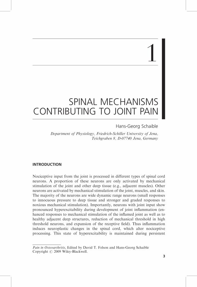
The articular nerves supplying the knee or elbow joint of rat, cat, and monkeyenter the spinal cord via several dorsal roots, thus projecting to several spinalsegments. Due to the widely distributed projection area, joint afferentsinfluence sensory neurons and reflex pathways in several spinal segments.Within the gray matter, knee joint afferents project to the superficial lamina Iand to the deep laminae V–VII.7 Figure 1.1A shows the spinal terminationfields of horseradish peroxidase-labeled knee joint afferents in the segment L7in the cat spinal cord. Correspondingly, spinal cord neurons that are synapti-cally activated by joint afferents can be identified in the superficial and deepdorsal horn and also in the ventral horn.9,10
Receptive Fields and Activation Thresholds of Neurons withJoint Input
In both cat and rat, mechanonociceptive inputs from the joint are processed indorsal horn neurons that respond solely to mechanical stimulation of deeptissue, or in neurons that respond to mechanical stimulation of both deep tissueand the skin. Receptive fields of single sensory neurons (regions from whichneurons can be activated) are usually not restricted to the joint but moreextended. Figure 1.1C shows the receptive field of a spinal cord neuron withconvergent inputs from skin, deep tissue, and the knee joint. The neuron wasactivated by pressure applied to the knee joint (capsule, ligaments) and also bycompression of the quadriceps muscle in the thigh and the gastrocnemius–soleus muscle in the lower leg, and in addition it had a cutaneous receptive fieldat the paw. However, many neurons have receptive fields that are restricted tothe deep tissue. Figure 1.1D shows the receptive field of a spinal cord neuronwith a receptive field in the deep tissue of the leg and in the knee joint. Someneurons have bilateral receptive fields.7
Concerning mechanical thresholds, neurons are either nociceptive-specific(NS) or wide-dynamic-range (WDR) neurons. Nociceptive-specific neuronsrespond only to intense pressure and/or to painful movements such as forcefulsupination and pronation. These stimuli elicit pain. WDR neurons respond toboth innocuous pressure and noxious pressure, encoding stimulus intensity by
SPINAL CORD NEURONS THAT RESPOND TO MECHANICAL STIMULATION 5
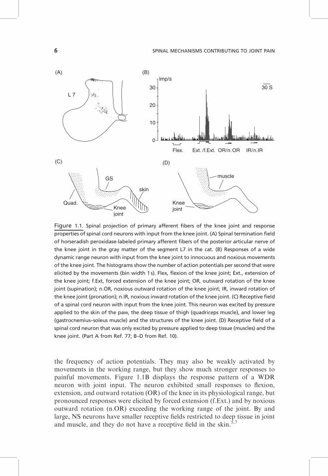
the frequency of action potentials. They may also be weakly activated bymovements in the working range, but they show much stronger responses topainful movements. Figure 1.1B displays the response pattern of a WDRneuron with joint input. The neuron exhibited small responses to flexion,extension, and outward rotation (OR) of the knee in its physiological range, butpronounced responses were elicited by forced extension (f.Ext.) and by noxiousoutward rotation (n.OR) exceeding the working range of the joint. By andlarge, NS neurons have smaller receptive fields restricted to deep tissue in jointand muscle, and they do not have a receptive field in the skin.5,7
(A)
(C)
(B)
(D)
L 730
20
10
0
lmp/s
Flex. Ext. /f.Ext. OR/n.OR IR/n.IR
30 S
GS
skin
Kneejoint
Kneejoint
Quad.
muscle
Figure 1.1. Spinal projection of primary afferent fibers of the knee joint and response
properties of spinal cord neurons with input from the knee joint. (A) Spinal termination field
of horseradish peroxidase-labeled primary afferent fibers of the posterior articular nerve of
the knee joint in the gray matter of the segment L7 in the cat. (B) Responses of a wide
dynamic range neuron with input from the knee joint to innocuous and noxious movements
of the knee joint. The histograms show the number of action potentials per second that were
elicited by the movements (bin width 1 s). Flex, flexion of the knee joint; Ext., extension of
the knee joint; f.Ext, forced extension of the knee joint; OR, outward rotation of the knee
joint (supination); n.OR, noxious outward rotation of the knee joint; IR, inward rotation of
the knee joint (pronation); n.IR, noxious inward rotation of the knee joint. (C) Receptive field
of a spinal cord neuron with input from the knee joint. This neuron was excited by pressure
applied to the skin of the paw, the deep tissue of thigh (quadriceps muscle), and lower leg
(gastrocnemius–soleus muscle) and the structures of the knee joint. (D) Receptive field of a
spinal cord neuron that was only excited by pressure applied to deep tissue (muscles) and the
knee joint. (Part A from Ref. 77; B–D from Ref. 10).
6 SPINAL MECHANISMS CONTRIBUTING TO JOINT PAIN
Supraspinal and Spinal Projections of Spinal Neurons with Joint Input
Neurons with joint input project to different supraspinal sites (cerebellum,spinocervical nucleus, thalamus, reticular formation) or to intraspinal (seg-mental) interneurons and motoneurons.7 Ascending projections to the thala-mus (in the spinothalamic tract) are important to activate the thalamocorticalsystems that generate the conscious pain sensation. In the cat, neurons wereidentified that have cell bodies in the ventral horn, belong to the spinoreticulartract, and are predominantly or exclusively excited by noxious stimulation ofdeep tissue.11,12 Segmental projections are important for the generation ofmotor and sympathetic reflexes. Spinal and supraspinal motor reflexes regulatemovements and exert protective functions including flexor reflexes uponnociceptive stimulation.4 Noxious stimulation of joint afferents can evokenociceptive withdrawal reflexes.7,13 During acute chemical stimulation of theknee and during inflammation in the joint, spinal motor reflexes are en-hanced.13–15 Articular dysfunction and ligamentous strain may cause musclespasms.16 However, there is some evidence that the reflex pattern ofg-motoneurons changes in the course of inflammation such that inhibitoryreflexes are generated.15 The latter may create a new motoric balance and allowthe leg with an inflamed knee to be kept in midposition. In midposition, thenociceptive outflow from the inflamed joint is at a minimum.
Inhibition by Heterotopic and Descending Inhibitory Systems
Neurons with joint input are inhibited by heterotopic noxious stimuli, in linewith the concept of diffuse noxious inhibitory controls (DNICs). The lattermeans that painful stimulation at one site of the body may reduce the pain atanother site of the body.17 In addition, most spinal cord neurons with joint inputare tonically inhibited by descending inhibitory systems that keep the spinal cordunder continuous control.18,19 The interruption of descending inhibition canlower the excitation threshold of spinal cord neurons for mechanical input fromthe knee, substantially increase the receptive fields of neurons, and cause(increased) ongoing discharges. Thus the response properties of neurons withjoint input are controlled by the primary afferent input, by intrinsic properties ofthe spinal cord neurons, by local circuits, and by descending pathways.
HYPEREXCITABILITY OF SPINAL CORD NEURONS DURINGINFLAMMATION IN THE JOINT
Experimental Models of Joint Inflammation
As described in the Introduction, pain and hyperalgesia are usually elicitedduring inflammation of the joint. Hence experimental models have been used to
HYPEREXCITABILITY OF SPINAL CORD NEURONS 7
study neuronal mechanisms underlying these pain symptoms. Acute inflamma-tion in the joint can be induced by the intra-articular injections of crystals suchas urate and kaolin or by carrageenan. The injection of kaolin and carrageenan(K/C) into the joint produces an edema and granulocytic infiltration within 1–3hours with a plateau after 4–6 hours. Awake animals show limping of theinjected leg and enhanced sensitivity to pressure onto the joint. By contrast, theinjection of Freundus complete adjuvant (FCA) into a single joint produces amonoarthritis that is present for 2–4 weeks. Usually the lesion is restricted tothe injected joint, although bilateral effects are observed sometimes. Hyper-algesia (limping or guarding of the leg, enhanced sensitivity to pressure onto thejoint) develops within a day, reaches a peak within 3 days, and is maintained tosome degree up to several weeks. When FCA is injected at a high dose into thetail base or lymph node, a polyarthritis develops.7 More recently, other modelssuch as collagen-induced polyarthritis20 and antigen-induced monoarthritis21,22
are also being used in order to investigate inflammatory pain.
Generation of Spinal Hyperexcitability (Central Sensitization)
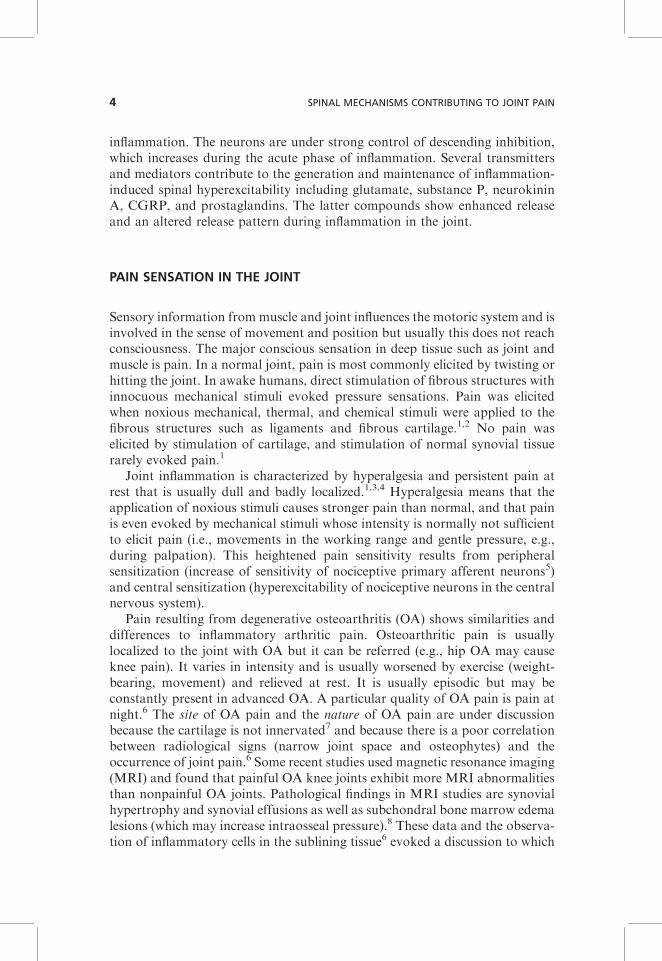
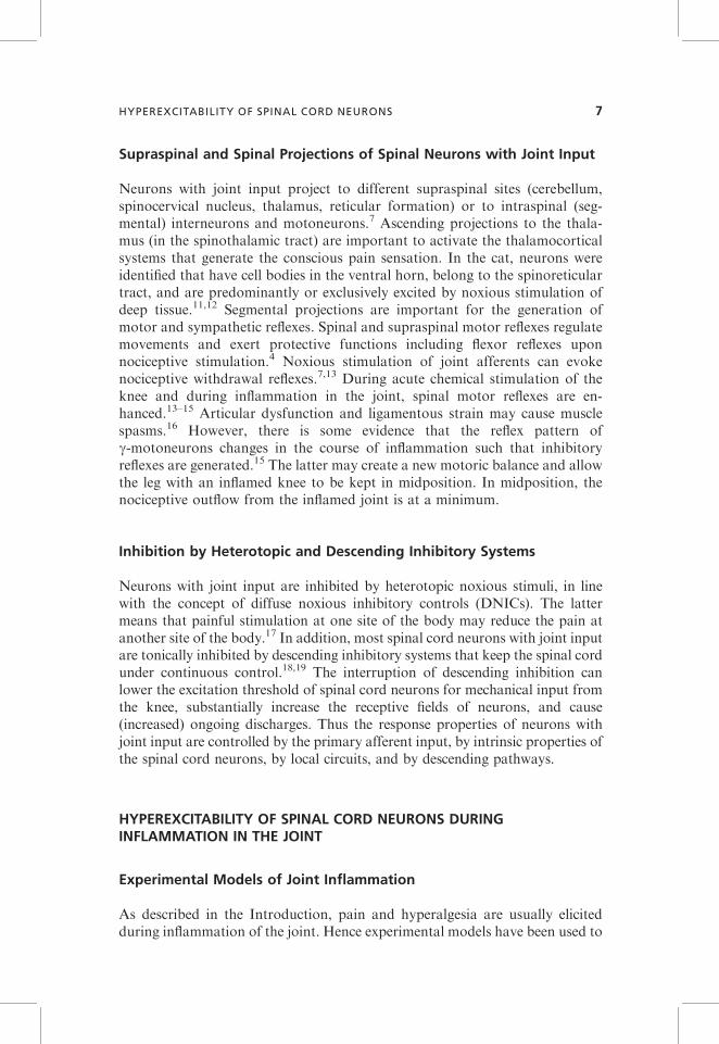
During the development of a K/C-induced inflammation in the joint, both NSand WDR neurons with joint input show within 1–3 hours enhanced responsesto noxious stimuli applied to the inflamed joint. NS neurons exhibit a reductionin their mechanical threshold such that the application of innocuous stimuli tothe inflamed joint is sufficient to excite the neurons. Figure 1.2A shows thegeneration of hyperexcitability in a spinal cord neuron with joint input.Initially, while the joint was normal, the neuron responded only to noxiouspressure applied to the knee (and adjacent muscles in thigh and lower leg, Fig.1.2B, left side). No responses were elicited by pressure onto the ankle and thepaw. After injection of kaolin and carrageenan (K/C) into the knee joint, theresponses to noxious compression of the knee increased markedly, and at alatency of about half an hour the neuron started also to respond to pressureapplied to the ankle and the paw. Thus the receptive field expanded from theknee toward the paw (Fig. 1.2B, right side), and the previously high thresholdneuron was then even activated by gentle innocuous pressure. The increasedresponses to stimuli applied to the inflamed joint result most likely from theenhanced synaptic input from afferent units that are sensitized during stimula-tion. However, the appearance of responses to stimulation of ankle and pawmust result from a mechanism in the spinal cord because these regions were notinflamed. Thus nociceptive spinal cord neurons obviously develop a state ofhyperexcitability in which the responsiveness to both inputs from inflamed andnoninflamed areas is increased.23–26 The increased responses to stimulation ofthe inflamed area are thought to be the neuronal mechanism of primaryhyperalgesia (hyperalgesia at the site of inflammation), whereas the increasedresponses to stimuli applied to healthy tissue are thought to be the neuronal
8 SPINAL MECHANISMS CONTRIBUTING TO JOINT PAIN
0
200
400
600
800
1000
1200
1400Im
p/15
sN
oxio
us p
ress
ure
kn
ee, a
nkle
, paw
K/C
knee
ankl
e
paw
norm
altis
sue
1 2 3 4
−180
−120
−60
060
120
180
240
300
min
(B)
Rec
eptiv
e fie
ld s
ize
cont
rol
3h p
ost K
/C
noxi
ous
pres
sure
inno
cuou
s pr
essu
re
1 2 3 4
(A)
(D)
(C)
in-
flam
ed
tissu
e
1 2 3 4
pre
ssure
1 2 3 4
pre
ssure
Fig
ure
1.2
.D
eve
lop
me
nt
of
infl
am
ma
tio
n-e
vok
ed
hyp
ere
xcit
ab
ilit
yin
asp
ina
lco
rdn
eu
ron
wit
hin
pu
tfr
om
the
kn
ee
join
t.(A
)H
isto
gra
m
sho
win
gth
ere
spo
nse
s(a
ctio
np
ote
nti
als
/re
spo
nse
)o
fth
en
eu
ron
ton
oxi
ou
sp
ress
ure
ap
pli
ed
toth
ek
ne
ejo
int,
the
an
kle
,a
nd
the
pa
w
be
fore
an
da
fte
rin
ject
ion
of
ka
oli
na
nd
carr
ag
ee
na
n(K
/C)
into
the
ipsi
late
ral
kn
ee
join
t.(B
)R
ece
pti
vefi
eld
(sh
ad
ed
are
a)
of
the
ne
uro
n
be
fore
(co
ntr
ol)
an
dd
uri
ng
kn
ee
join
tin
fla
mm
ati
on
(3h
po
stK
/C).
(C,D
)M
od
elsh
ow
ing
the
resp
on
ses
an
dth
ere
cep
tive
fie
ldo
fa
spin
alco
rd
ne
uro
nb
efo
rein
fla
mm
ati
on
(C)
an
da
fte
rd
eve
lop
me
nt
of
hyp
ere
xcit
ab
ilit
y(D
).B
efo
rein
fla
mm
ati
on
the
ne
uro
nw
as
on
lye
xcit
ed
by
pre
ssu
re
toth
ein
itia
lre
cep
tive
fie
ld(s
tim
ula
tio
nsi
tes
2a
nd
3).
Aft
er
infl
am
ma
tio
nth
en
eu
ron
wa
sa
ctiv
ate
dfr
om
ala
rge
ra
rea
(sti
mu
lati
on
site
s1
–4).
(Pa
rts
Aa
nd
Bfr
om
Re
f.2
5.)
9
mechanism underlying secondary hyperalgesia (hyperalgesia in healthy tissueadjacent to and remote from inflamed tissue).
Figure 1.2C,D shows the working hypothesis of how these changes areproduced. When the tissue is normal, the neuron is only excited by stimuliapplied to the restricted receptive field (circle in Fig. 1.2C) but not by stimuliapplied to adjacent areas. When an inflammation develops in the receptive field(shaded area, Fig. 1.2D), primary afferents in this region are sensitized and theyinduce a process of spinal sensitization. When the spinal neuron is hyperexci-table, it shows stronger responses to stimuli applied to the original receptivefield (stimulation sites 2 and 3), and in addition the neuron responds to inputsthat are normally too weak to excite the neuron above threshold (stimulationsites 1 and 4). Hence the receptive field expands (Fig. 1.2D). The spinal‘‘functional connection’’ between knee and paw and the change of synapticeffectiveness during inflammation was also shown in a recent study in whichfield potentials in the spinal cord were recorded. Electrical stimulation of theposterior articular nerve (PAN) of the knee joint evoked typical field potentialsin lumbar spinal segments. The elicited N2 and N3 waves (generated bysynaptic activation of dorsal horn neurons by thin myelinated PAN afferents)became gradually increased after induction of a local inflammation in the pawby capsaicin.27 These data show that a disease process in an area may changesynaptic processing from adjacent and even remote areas.
Central sensitization can persist during chronic inflammation. In rats withunilateral arthritis28 as well as in rats suffering from chronic polyarthritis,29
spinal cord neurons with joint input appear on average more sensitive and haveexpanded receptive fields. During chronic FCA-induced inflammation in theknee joint, secondary hyperalgesia at the ankle can also last several weeks, andfor a long time this hypersensitivity is associated with enhanced responses ofspinal cord neurons to A and C fiber inputs.30
Interestingly, the stimulation of primary afferents from deep tissue (muscleand joint) evokes more prolonged facilitation of a nociceptive flexor reflex thanstimulation of cutaneous afferents,13 and capsaicin injection into deep tissueelicits more prolonged hyperalgesia than injection of capsaicin into the skin,31
suggesting that deep input is particularly able to induce long-term changes inthe nociceptive system. However, spinal sensitization is counteracted to someextent by inhibitory influences. Descending inhibition19 as well as heterotopicinhibitory influences (see above) are increased during inflammation,32 at least inearly stages.
While spinal cord recordings can only be done in experimental studies,human studies lend support to the concept of central sensitization. In humans itis possible to map areas of referred pain upon noxious stimulation at arestricted site. When a noxious stimulus, (e.g., intramuscular injection of 6%NaCl) is applied to a muscle, the area in which pain is felt extends far beyondthe stimulation site. Interestingly, such areas were found to be significantlylarger during pathological conditions such as osteoarthritis.33 The enlargementof painful areas may correspond to the expansion of receptive fields of spinal
10 SPINAL MECHANISMS CONTRIBUTING TO JOINT PAIN
cord neurons. By and large, the described neuronal changes in the spinal cordare likely to account for deep referred pain and secondary hyperalgesia that areinduced in humans by noxious stimulation of deep tissue.34 Based on thisparadigm, numerous pathological conditions in humans such as inflammationand osteoarthritis seem to be associated with central sensitization, suggestingthat the spinal cord is indeed in a state of hyperexcitability.
MOLECULAR MECHANISMS OF SPINAL SENSITIZATION
In general, the process of spinal sensitization depends on several preconditions.First, nociceptive spinal cord neurons have the potential for activity-dependentneuroplastic changes. For example, repetitive electrical stimulation of C fibersat the same current can induce wind-up of the synaptic responses to electricalnerve stimulation (a short-lived increase of responsiveness, however, notoutlasting the stimulation protocol)35 or a long-term potentiation (a persistentincrease of synaptic responses to electrical stimulation outlasting the condi-tioning stimulus).36 Second, both an increase of excitatory mechanism as wellas a reduction of inhibition (e.g., by apoptosis of inhibitory interneurons underneuropathic conditions) may contribute to central sensitization.37 Third, bothneurons and glial cells may be involved in enhanced neuronal excitability.20,38,39
In the case of inflammation, peripheral nociceptive fibers play a key role intriggering the process of spinal sensitization. During developing inflammation,numerous nociceptive mechanosensitive joint afferents are sensitized to me-chanical stimulation such that innocuous stimuli (palpation of the joint andmovements in the working range) become sufficient to evoke action potentials.In addition, initially mechanoinsensitive nociceptive afferents are renderedmechanosensitive and contribute to the input into the spinal cord uponstimulation of the inflamed joint.5,37 Numerous inflammatory mediatorsincluding classical inflammatory mediators such as bradykinin and prostaglan-dins,5 as well as the cytokines interleukin-640 and TNF-a21, have the potentialto sensitize joint afferents for mechanical stimuli. As a consequence ofperipheral sensitization, the intraspinal release of glutamate (the main trans-mitter of nociceptive afferents),41 the neuropeptides substance P, neurokinin A,and CGRP (cotransmitters in primary afferents and interneurons), and spinalprostaglandins is enhanced, and these mediators are involved in the generation(and maintenance) of spinal hyperexcitability.
Excitatory Amino Acids (Glutamate)
As mentioned, glutamate is the major transmitter in the synaptic activation ofspinal cord neurons with joint input. On the postsynaptic site, glutamateactivates N-methyl-D-aspartate (NMDA) receptors and non-NMDA receptors.
MOLECULAR MECHANISMS OF SPINAL SENSITIZATION 11
The activation of non-NMDA receptors leads to basic excitation of neurons.By contrast, the activation of NMDA receptors leads to a calcium influx intoneurons and causes processes of neuronal plasticity such as long-term changesof responses in many neuronal circuits. The ionophoretic application ofantagonists at AMPA/kainate (non-NMDA) receptors close to neurons withjoint input reduced the responses to innocuous and noxious pressure, whereasthe application of NMDA receptor antagonists reduced only the responses tonoxious mechanical stimulation. Thus, in our hands, NMDA receptors are onlyactivated by noxious stimulation.25
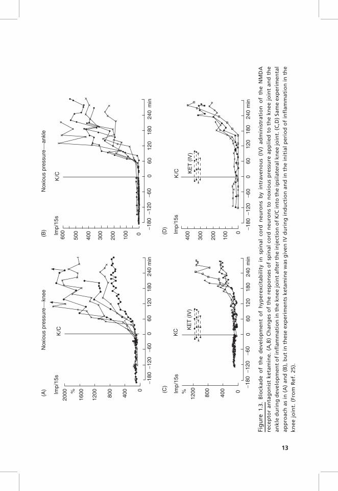
The ionophoretic application of NMDA antagonists at AMPA/kainate andNMDA receptors to spinal cord neurons as well as systemic application ofNMDA antagonists prevents the development of inflammation-evoked spinalhyperexcitability.25 Figure 1.3 shows the effect of ketamine, an antagonist atNMDA receptors. In six control neurons without ketamine, the induction ofinflammation in the knee joint by injection of kaolin/carrageenan (K/C) causedincreases of the responses to noxious pressure applied to the injected knee andthe noninjected ankle. When ketamine was administered before and duringinduction of inflammation, the development of inflammation in the kneejoint did not cause changes of responses as long as the antagonist was applied(Fig. 1.3C,D). Importantly, antagonists at both receptor types can reduceresponses of the neurons to mechanical stimulation of the joint also afterinflammation is established, and this is even seen in a chronic model ofinflammation.25,42 Thus glutamate receptors play a key role in the generationand maintenance of inflammation-evoked spinal hyperexcitability even in thelong-term range. In addition, NMDA receptors are involved in the regulationof spinal NOS isoforms during monoarthritis. During monoarthritis, theexpression of nNOS, iNOS, and eNOS in the dorsal horn was increased,and ketamine reduced nNOS expression and increased iNOS and eNOSexpression.43 However, the functional consequences of these changes are tobe determined.
Neuropeptides
Numerous joint afferents contain the neuropeptides substance P, neurokinin A,and CGRP that are coexpressed with glutamate. Noxious compression, but notinnocuous compression, of the normal joint enhances the intraspinal release ofthese peptides above baseline.44 This pattern of release changes when the jointis inflamed. During acute inflammation, release of neuropeptides occurs whenthe joint is stimulated at innocuous intensity. Thus under inflammatoryconditions, a ‘‘cocktail’’ of transmitters and/or modulators is released in thespinal cord, which changes synaptic processing.45–47 As a further indicatorof spinal release of substance P during arthritis, movements of an arthriticjoint was found to induce internalization of the neurokinin 1 receptor.48 The
12 SPINAL MECHANISMS CONTRIBUTING TO JOINT PAIN
2000
1600
1200 800
400 0
−180
−120
−60
060
120
180
240
−180
−120
−60
060
120
180
240
min
600
500
400
300
200
100 0
min
Imp/
15s
Imp/
15s
Imp/
15s
Imp/
15s
%
K/C
K/C
Nox
ious
pre
ssur
e
knee
Nox
ious
pre
ssur
e
ankl
e(A
) (C)
(D)
(B)
1200
800
400 0%
KC K
ET
(IV
)
−180
−120
−60
060
120
180
240
−180
−120
−60
060
120
180
240
min
K/C
400
300
200
100 0
KE
T (
IV)
min
Fig
ure
1.3
.B
lock
ad
eo
fth
ed
eve
lop
me
nt
of
hyp
ere
xcit
ab
ilit
yin
spin
al
cord
ne
uro
ns
by
intr
ave
no
us
(IV
)a
dm
inis
tra
tio
no
fth
eN
MD
A
rece
pto
ra
nta
go
nis
tk
eta
min
e.
(A,B
)C
ha
ng
es
of
the
resp
on
ses
of
spin
al
cord
ne
uro
ns
ton
oxio
us
pre
ssu
rea
pp
lie
dto
the
kn
ee
join
ta
nd
the
an
kle
du
rin
gd
eve
lop
me
nt
of
infl
am
ma
tio
nin
the
kn
ee
join
ta
fte
rth
ein
ject
ion
of
K/C
into
the
ipsi
late
ralk
ne
ejo
int.
(C,D
)Sa
me
exp
eri
me
nta
l
ap
pro
ach
as
in(A
)a
nd
(B),
bu
tin
the
see
xpe
rim
en
tsk
eta
min
ew
as
giv
en
IVd
uri
ng
ind
uct
ion
an
din
the
init
ial
pe
rio
do
fin
fla
mm
ati
on
inth
e
kn
ee
join
t.(F
rom
Re
f.2
5).
13
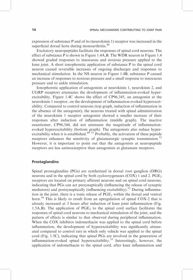
expression of substance P and of its (neurokinin 1) receptor was increased in thesuperficial dorsal horn during monoarthritis.48
Excitatory neuropeptides facilitate the responses of spinal cord neurons. Theeffect of substance P is shown in Figure 1.4A,B. The WDR neuron in Figure 1.4showed graded responses to innocuous and noxious pressure applied to theknee joint. A short ionophoretic application of substance P to the spinal cordneuron caused reversible increases of ongoing discharges and responses tomechanical stimulation. In the NS neuron in Figure 1.4B, substance P causedan increase of responses to noxious pressure and a small response to innocuouspressure and to ankle stimulation.
Ionophoretic application of antagonists at neurokinin 1, neurokinin 2, andCGRP receptors attenuates the development of inflammation-evoked hyper-excitability. Figure 1.4C shows the effect of CP96,345, an antagonist at theneurokinin 1 receptor, on the development of inflammation-evoked hyperexcit-ability. Compared to control neurons (top graph, induction of inflammation inthe absence of the antagonist), the neurons treated with spinal administrationof the neurokinin 1 receptor antagonist showed a smaller increase of theirresponses after induction of inflammation (middle graph). The inactiveenantiomer, CP96,344, did not attenuate the magnitude of inflammation-evoked hyperexcitability (bottom graph). The antagonists also reduce hyper-excitability when it is established.49–51 Probably, the activation of these peptidereceptors enhances the sensitivity of glutamatergic synaptic transmission.52
However, it is important to point out that the antagonists at neuropeptidereceptors are less antinociceptive than antagonists at glutamate receptors.
Prostaglandins
Spinal prostaglandins (PGs) are synthetized in dorsal root ganglion (DRG)neurons and in the spinal cord by both cyclooxygenases (COX) 1 and 2. PGE2
receptors are located on primary afferent neurons and on spinal cord neurons,indicating that PGs can act presynaptically (influencing the release of synapticmediators) and postsynaptically (influencing excitability).53 During inflamma-tion in the joint, there is a tonic release of PGE2 within the dorsal and ventralhorn.54 This is likely to result from an upregulation of spinal COX-2 that isalready increased at 3 hours after induction of knee joint inflammation (Fig.1.5A,B). The application of PGE2 to the spinal cord surface facilitates theresponses of spinal cord neurons to mechanical stimulation of the joint, and thepattern of effects is similar to that observed during peripheral inflammation.When the COX inhibitor indomethacin was applied to the spinal cord beforeinflammation, the development of hyperexcitability was significantly attenu-ated compared to control rats in which only vehicle was applied to the spinalcord (Fig. 1.5C), indicating that spinal PGs are involved in the generation ofinflammation-evoked spinal hyperexcitability.55 Interestingly, however, theapplication of indomethacin to the spinal cord, after knee inflammation and
14 SPINAL MECHANISMS CONTRIBUTING TO JOINT PAIN
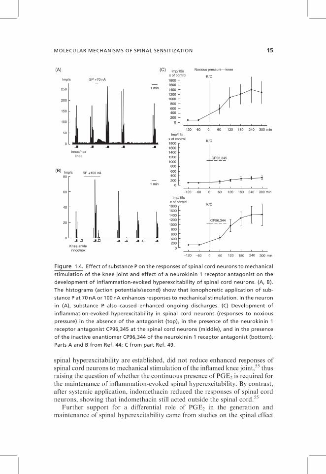
spinal hyperexcitability are established, did not reduce enhanced responses ofspinal cord neurons to mechanical stimulation of the inflamed knee joint,55 thusraising the question of whether the continuous presence of PGE2 is required forthe maintenance of inflammation-evoked spinal hyperexcitability. By contrast,after systemic application, indomethacin reduced the responses of spinal cordneurons, showing that indomethacin still acted outside the spinal cord.55
Further support for a differential role of PGE2 in the generation andmaintenance of spinal hyperexcitability came from studies on the spinal effect
18001600140012001000800600400200
−120 −60 0 60 120
0
180 240 300 min
Imp/15sx of control
Imp/15sx of control
18001600140012001000
800
600400200
0
K/C
K/C
Imp/15sx of control
Noxious pressureknee
CP96,345
−120 −60 0 60 120 180 240 300 min
18001600140012001000800600400200
0
−120 −60 0 60 120 180 240 300 min
K/C
CP96,344
250
200
150
100
50
0
Imp/s SP +70 nA
1 min
innoc/noxknee
1 min
SP +100 nAImp/s80
60
40
20
0
Knee ankleinnoc/nox
(A)
(B)
(C)
Figure 1.4. Effect of substance P on the responses of spinal cord neurons to mechanical
stimulation of the knee joint and effect of a neurokinin 1 receptor antagonist on the
development of inflammation-evoked hyperexcitability of spinal cord neurons. (A, B).
The histograms (action potentials/second) show that ionophoretic application of sub-
stance P at 70 nA or 100 nA enhances responses to mechanical stimulation. In the neuron
in (A), substance P also caused enhanced ongoing discharges. (C) Development of
inflammation-evoked hyperexcitability in spinal cord neurons (responses to noxious
pressure) in the absence of the antagonist (top), in the presence of the neurokinin 1
receptor antagonist CP96,345 at the spinal cord neurons (middle), and in the presence
of the inactive enantiomer CP96,344 of the neurokinin 1 receptor antagonist (bottom).
Parts A and B from Ref. 44; C from part Ref. 49.
MOLECULAR MECHANISMS OF SPINAL SENSITIZATION 15
COX-1
COX-1
COX-2
COX-2
Con 3h 6h 12h Std
Con 3h 6h 12h
(A)
(C)
(B)
0.3
0.2
0.1
0.0
Ave
rage
OD
uni
ts
K/C K/C
knee ankle
800
600
400
200
800
600
400
200
0
800
600
400
200
0
0
800
600
400
200
0
indo.controlnox.
innoc.
impu
lses
/15s
abo
ve B
Lim
puls
es/1
5s a
bove
BL
BL 1st 2nd 3rd 4th BL 1st 2nd 3rd 4th hour
Figure 1.5. Upregulation of cyclooxygenase 2 in the spinal cord during inflammation in
the knee joint and effect of spinal administration of indomethacin on the development
of inflammation-evoked hyperexcitability of spinal cord neurones. (A,B). During in-
flammation in the joint, mainly spinal cyclooxygenase 2 (COX-2) shows an increase. (C)
The spinal application of indomethacin, a blocker of COX-1 and COX-2, attenuates
spinal hyperexcitability. Open squares show the inflammation-evoked changes of
responses after kaolin/carrageenan injection in control neurons; filled squares show
the changes of the responses during development of inflammation after topical
administration of indomethacin to the spinal cord. Top graphs show responses to
noxious pressure, bottom graphs responses to innocuous pressure. (Parts A and B
from Ref. 54; Part C from Ref. 55).
16 SPINAL MECHANISMS CONTRIBUTING TO JOINT PAIN
of EP receptor agonists56 and on the inhibition of the transcription factorNFkB in the spinal cord.57 The enhancement of responses of spinal cordneurons to mechanical stimulation of the normal knee joint by spinal PGE2 wasmimicked by the spinal application of agonists at the EP1 receptor (whichenhances calcium influx in neurons), and by agonists at the EP2 and EP4receptors (which activate Gs proteins and adenylylcyclases). However, afterinflammation and spinal hyperexcitability were established, only the EP1receptor agonist further increased responses to mechanical stimulation of theinflamed knee, whereas the EP2 and EP4 agonists did not influence neuronalresponses. On the other hand, spinal application of an agonist at the EP3receptor (most isoforms are coupled to Gi proteins and reduce cAMP levels)had no influence on neuronal responses when the joint was normal but reducedthe responses to mechanical stimulation of the knee when it was spinallyapplied during established inflammation.56 Thus the status of the spinal cordmay determine which EP receptor agonist causes an effect upon spinalapplication, and the level of cAMP could be an important molecular factor.
The activation of COX-2 depends on the activation of the transcriptionfactor nuclear factor-kB (NFkB). In unstimulated tissue, NFkB is bound in thecytoplasma to IkBa and IkBb, which prevent it from entering the nucleus. Afterstimulation, IkB kinase (IKK) phosphorylates IkB and causes its degradation,thus allowing the unbound NFkB to enter the nucleus. Hence IKK inhibitorsreduce NFkB-mediated effects.58,59 Recent reports indicate a role of spinalNFkB activation in spinal mechanisms of nociception.60,61 A study on spinalmechanisms of joint nociception showed that spinal application of a specificIKK inhibitor before and early during development of inflammation totallyprevented spinal hyperexcitability during developing joint inflammation, sug-gesting an important role of spinal NFkB in this process. However, duringestablished inflammation, the IKK inhibitor did not reduce the responses ofneurons to mechanical stimulation of the inflamed knee within 2.5 hours afterspinal administration, thus suggesting that spinal hyperexcitability is notmaintained by continuous NFkB activation.57 The pattern of effect of theIKK inhibitor is similar to that of indomethacin (see above), and becauseNFkB inhibitors prevent the upregulation of spinal cyclooxygenases,60,61 thesedata collectively suggest spinal PGE2 is mainly important for the generation ofinflammation-evoked spinal hyperexcitability but not for its maintenance.
It should be noted, however, that other prostaglandins are also synthesizedin the spinal cord. The other major prostaglandin in the central nervous systemincluding the spinal cord is PGD2.
62 Electrophysiological recordings fromspinal cord neurons with knee input showed that topical application of PGD2
to the spinal cord at a high dose may cause a sensitization of spinal cordneurons for mechanical stimulation of the normal joint similar to PGE2. Thiseffect may result from synaptic facilitation due to an increase of the spinalrelease of substance P and CGRP from primary afferent neurons.63–65 How-ever, under conditions of joint inflammation, PGD2 dose-dependently reducedresponses of spinal cord neurons to stimulation of the inflamed knee joint, and
MOLECULAR MECHANISMS OF SPINAL SENSITIZATION 17
spinal application of an antagonist at the DP1 receptor increased responses tostimulation of the inflamed knee.66 Indeed, PGD2 can reduce the enhanceddischarges evoked by PGE2.
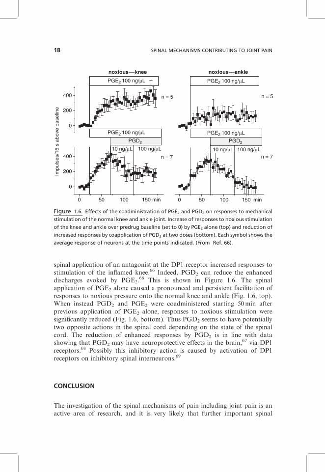
66 This is shown in Figure 1.6. The spinalapplication of PGE2 alone caused a pronounced and persistent facilitation ofresponses to noxious pressure onto the normal knee and ankle (Fig. 1.6, top).When instead PGD2 and PGE2 were coadministered starting 50min afterprevious application of PGE2 alone, responses to noxious stimulation weresignificantly reduced (Fig. 1.6, bottom). Thus PGD2 seems to have potentiallytwo opposite actions in the spinal cord depending on the state of the spinalcord. The reduction of enhanced responses by PGD2 is in line with datashowing that PGD2 may have neuroprotective effects in the brain,67 via DP1receptors.68 Possibly this inhibitory action is caused by activation of DP1receptors on inhibitory spinal interneurons.69
CONCLUSION
The investigation of the spinal mechanisms of pain including joint pain is anactive area of research, and it is very likely that further important spinal
0 50 100 150
n = 5
n = 7
0 50 100 150
0
200
400
n = 5
n = 7
0
200
400
PGD 2 PGD 2 PGD2 PGD2
100 ng/µL10 ng/µL100 ng/µL10 ng/µL
Impu
lses
/15
s ab
ove
base
line
noxiousknee noxiousankle
min min
PGE2 100 ng/µL
PGE2 100 ng/µL
PGE2 100 ng/µL
PGE2 100 ng/µL
Figure 1.6. Effects of the coadministration of PGE2 and PGD2 on responses to mechanical
stimulation of the normal knee and ankle joint. Increase of responses to noxious stimulation
of the knee and ankle over predrug baseline (set to 0) by PGE2 alone (top) and reduction of
increased responses by coapplication of PGD2 at two doses (bottom). Each symbol shows the
average response of neurons at the time points indicated. (From Ref. 66).
18 SPINAL MECHANISMS CONTRIBUTING TO JOINT PAIN
mechanisms of joint pain will be elucidated in the next few years. Of potentialinterest could be the role of glia cells, which produce a number of mediatorsincluding cytokines.39,70
While the spinal cord is an important site where neuronal plasticity such ascentral sensitization occurs, the conscious pain response is produced in thethalamocortical system. The thalamus and cortex contain nociceptive neuronsthat are activated by nociceptive deep input from muscle and joint. Most ofthese neurons have convergent inputs from skin and deep tissue, but smallproportions of neurons respond only to noxious stimulation of muscle andtendon.71–73 There is evidence that during arthritis neuroplastic changes arealso induced at the thalamocortical level.74–76 It is unknown whether thesealterations mirror the altered spinal processing or whether additional elementsof neuroplasticity are generated in the thalamus and cortex. In any case, theworking of the nociceptive system is substantially modified under clinical painconditions, and it is a continuous challenge to identify suitable target sites forpain therapy.
REFERENCES
1. Kellgren JH, Samuel EP. (1950). The sensitivity and innervation of the articular
capsule. J Bone Joint Surg 32-B:84–91.
2. Lewis T. (1942). Pain. London: MacMillan.
3. Kellgren JH. (1939). Some painful joint condition and their relation to osteoar-
thritis. Clin Sci 4:193–205.
4. Lewis T. (1938). Suggestions relating to the study of somatic pain. Br Med J
1:321–325.
5. Schaible H-G. (2006). Basic mechanisms of deep somatic pain. In McMahon SB and
Koltzenburg M (eds.), Wall and Melzack’s Textbook of Pain, 5th ed. Amsterdam:
Elsevier, Churchill Livingston, pp. 621–633.
6. Scott DL. (2006). Osteoarthritis and rheumatoid arthritis. In McMahon SB and
Koltzenburg M (eds.) Wall and Melzackus Textbook of Pain, 5th ed. Amsterdam:
Elsevier, pp. 653–667.
7. Schaible H-G, Grubb BD. (1993). Afferent and spinal mechanisms of joint pain.
Pain 55:5–54.
8. Felson DT. (2005). The sources of pain in knee osteoarthritis. Curr Opin Rheumatol
17:624–628.
9. Schaible H-G, Schmidt RF, Willis WD. (1986). Responses of spinal cord neurones
to stimulation of articular afferent fibres in the cat. J Physiol 372:575–593.
10. Schaible H-G, Schmidt RF, Willis WD. (1987). Convergent inputs from articular,
cutaneous and muscle receptors onto ascending tract cells in the cat spinal cord. Exp
Brain Res 66:479–488.
11. Fields HL, Clanton CH, Anderson SD. (1977). Somatosensory properties of
spinoreticular neurons in the cat. Brain Res 120:49–66.
REFERENCES 19
12. Meyers DER, Snow PJ. (1982). The responses to somatic stimuli of deep spinotha-
lamic tract cells in the lumbar spinal cord of the cat. J Physiol 329:355–371.
13. Woolf CJ, Wall PD. (1986). Relative effectiveness of C primary afferent fibres of
different origins in evoking a prolonged facilitation of the flexor reflex in the rat.
J Neurosci 6:1433–1442.
14. Ferrell WR, Wood L, Baxendale RH. (1998). The effect of acute joint inflammation
on flexion reflex excitability in the decerebrate, low spinal cat. Q J Exp Physiol
373:353–365.
15. He X, Proske U, Schaible H-G, Schmidt RF. (1988). Acute inflammation of the
knee joint in the cat alters responses of flexor motoneurones to leg movements.
J Neurophysiol 59:326–340.
16. Mense S. (1997). Pathophysiologic basis of muscle pain syndromes. Myofasc Pain
Update in Diagnosis and Treatment 8:23–53.
17. LeBars D, Villanueva L. (1988). Electrophysiological evidence for the activation of
descending inhibitory controls by nociceptive pathways. In Fields HL and Besson
J-M (eds.) Progress in Brain Research, vol. 77, Amsterdam: Elsevier, pp. 275–299.
18. Cervero F, Schaible H-G, Schmidt RF. (1991). Tonic descending inhibition of spinal
cord neurones driven by joint afferents in normal cats and in cats with an inflamed
knee joint. Exp Brain Res 83:675–678.
19. Schaible H-G, Neugebauer V, Cervero F, Schmidt RF. (1991). Changes in tonic
descending inhibition of spinal neurons with articular input during the development
of acute arthritis in the cat. J Neurophysiol 66:1021–1032.
20. Inglis JJ, Notley CA, Essex D, Wilson AW, Feldmann M, Anand P, Williams R.
(2007). Collagen-induced arthritis as a model of hyperalgesia. Functional and
cellular analysis of the analgesic actions of tumor necrosis factor blockade. Arthritis
Rheum 56:4015–4023.
21. Boettger MK, Hensellek S, Richter F, Gajda M, Stockigt R, Segond von Banchet G,
Brauer R, Schaible H-G. (2008). Antinociceptive effects of TNF-a neutralization in
a rat model of antigen-induced arthritis. Evidence for a neuronal target. Arthritis
Rheum 58:2368–2378.
22. Segond von Banchet G, Petrow PK, Brauer R, Schaible H-G. (2000). Monoarticular
antigen-induced arthritis leads to pronounced bilateral upregulation of the expres-
sion of neurokinin 1 and bradykinin 2 receptors in dorsal root ganglion neurones of
rats. Arthritis Res 2:424–427.
23. Dougherty PM, Sluka KA, Sorkin LS, Westlund KN, Willis WD. (1992). Neural
changes in the acute arthritis in monkeys. I. Parallel enhancement of responses of
spinothalamic tract neurons to mechanical stimulation and excitatory amino acids.
Brain Res Rev 17:1–13.
24. Neugebauer V, Schaible H-G. (1990). Evidence for a central component in the
sensitization of spinal neurons with joint input during development of acute arthritis
in cat’s knee. J Neurophysiol 64:299–311.
25. Neugebauer V, Lucke T, Schaible H-G. (1993). N-methyl-D-aspartate (NMDA) and
non-NMDA receptor antagonists block the hyperexcitability of dorsal horn
neurones during development of acute arthritis in rat’s knee joint. J Neurophysiol
70:1365–1377.
20 SPINAL MECHANISMS CONTRIBUTING TO JOINT PAIN
26. Schaible H-G, Schmidt RF, Willis WD. (1987). Enhancement of the responses of
ascending tract cells in the cat spinal cord by acute inflammation of the knee joint.
Exp Brain Res 66:489–499.
27. Rudomin P, Hernandez E. (2008). Changes in synaptic effectiveness of myelinated
joint afferents during capsaicin-induced inflammation of the footpad in the
anaesthetized cat. Exp Brain Res 187:71–84.
28. Grubb BD, Stiller RU, Schaible H-G. (1993). Dynamic changes in the receptive field
properties of spinal cord neurons with ankle input in rats with unilateral adjuvant-
induced inflammation in the ankle region. Exp Brain Res 92:441–452.
29. Menetrey D, Besson J-M. (1982). Electrophysiological characteristics of dorsal horn
cells in rats with cutaneous inflammation resulting from chronic arthritis. Pain
13:343–364.
30. Martindale JC, Wilson AW, Reeve AJ, Chessell IP, Headley PM. (2007). Chronic
secondary hypersensitivity of dorsal horn neurones following inflammation of the
knee joint. Pain 133:79–86.
31. Sluka KA. (2002). Stimulation of deep somatic tissue with capsaicin produces long-
lasting mechanical allodynia and heat hypoalgesia that depends on early activation
of the cAMP pathway. J Neurosci 22:5687–5693.
32. Calvino B, Villanueva L, LeBars D. (1987). Dorsal horn (convergent) neurones in
the intact anaesthetized arthritic rat. II. Heterotopic inhibitory influences. Pain
31:359–379.
33. Bajaj P, Bajaj P, Graven-Nielsen T, Arendt-Nielsen L. (2001). Osteoarthritis and its
association with muscle hyperalgesia: an experimental controlled study. Pain
93:107–114.
34. Arendt-Nielsen L, Laursen RJ, Drewes AM. (2000). Referred pain as an indicator
for neural plasticity. In Sandkuhler J, Bromm B and Gebhart GF (eds.) Progress in
Brain Research, Amsterdam: Elsevier, Vol. 129, pp. 343–356.
35. Mendell LM, Wall PD. (1965). Responses of single dorsal cord cells to peripheral
cutaneous unmyelinated fibers. Nature 206:97–99.
36. Sandkuhler J. (2000). Learning and memory in pain pathways. Pain 88:113–118.
37. Schaible H-G. (2006). Peripheral and central mechanisms of pain generation. In
Stein C (ed.) Handbook of Experimental Pharmacalogy, Berlin: Springer-Verlag,
Vol. 177, pp. 4–28.
38. Sun S, Cao H, Han M, Li TT, Pan HL, Zhao ZQ, Zhang YQ. (2007). New evidence
for the involvement of spinal fraktalkine receptor in pain facilitation and spinal glial
activation in rat model of monoarthritis. Pain 129:64–75.
39. Watkins LR, Maier SF. (2005). Glia and pain: past, present, and future. In Merskey
H, Loeser JD, and Dubner R (eds.), The Paths of Pain 1975–2005. Seattle: IASP
Press, pp. 165–175.
40. Brenn D, Richter F, Schaible H-G. (2007). Sensitization of unmyelinated sensory
fibers of the joint nerve for mechanical stimuli by interleukin-6 in the rat. An
inflammatory mechanism of joint pain. Arthritis Rheum 56:351–359.
41. Sorkin LS, Westlund KN, Sluka KA, Dougherty PM, Willis WD. (1992). Neural
changes in acute arthritis in monkeys. IV. Time course of amino acid release into the
lumbar dorsal horn. Brain Res Rev 17:39–50.
REFERENCES 21
42. Neugebauer V, Lucke T, Grubb BD, Schaible H-G. (1994). The involvement of N-
methyl-D-aspartate (NMDA) and non-NMDA receptors in the responsiveness of rat
spinal neurons with input from the chronically inflamed ankle. Neurosci Lett
170:237–240.
43. Infante C, Diaz M, Hernandez A, Constandil L, Pelissier T. (2007). Expression of
nitric oxide synthase isoforms in the dorsal horn of monoarthritic rats: effects of
competitive and uncompetitive N-methyl-D-aspartate antagonists. Arthritis Res
Ther 9:R53.
44. Neugebauer V, Schaible H-G, Weiretter F, Freudenberger U. (1994). The involve-
ment of substance P and neurokinin-1 receptors in the responses of rat dorsal horn
neurons to noxious but not to innocuous mechanical stimuli applied to the knee
joint. Brain Res 666:207–215.
45. Hope PJ, Jarrott B, Schaible H-G, Clarke RW, Duggan AW. (1990). Release and
spread of immunoreactive neurokinin A in the cat spinal cord in a model of acute
arthritis. Brain Res 533:292–299.
46. Schaible H-G, Jarrott B, Hope PJ, Duggan AW. (1990). Release of immunoreactive
substance P in the cat spinal cord during development of acute arthritis in cat’s knee:
a study with antibody bearing microprobes. Brain Res 529:214–223.
47. Schaible H-G, Freudenberger U, Neugebauer V, Stiller U. (1994). Intraspinal
release of immunoreactive calcitonin gene-related peptide during development of
inflammation in the joint in vivo—a study with antibody microprobes in cat and rat.
Neuroscience 62:1293–1305.
48. Sharif Naeini R, Cahill CM, Ribeiro-da-Silva A, Menard HA, Henry JL. (2005).
Remodelling of spinal nociceptive mechanisms in an animal model of monoarthritis.
Eur J Neurosci 22:2005–2015.
49. Neugebauer V, Weiretter F, Schaible H-G. (1995). The involvement of substance P
and neurokinin-1 receptors in the hyperexcitability of dorsal horn neurons during
development of acute arthritis in rat’s knee joint. J Neurophysiol 73:1574–1583.
50. Neugebauer V, Rumenapp P, Schaible H-G. (1996). The role of spinal neurokinin-2
receptors in the processing of nociceptive information from the joint and in the
generation and maintenance of inflammation-evoked hyperexcitability of dorsal
horn neurons in the rat. Eur J Neurosci 8:249–260.
51. Neugebauer V, Rumenapp P, Schaible H-G. (1996). Calcitonin gene-related peptide
is involved in the generation and maintenance of hyperexcitability of dorsal horn
neurons observed during development of acute inflammation in rat’s knee joint.
Neuroscience 71:1095–1109.
52. Ebersberger A, Charbel Issa P, Vanegas H, Schaible H-G. (2000). Differential
effects of CGRP and CGRP 8-37 upon responses to NMDA and AMPA in spinal
nociceptive neurons with knee input in the rat. Neuroscience 99:171–178.
53. Vanegas H, Schaible H-G. (2001). Prostaglandins and cyclooxygenases in the spinal
cord. Prog Neurobiol 64:327–363.
54. Ebersberger A, Grubb BD, Willingale HL, Gardiner NJ, Nebe J, Schaible H-G.
(1999). The intraspinal release of prostaglandin E2 in a model of acute arthritis is
accompanied by an upregulation of cyclooxygenase-2 in the rat spinal cord.
Neuroscience 93:775–781.
22 SPINAL MECHANISMS CONTRIBUTING TO JOINT PAIN
55. Vasquez E, Bar K-J, Ebersberger A, Klein B, Vanegas H, Schaible H-G. (2001).
Spinal prostaglandins are involved in the development but not the maintenance of
inflammation-induced spinal hyperexcitability. J Neurosci 21:9001–9008.
56. Bar K-J, Natura G, Telleria-Diaz A, Teschner P, Vogel R, Vasquez E, Schaible
H-G, Ebersberger A. (2004). Changes in the effect of spinal prostaglandin E2 during
inflammation—prostaglandin E (EP1-EP4) receptors in spinal nociceptive proces-
sing of input from the normal or inflamed knee joint. J Neurosci 24:642–651.
57. Ebersberger A, Buchmann M, Ritzeler O, Michaelis M, Schaible H-G. (2006).
The role of spinal nuclear factor-kB in spinal hyperexcitability. NeuroReport
17:1615–1618.
58. Barnes PJ, Karin M. (1997). Nuclear factor-kB—a pivotal transcription factor in
chronic inflammatory diseases. N Engl J Med 336:1066–1071.
59. Chen L-W, Egan L, Li Z-W, Greten FR, Kagnoff MF, Karin M. (2003). The
two faces of IKK and NF-kB inhibition: prevention of systemic inflammation
but increased local injury following intestinal ischemia-reperfusion. Nature Med
9:575–581.
60. Lee KM, Kang BS, Lee HL, Son SJ, Hwang SH, Kim DS, Park J-S, Cho H-J.
(2004). Spinal NF-kB activation induces COX-2 upregulation and contributes to
inflammatory pain hypersensitivity. Eur J Neurosci 19:3375–3381.
61. Tegeder I, Niederberger E, Schmidt R, Kunz S, Guhring H, Ritzeler O, Michaelis
M, Geisslinger G. (2004). Specific inhibition of IkB kinase reduces hyperalgesia in
inflammatory and neuropathic pain models in rats. J Neurosci 24:1637–1645.
62. Willingale HL, Gardiner NJ, McLymont N, Giblett S, Grubb BD. (1997).
Prostanoids synthesized by cyclo-oxygenase isoforms in rat spinal cord and their
contribution to the development of neuronal hyperexcitability. Br J Pharmacol
122:1593–1604.
63. Andreeva L, Rang HP. (1993). Effect of bradykinin and prostaglandins on the
release of calcitonin gene-related peptide-like immunoreactivity from the rat spinal
cord in vitro. Br J Pharmacol 108:185–190.
64. Jenkins DW, Feniuk W, Humphrey PP. (2001). Characterization of the prostanoid
receptor types involved in mediating calcitonin gene-related peptide release from
cultured rat trigeminal neurones. Br J Pharmacol 134:1296–1302.
65. Nakae K, Hayashi F, Hayashi M, Yamamoto N, Iino T, Yoshikawa S, Gupta J.
(2005). Functional role of prostacyclin receptor in rat dorsal root ganglion neurons.
Neurosci Lett 388:132–137.
66. Telleria-Diaz A, Ebersberger A, Vasquez E, Schache F, Kahlenbach J, Schaible
H-G. (2008). Different effects of spinally applied prostaglandin D2 (PGD2) on
responses of dorsal horn neurons with knee input in normal rats and in rats with
acute knee inflammation. Neuroscience 156:184–192.
67. Grill M, Heinemann A, Hoefler G, Peskar BA, Schuligoi R. (2008). Effect of
endotoxin treatment on the expression and localization of spinal cyclooxygenase,
prostaglandin synthases, and PGD2 receptors. J Neurochem 104:1345–1357.
68. Liang X, Wu L, Hand T, Andreasson K. (2005). Prostaglandin D2 mediates
neuronal protection via the DP1 receptor. J Neurochem 92:477–486.
REFERENCES 23
69. Minami T, Okuda-Ashitaka E, Nishizawa M, Mori H, Ito S. (1997). Inhibition of
nociceptin-induced allodynia in conscious mice by prostaglandin D2. Br J Pharma-
col 122:605–610.
70. Marchand F, Perretti M, McMahon SB. (2005). Role of the immune system in
chronic pain. Nat Rev Neurosci 6:521–532.
71. Guilbaud G, Peschanski M, Gautron M, Binder D. (1980). Neurones responding to
noxious stimulation in VB complex and caudal adjacent regions in the thalamus of
the rat. Pain 8:303–318.
72. HutchisonWD, LuhnMA, Schmidt RF. (1992). Knee joint input into the peripheral
region of the ventral posterior lateral nucleus of cat thalamus. J Neurophysiol
67:1092–1104.
73. Kniffki K-D, Mizumura K. (1983). Responses of neurons in VPL and VPL-VL
region of the cat to algesic stimulation of muscle and tendon. J Neurophysiol
49:649–661.
74. Gautron M, Guilbaud G. (1982). Somatic responses of ventrobasal thalamic
neurones in polyarthritic rats. Brain Res 237:459–471.
75. Lamour Y, Willer JC, Guilbaud G. (1983). Rat somatosensory (Sm I) cortex. II.
Laminar and columnar organization of noxious and non-noxious inputs. Exp Brain
Res 49:46–54.
76. Lamour Y, Willer JC, Guilbaud G. (1983). Altered properties and laminar
distribution of neuronal responses to peripheral stimulation in the Sm I cortex of
the arthritic rat. Brain Res 273:183–187.
77. Craig AD, Heppelmann B, Schaible H-G. (1988). The projection of the medial and
posterior articular nerves of the catus knee to the spinal cord. J Comp Neurol
276:279–288.
24 SPINAL MECHANISMS CONTRIBUTING TO JOINT PAIN


![Movilizacion Articular Del Complejo Articular Del Hombro [Compatibility Mode]](https://img.pdfslide.net/doc/110x75/54e27cb94a7959b7578b467a/movilizacion-articular-del-complejo-articular-del-hombro-compatibility-mode.jpg)