Embed Size (px)
Citation preview
The Nonribosomal Peptide Synthetase Enzyme DdaD TethersN�-Fumaramoyl-L-2,3-diaminopropionate for Fe(II)/
r-Ketoglutarate-Dependent Epoxidation by DdaC duringDapdiamide Antibiotic Biosynthesis
Marie A. Hollenhorst,† Stefanie B. Bumpus,‡ Megan L. Matthews,§
J. Martin Bollinger, Jr.,| Neil L. Kelleher,‡,⊥ and Christopher T. Walsh*,†
Department of Biological Chemistry and Molecular Pharmacology, HarVard Medical School,Boston, Massachusetts 02115, Department of Chemistry and Institute for Genomic Biology,UniVersity of Illinois at Urbana-Champaign, Urbana, Illinois 61801, and Departments of
Chemistry, Biochemistry, and Molecular Biology, The PennsylVania State UniVersity, UniVersityPark, PennsylVania 16802, United States
Received August 11, 2010; E-mail: [email protected]
Abstract: The gene cluster from Pantoea agglomerans responsible for biosynthesis of the dapdiamideantibiotics encodes an adenylation-thiolation didomain protein, DdaD, and an Fe(II)/R-ketoglutarate-dependent dioxygenase homologue, DdaC. Here we show that DdaD, a nonribosomal peptide synthetasemodule, activates and sequesters N�-fumaramoyl-L-2,3-diaminopropionate as a covalently tethered thioesterfor subsequent oxidative modification of the fumaramoyl group. DdaC catalyzes Fe(II)- and R-ketoglutarate-dependent epoxidation of the covalently bound N�-fumaramoyl-L-2,3-diaminopropionyl-S-DdaD species togenerate N�-epoxysuccinamoyl-DAP (DAP ) 2,3-diaminopropionate) in thioester linkage to DdaD. Afterhydrolytic release, N�-epoxysuccinamoyl-DAP can be ligated to L-valine by the ATP-dependent ligase DdaFto form the natural antibiotic N�-epoxysuccinamoyl-DAP-Val.
Introduction
The dapdiamide antibiotics are a family of five N-acylateddipeptides produced by Pantoea agglomerans (Figure 1A).1 The“dap” prefix refers to the presence of the nonproteinogenicamino acid 2,3-diaminopropionate (DAP; blue in Figure 1),while the “diamide” suffix reflects the two backbone amidebonds. The DAP moiety, which can be acylated either on N�
or on NR, is the first residue of the dipeptide and is attached viaa standard peptide linkage to a terminal valine (Val), isoleucine(Ile), or leucine (Leu) (red in Figure 1). The N-acyl moiety(green in Figure 1) is a fumaramoyl group in dapdiamides A-Dand an epoxysuccinamoyl group in dapdiamide E. The fuma-ramoyl or epoxysuccinamoyl functionality most likely providesthe electrophilic moiety that accounts for the antibiotic activityof this class of compounds.2 The dapdiamides are likely cleavedintracellularly to generate acyl-DAP warheads that targetglucosamine-6-phosphate synthase via capture of the nucleo-philic active site cysteine (Cys).2,3
The dapdiamides A-E were isolated by activity-basedcloning of dapdiamide biosynthetic genes from P. agglomeransCU0119 into Escherichia coli.1 This allowed for sequencingof the responsible gene cluster, which revealed nine genes,annotated as ddaA-I (Figure 1B), that are necessary andsufficient for E. coli to make the dapdiamides. In our initialstudies on the Dda enzymes, we determined that DdaG andDdaF are ATP-dependent ligases that build the N-acyldipeptidescaffolds.4 DdaG is an AMP-generating ligase that makes theregiospecifically N-acylated N�-fumaroyl-DAP (N�FmDAP). Ourdata indicated that DdaF catalyzes the last step in the pathwayand forms the dipeptide linkage of N-acyl-DAP with Val, Ile,or Leu. DdaF cleaves ATP to ADP (not AMP), generating theN-acyl-DAP-phosphate as an activated intermediate for captureby Val, Ile, or Leu. This enzyme accepts only N�-fumaramoyl-DAP (N�FmmDAP), not N�FmDAP, as the carboxylate sub-strate, suggesting DdaH, the putative fumaroyl to fumaramoylamide synthase, acts after DdaG but before DdaF.
In this study, we have turned our attention to the epoxidationevent in the dapdiamide biosynthetic pathway. An epoxysucci-namoyl moiety is present in both dapdiamide E and its N�-acyl-DAP isomer N�-epoxysuccinamoyl-DAP-Val (N�EpSmDAP-Val) [this compound has been referred to in the literaturevariously as CB-25-I, [2-amino-3-(oxirane-2,3-dicarboxami-do)propanoyl]valine, and herbicolin I], a natural productproduced by Serratia plymuthica and P. agglomerans strains
† Harvard Medical School.‡ University of Illinois at Urbana-Champaign.§ Department of Chemistry, The Pennsylvania State University.| Departments of Chemistry, Biochemistry, and Molecular Biology, The
Pennsylvania State University.⊥ Current address: Department of Chemistry, Northwestern University,
Evanston, IL 60208, United States.(1) Dawlaty, J.; Zhang, X.; Fischbach, M. A.; Clardy, J. J. Nat. Prod.
2010, 73, 441–446.(2) Kucharczyk, N.; Denisot, M. A.; Le Goffic, F.; Badet, B. Biochemistry
1990, 29, 3668–3676.(3) Milewski, S.; Andruszkiewicz, R.; Kasprzak, L.; Mazerski, J.; Mignini,
F.; Borowski, E. Antimicrob. Agents Chemother. 1991, 35, 36–43.(4) Hollenhorst, M. A.; Clardy, J.; Walsh, C. T. Biochemistry 2009, 48,
10467–10472.
Published on Web 10/14/2010
10.1021/ja1072367 2010 American Chemical Society J. AM. CHEM. SOC. 2010, 132, 15773–15781 9 15773
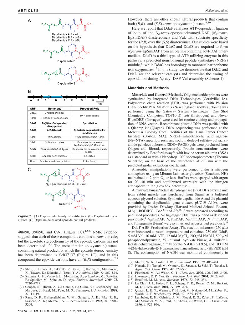
48b/90, 39b/90, and C9-1 (Figure 1C).1,5,6 NMR evidencesuggests that each of these compounds contains a trans-epoxide,but the absolute stereochemistry of the epoxide carbons has notbeen determined.1,5,6 The most similar epoxysuccin(am)ate-containing natural product for which the epoxide stereochemistryhas been determined is Sch37137 (Figure 1C), and in thiscompound the epoxide carbons have an (R,R) configuration.7,8
However, there are other known natural products that containboth (R,R)- and (S,S)-trans-epoxysuccin(am)ate.9,10
Here we report that DdaF catalyzes ATP-dependent ligationof both of the N�-trans-epoxysuccinamoyl-DAP (N�-trans-EpSmDAP) diastereomers and Val, with substrate specificityfor the (R,R) over the (S,S) diastereomer. Our studies were basedon the hypothesis that DdaC and DdaD are required to formN�-trans-EpSmDAP from an olefin-containing acyl-DAP inter-mediate. DdaD is a third type of ATP-utilizing enzyme in thispathway, a predicted nonribosomal peptide synthetase (NRPS)module,11 while DdaC has homology to mononuclear nonhemeiron oxygenases.12 In this study, we demonstrate that DdaC andDdaD are the relevant catalysts and determine the timing ofepoxidation during N�-acyl-DAP-Val assembly (Scheme 1).
Materials and Methods
Materials and General Methods. Oligonucleotide primers weresynthesized by Integrated DNA Technologies (Coralville, IA).Polymerase chain reaction (PCR) was performed with PhusionHigh-Fidelity PCR Mastermix (New England Biolabs). Cloning wasperformed using the Gateway System (Invitrogen). One ShotChemically Competent TOP10 E. coli (Invitrogen) and Nova-Blue(DE3) (Novagen) were used for routine cloning and propaga-tion of DNA vectors. Recombinant plasmid DNA was purified witha Qiaprep kit (Qiagen). DNA sequencing was performed at theMolecular Biology Core Facilities of the Dana Farber CancerInstitute (Boston, MA). Nickel-nitrilotriacetic acid agarose(Ni-NTA) superflow resin and sodium dodecyl sulfate-polyacryl-amide gel electrophoresis (SDS-PAGE) gels were purchased fromQiagen and Biorad, respectively. Protein concentrations weredetermined by Bradford assay13 with bovine serum albumin (BSA)as a standard or with a Nanodrop 1000 spectrophotometer (ThermoScientific) on the basis of the absorbance at 280 nm with thepredicted molar extinction coefficient.
Anaerobic manipulations were performed under a nitrogenatmosphere using an Mbraun Labmaster glovebox (Stratham, NH)maintained at 2 ppm O2 or less. Buffers were sparged with argonfor 20-30 min and equilibrated overnight with the nitrogenatmosphere in the glovebox before use.
A pyruvate kinase/lactate dehydrogenase (PK/LDH) enzyme mixfrom rabbit muscle was purchased from Sigma as a bufferedaqueous glycerol solution. Synthetic dapdiamide A and the plasmidcontaining the dapdiamide gene cluster, pUC19 A10A, wereprovided by Jessica Dawlaty (Harvard Medical School, Boston,MA).1 BODIPY-CoA14 and Sfp15,16 were prepared according topublished procedures. N-His6-tagged DdaF was purified as describedpreviously.4 N�FmDAP, NRFmDAP, N�FmmDAP, NRFmmDAP,and fumaramate (Fmm) were synthesized as described previously.4
DdaF ADP Production Assay. The reaction mixtures (250 µL)were incubated at room temperature and contained 250 nM DdaF,5 mM Val, 10 mM ATP, 12 mM MgCl2, 200 µM NADH, 500 µMphosphoenolpyruvate, 59 units/mL pyruvate kinase, 41 units/mLlactate dehydrogenase, 5 mM borate ·NaOH (pH 9.5), and 100 mM4-(2-hyhdroxyethyl)-1-piperazineethanesulfonic acid (HEPES) (pH8). The consumption of NADH was monitored continuously in
(5) Shoji, J.; Hinoo, H.; Sakazaki, R.; Kato, T.; Hattori, T.; Matsumoto,K.; Tawara, K.; Kikuchi, J.; Terui, Y. J. Antibiot. 1989, 42, 869–874.
(6) Sammer, U. F.; Volksch, B.; Mollmann, U.; Schmidtke, M.; Spiteller,P.; Spiteller, M.; Spiteller, D. Appl. EnViron. Microbiol. 2009, 75,7710–7717.
(7) Cooper, R.; Horan, A. C.; Gentile, F.; Gullo, V.; Loebenberg, D.;Marquez, J.; Patel, M.; Puar, M. S.; Truumees, I. J. Antibiot. 1988,41, 13–19.
(8) Rane, D. F.; Girijavallabhan, V. M.; Ganguly, A. K.; Pike, R. E.;Saksena, A. K.; McPhail, A. T. Tetrahedron Lett. 1993, 34, 3201–3204.
(9) Martin, W. R.; Foster, J. W. J. Bacteriol. 1955, 70, 405–414.(10) Hanada, K.; Tamai, M.; Ohmura, S.; Sawada, J.; Seki, T.; Tanaka, I.
Agric. Biol. Chem. 1978, 42, 529–536.(11) Fischbach, M. A.; Walsh, C. T. Chem. ReV. 2006, 106, 3468–3496.(12) Hausinger, R. P. Crit. ReV. Biochem. Mol. Biol. 2004, 39, 21–68.(13) Bradford, M. M. Anal. Biochem. 1976, 72, 248–254.(14) La Clair, J. J.; Foley, T. L.; Schegg, T. R.; Regan, C. M.; Burkart,
M. D. Chem. Biol. 2004, 11, 195–201.(15) Quadri, L. E. N.; Weinreb, P. H.; Lei, M.; Nakano, M. M.; Zuber, P.;
Walsh, C. T. Biochemistry 1998, 37, 1585–1595.(16) Lambalot, R. H.; Gehring, A. M.; Flugel, R. S.; Zuber, P.; LaCelle,
M.; Marahiel, M. A.; Reid, R.; Khosla, C.; Walsh, C. T. Chem. Biol.1996, 3, 923–936.
Figure 1. (A) Dapdiamide family of antibiotics. (B) Dapdiamide genecluster. (C) Dapdiamide-related epoxide natural products.
15774 J. AM. CHEM. SOC. 9 VOL. 132, NO. 44, 2010
A R T I C L E S Hollenhorst et al.
Plastibrand micro UV cuvettes in a Varian Cary 50 UV-visspectrophotometer by measuring the absorbance at 340 nm. Kineticconstants were derived from velocity versus substrate concentrationdata with GraphPad Prism using a nonlinear, least-squares fittingmethod for N�FmmDAP and N�-(R,R)-epoxysuccinamoyl-DAP(N�RREpSmDAP) and a linear regression for N�-(S,S)-epoxysuc-cinamoyl-DAP (N�SSEpSmDAP).
DdaD ATP-[32P]PPi Exchange Assays. The reaction mixtures(170-350 µL) contained 5 µM DdaD, 125 µM substrate, 1 mMATP, 1 mM MgCl2, 40 mM KCl, 5 mM Na[32P]PPi (approximately(2-4) × 107 counts per min (cpm)/mL), and 50 mM HEPES (pH7.5). The reactions were incubated at room temperature for 10 min,and then 50 µL aliquots were removed and quenched with 250 µLof a charcoal suspension (100 mM NaPPi, 350 mM HClO4, and 16g/L charcoal). The samples were mixed using a vortex and thencentrifuged at 16000g for 3 min. The pellets were washed twicewith 250 µL of wash solution (100 mM NaPPi, and 350 mMHClO4). Charcoal-bound radioactivity was measured on a BeckmanLS 6500 scintillation counter.
DdaC/D Enzymatic Assays with Anaerobically PurifiedDdaC. Phosphopantetheinylation reactions (12-270 µL) contained25 µM DdaD, 12 µM Sfp, 400 µM coenzyme A (CoA), 10 mMMgCl2, 1.5 mM dithiothreitol (DTT), and 50 mM HEPES (pH 7.5).The reactions were incubated at room temperature for 1 h. To formaminoacyl-S-DdaD, 120-250 µM substrate and 1 mM ATP wereadded (final volume 12.5-156 µL) and the reactions incubated foran additional 5-60 min at room temperature.
The following reagents were added to the N�FmmDAP-S-DdaDpreparations to the indicated final concentrations: 50 µMFe(NH4)2(SO4)2 (stock solution 2.5 mM in 200 µM HCl), 300 µMR-ketoglutarate (R-KG) (stock solution 6 mM in 50 mM HEPES(pH 8)), 4 mM ascorbic acid (stock solution 100 mM in 50 mMHEPES (pH 8)), and 10 µM DdaC (final volume 73-79 µL). Thereactions were incubated for 5-60 min at room temperature.
The reactions were quenched by flash freezing in N2(l) forsubsequent trypsin digestion or by the addition of an equal volumeof 25% formic acid followed by flash freezing in N2(l) for reversed-phase liquid chromatography (RPLC)-Fourier transform massspectrometry (FTMS) analysis of intact DdaD.
H218O incubations were carried out as described above with the
exception that the final reaction mixtures contained 63% H218O
(Cambridge Isotope Laboratories).Trypsin Digestion of DdaD-Containing Reactions. The fol-
lowing digestion procedure was used for analysis of (1) conversionof apo-DdaD to holo-DdaD (HS-DdaD), (2) loading of DdaD withN�FmmDAP to generate N�FmmDAP-S-DdaD, (3) conversion ofN�FmmDAP-S-DdaD to N�EpSmDAP-S-DdaD by DdaC, (4)dependence of epoxide formation on R-KG, (5) loading of dap-
diamide A onto HS-DdaD, and (6) incorporation of 18O intoN�EpSmDAP-S-DdaD using H2
18O.All reactions were stored at -80 °C until trypsin digestion. Prior
to trypsin digestion, the samples were thawed at room temperature.Trypsin (Promega Sequencing grade) was resuspended in the bufferprovided by the manufacturer to a final concentration of 1 µg/µLand incubated at 30 °C for 15 min. An aliquot of the reactioncontaining 50 µg of DdaD was removed and added to an equalvolume of 0.1 M NH4HCO3 (pH 7.8-8) and 2 mM Tris(2-carboxyethyl)phosphine (TCEP) (pH 6). This mixture was incubatedfor 4 min at room temperature, and trypsin was added at a massratio of 1:5 trypsin:total protein in the digestion reaction. Afteraddition of the trypsin, the reaction was incubated at 30 °C for 5min. The reaction was quenched by the addition of one-half reactionvolume of 25% formic acid and stored at -80 °C until massspectrometric analysis.
RPLC-FTMS Analysis of Trypsin Digests. All RPLC-FTMSanalyses were conducted using an Agilent 1200 high-performanceLC (HPLC) system with an autosampler coupled directly to aThermoFisher Scientific LTQ-FT hybrid linear ion trap FTMSsystem operating at 11 T. The mass spectrometer was calibratedweekly using the calibration mixture and instructions specified bythe manufacturer. All instrument parameters were tuned accordingto the manufacturer’s instructions (employing bovine ubiquitin(Sigma) for tuning purposes).
For all analyses of trypsin digests of DdaD-containing reactions,a 1 mm × 150 mm Jupiter C18 column (Phenomenex, 300 Å, 5µm) was connected in-line with the electrospray ionization (ESI)source (operated at ∼5 kV with a capillary temperature of 200-250°C) for the MS system. The 70 min separation gradient used forall RPLC analyses is shown in Table S1 (Supporting Information),where solvent A was H2O + 0.1% formic acid and solvent B wasacetonitrile (MeCN) + 0.1% formic acid. A trypsin-digestedreaction mixture was loaded onto the column using the autosamplerand separated according to the gradient shown.
All ionized peptide species entering the mass spectrometer weresubjected to an MS method with five MS and MS/MS events: (1)full scan measurement of all intact peptides (all ions detected inthe FTMS instrument in profile mode, resolution 100 000, m/z rangedetected 400-2000), (2) the phosphopantetheinyl (Ppant) ejectionassay using nozzle-skimmer dissociation (NSD) (all ions detectedin the FTMS instrument in profile mode, resolution 50 000, m/zrange 250-500, surface-induced dissociation (SID) 75 V), (3-5)data-dependent MS/MS on the first, second, and third most abundantions from scan 1 using collision-induced dissociation (CID) (allions detected in the FTMS instrument in profile mode, minimumtarget signal counts 5000, resolution 50 000, m/z range detecteddependent on target m/z, default charge state 2, isolation width 5
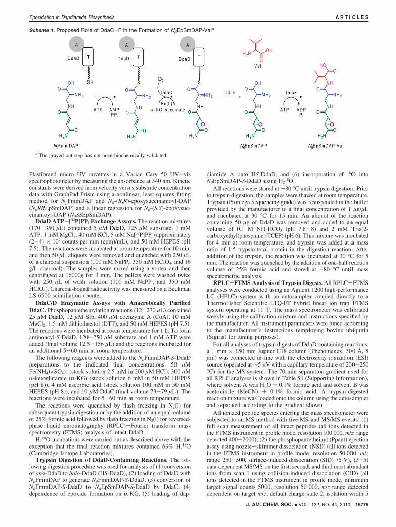
Scheme 1. Proposed Role of DdaC-F in the Formation of N�EpSmDAP-Vala
a The grayed-out step has not been biochemically validated.
J. AM. CHEM. SOC. 9 VOL. 132, NO. 44, 2010 15775
Epoxidation in Dapdiamide Biosynthesis A R T I C L E S
m/z, normalized collision energy (NCE) 35, activation q value 0.40,activation time 30 ms). During all analyses, dynamic exclusion wasenabled with the following settings: repeat count 2, repeat duration30 s, exclusion list size 300, exclusion duration 60 s.
All data were analyzed using Qualbrowser (Xcalibur) andProSightPC, both provided with the LTQ-FT system. ProsightPCwas used to search all MS/MS data against a database containingthe apo-DdaD protein sequence for peptide identification, with anintact peptide mass tolerance of 750 Da (which allowed forobservation of HS-DdaD and HS-DdaD loaded with a variety ofsubstrates) and a fragment ion mass tolerance of 10 ppm (the ∆mmode was enabled for all searches). A minimum of five matchingfragment ions were required for peptide identification. In Qual-browser, ions of interest were searched within a range of 0.01 m/zaround the isotopic peak of interest, within a tolerance of 5 ppm.
RPLC-FTMS Analysis of Intact DdaD. RPLC-FTMS analy-sis of intact DdaD by the Ppant ejection assay was employed foranalyses of the loading of N�RREpSmDAP onto HS-DdaD, theloading of N�SSEpSmDAP onto HS-DdaD, and 18O incorporationinto N�EpSmDAP-S-DdaD using 18O2. All reactions were quenchedwith an equal volume of 25% formic acid and stored at -80 °Cprior to analysis.
For all analyses of intact proteins in DdaD-containing reactions,a 1 mm ×150 mm Jupiter C4 column (Phenomenex, 300 Å, 5 µm)was connected in-line with the ESI source (operated at ∼5 kV witha capillary temperature of 200-250 °C) for the MS system. Areaction aliquot containing 12-15 µg of DdaD was injected foreach sample. The 45 min separation gradient used for all RPLCanalyses is shown in Table S2 (Supporting Information), wheresolvent A was H2O + 0.1% formic acid and solvent B was MeCN+ 0.1% formic acid. The reaction mixture was loaded onto thecolumn using the autosampler and separated according to thegradient shown.
All ionized protein species entering the mass spectrometer weresubjected to an MS method with two MS and MS/MS events: (1)full scan measurement of all intact peptides (all ions detected inthe ion trap MS instrument in profile mode, m/z range detected400-2000), (2) Ppant ejection assay using NSD (all ions detectedin the FTMS instrument in profile mode, resolution 50 000, m/zrange 250-500, SID ) 75 V). All data were analyzed usingQualbrowser (Xcalibur), provided for analysis with the LTQ-FTsystem. Ppant ejection ions of interest were searched within a rangeof 0.01 m/z around the isotopic peak of interest, within a toleranceof 5 ppm.
Determination of the Source of the Epoxide Oxygen by Useof 18O2(g). Phosphopantetheinylation reactions (506 µL) contained25 µM DdaD, 12 µM Sfp, 400 µM CoA, 1 mM MgCl2, 1.5 mMDTT, and 50 mM HEPES (pH 7.5). The reactions were incubatedat room temperature for 1 h. To form N�FmmDAP-S-DdaD, 123µM N�FmmDAP and 5.5 mM ATP were added (final volume 522µL) and the reactions incubated for an additional 5 min at roomtemperature.
O2-free solutions (prepared as previously described17) (225 µL)containing N�FmmDAP-S-DdaD and R-KG (0.13 mM) in theabsence or presence of DdaC (4.2 µM) were placed in septum-sealed flasks. The flasks were stirred on ice and briefly evacuatedto ∼30 Torr. One flask was subsequently refilled with 1.3 atm ofa gas mixture containing 80% 18O2(g) (95-98% isotopic enrich-ment) and 20% N2(g), and the second flask was refilled with 1.1atm of natural-abundance O2(g). Fe(NH4)2(SO4)2 and ascorbic acidwere subsequently added to the flasks (to concentrations of 21 µMand 1.7 mM, respectively) via a gastight syringe. The solutionswere stirred for 5 min at 0 °C to allow the reactions to reachcompletion. The flasks were opened to air, the reactions wereterminated by addition of each solution to an equal volume of 25%formic acid, and the acidified mixtures were flash frozen in N2(l).
Nucleotide Sequence Accession Number. The nucleotidesequence of the dapdiamide gene cluster from P. agglomerans strainCU0119 has been deposited in the NCBI GenBank database underaccession number HQ130277.
Results
DdaF Ligates N�EpSmDAP and Val in an ATP-DependentManner. Guided by the hypothesis that the dapdiamide pathwayproduces an antibiotic containing a trans-epoxysuccinamatemoiety, we synthesized both diastereomers of N�-trans-EpSm-DAP (see the Supporting Information for synthetic protocolsand characterization) and tested the activity of DdaF in ligatingthese compounds to Val.
Analysis of the enzymatic assays by LC-MS (see theSupporting Information for the method) revealed that DdaFcan catalyze the ligation of both N�-trans-EpSmDAP dia-stereomers to Val to produce the N�EpSmDAP-Val dipeptideantibiotics (Figure S1, Supporting Information). A coupledspectrophotometric ADP production assay was used tokinetically characterize the activity of DdaF with respect tothe two N�-trans-EpSmDAP diastereomers. DdaF was foundto use N�RREpSmDAP as a saturable substrate, whereassaturation was not achieved with N�SSEpSmDAP at concentra-tions up to 590 µM (Table 1; Figure S2, Supporting Informa-tion). These findings suggested that N�RREpSmDAP may bean on-pathway intermediate, with DdaF catalytic efficiency forthis substrate approximately 40-fold greater than that for thecorresponding (S,S)-epoxide.
Expression and Purification of DdaC and DdaD in E. coli.The ddaC and ddaD genes were amplified from pUC19 A10A,a plasmid containing the dapdiamide gene cluster,1 and clonedinto an expression vector encoding an N-terminal His6 tag forDdaC or a C-terminal His6 tag for DdaD. The proteins wereoverexpressed in E. coli BL21(DE3) and purified by Ni-NTAaffinity chromatography (see the Supporting Information for themethods, Figure S3). Yields ranged from 4 to 6 mg/L for DdaCand from 11 to 14 mg/L for DdaD.
Following Ni-NTA chromatography, DdaC was either flashfrozen to yield an aerobic preparation or gel filtered into ananaerobic atmosphere. The anaerobic preparations were incu-bated with Fe(NH4)2(SO4)2, R-KG, and DTT and subsequentlydesalted, giving a preparation with an iron occupancy of 47 (4% (average ( standard deviation, data from two independentexperiments) by ferene spectrophotometric assay.18
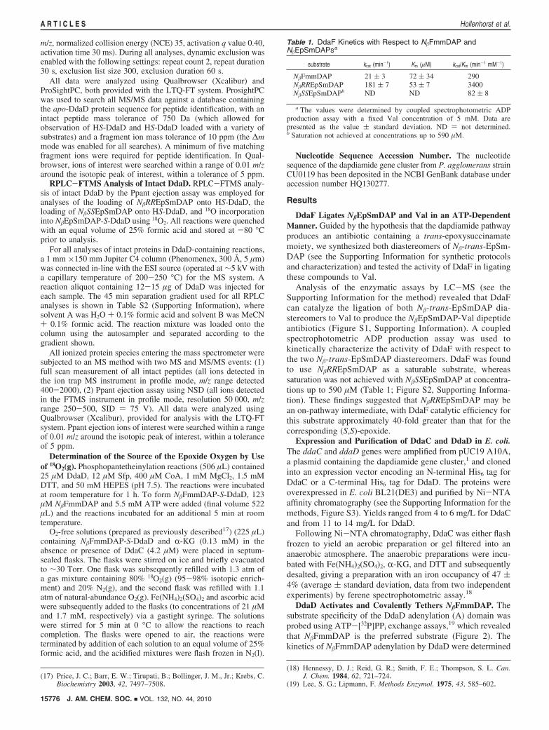
DdaD Activates and Covalently Tethers N�FmmDAP. Thesubstrate specificity of the DdaD adenylation (A) domain wasprobed using ATP-[32P]PPi exchange assays,19 which revealedthat N�FmmDAP is the preferred substrate (Figure 2). Thekinetics of N�FmmDAP adenylation by DdaD were determined
(17) Price, J. C.; Barr, E. W.; Tirupati, B.; Bollinger, J. M., Jr.; Krebs, C.Biochemistry 2003, 42, 7497–7508.
(18) Hennessy, D. J.; Reid, G. R.; Smith, F. E.; Thompson, S. L. Can.J. Chem. 1984, 62, 721–724.
(19) Lee, S. G.; Lipmann, F. Methods Enzymol. 1975, 43, 585–602.
Table 1. DdaF Kinetics with Respect to N�FmmDAP andN�EpSmDAPsa
substrate kcat (min-1) Km (µM) kcat/Km (min-1 mM-1)
N�FmmDAP 21 ( 3 72 ( 34 290N�RREpSmDAP 181 ( 7 53 ( 7 3400N�SSEpSmDAPb ND ND 82 ( 8
a The values were determined by coupled spectrophotometric ADPproduction assay with a fixed Val concentration of 5 mM. Data arepresented as the value ( standard deviation. ND ) not determined.b Saturation not achieved at concentrations up to 590 µM.
15776 J. AM. CHEM. SOC. 9 VOL. 132, NO. 44, 2010
A R T I C L E S Hollenhorst et al.
by this assay; the Km value is 420 ( 80 µM, and the kcat valueis 64 ( 5 min-1 (Figure S4, Supporting Information). DdaDwas also active with N�SSEpSmDAP but to a much lesser extent,with a kcat/Km of 2.3 min-1 mM-1 compared with 150 min-1
mM-1 for N�FmmDAP (Figure S4). No exchange was observedwith N�RREpSmDAP.
The competence of the DdaD T domain to be phosphopanteth-einylated (PPTated) was initially validated by the observation thatit could be modified by the promiscuous Bacillus subtilis phos-phopantetheinyl transferase (PPTase) Sfp15,16 with BODIPY-CoA14 to produce a fluorescent band during SDS-PAGEanalysis (Figure S5, Supporting Information).
Next we turned to FTMS and the Ppant ejection assay20,21
(Scheme S1, Supporting Information) to further characterizeintermediates tethered to DdaD. The experiments were carriedout in one of two fashions. For some analyses, enzymaticincubations were subjected to trypsin digestion, and trypticpeptides were separated by RPLC coupled directly to a hybridlinear ion trap FTMS system (ThermoFisher Scientific LTQ-FT), allowing determination of the masses of intact peptides,identification of the DdaD active site peptide (containing thesite of serine (Ser) phosphopantetheinylation (PPTation)), andobservation of Ppant ejection products. In other experiments,undigested reactions were subjected to RPLC-MS, and Ppantejection was observed directly from intact DdaD.
During LC-MS analysis of a tryptic digest of apo-DdaDincubated with Sfp and CoA, we observed a thiolation (T)
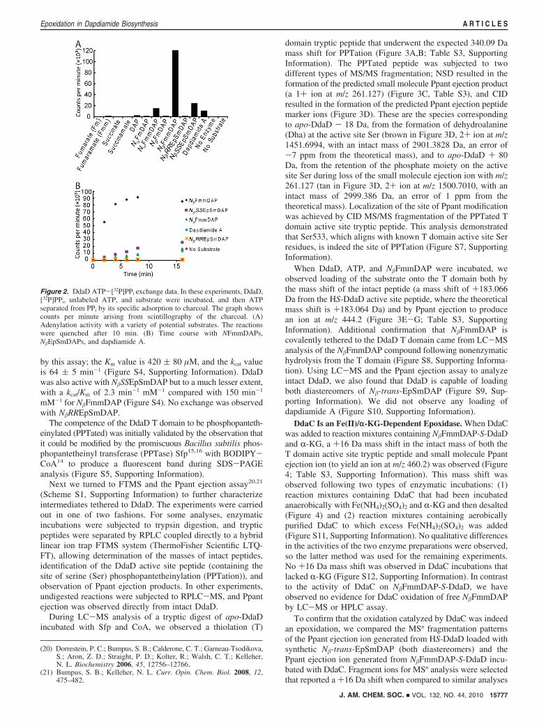
domain tryptic peptide that underwent the expected 340.09 Damass shift for PPTation (Figure 3A,B; Table S3, SupportingInformation). The PPTated peptide was subjected to twodifferent types of MS/MS fragmentation; NSD resulted in theformation of the predicted small molecule Ppant ejection product(a 1+ ion at m/z 261.127) (Figure 3C, Table S3), and CIDresulted in the formation of the predicted Ppant ejection peptidemarker ions (Figure 3D). These are the species correspondingto apo-DdaD - 18 Da, from the formation of dehydroalanine(Dha) at the active site Ser (brown in Figure 3D, 2+ ion at m/z1451.6994, with an intact mass of 2901.3828 Da, an error of-7 ppm from the theoretical mass), and to apo-DdaD + 80Da, from the retention of the phosphate moiety on the activesite Ser during loss of the small molecule ejection ion with m/z261.127 (tan in Figure 3D, 2+ ion at m/z 1500.7010, with anintact mass of 2999.386 Da, an error of 1 ppm from thetheoretical mass). Localization of the site of Ppant modificationwas achieved by CID MS/MS fragmentation of the PPTated Tdomain active site tryptic peptide. This analysis demonstratedthat Ser533, which aligns with known T domain active site Serresidues, is indeed the site of PPTation (Figure S7, SupportingInformation).
When DdaD, ATP, and N�FmmDAP were incubated, weobserved loading of the substrate onto the T domain both bythe mass shift of the intact peptide (a mass shift of +183.066Da from the HS-DdaD active site peptide, where the theoreticalmass shift is +183.064 Da) and by Ppant ejection to producean ion at m/z 444.2 (Figure 3E-G; Table S3, SupportingInformation). Additional confirmation that N�FmmDAP iscovalently tethered to the DdaD T domain came from LC-MSanalysis of the N�FmmDAP compound following nonenzymatichydrolysis from the T domain (Figure S8, Supporting Informa-tion). Using LC-MS and the Ppant ejection assay to analyzeintact DdaD, we also found that DdaD is capable of loadingboth diastereomers of N�-trans-EpSmDAP (Figure S9, Sup-porting Information). We did not observe any loading ofdapdiamide A (Figure S10, Supporting Information).
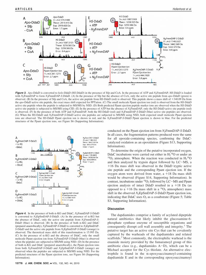
DdaC Is an Fe(II)/r-KG-Dependent Epoxidase. When DdaCwas added to reaction mixtures containing N�FmmDAP-S-DdaDand R-KG, a +16 Da mass shift in the intact mass of both theT domain active site tryptic peptide and small molecule Ppantejection ion (to yield an ion at m/z 460.2) was observed (Figure4; Table S3, Supporting Information). This mass shift wasobserved following two types of enzymatic incubations: (1)reaction mixtures containing DdaC that had been incubatedanaerobically with Fe(NH4)2(SO4)2 and R-KG and then desalted(Figure 4) and (2) reaction mixtures containing aerobicallypurified DdaC to which excess Fe(NH4)2(SO4)2 was added(Figure S11, Supporting Information). No qualitative differencesin the activities of the two enzyme preparations were observed,so the latter method was used for the remaining experiments.No +16 Da mass shift was observed in DdaC incubations thatlacked R-KG (Figure S12, Supporting Information). In contrastto the activity of DdaC on N�FmmDAP-S-DdaD, we haveobserved no evidence for DdaC oxidation of free N�FmmDAPby LC-MS or HPLC assay.
To confirm that the oxidation catalyzed by DdaC was indeedan epoxidation, we compared the MSn fragmentation patternsof the Ppant ejection ion generated from HS-DdaD loaded withsynthetic N�-trans-EpSmDAP (both diastereomers) and thePpant ejection ion generated from N�FmmDAP-S-DdaD incu-bated with DdaC. Fragment ions for MSn analysis were selectedthat reported a +16 Da shift when compared to similar analyses
(20) Dorrestein, P. C.; Bumpus, S. B.; Calderone, C. T.; Garneau-Tsodikova,S.; Aron, Z. D.; Straight, P. D.; Kolter, R.; Walsh, C. T.; Kelleher,N. L. Biochemistry 2006, 45, 12756–12766.
(21) Bumpus, S. B.; Kelleher, N. L. Curr. Opin. Chem. Biol. 2008, 12,475–482.
Figure 2. DdaD ATP-[32P]PPi exchange data. In these experiments, DdaD,[32P]PPi, unlabeled ATP, and substrate were incubated, and then ATPseparated from PPi by its specific adsorption to charcoal. The graph showscounts per minute arising from scintillography of the charcoal. (A)Adenylation activity with a variety of potential substrates. The reactionswere quenched after 10 min. (B) Time course with NFmmDAPs,N�EpSmDAPs, and dapdiamide A.
J. AM. CHEM. SOC. 9 VOL. 132, NO. 44, 2010 15777
Epoxidation in Dapdiamide Biosynthesis A R T I C L E S
conducted on the Ppant ejection ion from N�FmmDAP-S-DdaD.In all cases, the fragmentation patterns produced were the samefor all epoxide-containing species, confirming the DdaC-catalyzed oxidation as an epoxidation (Figure S13, SupportingInformation).
To determine the origin of the putative incorporated oxygen,DdaC incubations were carried out either in H2
18O or under an18O2 atmosphere. When the reaction was conducted in H2
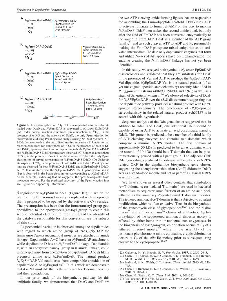
18Oand then analyzed by trypsin digest followed by LC-MS, a+16 Da mass shift was observed in the DdaD tryptic activesite peptide and the corresponding Ppant ejection ion. If theoxygen atom were derived from water, a +18 Da mass shiftwould be observed (Figure S14, Supporting Information). Incontrast, incubations under 18O2 followed by LC-MS and Ppantejection analysis of intact DdaD resulted in a +18 Da (asopposed to a +16 Da mass shift in a 16O2 atmosphere) massshift in the observed N�EpSmDAP-S-DdaD Ppant ejection ion,indicating that DdaC uses O2 as a cosubstrate (Figure 5; TableS3, Supporting Information).
Discussion
The dapdiamides comprise a family of acylated dipeptidenatural antibiotics that likely inhibit the glucosamine-6-phosphate synthase enzyme of susceptible organisms andconsequently disrupt cell wall assembly and integrity.1 Theputative target has an active site Cys that can be covalentlycaptured by the warheads of the dapdiamides and relatedscaffolds.2 Most commonly, the electrophilic warhead is theenamide moiety provided by the fumaramoyl group of thisantibiotic class (e.g., dapdiamides A-D), which can be aMichael acceptor for the Cys thiolate. An alternative elec-trophile is found in the R-epoxysuccinamoyl-containingdapdiamide E and in the corresponding epoxysuccinamoyl
Figure 4. In the presence of both R-KG and DdaC, N�FmmDAP-S-DdaDis converted to N�EpSmDAP-S-DdaD. (A) In the presence of R-KG butthe absence of DdaC, only the active site peptide from N�FmmDAP-S-DdaD (blue) is observed. (B) In the presence of both R-KG and DdaC(prepared anaerobically), N�FmmDAP-S-DdaD is converted to N�EpSmDAP-S-DdaD and the active site peptide from N�EpSmDAP-S-DdaD (orange) isobserved. The theoretical mass shift of this transformation is 15.995 Da.(C) In the presence of R-KG and the absence of DdaC, only the smallmolecule Ppant ejection ion from N�FmmDAP-S-DdaD (blue) is observedwhen the peptides are subjected to MS/MS using NSD. (D) In the presenceof both R-KG and DdaC (prepared anaerobically), the Ppant ejection ionsfrom both N�FmmDAP-S-DdaD and N�EpSmDAP-S-DdaD (orange) areobserved when the peptides are subjected to MS/MS using NSD. For thepredicted structures of the Ppant ejection ions, see Figure S6 (SupportingInformation).
Figure 3. Apo-DdaD is converted to holo-DdaD (HS-DdaD) in the presence of Sfp and CoA. In the presence of ATP and N�FmmDAP, HS-DdaD is loadedwith N�FmmDAP to form N�FmmDAP-S-DdaD. (A) In the presence of Sfp but the absence of CoA, only the active site peptide from apo-DdaD (green) isobserved. (B) In the presence of Sfp and CoA, the active site peptide from HS-DdaD (red) is observed. This peptide shows a mass shift of +340.09 Da fromthe apo-DdaD active site peptide, the exact mass shift expected for PPTation. (C) The small molecule Ppant ejection ion (red) is observed from the HS-DdaDactive site peptide when the peptide is subjected to MS/MS by NSD. (D) Both predicted Ppant ejection peptide marker ions are observed when the HS-DdaDactive site peptide is subjected to MS/MS using CID. (E) In the presence of ATP but the absence of N�FmmDAP, only the HS-DdaD active site peptide (red)is observed. (F) In the presence of both ATP and N�FmmDAP, both the HS-DdaD (red) and N�FmmDAP-S-DdaD (blue) active site peptides are observed.(G) When the HS-DdaD and N�FmmDAP-S-DdaD active site peptides are subjected to MS/MS using NSD, both expected small molecule Ppant ejectionions are observed. The HS-DdaD Ppant ejection ion is shown in red, and the N�FmmDAP-S-DdaD Ppant ejection is shown in blue. For the predictedstructures of the Ppant ejection ions, see Figure S6 (Supporting Information).
15778 J. AM. CHEM. SOC. 9 VOL. 132, NO. 44, 2010
A R T I C L E S Hollenhorst et al.
� regioisomer N�EpSmDAP-Val (Figure 1C), in which theolefin of the fumaramoyl moiety is replaced with an epoxidethat is proposed to be opened by the active site Cys residue.The presumption has been that the fumar(am)oyl group getsepoxidized to the epoxysuccin(am)oyl group to create thissecond potential electrophile; the timing and the identity ofthe catalysts responsible for this conversion are the subjectof this study.
Regiochemical variation is observed among the dapdiamideswith regard to which amino group of 2(R),3(�)-DAP thefumaramoyl/epoxysuccinamoyl moieties are attached in amidelinkage. Dapdiamides A-C have an N�FmmDAP attachment,while dapdiamide D has an NRFmmDAP linkage. DapdiamideE, with an epoxysuccinamoyl group in R amide linkage, couldin principle arise from epoxidation of dapdiamide D or its likelyprecursor amino acid NRFm(m)DAP. The natural productN�EpSmDAP-Val could arise from comparable epoxidation ofdapdiamide A or N�Fm(m)DAP. In this work we demonstratethat it is N�FmmDAP that is the substrate for T domain loadingand then epoxidation.
In our prior study of the biosynthetic pathway for thisantibiotic family, we demonstrated that DdaG and DdaF are
the two ATP-cleaving amide-forming ligases that are responsiblefor assembling the Fmm-dipeptide scaffold. DdaG uses ATPto activate fumarate to fumaroyl-AMP on the way to makingN�FmDAP. DdaF then makes the second amide bond, but onlyafter the acid of FmDAP has been converted enzymatically tothe amide in FmmDAP. DdaF is a member of the ATP graspfamily,22 and as such cleaves ATP to ADP and Pi, presumablymaking the FmmDAP-phosphate mixed anhydride as an acti-vated intermediate. To date only dapdiamide enzymes that formand utilize N�-acyl-DAP species have been characterized; theenzyme creating the NRFmmDAP linkage has not yet beenidentified.
In this study, we assayed both synthetic N�-trans-EpSmDAPdiastereomers and validated that they are substrates for DdaFin the presence of Val and ATP to produce the N�EpSmDAP-Val dipeptide. N�EpSmDAP-Val is the natural product (of asyet unassigned epoxide stereochemistry) recently identified inP. agglomerans strains (48b/90, 39b/90, and C9-1) as well as astrain of Serratia plymuthica.5,6 We observed selectivity of DdaFfor N�RREpSmDAP over the (S,S) diastereomer, suggesting thatthe dapdiamide pathway produces a natural product with (R,R)-epoxide stereochemistry. The precedence of (R,R)-epoxidestereochemistry in the related natural product Sch37137 is inaccord with this hypothesis.8
Sequence analysis of the Dda gene cluster suggested that, inaddition to DdaG and DdaF, one additional ORF should becapable of using ATP to activate an acid cosubstrate, namely,DdaD. This protein is predicted to be a member of a third familyof ATP-cleaving enzymes and contains two domains whichcomprise a minimal NRPS module. The first domain ofapproximately 50 kDa is predicted to be an A domain, whilethe second of 10 kDa should be a T domain that can be post-translationally primed with a Ppant group. The adjacent ORFDdaE, encoding a predicted thioesterase, is the only other NRPS-related ORF in the dapdiamide biosynthetic gene cluster,suggesting the adenylation-thiolation (A-T) didomain DdaDacts as a stand-alone module and not as part of a classical NRPSassembly line.
We have shown in several other contexts that stand-aloneA-T didomains (or isolated T domains) are used in bacterialmetabolism to sequester some fraction of an amino acid pool,tethered as the aminoacyl-S-pantetheinyl T domain thioester.The tethered aminoacyl-S-T domain is then subjected to covalentmodification, which is often oxidative. Thus, in the biosynthesisof the vancomycin class of glycopeptides23,24 and the nikko-mycin25 and aminocoumarin26 classes of antibiotics, C� hy-droxylation of the sequestered aminoacyl thioester moiety iseffected by either heme iron or nonheme iron oxygenases. Inthe biogenesis of syringomycin, chlorination occurs at C4 of atethered threonyl moiety,27 while in the assembly of thejasmonate phytohormone mimic coronatine, cryptic chlorinationoccurs at Cγ of the allo-Ile moiety prior to subsequent ringclosure to the cyclopropane.28,29
(22) Galperin, M. Y.; Koonin, E. V. Protein Sci. 1997, 6, 2639–2643.(23) Chen, H.; Thomas, M. G.; O’Connor, S. E.; Hubbard, B. K.; Burkart,
M. D.; Walsh, C. T. Biochemistry 2001, 40, 11651–11659.(24) Hubbard, B. K.; Walsh, C. T. Angew. Chem., Int. Ed. 2003, 42, 730–
765.(25) Chen, H.; Hubbard, B. K.; O’Connor, S. E.; Walsh, C. T. Chem. Biol.
2002, 9, 103–112.(26) Chen, H.; Walsh, C. T. Chem. Biol. 2001, 8, 301–312.(27) Vaillancourt, F. H.; Yin, J.; Walsh, C. T. Proc. Natl. Acad. Sci. U.S.A.
2005, 102, 10111–10116.
Figure 5. In an atmosphere of 18O2, 18O is incorporated into the substrateloaded onto DdaD and N�FmmDAP is converted to N�-trans-EpSmDAP.(A) Under normal reaction conditions (an atmosphere of 16O2), in thepresence of R-KG and the absence of DdaC, the only Ppant ejection ionobserved (blue) during Ppant ejection analysis (using NSD) of N�FmmDAP-S-DdaD corresponds to the unoxidized starting substrate. (B) Under normalreaction conditions (an atmosphere of 16O2), in the presence of both R-KGand DdaC, Ppant ejection ions corresponding to both N�FmmDAP-S-DdaDand N�EpSmDAP-S-DdaD (orange) are observed. (C) Under an atmosphereof 18O2, in the presence of R-KG but the absence of DdaC, the only Ppantejection ion observed corresponds to N�FmmDAP-S-DdaD. (D) Under anatmosphere of 18O2, in the presence of both R-KG and DdaC, Ppant ejectionions are observed for both N�FmmDAP-S-DdaD and N�EpSmDAP-S-DdaD.A +2 Da mass shift (from the N�EpSmDAP-S-DdaD Ppant ejection ion in(B)) is observed in the Ppant ejection ion corresponding to N�EpSmDAP-S-DdaD (purple), indicating that the oxygen in the epoxide originates frommolecular oxygen. For the predicted structures of the Ppant ejection ionssee Figure S6, Supporting Information.
J. AM. CHEM. SOC. 9 VOL. 132, NO. 44, 2010 15779
Epoxidation in Dapdiamide Biosynthesis A R T I C L E S
With such precedents, we sought an analogous role formodification of an aminoacyl thioester covalently attached toDdaD. The adjacent ORF DdaC has homology to the Fe(II)/R-KG-dependent dioxygenase family of enzymes, which typi-cally catalyze O2-dependent substrate hydroxylations.12 Ex-amples of members of this family include the syringomycinbiosynthetic enzyme SyrP30 and the kutzneride pathway en-zymes KtzO and P,31 which carry out �-hydroxylations of Tdomain-tethered aspartate and glutamate, respectively. In ad-dition to hydroxylations, members of this family have beenshown to carry out a range of other oxidative transformations.12
Evidence from bioconversion and cell extract studies hasimplicated Fe(II)/R-KG enzymes in epoxidation reactions,32,33
but to our knowledge no in vitro characterization of a purifiedepoxidase in this class has been reported previously.
In the context of the known dapdiamide family members(Figure 1A) it seemed likely that DdaC could be an epoxidasethat acts on the fumaroyl/fumaramoyl moiety of an intermediatetethered in thioester linkage to the T domain of DdaD. Inaddition, DdaE is a predicted thioesterase; thus, the tandemaction of DdaD, C, and E could be a branch pathway forselection and activation of an olefin-containing pathway inter-mediate, epoxidation, and then hydrolysis to produce anepoxysuccin(am)oyl building block for condensation withanother monomer via DdaG and/or DdaF. (We have not beenable to heterologously express DdaE in a soluble form in E.coli to establish such a thioesterase role.)
Validation of the proposed roles for DdaD and DdaC startedwith determination of the selectivity of the A domain of DdaD.Using the classical ATP-[32P]PPi exchange assay, diagnosticfor reversible formation of tightly held (amino)acyl-AMPs inenzyme active sites, DdaD showed clear preference forN�FmmDAP. The Km value for DdaD with respect toN�FmmDAP was found to be 420 µM, comparable to Km valuesreported for other NRPS A domains.28,34,35 These resultsprovided a key early insight: DdaD is indeed selecting an olefin-containing pathway intermediate for activation as the AMPmixed anhydride. This was strongly suggestive that the fuma-ramoyl moiety of thioester-tethered FmmDAP would be thespecies epoxidized.
To validate the second step of A-T didomain function, thepredicted covalent loading of N�FmmDAP-AMP onto the Ppantarm of the T domain of DdaD, we turned to mass spectrometry.We found that apo-DdaD could be post-translationally convertedto the Ppant-containing holo-DdaD by action of purified Sfp.Incubation of holo-DdaD with ATP and N�FmmDAP alloweddetection of the N�FmmDAP-S-Ppant adduct in the T domainby peptide mass analysis and by the release of the N�FmmDAP-S-Ppant thioester fragment ion. Thus, the second step of A-T
didomain function, the covalent tethering of the substrateactivated by the A domain, was operant.
When DdaC was incubated with the covalent N�FmmDAP-S-DdaD enzyme intermediate, a mass increase of +16 Da wasobserved for both the T domain active site tryptic peptidecontaining the tethered acyl-DAP thioester and the ejected Ppantion. We found that, as anticipated for a member of the Fe(II)/R-KG family, the activity of DdaC is dependent on R-KG.Additionally, incubation under 18O2(g) resulted in a +18 Damass shift, demonstrating that DdaC uses molecular oxygen asa cosubstrate.
The ejected pantetheinyl fragment from DdaC/D experimentshad the M + 16 Da mass increase anticipated for the epoxideproduct. However, it was formally possible that the introductionof one oxygen atom into the FmmDAP moiety arose not byepoxidation of the double bond but by C- or N-hydroxylationof the DAP residue. MSn fragmentation of Ppant ejection ionsfrom both HS-DdaD loaded with authentic N�-trans-EpSmDAPand from N�FmmDAP-S-DdaD incubated with DdaC resultedin the same fragmentation pattern, suggesting that DdaC indeedacts as an epoxidase.
Our studies of DdaC have generated a number of questionsto be answered in future investigations. We have notattempted to determine single-turnover kinetics of the enzymebecause of the difficulty of quantifying its substrate, thecovalent N-acylaminoacyl thioester adduct of DdaD. We havealso been unable to obtain sufficient N�EpSmDAP fromDdaC/D incubations to directly determine the stereochemistryof the epoxide carbons in the product, nor have we yetevaluated the epoxidation mechanism. In analogy to proposedmechanisms for Fe(II)/R-KG hydroxylases, an Fe(IV)-oxointermediate is the likely oxygen transfer species. However,whether C-O bond formations are stepwise and ionic orradical, as suggested in Scheme S2 (Supporting Information),is yet to be probed.
Additionally, the question arises of why P. agglomeransmakes both the enamide electrophile (fumaramoyl) and theepoxide electrophile (epoxysuccinamoyl) as parallel N-acylwarheads in this antibiotic family. Two future studies willcompare the epoxysuccinamoyl versus the fumaramoylgroups. First, minimum inhibitory concentration (MIC)determinations of the N� molecules dapdiamide A andN�EpSmDAP-Val will test for any differences in uptake bysusceptible bacteria and fungi. Once taken up by theoligopeptide permease systems, intracellular proteases arethought to liberate the N�-acyl-DAPs as the proximalinactivators for glucosamine synthase. Thus, it will be usefulto compare FmmDAP and EpSmDAP side by side againstthe target enzyme to determine inactivation efficiencies. Itis possible that the epoxide warhead is more selective thanthe enamide: the epoxide may require acid catalysis in theenzyme active site for covalent capture, whereas Michaeladdition to the fumaramoyl moiety may not. In that contexta proteomics36 study to evaluate how many proteins in asusceptible cell are targeted covalently would offer a globalcomparison of “off-target” labeling by the two types ofelectrophilic N-acyl warheads.
Acknowledgment. We thank Emily Balskus, Christopher Neu-mann, Elizabeth Sattely, and Albert Bowers for helpful discussions.We thank John Heemstra for providing synthetic BODIPY-CoA,
(28) Couch, R.; O’Connor, S. E.; Seidle, H.; Walsh, C. T.; Parry, R. J.Bacteriol. 2004, 186, 35–42.
(29) Vaillancourt, F. H.; Yeh, E.; Vosburg, D. A.; O’Connor, S. E.; Walsh,C. T. Nature 2005, 436, 1191–1194.
(30) Singh, G. M.; Fortin, P.; Koglin, A.; Walsh, C. T. Biochemistry 2008,47, 11310–11320.
(31) Strieker, M.; Nolan, E. M.; Walsh, C. T.; Marahiel, M. A. J. Am.Chem. Soc. 2009, 131, 13523–13530.
(32) Watanabe, M.; Sumida, N.; Murakami, S.; Anzai, H.; Thompson, C. J.;Tateno, Y.; Murakami, T. Appl. EnViron. Microbiol. 1999, 65, 1036–1044.
(33) Hashimoto, T.; Matsuda, J.; Yamada, Y. FEBS Lett. 1993, 329, 35–39.
(34) Mootz, H. D.; Marahiel, M. A. J. Bacteriol. 1997, 179, 6843–6850.(35) Ehmann, D. E.; Shaw-Reid, C. A.; Losey, H. C.; Walsh, C. T. Proc.
Natl. Acad. Sci. U.S.A. 2000, 97, 2509–2514.(36) Han, X.; Aslanian, A.; Yates, J. R., III. Curr. Opin. Chem. Biol. 2008,
12, 483–490.
15780 J. AM. CHEM. SOC. 9 VOL. 132, NO. 44, 2010
A R T I C L E S Hollenhorst et al.
Elizabeth Sattely for providing Sfp, and Jessica Dawlaty forproviding synthetic dapdiamide A and the pUC19 A10A plasmid.This work was supported in part by NIH Grant GM 20011 (C.T.W.),NIH Medical Scientist Training Program GM 07753 (M.A.H.), NIHGrant GM 067725-08 (N.L.K.), NSF Grant MCB-642058 (J.M.B.),and a fellowship from the American Chemical Society Division ofAnalytical Chemistry (S.B.B.).
Supporting Information Available: Additional materials andmethods, Tables S1-S5, Figures S1-S14, and Schemes S1-S2.This material is available free of charge via the Internet at http://pubs.acs.org.
JA1072367
J. AM. CHEM. SOC. 9 VOL. 132, NO. 44, 2010 15781
Epoxidation in Dapdiamide Biosynthesis A R T I C L E S