Embed Size (px)
Citation preview

THE JOURNAL OF BIOLOGICAL CHEMISTRY 0 1987 by The American Society of Biologicnl Cbemista, Inc. Vol. 262, No. 9, Issue of March 25, pp. 43604366,1987
Printed in U.S.A.
Lipid Asymmetry Induced by Transmembrane pH Gradients in Large Unilamellar Vesicles*
(Received for publication, June 24, 1986)
Michael J. Hope and Pieter R. Cullis$ From the Biochemistry Department, University of British Columbia, Vancouver, British Columbia V6T 1 W5 Canada
We have investigated the influence of transmem- brane pH gradients across large unilamellar vesicle membranes on the transbilayer distributions of simple lipids with weak base and weak acid characteristics. Trinitrobenzenesulfonic acid labeling results consist- ent with a rapid and complete migration of stearyla- mine and sphingosine to the inner monolayer of the large unilamellar vesicles are observed when the large unilamellar vesicles’ interior is acidic. Alternatively, when the vesicle interior is basic, oleic and stearic acid cannot be removed by external bovine serum albumin, indicating a localization in the inner monolayer. More- over, effects corresponding to the decrease in external surface charge predicted upon the migration of stea- rylamine or stearic acid to the inner monolayer are readily detected employing ion exchange chromatog- raphy. These results are consistent with transbilayer distributions of these agents dictated by a Henderson- Hasselbach equilibrium. The possible implications for metabolic regulation by pH gradients, as well as factors giving rise to phospholipid transbilayer asymmetry, are discussed.
It is now generally accepted that many biological mem- branes exhibit asymmetric transmembrane distributions of lipids (1,2). The erythrocyte membrane is best characterized in this regard, where it has been shown that the amino- containing phospholipids phosphatidylethanolamine (PE)’ and phosphatidylserine (PS) are primarily localized to the inner monolayer, whereas phosphatidylcholine (PC) and sphingomyelin are found in the outermost bilayer (1, 2). The mechanisms whereby these transmembrane distributions of lipid are developed and maintained are currently not under- stood.
The roles of transmembrane ion gradients as possible driv- ing forces for lipid asymmetry have not received detailed attention. This is despite the large body of work indicating that the transbilayer distributions of weak bases and weak acids are markedly sensitive to transmembrane pH gradients and the widespread occurrence of pH gradients across biolog-
* This work was supported by the Medical Research Council of Canada. The costs of publication of this article were defrayed in part by the payment of page charges. This article must therefore be hereby marked “advertisement” in accordance with 18 U.S.C. Section 1734 solely to indicate this fact.
$ MRC Scientist. The abbreviations used are: PE, phosphatidylethanolamine; PS,
phosphatidylserine; PC, phosphatidylcholine; LUV, large unilamellar vesicle; DOPC, dioleoylphosphatidylcholine; DOPE, dioleoylphos- phatidylethanolamine; EPC, egg phosphatidylcholine; HEPES, 4-(2- hydroxyethy1)-1-piperazineethanesulfonic acid; TNBS, trinitroben- zenesulfonic acid BSA, bovine serum albumin; [“C]TPP+, tetra- phenylphosphonium ion.
ical membranes (3). In this work, therefore, we have examined the influence of pH gradients on the transmembrane distri- butions of stearylamine, sphingosine, oleic acid, and stearic acid in large unilamellar vesicle (LUV) systems. We show that results consistent with immediate and complete seques- tration of stearylamine and sphingosine into the inner mon- olayer can be observed for LUVs with acidic interiors. Alter- natively, results consistent with a localization of oleic acid and stearic acid to the inner monolayer are observed for LUVs with basic interiors.
EXPERIMENTAL PROCEDURES
Lipids-Dioleoylphosphatidylcholine (DOPC), dioleoylphosphati- dylethanolamine (DOPE), and egg phosphatidylcholine (EPC) were purchased from Avanti Polar Lipids (Birmingham, Alabama). I4C- labeled DOPE was prepared from DOPC and [I4C]ethanolamine (New England Nuclear) using phospholipase D to exchange the choline head group for ethanolamine. The resulting I4C-labeled DOPE was purified on CM-cellulose. Cholesterol, stearylamine, sphingosine, stearic acid, and oleic acid were obtained from Sigma and used without further purification. [3H]dipalmitoylphosphatidylcholine, [“C]oleic acid, and [14C]stearic acid were purchased from New England Nuclear.
Preparation of Vesicles-Large unilamellar vesicles were prepared by extrusion techniques as previously described (4). The extrusion device was obtained from Lipex Biomembranes Inc., Vancouver, Canada. For chromatographic analysis following chemical labeling studies, DOPC and various amounts of [14C]DOPE were mixed in chloroform and dried to a film under vacuum. Buffer (150 mM NaCl, 20 mM HEPES (pH 7.0)) was added to the dried film to give a liposomal suspension of approximately 10 pmol of phospholipid/ml. The liposomes were then freeze-thawed five times employing alter- nate liquid nitrogen and warm water cycles and subsequently extruded 10 times through two stacked 0.1-pm pore size polycarbonate filters (Nucleopore). The resulting LUVs exhibited an average diameter of 100 nm (as determined by light scattering techniques), a trapped volume of approximately 1.5 pllpmol phospholipid, and have been well characterized elsewhere (4). It should be noted that this proce- dure does not cause any detectable degradation of lipid as determined by thin layer chromatography.
Chromatographic Assay for Trinitrobenzenesulfonic acid (TNBS) Labeling of PE-DOPC and DOPE (9:l mol ratio containing trace amounts of [“CIDOPE) were mixed in chloroform and LUVs pre- pared as described above. 2 pmol of the vesicle suspension (200 pl) were added to 800 p1 of 0.5 mM TNBS (Sigma) in 100 mM NaHC03, 50 mM H2BOa (pH 8.5). The mixture was incubated at room temper- ature for 30 min, and 20 p1 of 20 mM ethanolamine were added (to quench the TNBS reaction) with mixing. After 1 min, 200 p1 of 1 M HCl were added, the mixture was vortexed, and left to stand for approximately 2 min. Lipid was extracted from the aqueous mixture employing the Bligh and Dyer extraction procedure as described in Ref. 5. The resulting chloroform extract was dried under nitrogen and resuspended in 50 pl of chloroform. The total extract was applied to precoated Silica Gel 60 thin layer chromatography plates (20 X 20 cm; BDH, Vancouver, Canada) and subsequently developed in a running solvent consisting of chloroform, methanol, 27% ammonia solution, water (9003005753, v/v). When the chromatogram was complete, the plates were dried and sprayed with a solution of 0.2% Ninhydrin in saturated butanol. The glass plates were developed by beating until the distinct pink spots of amino-containing lipid could
4360

Lipid Asymmetry Induced by pH Gradients 4361
be seen. The trinitrophenylated phosphatidylethanolamine was visi- ble as a yellow spot near the solvent front. PE and trinitrophenylated PE were well separated, and the silica gel containing the two lipids was aspirated directly into 4 ml of chloroform/methanol/water (604010, v/v). After vortexing, the silica was pelleted by centrifuga- tion at 500 X g for 2 min and the solvent decanted into 20-ml scintillation vials. The silica pellet was extracted once more and the combined solvent dried under nitrogen at 50 ‘C. The activity of each sample was determined employing a Packard 2000CA scintillation analyzer (United Technologies, California).
Spectrophotometric Assay for TNBS Labeling-DOPC was mixed in chloroform with various amounts of DOPE, sphingosine, or stea- rylamine and dried under vacuum. The lipids were resuspended in buffer to a concentration of 20 pmol/ml at either pH 8.5 (100 mM NaHC03, 50 mM H3BO3) or pH 5.0 (150 mM sodium citrate). The vesicles were produced as described above and were passed down gel filtration columns (15 X 1.5 cm) containing Sephadex G-50 (Phar- macia, P-L Biochemicals, or Sigma) equilibrated with 100 mM Na- HC03, 50 mM at pH 8.5. A double-beam spectrophotometer (Pye Unicam SP8-500, Philips, or a Shimadzu UV-160) was employed to follow the TNBS labeling of available primary amine groups on the vesicles. A reference cuvette containing 2.5 ml of buffer (pH 8.5) was placed in the reference beam. The sample cuvette contained 2.5 ml of buffer (pH 8.5) containing 0.5 mM TNBS. Absorbance at 420 nm was followed to establish a base line, and subsequently 0.1-0.5- ml aliquots of vesicles were added to each cuvette. The increase in absorbance at 420 nm was recorded until the reaction was complete. 200 pl of 0.5% Triton X-100 was added to both cuvettes to solubilize the vesicles and thus expose all amino groups present to TNBS. The absorbance in the presence of detergent was taken as 100% labeling.
Analysis of Fatty Acid Distributions-DOPC was mixed with var- ious amounts of [“C]oleic acid or [“Clstearic acid and trace amounts of [3H]dipalmitoylphosphatidylcholine in chloroform. The mixture was dried under vacuum and the lipid resuspended in either 150 mM NaCl, 20 mM HEPES at pH 7.0, or 150 mM (pH 10). The liposomes were freeze-thawed and vesicles produced as described above. The vesicles were passed down Sephadex G-50 columns pre- equilibrated with 150 mM NaCl, 20 mM HEPES at pH 7.0. 200 pl of a solution of fat-free bovine serum albumin (BSA, Sigma) was added to 1 ml of the resulting vesicle preparations to give a final protein concentration of 5 mg/ml. The vesicle-albumin mixture was applied to a 15 X 1.5-cm column containing Sepharose 4B (Pharmacia) pre- equilibrated in buffer at pH 7.0. Fractions (0.5 ml) were collected, transferred to scintillation vials, and activities determined using a 3H/14C dual-label program employing the Packard 2000CA scintilla- tion analyzer.
Zon Exchange Chromatography-Mixtures of EPC containing trace amounts of [3H]dipalmitoylphosphatidylcholine and either 10 mol % stearylamine or 10 mol % [“Clstearic acid were used to prepare vesicles according to the procedures already described. 0.5 ml of each preparation at an approximate concentration of 50 pmollml phos- pholipid were passed down a gel filtration column (15 X 1.5 cm) containing Sephadex G-50 equilibrated with 10 mM HEPES (pH 7.0). 0.5 ml of the resulting vesicle suspension in low ionic strength buffer was applied to an ion exchange column (10 X 1.5 cm) equilibrated in the same buffer. Vesicles containing stearic acid were added to DEAE- Sephacel (Pharmacia), and vesicles containing stearylamine were added to a column of CM-Sepharose, CL-GB (Pharmacia). Flow rates were adjusted to 1-2 ml/min and 1-ml fractions collected. Vesicles bound to the column material were eluted employing 0.5 M NaCl, 10 mM HEPES at pH 7.0 (pH 8.5 for vesicles containing stearylamine). Fractions were counted in a Packard 2000CA scintillation analyzer.
Vesicle pH gradients were dissipated employing nigericin and valinomycin (Sigma) each at a concentration of 1 pglpmol lipid followed by the addition of 100 mM KC1 to a final concentration of 1 mM. In the case of vesicles containing 10 mol % stearylamine, the pH gradient was also dissipated (in the absence of ionophores) by subjecting the vesicles to five cycles of freezing and thawing followed by resizing using the extrusion process.
Determination of Membrane Potentiuls-[“C]Methylamine and [‘4C]tetraphenylphosphonium ion ([“C]TPP+) (New England Nu- clear) were used to detect the ApH and A$, respectively, for vesicles with an acidic interior. 1 pCi of [14C]methylamine or [14C]TPP+ was added to 1 ml of vesicles (-10 pmol of lipid/ml). After 15 min, 100- pl aliquots were removed and added to 1-ml tuberculin syringes packed with Sephadex G-50 and equilibrated with the vesicle suspen- sion buffer. Preparation of the minicolumns has been described elsewhere (4). After centrifugation at 500 X g for 2 min, the column
eluant was counted. The electrical potential was calculated employing the Nernst equation as described previously (4):
A$ (mV) = -59 log - [TPP’I, [TPP+]i
ApH was calculated from the distribution of the pH probe methyl- amine (5):
~ H I N - pHow = -4pH = log [meth~lamine]~ [methylamine],
For vesicles that had been labeled with TNBS, the procedure was the same. However, prior to the addition of [“C]TPP+, vesicles were passed down an additional Sephadex G-50 gel filtration column (15 X 1.5 cm) in order to remove excess TNBS.
To measure the presence of a pH gradient that was basic inside the vesicle, 14C-labeled potassium thiocyanate was employed to detect the positive membrane potential created by the proton diffusion gradient. 1 pCi of “C-labeled potassium thiocyanate was added to 1 ml of vesicles (10 pmol/ml), and the same procedure as described above was used to determine the distribution of the probe across the membrane:
RESULTS
TNBS Assay-The first stage of this investigation was aimed at establishing the validity of the TNBS labeling pro- cedures for assaying transmembrane distributions of lipids containing primary amino groups in large unilamellar vesicle systems. In particular, for equilibrium transmembrane distri- butions -50% of the amino lipid should be labeled by TNBS. Furthermore, the pH-sensitive TNBS reaction should not be inhibited by the presence of a p H gradient across the vesicle membrane (interior acidic). Our initial experiments were therefore aimed at showing that 50% of the PE in D0PE:DOPC (1:9) LUVs could be labeled employing the chromatographic assay procedure (see “Materials and Meth- od~’’) and that the extent and rate of labeling was not influ- enced by the presence of a pH gradient (interior acidic). As shown in Fig. 1, for vesicles with a p H of 8.5 inside and out, the TNBS-PE reaction was rapid (essentially complete within 10 min) and proceeds to an equilibrium value of 52 f 4%.
100
90
n 80 2
d 70
4 60
2 50 4
m
w 0
I-
4 0
E 30
20
n 0 - e .
lo t 5 IO 15 2 0 25 30
INCUBATION T I M E ( m i n )
FIG. 1. Time course for the reaction of TNBS with vesicles of DOPC containing 10 mol % DOPE. Labeling was carried out as described under “Materials and Methods.” Trinitrophenylated PE was determined either chromatographically (0) or spectrophotometri- cally by monitoring the absorbance at 420 nm (0). Vesicles of EPC:DOPE (91) with an interior pH of 5.0 and an exterior pH of 8.5 (ApH = -3.5); labeling was determined spectrophotometrically (m). Incubations were at room temperature.

4362 Lipid Asymmetry Induced by pH Gradients
This is consistent with an equilibrium transmembrane distri- bution of PE. Vesicles with an interior pH of 5.0 (ApH = -3.5) were labeled to a similar extent, achieving a value of 55 rt 3% after 30 min. Experiments were also performed employ- ing the spectrophotometric assay (see “Materials and Meth- ods”), which directly monitors the formation of the phen- ylated derivative of DOPE (absorbance maximum 420 nm) (7). As shown in Fig. 1, essentially equivalent results were obtained for vesicles with and without a proton gradient, demonstrating that the presence of a negative ApH does not affect the rate or extent of TNBS labeling.
Influence of Negative APE on the T r ~ ~ ~ i l a ~ e r D ~ t r i b u t ~ n of Amino Lipids-The next set of experiments was aimed at establishing the influence of pH gradients on the transbilayer distributions of the amino-containing lipids, stearylamine and sphingosine. The reasoning behind these experiments is based on the abilities of water-soluble weak bases such as methyl- amine to permeate vesicle membranes (in the neutral form) to achieve transmembrane (aqueous) concentrations obeying the Henderson-Hasselbach relation.
[AH+li W’I, [m+I0 - W I , (1)
where AH+ refers to the protonated amine and the subscripts i and o refer to the interior and exterior of the vesicle, respectively. In the case of amino lipids such as stearylamine and sphingosine, their localization to the membrane should result in similar redistributions where the transbilayer loca- tion of the protonated amine reflects the interior and exterior proton concentrations at the membrane interfaces.
These predictions were initially tested employing DOPC LUVs containing 5 mol % stearylamine. As shown in Fig. 24 for no transmembrane ApH (pH = 8.5 inside and out), the
. l o - E 0 N
.05 - vi .*
TXlOO
vesicles
TXIOO 6
vesicles
0 “
2 4 6 8 IO
MINUTES
FIG. 2. Influence of proton gradients (inside acidic) on the availability of stearylamine to TNBS in DOPC vesicles. A, DOPE vesicles containing 5 mol % stearylamine. These LUVs do not exhibit a proton gradient (pH = 8.5) at the vesicle interior and exterior. Absorbance at 420 nm is monitored throughout the incuba- tion. Foilowing the addition of vesicles, Triton X-100 (TX100) is employed to solubilize the membranes and expose all available amines. A slight decrease in absorbance is observed due to dilution and soIubili~tion effects. B, DOPC LUVs exhibit a pH gradient (pH 5.0 inside and p H 8.5 outside).
TNBS labeling of stearylamine proceeded to completion as assayed spectrophotometrically. This is consistent with a rapid redistribution of inner monolayer stearylamine to the outer monolayer as the outer monolayer stearylamine is de- pleted on reaction with TNBS. If more saturated phosphati- dylcholines (EPC or dipalmitoyl PC) were employed in place of DOPC, a clear break in the labeling rate could be observed after -50% of the stearylamine was reacted, although labeling still proceeded to completion (data not shown). However, as shown in Fig. 2B, when a pH gradient was present (interior pH 5.0, exterior pH 8.5) no labeling of stearylamine could be detected within 10 min. The subsequent addition of detergent to solubilize the vesicles resulted in complete labeling within 20 s. These results are clearly consistent with a localization of stearylamine to the inner monolayer in response to the pH gradient (interior acidic).
In order to confirm that a pH gradient was established in vesicles containing stearylamine and that this gradient was maintained during incubations with TNBS, two molecular probes were employed to measure the proton gradient estab- lished in EPCxtearylamine (9:l) LUVs (interior pH 5.0 and exterior pH 8.5, see “Materials and Methods”). The distribu- tion of the pH probe methylamine indicated a ApH of 2.6 rt 0.3, which was stable for at least 2 h at room temperature, demonstrating the stability of the pH gradient during the time course of the experiment. The fact that the measured ApH is somewhat less than the imposed ApH may arise due to reduction in the buffering capacity in the vesicle interior due to an inward flux of stearylamine.
The presence of a proton gradient can also be detected using TPP+, a probe of membrane potential (interior nega- tive). The concentration gradient of protons across the vesicle membrane gives rise to a proton diffusion potential (negative inside for vesicles with an acidic interior). For vesicles with an interior pH of 5.0 and an exterior pH of 8.5, a All; of -137 mV was calculated from the TPP+ distribution. Moreover, after incubating the vesicles for 15 min with TNBS (during which time no labeling of stearylamine could be detected), a All; of -159 mV was determined. These values are in agree- ment with the ApH measured using methylamine (ApH of 2.6 corresponds to 153 mV).
Similar labeling experiments were performed for sphingo- sine, a primary intermediate in the biosynthesis of sphingo- lipids. As shown in Fig. 3A, in the absence of a pH gradient (inside and outside pH = 8.5) TNBS labeling of -40% is achieved at 25 min, which rapidly proceeds to completion on addition of detergent. The decreased labeling rates as com- pared with stearylamine and PE likefy reflect a reduced accessibility as well as different reaction kinetics of the sphin- gosine primary amine (7). As shown in Fig. 3B, when a transmembrane pH gradient is applied (interior pH = 5.0) no TNBS labeling is observed until addition of detergent, con- sistent with localization of sphingosine to the inner monolayer in these systems.
Influence of Positive ApH on the Transbilayer Distribution of Fatty Acids-The ability of pH gradients to markedly influence the transmembrane distributions of these amine- containing lipids would imply that the transmembrane distri- butions of lipids which are weak acids, such as fatty acids, should also be strongly dependent on transmembrane pH gradients. Assuming that the neutral (protonated) form can permeate the membrane, this will result in transmembrane gradients according to
[RCOO-Ji [H’l0 [RCOO-1, [H+]i ” -
Lipid Asymmetry Induced by pH Gradients 4363
0.2
E * 0.1
0 N
yi 0 4
0
0.2
I
* 0.1
0 N
v; 4
0
A 1x100
v e s i c l e s 1
8 16 24 32 40
8
v e s i c l e s
1 4 8 12 16 20
MINUTES
FIG. 3. Influence of proton gradients (inside acidic) on the availability of sphingosine to TNBS in DOPC vesicles. A, DOPC vesicles containing 5 mol % sphingosine. These LUVs do not exhibit a proton gradient (pH = 8.5) at the vesicle interior and exterior. Absorbance at 420 nm is monitored throughout the incuba- tion. Following the addition of vesicles, Triton X-100 (TXIOO) is employed to solubilize the membranes and expose all available amines. B, vesicles exhibit a proton gradient (pH 5.0 inside and pH 8.5 outside).
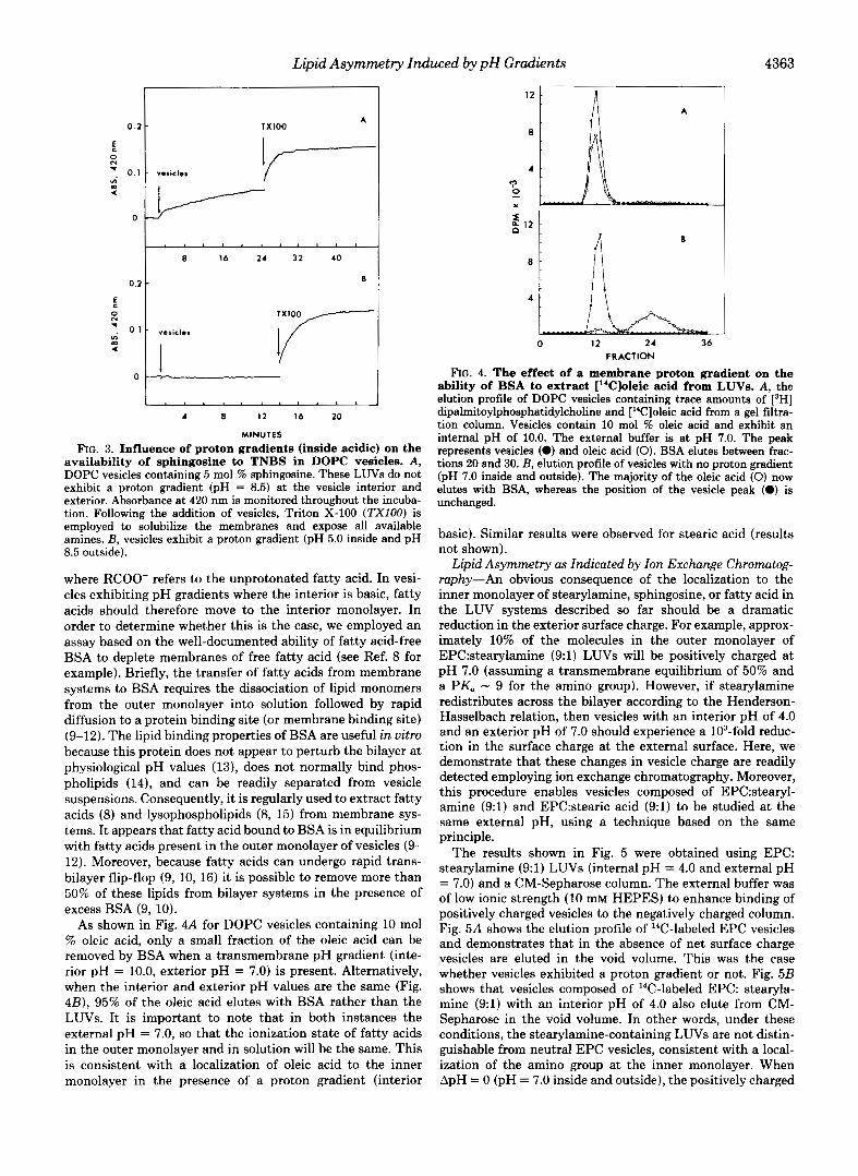
where RCOO- refers to the unprotonated fatty acid. In vesi- cles exhibiting pH gradients where the interior is basic, fatty acids should therefore move to the interior monolayer. In order to determine whether this is the case, we employed an assay based on the well-documented ability of fatty acid-free BSA to deplete membranes of free fatty acid (see Ref. 8 for example). Briefly, the transfer of fatty acids from membrane systems to BSA requires the dissociation of lipid monomers from the outer monolayer into solution followed by rapid diffusion to a protein binding site (or membrane binding site) (9-12). The lipid binding properties of BSA are useful in vitro because this protein does not appear to perturb the bilayer at physiological pH values (13), does not normally bind phos- pholipids (14), and can be readily separated from vesicle suspensions. Consequently, it is regularly used to extract fatty acids (8) and lysophospholipids (8, 15) from membrane sys- tems. It appears that fatty acid bound to BSA is in equilibrium with fatty acids present in the outer monolayer of vesicles (9- 12). Moreover, because fatty acids can undergo rapid trans- bilayer flip-flop (9, 10, 16) it is possible to remove more than 50% of these lipids from bilayer systems in the presence of excess BSA (9, 10).
As shown in Fig. 4A for DOPC vesicles containing 10 mol % oleic acid, only a small fraction of the oleic acid can be removed by BSA when a transmembrane pH gradient (inte- rior pH = 10.0, exterior pH = 7.0) is present. Alternatively, when the interior and exterior pH values are the same (Fig. 4B), 95% of the oleic acid elutes with BSA rather than the LUVs. It is important to note that in both instances the external pH = 7.0, so that the ionization state of fatty acids in the outer monolayer and in solution will be the same. This is consistent with a localization of oleic acid to the inner monolayer in the presence of a proton gradient (interior
12 n A
b 12 z
6
4
0 12 24 36 FRACTION
FIG. 4. The effect of a membrane proton gradient on the ability of BSA to extract ["C]oleic acid from LUVs. A , the elution profile of DOPC vesicles containing trace amounts of [3H] dipalmitoylphosphatidylcholine and ["Cloleic acid from a gel filtra- tion column. Vesicles contain 10 mol % oleic acid and exhibit an internal pH of 10.0. The external buffer is at pH 7.0. The peak represents vesicles (0) and oleic acid (0). BSA elutes between frac- tions 20 and 30. B, elution profile of vesicles with no proton gradient (pH 7.0 inside and outside). The majority of the oleic acid (0) now elutes with BSA, whereas the position of the vesicle peak (0) is unchanged.
basic). Similar results were observed for stearic acid (results not shown).
Lipid Asymmetry as Indicated by Ion Exchange Chromatog- raphy-An obvious consequence of the localization to the inner monolayer of stearylamine, sphingosine, or fatty acid in the LUV systems described so far should be a dramatic reduction in the exterior surface charge. For example, approx- imately 10% of the molecules in the outer monolayer of EPCxtearylamine (9:l) LUVs will be positively charged at pH 7.0 (assuming a transmembrane equilibrium of 50% and a PK, - 9 for the amino group). However, if stearylamine redistributes across the bilayer according to the Henderson- Hasselbach relation, then vesicles with an interior pH of 4.0 and an exterior pH of 7.0 should experience a 103-fold reduc- tion in the surface charge at the external surface. Here, we demonstrate that these changes in vesicle charge are readily detected employing ion exchange chromatography. Moreover, this procedure enables vesicles composed of EPC:stearyl- amine (9:l) and EPC:stearic acid (9:l) to be studied at the same external pH, using a technique based on the same principle.
The results shown in Fig. 5 were obtained using EPC: stearylamine (9:l) LUVs (internal pH = 4.0 and external pH = 7.0) and a CM-Sepharose column. The external buffer was of low ionic strength (10 mM HEPES) to enhance binding of positively charged vesicles to the negatively charged column. Fig. 5A shows the elution profile of I4C-labeled EPC vesicles and demonstrates that in the absence of net surface charge vesicles are eluted in the void volume. This was the case whether vesicles exhibited a proton gradient or not. Fig. 5B shows that vesicles composed of 14C-labeled EPC: stearyla- mine (9:l) with an interior pH of 4.0 also elute from CM- Sepharose in the void volume. In other words, under these conditions, the stearylamine-containing LUVs are not distin- guishable from neutral EPC vesicles, consistent with a local- ization of the amino group at the inner monolayer. When ApH = 0 (pH = 7.0 inside and outside), the positively charged
4364 Lipid Asymmetry Induced by p H Gradients
50
30
10
75
45
3 15 n
3 0
20
10
50
30
10
0 12 24 36 40
FRACTION
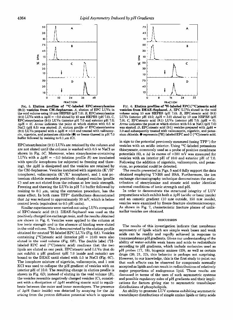
FIG. 5. Elution profiles of '"C-labeled EPC:stearylamine (9:l) vesicles from CM-Sepharose. A , elution of EPC LUVs in the void volume using 10 mM HEPES (pH 7.0). B, EPC:stearylamine (91) LUVs with a ApH = -3.0 eluted by 10 mM HEPES (pH 7.0). C, EPC:stearylamine (91) LUVs (interior pH 7.0 and exterior pH 7.0; ApH = 0). Arrow indicates the point at which elution with 0.5 M NaCl (pH 8.5) was started. D, elution profile of EPC:stearylamine (9:l) LUVs prepared with a ApH = -3.0 and treated with valinomy- cin, nigericin, and potassium chloride (0) or freeze-thawed in pH 7.0 buffer followed by resizing to 0.1 pm (0).
EPC:stearylamine (9:l) LUVs are retained by the column and are not eluted until the column is washed with 0.5 M NaCl as shown in Fig. 5C. Moreover, when stearylamine-containing LUVs with a ApH = -3.0 (elution profile B ) are incubated with specific ionophores (or subjected to freezing and thaw- ing), the ApH is dissipated and the vesicles are retained by the CM-Sepharose. Vesicles incubated with nigericin (K+/H+ ionophore), valinomycin (K+/K' ionophore), and 1 mM po- tassium chloride resemble positively charged vesicles (profile C ) and are not eluted from the column at low ionic strength. Freezing and thawing the LUVs in pH 7.0 buffer followed by resizing to 0.1 pm, using the extrusion procedure, has the same effect. In both cases TPP' distributions demonstrated that A$ was reduced to approximately 30 mV, which is below control levels (equivalent to 0.5 pH units).
Similar experiments were carried out using LUVs composed of EPC:stearic acid (9:l). DEAE-Sephacel was used as the positively charged ion exchange resin, and the results obtained are shown in Fig. 6. Vesicles were applied to the column at low ionic strength and in the absence of binding were eluted in the void volume. This is demonstrated by the elution profile obtained for neutral 3H-labeled EPC LUVs (Fig. 6A). Vesicles containing [14C]stearic acid (interior pH = 10.0) were also eluted in the void volume (Fig. 6B). The double label (3H- labeled EPC and [14C]stearic acid) confirms that the two lipids are eluted as one peak. EPC:stearic acid LUVs that do not exhibit a pH gradient (pH 7.0 inside and outside) are bound to the DEAE until eluted with 0.5 M NaCl (Fig. 6C). The ionophore mixture of nigericin, valinomycin, and 1 mM KC1 was used to collapse the pH gradient of vesicles with an interior pH of 10.0. The resulting change in elution profile is shown in Fig. 6D; instead of eluting in the void volume ( B ) , the vesicles resemble negatively charged vesicles (C), consist- ent with a dissipation of ApH enabling stearic acid to equili- brate between the outer and inner monolayers. The presence of ApH (basic inside) was detected by assaying for the A$ arising from the proton diffusion potential which is opposite
A
0 a 16 24 32
FRACTION FIG. 6. Elution profiles of aH-labeled EPC:['"C]stearic acid
vesicles from DEAE-Sephacel. A, EPC LUVs eluted in the void volume using 10 mM HEPES (pH 7.0). B, EPC:stearic acid (9:l) LUVs (interior pH 10.0; ApH = 3.0) eluted by 10 mM HEPES (pH 7.0). C, EPC:stearic acid (91) LUVs (interior pH 7.0; ApH = 0). Arrow indicates the point at which elution with 0.5 M NaCl (pH 7.0) was started. D, EPC:stearic acid (91) vesicles prepared with ApH = 3.0 and subsequently treated with valinomycin, nigericin, and potas- sium chloride. 0 represents [3H]-labeled EPC and 0 ["Clstearic acid.
in sign to the potential previously measured (using TPP+) for vesicles with an acidic interior. Using 14C-labeled potassium thiocyanate, commonly used as a probe of positive membrane potentials (6) , a A$ in excess of +200 mV was measured for vesicles with an interior pH of 10.0 and exterior pH of 7.0. Following the addition of nigericin, valinomycin, and potas- sium, no potential could be detected.
The results presented in Figs. 5 and 6 fully support the data obtained employing TNBS and BSA. Furthermore, the ion exchange chromatography technique demonstrates the redis- tribution of stearylamine and stearic acid under identical external conditions of ionic strength and pH.
In order to demonstrate the structural integrity of LUV preparations which exhibit both a ApH (acidic or basic inside) and an osmotic gradient (10 mM outside, 150 mM inside), vesicles were examined by freeze-fracture electromicroscopy. As shown in Fig. 7, characteristic fracture planes of unila- mellar vesicles are obtained.
DISCUSSION
The results of this investigation indicate that membrane asymmetry of lipids which are simple weak bases and weak acids can be readily and rapidly achieved in response to transmembrane pH gradients. Given our understanding of the ability of water-soluble weak bases and acids to redistribute according to pH gradients, which include molecules used as pH probes (17, 18), biogenic amines (19), as well as certain drugs (20, 21, 22), this behavior is perhaps not surprising. However, to our knowledge, this is the first study to point out that such effects can be observed for compounds normally accepted as lipids and can result in redistributions of relatively major proportions of endogenous lipid. These results are discussed in terms of the uses of such asymmetric systems and possible regulatory roles of pH gradients and their impli- cations for factors giving rise to asymmetric transbilayer distributions of phospholipids.
An ability to generate LUV systems exhibiting asymmetric transbilayer distributions of simple amino lipids or fatty acids

Lipid Asymmetry Induced by p H Gradients 4365
FIG. 7. Freeze-fracture electron micrograph of vesicles used for ion exchange chromatography. A, EPC:stearylamine (91). 150 mM NaCI, 10 mM HEPES (pH 7.0 inside) and 10 mM HEPES (pH 7.0 outside; ApH = 0). B, EPC:stearic acid (9:l). 150 mM NaCI, 10 mM HEPES (pH 7.0 inside) and 10 mM HEPES (pH 7.0 outside; ApH = 0). c, EPC:stearylamine (91). 150 mM citric acid (pH 4.0 inside) and 10 mM HEPES (pH 7.0 outside; ApH = -3.0). D, EPCstearic acid (91). 150 mM boric acid (pH 10.0 inside) and 10 mM HEPES (pH 7.0 outside; ApH = 3.0).
may have utility in a number of applications. These include sampling of lipid motion and "order" in the hydrocarbon regions of outer and inner monolayers separately (employing deuterated fatty acids and *H NMR techniques, for example) as well as generating delivery systems which exhibit surface charges which change as a function of time. However, the implications of the results presented for the regulation of the metabolism of lipids such as sphingosine and fatty acid is perhaps of more immediate interest. For example, the trans- bilayer localization of sphingosine in the endoplasmic reticu- lum membrane will likely be sensitive to pH gradients. An acidic pH in the lumen will lead to decreased availability in the outer monolayer. Conversely, in the case of fatty acids, such pH gradients would lead to increased availability on the cytoplasmic side. In lysosomal systems, catabolism of endo- cytosed lipids would be expected to lead to immediate trans- location of resulting fatty acids to the cytoplasmic monolayer due to the transmembrane pH gradients (acidic inside) main- tained by these organelles.
The ability of fatty acids to translocate in response to pH gradients has interesting implications with respect to several observations previously noted for the transfer of fatty acids between liposomes and fatty acid binding proteins (9, 10) and the exchange of fluorescent analogues of fatty acids between unilamellar vesicles (12). All report a reduced exchange as the external pH is decreased, which is interpreted to mean that the protonated form of the fatty acid is less likely to dissociate from the bilayer and diffuse to another binding site. However, the results presented in this paper clearly demonstrate the importance of ensuring that when adjustments are made to the external pH a transmembrane proton gradient is not created. This can best be achieved by hydrating the memhrane system at each pH being investigated. Lowering the external pH could create a ApH that is basic inside with respect to the outside medium. Consequently, the concentration of fatty acid available for dissociation from the outer monolayer into so- lution will be reduced. This dissociation step is thought to be rate-limiting in the exchange of fatty acids between mem-
brane systems (11, 12). I t is also interesting to note that the flow of fatty acid from BSA to cells in vitro has been reported to increase as the external pH is lowered from pH 7.4 (23). Assuming the cytoplasmic pH = 7.4, then a basic positive ApH might be expected to develop, decreasing the fatty acid concentration in the outer monolayer of plasma membranes and increasing the inner monolayer concentration. The fatty acid equilibrium between the membrane and BSA would now favor the membrane.
The relation between the results presented here and factors giving rise to phospholipid asymmetry are not immediately obvious. However, it is intriguing that the amino-containing phospholipids PE and PS are both preferentially localized on the same side of plasma membranes such as that of the erythrocyte (1, 2). This could be taken to suggest that the transbilayer distributions of P E and/or PS result from a transbilayer pH gradient (interior acidic). Indeed, due to the presence of the negatively charged PS on the inner monolayer of the erythrocyte membrane, a surface potential of -69 mV is expected which would give rise to an interfacial pH gradient in excess of 1 pH unit (interior acidic). This could be hypoth- esized to influence the transbilayer distribution of PE. How- ever, the situation is clearly more complicated, as the presence of a pH gradient alone does not cause PE to migrate to the inner monolayer of the PE:PC (1:9) systems investigated here. This almost certainly arises from a high energy barrier for the transbilayer transport of the phospholipid phosphate group (16). In model membrane systems transbilayer move- ment of phospholipids is extremely slow, and in the PC:PE systems used in this study flip-flop could not be detected. However, biological membranes often exhibit relatively rapid transbilayer movement of diacylphospholipids (24-26) and lysophospholipids (15, 27). Rat liver microsomes have been reported to possess a phosphatidylcholine transporter which enables newly synthesized lipid at the cytoplasmic monolayer to flip to the luminal monolayer (24). There is also evidence that the erythrocyte membrane might contain a similar pro- tein, specific for amino phospholipids (26, 28). For example,
4366 Lipid Asymmetry Induced by pH Gradients
it has been demonstrated that phosphatidylserine and phos- phatidylethanolamine move to the inner monolayer when added exogenously (25, 26) but that phosphatidylcholine and sphingomyelin remain in the outer monolayer (25). Similar results have been observed for spin-labeled analogues of phos- pholipids (28). These observations lend support to the idea of an amino phospholipid transfer which enables the movement of amino lipids to the inner monolayer but limited access to the return cycle (26, 28). Such a mechanism appears to be energy-dependent and might underlie the observed lipid asymmetry in the erythrocyte membrane. The results we have presented suggest that if a protein-dependent pathway for flip-flop is available to phospholipids in biological mem- branes, then transbilayer distributions of phospholipid could be dictated by amino functions responding to ATP-dependent ion gradients.
Acknowledgment-We wish to thank T. Redelmeier for helpful suggestions.
REFERENCES
1. Houslay, M. D., and Stanley, K. K. (1982) Dynamics of Biological
2. Op den Kamp, J. A. F. (1979) Annu. Rev. Biochem. 48,47-71 3. Deamer, D. W. (1982) in Intracellular pH: Its Measurements,
Regulation and Utilization in Cellular Functions (Lisas, A., ed), Elsevier Scientific Publishing Co., New York
4. Hope, M. J., Bally, M. B., Webb, G., and Cullis, P. R. (1985) Biochim. Biophys. Acta 812,55-65
5. Kates, M. (1972) in Techniques of Lipidology: Isolation, Analysis and Identification of Lipids (Work, T. S., and Work, E., eds), Elsevier Scientific Publishing Co., New York
Membranes, John Wiley and Sons, Toronto
7. Means, G. E., Congdon, W. I., and Bender, M. L. (1972) Biochem-
8. Haest, C. W. M., Plasa, G., and Deuticke, B. (1981) Biochim.
9. Brecher, P., Saouaf, R., Sugerman, J. M., Eisenberg, D., and
10. Hamilton, J. A., and Cistola, D. P. (1986) Proc. Natl. Acad. Sci.
11. Nichols, J. W., and Pagano, R. E. (1982) Biochemistry 21 , 1720-
12. Doody, M. C., Pownall, H. J., Kao, Y. J., and Smith, L. C. (1980)
13. Kimelberg, H. K., and Papahadjopoulos, D. (1971) Biochim. Bio-
14. Jonas, A. (1975) Biochem. Biophys. Res. Commun. 64,1003-1008 15. Mohandas, N., Wyatt, J., Mel, S. F., Rossi, M. E., and Shohet, S.
16. Ganong, B. R., and Bell, R. M. (1984) Biochemistry 2 3 , 4977-
17. Nicholls, D. G. (1982) Bioenergetics: An Introduction to the Chem-
18. Boron, W. F. (1983) J. Membr. Biol. 72 , 1-16 19. Nichols, J. W., and Deamer, D. W. (1976) Biochim. Biophys. Acta
20. Maver. L. D.. Ballv. M. B.. Houe. M. J.. and Cullis. P. R. (1985)
istry 19,3564-3571
Biophys. Acta 6 4 9 , 701-708
LaRosa, K. (1984) J. Biol. Chem. 2 5 9 , 13395-13401
U. S. A. 8 3 , 82-86
1726
Biochemistry 19,108-116
phys. Acta 233,805-809
B. (1982) J. Biol. Chem. 267,6537-6543
4983
iosmotic Theory, Academic. Press, Orlando, FL
455,269-271
Biol. Chem. 260,8021808 '
. ,
21. Ballv. M. B.. Houe. M. J.. Van Echteld. C. J. A.. and Cullis. P. R."(1985) bioc i ih . Biophys. Acta 812; 66-76 '
22. Mayer, L. D., Bally, M. B., Hope, M. J., and Cullis, P. R. (1985)
23. Spector, A. A. (1969) J. Lipid Res. 10 , 207-215 Biochim. Biophys. Acta 8 1 6 , 294-302
24. Bishop, W. R., and Bell, R. M. (1985) Cell 42,51-60 25. Tilley, L., Cribier, S., Roelofsen, B., Op den Kamp, J. A. F., and
26. Daleke, D. L., and Huestis, W. H. (1985) Biochemistry 24,5406-
27. Bergmann, W. L., Dressler, V., Haest, C. W. M., and Deuticke,
van Deenen, L. L. M. (1986) FEBS Lett. 194, 21-27
5416
B. (1984) Biochim. BioDhvs. Acta 769.390-398
6. Rottenberg, H. (1979) MethodsEnzyml. 5 5 , 547-569 28. Zachowski; A., Favre, E.; Cribier, S., Herve, P., and Devaux, P.
F. (1986) Biochemistry 25, 2585-2590


![A Diet Enriched in Stearic Acid Protects Against the Progression[1]](https://img.pdfslide.net/doc/110x75/577cd9521a28ab9e78a33ba7/a-diet-enriched-in-stearic-acid-protects-against-the-progression1.jpg)