Embed Size (px)
Citation preview
Turk J Med Sci2006; 36 (6): 387-392© TÜB‹TAKE-mail: [email protected]
387
CASE REPORT
The Oncocytic Variant of Papillary Thyroid Carcinoma*
Abstract: Papillary thyroid carcinoma (PTC) has many subtypes. A rare subtype consisting of papillaryprojections covered by cells oncocytic in character has been defined in recent years. Oncocytes withcharacteristic nuclear features of PTC are seen in the thyroid aspiration cytology of this subtype. In thecytological diagnosis, this variant could be confused with other oncocytic lesions of the thyroid.
We discuss here the cytological and histopathological features of the oncocytic variant of PTC diagnosed in theaspiration biopsy and thyroid resection material in a 42-year-old female patient.
Key Words: Papillary thyroid carcinoma, oncocytic variant, aspiration cytology, histopathology
Papiller Tiroid Karsinomu Onkositik Varyant› Bir Olgu Sunumu
Özet: Papiller tiroid karsinomunun bir çok alttipi vard›r. Son y›llarda onkositik karakterde hücreler ile döflelipapiller yap›lardan ibaret olan nadir bir alt tip tan›mlanm›flt›r. Tiroid aspirasyon sitolojisinde papiller tiroidkarsinomunun karakteristik nükleer özelliklerine sahip onkositler görülmektedir. Sitolojik incelemede bu alttip, tiroidin di¤er onkositik lezyonlar› ile kar›flabilir.
Burada, 42 yafl›ndaki kad›n hastada tan›s› konan onkositik varyant papiller tiroid karsinomunun aspirasyonyaymalar›ndaki sitolojik özellikleri ve rezeksiyon materyalindeki histopatolojik özelliklerini tart›fl›yoruz.
Anahtar Sözcükler: Tiroid papiller karsinoma, onkositik varyant, aspirasyon sitolojisi, histopatoloji.
Reflit Do¤an KÖSEO⁄LU
Nurper ONUK F‹L‹Z
Department of Pathology, Faculty of Medicine, Gaziosmanpafla University, Tokat - TURKEY
Received: June 12, 2006Accepted: December 07, 2006
Correspondence
R. Do¤an KÖSEO⁄LUGaziosmanpafla University,
Faculty of Medicine,Department of Pathology60030, Tokat - TURKEY
Introduction
The oncocytic variant of papillary thyroid carcinoma (oncocytic variant of PTC) is avery rare subtype of PTC. This variant has also been termed as Hürthle cell variant oroxyphilic variant. Although this variant has been known for about 35 years, it has notbeen sufficiently reported in the literature. Its biological behavior has been poorlydefined because histological and cytological criteria have not been clearly determined forthe diagnosis (1).
We report our case diagnosed as oncocytic variant of PTC on both cytological andhistopathological bases, and we analyze the cytological and histopathological features ofour case, with an accompanying review of the related literature.
Case Report
A 42-year-old female patient was admitted with complaints of palpitation, sweatingand swelling of the neck for two years. There was nothing remarkable in her family’smedical history. In physical examination, asymmetry in the thyroid was noted. Inaddition, a mobile nodule with firm consistency involving both thyroid lobes wasdetermined. The neck computerized tomography revealed the cystic lesion, 3x2x1.5 cmin size, as a centrally placed solid nodule in the isthmus. In addition, multiple bilateralcervical lymphadenopathies were determined. Fine needle aspiration biopsy performedon the cystic lesion revealed cellular, three-dimensional groups (Figure 1A). Some ofthese groups were papillary in character and others showed follicular organization. The
* This manuscript was presented as a poster at the 1st National Congress of Cytopathology, 2-5 March2006, Antalya - TURKEY
388
KÖSEO⁄LU, RD et al. Oncocytic Papillary Carcinoma Turk J Med Sci
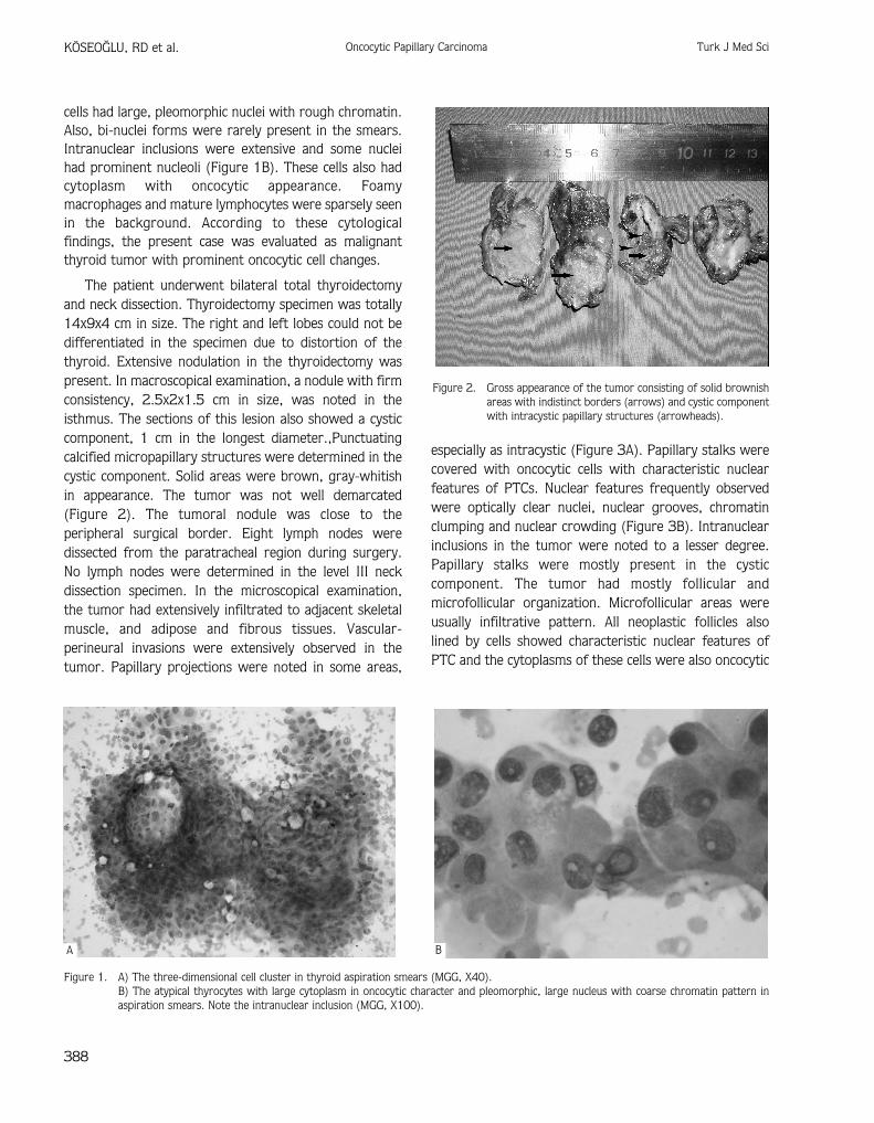
cells had large, pleomorphic nuclei with rough chromatin.Also, bi-nuclei forms were rarely present in the smears.Intranuclear inclusions were extensive and some nucleihad prominent nucleoli (Figure 1B). These cells also hadcytoplasm with oncocytic appearance. Foamymacrophages and mature lymphocytes were sparsely seenin the background. According to these cytologicalfindings, the present case was evaluated as malignantthyroid tumor with prominent oncocytic cell changes.
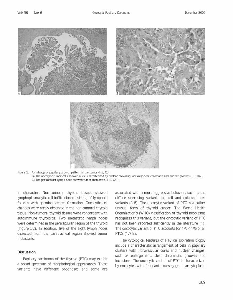
The patient underwent bilateral total thyroidectomyand neck dissection. Thyroidectomy specimen was totally14x9x4 cm in size. The right and left lobes could not bedifferentiated in the specimen due to distortion of thethyroid. Extensive nodulation in the thyroidectomy waspresent. In macroscopical examination, a nodule with firmconsistency, 2.5x2x1.5 cm in size, was noted in theisthmus. The sections of this lesion also showed a cysticcomponent, 1 cm in the longest diameter.,Punctuatingcalcified micropapillary structures were determined in thecystic component. Solid areas were brown, gray-whitishin appearance. The tumor was not well demarcated(Figure 2). The tumoral nodule was close to theperipheral surgical border. Eight lymph nodes weredissected from the paratracheal region during surgery.No lymph nodes were determined in the level III neckdissection specimen. In the microscopical examination,the tumor had extensively infiltrated to adjacent skeletalmuscle, and adipose and fibrous tissues. Vascular-perineural invasions were extensively observed in thetumor. Papillary projections were noted in some areas,
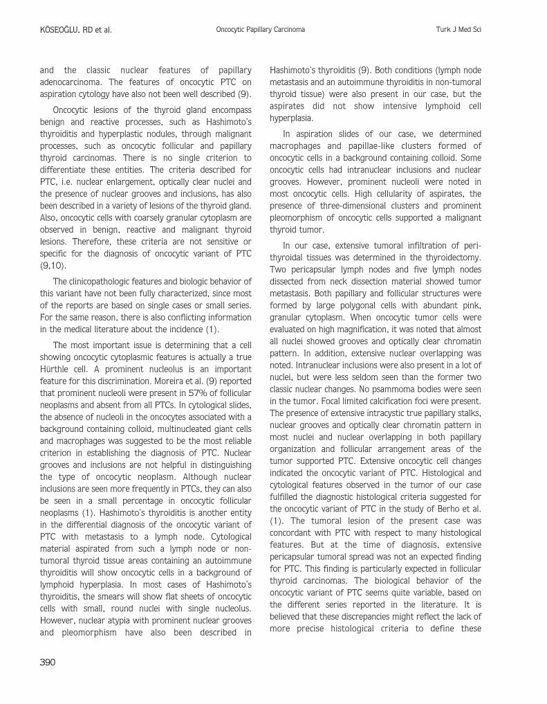
especially as intracystic (Figure 3A). Papillary stalks werecovered with oncocytic cells with characteristic nuclearfeatures of PTCs. Nuclear features frequently observedwere optically clear nuclei, nuclear grooves, chromatinclumping and nuclear crowding (Figure 3B). Intranuclearinclusions in the tumor were noted to a lesser degree.Papillary stalks were mostly present in the cysticcomponent. The tumor had mostly follicular andmicrofollicular organization. Microfollicular areas wereusually infiltrative pattern. All neoplastic follicles alsolined by cells showed characteristic nuclear features ofPTC and the cytoplasms of these cells were also oncocytic
Figure 1. A) The three-dimensional cell cluster in thyroid aspiration smears (MGG, X40).B) The atypical thyrocytes with large cytoplasm in oncocytic character and pleomorphic, large nucleus with coarse chromatin pattern inaspiration smears. Note the intranuclear inclusion (MGG, X100).
Figure 2. Gross appearance of the tumor consisting of solid brownishareas with indistinct borders (arrows) and cystic componentwith intracystic papillary structures (arrowheads).
A B
in character. Non-tumoral thyroid tissues showedlymphoplasmacytic cell infiltration consisting of lymphoidfollicles with germinal center formation. Oncocytic cellchanges were rarely observed in the non-tumoral thyroidtissue. Non-tumoral thyroid tissues were concordant withautoimmune thyroiditis. Two metastatic lymph nodeswere determined in the pericapsular region of the thyroid(Figure 3C). In addition, five of the eight lymph nodesdissected from the paratracheal region showed tumormetastasis.
Discussion
Papillary carcinoma of the thyroid (PTC) may exhibita broad spectrum of morphological appearances. Thesevariants have different prognoses and some are
associated with a more aggressive behavior, such as thediffuse sclerosing variant, tall cell and columnar cellvariants (2-6). The oncocytic variant of PTC is a ratherunusual form of thyroid cancer. The World HealthOrganization’s (WHO) classification of thyroid neoplasmsrecognizes this variant, but the oncocytic variant of PTChas not been reported sufficiently in the literature (1).The oncocytic variant of PTC accounts for 1%-11% of allPTCs (1,7,8).
The cytological features of PTC on aspiration biopsyinclude a characteristic arrangement of cells in papillaryclusters with fibrovascular cores and nuclear changes,such as enlargement, clear chromatin, grooves andinclusions. The oncocytic variant of PTC is characterizedby oncocytes with abundant, coarsely granular cytoplasm
389
Vol: 36 No: 6 Oncocytic Papillary Carcinoma December 2006
Figure 3. A) Intracystic papillary growth pattern in the tumor (HE, X5)B) The oncocytic tumor cells showed nuclei characterized by nuclear crowding, optically clear chromatin and nuclear grooves (HE, X40).C) The pericapsular lymph node showed tumor metastasis (HE, X5).
A B
C
and the classic nuclear features of papillaryadenocarcinoma. The features of oncocytic PTC onaspiration cytology have also not been well described (9).
Oncocytic lesions of the thyroid gland encompassbenign and reactive processes, such as Hashimoto’sthyroiditis and hyperplastic nodules, through malignantprocesses, such as oncocytic follicular and papillarythyroid carcinomas. There is no single criterion todifferentiate these entities. The criteria described forPTC, i.e. nuclear enlargement, optically clear nuclei andthe presence of nuclear grooves and inclusions, has alsobeen described in a variety of lesions of the thyroid gland.Also, oncocytic cells with coarsely granular cytoplasm areobserved in benign, reactive and malignant thyroidlesions. Therefore, these criteria are not sensitive orspecific for the diagnosis of oncocytic variant of PTC(9,10).
The clinicopathologic features and biologic behavior ofthis variant have not been fully characterized, since mostof the reports are based on single cases or small series.For the same reason, there is also conflicting informationin the medical literature about the incidence (1).
The most important issue is determining that a cellshowing oncocytic cytoplasmic features is actually a trueHürthle cell. A prominent nucleolus is an importantfeature for this discrimination. Moreira et al. (9) reportedthat prominent nucleoli were present in 57% of follicularneoplasms and absent from all PTCs. In cytological slides,the absence of nucleoli in the oncocytes associated with abackground containing colloid, multinucleated giant cellsand macrophages was suggested to be the most reliablecriterion in establishing the diagnosis of PTC. Nucleargrooves and inclusions are not helpful in distinguishingthe type of oncocytic neoplasm. Although nuclearinclusions are seen more frequently in PTCs, they can alsobe seen in a small percentage in oncocytic follicularneoplasms (1). Hashimoto’s thyroiditis is another entityin the differential diagnosis of the oncocytic variant ofPTC with metastasis to a lymph node. Cytologicalmaterial aspirated from such a lymph node or non-tumoral thyroid tissue areas containing an autoimmunethyroiditis will show oncocytic cells in a background oflymphoid hyperplasia. In most cases of Hashimoto’sthyroiditis, the smears will show flat sheets of oncocyticcells with small, round nuclei with single nucleolus.However, nuclear atypia with prominent nuclear groovesand pleomorphism have also been described in
Hashimoto’s thyroiditis (9). Both conditions (lymph nodemetastasis and an autoimmune thyroiditis in non-tumoralthyroid tissue) were also present in our case, but theaspirates did not show intensive lymphoid cellhyperplasia.
In aspiration slides of our case, we determinedmacrophages and papillae-like clusters formed ofoncocytic cells in a background containing colloid. Someoncocytic cells had intranuclear inclusions and nucleargrooves. However, prominent nucleoli were noted inmost oncocytic cells. High cellularity of aspirates, thepresence of three-dimensional clusters and prominentpleomorphism of oncocytic cells supported a malignantthyroid tumor.
In our case, extensive tumoral infiltration of peri-thyroidal tissues was determined in the thyroidectomy.Two pericapsular lymph nodes and five lymph nodesdissected from neck dissection material showed tumormetastasis. Both papillary and follicular structures wereformed by large polygonal cells with abundant pink,granular cytoplasm. When oncocytic tumor cells wereevaluated on high magnification, it was noted that almostall nuclei showed grooves and optically clear chromatinpattern. In addition, extensive nuclear overlapping wasnoted. Intranuclear inclusions were also present in a lot ofnuclei, but were less seldom seen than the former twoclassic nuclear changes. No psammoma bodies were seenin the tumor. Focal limited calcification foci were present.The presence of extensive intracystic true papillary stalks,nuclear grooves and optically clear chromatin pattern inmost nuclei and nuclear overlapping in both papillaryorganization and follicular arrangement areas of thetumor supported PTC. Extensive oncocytic cell changesindicated the oncocytic variant of PTC. Histological andcytological features observed in the tumor of our casefulfilled the diagnostic histological criteria suggested forthe oncocytic variant of PTC in the study of Berho et al.(1). The tumoral lesion of the present case wasconcordant with PTC with respect to many histologicalfeatures. But at the time of diagnosis, extensivepericapsular tumoral spread was not an expected findingfor PTC. This finding is particularly expected in follicularthyroid carcinomas. The biological behavior of theoncocytic variant of PTC seems quite variable, based onthe different series reported in the literature. It isbelieved that these discrepancies might reflect the lack ofmore precise histological criteria to define these
390
KÖSEO⁄LU, RD et al. Oncocytic Papillary Carcinoma Turk J Med Sci
neoplasms, leading to inclusion of a broad range ofoncocytic tumors within such studies. As a result of thelack of more precise criteria, the line that divides “true”oncocytic PTC from other oncocytic thyroid tumors(particularly papillary Hürthle cell tumors) has been veryindistinct (1). The studies in which papillary configurationrather than characteristic nuclear features was used asthe overriding criterion for diagnosis of PTC are noted inthe literature (11-13). It thus appears that the studiespreviously reported concerning the oncocytic variant ofPTC might contain a heterogeneous mixture of tumors ofvarying biological potential sharing in common only theironcocytic cytoplasmic features and papillary architecture.Distinguishing the oncocytic variant of PTC from otheroncocytic thyroid neoplasms with different biologicalbehavior is important. The most important entity amongthese neoplasms is papillary Hürthle cell carcinoma(14,15), which has different nuclear features from PTC,consisting mainly of round-shape, vesicular chromatinpattern and prominent centrally placed nucleolus. Focalhyperchromasia, binucleation and marked nuclear atypiaare frequent accompanying nuclear features of these cells(16). Infiltration of the soft tissues surrounding thethyroid is not an uncommon event in these neoplasms.Distant metastasis, especially to lung and bones, occursmore frequently in Hürthle cell carcinomas. The overallfive-year survival for Hürthle cell carcinoma ranges from50% to 60% in most series. However, the prognosis ofPTC is excellent (1). In our case, peri-thyroidal soft tissueinfiltration was noted on the histopathologicalexamination. This feature is more likely in Hürthle cell
carcinoma. In addition, prominent nucleoli and vesicularchromatin pattern also suggested Hürthle cell carcinoma.Our case also frequently showed prominent nucleoli in thehistologic sections. However, we evaluated our case asPTC because extensive nuclear groove formation andintranuclear inclusions, extensive papillary structures anda cystic component were present in the tumor. Regardinghistopathological and cytological features and the clinicalsituation, the present case could be evaluated as a hybridneoplasm sharing the features of both PTC and Hürthlecell carcinoma.
Other diagnostic considerations in the histologicaldifferential diagnosis of these tumors include the tall cellvariant of PTC and the oncocytic variant of medullarycarcinoma of the thyroid. Neoplastic cells of tall cellvariant of PTC are twice as tall as wide, with elongateddark nuclei. The oncocytic variant of medullary carcinomais characterized by a solid or follicular growth patternwith prominent oncocytic cell changes, absence of thecharacteristic nuclear features of PTC, and the presenceof more conventional areas of medullary carcinomawithin the tumor (4,5,17,18).
In conclusion, the oncocytic variant of PTC is amorphologically distinct and rare variant of papillarythyroid cancer. Additional studies with longer follow-upperiods are necessary to further define the spectrum ofbiological activity and metastatic potential of theseneoplasms. In addition, strict histological criteria shouldbe established to separate these lesions from otherthyroid neoplasms characterized by oncocytic cellchanges.
391
Vol: 36 No: 6 Oncocytic Papillary Carcinoma December 2006
References1. Berho M, Suster S. The oncocytic variant of papillary carcinoma of
the thyroid: a clinicopathologic study of 15 cases. Hum Pathol1997; 28: 47-53.
2. Carcangiu ML, Bianchi S. Diffuse sclerosing variant of papillarythyroid carcinoma: clinicopathologic study of 15 cases. Am J SurgPathol 1989; 3: 1041-1049.
3. Fujimoto Y, Obara T, Ito Y, Kodama T, Aiba M, Yamaguchi K.Diffuse sclerosing variant of papillary carcinoma of the thyroid.Cancer 1990; 66: 2306-2312.
4. Hawk WA, Hazard JB. The many appearances of papillarycarcinoma of the thyroid: comparison with the common form ofpapillary carcinoma by DNA and morphometric analysis. Cleve ClinQ 1976; 43: 207-215.
5. Johnson TL, Lloyd R, Thompson NW, Beierwaltes WH, Sisson JC.Prognostic implications of the tall cell variant of papillary thyroidcarcinoma. Am J Surg Pathol 1988; 12: 22-27.
6. Evans HL. Columnar cell carcinoma of the thyroid: a report of twocases of an aggressive variant of thyroid carcinoma. Am J ClinPathol 1986; 85: 77-80.
7. Selzer G, Kahn LB, Alberttyn L. Primary malignant tumors of thethyroid: a clinicopathologic study of 254 cases. Cancer 1977; 40:1501-1511.
8. Woolner LB, Beahrs OH, Black BM, McConahey WM, Keating FRJr. Classification and prognosis of thyroid carcinoma: a study of885 cases observed in a 30 year period. Am J Surg 1961; 102:354-387.
9. Moreira AL, Waisman J, Cangiarella JF. Aspiration cytology of theoncocytic variant of papillary adenocarcinoma of the thyroidgland. Acta Cytol 2004; 48: 137-141.
10. Lew W, Orell S, Henderson DW. Intranuclear vacuoles in non-papillary carcinoma of the thyroid gland: a report of three cases.Acta Cytol 1984; 28: 581-586.
392
KÖSEO⁄LU, RD et al. Oncocytic Papillary Carcinoma Turk J Med Sci
11. Tschol-Ducommun J, Hedinger CE. Papillary thyroid carcinomas:morphology and prognosis. Virchows Arch [A] 1982; 396: 19-39.
12. Herrera MF, Hay ID, WU PS, Goellner JR, Ryan JJ, Ebersold JRet al. Hurthle cell (oxyphilic) papillary carcinoma of the thyroid: avariant with more aggressive biologic behavior. World J Surg1992; 16: 669-675.
13. Schroder S, Dralle H, Rehpenning W, Bocker W.Prognosekriterien des Papillaren Schilddrusencarcinomas:Morphologisch-klinische analyse von 202 tumor fallen.Langenbecks Arch Chir 1987; 371: 263-280.
14. Barbuto D, Carcangiu ML, Rosai J. Papillary Hurthle cellneoplasms of the thyroid gland: a study of 20 cases (Abstract).Mod Pathol 1990; 3: 7A.
15. Apel RL, Asa SL, LiVolsi VA. Papillary Hurthle cell carcinoma withlymphocytic stroma: “Warthin-like tumor” of the thyroid. Am JSurg Pathol 1995; 19: 810-814.
16. Flint A, Llyod RV. Hurthle-cell neoplasms of the thyroid gland.Pathol Annu 1990; 19: 37-52.
17. Dominquez-Malagon H, Delgado-Chavez R, Torres-Najera M,Gould E, Albores-Saavedra J. Oxyphilic and squamous variants ofmedullary carcinoma of the thyroid. Cancer 1989; 63: 1183-1188.
18. Harach HR, Bergholm U. Medullary (C cell) carcinoma of thethyroid with features of follicular oxyphilic cell tumors.Histopathology 1988; 13: 645-656.


![Clinical impact of follicular oncocytic (Hürthle cell ... · oxyphilic or oncocytic cell follicular thyroid carcinoma, rep-resents about 3–5% of thyroid carcinomas [5–8]. Traditionally,](https://img.pdfslide.net/doc/110x75/5f96415ab1c35b1da41c4408/clinical-impact-of-follicular-oncocytic-hrthle-cell-oxyphilic-or-oncocytic.jpg)