Embed Size (px)
Citation preview

Send Orders for Reprints to [email protected]
The Open Ophthalmology Journal, 2016, 10, 111-118 111
1874-3641/16 2016 Bentham Open
The Open Ophthalmology Journal
Content list available at: www.benthamopen.com/TOOPHTJ/
DOI: 10.2174/1874364101610010111
Long Term Clinical and Visual Outcomes of Retrofixated Iris ClawLenses Implantation in Complicated Cases
Sri Ganesh, Sheetal Brar and Kirti Relekar*
Nethradhama Superspeciality Eye Hospital, Bengaluru, India
Received: December 1, 2015 Revised: December 30, 2015 Accepted: January 19, 2016
Abstract:
Aims:
To evaluate the visual outcomes and complications after implantation of retrofixated iris claw (RFIC) lens in various challengingsituations.
Settings and Design:
Retrospective, single centre, 8 year clinical audit.
Materials and Methods:
A retrospective analysis of cases who underwent RFIC lens implantation alone (group 1) or in combination with vitreoretinal (VR),corneal or glaucoma procedures (group 2) was performed. The main outcomes evaluated were corrected distant visual acuity(CDVA) and postoperative complications. The mean follow up was 13.09±6.8 (range 6-24) months.
Results:
The study involved 100 eyes of 83 patients with mean age of 51.1±25.4 years. Group 1 included 59 eyes and group 2 had 41 eyes. Ingroup 1, the mean CDVA improved from 0.86±0.81 to 0.38±0.51 LogMAR (p<0.001) with 72.8% eyes having gain in lines (≥ 2lines) of CDVA with safety index of 1.73. The mean CDVA in group 2 improved from 0.71±0.65 to 0.38±0.34 LogMAR (p=0.003)with 65.8% eyes having gain in lines (≥ 2 lines) of CDVA with a safety index of 1.54. Group 2 showed a higher complication rate of36.59% compared to group 1 (20.34%). Significant complications noted were secondary glaucoma (8%), disenclavation of haptic(4%), subluxation of RFIC lens (1%).
Conclusion:
The visual outcome with RFIC lenses when combined with other intraocular procedures is mainly affected by the complexity of co-existing pathologies .The complications are more related to the combined procedures performed rather than RFIC lens implantationalone. This may still be acceptable when complication profile of other intraocular lenses is evaluated in similar challengingsituations.
Keywords: Combined, enclavation, intraocular lens, iris claw, retrofixated, vitreoretinal.
INTRODUCTION
The surgical correction of aphakia, crystalline lens dislocation, or dislocation of an intraocular lens (IOL) in theabsence of adequate capsular support remains challenging. In these situations, an angle supported or iris supportedanterior chamber intraocular lens (ACIOL), a scleral fixated posterior chamber IOL (SFIOL), a fibrin glue assisted
* Address correspondence to this author at 256/14, Kanakapura main road, 7th block, Jayanagar, Bengaluru-560070, India; Tel: 09820516075; Fax:26633770; E-mail: [email protected]

112 The Open Ophthalmology Journal, 2016, Volume 10 Ganesh et al.
suture less posterior chamber intraocular lens (PCIOL) - Glued IOL, or an iris fixated PCIOL have been evaluated forvisual restoration [1 - 3]. However, most of these IOLs are associated with certain risks with respect to long term safety.Due to complications like corneal decompensation, cystoid macular edema (CME), secondary glaucoma, uveitis andretinal detachment (RD), use of an ACIOL in complicated situations is not recommended [1, 2]. SFIOLs althoughpreserve the anatomy of eye and cause less corneal endothelial damage, are technically more challenging, require moresurgical time and are associated with a high incidence of complications such as lens tilting, decentration, choroidalhaemorrhage, RD and CME [1, 2, 4]. Glued IOLs require the creation of scleral flaps and tucking of the IOL hapticsunder the flaps which can be associated with haptic related complications such as IOL decentration, haptic extrusionand subconjunctival haptic in the long term [5]. RFIC lenson the other hand, has the advantage of retropupillaryposterior chamber location and a shorter learning curve [6 - 8]. Various studies done previously have mainly studied theoutcome of RFIC lens implantation with aphakia being the primary indication [6]. However, they have not evaluated theoutcomes of RFIC lens when combined with additional ocular surgical procedures such as vitreoretinal, corneal andglaucoma procedures. Hence this comprehensive retrospective review was conducted to analyze various indications,surgical difficulties, long term safety and visual outcomes of RFIC lens implantation when performed alone forcorrection of aphakia as a primary indication as well as when performed in combination with other ocular surgeries indifferent complicated scenarios.
MATERIALS AND METHODS
This retrospective study was approved by Institutional ethics committee and abided by the tenets of Declaration ofHelsinki. The study included all patients who had undergone RFIC lens implantation with or without a combinedprocedure from January 2007 to December 2014. Electronic medical records of these patients were reviewed for datacollection and analysis. The various combined procedures performed were (a) vitreoretinal (VR) procedures like parsplana vitrectomy (PPV), pars plana lensectomy (PPL) (b) corneal procedures like optical penetrating keratoplasty (PKP)and ACIOL explantation (c) glaucoma procedures like trabeculectomy for medically uncontrolled open angle glaucoma.
The RFIC lens used in study was OV lens (Care Group, India) which is a polymethylmethacrylate (PMMA) lens,style- ICLIP-5 with an optic size 5.0mm and overall diameter of 8.5mm. The recommended A-constant for anteriorfixation is 114.9. However, for retrofixation an A constant of 117.20 was calculated for all cases. Only cases withminimum follow up of 6 months were included. On each follow up, uncorrected distant visual acuity (UDVA),corrected distant visual acuity (CDVA), detailed anterior and posterior segment evaluation, intraocular pressure (IOP)measurements with non-contact tonometery and specular micoscopy were performed. Eligibility criteria for RFIC lensimplantation were presence of healthy and sufficient iris tissue and endothelial cell count of more than 1500 cells/mm2.
Surgical Technique:
All procedures were performed by a single experienced surgeon (S.G.) using a standard surgical technique. All theRFIC lens implantations were combined with other procedures in the same/single setting.
Fig. (1). Associated conditions.
������������������
��
��
��
� � �
�
����� ��������������
��
��
��
�
�
������������ ����
�������� �
����������� ��
! � ���������
"��������� ��������
#������$�����
!��� ��� � %�

Retrospective Retrofixated Iris Claw Complicated Cases Contributors The Open Ophthalmology Journal, 2016, Volume 10 113
Suitable anaesthesia local/general was administered. After a conjunctival peritomy, a diamond knife was used tocreate a superior scleral incision (5-5.5mm). The sclerocorneal tunnel was fashioned with disposable crescent blade.Two paracentesis 90 degrees from the scleral tunnel at 3 and 9 ‘O’ clock positions were made using a 1mm diamondknife. This was followed by automated anterior vitrectomy (AV) with a high cutting rate of 800 cpm (cuts per minute),wherever indicated. Intra-cameral pilocarpine was injected for pupillary miosis. Iris claw IOL was introduced into theanterior chamber with Budo’s lens holding forceps. Hypromellose 2% (Viscomet PF, Unimed technologies) wasinjected at each stage to deepen the anterior chamber. Lens was aligned with the claws oriented at 3 and 9 ‘O’ clockposition. While holding the optic with the Budo's forceps, one haptic was tilted down and pushed under the iris withgentle manipulation. Simultaneously a fine tip titanium enclavation forceps was introduced through paracentesis on thesame side. Once the haptic of IOL was behind iris, the haptic was tilted up to produce an indent on the iris. The iris wasenclavated into the haptic claw by gentle push with the enclavation forceps. While the Budo’s forceps still holding theoptic, it was then gently shifted to other hand and similar manoeuvre was performed to achieve enclavation of haptic onthe other side.
Statistical Methods Used:
SPSS ver. 20 (Statistical Package for Social Sciences) has been used for Statistical Analysis. All Quantitative dataare analysed using paired ‘t’ test and for comparisons between groups Mann Whitney test is used. A probability p valueof ≤ 0.05 was considered statistically significant.
RESULTS
The study included 100 eyes of 83 patients (60 males, 23 females) with mean age of 51.1±25.4 years (range 6-94years). 17 patients underwent bilateral implantation of RFIC lens while 66 patients had unilateral implantation of RFIClens. The mean age of the 17 patients with bilateral RFIC lens implantation was 24±3.1 years (range 11-35 years). Themean follow up was 13.09±6.8 months (range 6-24 months).
Indications and Associated Conditions:
Table 1 shows the various indications for which RFIC lens implantation was performed. Surgical aphakia (due tovarious reasons) and subluxated clear lens formed the major indications. Fig. (1) highlights the various associatedconditions found in eyes that underwent RFIC lens implantation in the study period. Sixty percent eyes were associatedwith significant systemic and ocular conditions mainly high myopia (20%), pseudoexfoliation (17%) and previousocular trauma (10%).
Table 1. Indications.
Indication Number of eyes (n) Percentage (%)
Secondary procedure forSurgical aphakia
Post cataract surgery 15 15Post lensectomy for subluxated lens 4 4
Post vitreo retinal surgery for retinal detachment 3 3Subluxated clear lens 21 21
Subluxated cataractPost trauma 4 4
With pseudoexfoliation 13 13Traumatic posterior dislocated lens 1 1
Subluxated IOL 12 12Subluxated IOL with endocapsular ring 1 1
Posterior dislocated IOL 16 16Primary procedure for intraoperative compromised capsular support 6 6
OthersACIOL with pseudophakic bullous keratopathy 3 3
Failed graft with ACIOL 1 1Total 100 100
Types of Procedures Performed:
The various surgical procedures performed during the study period were broadly categorised into 2 groups-group 1consisting of eyes with RFIC lens implantation performed alone and group 2 having RFIC lens implantation done withcombined procedures. Group 2 was further subdivided according to the combined procedure performed along with

114 The Open Ophthalmology Journal, 2016, Volume 10 Ganesh et al.
RFIC lens implantation as group 2a-combined vitreoretinal, group 2b-combined penetrating keratoplasty and group 2c-combined trabeculectomy. The distribution of eyes falling in various groups is as shown in Fig. (2). Majority (59%) ofeyes had only an RFIC lens implantation while the rest had RFIC lens implantation combined with either VR (36%),corneal (4%) or glaucoma (1%) procedure.
Fig. (2). Procedures performed.Group 1-anterior segment procedure (ASP) ± anterior vitrectomy (AV)+Retrofixated iris claw (RFIC) lens.Group 2-combined group.Group 2a-Vitreoretinal procedure+RFIC lens.Group 2b-penetrating keratoplasty(PKP)+ACIOL Explantation+AV+RFIC lens.Group 2c-ASP+AV+RFIC lens+Trabeculectomy(TRAB).
Visual Outcomes:
Table 2 and Fig. (3) show the comparison of CDVA preoperatively and postoperatively in the various groups.Group 1 and group 2 showed a significant improvement in CDVA post operatively when compared to preoperativevalues (probability p values < 0.05). The postoperative refractive errors were within ±2.25 diopter (D) of emmetropia in95% of eyes. A high safety index was observed being 1.73 for group 1 and 1.54 for group 2. In group 1, 72.8% of eyeshad gain in lines (≥ 2 lines) of CDVA while 3.38% of eyes had loss of lines (≤ 3 lines) of CDVA. In group 2, gain inlines of CDVA was observed in 65.8% of eyes while 17.07% of eyes had loss of lines of CDVA.
Fig. (3). Comparison of preoperative and postoperative corrected distant visual acuity (CDVA).
��
��
��
��
��
��
��� �� �� ��� �� ��� �� ���
��
��
��
���������������������
��������������� ��
�� ���������������������������������������
��
��
��
��
��
��
��
��
�
�
� ���
� �� ���
��
��
�� �� �� ��� �� ��� �� ���
���� �����!��"#$%&����!��"#$%�!��!��"#$%
��
Retrospective Retrofixated Iris Claw Complicated Cases Contributors The Open Ophthalmology Journal, 2016, Volume 10 115
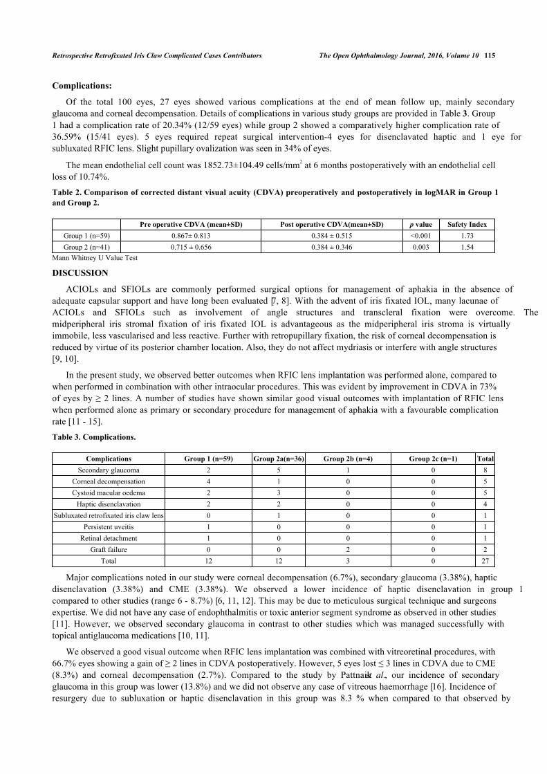
Complications:
Of the total 100 eyes, 27 eyes showed various complications at the end of mean follow up, mainly secondaryglaucoma and corneal decompensation. Details of complications in various study groups are provided in Table 3. Group1 had a complication rate of 20.34% (12/59 eyes) while group 2 showed a comparatively higher complication rate of36.59% (15/41 eyes). 5 eyes required repeat surgical intervention-4 eyes for disenclavated haptic and 1 eye forsubluxated RFIC lens. Slight pupillary ovalization was seen in 34% of eyes.
The mean endothelial cell count was 1852.73±104.49 cells/mm2 at 6 months postoperatively with an endothelial cellloss of 10.74%.
Table 2. Comparison of corrected distant visual acuity (CDVA) preoperatively and postoperatively in logMAR in Group 1and Group 2.
Pre operative CDVA (mean±SD) Post operative CDVA(mean±SD) p value Safety IndexGroup 1 (n=59) 0.867± 0.813 0.384 ± 0.515 <0.001 1.73Group 2 (n=41) 0.715 ± 0.656 0.384 ± 0.346 0.003 1.54
Mann Whitney U Value Test
DISCUSSION
ACIOLs and SFIOLs are commonly performed surgical options for management of aphakia in the absence ofadequate capsular support and have long been evaluated [7, 8]. With the advent of iris fixated IOL, many lacunae ofACIOLs and SFIOLs such as involvement of angle structures and transcleral fixation were overcome. Themidperipheral iris stromal fixation of iris fixated IOL is advantageous as the midperipheral iris stroma is virtuallyimmobile, less vascularised and less reactive. Further with retropupillary fixation, the risk of corneal decompensation isreduced by virtue of its posterior chamber location. Also, they do not affect mydriasis or interfere with angle structures[9, 10].
In the present study, we observed better outcomes when RFIC lens implantation was performed alone, compared towhen performed in combination with other intraocular procedures. This was evident by improvement in CDVA in 73%of eyes by ≥ 2 lines. A number of studies have shown similar good visual outcomes with implantation of RFIC lenswhen performed alone as primary or secondary procedure for management of aphakia with a favourable complicationrate [11 - 15].
Table 3. Complications.
Complications Group 1 (n=59) Group 2a(n=36) Group 2b (n=4) Group 2c (n=1) TotalSecondary glaucoma 2 5 1 0 8
Corneal decompensation 4 1 0 0 5Cystoid macular oedema 2 3 0 0 5
Haptic disenclavation 2 2 0 0 4Subluxated retrofixated iris claw lens 0 1 0 0 1
Persistent uveitis 1 0 0 0 1Retinal detachment 1 0 0 0 1
Graft failure 0 0 2 0 2Total 12 12 3 0 27
Major complications noted in our study were corneal decompensation (6.7%), secondary glaucoma (3.38%), hapticdisenclavation (3.38%) and CME (3.38%). We observed a lower incidence of haptic disenclavation in group 1compared to other studies (range 6 - 8.7%) [6, 11, 12]. This may be due to meticulous surgical technique and surgeonsexpertise. We did not have any case of endophthalmitis or toxic anterior segment syndrome as observed in other studies[11]. However, we observed secondary glaucoma in contrast to other studies which was managed successfully withtopical antiglaucoma medications [10, 11].
We observed a good visual outcome when RFIC lens implantation was combined with vitreoretinal procedures, with66.7% eyes showing a gain of ≥ 2 lines in CDVA postoperatively. However, 5 eyes lost ≤ 3 lines in CDVA due to CME(8.3%) and corneal decompensation (2.7%). Compared to the study by Pattnaik et al., our incidence of secondaryglaucoma in this group was lower (13.8%) and we did not observe any case of vitreous haemorrhage [16]. Incidence ofresurgery due to subluxation or haptic disenclavation in this group was 8.3 % when compared to that observed by
116 The Open Ophthalmology Journal, 2016, Volume 10 Ganesh et al.
Ramon et al. (15.6%) [18]. In all such cases who underwent resurgery, the RFIC lens remained stable until the lastfollow up and the visual outcomes were not affected. Previous studies have evaluated the safety and efficacy of RFIClens in patients requiring concomitant vitreoretinal procedures and have shown favourable results [16 - 24].
In our study, 4 eyes underwent combined penetrating keratoplasty, ACIOL explantation and RFIC lens implantationfor bullous keratopathy secondary to ACIOL. Visual acuity improved from counting finger at 1 meter to 6/24 in 2 eyes.However, 2 eyes did not show improvement in visual acuity due to subsequent graft failure. Previous studies haveshown good visual results and favourable complication rate with RFIC lens implantation combined with PKP forvarious indications [25 - 27]. The most common indication for combining PKP with RFIC lens in previous studies wasalso pseudophakic bullous keratopathy [25, 26].
One patient in this series had secondary elevation of IOP, which was medically controlled and did not lead to graftfailure. In a series of 12 eyes combining PKP+ RFIC lens implantation by Rufer et al. the most frequent complicationencountered was postoperative glaucoma [27]. However, Gonnermann et al. did not observe any significant change inIOP in their study [26].
Studies combining PKP with SFIOL have also demonstrated good clinical outcome [26 - 28]. However, certaincomplications such as vitreous haemorrhage, postoperative endophthalmitis and partial IOL dislocation have beenreported. Vitreous haemorrhage due to trauma to ciliary body and root of iris may occur at the time of surgery or inimmediate postoperative period. Exposure of scleral suture may increase the risk of endophthalmitis [28 - 30]. Unequalplacement or tying of scleral fixation suture may lead to partial IOL dislocation requiring refixation [28, 31]. RFIC lensmay be safer in this scenario as most of the suture related complications [32]. associated with SFIOLs can be avoidedimproving long term safety.
Moreover, ultrasound biomicroscopy studies on eyes with combined PKP with RFIC lens have shown thatenclavation to the posterior plane of iris preserves the anatomy of the anterior segment with respect to the iridocornealangle [25, 33]. This may also be relevant in eyes requiring combined trabeculectomy and RFIC lens implantation. In ourseries, we had only one such eye that underwent combined trabeculectomy with Mitomycin C and RFIC lensimplantation for a grossly subluxated cataract with pseudoexfoliation glaucoma. The postoperative outcomes were goodwith improvement in CDVA from counting finger 2 meter to 6/7.5.
Literature does not report outcomes of combined glaucoma filtration surgery simultaneously with RFIC lensimplantation. However, one case undergoing RFIC lens implantation following traumatic wound dehiscence oftrabeculectomy scleral flap has been reported [34]. Theoretically, RFIC lens would be a better option compared toSFIOL and glued IOL in glaucomatous eyes due to advantages of being sutureless, preservation of trabecular meshworkstructures and avoidance of creation of additional scleral flaps, thus ensuring long term safety and structural integrity ofthe eye.
To the best of our knowledge, this is the first study evaluating the outcome of RFIC lens implantation in differentchallenging cases in a long term (8 years) retrospective clinical audit. The results support the superiority of retrofixatediris claw IOL over other modalities (SFIOL, glued IOL) in similar situations. However, we observed a comparativelyhigher complication rate (36.59%) when RFIC lens implantation was combined with additional intraocular procedures(pars plana vitrectomy, penetrating keratoplasty, trabeculectomy) compared to when performed alone (20.34%). This isanticipated, since by combining two intraocular procedures, the overall risk of complications is expected to increase.This may also explain the comparatively higher percentage of eyes with loss of best corrected visual acuity in thecombined group. Nevertheless, the long term visual outcomes were satisfactory with good safety profile and withoverall low risk of serious and sight threatening complications.
CONFLICT OF INTEREST
The authors confirm that this article content has no conflict of interest.
ACKNOWLEDGEMENTS
Declared none.
REFERENCES
[1] Dick HB, Augustin AJ. Lens implant selection with absence of capsular support. Curr Opin Ophthalmol 2001; 12(1): 47-57.[http://dx.doi.org/10.1097/00055735-200102000-00009] [PMID: 11150081]
Retrospective Retrofixated Iris Claw Complicated Cases Contributors The Open Ophthalmology Journal, 2016, Volume 10 117
[2] Wagoner MD, Cox TA, Ariyasu RG, Jacobs DS, Karp CL. Intraocular lens implantation in the absence of capsular support: a report by theAmerican Academy of Ophthalmology. Ophthalmology 2003; 110(4): 840-59.[http://dx.doi.org/10.1016/S0161-6420(02)02000-6] [PMID: 12689913]
[3] Agarwal A, Kumar DA, Jacob S, Baid C, Agarwal A, Srinivasan S. Fibrin glue-assisted sutureless posterior chamber intraocular lensimplantation in eyes with deficient posterior capsules. J Cataract Refract Surg 2008; 34(9): 1433-8.[http://dx.doi.org/10.1016/j.jcrs.2008.04.040] [PMID: 18721701]
[4] Dadeya S, Kamlesh K, Kumari SP. Secondary intraocular lens (IOL) implantation: anterior chamber versus scleral fixation long-termcomparative evaluation. Eur J Ophthalmol 2003; 13(7): 627-33.[PMID: 14552597]
[5] Kumar DA, Agarwal A, Packiyalakshmi S, Jacob S, Agarwal A. Complications and visual outcomes after glued foldable intraocular lensimplantation in eyes with inadequate capsules. J Cataract Refract Surg 2013; 39(8): 1211-8.[http://dx.doi.org/10.1016/j.jcrs.2013.03.004] [PMID: 23726133]
[6] Mohr A, Hengerer F, Eckardt C. Retropupillary fixation of the iris claw lens in aphakia. 1 year outcome of a new implantation techniques.Ophthalmologe 2002; 99(7): 580-3.[http://dx.doi.org/10.1007/s00347-001-0563-z] [PMID: 12148307]
[7] Menezo JL, Martinez MC, Cisneros AL. Iris-fixated Worst claw versus sulcus-fixated posterior chamber lenses in the absence of capsularsupport. J Cataract Refract Surg 1996; 22(10): 1476-84.[http://dx.doi.org/10.1016/S0886-3350(96)80151-9] [PMID: 9051506]
[8] Hsing YE, Lee GA. Retropupillary iris claw intraocular lens for aphakia. Clin Experiment Ophthalmol 2012; 40(9): 849-54.[http://dx.doi.org/10.1111/j.1442-9071.2012.02808.x] [PMID: 22594520]
[9] Güell JL, Velasco F, Malecaze F, Vázquez M, Gris O, Manero F. Secondary Artisan-Verysise aphakic lens implantation. J Cataract RefractSurg 2005; 31(12): 2266-71.[http://dx.doi.org/10.1016/j.jcrs.2005.06.047] [PMID: 16473216]
[10] Baykara M, Ozcetin H, Yilmaz S, Timuçin OB. Posterior iris fixation of the iris-claw intraocular lens implantation through a scleral tunnelincision. Am J Ophthalmol 2007; 144(4): 586-91.[http://dx.doi.org/10.1016/j.ajo.2007.06.009] [PMID: 17692274]
[11] Gonnermann J, Klamann MK, Maier AK, et al. Visual outcome and complications after posterior iris-claw aphakic intraocular lensimplantation. J Cataract Refract Surg 2012; 38(12): 2139-43.[http://dx.doi.org/10.1016/j.jcrs.2012.07.035] [PMID: 23036355]
[12] De Silva SR, Arun K, Anandan M, Glover N, Patel CK, Rosen P. Iris-claw intraocular lenses to correct aphakia in the absence of capsulesupport. J Cataract Refract Surg 2011; 37(9): 1667-72.[http://dx.doi.org/10.1016/j.jcrs.2011.03.051] [PMID: 21855764]
[13] Rao R, Sasidharan A. Iris claw intraocular lens: a viable option in monocular surgical aphakia. Indian J Ophthalmol 2013; 61(2): 74-5.[http://dx.doi.org/10.4103/0301-4738.107198] [PMID: 23412525]
[14] Lett KS, Chaudhuri PR. Visual outcomes following Artisan aphakia iris claw lens implantation. Eye (Lond) 2011; 25(1): 73-6.[http://dx.doi.org/10.1038/eye.2010.146] [PMID: 20948556]
[15] Gonnermann J, Torun N, Klamann MK, et al. Posterior iris-claw aphakic intraocular lens implantation in children. Am J Ophthalmol 2013;156(2): 382-386.e1.[http://dx.doi.org/10.1016/j.ajo.2013.03.002] [PMID: 23721944]
[16] Pattnaik L, Almozawak K, Binder S. Pars plana vitrectomy and artisan iris fixated intraocular lens for aphakia in complicated vitreoretinalreferrals. J Acute Dis 2013; 2(2): 109-14.[http://dx.doi.org/10.1016/S2221-6189(13)60109-2]
[17] Patil KB, Meleth P, Shanker MP. Pars plana vitrectomy with posterior iris claw implantation for posteriorly dislocated nucleus and intraocularlens. Indian J Ophthalmol 2011; 59(6): 497-500.[http://dx.doi.org/10.4103/0301-4738.86321] [PMID: 22011497]
[18] Anglada R, Castellví J, Parera A, Sabala A. Inverted implantation of posterior iris-fixated intraocular lens with 23G transconjunctivalvitrectomy in the management of secondary implant-Technique and stability, astigmatism and endothelial loss outcomes. J Emmetropia 2014;5(3): 133-43.
[19] Kodjikian L, Beby F, Spire M, et al. Combined pars plana phacofragmentation, vitrectomy, and Artisan lens implantation for traumaticsubluxated cataracts. Retina 2006; 26(8): 909-16.[http://dx.doi.org/10.1097/01.iae.0000250005.38037.da] [PMID: 17031292]
[20] van der Meulen IJ, Gunning FP, Vermeulen MG, de Smet MD. Artisan lens implantation to correct aphakia after vitrectomy for retainednuclear lens fragments. J Cataract Refract Surg 2004; 30(12): 2585-9.[http://dx.doi.org/10.1016/j.jcrs.2004.04.050] [PMID: 15617928]
[21] Riazi M, Moghimi S, Najmi Z, Ghaffari R. Secondary Artisan-Verysise intraocular lens implantation for aphakic correction in post-traumaticvitrectomized eye. Eye (Lond) 2008; 22(11): 1419-24.[http://dx.doi.org/10.1038/eye.2008.271] [PMID: 18756286]
118 The Open Ophthalmology Journal, 2016, Volume 10 Ganesh et al.
[22] Acar N, Kapran Z, Altan T, Kucuksumer Y, Unver YB, Polat E. Secondary iris claw intraocular lens implantation for the correction ofaphakia after pars plana vitrectomy. Retina 2010; 30(1): 131-9.[http://dx.doi.org/10.1097/IAE.0b013e3181b32eef] [PMID: 19834354]
[23] Català-Mora J, Díaz-Cascajosa J, Ferreruela-Sanfeliu G, Castany-Aregall M, Prat-Bartomeu J, García-Arumí J. 23-G pars plana vitrectomy,lensectomy, and artisan IOL implantation for the management of nontraumatic ectopia lentis: a new iris enclavation technique for iris clawlens. Retina 2012; 32(6): 1214-6.[http://dx.doi.org/10.1097/IAE.0b013e31824d4f06] [PMID: 22643806]
[24] Farrahi F, Feghhi M, Haghi F, Kasiri A, Afkari A, Latifi M. Iris claw versus scleral fixation intraocular lens implantation during pars planavitrectomy. J Ophthalmic Vis Res 2012; 7(2): 118-24.[PMID: 23275819]
[25] Dighiero P, Guigou S, Mercie M, Briat B, Ellies P, Gicquel JJ. Penetrating keratoplasty combined with posterior Artisan iris-fixatedintraocular lens implantation. Acta Ophthalmol Scand 2006; 84(2): 197-200.[http://dx.doi.org/10.1111/j.1600-0420.2005.00573.x] [PMID: 16637836]
[26] Gonnermann J, Torun N, Klamann MK, et al. Visual outcomes and complications following posterior iris-claw aphakic intraocular lensimplantation combined with penetrating keratoplasty. Graefes Arch Clin Exp Ophthalmol 2013; 251(4): 1151-6.[http://dx.doi.org/10.1007/s00417-012-2226-y] [PMID: 23250481]
[27] Rüfer F, Saeger M, Nölle B, Roider J. Implantation of retropupillar iris claw lenses with and without combined penetrating keratoplasty.Graefes Arch Clin Exp Ophthalmol 2009; 247(4): 457-62.[http://dx.doi.org/10.1007/s00417-008-0940-2] [PMID: 18787833]
[28] Koçak-Altintas AG, Koçak-Midillioglu I, Dengisik F, Duman S. Implantation of scleral-sutured posterior chamber intraocular lenses duringpenetrating keratoplasty. J Refract Surg 2000; 16(4): 456-8.[PMID: 10939726]
[29] Davis RM, Best D, Gilbert GE. Comparison of intraocular lens fixation techniques performed during penetrating keratoplasty. Am JOphthalmol 1991; 111(6): 743-9.[http://dx.doi.org/10.1016/S0002-9394(14)76783-2] [PMID: 2039047]
[30] Soong HK, Musch DC, Kowal V, Sugar A, Meyer RF. Implantation of posterior chamber intraocular lenses in the absence of lens capsuleduring penetrating keratoplasty. Arch Ophthalmol 1989; 107(5): 660-5.[http://dx.doi.org/10.1001/archopht.1989.01070010678026] [PMID: 2655567]
[31] Schein OD, Kenyon KR, Steinert RF, et al. A randomized trial of intraocular lens fixation techniques with penetrating keratoplasty.Ophthalmology 1993; 100(10): 1437-43.[http://dx.doi.org/10.1016/S0161-6420(93)31458-2] [PMID: 8414402]
[32] Price MO, Price FW Jr, Werner L, Berlie C, Mamalis N. Late dislocation of scleral-sutured posterior chamber intraocular lenses. J CataractRefract Surg 2005; 31(7): 1320-6.[http://dx.doi.org/10.1016/j.jcrs.2004.12.060] [PMID: 16105601]
[33] Gicquel JJ, Guigou S, Bejjani RA, Briat B, Ellies P, Dighiero P. Ultrasound biomicroscopy study of the Verisyse aphakic intraocular lenscombined with penetrating keratoplasty in pseudophakic bullous keratopathy. J Cataract Refract Surg 2007; 33(3): 455-64.[http://dx.doi.org/10.1016/j.jcrs.2006.11.017] [PMID: 17321397]
[34] Rubinstein A, Salmon JF. Late traumatic scleral flap dehiscence following trabeculectomy. Eye (Lond) 2007; 21(1): 145-6.[http://dx.doi.org/10.1038/sj.eye.6702476] [PMID: 16763651]
© Ganesh et al.; Licensee Bentham Open.
This is an open access article licensed under the terms of the Creative Commons Attribution-Non-Commercial 4.0 International Public License(CC BY-NC 4.0) (https://creativecommons.org/licenses/by-nc/4.0/legalcode), which permits unrestricted, non-commercial use, distribution andreproduction in any medium, provided the work is properly cited.