Embed Size (px)
Citation preview
97
Case report
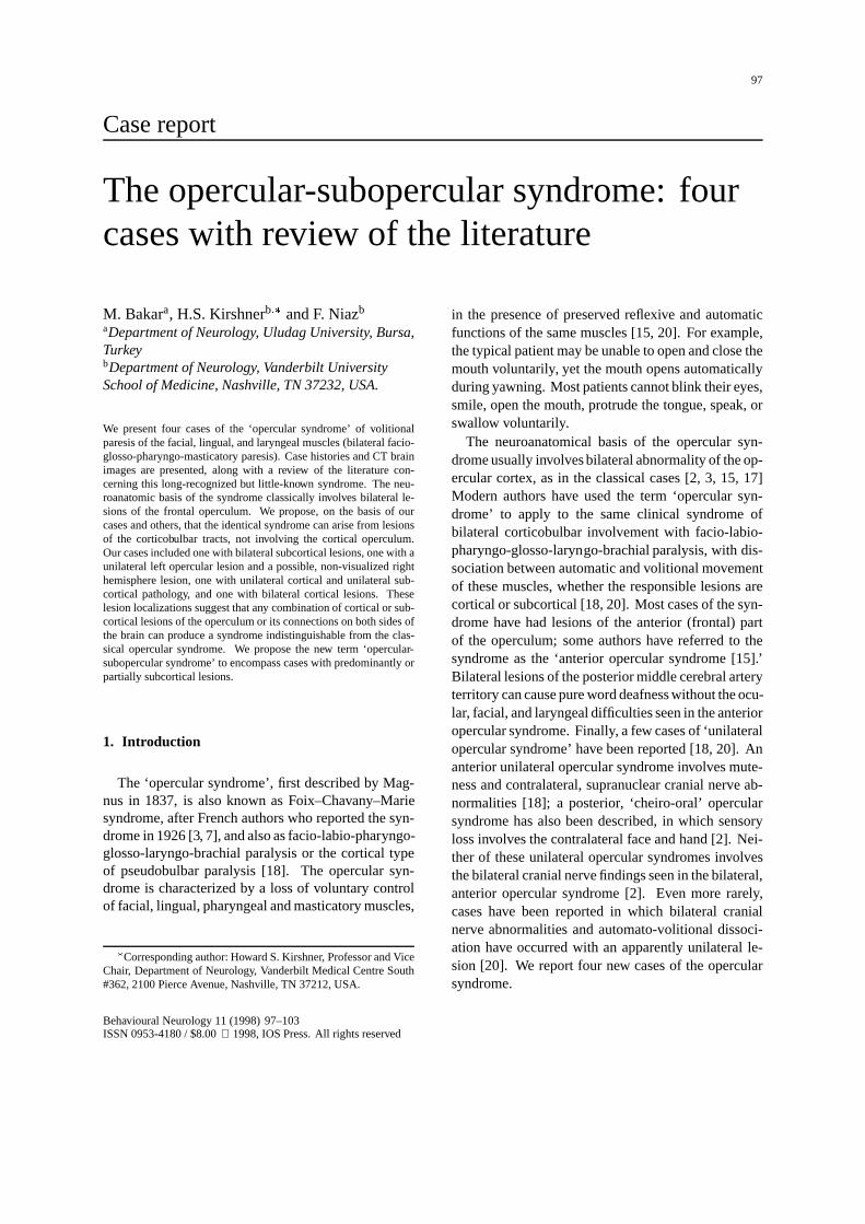
The opercular-subopercular syndrome: fourcases with review of the literature
M. Bakara, H.S. Kirshnerb ��� and F. Niazb
aDepartment of Neurology, Uludag University, Bursa,TurkeybDepartment of Neurology, Vanderbilt UniversitySchool of Medicine, Nashville, TN 37232, USA.
We present four cases of the ‘opercular syndrome’ of volitionalparesis of the facial, lingual, and laryngeal muscles (bilateral facio-glosso-pharyngo-masticatory paresis). Case histories and CT brainimages are presented, along with a review of the literature con-cerning this long-recognized but little-known syndrome. The neu-roanatomic basis of the syndrome classically involves bilateral le-sions of the frontal operculum. We propose, on the basis of ourcases and others, that the identical syndrome can arise from lesionsof the corticobulbar tracts, not involving the cortical operculum.Our cases included one with bilateral subcortical lesions, one with aunilateral left opercular lesion and a possible, non-visualized righthemisphere lesion, one with unilateral cortical and unilateral sub-cortical pathology, and one with bilateral cortical lesions. Theselesion localizations suggest that any combination of cortical or sub-cortical lesions of the operculum or its connections on both sides ofthe brain can produce a syndrome indistinguishable from the clas-sical opercular syndrome. We propose the new term ‘opercular-subopercular syndrome’ to encompass cases with predominantly orpartially subcortical lesions.
1. Introduction
The ‘opercular syndrome’, first described by Mag-nus in 1837, is also known as Foix–Chavany–Mariesyndrome, after French authors who reported the syn-drome in 1926 [3, 7], and also as facio-labio-pharyngo-glosso-laryngo-brachial paralysis or the cortical typeof pseudobulbar paralysis [18]. The opercular syn-drome is characterized by a loss of voluntary controlof facial, lingual, pharyngeal and masticatory muscles,
�Corresponding author: Howard S. Kirshner, Professor and Vice
Chair, Department of Neurology, Vanderbilt Medical Centre South#362, 2100 Pierce Avenue, Nashville, TN 37212, USA.
in the presence of preserved reflexive and automaticfunctions of the same muscles [15, 20]. For example,the typical patient may be unable to open and close themouth voluntarily, yet the mouth opens automaticallyduring yawning. Most patients cannot blink their eyes,smile, open the mouth, protrude the tongue, speak, orswallow voluntarily.
The neuroanatomical basis of the opercular syn-drome usually involves bilateral abnormality of the op-ercular cortex, as in the classical cases [2, 3, 15, 17]Modern authors have used the term ‘opercular syn-drome’ to apply to the same clinical syndrome ofbilateral corticobulbar involvement with facio-labio-pharyngo-glosso-laryngo-brachial paralysis, with dis-sociation between automatic and volitional movementof these muscles, whether the responsible lesions arecortical or subcortical [18, 20]. Most cases of the syn-drome have had lesions of the anterior (frontal) partof the operculum; some authors have referred to thesyndrome as the ‘anterior opercular syndrome [15].’Bilateral lesions of the posterior middle cerebral arteryterritory can cause pure word deafness without the ocu-lar, facial, and laryngeal difficulties seen in the anterioropercular syndrome. Finally, a few cases of ‘unilateralopercular syndrome’ have been reported [18, 20]. Ananterior unilateral opercular syndrome involves mute-ness and contralateral, supranuclear cranial nerve ab-normalities [18]; a posterior, ‘cheiro-oral’ opercularsyndrome has also been described, in which sensoryloss involves the contralateral face and hand [2]. Nei-ther of these unilateral opercular syndromes involvesthe bilateral cranial nerve findings seen in the bilateral,anterior opercular syndrome [2]. Even more rarely,cases have been reported in which bilateral cranialnerve abnormalities and automato-volitional dissoci-ation have occurred with an apparently unilateral le-sion [20]. We report four new cases of the opercularsyndrome.
ISSN 0953-4180 / $8.00 1998, IOS Press. All rights reservedBehavioural Neurology 11 (1998) 97–103
98 M. Bakar et al. / The opercular-subopercular syndrome: four cases with review of the literature
2. Report of cases
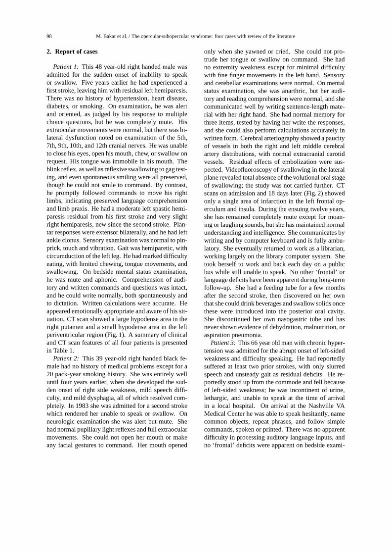
Patient 1: This 48 year-old right handed male wasadmitted for the sudden onset of inability to speakor swallow. Five years earlier he had experienced afirst stroke, leaving him with residual left hemiparesis.There was no history of hypertension, heart disease,diabetes, or smoking. On examination, he was alertand oriented, as judged by his response to multiplechoice questions, but he was completely mute. Hisextraocular movements were normal, but there was bi-lateral dysfunction noted on examination of the 5th,7th, 9th, 10th, and 12th cranial nerves. He was unableto close his eyes, open his mouth, chew, or swallow onrequest. His tongue was immobile in his mouth. Theblink reflex, as well as reflexive swallowing to gag test-ing, and even spontaneous smiling were all preserved,though he could not smile to command. By contrast,he promptly followed commands to move his rightlimbs, indicating preserved language comprehensionand limb praxis. He had a moderate left spastic hemi-paresis residual from his first stroke and very slightright hemiparesis, new since the second stroke. Plan-tar responses were extensor bilaterally, and he had leftankle clonus. Sensory examination was normal to pin-prick, touch and vibration. Gait was hemiparetic, withcircumduction of the left leg. He had marked difficultyeating, with limited chewing, tongue movements, andswallowing. On bedside mental status examination,he was mute and aphonic. Comprehension of audi-tory and written commands and questions was intact,and he could write normally, both spontaneously andto dictation. Written calculations were accurate. Heappeared emotionally appropriate and aware of his sit-uation. CT scan showed a large hypodense area in theright putamen and a small hypodense area in the leftperiventricular region (Fig. 1). A summary of clinicaland CT scan features of all four patients is presentedin Table 1.
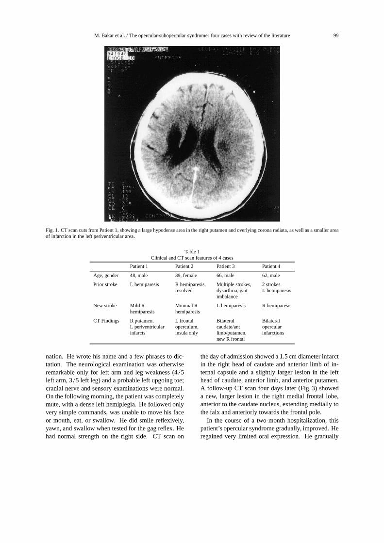
Patient 2: This 39 year-old right handed black fe-male had no history of medical problems except for a20 pack-year smoking history. She was entirely welluntil four years earlier, when she developed the sud-den onset of right side weakness, mild speech diffi-culty, and mild dysphagia, all of which resolved com-pletely. In 1983 she was admitted for a second strokewhich rendered her unable to speak or swallow. Onneurologic examination she was alert but mute. Shehad normal pupillary light reflexes and full extraocularmovements. She could not open her mouth or makeany facial gestures to command. Her mouth opened
only when she yawned or cried. She could not pro-trude her tongue or swallow on command. She hadno extremity weakness except for minimal difficultywith fine finger movements in the left hand. Sensoryand cerebellar examinations were normal. On mentalstatus examination, she was anarthric, but her audi-tory and reading comprehension were normal, and shecommunicated well by writing sentence-length mate-rial with her right hand. She had normal memory forthree items, tested by having her write the responses,and she could also perform calculations accurately inwritten form. Cerebral arteriography showed a paucityof vessels in both the right and left middle cerebralartery distributions, with normal extracranial carotidvessels. Residual effects of embolization were sus-pected. Videofluoroscopy of swallowing in the lateralplane revealed total absence of the volutional oral stageof swallowing; the study was not carried further. CTscans on admission and 18 days later (Fig. 2) showedonly a single area of infarction in the left frontal op-erculum and insula. During the ensuing twelve years,she has remained completely mute except for moan-ing or laughing sounds, but she has maintained normalunderstanding and intelligence. She communicates bywriting and by computer keyboard and is fully ambu-latory. She eventually returned to work as a librarian,working largely on the library computer system. Shetook herself to work and back each day on a publicbus while still unable to speak. No other ‘frontal’ orlanguage deficits have been apparent during long-termfollow-up. She had a feeding tube for a few monthsafter the second stroke, then discovered on her ownthat she could drink beverages and swallow solids oncethese were introduced into the posterior oral cavity.She discontinued her own nasogastric tube and hasnever shown evidence of dehydration, malnutrition, oraspiration pneumonia.
Patient 3: This 66 year old man with chronic hyper-tension was admitted for the abrupt onset of left-sidedweakness and difficulty speaking. He had reportedlysuffered at least two prior strokes, with only slurredspeech and unsteady gait as residual deficits. He re-portedly stood up from the commode and fell becauseof left-sided weakness; he was incontinent of urine,lethargic, and unable to speak at the time of arrivalin a local hospital. On arrival at the Nashville VAMedical Center he was able to speak hesitantly, namecommon objects, repeat phrases, and follow simplecommands, spoken or printed. There was no apparentdifficulty in processing auditory language inputs, andno ‘frontal’ deficits were apparent on bedside exami-
M. Bakar et al. / The opercular-subopercular syndrome: four cases with review of the literature 99
Fig. 1. CT scan cuts from Patient 1, showing a large hypodense area in the right putamen and overlying corona radiata, as well as a smaller areaof infarction in the left periventricular area.
Table 1Clinical and CT scan features of 4 cases
Patient 1 Patient 2 Patient 3 Patient 4
Age, gender 48, male 39, female 66, male 62, male
Prior stroke L hemiparesis R hemiparesis, Multiple strokes, 2 strokesresolved dysarthria, gait L hemiparesis
imbalance
New stroke Mild R Minimal R L hemiparesis R hemiparesishemiparesis hemiparesis
CT Findings R putamen, L frontal Bilateral BilateralL periventricular operculum, caudate/ant opercularinfarcts insula only limb/putamen, infarctions
new R frontal
nation. He wrote his name and a few phrases to dic-tation. The neurological examination was otherwiseremarkable only for left arm and leg weakness (4
�5
left arm, 3�5 left leg) and a probable left upgoing toe;
cranial nerve and sensory examinations were normal.On the following morning, the patient was completelymute, with a dense left hemiplegia. He followed onlyvery simple commands, was unable to move his faceor mouth, eat, or swallow. He did smile reflexively,yawn, and swallow when tested for the gag reflex. Hehad normal strength on the right side. CT scan on
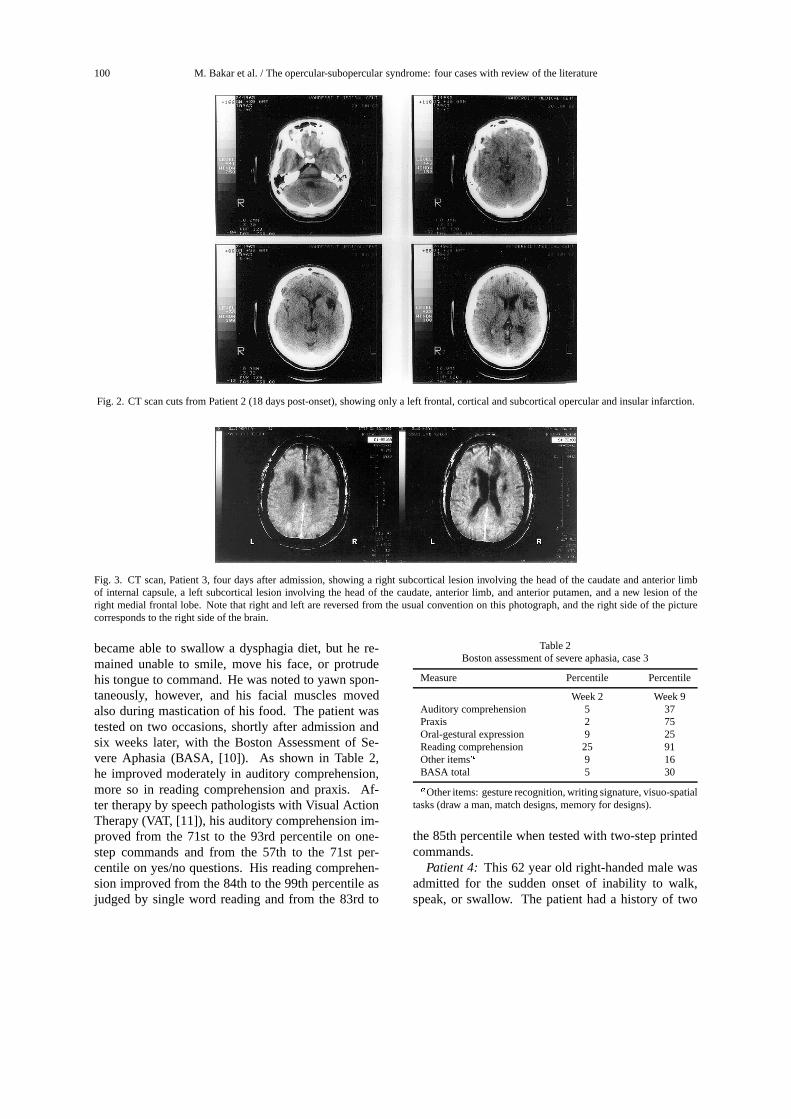
the day of admission showed a 1.5 cm diameter infarctin the right head of caudate and anterior limb of in-ternal capsule and a slightly larger lesion in the lefthead of caudate, anterior limb, and anterior putamen.A follow-up CT scan four days later (Fig. 3) showeda new, larger lesion in the right medial frontal lobe,anterior to the caudate nucleus, extending medially tothe falx and anteriorly towards the frontal pole.
In the course of a two-month hospitalization, thispatient’s opercular syndrome gradually, improved. Heregained very limited oral expression. He gradually
100 M. Bakar et al. / The opercular-subopercular syndrome: four cases with review of the literature
Fig. 2. CT scan cuts from Patient 2 (18 days post-onset), showing only a left frontal, cortical and subcortical opercular and insular infarction.
Fig. 3. CT scan, Patient 3, four days after admission, showing a right subcortical lesion involving the head of the caudate and anterior limbof internal capsule, a left subcortical lesion involving the head of the caudate, anterior limb, and anterior putamen, and a new lesion of theright medial frontal lobe. Note that right and left are reversed from the usual convention on this photograph, and the right side of the picturecorresponds to the right side of the brain.
became able to swallow a dysphagia diet, but he re-mained unable to smile, move his face, or protrudehis tongue to command. He was noted to yawn spon-taneously, however, and his facial muscles movedalso during mastication of his food. The patient wastested on two occasions, shortly after admission andsix weeks later, with the Boston Assessment of Se-vere Aphasia (BASA, [10]). As shown in Table 2,he improved moderately in auditory comprehension,more so in reading comprehension and praxis. Af-ter therapy by speech pathologists with Visual ActionTherapy (VAT, [11]), his auditory comprehension im-proved from the 71st to the 93rd percentile on one-step commands and from the 57th to the 71st per-centile on yes/no questions. His reading comprehen-sion improved from the 84th to the 99th percentile asjudged by single word reading and from the 83rd to
Table 2Boston assessment of severe aphasia, case 3
Measure Percentile Percentile
Week 2 Week 9Auditory comprehension 5 37Praxis 2 75Oral-gestural expression 9 25Reading comprehension 25 91Other items � 9 16BASA total 5 30
� Other items: gesture recognition, writing signature, visuo-spatialtasks (draw a man, match designs, memory for designs).
the 85th percentile when tested with two-step printedcommands.
Patient 4: This 62 year old right-handed male wasadmitted for the sudden onset of inability to walk,speak, or swallow. The patient had a history of two
M. Bakar et al. / The opercular-subopercular syndrome: four cases with review of the literature 101
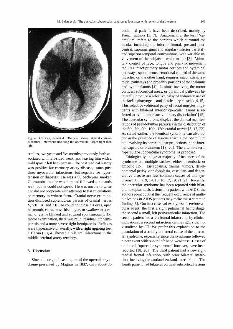
Fig. 4. CT scan, Patient 4. The scan shows bilateral cortical-subcortical infarctions involving the operculum, larger right thanleft.
strokes, two years and five months previously, both as-sociated with left-sided weakness, leaving him with amild spastic left hemiparesis. The past medical historywas positive for coronary artery disease, status postthree myocardial infarctions, but negative for hyper-tension or diabetes. He was a 90 pack-year smoker.On examination, he was alert and followed commandswell, but he could not speak. He was unable to writeand did not cooperate with attempts to test calculationsor memory in written form. Cranial nerve examina-tion disclosed supranuclear paresis of cranial nervesV, VII, IX, and XII. He could not close his eyes, openhis mouth, chew, move his tongue, or swallow to com-mand, yet he blinked and yawned spontaneously. Onmotor examination, there was mild, residual left hemi-paresis and a more severe right hemiparesis. Reflexeswere hyperactive bilaterally, with a right upgoing toe.CT scan (Fig. 4) showed a bilateral infarctions in themiddle cerebral artery territory.
3. Discussion
Since the original case report of the opercular syn-drome presented by Magnus in 1837, only about 30
additional patients have been described, mainly byFrench authors [3, 7]. Anatomically, the term ‘op-erculum’ refers to the cortices which surround theinsula, including the inferior frontal, pre-and post-central, supramarginal and angular (inferior parietal),and superior temporal convolutions, with variable in-volvement of the subjacent white matter [3]. Volun-tary control of face, tongue and pharynx movementrequires intact primary motor cortices and pyramidalpathways; spontaneous, emotional control of the samemuscles, on the other hand, requires intact extrapyra-midal pathways and probably portions of the thalamusand hypothalamus [4]. Lesions involving the motorcortices, subcortical areas, or pyramidal pathways bi-laterally produce a selective palsy of voluntary use ofthe facial, pharyngeal, and masticatory muscles [4, 15].This selective volitional palsy of facial muscles in pa-tients with bilateral anterior opercular lesions is re-ferred to as an ‘automato-voluntary dissociation’ [15].The opercular syndrome displays the clinical manifes-tations of pseudobulbar paralysis in the distribution ofthe 5th, 7th, 9th, 10th, 12th cranial nerves [3, 17, 22].As stated earlier, the identical syndrome can also oc-cur in the presence of lesions sparing the operculumbut involving its corticobulbar projections in the inter-nal capsule or brainstem [18, 20]. The alternate term‘opercular-subopercular syndrome’ is proposed.
Etiologically, the great majority of instances of thesyndrome are multiple strokes, either thrombotic orembolic [15]. Encephalitis, trauma, tumors, devel-opmental perisylvian dysplasia, vasculitis, and degen-erative disease are less common causes of this syn-drome [3, 6, 7, 9, 14, 15, 16, 17, 19, 21, 23]. Recently,the opercular syndrome has been reported with bilat-eral toxoplasmosis lesions in a patient with AIDS; theauthors point out that the frequent occurrence of multi-ple lesions in AIDS patients may make this a commonfinding [8]. Our first case had two types of cerebrovas-cular event, the first a right putamenal hemorrhage,the second a small, left periventricular infarction. Thesecond patient had a left frontal infarct and, by clinicalindications, a second infarction on the right side, notvisualized by CT. We prefer this explanation to thepostulation of a strictly unilateral cause of the opercu-lar syndrome, especially since the syndrome followeda new event with subtle left hand weakness. Cases ofunilateral ‘opercular syndrome,’ however, have beenreported [18, 20]. The third patient had a new rightmedial frontal infarction, with prior bilateral infarc-tions involving the caudate head and anterior limb. Thefourth patient had bilateral cortical-subcortical lesions
102 M. Bakar et al. / The opercular-subopercular syndrome: four cases with review of the literature
involving the middle cerebral artery territory. Inter-estingly, this is the only case in our series with theclassical finding of bilateral, cortical opercular lesions.Our cases all had CT scans, since this was the onlymodality available at the time of presentation in Case2, and because MRI scans were not routinely avail-able in Bursa, Turkey or at the Nashville VA MedicalCenter when Cases 1, 3, and 4 presented. MRI mayreveal additional lesions as compared to CT in patientswith opercular syndrome secondary to stroke, and wesuspect that an additional, contralateral lesion mighthave been detected by MRI in Case 2.
The opercular syndrome must be distinguishedfrom catatonia, akinetic mutism, oral buccal apraxia,Broca’s aphasia, pseudobulbar palsy, and bulbar palsysecondary to myasthenia gravis, Guillain–Barre syn-drome, and brainstem strokes. The prompt ability ofthe patient to follow commands involving limb mus-cles differentiates the patient with the opercular syn-drome from those with catatonia or akinetic mutism.Broca’s aphasia, even when associated with muteness,does not involve the total loss of voluntary movementin the cranial musculature. The opercular syndromecan be considered a form of pseudobulbar palsy, interms of the muteness and aphagia, and often increasedjaw and gag reflexes. The involuntary laughter andcrying which are often a part of pseudobulbar palsy arenot seen in all cases of either the opercular syndromeor pseudobulbar palsy. The only real difference be-tween the opercular syndrome and pseudobulbar palsyis that the opercular syndrome implies a complete syn-drome of muteness and inability to move the facial,buccal, lingual and pharyngeal muscles, while pseu-dobulbar palsy includes more partial bipyramidal syn-dromes with dysarthria. Buccal or buccofacial apraxiarefers to the inability to follow commands related tothe oral apparatus, while these muscles can be usedspontaneously. The automato-voluntary dissociationthus applies both to the opercular syndrome and buccalapraxia. Buccal apraxia clearly can be a part of theopercular syndrome, though most patients with buccalapraxia are not completely mute [5], nor in most casesare they completely unable to move the facial, lingual,and pharyngeal muscles. Speech apraxia, also calledverbal apraxia, refers to syndromes of errors in theproduction of multisyllabic utterances, usually associ-ated with single, left hemisphere lesions [12, 13]; sucha deficit cannot even be tested in a mute patient. Theopercular syndrome differs from bulbar palsy by thelack of fasciculations, atrophy, and absent jaw and gagreflexes as would be expected in a lower motor neuron
syndrome. Bulbar palsy secondary to the Guillain–Barre syndrome and myasthenia gravis are also differ-entiated from the opercular syndrome by the lack ofthe automato-voluntary dissociation in eye and facialmovements [15, 17, 22].
The opercular syndrome is classically ascribed todirect involvement of the opercular cerebral cortex.As described above, however, subcortical lesions canproduce the syndrome by destroying fibers connect-ing cortical and subcortical structures and descendingpyramidal pathways, in the absence of cortical dam-age [18, 23]. A few cases of unilateral opercular syn-drome secondary to a unilateral cortical lesion havebeen described [3], whereas only one case has beendescribed of the opercular syndrome secondary to aunilateral subcortical stroke [18]; this patient did nothave a complete, bilateral syndrome. To our knowl-edge, our first case is the first reported instance ofthe bilateral opercular syndrome secondary to bilat-eral subcortical strokes, though cases of pseudobul-bar palsy have been reported with bilateral subcorticallesions [1]. The other, previously reported case of abilateral subcortical opercular syndrome was associ-ated with a progressive upper motor neuron degenera-tive syndrome, primary lateral sclerosis [23]. Anotherpoint worth emphasizing is the surprisingly small sizeof the lesion associated with the second stroke in ourfirst two cases, in view of the devastating, bilateralcranial nerve syndromes which resulted. The secondstroke, which produced no disabling weakness or sen-sory impairment in the limbs, resulted in severe, total,bilateral upper-motor-neuron involvement of the 5th,7th, 9th, 10th, and 12th cranial nerves. It appears thatthe other hemisphere has a large capacity to compen-sate for a unilateral lesion, but a small lesion in the in-tact hemisphere can then bring out multiple pseudob-ulbar findings on both sides, not evident after the firststroke. Another possible explanation for the profoundeffects of the second, left hemisphere stroke in thesepatients would be a relative dominance of one hemi-sphere for these cranial nerves functions [17]. Usually,this dominant hemisphere is thought to be the left. Thecase of bilateral opercular syndrome secondary to aunilateral lesion reported by Starkstein et al. [20] hada lesion involving the right insula, with extension tothe frontoparietal operculum; no left hemisphere le-sion was evident at autopsy, approximately one monthpost-onset. The patient was beginning to speak at thetime of death, so the permanence of the unilateral syn-drome is uncertain. Nonetheless, this case provides thebest evidence that a unilateral lesion can produce a bi-

M. Bakar et al. / The opercular-subopercular syndrome: four cases with review of the literature 103
lateral opercular syndrome. We suspect that our Case2, as well as most other cases of the bilateral opercu-lar syndrome secondary to a unilateral lesion, actuallyhave a subtle lesion on the second side. The naturalhistory of the opercular syndrome has not been studiedin detail. Our second case has had a stable syndromeof near-muteness for twelve years. The response tospeech therapy with Visual Action Therapy [10] is ofinterest, but the applicability of this therapy techniquein cases of the opercular syndrome will require furtherstudy.
In conclusion, our cases and those cited from theliterature suggest that the opercular syndrome can beproduced by any combination of cortical or subcor-tical lesions affecting the corticobulbar pathways forthe cranial nerves in both hemispheres. The term‘opercular-subopercular’ syndrome’ would take intoaccount subcortical localizations for the syndrome.
References
[1] G. Besson, J. Bogousslavsky, F. Regli and P. Maeder, Acutepseudobulbar or supranuclear bulbar palsy, Arch. Neurol. 48(1991), 501–507.
[2] J. Bogousslavsky, K. Dizerens and F. Regli, Despland P-A.Opercular cheiro-oral syndrome, Arch. Neurol. 48 (1991),658–661.
[3] G.W. Bruyn and J.C. Gathier, The operculum syndrome,in: Handbook of clinical Neurology, Vol 2, P.J. Vinken, G.W.Bruyn, ed., Amsterdam: North Holland, 1969, pp. 776–783.
[4] R.N. Dejong, The neurologic examination, MD: Harper &Row, 4th ed. Hagerstown, 1979, 190.
[5] E. DeRenzi, A. Pieczuro and L.A. Vignolo, Oral apraxia andaphasia, Cortex 2 (1966), 50–73.
[6] B. Duche and C. Marchal, P. Loiseau and C. Louvet Gien-daj, Facio-Linguo-Masticatory diplegia and epilepsy. Corti-cal dysplasia, Rev. Neurol. 148(8–9) (1992), 562–565.
[7] N.R. Graff-Radford, E.P. Bosch, J.C. Stears and D. Tranel,Developmental Foix-Chavany-Marie syndrome in identicaltwins, Ann. Neurol. 20 (1986), 632–635.
[8] M.P. Grassi, M. Borella, F. Clerici, C. Perin, M.T. Bini and A.Mangoni, Reversible bilateral opercular syndrome secondary
to AIDS-associated cerebral toxoplasmosis, Ital. J. Neurol.Sci. 15 (1994), 115–117.
[9] Z. Grosswasser, I. Grosswasser Reider and C. Korn, Bioper-cular lesions and acquired mutism in a young patient, BrainInj. 5(3) (1991), 331–334.
[10] N.A. Helm-Estabrooks, P.M. Fitzpatrick and B. Barresi, Vi-sual action therapy for global aphasia, J. Sp. Hearing Dis. 47(1982), 385–389.
[11] N. Helm-Estabrooks, A.R. Ramsberger and M.N. Morgan,BASA: Boston assessment of severe aphasia, Special PressInc., San Antonio, TX, 1989.
[12] D.F. Johns and F.L. Darley, Phonemic variability in apraxiaof speech, J. Speech Hearing Res. 13 (1970), 556–583.
[13] H.S. Kirshner, Apraxia of speech: A linguistic enigma. Aneurologist’s perspective, Sem. Speech Lang. 13 (1992), 14–24.
[14] C. Lang, J. Rechwein, H. Iro and T. Treig, Foix-Chavany-Marie syndrome. Neurological, neuropsychological, CT,MRI, and SPECT findings in a case progressive for more than10 years, Eur. Arch. Psychiatry 239(3) (1989), 188–193.
[15] C.C. Mao, B.M. Coull, L.C. Golper and M.T. Rau, Anterioropercular syndrome, Neurology 39 (1989), 1169–1172.
[16] G.C. Marchiori, G. Trincia and S. Cusumano, Labio-glosso-pharyngo laringealparalysis caused by two brain lesions: Cor-tical and subcortical, Ital. J. Neurol. Sci. 12(4) (1991), 419–422.
[17] C. Mariani, H. Spinnler, R. Sterzi and G. Vallar, Bilateralperisylvian softenings: Bilateral anterior opercular syndrome(Foix-Chavany-Marie syndrome), J. Neurol. 223 (1980),269–284.
[18] L. Posteraro, F. Pezzoni, E. Varalda, G. Fugazzo and A. Maz-zucchi, A case of unilateral opercular syndrome associatedwith subcortical lesion, J. Neurol. 238 (1991), 337–339.
[19] M.I. Shevell, L. Carmont, K. Meagher Willemure, Devel-opmental bilateral perisylvian dysplasia, Ped. Neurol. 8(4)(1992), 299–302.
[20] S.E. Starkstein, M. Berthier and R. Leiguarda, Bilateral op-ercular syndrome and crossed aphemia due to a right insularlesion: A clinicopathological study, Brain and Language 34(1988), 253–261.
[21] J.C. Van der Poel, C.A. Haenggeli and W.C. Overweg-Plandsoen, Operculum syndrome: unusual feature of herpessimplex encephalitis, Pediatr. Neurol. 12 (1995), 246–249.
[22] M. Weller, How to define the opercular syndrome? (letter),J. Neurol. 239(5) (1992), 294–95.
[23] M. Weller, M. Poremba and J. Dichgans, Opercular syndromewithout opercular lesions: Foix-Chavany-Marie syndromein progressive supranuclear motor system degeneration, Eur.Arch. Psychiatry Neurol. Sci. 239(6) (1990), 370–372.

Submit your manuscripts athttp://www.hindawi.com
Stem CellsInternational
Hindawi Publishing Corporationhttp://www.hindawi.com Volume 2014
Hindawi Publishing Corporationhttp://www.hindawi.com Volume 2014
MEDIATORSINFLAMMATION
of
Hindawi Publishing Corporationhttp://www.hindawi.com Volume 2014
Behavioural Neurology
EndocrinologyInternational Journal of
Hindawi Publishing Corporationhttp://www.hindawi.com Volume 2014
Hindawi Publishing Corporationhttp://www.hindawi.com Volume 2014
Disease Markers
Hindawi Publishing Corporationhttp://www.hindawi.com Volume 2014
BioMed Research International
OncologyJournal of
Hindawi Publishing Corporationhttp://www.hindawi.com Volume 2014
Hindawi Publishing Corporationhttp://www.hindawi.com Volume 2014
Oxidative Medicine and Cellular Longevity
Hindawi Publishing Corporationhttp://www.hindawi.com Volume 2014
PPAR Research
The Scientific World JournalHindawi Publishing Corporation http://www.hindawi.com Volume 2014
Immunology ResearchHindawi Publishing Corporationhttp://www.hindawi.com Volume 2014
Journal of
ObesityJournal of
Hindawi Publishing Corporationhttp://www.hindawi.com Volume 2014
Hindawi Publishing Corporationhttp://www.hindawi.com Volume 2014
Computational and Mathematical Methods in Medicine
OphthalmologyJournal of
Hindawi Publishing Corporationhttp://www.hindawi.com Volume 2014
Diabetes ResearchJournal of
Hindawi Publishing Corporationhttp://www.hindawi.com Volume 2014
Hindawi Publishing Corporationhttp://www.hindawi.com Volume 2014
Research and TreatmentAIDS
Hindawi Publishing Corporationhttp://www.hindawi.com Volume 2014
Gastroenterology Research and Practice
Hindawi Publishing Corporationhttp://www.hindawi.com Volume 2014
Parkinson’s Disease
Evidence-Based Complementary and Alternative Medicine
Volume 2014Hindawi Publishing Corporationhttp://www.hindawi.com