Embed Size (px)
Citation preview

Mol Genet Genomics (2012) 287:95–109
DOI 10.1007/s00438-011-0663-7REVIEW
The optogenetic (r)evolution
Martin L. Rein · Jan M. Deussing
Received: 25 August 2011 / Accepted: 30 November 2011 / Published online: 20 December 2011© The Author(s) 2011. This article is published with open access at Springerlink.com
Abstract Optogenetics is a rapidly evolving Weld of tech-nology that allows optical control of genetically targetedbiological systems at high temporal and spatial resolution.By heterologous expression of light-sensitive microbialmembrane proteins, opsins, cell type-speciWc depolariza-tion or silencing can be optically induced on a millisecondtime scale. What started in a petri dish is applicable todayto more complex systems, ranging from the dissection ofbrain circuitries in vitro to behavioral analyses in freelymoving animals. Persistent technical improvement hasfocused on the identiWcation of new opsins, suitable foroptogenetic purposes and genetic engineering of existingones. Optical stimulation can be combined with variousreadouts deWned by the desired resolution of the experi-mental setup. Although recent developments in optogenet-ics have largely focused on neuroscience it has lately beenextended to other targets, including stem cell research andregenerative medicine. Further development of optogeneticapproaches will not only highly increase our insight intohealth and disease states but might also pave the way for afuture use in therapeutic applications.
Keywords Optogenetics · Rhodopsin · Channelrhodopsin · Halorhodopsin · Optical tools · Arch · ChR2 · NpHR
Introduction
In recent years, optical control of genetically targeted bio-logical systems has been a fast moving Weld of continuousprogress. The combination of optics, genetics and bioengi-neering to either stimulate or inhibit cellular activity vialight-sensitive microbial membrane proteins (opsins) gavebirth to a new research discipline named “optogenetics”(Nagel et al. 2002, 2003; Boyden et al. 2005; Deisserothet al. 2006). Optical control by microbial opsins has severaladvantages in comparison to classical electrical or multi-component manipulation techniques. By genetic targeting,optogenetic stimulation and even inhibition of heteroge-neous brain tissue can be achieved in a cell type-speciWcmanner. In contrast, electrical stimulation unselectivelyinterferes with all present cell types, regardless of their ana-tomical or genetic entity (for example excitatory versusinhibitory neurons and local neurons versus projections),thereby diluting the contribution of individual elements onbrain circuitries on the overall eVect. In opsins, sensor andeVector are combined in a monocomponent system and noexogenous genetical or chemical substitution is necessarywhat makes it more suitable for in vivo experiments in con-trast to multicomponent systems, which are limited rather to invitro applications. As light of moderate intensity does notinterfere with neuronal function and opsin latency upon illu-mination is very short, optogenetics uniquely combines celltype-speciWc control with millisecond time scale temporalresolution in a fully reversible manner. Once channelrho-dopsin 2 (ChR2) and halorhodopsin (NpHR) had been
Communicated by J. Graw.
M. L. Rein · J. M. Deussing (&)Research Group “Molecular Neurogenetics”, Max Planck Institute of Psychiatry, Kraepelinstr. 2-10, 80804 Munich, Germanye-mail: [email protected]: http://www.mpipsykl.mpg.de/en/research/groups/deussing/index.html
J. M. DeussingClinical Cooperation Group Molecular Neurogenetics, Institute of Developmental Genetics, Helmholtz Center Munich, Ingolstaedter Landstr. 1, 85764 Neuherberg, Germany
123

96 Mol Genet Genomics (2012) 287:95–109
recognized as multimodal optical interrogation tools inneuroscience, signiWcant eVorts have been made to lift theoptogenetic approach to a level of broader applicability(Nagel et al. 2003; Boyden et al. 2005; Deisseroth et al.2006; Zhang et al. 2007a). Bioengineering of existing opsingenes from diVerent microorganisms generated a variety ofchimeric rhodopsin versions with modiWed propertiesregarding traYcking, kinetics and light responsivity (Zhaoet al. 2008; Airan et al. 2009; Berndt et al. 2008, 2011; Linet al. 2009; Bamann et al. 2010; Gunaydin et al. 2010;Oh et al. 2010; Han et al. 2011; Kleinlogel et al. 2011;Schultheis et al. 2011; Yizhar et al. 2011a, b). In parallel,opsins from various species were screened for their optoge-netic suitability. Channelrhodopsin from diVerent speciesextended the optical spectrum of cellular excitation and thelight-driven proton pumps Arch and ArchT enabled neuro-nal silencing in addition to halorhodopsin (Zhang et al.2008; Chow et al. 2010; Govorunova et al. 2011; Han et al.2011). Starting with proof of principle experiments in frogoocytes, optogenetics has been intensively used in vitro(Nagel et al. 2002; Boyden et al. 2005). With the creationof an optical neural interface, freely moving mammals can bestudied in vivo, upgrading the optogenetic toolbox to aninstrument for the analysis of such complex biologicalmechanisms such as animal behavior (for example Aravaniset al. 2007; Adamantidis et al. 2007; Tsai et al. 2009; Carteret al. 2010; Ciocchi et al. 2010; Tye et al. 2011). Beside itsrole in neuroscience, other research Welds recently havestarted to illuminate ChR2-targeted tissues such as cardio-myocytes and embryonic stem cells, proving the universalcapabilities of optogenetics (Bruegmann et al. 2010; Strohet al. 2011). Continuous progress in optical technologies andopsin engineering will not only further consolidate optoge-netics as an excellent experimental tool but will also lay thefoundation for potential future therapeutic applications (Biet al. 2006; Lagali et al. 2008; Llewellyn et al. 2010; Weicket al. 2010; Kleinlogel et al. 2011) (Table 1).
Exploiting light-sensitive microbial membrane proteins
With the identiWcation of the gene for a light-responsivemembrane protein in microorganisms called bacteriorho-dopsin (BR), Stoeckenius and Oesterhelt (1971) set thefoundation for a technology, which is today described by theterm “optogenetics” (Oesterhelt and Stoeckenius 1973;Deisseroth et al. 2006). Three decades had passed until sci-entists were able to take advantage of the potential based onthe working principle of this seven-transmembrane domain-containing light-transducing proton pump, which in natureserves microbes, among other things, as a regulator ofhomeostasis and phototrophy (Beja et al. 2000). Encoded bya single open reading frame, it works as an optical controller
of transmembrane ion Xow combining fast action andcoupling of sensor and eVector in a monocomponentsystem (Oesterhelt and Stoeckenius 1971). Later rhodopsin-mediated responses were also studied in the algaChlamydomonas (Harz and Hegemann 1991) and since thephotocurrets were ultra fast the authors claimed that rho-dopsin and channel were intimately linked (Holland et al.1996). Then Nagel and colleagues proved this concept byshowing that the Rhodopsins are directly light-gated ionchannels. They used amphibian and mammalian cells ashosts to express channelrhodopsin-1 (ChR1), a light-gatedproton channel from the green algae Chlamydomonas rein-hardtii. In a modiWed patch clamp set-up they substantiatedthat rhodopsins react functionally upon laser illuminationalso in mammalian cells (Nagel et al. 2002). What wasinitially used as an experimental model system to studychannelrhodopsin function turned into the precursor of anovel scientiWc tool. With the functional characterization ofChR2, a directly light-gated cation-selective membranechannel, Nagel et al. (2003) showed that mammalian cellsexpressing ChR2 could be depolarized “simply by illumi-nation”. The full conversion of this approach into an opticalcontrol device of cell activity happened in 2005 when itfound its way into neuroscience. The Deisseroth lab appliedlentiviral gene delivery to express ChR2 in rat hippocampalneurons. In combination with high-speed optical switching,neuronal spiking was elicited by photostimulation in a mil-lisecond timescale (Boyden et al. 2005). In parallel, the labsof Yawo, Herlitze and Gottschalk independently demon-strated the feasibility of ChR2-based optical neuronalmanipulation (Li et al. 2005; Nagel et al. 2005; Ishizukaet al. 2006). The interplay of optics, genetics, and bioengi-neering in this novel approach was the inspiration to cointhe term “optogenetics”, which can be deWned as opticalcontrol of cells achieved by the genetic introduction oflight-sensitive proteins (Deisseroth et al. 2006). Anothermilestone in the evolution of optical control was the discov-ery of an inhibitory opponent of ChR2. Although relativereduction of neuronal activity had previously been shownfor the Gi/o protein-coupled vertebrate rat opsin (Ro4, atype II opsin) by activating G protein-gated rectifying potas-sium channels and voltage-gated calcium channels (Li et al.2005), complete and fast silencing was achieved by using amicrobial (type I opsin) chloride pump. Halorhodopsin(HR) had been discovered decades before its conversioninto a research tool (Matsuno-Yagi and Mukohata 1977).Expression of Natromonas pharaonis HR (NpHR) in neuronaltissue enables optically induced cellular hyperpolarizationby pumping chloride into the cell. Consecutive inhibition ofspontaneous neuronal Wring can be achieved in a millisec-ond time scale (Zhang et al. 2007a, b; Han and Boyden2007). A major advantage compared to Ro4 is the manipu-lation via Cl¡, an ion that is not involved in intracellular
123

Mol Genet Genomics (2012) 287:95–109 97
Tab
le1
Sum
mar
y of
opt
ogen
etic
tool
s
Ops
inG
ener
al d
escr
iptio
nOV
-kin
etic
s� m
ax (
nm)
Pro
pert
ies
Ref
eren
ce
Fas
t ac
tiva
tors
(w
ild-
type
, chi
mer
as, m
utan
ts a
nd d
oubl
e m
utan
ts)
ChR
2N
atur
ally
occ
urri
ng li
ght-
sens
itive
ca
tion
chan
nel
10§
1m
s47
0S
tand
ard,
wild
-typ
eL
ow p
hoto
curr
ents
, slo
w r
ecov
ery,
sp
ike
fail
ure
>20
Hz
Nag
el e
tal.
(200
3)B
oyde
n et
al. (
2005
)
ChR
2 (H
134R
)S
ingl
e m
utat
ed C
hR2
vari
ant
19§
2m
s45
0P
hoto
curr
ents
"S
low
kin
etic
s, n
ot s
uita
ble
for
>20
Hz
stim
ulat
ion
Nag
el e
tal.
(200
5)G
radi
naru
eta
l. (2
007)
ChE
TA
(E
123T
)S
ingl
e m
utat
ed C
hR2
vari
ant
5§
1m
s50
0R
elia
ble
spik
ing ·
200
Hz
Red
uced
pho
tocu
rren
ts if
not
com
bine
d w
ith
H13
4R o
r T
159C
Gun
aydi
n et
al. (
2010
)
ChR
2 (T
159C
)S
ingl
e m
utat
ed C
hR2
vari
ant
26m
s47
0P
hoto
curr
ents
"L
ight
sen
sitiv
ity ""
No
high
fre
quen
cy s
piki
ng
Ber
ndt e
tal.
(201
1)
Cat
CH
(L
132C
)S
ingl
e m
utat
ed C
hR2
vari
ant
16§
3m
sa47
4P
hoto
curr
ents
", l
ight
sen
sitiv
ity ""
Rel
iabl
e sp
ikin
g ·
50
Hz
Cel
l tol
eran
ce to
incr
ease
d in
trac
ellu
lar
calc
ium
?
Kle
inlo
gel e
tal.
(201
1)
E12
3T+
T15
9CD
oubl
e m
utan
tD
oubl
e m
utat
ed C
hR2
vari
ant
8m
s50
5P
hoto
curr
ents
= w
ild-
type
Rel
iabl
e sp
ikin
g ·
40H
zL
imit
ed s
pect
ral s
epar
atio
n fr
om in
hibi
tory
ops
ins
Ber
ndt e
tal.
(201
1)
ChI
EF
Chi
mer
ic C
hR1/
ChR
2 va
rian
t10
§1
ms
450
Pho
tocu
rren
ts "
Rel
iabl
e sp
ikin
g ·
25H
zR
educ
ed li
ght s
ensi
tivity
Lin
eta
l. (2
009)
ChR
1/2 5
/2C
hR1/
2 2/5
Chi
mer
ic C
hR1/
ChR
2 va
rian
ts47
5 (5
05)
470
(485
)cM
ight
be
used
as
a pH
-sen
sitiv
e lig
ht-g
ated
cha
nnel
Tsu
noda
and
Heg
eman
n (2
009)
ChR
GR
Chi
mer
ic C
hR1/
ChR
2 va
rian
t8–
10m
s50
5P
hoto
curr
ents
"S
low
kin
etic
sW
ang
etal
. (20
09)
Wen
eta
l. (2
010)
VC
hR1
Nat
ural
ly o
ccur
ring
ligh
t-se
nsiti
ve
catio
n ch
anne
l13
3m
s54
5R
ed-s
hift
ed s
pect
rum
, red
uced
pho
tocu
rren
tsW
eak
mem
bran
e ex
pres
sion
Slo
w k
inet
ics
Zha
ng e
tal.
(200
8)
C1V
1C
him
eric
ChR
1/V
ChR
2 va
rian
t15
6m
s54
0R
ed-s
hift
ed s
pect
rum
Pho
tocu
rren
ts "
Impr
oved
exp
ress
ion
Yiz
har
etal
. (20
11a,
b)
MC
hR1b
Nat
ural
ly o
ccur
ring
ligh
t-se
nsiti
ve
catio
n ch
anne
l27
ms
528
Red
-shi
fted
spe
ctru
m, f
aste
r th
an V
ChR
2R
educ
ed p
hoto
curr
ents
, not
test
ed in
neu
rons
Gov
orun
ova
etal
. (20
11)
123

98 Mol Genet Genomics (2012) 287:95–109
Tab
le1
cont
inue
d
aOV
dat
a fo
r C
atC
H w
as m
easu
red
in X
. lae
vis
oocy
tes,
not
neu
rons
bD
ata
for
MC
hR1
is f
rom
HE
K 2
93 c
ells
and
MC
hR1
has
not b
een
eval
uate
d in
neu
rons
cM
easu
rmen
ts w
ere
perf
orm
ed a
t pH
7.5
(an
d pH
4.0
)
" in
dica
tes
impr
ovem
ent i
n co
mpa
riso
n to
wild
-typ
e va
rian
t
Ops
inG
ener
al d
escr
iptio
nOV
-kin
etic
s� m
ax (
nm)
Pro
pert
ies
Ref
eren
ce
Slow
act
ivat
ors
(Bis
tabl
e, s
tep-
func
tion
ops
ins)
ChR
2 C
128S
Sing
le m
utat
ed C
hR2
vari
ant
106 §
9s
On
470
OV
»56
0L
ight
sen
siti
vity
"""
"L
ong
term
dep
olar
isat
ion
Not
sui
ted
for
repe
ated
sti
mul
atio
n
Ber
ndt e
tal.
(200
8)B
aman
n et
al. (
2010
)
ChR
2 D
156A
Sing
le m
utat
ed C
hR2
vari
ant
414
sO
n 48
0OV
593
Lig
ht s
ensi
tivi
ty """
"L
ong
term
dep
olar
isat
ion
Not
sui
ted
for
repe
ated
sti
mul
atio
n
Bam
ann
etal
. (20
10)
C12
8S +
D15
6AD
oubl
e m
utan
tD
oubl
e m
utat
ed C
hR2
vari
ant
29m
inO
n 44
5OV
590
Lig
ht s
ensi
tivi
ty """
"L
ong
term
dep
olar
isat
ion
Not
sui
ted
for
repe
ated
sti
mul
atio
n
Yiz
har
etal
. (20
11a,
b)
Inhi
bito
rs
NpH
RN
atur
ally
occ
urri
ng li
ght-
sens
itiv
e ch
lori
de p
ump
41m
s58
9S
tand
ard,
wild
-typ
eIn
trac
ellu
lar
bleb
bing
, poo
r tr
aYck
ing
Inco
mpl
ete
sile
ncin
g
Zha
ng e
tal.
(200
7a)
eNpH
R 3
.0M
utat
ed h
alor
hodo
psin
4.2
ms
590
Lig
ht s
ensi
tivi
ty ""
Pho
tocu
rren
ts """
Mem
bran
e tr
aYck
ing ""
"
Gra
dina
ru e
tal.
(201
0)
Arc
hA
rcha
erho
dops
in-3
Nat
ural
ly o
ccur
ring
ligh
t-se
nsit
ive
prot
on p
ump
19m
s56
6L
ight
sen
siti
vity
""
Pho
tocu
rren
ts """
Fas
t rec
over
yS
ubop
tim
al tr
aYck
ing
Con
stan
t illu
min
atio
n re
quir
ed
Cho
w e
tal.
(201
0)
Arc
h T
Arc
haer
hodo
psin
Nat
ural
ly o
ccur
ring
ligh
t-se
nsit
ive
prot
on p
ump
15 §
4m
s56
6L
ight
sen
siti
vity
"""
, pho
tocu
rren
ts """
, fas
t rec
over
yS
ubop
tim
al tr
aYck
ing
Con
stan
t illu
min
atio
n re
quir
ed
Han
eta
l. (2
011)
Mod
ulat
ors
of in
trac
ellu
lar
sign
allin
g, li
ght-
sens
itiv
e G
pro
tein
-cou
pled
rec
epto
rs
Opt
o-� 1
AR
Rho
dops
in/�
1 ad
rene
rgic
re
cept
or c
him
era
3s
500
Alp
ha1-
adre
nerg
ic r
ecep
tor )
act
ivat
ion
of G
q pr
otei
n si
gnal
ing )
indu
ctio
n of
IP
3AV
ects
beh
avio
r of
fre
ely
mov
ing
mic
e.
Air
an e
tal.
(200
9)
Opt
o-� 2
AR
Rho
dops
in/�
2 ad
rene
rgic
re
cept
or c
him
era
500
ms
500
Bet
a 2-a
dren
ergi
c re
cept
or )
act
ivat
ion
of G
s pr
otei
n si
gnal
ing )
indu
ctio
n of
cA
MP
Air
an e
tal.
(200
9)
Rh-
CT
5-H
T1A
Rho
dops
in/s
erot
oner
gic
1A r
ecep
tor
chim
era
3s
485
5-H
T1A
rec
epto
r )
act
ivat
ion
of G
i/o
prot
ein
sign
alin
g )
rep
ress
ion
of c
AM
P.
Indu
ctio
n of
GIR
K c
hann
el in
duce
d hy
perp
olar
izat
ion
in n
euro
ns o
f ro
dent
s.O
h et
al. (
2010
)
b-P
AC
Mic
robi
al p
hoto
-act
ivat
ed
aden
ylyl
cyc
lase
12s
453
Lig
ht-i
nduc
ed in
duct
ion
of c
AM
P in
fro
g oo
cyte
s, r
oden
t neu
rons
EV
ect o
n be
havi
or o
f fr
eely
mov
ing
D. m
elan
ogas
ter.
Stie
rl e
tal.
(201
1)
123

Mol Genet Genomics (2012) 287:95–109 99
signaling such as calcium and the use of all-trans retinalinstead of 11-cis which is functioning as a chromophore inRo4. Due to spectrally separated activation maxima ofChR2 and NpHR bidirectional optical modulation is possi-ble, oVering excitation and silencing of the same target cell.Even more eYcient optical silencers, the proton pumpsArch and ArchT, with similar activation spectra were foundin archaebacteria after screening various species (prokary-otes, algae and fungi) for microbial type I opsins (Chowet al. 2010; Han et al. 2011). In an attempt to establish asimultaneous multi-excitatory optical control system byusing spectrally separated opsins, red-shifted ChR1 fromVolvox carteri (VChR1) was identiWed. VChR1 works likeChR2 as a cation channel and extended the optical spectrum ofcellular excitation toward green light (Zhang et al. 2008).Yet VChR1 applicability in mammalian cells was stronglylimited by small photocurrents due to insuYcient mem-brane expression; a limitation that has been circumventedby the generation of a chimeric channelrhodopsin (C1V1)composed of channelrhodopsin-1 parts from Chlamydo-monas reinhardtii and Volvox carteri (Yizhar et al. 2011b).In a very recent publication a new channelrhodopsin fromMesostigma viride (MChR1) with a similarly red-shiftedaction spectrum has been identiWed and mutated (Govoru-nova et al. 2011). Heterologous expression of nativeMChR1 in HEK293 cells indicated peak currents compara-ble to VChR1 with faster current kinetics making MChR1 apotential candidate for red-shifted optogenetic control,although toxicity and expression levels in neuronal tissueremain to be analyzed. Channelrhodopsins show structuralhomology to other type I opsins, however, in their workingprinciple they fundamentally diVer from NpHR and BRslike Arch and ArchT (Fig. 1). ChRs are non-selective cationchannels that open when illuminated by green light withpassive inXux of predominantly Na+ and, to a lesser extent,Ca2+ ions along a membrane gradient resulting in the depo-larization of cells expressing these molecules (Nagel et al.2003). In contrast, HR and BR are ion pumps that workagainst an electrochemical gradient across the membrane(Racker and Stoeckenius 1974, Schobert and Lanyi 1982).With an excitation maximum at 589 nm NpHR pumpschloride into the cell when activated by yellow light caus-ing a hyperpolarization with consecutive silencing of thetarget cell. Arch and ArchT also hyperpolarize cells, but bypumping H+ outwards and in a more rapidly recoveringmanner. Excitation maxima are at approximately 566 nmfor Arch and ArchT (Chow et al. 2010; Han et al. 2011).
Adjusting microbial opsins to optogenetic needs
While opsins are designed as the perfect single unit opticalregulators of membrane Xow in microbes, more complex
organisms such as vertebrates in contrast use multicompo-nent systems of information signaling or energy transduc-tion. After the identiWcation of promising optogeneticcandidates, several hurdles had to be smoothed out in orderto maximize the compatibility of membrane proteins ofmicrobial origin with their use in mammalian cells. In thisregard the transformation of the optogenetic silencing toolNpHR into its updated version eNpHR 3.0, very well exem-pliWes several aspects of genetic opsin engineering. In orderto optimize opsin expression in mammalian cells a codon-optimized variant of NpHR was created in analogy to ChR2(Boyden et al. 2005; Zhang et al. 2007a). In contrast to theeYcient expression of NpHR-EYFP fusion proteins in rathippocampal neurons in vitro, expression of native NpHRunder a strong ubiquitous promoter in mammalian cells invivo lead to retention in the endoplasmic reticulum causingtoxic intracellular accumulation (Zhang et al. 2007a; Zhaoet al. 2008). By adding a vertebrate export motif c-termi-nally, eNpHR 2.0 was generated, resulting in decreasedintracellular blebbing and improved membrane traYcking(Gradinaru et al. 2008). After analyzing various transportsignals, another c-terminal modiWcation Wnally brought thebreakthrough. eNpHR 3.0 contains an additional traYckingsignal leading to improved membrane targeting both at thesoma as well as in cellular processes. Consecutive to welltolerated high level in vivo expression of eNpHR 3.0,higher photocurrents can be measured in targeted mamma-lian neuronal tissue compared to the non-tuned NpHR(Gradinaru et al. 2010). Up to now bioengineering of opto-genetic silencers focused on improving opsin expressionand the mentioned principles seem applicable to protonpumps such as Arch as well. Indeed, equipping the originalversion of BR with an export motive and traYcking signalconverted BR from an intracellular accumulator into theoptogenetic tool eBR (Gradinaru et al. 2010). In contrast,optimizing the photocycle kinetics in pumps is more diY-cult since a slow down of the photocycle automaticallytranslates into a lower pump activity.
Creating high-end optogenetic tools
Engineering of rhodopsins was Wrst introduced by Nagelet al. (2003). Mutating the amino acid 123 from glutamateto glutamine resulted in altered photocurrents of ChR2when expressed in oocytes from Xenopus laevis. For theuse in mammalian neurons codon optimization led to sig-niWcantly higher expression levels compared to codonsused in algae (Boyden et al. 2005; Gradinaru et al. 2007).As high opsin expression is a prerequisite for suprathresh-old depolarization, initial modiWcations focused on boost-ing photocurrents. Substitution of arginine by histidine atposition 134 (H134R) doubled current size by delayed
123

100 Mol Genet Genomics (2012) 287:95–109
channel closure, but the doubled oV-kinetics led to impairedtemporal precision (Boyden et al. 2005; Gradinaru et al.2007; Lin et al. 2009). Wild-type hChR2 works reliably ina frequency ranging from 1 to 20 Hz. In order to improvechannelrhodopsin accuracy also in a higher frequencyrange, site-directed mutated variants of ChR2 were testedfor their optogenetic qualities, identifying CHETA. Muta-tion at position 123 in CHETA (E123T or A) led toincreased steady state-to-peak current ratio and faster oV-kinetics, resulting in precise spike trains up to 120 Hz. Inline with the aforementioned example the advantage ofhigh frequent and temporally precise stimulation wastraded against a reduced peak current (Gunaydin et al.2010). An approach to circumvent loss of current and repet-itive spiking Wdelity by improving frequency was the gen-eration of chimeric hybrids of ChR1 and ChR2 (ChIEF)with reduced desensitization, stronger currents and reliablespiking beyond 20 Hz (Lin et al. 2009). After transferringcrystallographic observations made in BRs to channelrho-dopsins, step-function opsins (SFOs) were generated bysite-directed point mutation of the cytosine in position 128(Berndt et al. 2008; Bamann et al. 2010). Modifying the
hypothetical chromophore binding pocket slowed down thephotocycle kinetics by means of extending the conductingstate up to nearly 2 min for certain mutations (C128S).Thereby, long-time depolarization is induced far beyondthe pulse duration of illumination. Inactivation during thisperiod of prolonged activation can be achieved by pulses of560 nm light. In a recent publication, C128 mutants havebeen used for the induction of prolonged depolarization (upto even days after repetitive stimulation) in muscle cellsand neurons from Caenorhabditis elegans, thereby trigger-ing long-term behavioral eVects and inXuencing animaldevelopment (Schultheis et al. 2011). Similar eVects areobserved with the D156A mutation in the fourth transmem-brane domain (Bamann et al. 2010) and the combination ofboth SFO mutations resulted in remarkable stability of theactive state up to 30 min (Yizhar et al. 2011b).
Another possibility of long-term manipulation is the tar-geting of G protein-mediated intracellular signaling cas-cades with novel optogenetic tools such as chimericrhodopsins fused to G protein-coupled receptors alsoknown as optoXRs or RH-CTs. By exchanging the intracel-lular loops of the native rhodopsin with corresponding ones
Fig. 1 The optogenetic princi-ple: changing the membrane voltage potential of excitable cells. a Activating tools—chan-nelrhodopsins: channelrhodop-sin-2 from Chlamydomoas reinhardtii (ChR2) and channel-rhodopsin-1 Volvox carteri (VChR1) from nonselective cation channels leading to depolarization of target cells. Silencing tools—ion pumps: archaerhodopsin-3 (Arch) from Halorubrum sodomense works as a proton pump and leads to hyperpolarization of the target cell such as the chloride pump NpHR (NpHR) from Natrono-monas pharaonis. b Spectral working properties of light-sensitive membrane proteins
-CI
H+K+
Na+ Ca2+ Na+ Ca2+
K+
NpHRVChR1 ArchChR2
1
0.8
0.6
0.4
325 425 525 625 725
0.2
Rel
ativ
e A
ctiv
ity
Wavelength (nm)
ChR2
VChR1
Arch
NpHR
a
b
123

Mol Genet Genomics (2012) 287:95–109 101
from �1-adrenergic (Opto-�1AR)/�2-adrenergic receptors(Opto-�2AR) or the c-terminal domain of the 5-HT1Areceptor (Rh-CT5-HT1A), intracellular signaling via PLC oradenylyl cyclase could be elicited (Airan et al. 2009, Ohet al. 2010). In the case of Opto-�1AR light-induced Gq pro-tein-mediated inositol triphosphate elevation led to behav-ioral changes in freely moving mice. However, the need of11-cis retinal is a problem for the in vivo application ofthese rhodopsin hybrids. Direct optical control over secondmessengers and consecutive behavioral changes in dro-sophila were also achieved by the identiWcation and heter-ologous expression of directly photoactivated adenylylcyclcases such as bPAC from the bacterium Beggiatoa sp.(Stierl et al. 2011). Optogenetic manipulation of intracellu-lar signaling allows phasic as well as tonic stimulation, aninteresting advantage over pharmacological approaches.Recently another example of rhodopsin optimization wasreported by the Bamberg lab. On the search for ChR2mutants with higher light sensitivity they identiWed a newprinciple by indirectly increasing light sensitivity throughelevating calcium permeability. As described above for the123 and 128 mutants higher light sensitivity achieved byincreasing the open state of the channel and elevating Na+
inXux was traded in for slower response kinetics. By indi-vidually replacing each amino acid from Arg115 to Thr139,which are located in the third transmembrane domain, theL132C (CatCh) mutation was identiWed. CatCh has a 2.6-fold higher Ca2+ permeability compared to wild-type ChR2.The activation of voltage-gated Na+ channels, which isfacilitated by an increase in [Ca2+]I, elevates the internalsurface potential and thereby indirectly increases light sen-sitivity. Moreover, the Ca2+-mediated increase in Na+ sensi-tivity results in a faster response kinetics and improvedspike reliability of CatCh in comparison to the wild-typeChR2 (Kleinlogel et al. 2011). Yet long-term tolerabilityand functionality have to be analyzed in the living mamma-lian brain, as calcium interference is likely to aVect intracel-lular signaling cascades.
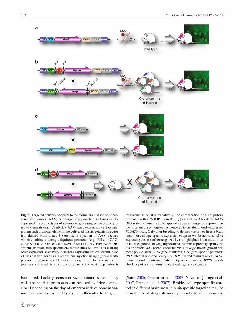
Targeted delivery of optogenetic tools
For most of the targeting, in vivo viral gene transfer hasbeen eVectively used in mice, rats and non human primates,especially adeno-associated viruses (AAV; e.g., Sohal et al.2009; Carter et al. 2010) lentiviruses (e.g., Adamantidiset al. 2007; Han et al. 2009), and to a lesser extent also her-pes simplex virus (HSV; e.g., Covington et al. 2010; Loboet al. 2010). Advantages of viral expression are a straight-forward experimental approach, which takes approximately6 weeks from virus production to extensive long lastingopsin expression (four more weeks for HSV) and a broadapplicableness regarding the subject, ranging from rodents
to primates. Depending on the experimental setup eitherstrong ubiquitous promoters such as elongation factor1-alpha (EF1�), human synapsin, human thymocyte diVer-entiation antigen 1 (Thy-1), a combination of chicken beta-actin promoter and cytomegalovirus immediate-earlyenhancer (CAG) or weaker cell type-speciWc promoters, forexample �-calcium/calmodulin-dependent protein kinase II(CamKII�) can be used (Fig. 2a; Xu et al. 2001; Jakobssonet al. 2003; Dittgen et al. 2004). AAVs are to some extentsuperior to lentiviruses due to the fact that AAV-based vec-tors remain episomally while lentiviral vectors are inte-grated into the host genome. Thus, the expression fromlentiviral vectors is prone to inXuences of the surroundingchromatin and the integration could lead to an undesireddisruption of host genes. General drawbacks are a limitedpacking volume of AAVs and lentiviruses and weak celltype-speciWc transcriptional promoter activity. For exam-ple, the smallest promoter targeting selectively inhibitoryneurons is approximately 3 kilobase pairs (kb) in length,which interferes with the maximal capacity of AAVs andlentiviruses, which is about 5 and 8 kb, respectively(Nathanson et al. 2009; Dong et al. 2010). A limitationoften seen in experiments using cell type-speciWc promot-ers is the need of high irradiance for optimal stimulation oflarger brain areas. As light penetration is low in brain tis-sue, reaching deeper regions optically requires either higherexpression levels of the rhodopsin (e.g., by using a strongexogenous promoter as indicated above) or higher levels oflight energy. As an alternative to moderately active tissue-or cell type-speciWc promoters, strong conditional rhodop-sin expression can be achieved by the use of transgenic crerecombinase driver mice (Fig. 2; Geschwind 2004; Gonget al. 2007). By using a cre-dependent viral construct likeAAV-FlEx (Flip-Excision) or AAV-DIO (double Xoxedinverse open reading frame) driven by a strong promotersuch as EF1� or CAG, cell and tissue speciWcity is providedby selective cre activity (Fig. 2b; Atasoy et al. 2008; Sohalet al. 2009). An alternative to viral injections is the use oftransgenic animals, where rhodopsin expression occursearly in development. Several transgenic mouse lines exist,expressing channelrhodopsin or HR under the panneuronalThy-1 promoter (Arenkiel et al. 2007; Wang et al. 2007;Zhao et al. 2008) or the glutamatergic Vglut2 promoter(Fig. 2c; Hagglund et al. 2010). In analogy to the above-mentioned cre-dependent conditional approach utilizingviral gene delivery systems, a cell type-speciWc expressioncould be achieved by using a strong panneuronal promotersuch as EF1� or CAG in combination with the cre-loxP sys-tem. Breeding to selective cre driver mice will result inexpression of ChR2 or NpHR driven by the panneural pro-moter, whereas the temporal and spatial expression willdepend on the used cre driver line (Fig. 2d). For in vivo celltype-speciWc opsin expression in utero electroporation has
123
102 Mol Genet Genomics (2012) 287:95–109
been used. Lacking construct size limitations even largecell type-speciWc promoters can be used to drive expres-sion. Depending on the day of embryonic development var-ious brain areas and cell types can eYciently be targeted
(Saito 2006; Gradinaru et al. 2007; Navarro-Quiroga et al.2007; Petreanu et al. 2007). Besides cell type-speciWc con-trol in diVerent brain areas, circuit-speciWc targeting may bedesirable to distinguish more precisely between neurons,
Fig. 2 Targeted delivery of opsins to the mouse brain based on adeno-associated viruses (AAV) or transgenic approaches. a Opsins can beexpressed in speciWc types of neurons or glia using gene-speciWc pro-moter elements (e.g., CamKII�). AAV-based expression vectors inte-grating such promoter elements are delivered via stereotactic injectioninto desired brain areas. b Stereotactic injection of AAV vectors,which combine a strong ubiquitous promoter (e.g., Ef1� or CAG)either with a “STOP” cassette (top) or with an AAV-FlEx/AAV-DIOsystem (bottom), into speciWc cre mouse lines will result in a strongopsin expression selectively in neurons expressing the cre recombinase.c Classical transgenesis via pronucleus injection using a gene-speciWcpromoter (top) or targeted knock-in strategies in embryonic stem cells(bottom) will result in a neuron- or glia-speciWc opsin expression in
transgenic mice. d Alternatively, the combination of a ubiquitouspromoter with a “STOP” cassette (top) or with an AAV-FlEx/AAV-DIO system (bottom) can be applied also in a transgenic approach ei-ther in a random or targeted fashion, e.g., to the ubiquitously expressedROSA26 locus. Only after breeding to desired cre driver lines a brainregion- or cell type-speciWc expression of opsins will be activated. Miceexpressing opsins can be recognized by the highlighted brain and an insetin the background showing hippocampal neurons expressing opsin-GFPfusion protein. AAV adeno-associated virus, BGHpA bovine growth hor-mone poly A signal, GOI gene of interest, GSP gene-speciWc promoter,IRES internal ribosomal entry side, ITR inverted terminal repeat, STOPtranscriptional terminator, UBP: ubiquitous promoter, WPRE wood-chuck hepatitis virus posttranscriptional regulatory element
UBP Opsin BGHpA
ITR UBP Opsin WPRE BGHpA ITR
ITR UBP STOP WPRE BGHpA ITR
ITR GSP Opsin WPRE BGHpA ITR
Opsin
AAV
Cre driver lineof interest
a
c
UBP STOP BGHpA
GSP Opsin BGHpA
Opsin
or
b
IRES Opsin BGHpAGOI
or X
d
AAV
Cre deriver lineof interest
loxP
loxP
loxPlox2722
loxPlox2722
or
wild-type
123

Mol Genet Genomics (2012) 287:95–109 103
which, although carrying the same cell-speciWc set ofgenetic markers, are heterogeneous regarding their aVerentor eVerent inXuences. By fusing cre recombinase to axo-nally transported proteins such as wheat germ agglutinin(WGA) and tetanus toxin fragment C (TTC) or using idio-syncratic antero-/retrograde viral axonal delivery, circuitconnectivity can be analyzed by optogenetics in a transneu-ronal fashion. This transcellular approach in combinationwith conditional cre-dependent expression vectors repre-sents a versatile tool for circuit mapping independent of theuse of cre animals, extending the cell type-speciWc optoge-netic approach to animals such as rats and primates, whichhave not been amenable to large-scale genetic manipulation(Gradinaru et al. 2010).
Optogenetics is compatible with a plethora of functional readouts
The major advantage of optogenetics, besides genetic tar-geting, is stimulation with light, as it oVers the possibilityof simultaneous on-site electrical measurement. Dependingon the desired resolution, optogenetics can be combinedwith a variety of readouts. For in vitro proof-of-conceptexperiments the most widely used methods are whole-cellpatch clamp and Weld potential recordings in combinationwith optical illumination in brain slices (for example Nagelet al. 2003; Boyden et al. 2005; Deisseroth et al. 2006;Zhang et al. 2007a, b). To study neural activity beyond sin-gle cells on a network level calcium imaging can be used asa multisite indirect measurement of action potential activityin a full optical approach (Knopfel et al. 2006). The Wrstexample of postsynaptic calcium imaging following light-induced action potentials in ChR2-targeted presynapticaxons was demonstrated by Zhang and Oertner (2007) tostudy plasticity at single-synapse level. By using a spec-trally compatible dye (Fluo-5F) and two-photon excitationat 810 nm in rat hippocampal slice cultures targeted withChR2-tdimer2, synaptic contacts between labeled cellscould be identiWed by imaging optically induced spinal cal-cium release. In addition, long-term potentiation of preva-lent synaptic connections was induced (Zhang and Oertner2007). An all-optical approach was also applied in the ini-tial description of bidirectional optical control of neural cir-cuits. After lentiviral transduction of murine acute corticalslices with CaMKII�::ChR2-mCherry and EF1�::NpHR-EYFP Fura-2 calcium imaging was used to measure theeVects of optical excitation and inhibition (Zhang et al.2007a). Because of its UV-shifted spectral properties (exci-tation maximum at 340 nm) Fura-2 has been successfullyused in concert with ChR2 (excitation maximum at470 nm), NpHR (excitation maximum at 590 nm) and theOptoXRs (excitation maximum at 500 nm; Airan et al.
2009). Yet, although spectrally overlapping, geneticallyencoded calcium sensors such as G-CaMP can be applied inconcert with ChR2. In a technically elegant study Guo et al.(2009) combined in vivo optical stimulation of ChR2 wave-length with simultaneous calcium imaging at 488 nm forfunctional connectivity mapping in C. elegans neurons. Byseparating an epiXuorescent high-power stimulation pathfor ChR2 activation from a low-intensity laser light imag-ing path for G-CaMP Xuorescence, unspeciWc ChR2 activa-tion during imaging at 488 nm could be avoided althoughChR2 has its peak activation at 470 nm (Guo et al. 2009,see also Fig. 1). Laser scanning photostimulation (LSPS)has previously also been used in combination with gluta-mate uncaging and whole-cell recordings to analyze synap-tic connectivity in brain slices (Katz and Dalva 1994;Shepherd and Svoboda 2005; Svoboda and Yasuda 2006).By exchanging glutamate uncaging against light activationthrough targeted axonal photosensitivity via ChR2, thegroup of Svoboda developed ChR2-assisted circuit map-ping (CRACM; Petreanu et al. 2007). Using in utero elec-troporation in mice layer, 2/3 cortical neurons were targetedwith ChR2-venus and photostimulation was performedthroughout a grid covering the entire depth of the cortex.Whole-cell current clamp recordings of Xuorescent ChR2positive cells revealed their depolarization and detectedaction potentials after perisomatic and axonal LSPS. Map-ping postsynaptic targets of L2/3 neurons in several corticallayers in the ipsi- and contralateral hemisphere indicatedlaminar speciWcity of local and long-range cortical projec-tions (Petreanu et al. 2007). A modiWed approach(sCRACM) was used to study the subcellular organizationof excitatory pyramidal neurons. Local glutamate releasewas measured after LSPS in conditions blocking actionpotentials. EPSPs as a measure of light-induced activationwere detected only in the case of overlap between therecorded cell and ChR2-targeted presynaptic axons.Interestingly, subcellular mapping of neural circuitry bysCRACM revealed that the structural connectivity of neu-rons, as indicated by overlap of axons and dendrites, is notalways reXected by their connection on a functional level(Petreanu et al. 2009). To study whole circuit propertiesvoltage-sensitive dyes (VSD) can be applied in brain slicesfor an all-optical approach. As VSDs change their lightemission properties in a voltage-dependent and most nota-bly in a millisecond time scale they are potentially wellsuited to match the high temporal resolution achieved byoptogenetics (Grinvald and Hildesheim 2004). Althoughcompatibility can be achieved with spectrally separatedinfrared dyes such as RH-155 and VSD imaging followingoptical stimulation has been established successfully, it hasrarely been used experimentally up to now, most probablydue to a less favorable signal-to-noise ratio in comparisonto calcium imaging (Airan et al. 2007; Zhang et al. 2010).
123

104 Mol Genet Genomics (2012) 287:95–109
Another example for the study of macrocircuits and globalin vivo mapping is optical stimulation in combination withhigh-resolution fMRI. Lee and colleagues showed in aWrst publication that blood oxygenation level-dependent(BOLD) signals similar to classical stimulus-evoked BOLDfMRI responses could be elicited optically. This eVect wasseen not only locally at the expression site of ChR2 in M1CaMKII� positive excitatory neurons, but also in distinctthalamic neurons deWned by the corticofugal axonal projec-tions (Lee et al. 2010). In a reverse approach optical stimu-lation of ChR2 expressing cortico-thalamic projectionWbers at the level of the thalamus was suYcient to drive notonly local thalamic BOLD signals, but also cortical signals,most probably in an antidromic axosomatic manner. “Opto-fMRI” or “ofMRI” has also been used to identify down-stream targets of the primary sensory cortex (SI). Opticalstimulation of a ChR2-targeted subpopulation of SI pyrami-dal cells evoked local BOLD signals and also in function-ally connected network regions, with diVerences in awakecompared to anesthetized mice (Desai et al. 2011). Eventhough widely hypothesized before, these experiments indi-cate a most likely causal link between local neuronal exci-tation and positive hemodynamic responses, although thegeneration of BOLD signals may have a more complexnature. However, functional mapping using optogenetics incombination with fMRI not only integrates cell type speci-Wcity and anatomical conjunction, but can also be used tofunctionally dissect brain circuitries based on their connec-tion topology (Leopold 2010). For global measurements ofbrain activity in living animals, electroencephalography(EEG) has been combined with optogenetic stimulation. Ina Wrst experiment Adamantidis et al. (2007, 2010) identiWedthe eVect of hypocretin-producing neurons in the lateralhypothalamus on sleep to wake transition by analyzingsleep recordings of mice with chronically implanted EEGelectrodes. In a bidirectional in vivo approach usingeNpHR and ChR2 the same group studied the eVect oflocus coeruleus neurons on wakefulness by applying EEGand in vivo microdialysis as a functional readout (Carteret al. 2010). For in vivo extracellular recording upon tar-geted optical stimulation, a device called “optrode” wascreated fusing a stimulating Wberoptic with a recordingelectrode (Gradinaru et al. 2007, 2009). Constant technicaldevelopment even allows simultaneous recordings at multi-ple sites in the living animal (Zhang et al. 2009; Royer et al.2010). The foundation for using optogenetics in freelymoving rodents was set by Aravanis et al. (2007). Via alightweight, Xexible optical Wber called optical neural inter-face (ONI) excitatory motor cortex neurons could selec-tively be activated in rats and mice as measured by theirwhisker movement. Several successive studies focused onmore complex behavioral paradigms as readouts for opto-genetical manipulation such as motor behavior in animal
models of Parkinson’s disease (Gradinaru et al. 2009;Kravitz et al. 2010), reward motivated behavior and addic-tion (Airan et al. 2009; Tsai et al. 2009; Witten et al. 2010;Stuber et al. 2011), fear conditioning and anxiety (Ciocchiet al. 2010; Johansen et al. 2010; Tye et al. 2011) and othercerebral targets such as prefrontal cortex or hypothalamusfor the study of psychiatric diseases and aggressive behav-ior (Covington et al. 2010; Lin et al. 2011; Yizhar et al.2011a, b).
From microbes to model organisms and beyond
Optogenetics is applicable to diverse species and animalmodels. For the initial functional characterization of ChR1and ChR2 a host model was applied that had previouslybeen successfully used for diVerent rhodopsins. Expressionof ChR1 and ChR2 in oocytes from Xenopus laevis in thepresence of all-trans retinal was the Wrst example of func-tional heterologous channelrhodopsin expression outside ofalgae (Nagel et al. 2002, 2003). At that moment the equa-tion was: CHOP 1 or 2 + retinal = ChR1 or ChR2, as thephotoreceptor system from Chlamydomonas reinhardtiiwas thought to be composed of channelopsin (from acDNA originally named CHOP1 and CHOP2 but alsotermed Chlamyopsin, Cop3 or Cop4) and all-trans retinal(Nagel et al. 2003). Deisseroth and colleagues redid themath and prove that “CHOP2” successfully worked in rathippocampal primary neurons after lentiviral transductionwithout adding all-trans retinal at all (although the culturemedia contained little amounts of the precursor retinyl ace-tate). By this, ChR2 was established as a new one-compo-nent functional tool for neuroscience in mammals neitherrequiring the substitution of any synthetic chemical sub-strate nor genetic orthogonality between the rhodopsin andthe targeted host organism, even more important as there isno mammalian analog of the algal gene CHOP2 (Boydenet al. 2005). The Wrst in vivo applications in living animalswere put into practice in parallel by Li et al. (2005) andNagel et al. (2005). Transgenic expression of ChR2 speciW-cally in cholinergic motorneurons of C. elegans allowed toelicit behavioral responses via muscular contractions byblue light illumination (Nagel et al. 2005). Bidirectional,both inhibitory and excitatory in vivo control of spontane-ous chick spinal cord activity was performed in chickenembryos after in ovo electroporating and transient expres-sion of ChR2 and vertebrate Ro4, respectively (Li et al.2005). Another example of bidirectional in vivo control ofC. elegans locomotion was achieved after simultaneousexpression of ChR2 and NpHR using the muscle-speciWcmyosin promoter Pmyo-3 (Zhang et al. 2007a). As men-tioned above C. elegans has also been used for an in vivoall-optical interrogation of neural circuits using ChR2 and
123

Mol Genet Genomics (2012) 287:95–109 105
G-CaMP (Guo et al. 2009). The Wrst use of optogenetics inzebraWsh revealed that activation of zebraWsh trigeminalneurons by ChR2 induces escape behavior even upon singlespikes, as published by Douglass et al. (2008). By usingGal4 enhancer trapping in zebraWsh, transgenic animals canbe generated in a feasible way (Scott et al. 2007). TheBayer lab eYciently used the GAL4/UAS system to selec-tively express ChR2 and NpHR in speciWc neuronal sub-populations of zebraWsh larvae. Owing to the transparentnature of the animal in vivo functional mapping at a singleneuron resolution was performed dissecting neuronal popu-lations involved in swim behavior, saccade generation aswell cardiac development and function (Arrenberg et al.2009, 2010; Schoonheim et al. 2010). GAL4/UAS was alsoused to introduce ChR2 into speciWc subsets of neuronalpopulations (painless) in Drosophila melanogaster bycrossing transgenic UAS-ChR2 Xies with neuronspeciWcGal4 transgenic lines (Zhang et al. 2007c). Optical stimula-tion of nociceptive neurons in transgenic larvae by ChR2induced behavioral responses. However, it has to be takeninto account that D. melanogaster and zebraWsh containhydroxyretinal which does not enter the ChR or HR bindingsites making a substitution of all-trans retinal necessary(for example by food supplement). Moreover, interpreta-tion of behavioral data in D. melanogaster can be conXict-ing as the animal has an innate behavioral light response(Pulver et al. 2009). The species most widely investigatedwith optogenetic technologies is the mouse. This is largelyowing to the vast availability of transgenic cre driver lines,which can readily be combined with cre-dependent viralvectors in order to facilitate strong cell-/tissue-speciWc rho-dopsin expression (Fig. 2). As mentioned above, in micealso classical transgenic approaches have been applied toexpress ChR2 and NpHR under the control of the Thy-1promoter. Arenkiel et al. (2007) showed strong and func-tional, regionally restricted, channelrhodopsin expressionin Thy1::ChR2-EYFP mouse strains. By presynaptic opti-cal manipulation of targeted cells in the bulbar-cortical cir-cuit of the olfactory system they highlighted the potential ofoptogenetics for complex circuit analysis in brain slices andin vivo. Wang et al. (2007) used transgenic Thy1-ChR2-YFP mice for the mapping of functional connectivity in theneural circuitry of cortical layer VI neurons. In order tostudy the role of fast spiking parvalbumin interneurons inthe generation of gamma oscillations, Thy1::ChR2-EYFPmice were analyzed in combination with a cre-dependentapproach (Sohal et al. 2009). This animal model in combi-nation with a cell type-speciWc lentiviral approach was alsosuccessfully used in the systematic dissection of circuitsand deciphering of potential targets related to deep brainstimulation in a Parkinson mouse model (Gradinaru et al.2009). Hagglund and colleagues used BAC transgenesis toexpress ChR2-EYFP under the control of the vesicular glu-
tamate receptor two promoter. These transgenic miceexpress ChR2 in glutamatergic neurons of the hindbrainand spinal cord. By optical stimulation of the lumbar regionof the spinal cord and also the caudal hindbrain rhythmgeneration was observed, suggesting a role of glutamatergicneurons in central pattern generators of locomotion (Hagglundet al. 2010). Various examples in nematodes, mice and ratshave demonstrated a direct eVect of in vivo optogeneticstimulation on neuronal activity and behavioral responses(Nagel et al. 2005; Gradinaru et al. 2009; Tsai et al. 2009;Lee et al. 2010). Yet optical control of cortical circuits inhigher organisms might be more complex: the Wrst optoge-netic approach in primates was performed in the rhesusmacaque. Boyden and colleagues targeted speciWcallyexcitatory neurons of the frontal cortex by stereotactic len-tiviral gene delivery of ChR2-GFP driven by the CamKII�promoter and recorded neuronal activity after simultaneousoptical stimulation via an optrode (Han et al. 2009).Besides showing the feasibility and safety of optogeneticsin primates and thereby implementing a potential clinicaltranslation for the use in humans, the experiments showed aslightly discouraging, but seminal, result. Although ChR2was targeted speciWcally to excitatory neurons a signiWcantproportion of recorded cells showed decreased activity.This Wnding was previously also observed in transgenicThy1-ChR2 mice and is comparable to heterogeneous tis-sue activation in the case of electrical stimulation. Thus,targeting cortical circuits in a cell type-speciWc manner can-not precisely predict a speciWc psychological response,respectively, behavioral or clinical outcome. The impact ofoptogenetic manipulation on cortical circuitries seems to beheavily dependent on the neural network, in which the tar-get cells are embedded. Nevertheless, the results indicateimportant aspects for therapeutic options and suggest len-tiviral rhodopsin delivery as a well immuno-toleratedmethod for a putative application in humans.
Potential clinical applications of optoprosthetics
Recent publications underpin the possible role of optoge-netics as a potential future therapeutic application. Forexample, electrical peripheral nerve stimulation has beenused as an experimental treatment of patients with paralysisand muscle disease. A major drawback is random or reversemuscle recruitment by heterogeneous tissue excitation,resulting in high fatigability. Thy-1::ChR2YFP transgenicmice express ChR2-YFP in both the central and peripheralnervous system as well as in lower motor and dorsal rootganglion neurons. By an optical approach motor units canbe recruited in an orderly manner favoring small, slowmuscle Wbers in comparison to random recruitment by elec-trical stimulation. This results in a markedly enhanced
123

106 Mol Genet Genomics (2012) 287:95–109
functional performance as muscle fatigability is stronglyreduced by a more physiological recruitment pattern viaoptical stimulation. Taking into account recent advances inhuman gene therapy the authors point out an eventual thera-peutic use of optogenetics as neuroprosthetics and suggestNpHR in the treatment of spastic movement disorders(Llewellyn et al. 2010). Another promising example ofoptical control replacing electrical stimulation is light-inducedstimulation of the heart muscle. Bruegmann et al. (2010)transferred the optogenetic principle from neurons tocardiomyocytes. A ChR2-EYFP construct driven by theCAG promoter was electroporated into embryonic stemcells (ESCs). After ESC diVerentiation into cardiomyocytesChR2 expression was limited to the cell membrane andpulsed in vitro optical stimulation triggered action poten-tial-induced contractions with intercellular electrical spreading.The authors also generated transgenic CAG-ChR2-EYFPmice that showed robust rhodopsin expression in atrial andventricular cardiomyoctes. By illuminating an area of only0.05–0.2 mm with pulses as short as 1 ms, reliable actionpotentials were generated in a minimally delayed 1:1 man-ner in vitro and in vivo, allowing fast optical pacing limitedonly by the natural refractoriness of the cardiac cells. Thus,optogenetics could serve as an analytical tool for the studyof pacemaking and arrhythmia in cardiology with a highpotential for therapeutic use. There are several putativeclinical applications of optogenetics also in neurodegenera-tive disorders. A recent work from Stroh et al. (2011)focuses on a restorative approach. Mouse ESCs were trans-duced in vitro via lentiviruses to express ChR2-YFP underthe EF1� promoter. After a retinoic acid-based diVerentia-tion protocol, targeted cells were viable and electrophysio-logically mature, similar to native cells. After FACS sortingChR2-YFP expressing cells were transplanted into ratmotor cortex and reacted toward blue light illuminationafter integration into the host environment. Depolarizationand direct Ca2+ inXux via light-gated membrane proteinssuch as ChR2 could serve as a new tool for stem cell diVer-entiation into neural and neuronal cells in vitro and in vivo,as various facts indicate an important role of Ca2+ dependentcellular processes driving diVerentiation (e.g., D’Ascenzoet al. (2006). As functional integration of the transplant isthe major goal of regenerative medicine, optogeneticallyinduced diVerentiation, compared to unspeciWc chemicallyor electrically based protocols, just aVects the geneticallytargeted graft cells within a heterogeneous cellular hostenvironment, reducing undesirable side eVects such as celldeath or tumor growth (Stroh et al. 2011). In a previousstudy neurons derived from human ESCs were transducedwith ChR2-mCherry and mature neurons were analyzed forsynaptic integration and functional connectivity of thetargeted graft cells into the host circuitry of immuno-depressed neonatal SCID mice (Weick et al. 2010). In vitro
postmitotic neurons, both glutamatergic and GABAergic,showed typical electrophysiological responses upon illumi-nation. When transplanted, ChR2 positive matured neuronsnot only displayed spontaneous action potentials and post-synaptic currents (PSCs) as a proof of input-speciWc graftintegration, but furthermore, adjacent ChR2-negative cellslikewise displayed PSCs upon light stimulation, indicatinggraft to host microcircuit connectivity also in an output-speciWc manner. By generating a pluripotent human ESCline, expressing ChR2-mCherry constitutively under thesynapsin promoter, the authors could improve severaldrawbacks of viral based approaches such as low transduc-tion eYciency. Yet further research is necessary to establishoptogenetics as a standard examination and manipulationtool of functional graft to host integration and circuit plas-ticity in the context of stem cell based neuroregenerativestrategies. Another example for a clinical application couldbe vision restoration in human subjects with retinal degen-eration. ChR2- and NpHR-based approaches would, due totheir simple monocomponent working principle with goodimmune compatibility, cell type speciWcity and high spatialresolution, compete with electrical stimulation in their abil-ity of restoring photosensitivity. In blind mice lacking reti-nal photoreceptors (rd1 or Pde6brd1 mouse model, Boweset al. 1990) photosensitivity was restored by heterologousexpression of ChR2, either by transducing inner retinal neu-rons in a non-selective viral based strategy or by in vivoelectroporating exclusively ON bipolar retinal cells using aspeciWc promoter sequence (Bi et al. 2006, Lagali et al.2008). Positive changes in a behavioral readout indicatedimproved vision and suggest the use of optogenetics in atranslational approach also in humans. A major step towardapplications in vivo, especially for therapeutic use inhumans, would be the employment of highly light-sensitiverhodopsins with speciWcally deWned response kinetics sup-porting optical control at a cell type-speciWc resolution. Ingeneral optogenetics would signiWcantly beneWt fromChRs with larger conductance and higher selectivity forCa2+, Na+ or K+ in combination with appropriate color andkinetics.
Conclusions
Optogenetics has matured within a few years into a state-of-the-art technology. This technological breakthroughallows to tackle biological questions, which were previ-ously out of reach of our imaginations. The major impact ofoptogenetics in the upcoming years will most likely bedominated by approaches fundamentally increasing ourcomprehension of neural, as well as non-neural systems, ona circuit level. A deeper understanding of these intricateregulatory networks might unravel novel targets within
123
Mol Genet Genomics (2012) 287:95–109 107
such complex circuits, which could be suitable for thera-peutic intervention. Optogenetics has demonstrated itsenormous potential in animal models including nonhumanprimates and thereby has raised considerable expectationswith respect to future clinical applications. However,whether optogenetics will cross the Rubicon and will beamenable to application in human patients will primarilydepend on similar obstacles that gene therapy has beenstruggling with all along, namely eYcient and safe genedelivery strategies.
Open Access This article is distributed under the terms of the Crea-tive Commons Attribution Noncommercial License which permits anynoncommercial use, distribution, and reproduction in any medium,provided the original author(s) and source are credited.
References
Adamantidis AR, Zhang F, Aravanis AM, Deisseroth K, de Lecea L(2007) Neural substrates of awakening probed with optogeneticcontrol of hypocretin neurons. Nature 450:420–424
Adamantidis AR, Carter MC, de Lecea L (2010) Optogenetic deconstruc-tion of sleep-wake circuitry in the brain. Front Mol Neurosci 2:31
Airan RD, Hu ES, Vijaykumar R, Roy M, Meltzer LA, Deisseroth K(2007) Integration of light-controlled neuronal Wring and fast cir-cuit imaging. Curr Opin Neurobiol 17:587–592
Airan RD, Thompson KR, Fenno LE, Bernstein H, Deisseroth K(2009) Temporally precise in vivo control of intracellular signal-ling. Nature 458:1025–1029
Aravanis AM, Wang LP, Zhang F, Meltzer LA, Mogri MZ, SchneiderMB, Deisseroth K (2007) An optical neural interface: in vivo con-trol of rodent motor cortex with integrated Wberoptic and optoge-netic technology. J Neural Eng 4:S143–S156
Arenkiel BR, Peca J, Davison IG, Feliciano C, Deisseroth K, Augus-tine GJ, Ehlers MD, Feng G (2007) In vivo light-induced activa-tion of neural circuitry in transgenic mice expressingchannelrhodopsin-2. Neuron 54:205–218
Arrenberg AB, Del BF, Baier H (2009) Optical control of zebraWshbehavior with halorhodopsin. Proc Natl Acad Sci USA106:17968–17973
Arrenberg AB, Stainier DY, Baier H, Huisken J (2010) Optogeneticcontrol of cardiac function. Science 330:971–974
Atasoy D, Aponte Y, Su HH, Sternson SM (2008) A FLEX switch tar-gets Channelrhodopsin-2 to multiple cell types for imaging andlong-range circuit mapping. J Neurosci 28:7025–7030
Bamann C, Gueta R, Kleinlogel S, Nagel G, Bamberg E (2010) Struc-tural guidance of the photocycle of channelrhodopsin-2 by an in-terhelical hydrogen bond. Biochemistry 49:267–278
Beja O, Aravind L, Koonin EV, Suzuki MT, Hadd A, Nguyen LP,Jovanovich SB, Gates CM, Feldman RA, Spudich JL, SpudichEN, DeLong EF (2000) Bacterial rhodopsin: evidence for a newtype of phototrophy in the sea. Science 289:1902–1906
Berndt A, Yizhar O, Gunaydin LA, Hegemann P, Deisseroth K (2008)Bi-stable neural state switches. Nat Neurosci 12:229–234
Berndt A, Schoenenberger P, Mattis J, Tye KM, Deisseroth K, Hege-mann P, Oertner TG (2011) High-eYciency channelrhodopsinsfor fast neuronal stimulation at low light levels. Proc Natl AcadSci USA 108:7595–7600
Bi A, Cui J, Ma YP, Olshevskaya E, Pu M, Dizhoor AM, Pan ZH(2006) Ectopic expression of a microbial-type rhodopsin restoresvisual responses in mice with photoreceptor degeneration. Neu-ron 50:23–33
Bowes C, Li T, Danciger M, Baxter LC, Applebury ML, Farber DB(1990) Retinal degeneration in the rd mouse is caused by a defectin the beta subunit of rod cGMP-phosphodiesterase. Nature347:677–680
Boyden ES, Zhang F, Bamberg E, Nagel G, Deisseroth K (2005)Millisecond-timescale, genetically targeted optical control ofneural activity. Nat Neurosci 8:1263–1268
Bruegmann T, Malan D, Hesse M, Beiert T, Fuegemann CJ, Fleisch-mann BK, Sasse P (2010) Optogenetic control of heart muscle invitro and in vivo. Nat Methods 7:897–900
Carter ME, Yizhar O, Chikahisa S, Nguyen H, Adamantidis A, NishinoS, Deisseroth K, de Lecea L (2010) Tuning arousal with optoge-netic modulation of locus coeruleus neurons. Nat Neurosci13:1526–1533
Chow BY, Han X, Dobry AS, Qian X, Chuong AS, Li M, HenningerMA, Belfort GM, Lin Y, Monahan PE, Boyden ES (2010) High-performance genetically targetable optical neural silencing bylight-driven proton pumps. Nature 463:98–102
Ciocchi S, Herry C, Grenier F, WolV SB, Letzkus JJ, Vlachos I,Ehrlich I, Sprengel R, Deisseroth K, Stadler MB, Muller C, LuthiA (2010) Encoding of conditioned fear in central amygdala inhib-itory circuits. Nature 468:277–282
Covington HE III, Lobo MK, Maze I, Vialou V, Hyman JM, Zaman S,LaPlant Q, Mouzon E, Ghose S, Tamminga CA, Neve RL, Deis-seroth K, Nestler EJ (2010) Antidepressant eVect of optogeneticstimulation of the medial prefrontal cortex. J Neurosci 30:16082–16090
D’Ascenzo M, Piacentini R, Casalbore P, Budoni M, Pallini R, AzzenaGB, Grassi C (2006) Role of L-type Ca2+ channels in neuralstem/progenitor cell diVerentiation. Eur J Neurosci 23:935–944
Deisseroth K, Feng G, Majewska AK, Miesenbock G, Ting A, SchnitzerMJ (2006) Next-generation optical technologies for illuminatinggenetically targeted brain circuits. J Neurosci 26:10380–10386
Desai M, Kahn I, Knoblich U, Bernstein J, Atallah H, Yang A, KopellN, Buckner RL, Graybiel AM, Moore CI, Boyden ES (2011)Mapping brain networks in awake mice using combined opticalneural control and fMRI. J Neurophysiol 105:1393–1405
Dittgen T, Nimmerjahn A, Komai S, Licznerski P, Waters J, MargrieTW, Helmchen F, Denk W, Brecht M, Osten P (2004) Lentivirus-based genetic manipulations of cortical neurons and their opticaland electrophysiological monitoring in vivo. Proc Natl Acad SciUSA 101:18206–18211
Dong B, Nakai H, Xiao W (2010) Characterization of genome integrityfor oversized recombinant AAV vector. Mol Ther 18:87–92
Douglass AD, Kraves S, Deisseroth K, Schier AF, Engert F (2008) Escapebehavior elicited by single, channelrhodopsin-2-evoked spikesin zebraWsh somatosensory neurons. Curr Biol 18:1133–1137
Geschwind D (2004) GENSAT: a genomic resource for neuroscienceresearch. Lancet Neurol 3:82
Gong S, Doughty M, Harbaugh CR, Cummins A, Hatten ME, HeintzN, Gerfen CR (2007) Targeting Cre recombinase to speciWc neu-ron populations with bacterial artiWcial chromosome constructs.J Neurosci 27:9817–9823
Govorunova EG, Spudich EN, Lane CE, Sineshchekov OA, SpudichJL (2011) New channelrhodopsin with a red-shifted spectrum andrapid kinetics from Mesostigma viride. MBio 2(3):e00115-11
Gradinaru V, Thompson KR, Zhang F, Mogri M, Kay K, SchneiderMB, Deisseroth K (2007) Targeting and readout strategies for fastoptical neural control in vitro and in vivo. J Neurosci 27:14231–14238
Gradinaru V, Thompson KR, Deisseroth K (2008) eNpHR: a Natrono-monas halorhodopsin enhanced for optogenetic applications.Brain Cell Biol 36:129–139
Gradinaru V, Mogri M, Thompson KR, Henderson JM, Deisseroth K(2009) Optical deconstruction of parkinsonian neural circuitry.Science 324:354–359
123

108 Mol Genet Genomics (2012) 287:95–109
Gradinaru V, Zhang F, Ramakrishnan C, Mattis J, Prakash R, DiesterI, Goshen I, Thompson KR, Deisseroth K (2010) Molecular andcellular approaches for diversifying and extending optogenetics.Cell 141:154–165
Grinvald A, Hildesheim R (2004) VSDI: a new era in functional imag-ing of cortical dynamics. Nat Rev Neurosci 5:874–885
Gunaydin LA, Yizhar O, Berndt A, Sohal VS, Deisseroth K, Hege-mann P (2010) Ultrafast optogenetic control. Nat Neurosci13:387–392
Guo ZV, Hart AC, Ramanathan S (2009) Optical interrogation of neu-ral circuits in Caenorhabditis elegans. Nat Methods 6:891–896
Hagglund M, Borgius L, Dougherty KJ, Kiehn O (2010) Activation ofgroups of excitatory neurons in the mammalian spinal cord orhindbrain evokes locomotion. Nat Neurosci 13:246–252
Han X, Boyden ES (2007) Multiple-color optical activation, silencing,and desynchronization of neural activity, with single-spike tem-poral resolution. PLoS One 2:e299
Han X, Qian X, Bernstein JG, Zhou HH, Franzesi GT, Stern P, Bron-son RT, Graybiel AM, Desimone R, Boyden ES (2009) Millisec-ond-timescale optical control of neural dynamics in thenonhuman primate brain. Neuron 62:191–198
Han X, Chow BY, Zhou H, Klapoetke N, Boyden E et al (2011) Ahigh-light sensitivity optical neural silencer: development andapplication to optogenetic control of nun-human primate cortex.Front Sys Neurosci 5:1–8
Harz H, Hegemann P (1991) Rhodopsin-regulated calcium currents inChlamydomonas. Nature 351:489–491
Holland EM, Braun FJ, Nonnengässer C, Harz H, Hegemann P (1996)The nature of rhodopsin-triggered photocurrents in Chlamydo-monas. I. Kinetics and inXuence of divalent ions. Biophys J70:924–931
Ishizuka T, Kakuda M, Araki R, Yawo H (2006) Kinetic evaluation ofphotosensitivity in genetically engineered neurons expressinggreen algae light-gated channels. Neurosci Res 54:85–94
Jakobsson J, Ericson C, Rosenqvist N, Lundberg C (2003) Lentiviralvectors. Int Rev Neurobiol 55:111–122
Johansen JP, Hamanaka H, MonWls MH, Behnia R, Deisseroth K, BlairHT, LeDoux JE (2010) Optical activation of lateral amygdalapyramidal cells instructs associative fear learning. Proc Natl AcadSci USA 107:12692–12697
Katz LC, Dalva MB (1994) Scanning laser photostimulation: a new ap-proach for analyzing brain circuits. J Neurosci Methods 54:205–218
Kleinlogel S, Feldbauer K, Dempski RE, Fotis H, Wood PG, BamannC, Bamberg E (2011) Ultra light-sensitive and fast neuronal acti-vation with the Ca(2)+-permeable channelrhodopsin CatCh. NatNeurosci 14:513–518
Knopfel T, ez-Garcia J, Akemann W (2006) Optical probing of neuro-nal circuit dynamics: genetically encoded versus classical Xuores-cent sensors. Trends Neurosci 29:160–166
Kravitz AV, Freeze BS, Parker PR, Kay K, Thwin MT, Deisseroth K,Kreitzer AC (2010) Regulation of parkinsonian motor behavioursby optogenetic control of basal ganglia circuitry. Nature 466:622–626
Lagali PS, Balya D, Awatramani GB, Munch TA, Kim DS, BusskampV, Cepko CL, Roska B (2008) Light-activated channels targetedto ON bipolar cells restore visual function in retinal degeneration.Nat Neurosci 11:667–675
Lee JH, Durand R, Gradinaru V, Zhang F, Goshen I, Kim DS, FennoLE, Ramakrishnan C, Deisseroth K (2010) Global and local fMRIsignals driven by neurons deWned optogenetically by type andwiring. Nature 465:788–792
Leopold DA (2010) Neuroscience: fMRI under the spotlight. Nature465:700–701
Li X, Gutierrez DV, Hanson MG, Han J, Mark MD, Chiel H,Hegemann P, Landmesser LT, Herlitze S (2005) Fast noninvasive
activation and inhibition of neural and network activity by verte-brate rhodopsin and green algae channelrhodopsin. Proc NatlAcad Sci USA 102:17816–17821
Lin JY, Lin MZ, Steinbach P, Tsien RY (2009) Characterization ofengineered channelrhodopsin variants with improved propertiesand kinetics. Biophys J 96:1803–1814
Lin D, Boyle MP, Dollar P, Lee H, Lein ES, Perona P, Anderson DJ(2011) Functional identiWcation of an aggression locus in themouse hypothalamus. Nature 470:221–226
Llewellyn ME, Thompson KR, Deisseroth K, Delp SL (2010) Orderlyrecruitment of motor units under optical control in vivo. Nat Med16:1161–1165
Lobo MK, Covington HE III, Chaudhury D, Friedman AK, Sun H,mez-Werno D, Dietz DM, Zaman S, Koo JW, Kennedy PJ, Mou-zon E, Mogri M, Neve RL, Deisseroth K, Han MH, Nestler EJ(2010) Cell type-speciWc loss of BDNF signaling mimics optoge-netic control of cocaine reward. Science 330:385–390
Matsuno-Yagi A, Mukohata Y (1977) Two possible roles of bacterio-rhodopsin; a comparative study of strains of Halobacterium hal-obium diVering in pigmentation. Biochem Biophys Res Commun78:237–243
Nagel G, Ollig D, Fuhrmann M, Kateriya S, Musti AM, Bamberg E,Hegemann P (2002) Channelrhodopsin-1: a light-gated protonchannel in green algae. Science 296:2395–2398
Nagel G, Szellas T, Huhn W, Kateriya S, Adeishvili N, Berthold P, Ol-lig D, Hegemann P, Bamberg E (2003) Channelrhodopsin-2, a di-rectly light-gated cation-selective membrane channel. Proc NatlAcad Sci USA 100:13940–13945
Nagel G, Brauner M, Liewald JF, Adeishvili N, Bamberg E, Gotts-chalk A (2005) Light activation of channelrhodopsin-2 in excit-able cells of Caenorhabditis elegans triggers rapid behavioralresponses. Curr Biol 15:2279–2284
Nathanson JL, Jappelli R, ScheeV ED, Manning G, Obata K, BrennerS, Callaway EM (2009) Short promoters in viral vectors driveselective expression in mammalian inhibitory neurons, but do notrestrict activity to speciWc inhibitory cell-types. Front Neural Cir-cuits 3:19
Navarro-Quiroga I, Chittajallu R, Gallo V, Haydar TF (2007) Long-term, selective gene expression in developing and adult hippo-campal pyramidal neurons using focal in utero electroporation.J Neurosci 27:5007–5011
Oesterhelt D, Stoeckenius W (1971) Rhodopsin-like protein from thepurple membrane of Halobacterium halobium. Nat New Biol233:149–152
Oesterhelt D, Stoeckenius W (1973) Functions of a new photoreceptormembrane. Proc Natl Acad Sci USA 70:2853–2857
Oh E, Maejima T, Liu C, Deneris E, Herlitze S (2010) Substitution of5-HT1A receptor signaling by a light-activated G protein-coupledreceptor. J Biol Chem 285:30825–30836
Petreanu L, Huber D, Sobczyk A, Svoboda K (2007) Channelrhodop-sin-2-assisted circuit mapping of long-range callosal projections.Nat Neurosci 10:663–668
Petreanu L, Mao T, Sternson SM, Svoboda K (2009) The subcellularorganization of neocortical excitatory connections. Nature 457:1142–1145
Pulver SR, Pashkovski SL, Hornstein NJ, Garrity PA, GriYth LC(2009) Temporal dynamics of neuronal activation by Channelrho-dopsin-2 and TRPA1 determine behavioral output in Drosophilalarvae. J Neurophysiol 101:3075–3088
Racker E, Stoeckenius W (1974) Reconstitution of purple membranevesicles catalyzing light-driven proton uptake and adenosine tri-phosphate formation. J Biol Chem 249:662–663
Royer S, Zemelman BV, Barbic M, Losonczy A, Buzsaki G, Magee JC(2010) Multi-array silicon probes with integrated optical Wbers:light-assisted perturbation and recording of local neural circuits inthe behaving animal. Eur J Neurosci 31:2279–2291
123

Mol Genet Genomics (2012) 287:95–109 109
Saito T (2006) In vivo electroporation in the embryonic mouse centralnervous system. Nat Protoc 1:1552–1558
Schobert B, Lanyi JK (1982) Halorhodopsin is a light-driven chloridepump. J Biol Chem 257:10306–10313
Schoonheim PJ, Arrenberg AB, Del BF, Baier H (2010) Optogeneticlocalization and genetic perturbation of saccade-generating neu-rons in zebraWsh. J Neurosci 30:7111–7120
Schultheis C, Liewald JF, Bamberg E, Nagel G, Gottschalk A (2011)Optogenetic long-term manipulation of behavior and animaldevelopment. PLoS One 6:e18766
Scott EK, Mason L, Arrenberg AB, Ziv L, Gosse NJ, Xiao T, Chi NC,Asakawa K, Kawakami K, Baier H (2007) Targeting neural cir-cuitry in zebraWsh using GAL4 enhancer trapping. Nat Methods4:323–326
Shepherd GM, Svoboda K (2005) Laminar and columnar organizationof ascending excitatory projections to layer 2/3 pyramidal neu-rons in rat barrel cortex. J Neurosci 25:5670–5679
Sohal VS, Zhang F, Yizhar O, Deisseroth K (2009) Parvalbumin neu-rons and gamma rhythms enhance cortical circuit performance.Nature 459:698–702
Stierl M, Stumpf P, Udwari D, Gueta R, Hagedorn R, Losi A, GartnerW, Petereit L, Efetova M, Schwarzel M, Oertner TG, Nagel G,Hegemann P (2011) Light modulation of cellular cAMP by asmall bacterial photoactivated adenylyl cyclase, bPAC, of the soilbacterium Beggiatoa. J Biol Chem 286:1181–1188
Stroh A, Tsai HC, Ping WL, Zhang F, Kressel J, Aravanis A, Santha-nam N, Deisseroth K, Konnerth A, Schneider MB (2011) Track-ing stem cell diVerentiation in the setting of automatedoptogenetic stimulation. Stem Cells 29:78–88
Stuber GD, Sparta DR, Stamatakis AM, van Leeuwen WA, Hard-joprajitno JE, Cho S, Tye KM, Kempadoo KA, Zhang F, Deisse-roth K, Bonci A (2011) Excitatory transmission from theamygdala to nucleus accumbens facilitates reward seeking. Na-ture 475:377–380
Svoboda K, Yasuda R (2006) Principles of two-photon excitation micros-copy and its applications to neuroscience. Neuron 50:823–839
Tsai HC, Zhang F, Adamantidis A, Stuber GD, Bonci A, de Lecea L,Deisseroth K (2009) Phasic Wring in dopaminergic neurons issuYcient for behavioral conditioning. Science 324:1080–1084
Tsunoda SP, Hegemann P (2009) Glu 87 of channelrhodopsin-1 causespH-dependent color tuning and fast photocurrent inactivation.Photochem Photobiol 85:564–569
Tye KM, Prakash R, Kim SY, Fenno LE, Grosenick L, Zarabi H,Thompson KR, Gradinaru V, Ramakrishnan C, Deisseroth K(2011) Amygdala circuitry mediating reversible and bidirectionalcontrol of anxiety. Nature 471:358–362
Wang H, Peca J, Matsuzaki M, Matsuzaki K, Noguchi J, Qiu L, WangD, Zhang F, Boyden E, Deisseroth K, Kasai H, Hall WC, Feng G,Augustine GJ (2007) High-speed mapping of synaptic connectiv-ity using photostimulation in channelrhodopsin-2 transgenicmice. Proc Natl Acad Sci USA 104:8143–8148
Wang H, Sugiyama Y, Hikima T, Sugano E, Tomita H, Takahashi T,Ishizuka T, Yawo H (2009) Molecular determinants diVerentiat-
ing photocurrent properties of two channelrhodopsins from chla-mydomonas. J Biol Chem 284:5685–5696
Weick JP, Johnson MA, Skroch SP, Williams JC, Deisseroth K, ZhangSC (2010) Functional control of transplantable human ESC-de-rived neurons via optogenetic targeting. Stem Cells 28:2008–2016
Wen L, Wang H, Tanimoto S, Egawa R, Matsuzaka Y, Mushiake H,Ishizuka T, Yawo H (2010) Opto-current-clamp actuation of cor-tical neurons using a strategically designed channelrhodopsin.PLoS One 5:e12893
Witten IB, Lin SC, Brodsky M, Prakash R, Diester I, Anikeeva P, Gra-dinaru V, Ramakrishnan C, Deisseroth K (2010) Cholinergicinterneurons control local circuit activity and cocaine condition-ing. Science 330:1677–1681
Xu ZL, Mizuguchi H, Ishii-Watabe A, Uchida E, Mayumi T, Hayaka-wa T (2001) Optimization of transcriptional regulatory elementsfor constructing plasmid vectors. Gene 272:149–156
Yizhar O, Fenno LE, Davidson TJ, Mogri M, Deisseroth K (2011a)Optogenetics in neural systems. Neuron 71:9–34
Yizhar O, Fenno LE, Prigge M, Schneider F, Davidson TJ, O’Shea DJ,Sohal VS, Goshen I, Finkelstein J, Paz JT, Stehfest K, Fudim R,Ramakrishnan C, Huguenard JR, Hegemann P, Deisseroth K(2011b) Neocortical excitation/inhibition balance in informationprocessing and social dysfunction. Nature 477(7363):171–178
Zhang YP, Oertner TG (2007) Optical induction of synaptic plasticityusing a light-sensitive channel. Nat Methods 4:139–141
Zhang F, Wang LP, Brauner M, Liewald JF, Kay K, Watzke N, WoodPG, Bamberg E, Nagel G, Gottschalk A, Deisseroth K (2007a)Multimodal fast optical interrogation of neural circuitry. Nature446:633–639
Zhang F, Aravanis AM, Adamantidis A, de Lecea L, Deisseroth K(2007b) Circuit-breakers: optical technologies for probing neuralsignals and systems. Nat Rev Neurosci 8:577–581
Zhang W, Ge W, Wang Z (2007c) A toolbox for light control of Dro-sophila behaviors through channelrhodopsin 2-mediated photoac-tivation of targeted neurons. Eur J Neurosci 26:2405–2416
Zhang F, Prigge M, Beyriere F, Tsunoda SP, Mattis J, Yizhar O, He-gemann P, Deisseroth K (2008) Red-shifted optogenetic excita-tion: a tool for fast neural control derived from Volvox carteri. NatNeurosci 11:631–633
Zhang J, Laiwalla F, Kim JA, Urabe H, Van WR, Song YK, ConnorsBW, Zhang F, Deisseroth K, Nurmikko AV (2009) Integrated de-vice for optical stimulation and spatiotemporal electrical record-ing of neural activity in light-sensitized brain tissue. J Neural Eng6:055007
Zhang F, Gradinaru V, Adamantidis AR, Durand R, Airan RD, de Le-cea L, Deisseroth K (2010) Optogenetic interrogation of neuralcircuits: technology for probing mammalian brain structures. NatProtoc 5:439–456
Zhao S, Cunha C, Zhang F, Liu Q, Gloss B, Deisseroth K, AugustineGJ, Feng G (2008) Improved expression of halorhodopsin forlight-induced silencing of neuronal activity. Brain Cell Biol36:141–154
123