Embed Size (px)
Citation preview

• The Orbital Region
The orbits are a pair of bony cavities that contain the eyeballs; their associated muscles, nerves, vessels, and fat; and most of the lacrimal apparatus
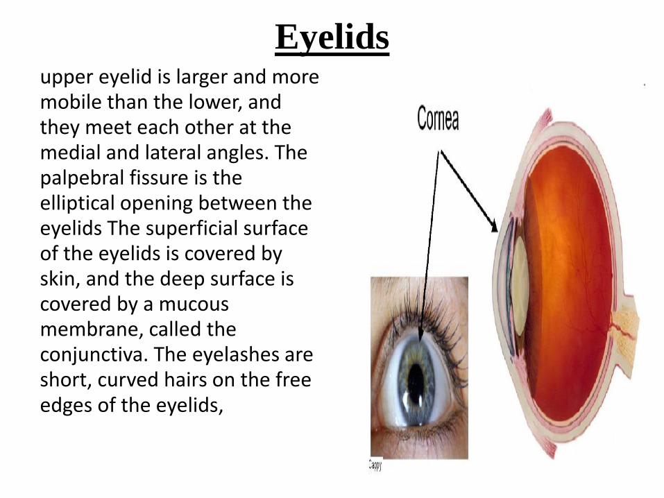
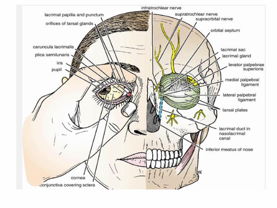
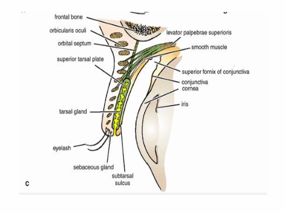
Eyelidsupper eyelid is larger and more mobile than the lower, and they meet each other at the medial and lateral angles. The palpebral fissure is the elliptical opening between the eyelids The superficial surface of the eyelids is covered by skin, and the deep surface is covered by a mucous membrane, called the conjunctiva. The eyelashes are short, curved hairs on the free edges of the eyelids,

• The sebaceous glands (glands of Zeis) open directly into the eyelash follicles, ciliary glands (glands of Moll) are modified sweat glands that open separately between adjacent lashes. The tarsal glands are long, modified sebaceous glands that pour their oily secretion onto the margin of the lid; their openings lie behind the eyelashes,

The framework of the eyelids is formed by a fibrous sheet, the
orbital septum . This is attached to the periosteum at the
orbital margins. The orbital septum is thickened at the margins
of the lids to form the superior and inferior tarsal plates. The
superficial surface of the tarsal plates and the orbital septum
are covered by the palpebral fibers of the orbicularis oculi
muscle .


Movements of the Eyelids
• The position of the eyelids at rest depends on
the tone of the orbicularis oculi and the levator
palpebrae superioris muscles and the position
of the eyeball. The eyelids are closed by the
contraction of the orbicularis oculi and the
relaxation of the levator palpebrae superioris
muscles. The eye is opened by the levator
palpebrae superioris raising the upper lid.

Lacrimal ApparatusLacrimal Gland
• The lacrimal gland consists of a large orbital
part and a small palpebral part It is situated
above the eyeball in the anterior and upper
part of the orbit posterior to the orbital
septum . The gland opens into the lateral part
of the superior fornix of the conjunctiva by
12ducts.
• parasympathetic secretomotor nerve supply is
derived from the lacrimal nucleus of the facial
nerve.

• The sympathetic postganglionic nerve supply is from the
internal carotid plexus and travels in the deep petrosal
nerve, the nerve of the pterygoid canal,

• . Lacrimal Ducts
• The tears circulate across the cornea and enter the canaliculi
lacrimales through the puncta lacrimalis. The canaliculi
lacrimales pass medially and open into the lacrimal sac
which lies in the lacrimal groove behind the medial
palpebral ligament and is the upper blind end of thenasolacrimal duct.
• The nasolacrimal duct is about (1.3 cm) long and emerges
from the lower end of the lacrimal sac . The duct descends
downward, backward, and laterally in a bony canal and
opens into the inferior meatus of the nose. The opening is
guarded by a fold of mucous membrane known as the
lacrimal fold. This prevents air from being forced up theduct into the lacrimal sac on blowing the nose.


The Orbit
Roof: Formed by the orbital plate of the frontal bone, which
separates the orbital cavity from the anterior cranial fossa and thefrontal lobe of the cerebral hemisphere
Lateral wall: Formed by the zygomatic bone and the greater wing ofthe sphenoid
Floor: Formed by the orbital plate of the maxilla, which separatesthe orbital cavity from the maxillary sinus
Medial wall: Formed from before backward by the frontal process of
the maxilla, the lacrimal bone, the orbital plate of the ethmoid
(which separates the orbital cavity from the ethmoid sinuses), andthe body of the sphenoid

Openings Into the Orbital Cavity
Orbital opening: Lies anteriorly
Supraorbital notch (Foramen): The supraorbital notch is situated on
the superior orbital margin . It transmits the supraorbital nerve andblood vessels.
Infraorbital groove and canal: Situated on the floor of the orbit in the
orbital plate of the maxilla they transmit the infraorbital nerve (acontinuation of the maxillary nerve) and blood vessels.
Nasolacrimal canal: Located anteriorly on the medial wall; it
communicates with the inferior meatus of the nose ). It transmitsthe nasolacrimal duct.

Inferior orbital fissure: Located posteriorly between the maxilla and
the greater wing of the sphenoid it communicates with the
pterygopalatine fossa. It transmits the maxillary nerve and its
zygomatic branch, the inferior ophthalmic vein, and
sympathetic nerves.
Superior orbital fissure: Located posteriorly between the greater and
lesser wings of the sphenoid it communicates with the middle
cranial fossa. It transmits the lacrimal nerve, the frontal nerve, the
trochlear nerve, the oculomotor nerve (upper and lower
divisions), the abducent nerve, the nasociliary nerve, and the
superior ophthalmic vein.
Optic canal: Located posteriorly in the lesser wing of the sphenoid
it communicates with the middle cranial fossa. It transmits the
optic nerve and the ophthalmic artery.

Optic Nerve
• The optic nerve enters the orbit from the middle
cranial fossa by passing through the optic canal
It is accompanied by the ophthalmic artery, which
lies on its lower lateral side. The nerve is
surrounded by sheaths of pia mater, arachnoid
mater, and dura mater, A rise in pressure of the
cerebrospinal fluid within the cranial cavity
therefore is transmitted to the back of the eyeball

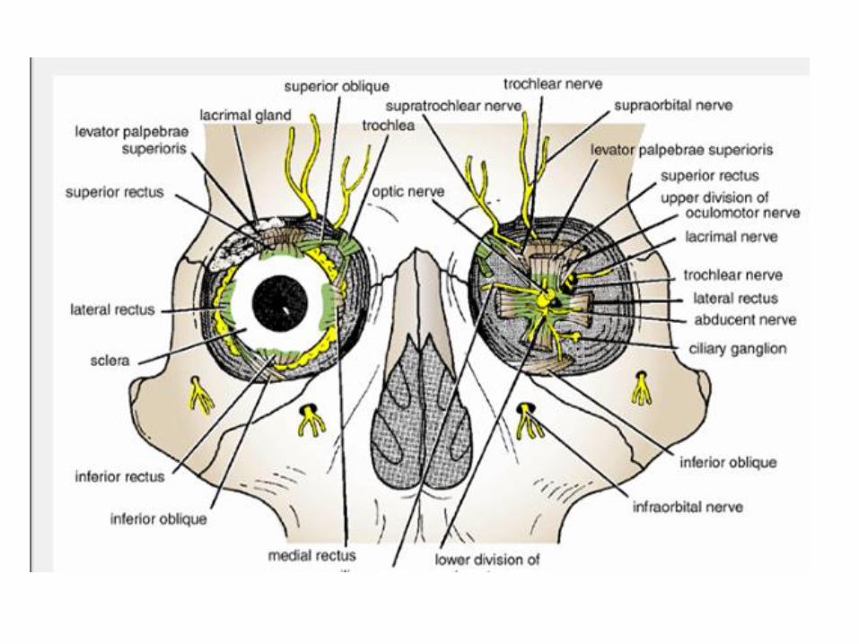
• The lacrimal nerve
• arises from the ophthalmic division of the
trigeminal nerve. It enters the orbit through
the upper part of the superior orbital fissure,
The lacrimal nerve ends by supplying the skin
of the lateral part of the upper lid.

• Frontal Nerve
• The frontal nerve arises from the ophthalmic
division of the trigeminal nerve. It enters the orbit
through the upper part of the superior orbitalfissure
• It divides into the supratrochlear and supraorbital nerves that wind around the upper margin of the orbital cavity to supply the skin of the forehead; the supraorbital nerve also supplies the mucous membrane of the frontal air sinus

• Trochlear Nerve
• The trochlear nerve enters the orbit through the
upper part of the superior orbital fissure . It runs
forward and supplies the superior oblique
muscle ,
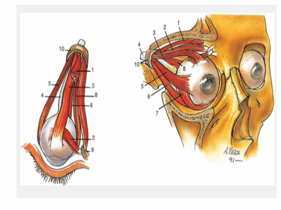
• Oculomotor Nerve
• The superior ramus of the oculomotor nerve
enters the orbit through the lower part of the
superior orbital fissure . It supplies the superior
rectus muscle, then pierces it, and supplies the
levator palpebrae superioris muscle .

• Nasociliary Nerve
• The nasociliary nerve arises from the ophthalmic division of the
trigeminal nerve. It enters the orbit through the lower part of thesuperior orbital fissure
• crosses above the optic nerve, runs forward along the upper marginof the medial rectus muscle, and ends by dividing into the anteriorethmoidal and infratrochlear nerves
•
• Branches of the Nasociliary Nerve
communicating branch to the ciliary ganglion.
long ciliary nerves
posterior ethmoidal nerve
infratrochlear nerve
• anterior ethmoidal nerve

• Abducent Nerve
• The abducent nerve enters the orbit
through the lower part of the superior
orbital fissure . It supplies the lateral rectus
muscle.

• Ciliary Ganglion
• The ciliary ganglion is a parasympathetic ganglion about
the size of a pinhead and situated in the posterior part of
the orbit. It receives its preganglionic parasympathetic
fibers from the oculomotor nerve via the nerve to the
inferior oblique. The postganglionic fibers leave the
ganglion in the short ciliary nerves, which enter the back of
the eyeball and supply the sphincter pupillae and the ciliary
muscle.A number of sympathetic fibers pass from the
internal carotid plexus into the orbit and run through theganglion without interruption


![healthy recipes - The Eye1].pdf · Healthy Snacks and Desserts 109 Recipe Resources 124. Introduction Welcome to BodyWorks Healthy Recipes, where you’ll find simple, low-cost recipes](https://img.pdfslide.net/doc/110x75/60532e81360fc7192751a9eb/healthy-recipes-the-eye-1pdf-healthy-snacks-and-desserts-109-recipe-resources.jpg)