Embed Size (px)
Citation preview
JOURNAL OF VIROLOGY, Jan. 2004, p. 320–328 Vol. 78, No. 10022-538X/04/$08.00�0 DOI: 10.1128/JVI.78.1.320–328.2004Copyright © 2004, American Society for Microbiology. All Rights Reserved.
The ORF2 Protein of Hepatitis E Virus Binds the 5� Region ofViral RNA
Milan Surjit, Shahid Jameel, and Sunil K. Lal*Virology Group, International Centre for Genetic Engineering & Biotechnology,
New Delhi 110067, India
Received 6 May 2003/Accepted 5 September 2003
Hepatitis E virus (HEV) is a major human pathogen in much of the developing world. It is a plus-strandRNA virus with a 7.2-kb polyadenylated genome consisting of three open reading frames, ORF1, ORF2, andORF3. Of these, ORF2 encodes the major capsid protein of the virus and ORF3 encodes a small protein ofunknown function. Using the yeast three-hybrid system and traditional biochemical techniques, we havestudied the RNA binding activities of ORF2 and ORF3, two proteins encoded in the 3� structural part of thegenome. Since the genomic RNA from HEV has been postulated to contain secondary structures at the 5� and3� ends, we used these two terminal regions, besides other regions within the genome, in this study. Experi-ments were designed to test for interactions between the genomic RNA fusion constructs with ORF2 and ORF3hybrid proteins in a yeast cellular environment. We show here that the ORF2 protein contains RNA bindingactivity. The ORF2 protein specifically bound the 5� end of the HEV genome. Deletion analysis of this proteinshowed that its RNA binding activity was lost when deletions were made beyond the N-terminal 111 aminoacids. Finer mapping of the interacting RNA revealed that a 76-nucleotide (nt) region at the 5� end of the HEVgenome was responsible for binding the ORF2 protein. This 76-nt region included the 51-nt HEV sequence,conserved across alphaviruses. Our results support the requirement of this conserved sequence for interactionwith ORF2 and also indicate an increase in the strength of the RNA-protein interaction when an additional 44bases downstream of this 76-nt region were included. Secondary-structure predictions and the location of theORF2 binding region within the HEV genome indicate that this interaction may play a role in viralencapsidation.
Hepatitis E virus (HEV), the causative agent of hepatitis E,is transmitted via the fecal-oral route, predominantly throughcontaminated water, and is responsible for sporadic infectionsas well as large epidemics of acute viral hepatitis in developingcountries (3, 4, 11, 12, 13, 21, 22, 23, 36). The HEV genomeorganization resembles that of many alphaviruses, with non-structural genes at the 5� end and structural genes at the 3� end(13, 23), and is currently classified in a separate unclassifiedgroup of hepatitis E-like viruses (2). It has a single-strandedpositive-sense RNA genome of 7.2 kb with three forward openreading frames (ORF1, ORF2, and ORF3) encoding three dif-ferent proteins (Fig. 1A) (13, 24, 29, 31). The 5� untranslatedregion (UTR) is 28 nucleotides (nt) in length, and bases 2through 26 can potentially assume a complex secondary struc-ture or hairpin (7). Bases 150 to 208 form a sequence that ishomologous to the 51-nt sequence, conserved across alphavi-ruses. The 3� UTR is 68 nt long and has been postulated toform stem-loop structures (29).
In the absence of a reliable culture system for HEV, funda-mental studies of its replication and expression strategy havebeen restricted to foreign gene expression systems. ORF2 andORF3 have been expressed in Escherichia coli, animal cells,baculovirus, yeast and in vitro in a coupled transcription-trans-lation system (6, 8, 9, 15, 19).
ORF1, the putative nonstructural gene, begins 28 nt from the5� end of the HEV genome, spans 5,079 bases before termi-nating at nt 5107, and codes for a polypeptide of 1,693 aminoacid residues. ORF2 starts from nt 5147, extends 1,980 basesbefore ending at nt 7127, and codes for the major structuralprotein of 660 amino acids (88 kDa) that is expressed intra-cellularly as well as on the cell surface. It is synthesized as aprecursor and is processed through signal sequence cleavageinto the mature protein, which is capable of self-associationand glycosylation (10, 30, 32). The third positive-polarity read-ing frame of 369 bases (ORF3) overlaps ORF1 at its 5� end by1 nt, significantly overlaps ORF2 (in a different frame), andcodes for a protein of 123 amino acids (13.5 kDa). ORF3encodes a phosphoprotein that is expressed intracellularly,fractionates with the cytoskeletal and membrane fractions, andshows no major processing (1, 20, 37). The ORF3 proteindimerizes using a 43-amino-acid interaction domain, interactswith proteins containing SH3 domains, and activates mitogen-activated protein kinase (14, 32). Recently, we have shown thatthe phosphorylated form of the ORF3 protein interacts withthe nonglycosylated form of the major capsid protein, ORF2 ofHEV (34).
Since HEV is a plus-strand RNA virus, it is expected thateither one of the two proteins encoded in the structural part ofthe genome, ORF2 or ORF3, would fulfill the role of genomicRNA binding for viral encapsidation leading to headfull pack-aging of new HEV particles in the infected hepatocyte. Thisfundamental aspect of the HEV life cycle has not yet beenelucidated.
* Corresponding author. Mailing address: Virology Group, Interna-tional Centre for Genetic Engineering & Biotechnology, P.O. Box10504, Aruna Asaf Ali Road, New Delhi 110067, India. Phone: 91-11-26177357. Fax: 91-11-26162316. E-mail: [email protected].
320
Dow
nloa
ded
from
http
s://j
ourn
als.
asm
.org
/jour
nal/j
vi o
n 20
Nov
embe
r 20
21 b
y 17
7.66
.203
.174
.

We have used the yeast three-hybrid system (26) to detectthe RNA binding properties of these two proteins of HEV andmap the interaction domains for the interacting protein andgenomic RNA. The yeast three-hybrid system is a genetic assayin which specific RNA-protein interactions can be detectedrapidly in yeast, in a fashion that is independent of the biolog-ical role of the RNA or protein. This approach is based on theyeast two-hybrid system, in principle, which detects protein-protein interactions. The three-hybrid system allows simplephenotypic properties of yeast, such as the ability to grow or tometabolize a chromogenic compound, to be used to detect andanalyze an RNA-protein interaction. In the cotransformedyeast cell, a fusion RNA molecule bridges two hybrid proteins,one containing a DNA binding domain and the other contain-
ing a transcriptional activation domain, resulting in the tran-scriptional activation of HIS3 and lacZ reporter genes down-stream of the binding site for the DNA binding domain. Toapply this system to HEV, we designed constructs fusing MS2-RNA with the 5� HEV (nt 1 to 910) genome and the 3� HEV(nt 6807 to 7184) genome in two separate constructs (Fig. 1B).The HEV ORF2 and ORF3 genes were cloned in-frame withthe Gal4 activation domain (Gal4 AD) (Fig. 1C) in two sepa-rate constructs.
From the two ORFs tested, using the yeast three-hybridassay, we found the ORF2 protein to be capable of binding tothe 5� end of the HEV genome. These results were verified byelectrophoretic mobility shift assays (EMSA). Further, we havemapped the interaction domains of the genomic RNA and the
FIG. 1. HEV genome and yeast three-hybrid constructs used to study RNA-protein interactions. (A) Genes and genome organization of HEV.Start sites for all three ORFs and both 3� and 5� UTRs are shown in red. A(n) represents the poly(A) tail (blue). Predicted stem-loop structures(SL) are shown schematically and numbered across the HEV genome. (B) Fusion RNA constructs designed to express fusion transcripts withinthe yeast cell. The MS2 RNA coding region (yellow) was cloned with two different HEV genomic regions (green). Predicted mRNA stem-loopstructures are shown schematically as fusion transcripts. (C) Hybrid protein constructs to test the RNA binding activity of the ORF2 and ORF3proteins of HEV using the yeast three-hybrid system. The schematic diagram shows the Gal4 Gal4 AD fused in frame to the ORF2 and ORF3genes of HEV, thus expressing fusion proteins in yeast cells. (D) Schematic diagram of the yeast three-hybrid system showing the differentfusion-RNA and hybrid-protein constructs being examined. P, promoter; Ter, terminator; SL, stem-loop structure.
VOL. 78, 2004 HEV ORF2 PROTEIN BINDS 5� REGION OF VIRAL RNA 321
Dow
nloa
ded
from
http
s://j
ourn
als.
asm
.org
/jour
nal/j
vi o
n 20
Nov
embe
r 20
21 b
y 17
7.66
.203
.174
.

interacting protein, ORF2. Deletion analysis of ORF2 showedthat the RNA binding activity of the protein was lost whendeletions were made beyond amino acid 111 from the N ter-minal, suggesting that its RNA binding activity is secondary-structure dependent rather than sequence specific. On theother hand, finer mapping of the 5� genomic RNA revealedthat a 76-nt region was responsible for this interaction. Al-though this 76-nt region was the smallest identifiable interac-tion domain that binds ORF2, genomic sequences up to 44 ntdownstream of this region contributed to the strengthening ofthe RNA protein interaction. A detailed RNA secondary-structure model for the interaction domain has been postu-lated, and the functional significance of this interaction duringthe viral life cycle has been discussed.
MATERIALS AND METHODS
Growth media, yeast strains, and plasmids. Yeast cells were grown either insynthetic media lacking the indicated nutrients or in rich media (25). The com-ponents for the three-hybrid selection were provided by the laboratory of M.Wickens (University of Wisconsin, Madison), and the selection was performedessentially as described elsewhere (26, 27, 38). The yeast strains, plasmids, andplasmid constructs used in this study are listed in Table 1.
Yeast three-hybrid techniques. The yeast host strain L40-coat constitutivelyexpresses the LexA-MS2 hybrid coat protein in which the LexA DNA bindingdomain is at the N terminal and the MS2 coat protein is at the C terminal. Boththe RNA plasmid (pIIIMS2-2) and its fusion constructs and the AD vector(pACTII) and its hybrid protein constructs were transformed in corresponding
pairs to study RNA-protein interactions (Fig. 1D). HIS3 and lacZ serve asreporter genes for the three-hybrid assay. �-Galactosidase expression levels weredetermined either by a 5-bromo-4-chloro-3-indolyl-�-D-galactoside (X-Gal) filterassay or by a liquid assay using o-nitrophenyl-D-galactoside (5, 35, 36). ConstructsMS2-IRE RNA, in which the MS2 protein is 5� of the iron response element(IRE) and construct AD-IRP, in which the iron regulatory protein (IRP) iscloned in frame downstream of the activation domain (AD), have been used asa positive control for the yeast three-hybrid experiments (26).
Gel shift assay (EMSA). EMSA was performed as described by Martin et al.(17). Uniformly 32P-labeled RNA was produced by T7 RNA polymerasetranscription from inserts cloned into the pGEMT Easy plasmid (Promega)and purified using the RN Easy kit (Qiagen). Purified probes were checked ona 6% urea–acrylamide gel to verify the integrity of RNA transcripts. Full-length ORF2 and ORF3 proteins and deletions of ORF2 were produced usingan in vitro-coupled transcription-translation rabbit reticulocyte system (Pro-mega) and verified by immunoprecipitation with anti-ORF2 and anti-ORF3antibodies.
To detect RNA-protein interactions by EMSA, 8 �g of total protein was mixedwith 50,000 cpm of uniformly 32P-labeled RNA and 20 �g of yeast tRNA andincubated for 20 min in 10 mM Tris-HCl (pH 7.5)–50 mM KCl–1 mM dithio-threitol–10% glycerol in 20 �l on ice. Reaction products were analyzed byelectrophoresis on native 5 or 4% polyacrylamide gels using 50 mM Tris-glycineas a buffer and visualized by autoradiography. Mock lysates were incubated alongwith the labeled probe in a similar manner to rule out the possible binding ofendogenous protein from the rabbit reticulocyte lysate (data not shown). Forcompetitor binding assay, a 100-fold excess of unlabeled RNAs was incubatedalong with the regular reaction mixture described above.
RNA secondary-structure analysis. RNA secondary structure was analyzedusing the mfold program (http://bioinfo.rpi.edu/applications/mfold/old/RNA),based on minimum free energy calculations at 25°C.
TABLE 1. Yeast strains, plasmids, and recombinant plasmid constructs used in this study
Strain, plasmid, or constructa Genotype and description
StrainL40-coat........................................................MATa ura3-52 leu2-3,112 his3�200 trp1�1 ade2 LYS::(LexA op)-HIS3 ura3::(LexA-op)-LacZ, LexA-
MS2 coat (TRP1)
PlasmidspACT2 ..........................................................GAL4 AD vector [GAL4(768–881)]; LEU2, 2�m, Ampr
pIII/MS2-2....................................................Derived from pIIIEx426RPR
ConstructspAS2-ORF2 .................................................pMT-ORF2 digested with NcoI and BamHI, fragment ligatedpACT-ORF2................................................pAS2-ORF2 digested with NcoI and BamHI, fragment ligatedpSG112-660 ORF2......................................Described in reference 8pACT-ORF2 112-660 .................................pSG-ORF2 1–110 digested with EcoRI and BamHI, fragment religatedpACT-ORF2 1-226 .....................................pACT-ORF2 digested with EcoRI, vector religatedpACT2-ORF2 1-358 ...................................pACT2-ORF2 digested with NcoI and ScaI, fragment religatedpACT2-ORF2 1-586 ...................................pACT2-ORF2 digested with BstXI and NcoI and religatedpSG-ORF2 ...................................................Described in reference 8pSG-ORF2 1-226 ........................................pSG-ORF2 digested with EcoRI and PstI, fragment religatedpSG-ORF2 1-358 ........................................pSG-ORF2 digested with ScaI and PstI, fragment religatedpSG-ORF2 1-586 ........................................pSG-ORF2 digested with BstXI and PstI, fragment religatedpSGIb ............................................................pSGI vector digested with EcoRI and HindIII, blunt ended, and vector religatedpSG-ORF2b..................................................pSG-ORF2 digested with PstI and BamHI, fragment religated into pSGIb
pSG-ORF2 228-660 ....................................pSG-ORF2b digested with BstEII and EcoRI, blunt ended, and vector religatedpIIIMS2-2 10-910 HEV ..............................Full-length HEV genome digested with SmaI and NdeI, fragment religatedpIIIMS2-1-250 HEV....................................Fragment from bp 1 to 250 PCR amplified and cloned into SmaI site of vectorpIIIMS2-2 1-130 HEV ................................Fragment from bp 1 to 130 PCR amplified and cloned into SmaI and ShpI site of vectorpIIIMS2-2 1-120 HEV ................................Fragment from bp 130 to 250 PCR amplified and cloned into SmaI and ShpI sites of vectorpGEMTeasy 1-250 HEV ............................Fragment from bp 1 to 250 PCR amplified and cloned using TA overhang ligationpGEMT easy 130-250 HEV .......................pGEMT easy 1–250 digested with StuI and PstI, fragment religatedpGEMT 3� 6807-7184 HEV .......................Fragment of 377 bp PCR amplified from pSG-ORF2 and cloned into pGEMT Easy vectorpMS2-2 3� 6807-7184 HEV ........................Fragment of 377 bp PCR amplified from pSG-ORF2 and cloned into SmaI site of vectorpSG-ORF3 ...................................................Described in reference 8pACT ORF3................................................pSG-ORF3 digested with SmaI and XhoI, fragment religated
a Numbers in italics represent the RNA sequence of the HEV genome.b Intermediate vector constructs.
322 SURJIT ET AL. J. VIROL.
Dow
nloa
ded
from
http
s://j
ourn
als.
asm
.org
/jour
nal/j
vi o
n 20
Nov
embe
r 20
21 b
y 17
7.66
.203
.174
.

RESULTS
The 5� end of the HEV genomic RNA interacts with theORF2 protein. The different regions of the HEV genome werecloned into yeast three-hybrid vectors so as to express fusionRNA transcripts in yeast cells. The 5� end (bases 1 to 910) ofthe HEV genome was cloned into the pIIIMS2-2 yeast three-hybrid vector (Table 1). Similarly, the 3� end (bases 6807 to7184) and the 572 nt from the middle region (bases 5108 to5680) of the HEV genome were cloned into the pIIIMS2-2yeast three-hybrid vector (Table 1). All the constructs weretransformed into the L40-coat yeast three-hybrid host strainand grown on auxotrophic media. Independent transformantswere picked and their total RNA was isolated. Transcription ofthe fusion RNA was checked by reverse transcription-PCRanalysis. Further verification of the fusion RNA was performedby cloning each insert into the SmaI site of the IRE sequencein the pIIIMS2-2 vector (26) and checking for an interactionbetween IRE and IRP in the presence of 25 mM 3-aminotria-zole (3-AT) on SD-His growth medium (see the legend to Fig.2) by using the yeast three hybrid system (data not shown).3-AT was used to measure the strength of the RNA-proteininteraction. Similarly, full-length ORF2 and ORF3 genes werecloned in frame with the Gal4 activation domain to express
hybrid proteins in the yeast cells as reported previously (32, 33,34).
Single transformants and cotransformants were obtained indifferent combinations and tested for RNA-protein interac-tions (Fig. 2). The host yeast strain (L40-coat) showed negli-gible background His� phenotype. When singly transformedhost cells were analyzed, low background reporter gene activitywas detected on His� plus 5 mM 3-AT medium and in �-ga-lactosidase assays. The MS2-5�HEV/AD-ORF2 cotransfor-mants clearly showed strong His� prototrophy up to 15 mM3-AT. This clone was strongly positive when tested for �-ga-lactosidase activity, in both filter and liquid assays. However,the MS2-3�HEV/AD-ORF2 cotransformants showed no re-porter gene activity. The HEV genomic RNA (nt 5108 to 5680)was also tested for interaction with AD-ORF2 and AD-ORF3.Both assays gave negative results (data not shown). Similarly,AD-ORF3, when cotransformed separately with MS2-5� HEVand MS2-3�HEV, failed to show increased reporter gene ac-tivity.
The protein binding domain was subsequently shortened tont 1 to 250nt from nt 1 to 910, subcloned into the pIIIMS2-2vector (Table 1), and checked for interaction with ORF2. Sincethis shorter RNA region showed positive interaction with the
FIG. 2. Results from the three-hybrid analysis showing 5� HEV genomic RNA interacting with the ORF2 protein. YPD, yeast extract peptonedextrose media (nonselective); Leu�, Ura�, LU� represent SD-Leu� (synthetic dextrose complete medium lacking leucine), SD-Ura� (syntheticdextrose complete media lacking uracil), and SD-Leu� Ura� synthetic growth media. LUHis��3-AT (synthetic dextrose complete media lackinghistidine, leucine, and uracil with 3-aminotrizole) represents SD-Leu� Ura� His� synthetic medium with 0, 5, 10, and 25 mM 3-aminotrizole(3-AT) added. �F represents results from the �-galactosidase filter assay, and the bar graph represents relative �-galactosidase units from the liquid�-galactosidase assay. L40-coat is the untransformed yeast host strain. MS2-IRE/AD-IRP is the postive control used in the assay (26).
VOL. 78, 2004 HEV ORF2 PROTEIN BINDS 5� REGION OF VIRAL RNA 323
Dow
nloa
ded
from
http
s://j
ourn
als.
asm
.org
/jour
nal/j
vi o
n 20
Nov
embe
r 20
21 b
y 17
7.66
.203
.174
.
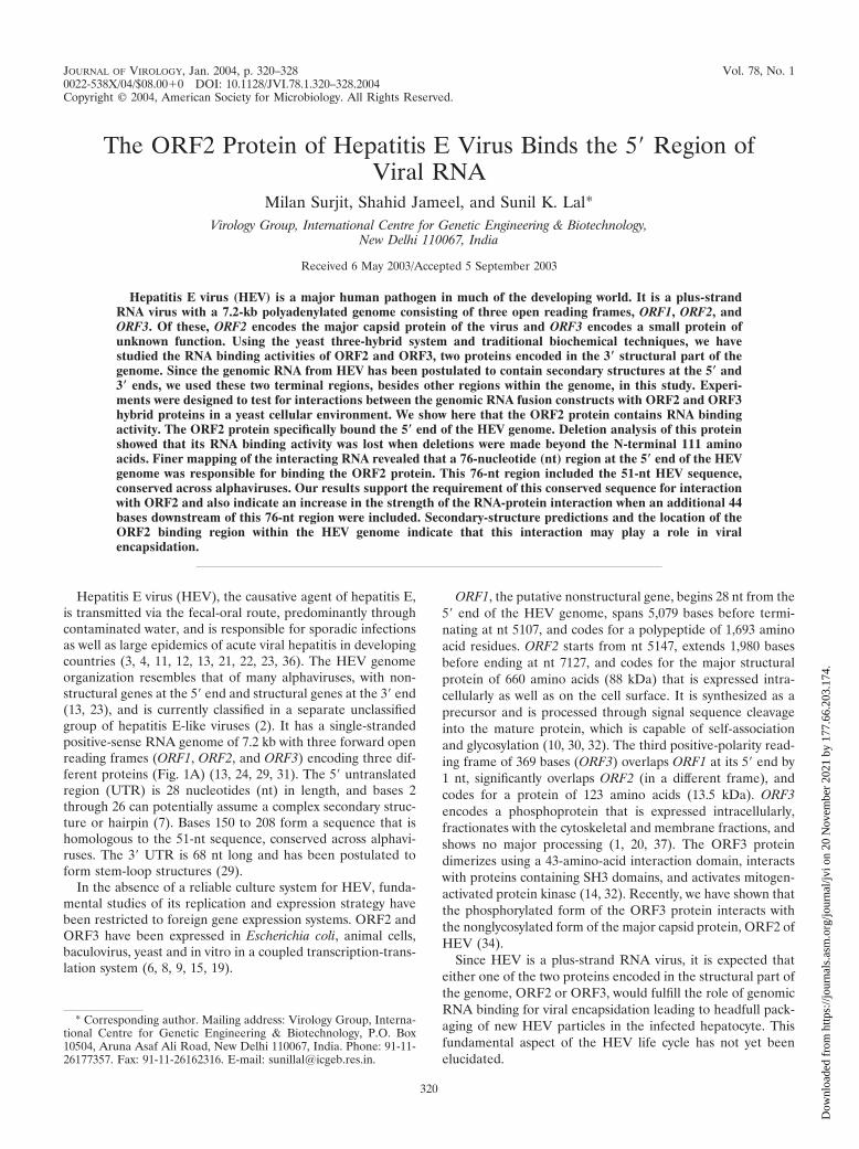
ORF2 protein, albeit of equal strength, subsequent EMSAexperiments were performed using this nt 1 to 250 5� HEVRNA. The 5� and 3� regions of the HEV genome were tran-scribed as 32P-labeled transcripts and, after purification, wereincubated with unlabeled ORF2 protein in separate tubes. Asnegative controls, the 32P-labeled transcripts from the 5� and 3�genomic regions of HEV were analyzed separately on a non-denaturing 6% polyacrylamide gel. Clearly, the 5� HEVgenomic transcript containing nt 1 to 250 showed a mobilityshift, indicating that the ORF2 protein was interacting with it(Fig. 3, lanes 2 and 3), in contrast to lane 1, where no proteinwas present. On the other hand, the 3� genomic region and thent 5108 to 5680 midgenomic region of HEV showed no bindingto the ORF2 protein (lane 4). Similar experiments were re-peated with the ORF3 protein, which showed a negative inter-action with both the 5� and 3� genomic regions of HEV (datanot shown).
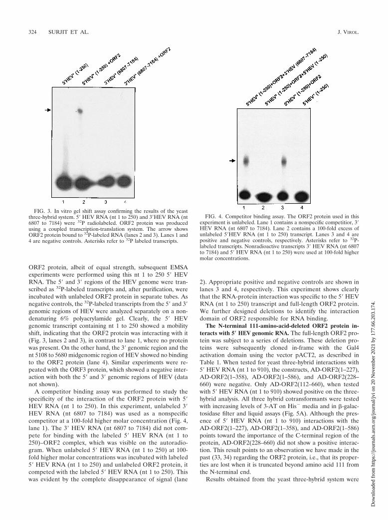
A competitor binding assay was performed to study thespecificity of the interaction of the ORF2 protein with 5�HEV RNA (nt 1 to 250). In this experiment, unlabeled 3�HEV RNA (nt 6807 to 7184) was used as a nonspecificcompetitor at a 100-fold higher molar concentration (Fig. 4,lane 1). The 3� HEV RNA (nt 6807 to 7184) did not com-pete for binding with the labeled 5� HEV RNA (nt 1 to250)–ORF2 complex, which was visible on the autoradio-gram. When unlabeled 5� HEV RNA (nt 1 to 250) at 100-fold higher molar concentrations was incubated with labeled5� HEV RNA (nt 1 to 250) and unlabeled ORF2 protein, itcompeted with the labeled 5� HEV RNA (nt 1 to 250). Thiswas evident by the complete disappearance of signal (lane
2). Appropriate positive and negative controls are shown inlanes 3 and 4, respectively. This experiment shows clearlythat the RNA-protein interaction was specific to the 5� HEVRNA (nt 1 to 250) transcript and full-length ORF2 protein.We further designed deletions to identify the interactiondomain of ORF2 responsible for RNA binding.
The N-terminal 111-amino-acid-deleted ORF2 protein in-teracts with 5� HEV genomic RNA. The full-length ORF2 pro-tein was subject to a series of deletions. These deletion pro-teins were subsequently cloned in-frame with the Gal4activation domain using the vector pACT2, as described inTable 1. When tested for yeast three-hybrid interactions with5� HEV RNA (nt 1 to 910), the constructs, AD-ORF2(1–227),AD-ORF2(1–358), AD-ORF2(1–586), and AD-ORF2(228–660) were negative. Only AD-ORF2(112–660), when testedwith 5� HEV RNA (nt 1 to 910) showed positive on the three-hybrid analysis. All three hybrid cotransformants were testedwith increasing levels of 3-AT on His� media and in �-galac-tosidase filter and liquid assays (Fig. 5A). Although the pres-ence of 5� HEV RNA (nt 1 to 910) interactions with theAD-ORF2(1–227), AD-ORF2(1–358), and AD-ORF2(1–586)points toward the importance of the C-terminal region of theprotein, AD-ORF2(228–660) did not show a positive interac-tion. This result points to an observation we have made in thepast (33, 34) regarding the ORF2 protein, i.e., that its proper-ties are lost when it is truncated beyond amino acid 111 fromthe N-terminal end.
Results obtained from the yeast three-hybrid system were
FIG. 3. In vitro gel shift assay confirming the results of the yeastthree-hybrid system. 5� HEV RNA (nt 1 to 250) and 3�HEV RNA (nt6807 to 7184) were 32P radiolabeled. ORF2 protein was producedusing a coupled transcription-translation system. The arrow showsORF2 protein bound to 32P-labeled RNA (lanes 2 and 3). Lanes 1 and4 are negative controls. Asterisks refer to 32P labeled transcripts.
FIG. 4. Competitor binding assay. The ORF2 protein used in thisexperiment is unlabeled. Lane 1 contains a nonspecific competitior, 3�HEV RNA (nt 6807 to 7184). Lane 2 contains a 100-fold excess ofunlabeled 5�HEV RNA (nt 1 to 250) transcript. Lanes 3 and 4 arepositive and negative controls, respectively. Asterisks refer to 32P-labeled transcripts. Nonradioactive transcripts 3� HEV RNA (nt 6807to 7184) and 5� HEV RNA (nt 1 to 250) were used at 100-fold highermolar concentrations.
324 SURJIT ET AL. J. VIROL.
Dow
nloa
ded
from
http
s://j
ourn
als.
asm
.org
/jour
nal/j
vi o
n 20
Nov
embe
r 20
21 b
y 17
7.66
.203
.174
.

verified by conventional in vitro techniques. All ORF2 dele-tions were expressed in a coupled transcription-translation sys-tem, checked for expressed protein (as shown in Fig. 5B), andused for EMSA with the 5� HEV RNA transcript (Fig. 5c).Results obtained from EMSA exactly matched our observa-tions from the yeast three-hybrid system, proving that only the
full-length protein and the ORF2(112–660) deletion were ca-pable of interacting with the 5� genomic RNA. Due to theinherent property of the ORF2 protein losing its RNA bindingactivity when truncated beyond the N-terminal amino acid 111,we were unable to perform a finer mapping of the interactiondomain for the ORF2 protein.
FIG. 5. Mapping the interaction domain for the ORF2 protein. (A) Amino acids 112 to 660 from the ORF2 protein are required for interactionwith the 5� HEV RNA (nt 1 to 910) region. Dotted boxes represent the AD regions which were fused in frame with the ORF2 protein (full lengthor deletions) shown in boxed regions with vertical lines. Checkered boxes show the MS2 regions fused with the 5� HEV RNA (nt 1 to 910), shownas horizontal lines. Open boxes represent regions that were deleted from ORF2. The numbers above the boxed regions with vertical lines representthe first and last nucleotides of the regions included in the ORF2 deletion constructs. YPD, yeast extract peptone dextrose media (nonselective);LU�, SD-Leu� Ura� synthetic growth medium; LUHis��3-AT, SD-Leu� Ura� His� synthetic medium with 0, 5, 10, and 25 mM 3-AT added.�F represents results from the �-galactosidase filter assay, and the bar graph represents relative �-galactosidase units from the liquid �-galactoseassay. (B) Control gel showing ORF2 deletions expressed using a coupled transcription-translation expression system. Major bands show theexpressed protein of interest and correspond to their calculated molecular masses. Weaker bands in each lane show nonspecific translation of rabbitreticulocyte proteins. (C) EMSA showing ORF2(112–660) interacting with the 5� genomic region of HEV. Asterisks refer to 32P-labeled transcript.
VOL. 78, 2004 HEV ORF2 PROTEIN BINDS 5� REGION OF VIRAL RNA 325
Dow
nloa
ded
from
http
s://j
ourn
als.
asm
.org
/jour
nal/j
vi o
n 20
Nov
embe
r 20
21 b
y 17
7.66
.203
.174
.

A 76-nt conserved domain from the 5� HEV genomic RNAinteracts with the ORF2 protein. The 5� HEV RNA (nt 1 to910) was subcloned into smaller fragments. 5� HEV RNA (nt1 to 250), 5� HEV RNA (nt 1 to 130), 5� HEV RNA (nt 130 to250), and 5� HEV RNA (nt 130 to 206) were deletions of thefull-length 5� HEV RNA (nt 1 to 910) as described in Table 1.Each of these deletions was individually tested for interactionwith the AD-ORF2 protein in the yeast three-hybrid assay plusEMSA. Interestingly, the 5� HEV RNA (nt 1 to 250) transcriptshowed a positive interaction, as had been observed with ourEMSA results described in the previous experiments. Subse-quently, the two deletion transcripts, 5� HEV RNA (nt 1 to130) and 5� HEV RNA (nt 130 to 250), which split the regionfrom nt 1 to 250, were tested for interaction with ORF2. Fromthis pair, the 5� HEV RNA (nt 130 to 250) transcript interactedwith ORF2 (Fig. 6A). The 5� HEV RNA (nt 130 to 250)region, consisting of 120 bases, showed a considerably strongerinteraction with the ORF2 protein compared with that of the5� HEV RNA (nt 130 to 206) region, consisting of only 76nucleotides (Fig. 6B). Hence, the nt 130 to 206 (76 nt) of theHEV genome may contain the major interaction domain re-quired for binding to ORF2; however, genomic sequences 44nt downstream of this region contribute significantly to anincrease in the strength of this RNA-protein interaction sig-nificantly.
DISCUSSION
We have used the yeast three-hybrid system to show that theORF2 protein of HEV is capable of binding its viral genome atthe 5� end. The ORF3 protein does not possess this activity.Also, the ORF2-RNA interaction is specific to the 5� end ofthe HEV genome and not to the 3� end or any other regiontested. Finally, we have mapped the ORF2 protein bindingregion on the RNA genome to a 76-nt region [5� HEV RNA(nt 130 to 206)]. This 76-nt region of the viral genome iscapable of binding the full-length ORF2 protein and its N-terminal deletion from nt 1 to 111 as well. Our previous ob-
servations of the dimerization of ORF2 (33) and its heterotypicinteraction with ORF3 (34) have shown similar results, sug-gesting that the ORF2 protein loses its activity when truncatedbeyond amino acid 111 from the N terminus.
We used the mfold program to predict the RNA secondarystructure for the HEV genomic region from nt 130 to 250(which includes the 76-nt region plus the downstream 44 nt),based on minimum free energy calculations (Fig. 7). Interest-ingly enough, the 76-nt region, which we have shown to beresponsible for binding the ORF2 protein, completely encom-passes the HEV homologue of the alphavirus consensus 51-ntsequence (bases 150 to 208) (18, 20). This 51-nt conserved
FIG. 6. Mapping the interaction domain for the 5� HEV RNA. (A) Mapping of the interaction domain of the 5� HEV RNA (nt 1 to 910) region.The hatched box represents the alphavirus consensus sequence. Plus signs shows a summarized result of the yeast three-hybrid interactions.(B) EMSA for the different RNA deletions from the 5� HEV genome. Asterisks refer to 32P-labeled transcript.
FIG. 7. Secondary-structure prediction of the 5� HEV genomic re-gion from nt 130 to 250. Numbers correspond to the numbers on theHEV genome. SL I, SL II, and SL III represent the three stem-loopstructures shown in the figure. The highlighted region represents the51-nt conserved region from alphaviruses.
326 SURJIT ET AL. J. VIROL.
Dow
nloa
ded
from
http
s://j
ourn
als.
asm
.org
/jour
nal/j
vi o
n 20
Nov
embe
r 20
21 b
y 17
7.66
.203
.174
.

region is highlighted and is part of two stem-loop structures(SLI and SLII), similar to other alphaviruses such as Sindbisvirus (18), Highlands J virus (28), and Semliki Forest virus(28). Looking at the secondary structures, it becomes obviousthat sequences downstream of base 208 contribute to thestrength of SLII. Our experimental data on the HEV RNA-ORF2 protein interaction fall in line with the in silico second-ary-structure prediction, suggesting that sequences down-stream of base 208 contribute to increased strength of SLII,thus strengthening the RNA-protein interaction. The 4-nt re-gion (nt 209 to 212 in SLII) may not be essential for theRNA-protein interaction but may contribute to increasing thebinding strength of the RNA-protein interaction in question.The 5� HEV region from nt 130 to 250 also forms a thirdstem-loop structure (SLIII). Although not essential, this stem-loop structure may contribute significantly toward increasingthe overall strength of the HEV genomic RNA-ORF2 proteininteraction.
Since HEV is a plus-strand RNA virus, one of its structuralproteins is required to show RNA binding activity for twoessential viral functions - viral replication and packaging of itsgenome into the capsid during viral assembly. Since ORF2 isthe major capsid protein, it would be the most likely candidateto bind the genomic RNA for viral packaging. We have exper-imentally shown that the ORF2 protein binds to the 5�-termi-nal region of the viral genome, thus becoming the most likelycandidate to perform this biological function.
HEV is postulated to form subgenomic RNA transcripts(�3.7 and �2 kb) from the 3� (structural) region of the ge-nome (16). Hence, it seems like a good strategic option for thevirus to have its RNA encapsidation signal at the 5� end of thegenome. This will result in only the full-length genomic RNA(�7.2 kb) being differentially recognized by the capsid proteinORF2 for headfull packaging during viral assembly in thehepatocyte.
Although data presented in this publication point to a fun-damental viral process, i.e., genome encapsidation, andstrongly indicates the possibility that the ORF2 protein may beresponsible for bringing the genomic RNA into the capsidduring assembly, direct biological evidence is difficult to obtaindue to the absence of an in vitro culture system for HEV.Indirect approaches using mutational knockout of the identi-fied interaction domain and restoration by complementary mu-tations are under investigation.
ACKNOWLEDGMENTS
Technical help from Shweta Tyagi is gratefully acknowledged.M.S. is a CSIR Junior Research Fellow and S.J. is an International
Senior Research Fellow in Biomedical Sciences of the WellcomeTrust. Support for this study was provided through internal funds fromthe ICGEB and partially through a grant to S.J. from the WelcomeTrust.
REFERENCES
1. Aye, T. T., T. Uchida, X. Z. Ma, F. Iida, T. Shikata, H. Zhuang, and K. M.Win. 1992. Complete nucleotide sequence of a hepatitis E virus isolated fromthe Xinjiang epidemic (1986–1988) of China. Nucleic Acids Res. 20:3512.
2. Berke, T., and D. O. Matson. 2000. Reclassification of the Calciviridae intodistinct genera and exclusion of hepatitis E virus from the family on the basisof comparative phylogenetic analysis. Arch. Virol. 145:1421–1436.
3. Bradley, D. W. 1990. Enterically-transmitted non-A, non-B hepatitis. Br.Med. Bull. 46:442–461.
4. Bradley, D. W., and M. A. Purdy. 1994. Molecular and serological charac-
teristics of hepatitis E virus, p 42–45. In K. Nishioka, H. Suzuki, S. Mishiro,et al. (ed.) Viral hepatitis and liver disease. Springer-Verlag, Tokyo, Japan.
5. Guarente, L. 1983. Yeast promoters and lacZ fusions designed to studyexpression of cloned genes in yeast. Methods Enzymol. 101:181–191.
6. He, J., A. W. Tam, P. O. Yarbough, G. R. Reyes, and M. Carl. 1993. Expres-sion and diagnostic utility of hepatitis E virus putative structural proteinsexpressed in insect cells. J. Clin. Microbiol. 31:2167–2173.
7. Huang, C., D. Nguyen, J. Fernandez, K. Y. Yun, K. E. Fry, D. W. Bradley,A. W. Tam, and G. R. Reyes. 1992. Molecular cloning and sequencing of theMexico isolate of hepatitis E virus (HEV). Virology 191:550–558.
8. Jameel, S., M. Zafrullah, M. H. Ozdener, and S. K. Panda. 1996. Expressionin animal cells and characterization of the hepatitis E virus structural pro-teins. J. Virol. 70:207–216.
9. Khudyakov, Y. E., M. O. Favorov, D. L. Jue, T. K. Hine, and H. A. Fields.1994. Immunodominant antigenic regions in a structural protein of thehepatitis E virus. Virology 198:390–393.
10. Khudyakov, Y. E, N. S. Khudyakov, H. A. Fields, D. Jue, C. Starling, M. O.Favorov, K. Krawczynski, L. Polish, E. Mast, and H. Margolish. 1993.Epitope mapping in proteins of hepatitis E virus. Virology 194:89–96.
11. Khuroo, M. S. 1980. Study of an epidemic of non-A, non-B hepatitis: pos-sibility of another human hepatitis virus distinct from post-transfusionnon-A, non-B type. Am. J. Med. 68:818–823.
12. Khuroo, M. S., M. R. Teli, S. Skidmore, M. A. Sofi, and M. Khuroo. 1981.Incidence and severity of viral hepatitis in pregnancy. Am. J. Med. 70:252–255.
13. Koonin, E. V., A. E. Gorbalenya, M. A. Purdy, M. N. Rozanov, G. R. Reyes,and D. W. Bradley. 1992. Computer-assisted assignment of functional do-mains in the nonstructural polyprotein of hepatitis E virus: delineation of anadditional group of positive-stranded RNA plant and animal viruses. Proc.Natl. Acad. Sci. USA 89:8259–8263.
14. Korkaya, H., S. Jameel, D. Gupta, S. Tyagi, R. Kumar, M. Zafrullah, M.Mazumdar, S. K. Lal, L. Xiaofang, D. Sehgal, S. R. Das, and D. Sahal. 2001.The ORF3 protein of hepatitis E virus binds to Src homology 3 domains andactivates MAPK. J. Biol. Chem. 276:42389–42400.
15. Lal, S. K., P. Tulasiram, and S. Jameel. 1997. Expression and characteriza-tion of the hepatitis E virus ORF3 protein in the methylotrophic yeast, Pichiapastoris. Gene 190:63–67.
16. Li, F., H. Zhuang, S. Kolivas, S. A. Locarnini, and D. A. Anderson. 1994.Persistent and transient antibody responses to hepatitis E virus detected byWestern immunoblot using open reading frame 2 and 3 and glutathioneS-transferase fusion proteins. J. Clin. Microbiol. 32:2060–2066.
17. Martin, F., A. Schaller, S. Eglite, D. Schumperli, and B. Muller. 1997. Thegene for histone RNA hairpin binding protein is located on human chromo-some 4 and encodes a novel type of RNA binding protein. EMBO J. 16:769–778.
18. Niesters, H. G. M., and J. H. Strauss. 1990. Mutagenesis of the conserved51-nucleotide region of Sindbis virus. J. Virol. 64:1639–1647.
19. Panda, S. K., I. H. Ansari, H. Durgapal, S. Agrawal, and S. Jameel. 2000.The in vitro-synthesized RNA from a cDNA clone of hepatitis E virus isinfectious. J. Virol. 74:2430–2437.
20. Panda, S. K., and S. Jameel. 1997. Hepatitis E virus: from epidemiology tomolecular biology. Viral Hepatitis Rev. 3:227–251.
21. Purcell, R. H. 2001. Hepatitis E virus, p. 3051–3061. In D. M. Knipe andP. M. Howley (ed.), Fields virology, 4th ed. Lippinscott-Raven, Philadelphia,Pa.
22. Purcell, R. H., and J. R. Ticehurst. 1988. Enterically transmitted non-A,non-B hepatitis: epidemiology and clinical characteristics, p. 131–137. InA. J. Zuckerman (ed.), Viral hepatitis and liver disease. Alan R. Liss, Inc.,New York, N.Y.
23. Purdy, M. A., A. W. Tam, C. C. Huang, P. O. Yarbough, and G. R. Reyes.1993. Hepatitis E virus: a non-enveloped member of the “alpha-like” RNAvirus supergroup? Semin. Virol. 4:319–326.
24. Reyes, G. R., C. C. Huang, A. W. Tam, and M. A. Purdy. 1993. Molecularorganization and replication of hepatitis E virus (HEV). Arch. Virol. 7:15–25.
25. Rose, M. D., F. Winston, and P. Hieter. 1990. Methods in yeast genetics.Cold Spring Harbor Laboratory, Cold Spring Harbor, N.Y.
26. Sengupta, D. J., B. Zhang, B. Kraemer, P. Pochart, S. Fields, and M.Wickens. 1996. A three-hybrid system to detect RNA-protein interactions invivo. Proc. Natl. Acad. Sci. USA 93:8496–8501.
27. Srivastava, R., and S. K. Lal. 2002. A liquid synchronized-growth cultureassay for the identification of true positive and negative yeast three-hybridtransformants. Lett. Appl. Microbiol. 34:300–303.
28. Strauss E. G., and J. H. Strauss. 1986. Structure and replication of thealphavirus genome, p. 35–90, In S. Schlesinger and M. J. Schlesinger (ed.),The Togaviridae and Flaviviridae. Plenum Publishing Corp., New York, N.Y.
29. Tam, A. W., M. M. Smith, M. E. Guerra, C. C. Huang, D. W. Bradley, K. E.Fry, and G. R. Reyes. 1991. Hepatits E virus (HEV): molecular cloning andsequencing of the full-length viral genome. Virology 185:120–131.
30. Torresi, J., F. Li, S. A. Locamini, and D. A. Anderson. 1999. Only the
VOL. 78, 2004 HEV ORF2 PROTEIN BINDS 5� REGION OF VIRAL RNA 327
Dow
nloa
ded
from
http
s://j
ourn
als.
asm
.org
/jour
nal/j
vi o
n 20
Nov
embe
r 20
21 b
y 17
7.66
.203
.174
.

non-glycosylated fraction of hepatitis E virus capsid (open reading frame 2)protein is stable in mammalian cells. J. Gen. Virol. 80:1185–1188.
31. Tsarev, S. A., S. U. Emerson, G. R. Reyes, T. S. Tsareva, L. J. Legters, I. A.Malik, M. Iqbal, and R. H. Purcell. 1992. Characterization of a prototypestrain of hepatitis E virus. Proc. Natl. Acad. Sci. USA 89:559–563.
32. Tyagi, S., S. Jameel, and S. K. Lal. 2001. Self-association and mapping of theinteraction domain of hepatitis E virus ORF3 protein. J. Virol. 75:2493–2498.
33. Tyagi, S., S. Jameel, and S. K. Lal. 2001. A yeast two-hybrid study onself-association of the ORF2 protein of hepatitis E virus. Biochem. Biophys.Res. Commun. 284:614–621.
34. Tyagi, S., H. Korkaya, M. Zafrullah, S. Jameel, and S. K. Lal. 2002. Thephosphorylated form of the ORF3 protein of hepatitis E virus interacts with
its non-glycosylated form of the major capsid protein, ORF2. J. Biol. Chem.277:22759–22767.
35. Tyagi, S., and S. K. Lal. 2000. Combined transformation and genetic tech-nique verification of protein-protein interactions in the yeast two-hybridsystem. Biochem. Biophys. Res. Commun. 277:589–593.
36. Tyagi, S., J. P. Salier and S. K. Lal. 2002. The liver-specific human alpha(1)-microglobulin/bikunin precursor (AMBP) is capable of self-association.Arch. Biochem. Biophys. 399:66–72.
37. Zafrullah, M., M. H. Ozdener, S. K. Panda, and S. Jameel. 1997. The ORF3protein of hepatitis E virus is a phosphoprotein that associates with thecytoskeleton. J. Virol. 71:9045–9053.
38. Zhang, B., M. Gallegos, A. Puoti, E. Durkin, S. Fields, J. Kimble, and M. P.Wickens. 1997. A conserved RNA-binding protein that regulates sexual fatesin the C. elegans hermaphrodite germ line. Nature 390:477–484.
328 SURJIT ET AL. J. VIROL.
Dow
nloa
ded
from
http
s://j
ourn
als.
asm
.org
/jour
nal/j
vi o
n 20
Nov
embe
r 20
21 b
y 17
7.66
.203
.174
.