Embed Size (px)
Citation preview

The orphan nuclear receptor chicken ovalbuminupstream promoter-transcription factor II isa critical regulator of adipogenesisZhao Xu, Songtao Yu*, Chung-Hsin Hsu, Jun Eguchi, and Evan D. Rosen†
Division of Endocrinology, Beth Israel Deaconess Medical Center, Harvard Medical School, Boston, MA 02215
Edited by Ronald M. Evans, Salk Institute for Biological Studies, San Diego, CA, and approved December 19, 2007 (received for review July 30, 2007)
The orphan nuclear receptor chicken ovalbumin upstream promoter-transcription factor II (COUP-TFII; Nr2f2) is expressed in adipose tissuein vivo and declines during differentiation. Overexpression of COUP-TFII prevents adipogenesis, whereas shRNA-mediated reduction ofCOUP-TFII promotes differentiation, as shown by increased lipidaccumulation and elevated expression of fat cell marker proteins.Furthermore, reduction of COUP-TFII allows uncommitted fibroblaststo be differentiated into fat cells. COUP-TFII represses the expressionof a number of proadipogenic factors in adipocytes, with direct actionnoted at the CAAT enhancer-binding protein ! promoter. We showthat COUP-TFII acts downstream of hedgehog signaling and is re-quired for the full antiadipogenic effect of this pathway. This effectis mediated in part by interaction with GATA factors. COUP-TFII andGATA2 are physically associated and repress target gene expressionin an additive manner. Taken together, our data demonstrate thatCOUP-TFII represents an endogenous suppressor of adipogenesis,linking antiadipogenic extracellular signals to the core transcriptionalcascade.
Repressor ! differentiation ! Nr2f2 ! GATA
Adipocyte differentiation is a highly regulated process con-trolled by a complex transcriptional cascade (1–4). A wide
array of transcription factors participate in adipogenesis, althoughmost attention has focused on several members of the CAATenhancer-binding protein (C/EBP) family and the nuclear receptorperoxisome proliferator-activated receptor ! (PPAR!). C/EBP"and C/EBP# are induced very early during differentiation (5).These early regulators in turn activate two critical proadipogenictranscription factors, PPAR! and C/EBP$, which mutually stimu-late each other and drive the transition of preadipocytes to matureadipocytes by activating a variety of genes required for maintainingthe adipocyte phenotype (6). Recently, a number of transcriptionfactors have been identified as regulators of adipogenesis, includingGATA2 and GATA3 (7, 8), certain members of the Kruppel-likefactor (KLF) family (9–11), and early B cell factors (EBF) 1 and 2(12, 13).
We have sought to identify transcriptional pathways in adi-pogenesis by using a systematic approach based on DNase-hypersensitivity analysis (51). Briefly, we used an integratedexperimental and computational strategy to identify overrepre-sented motifs in differentiation-dependent DNase-hypersensi-tive sites flanking adipocyte-selective genes. Among these motifswere sequences with a high degree of similarity to binding sitesfor the orphan nuclear receptor chicken ovalbumin upstreampromoter transcription factor (COUP-TF). First identified as anactivator of the chicken ovalbumin gene, COUP-TF was shownto bind to an imperfect direct repeat of the AGGTCA motif (14,15). Shortly thereafter, three mammalian orthologs were iden-tified, COUP-TFI (also known as Nr2f1 or EAR3), COUP-TFII(ARP-1, Nr2f2), and the more distantly related COUP-TFIII(also known as Nr2f6 or EAR2) (16–20).
We focused our attention on COUP-TFII because expressiondata suggested a role for this isoform in adipocyte biology (seebelow). COUP-TFII can act as either a positive or negative
regulator of transcription, although the latter appears to be moretypical (21). COUP-TFII is expressed widely during develop-ment (22), and loss-of-function studies in mice have confirmeda critical role in organogenesis. COUP-TFII-deficient mice diein utero with defects in heart development and angiogenesis (23).Tissue-specific knockout studies show that COUP-TFII is re-quired for the development of limb, skeletal muscles, andstomach (24, 25) and plays a critical role in determining veinidentity (26).
Here, we show that COUP-TFII is expressed in adipose tissuesand in cultured adipocyte models. Gain-of-function and loss-of-function studies demonstrate that COUP-TFII is a dominantrepressor of differentiation in adipocytes. We also show thatCOUP-TFII is required for other antiadipogenic factors, includ-ing sonic hedgehog (Shh) and GATA, to exert their full effect.COUP-TFII acts, in part, through physical and functional inter-actions with GATA factors. Taken together, these studies iden-tify a repressor of adipogenesis with links to upstream pathwaysand downstream effectors of this critical developmental process.
ResultsCOUP-TFII Is Expressed in Adipose Tissue and Is DevelopmentallyRegulated. To determine the tissue specificity of COUP-TFII, weisolated mRNA and protein from tissues of wild-type adult maleC57BL/6 mice and performed Northern and Western blotting.COUP-TFII mRNA [supporting information (SI) Fig. 5A] andprotein (Fig. 1A) are expressed at high levels in a number oftissues, including lung, kidney, and spleen. Moreover, COUP-TFII is expressed abundantly in white adipose tissue and, to alesser degree, in brown adipose tissue. Fractionation of whiteadipose tissue by low-speed centrifugation allowed us to identifythe stromal–vascular fraction (SVF) as the dominant site ofCOUP-TFII protein expression within the fat pad (Fig. 1B).
We next used murine 3T3-L1 cells to assess COUP-TFIIexpression over the course of adipogenesis. As differentiationproceeds, COUP-TFII mRNA is decreased by !50% (SI Fig.5B). COUP-TFII protein is decreased during adipogenesis aswell (Fig. 1C); interestingly, the magnitude of the effect appearsto be greater at the protein level than at the mRNA level,implying posttranscriptional regulation of COUP-TFII levelsduring differentiation. This notion is supported by what appearsto be an isoform shift favoring more mobile species of COUP-TFII later in differentiation. It is not yet clear what these
Author contributions: Z.X. and S.Y. contributed equally to this work; Z.X., S.Y., and E.D.R.designed research; Z.X., S.Y., C.-H.H., and J.E. performed research; Z.X., S.Y., and E.D.R.analyzed data; and Z.X. and E.D.R. wrote the paper.
The authors declare no conflict of interest.
This article is a PNAS Direct Submission.
*Present address: Department of Pathology, Northwestern University, Chicago, IL 60611.†To whom correspondence should be addressed. E-mail: [email protected].
This article contains supporting information online at www.pnas.org/cgi/content/full/0707082105/DC1.
© 2008 by The National Academy of Sciences of the USA
www.pnas.org"cgi"doi"10.1073"pnas.0707082105 PNAS ! February 19, 2008 ! vol. 105 ! no. 7 ! 2421–2426
CELL
BIO
LOG
Y

different isoforms represent, and this question remains a subjectof ongoing investigation. The reduction in COUP-TFII levelsseen during adipose conversion is consistent with the results inFig. 1B showing that the SVF, which includes preadipocytes, isthe major site of COUP-TFII expression within the fat pad.
COUP-TFII Is a Potent Suppressor of Adipogenesis. The developmen-tal pattern of COUP-TFII expression suggests that this factormight act as a repressor of adipogenesis. To test this hypothesis,we first performed gain-of-function experiments by using retro-viral delivery of COUP-TFII into 3T3-L1 preadipocytes. Asshown in SI Fig. 6A, COUP-TFII protein levels were increasedby !10-fold in COUP-TFII transgenic cells relative to cellstransduced with control virus. Strikingly, overexpression ofCOUP-TFII in these cells completely blocked adipogenesis (Fig.2A), as shown by oil red O staining of neutral lipids. Adipocytemarkers, such as Glut4, LPL, PPAR!, adiponectin, and aP2 (Fig.2B), were significantly decreased in COUP-TFII-overexpressingcells, further confirming an antiadipogenic effect.
Experimental manipulations that inhibit adipogenesis need tobe interpreted with caution, for the simple reason that differ-entiation is a complex process requiring a plethora of factors toturn on and off with precise timing; it is easy to imagine thatmanipulation might disrupt the process through nonspecificmechanisms. We therefore sought confirmation of COUP-TFIIantiadipogenic activity through loss-of-function studies, in whichwe would predict enhanced adipose conversion. We used retro-viral delivery of short-hairpin RNA (shRNA) to knock downCOUP-TFII in 3T3-L1 preadipocytes. As shown in SI Fig. 6B,doing so resulted in reduction of COUP-TFII protein by "80%.Cells with reduced COUP-TFII demonstrated enhanced adipo-genic potential, including greater lipid accumulation (Fig. 2C)and increased expression of adipocyte marker genes (Fig. 2D).
NIH 3T3 and other noncommitted fibroblast lines express highendogenous levels of COUP-TFII (data not shown), leading usto speculate that this expression might explain part of theinability of these cells to form adipocytes upon induction withdifferentiation mixture. Similar to what we observed in 3T3-L1preadipocytes, knockdown of COUP-TFII enhanced adipogen-esis in NIH 3T3 fibroblasts. Approximately 10% of cells express-ing a retrovirus carrying a COUP-TFII-specific shRNA could beinduced to differentiate into adipocytes, whereas none of thecells expressing a control shRNA displayed evidence of differ-
entiation (SI Fig. 7A). In addition to the enhanced lipid accu-mulation, the adipocyte markers adiponectin and Glut4 weredramatically induced in COUP-TFII knockdown cells induced todifferentiate (data not shown).
To date, PPAR! is the only protein known to be bothnecessary and sufficient for adipogenesis. To assess whetherreduction of COUP-TFII is sufficient to promote adipogenesisindependent of PPAR!, we knocked down COUP-TFII inPPAR!f lox/# or PPAR!#/# mouse embryonic fibroblasts(MEFs) (27). Knockdown of COUP-TFII in PPAR!f lox/# cellsenhanced adipogenesis to a degree similar to that seen in NIH3T3 cells but had no effect in PPAR!#/#cells (SI Fig. 7B). Thisresult is consistent with multiple lines of prior data suggesting apreeminent position in the adipogenic cascade for PPAR!.
COUP-TFII Represses a Number of Proadipogenic Factors. Our dataindicate that COUP-TFII is an endogenous suppressor of adi-pogenesis in multiple cell lines. We next sought to investigate themechanism by which COUP-TFII negatively regulates adipo-genesis. The developmental pattern of COUP-TFII expression inadipogenesis suggests a role in the first few days after theinitiation of differentiation, around the time that proadipogenicfactors such as C/EBP$, PPAR!, EBF1, EBF2, and others arefirst expressed. We used transient transfection to overexpressCOUP-TFII in mature 3T3-L1 adipocytes and examined mRNAlevels of some of these factors. EBF1, which was recentlyimplicated as an activator of adipogenic program (12, 13), wassignificantly down-regulated upon overexpression of COUP-TFII (SI Fig. 8A), although the equally proadipogenic EBF2 wasnot (data not shown). Other proadipogenic factors, such asKLF15, SREBP1c, PPAR!1, PPAR!2, and C/EBP$, were alsorepressed by COUP-TFII.
To address whether COUP-TFII directly represses the expres-sion of some of these key transcription factors, we first per-formed reporter assays in NIH 3T3 cells by using luciferase
Fig. 1. COUP-TFII is expressed in adipose tissue in vivo and in vitro. (A)COUP-TFII protein levels in different tissues of adult C57BL/6 mice weredetermined by Western blotting. B, brain; H, heart; Lu, lung; K, kidney; Li, liver;Sk, skeletal muscle; Sp, spleen; WAT, white adipose tissue; BAT, brown adiposetissue; T, testis. (B) Ovarian white adipose tissue was further separated into SVFand adipocytes. Protein lysates from both fractions were subjected to Westernblotting. (C) The 3T3-L1 preadipocytes were differentiated with DMI mixture.Protein lysates were prepared at the indicated time points and were subjectedto Western blotting. Ruby staining (B) and Ponceau S staining (C) were used todemonstrate equal loading.
Fig. 2. Overexpression of COUP-TFII in 3T3-L1 cells suppresses adipogenesis,whereas RNAi-mediated knockdown of COUP-TFII promotes adipogenesis. (Aand B) The 3T3-L1 preadipocytes were transduced with a retrovirus expressingCOUP-TFII or empty pMSCV vector. (A) The cells were induced with DMImixture. Oil red O staining was performed on day 7 after induction. (B) mRNAlevels of adipocyte genes Glut4, adiponectin, LPL, aP2, and PPAR! wereanalyzed with Q-PCR on days 0, 2, 4, and 7 after induction. (C and D) The 3T3-L1preadipocytes were transduced with a retrovirus expressing a shRNA specificfor COUP-TFII (shCOUP) or luciferase (shLuc). (C) Oil red O staining wasperformed on days 4 and 7 after DMI induction. (D) mRNA levels of Glut4,adiponectin, LPL, aP2, and PPAR! were analyzed with Q-PCR on days 0, 2, 4,and 7 after induction. Data are shown as mean $ SD of three biologicalreplicates. *, P % 0.05; **, P % 0.01; ***, P % 0.001.
2422 ! www.pnas.org"cgi"doi"10.1073"pnas.0707082105 Xu et al.
constructs driven by the #1.8-kb region of the PPAR!1 pro-moter, the #900-bp region of the PPAR!2 promoter, and the#300-bp region of the C/EBP$ promoter. We found that thepromoter activity of C/EBP$ was consistently and significantlyrepressed by COUP-TFII (SI Fig. 8B), but we saw no significanteffects on the promoters for PPAR!1 and PPAR!2 (data notshown). COUP-TFII also repressed the positive actions ofC/EBP" on this promoter (SI Fig. 8B).
Computer-assisted searching of the #300-bp region of theC/EBP$ promoter does not reveal an obvious COUP-TFII-binding motif. To provide further evidence that this regionrepresents a bona fide COUP-TFII target gene, we performeda chromatin immunoprecipitation (ChIP) assay in 3T3-L1 prea-dipocytes to test for the presence of endogenous COUP-TFII onthe C/EBP$ promoter. As shown in SI Fig. 8C, COUP-TFII wasspecifically immunoprecipitated from this region. COUP-TFIIwas not precipitated by a nonspecific antibody, nor was it foundassociated with a distal, irrelevant region of the same gene. Itremains unclear whether COUP-TFII directly binds a crypticbinding site in the C/EBP$ promoter or whether it associateswith the promoter through interactions with other transcriptionfactors.
COUP-TFII also represses the proadipogenic pRB proteinand increases the level of necdin; these actions may antagonizedifferentiation (SI Fig. 9).
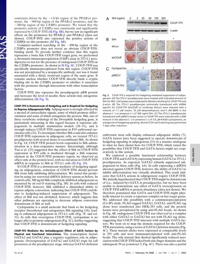
COUP-TFII Is Downstream of Hedgehog and Is Required for Hedgehogto Repress Adipogenesis Fully. Adipogenesis is strongly affected bya myriad of extracellular signals, some of which promote differ-entiation and some of which antagonize the process. Shh, one ofthree vertebrate orthologs of the Drosophila hedgehog gene, isparticularly interesting in this regard because it represses adi-pogenesis in multiple mammalian models (28–31), and itstrongly induces COUP-TFII expression in P19 embryonal car-cinoma cells (32). To investigate whether Shh could also enhanceCOUP-TFII expression in adipogenic cells, we treated 3T3-L1preadipocytes with several doses of recombinant Shh. As shownin Fig. 3A, COUP-TFII protein levels responded to Shh admin-istration in a dose-responsive manner. Interestingly, althoughTsai et al. (33) suggested that the effect of Shh on COUP-TFIIexpression in P19 cells is mediated through a Shh responseelement in the COUP-TFII promoter region, we observed aneffect only at the protein level, with no elevation in COUP-TFIImRNA in response to Shh in 3T3-L1 cells (SI Fig. 10).
If COUP-TFII is a downstream mediator of hedgehog signal-ing on adipogenesis, reduction of COUP-TFII should preventShh from fully inhibiting differentiation. We tested this predic-tion by using our retroviral shRNA delivery system as before. Incontrol cells, 300 ng/ml Shh completely inhibited adipogenesis asmeasured by oil red O staining (Fig. 3B). In cells with reducedCOUP-TFII, however, Shh exhibited a diminished ability torepress adipose conversion, indicating that COUP-TFII contrib-utes to hedgehog-induced suppression of adipogenesis. Theeffect of Shh was not abolished, however, which suggests thatother pathways are operating to decrease adipose conversiondownstream of Shh as well.
Cyclopamine is a small molecule that binds to the hedgehogreceptor Smoothened and antagonizes Shh signaling (34), lead-ing to enhanced adipogenesis in 3T3-L1 cells (Fig. 3C and ref.31). In cells that overexpress COUP-TFII, cyclopamine is nolonger able to promote adipogenesis (Fig. 3C), further suggestingthat COUP-TFII is downstream of hedgehog signaling.
COUP-TFII Mediates the Antiadipogenic Effect of GATA Factors byPhysical and Functional Interactions. The transcription factorsGATA2 and GATA3 play a negative regulatory role in adipo-genesis. Overexpression of GATA2 and GATA3 traps fat cellprecursors at the preadipocyte stage, whereas GATA3-deficient
embryonic stem cells display enhanced adipogenic ability (7).GATA factors have been suggested to operate downstream ofhedgehog signaling in adipogenesis (31) in a manner analogousto what we have shown here for COUP-TFII, which raised thepossibility that COUP-TFII and GATA factors might act coop-eratively in this system.
We explored a possible functional relationship betweenCOUP-TFII and GATA by expressing human GATA2 in 3T3-L1preadipocytes. As expected, GATA2 robustly suppressed adi-pogenesis in these cells (Fig. 4A). In cells expressing a shRNAdirected against COUP-TFII, however, the ability of GATA2 toinhibit differentiation was virtually abolished. This result indi-cates that GATA actions in adipogenesis require COUP-TFII.We initially hypothesized that COUP-TFII might be downstreamof (i.e., induced by) GATA in preadipocytes, but we have beenunable to demonstrate any effect of GATA overexpression onCOUP-TFII mRNA or protein abundance (data not shown). Wetherefore postulated that GATA and COUP-TFII might phys-ically interact to create a repressor complex on key target genes.We addressed this possibility with a coimmunoprecipitation(Co-IP) study. FLAG-tagged GATA2, GATA3, and FLAG tagalone were transfected into HEK-293 cells, and the proteinlysates were precipitated by using anti-FLAG beads. As shownin Fig. 4B, endogenous COUP-TFII was observed in a complexwith either GATA2 or GATA3 but not with FLAG tag alone,suggesting that COUP-TFII interacts with multiple GATA fac-tors. We went on to map the domain required for the COUP-TFII association, using a series of GATA2 deletion mutants (Fig.4C). These mutant alleles were expressed at comparable levelsin 293 cells and were immunoprecipitated with anti-FLAGbeads. The only mutant that showed significantly reduced asso-ciation with COUP-TFII lacked both zinc finger domains and thesubsequent 39 aa (construct 5; Fig. 4C). There was also a partial
Fig. 3. COUP-TFII is required for hedgehog-mediated suppression of adipo-genesis. (A) The 3T3-L1 preadipocytes were treated with indicated amount ofShh for 48 h. Cell lysates were subjected to Western blotting for COUP-TFII and"-actin. (B) The 3T3-L1 preadipocytes retrovirally transduced with shRNAspecific for COUP-TFII (shCOUP) or luciferase (shLuc) were induced with amixture of 1.7 %M insulin, 10 %M dexamethasone, and 5 nM IBMX in theabsence (#) or presence (&) of 300 ng/ml Shh. (C) The 3T3-L1 preadipocytestransduced with pMSCV empty vector or COUP-TFII were induced with a DMImixture in the absence (#) or presence (&) of 3.6 %M KAAD-cyclopamine, anantagonist of hedgehog signaling. (B and C) Oil red O staining was performed7 days after induction.
Xu et al. PNAS ! February 19, 2008 ! vol. 105 ! no. 7 ! 2423
CELL
BIO
LOG
Y

reduction in the COUP-TFII/GATA2 interaction with deletionof the N-terminal 289 aa (construct 2) or the 39 aa immediatelyafter the zinc fingers (construct 6). Taken together, these datademonstrate a physical interaction between these two antiadi-pogenic factors.
Because COUP-TFII and GATA2 are tightly associatedwith each other both functionally (Fig. 4A) and physically (Fig.4 B and C), we next asked whether they can cooperate in therepression of individual adipocyte target genes. We found thatCOUP-TFII and GATA2 were individually able to repressthe expression of endogenous C/EBP$ and Glut4 (Fig. 4D).When cotransfected (taking care to keep total repressor levelsequivalent), GATA2 and COUP-TFII were able to reduceexpression of these genes to a greater degree than eitherfactor alone.
DiscussionWe have sought to identify transcriptional pathways in adipo-genesis by using multiple experimental and computational tech-niques. These studies have led us to consider a role for theorphan nuclear receptor COUP-TFII in adipocyte differentia-tion. COUP-TFII is well known to play a significant role in thedevelopment of multiple organs and tissues, including heart,blood vessels, muscle, limb, and stomach (23–26). In the presentwork, we show that COUP-TFII is expressed in adipose tissuesand in cultured adipocytes. Gain-of-function and loss-of-function experiments were performed in several models ofadipogenesis. Overexpression of COUP-TFII in 3T3-L1 preadi-pocytes suppresses adipogenesis. Conversely, knockdown ofCOUP-TFII in 3T3-L1 preadipocytes, NIH 3T3 fibroblasts, andMEFs enhances fat differentiation (Fig. 2 and SI Fig. 7). NIH3T3 cells and MEFs are generally nonadipogenic and have beenshown to differentiate into fat cells only when strongly proadi-pogenic transcription factors are ectopically overexpressed (35).The fact that knockdown of COUP-TFII promotes adipogenesisin these cells indicates that COUP-TFII is a potent suppressor ofadipogenesis. Taken together, our data from multiple modelsprove that COUP-TFII represents an endogenous suppressor ofadipogenesis.
We have shown that levels of many proadipogenic transcrip-tion factors are reduced by transfection of COUP-TFII, althoughat present we do not know whether these actions are immediatelydownstream of COUP-TFII. COUP-TFII exerts a repressiveeffect on the C/EBP$ promoter in a reporter assay and localizesto this region in 3T3-L1 preadipocytes. Nonetheless, our inabilityto locate an obvious COUP-TF response element in this regionmakes it unclear whether the effect on C/EBP$ transcription ismediated by direct COUP-TFII binding or by an indirect actionthrough other binding partners. Both mechanisms have beenshown to be relevant to COUP-TFII action in other cell types(36–41). COUP-TFII has been shown to inhibit the ability ofPPAR! to induce phosphoenolpyruvate carboxykinase expres-sion in NIH 3T3 fibroblasts, perhaps by direct competition forthe PPAR! response element (42). It is also worth pointing outthat an effect on the C/EBP$ promoter is unlikely to accountfully for the antiadipogenic action of COUP-TFII becauseshRNA-mediated reduction of COUP-TFII in NIH 3T3 cells,which are functionally C/EBP$-deficient (43), is able to promoteadipogenesis. The identification of direct COUP-TFII targetgenes relevant to adipocyte differentiation is currently a priorityfor our laboratory.
Transcription factors regulate developmental events such asadipogenesis under the influence of signaling pathways, whichinclude such ancient pathways as Wnt and hedgehog, both ofwhich act to repress adipogenesis in mammalian systems. In mostcases, the link between the upstream signaling events and effectson the transcriptional cascades that govern differentiation ispoorly understood. We were struck, however, by the reportedrelationship between COUP-TFII and hedgehog signaling inheterologous cells (32), which suggested that there might be asimilar link in developing adipocytes. In fact, this suggestionproved to be true because COUP-TFII acts downstream ofhedgehog signaling and is required for the full expression of theantiadipogenic effect driven by Shh. Others have suggested a rolefor GATA factors as downstream mediators of hedgehog activityin developing adipocytes (31). Interestingly, we were able toshow that COUP-TFII and GATA2 demonstrate significantinterdependence, with both physical and functional interactionsbetween the two proteins. Thus, GATA2 cannot fully inhibitadipogenesis in the absence of COUP-TFII, and GATA2 andCOUP-TFII show additivity in repressing the expression of keyadipocyte genes such as C/EBP$ and Glut4.
Fig. 4. COUP-TFII is required for GATA-mediated suppression of adipogen-esis. (A) The 3T3-L1 preadipocytes were transduced with GATA2-pMSCV orempty vector, selected, and then transduced a second time with a virusexpressing shRNA specific for luciferase (shLuc) or COUP-TFII (shCOUP). Oil redO staining was performed 7 days after DMI induction. (B) HEK-293 cells weretransfected with FLAG empty vector (Flag), FLAG-GATA2, or FLAG-GATA3.Co-IP analysis was performed with anti-FLAG beads. Ten percent input and theSDS eluate were subjected to Western blotting with polyclonal antibodiesagainst FLAG or COUP-TFII. (C) The 293 cells were transfected with FLAG emptyvector (EV) or FLAG-tagged deletion mutants of GATA2. Co-IP analysis wasperformed as described above. (D) The 3T3-L1 adipocytes (day 5 after DMIinduction) were transfected with 1 %g of pCDNA3 (EV), 1 %g of COUP-TFII(COUP), 1 %g of GATA2 (GATA), or 0.5 %g of COUP-TFII plus 0.5 %g of GATA2(COUP & GATA). Relative mRNA levels of C/EBP$ and Glut4 were analyzed byusing Q-PCR. Data are shown as mean $ SD of three biological replicates. *, P %0.05; **, P % 0.01.
2424 ! www.pnas.org"cgi"doi"10.1073"pnas.0707082105 Xu et al.

COUP-TFII is exceptionally well conserved among species(22). Mouse and human COUP-TFII are identical, andCOUP-TF orthologs ranging from human to Caenorhabditiselegans and Drosophila display the highest degree of interspeciesconservation found in the nuclear hormone receptor superfam-ily (22). Intriguingly, the Drosophila ortholog of COUP-TF,seven-up, has been shown to play an essential role in fat bodydevelopment in flies (44). Disruption of seven-up results in lossor reduced expression of two terminal marker genes, Adh andDcg1, specifically in the larval fat body (44). Interestingly,serpent, the Drosophila ortholog of GATA, is also required for fatcell differentiation in flies (45). In particular, serpent was iden-tified as a transcriptional activator of Adh gene (46). Althoughthe molecular connection between seven-up and serpent inDrosophila remains unknown, their mammalian orthologs,COUP-TFII and GATA, appear to function cooperatively inhigher organisms, albeit they have the opposite effect on fatdevelopment versus that seen in the invertebrate models. Theconserved and integrated function of COUP-TFII and GATAduring evolution highlights their position as a team of keyregulators of fat cell differentiation.
COUP-TFs have been shown to affect gene expression anddevelopment by multiple molecular mechanisms, some of whichrequire DNA binding and some of which do not. For example,COUP-TFII can activate or repress gene expression throughbinding to COUP-TF motifs, such as those found in the ovalbu-min gene (47) and the apolipoprotein AI gene (48). Alterna-tively, COUP-TFII can also bind to other transcription factorsand influence gene expression as a cofactor (37–40). Finally,COUP-TFII can also compete for binding to the motifs of othernuclear receptors or can compete for other key cofactors,such as retinoic X receptor. We anticipate that the antiadipo-genic effects of COUP-TFII require more than one of thesemechanisms.
A major challenge in the field of adipogenesis is integratingtranscriptional effectors into the existing signaling and transcrip-tional cascade. This work establishes COUP-TFII as a criticalregulator of adipogenesis in multiple cell autonomous systems.Equally as important, however, is the identification of hedgehogas an upstream inducer of COUP-TFII and the identification ofC/EBP$ as a downstream target of COUP-TFII. Although it isvirtually certain that there will be other targets of COUP-TFIIin adipogenesis and other upstream modulators of COUP-TFIIexpression and activity, this work describes one of the first directconnections between extracellular signals and the classic tran-scriptional effectors in adipocyte differentiation.
MethodsFor additional materials and procedures, see SI Materials and Methods.
Materials and Reagents. The full-length mouse COUP-TFII cDNA was purchasedfrom American Type Culture Collection. The coding region was excised withSspI and MslI and cloned into pMSCV-puro (Clontech) at the HpaI site by bluntend ligation.
To construct the shRNA plasmids, DNA oligonucleotides were synthesized,annealed, and cloned into pSIREN vector (Clontech) at BamHI and EcoRI sites.Two COUP-TFII shRNA constructs were used in our studies and showed similareffects on adipogenesis (data not shown). The target sequence of the con-struct used in these experiments is 5'-AGCTCTTGCTTCGTCTCCC. FLAG-taggedGATA2 mutant constructs were the kind gift of Gokhan Hotamisligil (HarvardSchool of Public Health, Boston).
Rabbit anti-COUP-TFII antibody was a generous gift from Sotirios Karatha-nasis (Lilly). Rabbit anti-Glut4 antibody was a gift from Barbara Kahn (BethIsrael Deaconess Medical Center, Boston). Antibodies against "-actin, FLAG,and GAPDH were purchased from Santa Cruz Biotechnology.
Cell Culture. HEK-293 cells and Phoenix packaging cells were maintained inDMEM supplemented with 10% FBS. NIH 3T3 fibroblasts were maintained inDMEM supplemented with 10% calf serum. 3T3-L1 preadipocytes and MEFswere maintained and differentiated as described in refs. 27 and 49. Briefly,
3T3-L1 preadipocytes were grown to confluence in DMEM supplemented with10% calf serum. Two days after confluence, cells were supplied with differ-entiation medium [DMEM containing 10% FBS plus 1.7 %M insulin, 10 %Mdexamethasone, and 0.5 mM 3-isobutyl-1-methylxanthine (DMI)]. Forty-eighthours after induction, cells were fed maintenance medium (DMEM containing10% FBS plus 0.8 %M insulin), and the medium was replaced every 2 days. Fordifferentiation of NIH 3T3 cells and MEFs, differentiation medium was sup-plemented with 10 %M rosiglitazone, and the induction time was prolongedto 72 h.
Retrovirus Preparation and Infection. Retrovirus preparation and infectionwere performed as described in ref. 27. Briefly, pMSCV, pSIREN empty vectors,or their derivatives containing specific cDNA or shRNA, along with group-specific antigens and reverse transcriptase (gag-pol) and VSV-G-expressingplasmids, was transfected into Phoenix packaging cells with the CellPhecttransfection kit (Amersham Biosciences). Viral supernatant was collected 48 hafter transfection, filtered through 0.45-%m filters, and added to target cellsfor 12 h along with 8 %g/ml Polybrene. Cells were selected with 4 %g/mlpuromycin or 400 %g/ml hygromycin to make stable lines and were maintainedin media containing appropriate antibiotics.
Adipocyte Transfection. 3T3-L1 adipocytes were differentiated as described.On day 5 after DMI induction, the cells were trypsinized, and 1 %g of DNA wastransferred into 5 ( 106 cells with the Amaxa nucleofection device (AmaxaBiosystems) according to manufacturer’s instruction. mRNA was extracted24 h posttransfection.
Adipose Tissue Fractionation. Ovarian fat pads were dissected from 12-week-old C57BL/6J mice. The isolated white adipose tissue was subjected to a 45-mindigestion with 0.12 unit/ml collagenase at 37°C in a shaker at 25 rpm. Thesamples were then filtered through 300-%m nylon meshes (Spectrum Labora-tories) and were subjected to centrifugation at 500 ( g for 5 min. The floatingfraction (comprised of white adipocytes) and the pellet fraction (containingthe SVF) were dissolved in TRIzol reagent (Invitrogen) for RNA preparation orin TNN lysis buffer [50 mM Tris!HCl (pH 8.0), 150 mM NaCl, 0.5% Nonidet P-40]plus a protease inhibitor mixture (Roche) for Western blotting.
RNA Preparation and Quantitative PCR (Q-PCR). Total RNA was extracted frommouse tissues or cells with TRIzol reagent according to the manufacturer’sinstructions. cDNA was reverse-transcribed from 2 %g of RNA by using theRETROscript first-strand synthesis kit (Ambion). Q-PCR was performed withBrilliant SYBR Green QPCR Master Mix (Stratagene) and an Mx3000P thermalcycler (Stratagene). The relative amount of mRNA normalized to cyclophilin Bwas calculated by using the comparative Ct method.
Western Blotting. Cell lysates were prepared in TNN buffer with proteaseinhibitor mixture (Roche) unless described otherwise. Twenty micrograms ofprotein lysate was resolved by 10% SDS/PAGE and transferred onto PVDFmembranes. Ponceau S (Boston Bioproducts) staining was performed accord-ing to the manufacturer’s instructions. In some cases, a separate gel was runin parallel and was subjected to Ruby staining (Invitrogen) according to themanufacturer’s instructions. Membranes were blocked in PBS supplementedwith 0.5% Tween 20 (PBST) plus 10% nonfat milk for 1 h followed by incu-bation with primary (1:2,000) and secondary antibodies (1:2,000) for 1 h eachwith PBST washes in between. Blots were then exposed to enhanced chemi-luminescence substrate and exposed to film.
Co-IP Analysis. Co-IP analysis was performed according to a modified protocoldescribed by Xu et al. (50). Briefly, HEK-293 cells were transfected with FLAGempty vector, FLAG-tagged GATA2, GATA3, or truncated mutants of GATA2by using Lipofectamine2000 (Invitrogen) according to the manufacturer’sinstructions. Forty-eight hours later, cells were lysed with TD buffer containing1% Triton X-100, 50 mM Tris (pH 7.5), 250 mM NaCl, 5 mM EDTA, 50 mM NaF,plus protease inhibitor mixture (Roche). Cell lysates were diluted 1:1 withdilution buffer (1% Triton plus 20% glycerol) and were incubated withanti-FLAG beads (Sigma) overnight. The beads were eluted with nonreducingSDS/PAGE loading buffer after extensive washes with Tris-buffered saline andwere subjected to SDS/PAGE and Western blotting.
Oil Red O Staining. Cells were fixed with 4% Formalde-Fresh (Fisher Scientific)for 15 min at room temperature and stained with oil red O solution (0.5% oilred O in isopropyl alcohol/water ) 3:2) for 2 h. Cells were washed twice withdistilled water before photography.
Xu et al. PNAS ! February 19, 2008 ! vol. 105 ! no. 7 ! 2425
CELL
BIO
LOG
Y

ACKNOWLEDGMENTS. We are grateful to Dr. Gokhan Hotamisligil forproviding FLAG-tagged full-length and deleted GATA constructs and toDrs. Barbara Kahn and Sotirios Karathanasis for their generous gift of
reagents. This work was funded by National Institutes of Health GrantDK63906 (to E.D.R.) and an American Heart Association postdoctoral award(to Z.X.).
1. Rosen ED, Walkey CJ, Puigserver P, Spiegelman BM (2000) Genes Dev 14:1293–1307.2. Tong Q, Hotamisligil GS (2001) Rev Endocr Metab Disord 2:349–355.3. Rosen ED, MacDougald OA (2006) Nat Rev Mol Cell Biol 7:885–896.4. Farmer SR (2006) Cell Metab 4:263–273.5. Cao Z, Umek RM, McKnight SL (1991) Genes Dev 5:1538–1552.6. Wu Z, Rosen ED, Brun R, Hauser S, Adelmant G, Troy AE, McKeon C, Darlington GJ,
Spiegelman BM (1999) Mol Cell 3:151–158.7. Tong Q, Dalgin G, Xu H, Ting CN, Leiden JM, Hotamisligil GS (2000) Science 290:134–
138.8. Tong Q, Tsai J, Tan G, Dalgin G, Hotamisligil GS (2005) Mol Cell Biol 25:706–715.9. Wu J, Srinivasan SV, Neumann JC, Lingrel JB (2005) Biochemistry 44:11098–11105.
10. Banerjee SS, Feinberg MW, Watanabe M, Gray S, Haspel RL, Denkinger DJ, KawaharaR, Hauner H, Jain MK (2003) J Biol Chem 278:2581–2584.
11. Oishi Y, Manabe I, Tobe K, Tsushima K, Shindo T, Fujiu K, Nishimura G, Maemura K,Yamauchi T, Kubota N, et al. (2005) Cell Metab 1:27–39.
12. Akerblad P, Lind U, Liberg D, Bamberg K, Sigvardsson M (2002) Mol Cell Biol 22:8015–8025.
13. Jimenez MA, Akerblad P, Sigvardsson M, Rosen ED (2007) Mol Cell Biol 27:743–757.14. Pastorcic M, Wang H, Elbrecht A, Tsai SY, Tsai MJ, O’Malley BW (1986) Mol Cell Biol
6:2784–2791.15. Wang LH, Tsai SY, Sagami I, Tsai MJ, O’Malley BW (1987) J Biol Chem 262:16080–16086.16. Jonk LJ, de Jonge ME, Pals CE, Wissink S, Vervaart JM, Schoorlemmer J, Kruijer W (1994)
Mech Dev 47:81–97.17. Qiu Y, Cooney AJ, Kuratani S, DeMayo FJ, Tsai SY, Tsai MJ (1994) Proc Natl Acad Sci USA
91:4451–4455.18. Wang LH, Tsai SY, Cook RG, Beattie WG, Tsai MJ, O’Malley BW (1989) Nature 340:163–
166.19. Wehrenberg U, Ivell R, Walther N (1992) Biochem Biophys Res Commun 189:496–503.20. Miyajima N, Kadowaki Y, Fukushige S, Shimizu S, Semba K, Yamanashi Y, Matsubara
K, Toyoshima K, Yamamoto T (1988) Nucleic Acids Res 16:11057–11074.21. Pereira FA, Qiu Y, Tsai MJ, Tsai SY (1995) J Steroid Biochem Mol Biol 53:503–508.22. Qiu Y, Krishnan V, Pereira FA, Tsai SY, Tsai MJ (1996) J Steroid Biochem Mol Biol
56:81–85.23. Pereira FA, Qiu Y, Zhou G, Tsai MJ, Tsai SY (1999) Genes Dev 13:1037–1049.24. Lee CT, Li L, Takamoto N, Martin JF, Demayo FJ, Tsai MJ, Tsai SY (2004) Mol Cell Biol
24:10835–10843.
25. Takamoto N, You LR, Moses K, Chiang C, Zimmer WE, Schwartz RJ, DeMayo FJ, Tsai MJ,Tsai SY (2005) Development 132:2179–2189.
26. You LR, Lin FJ, Lee CT, DeMayo FJ, Tsai MJ, Tsai SY (2005) Nature 435:98–104.27. Rosen ED, Hsu CH, Wang X, Sakai S, Freeman MW, Gonzalez FJ, Spiegelman BM (2002)
Genes Dev 16:22–26.28. Zehentner BK, Leser U, Burtscher H (2000) DNA Cell Biol 19:275–281.29. Spinella-Jaegle S, Rawadi G, Kawai S, Gallea S, Faucheu C, Mollat P, Courtois B, Bergaud
B, Ramez V, Blanchet AM, et al. (2001) J Cell Sci 114:2085–2094.30. van der Horst G, Farih-Sips H, Lowik CW, Karperien M (2003) Bone 33:899–910.31. Suh JM, Gao X, McKay J, McKay R, Salo Z, Graff JM (2006) Cell Metab 3:25–34.32. Krishnan V, Pereira FA, Qiu Y, Chen CH, Beachy PA, Tsai SY, Tsai MJ (1997) Science
278:1947–1950.33. Krishnan V, Elberg G, Tsai MJ, Tsai SY (1997) Mol Endocrinol 11:1458–1466.34. Chen JK, Taipale J, Cooper MK, Beachy PA (2002) Genes Dev 16:2743–2748.35. Tontonoz P, Hu E, Spiegelman BM (1994) Cell 79:1147–1156.36. Cooney AJ, Tsai SY, O’Malley BW, Tsai MJ (1992) Mol Cell Biol 12:4153–4163.37. Cooney AJ, Leng X, Tsai SY, O’Malley BW, Tsai MJ (1993) J Biol Chem 268:4152–4160.38. Kliewer SA, Umesono K, Heyman RA, Mangelsdorf DJ, Dyck JA, Evans RM (1992) Proc
Natl Acad Sci USA 89:1448–1452.39. Tran P, Zhang XK, Salbert G, Hermann T, Lehmann JM, Pfahl M (1992) Mol Cell Biol
12:4666–4676.40. Leng X, Cooney AJ, Tsai SY, Tsai MJ (1996) Mol Cell Biol 16:2332–2340.41. Tsai SY, Tsai MJ (1997) Endocr Rev 18:229–240.42. Eubank DW, Duplus E, Williams SC, Forest C, Beale EG (2001) J Biol Chem 276:30561–
30569.43. El-Jack AK, Hamm JK, Pilch PF, Farmer SR (1999) J Biol Chem 274:7946–7951.44. Hoshizaki DK, Blackburn T, Price C, Ghosh M, Miles K, Ragucci M, Sweis R (1994)
Development 120:2489–2499.45. Hayes SA, Miller JM, Hoshizaki DK (2001) Development 128:1193–1200.46. Abel T, Michelson AM, Maniatis T (1993) Development 119:623–633.47. Sagami I, Tsai SY, Wang H, Tsai MJ, O’Malley BW (1986) Mol Cell Biol 6:4259–4267.48. Ladias JA, Karathanasis SK (1991) Science 251:561–565.49. Xu Z, Kandror KV (2002) J Biol Chem 277:47972–47975.50. Xu YX, Hirose Y, Zhou XZ, Lu KP, Manley JL (2003) Genes Dev 17:2765–2776.51. Eguchi J, Yan QW, Schones DE, Kamal M, Hsu CH, Zhang MQ, Crawford GE, Rosen ED
(2008) Cell Metab 7:86–94.
2426 ! www.pnas.org"cgi"doi"10.1073"pnas.0707082105 Xu et al.
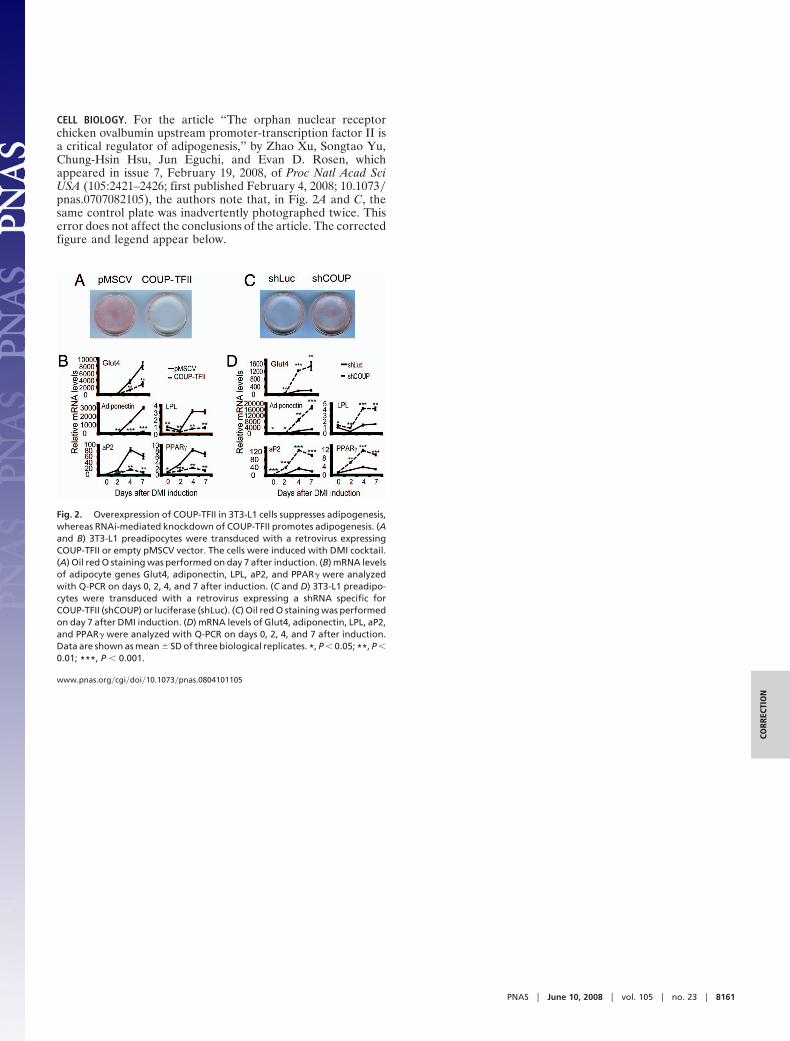
CELL BIOLOGY. For the article ‘‘The orphan nuclear receptorchicken ovalbumin upstream promoter-transcription factor II isa critical regulator of adipogenesis,’’ by Zhao Xu, Songtao Yu,Chung-Hsin Hsu, Jun Eguchi, and Evan D. Rosen, whichappeared in issue 7, February 19, 2008, of Proc Natl Acad SciUSA (105:2421–2426; first published February 4, 2008; 10.1073!pnas.0707082105), the authors note that, in Fig. 2A and C, thesame control plate was inadvertently photographed twice. Thiserror does not affect the conclusions of the article. The correctedfigure and legend appear below.
Fig. 2. Overexpression of COUP-TFII in 3T3-L1 cells suppresses adipogenesis,whereas RNAi-mediated knockdown of COUP-TFII promotes adipogenesis. (Aand B) 3T3-L1 preadipocytes were transduced with a retrovirus expressingCOUP-TFII or empty pMSCV vector. The cells were induced with DMI cocktail.(A) Oil red O staining was performed on day 7 after induction. (B) mRNA levelsof adipocyte genes Glut4, adiponectin, LPL, aP2, and PPAR! were analyzedwith Q-PCR on days 0, 2, 4, and 7 after induction. (C and D) 3T3-L1 preadipo-cytes were transduced with a retrovirus expressing a shRNA specific forCOUP-TFII (shCOUP) or luciferase (shLuc). (C) Oil red O staining was performedon day 7 after DMI induction. (D) mRNA levels of Glut4, adiponectin, LPL, aP2,and PPAR! were analyzed with Q-PCR on days 0, 2, 4, and 7 after induction.Data are shown as mean ! SD of three biological replicates. *, P " 0.05; **, P "0.01; ***, P " 0.001.
www.pnas.org!cgi!doi!10.1073!pnas.0804101105
PNAS " June 10, 2008 " vol. 105 " no. 23 " 8161
CORR
ECTI
ON