Embed Size (px)
Citation preview

2DX-RAY
SIRONAUSA.COM
T h e D e n t a l C o m p a n y
THE ORTHOPHOS® FAMILY
IMAGING YOU RELY ON.

AS VERSATILE AS YOUR PRACTICE.The ORTHOPHOS family offers the right solution for every practice. From entry into digital radiography to the perfect solution for your area of specialty–Sirona offers a product family sophisticated in every way with the ideal solution for every dentist. Enjoy every day. With Sirona.
2/3
OPTIMUMWORKFLOW
UNPARALLELEDIMAGE QUALITY
PROVENSOLUTION
UNPARALLELED DIGITAL ADVANTAGES.Digital imaging is becoming the standard in more and more dental practices. The benefits are obvious: lower radiation exposure, and more brilliant images accompanied by a more efficient practice workflow. At the same time, both treatment methods and practice services are easily accepted by the patient.
Low dose���ORTHOPHOS offers the best image quality
at the lowest achievable dose���Quickshot mode reduce dose even further��Reliable positioning prevents errors and repeat imaging���Optimal radiation management through
panoramic and cephalometric collimation��Automatic radiation management for differentiated image indications
Easy and efficient workflow���Logical symbols on the control panel prevent
operator errors���Efficient software analysis tools for quick and
clear diagnosis���Easy data exchange with practice management
systems (DICOM compliant)���All the advantages of SIDEXIS 4 software
4/5
Best image quality��DCS and Sharp Layer Technology provide never-before-seen imaging definition*��Automatic adjustment to the jaw width ensures high image sharpness��Automatic image processing assists with detailed image visualization��3-point patient stabilization prevents motion blurring��Automatic patient positioning for panoramic images with auto positioner**
*Standard with ORTHOPHOS SL DCS** Standard with ORTHOPHOS SL DCS and ORTHOPHOS XG 3Dready
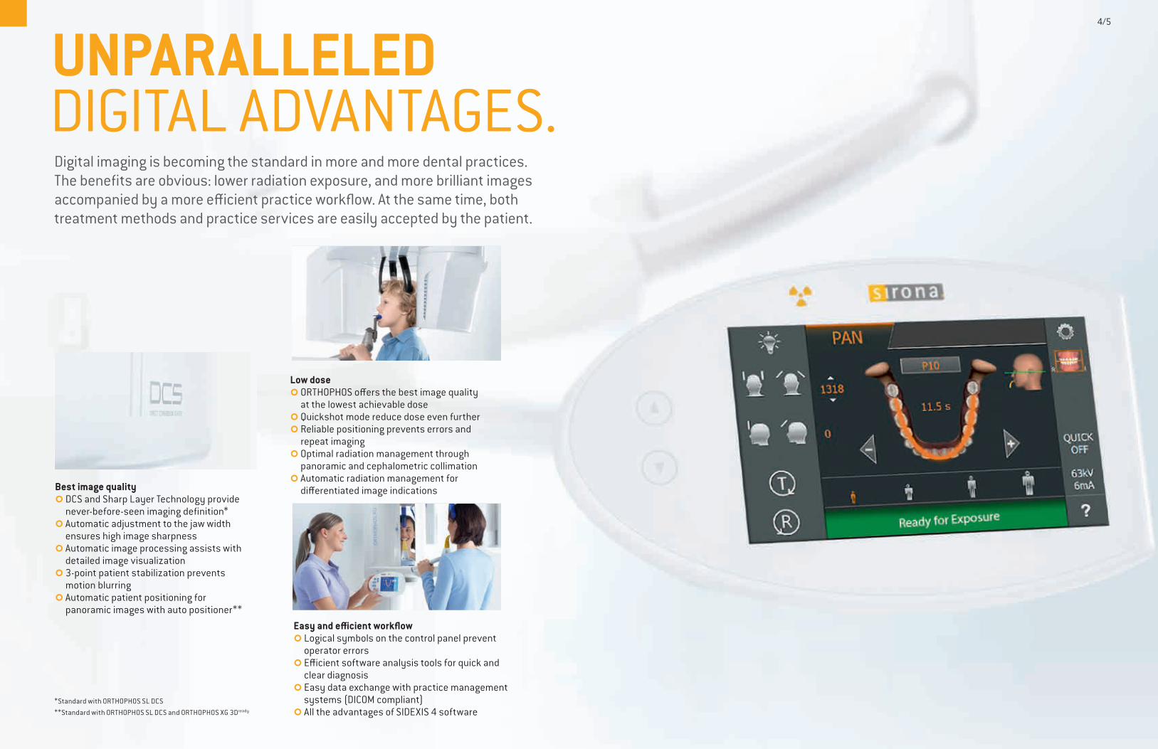
SIDEXIS 4–this is the core of the digital workflow with Sirona.
SIDEXIS 4 is the new standard in clinical diagnosis and patient communication. The software with its intuitive user interface has a very simple structure: it follows your work processes and provides you at all times with all visual data of your patients seamlessly and at a glance–whether 2D, 3D or intraoral. This integrates your patients optimally and thus results in a high acceptance of your treatment proposal. SIDEXIS 4 stands for real imaging efficiency.
WORKING DIGITALLY IS SO EASY.
Simple overview of the patient historyThe timeline gives us a quick overview of the entire history of the patient. This allows you to add a time dimension to your diagnostic options in a very intuitive way.
Compare images directlyIdeal for a comprehensive diagnosis: In the Lightbox, 2D and 3D images as well as camera images and FaceScan data can be compared side-by-side.
�� Modern design�� Software platform for all Sirona x-ray units�� Intuitive operation, optimally coordinated workflows�� Simple overview of the patient history thanks to the intuitive timeline�� Easy export of DICOM data sets�� Interface of the integrated solutions from Sirona
Clear and understandable workflows The software structure with easy-to-understand symbols makes it simple to use. It is geared to your practice workflows and it helps the entire practice team to use the software intuitively.
6/7
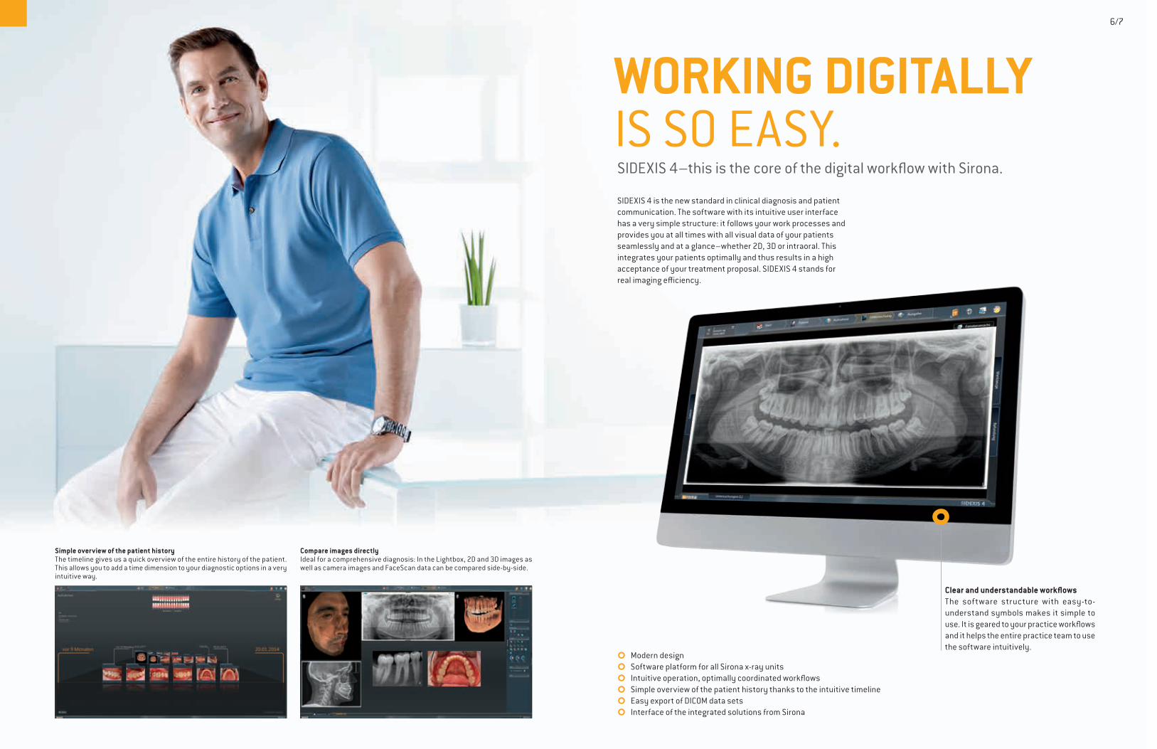
MAXIMUM IMAGE QUALITY WITH THE LOWEST DOSE.The ORTHOPHOS® family has been developed according to the ALARA principle to allow the best x-ray images with the lowest radiation dose. All programs and image parameters are tailored to the specific diagnosis tasks while offering you more diagnostic options and treatment at the same time.
QUICKSCAN FUNCTIONThe QuickScan function allows faster exposure cycles at a low radiation dose. This makes it easier to take panorama and cephalometric images of children, for example.
LOWER DOSE THANKS TO THE COLLIMATOR FUNCTIONUse the collimator function to select the region of interest if only a specific area of the volume is required for diagnosis. You can focus in on a more narrow region while also achieving a corresponding even lower dose.
PEDIATRIC PANORAMIC IMAGESThe horizontally and vertically reduced pediatric panoramic program achieves outstanding image quality with lower dose.
8/9
IMAGE
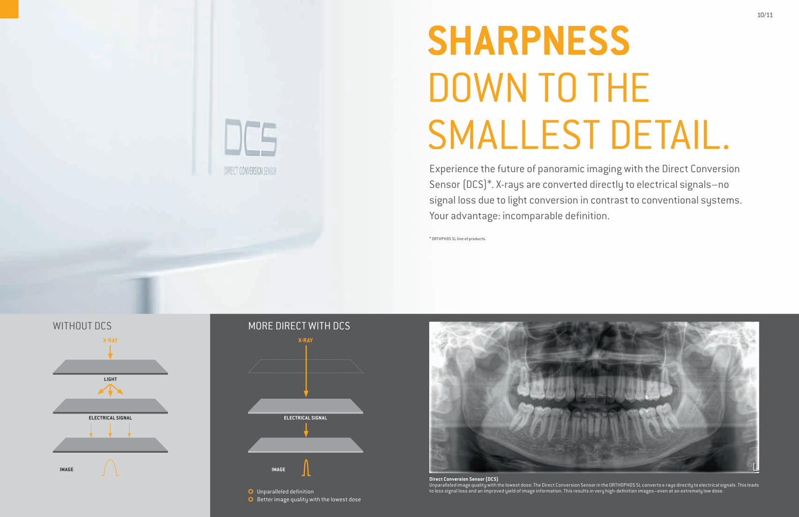
WITHOUT DCSX-RAY
ELECTRICAL SIGNAL
LIGHT
MORE DIRECT WITH DCS
ELECTRICAL SIGNAL
IMAGE
X-RAY
�� Unparalleled definition�� Better image quality with the lowest dose
Experience the future of panoramic imaging with the Direct Conversion Sensor (DCS)*. X-rays are converted directly to electrical signals–no signal loss due to light conversion in contrast to conventional systems. Your advantage: incomparable definition.
* ORTHPHOS SL line of products.
SHARPNESS DOWN TO THE SMALLEST DETAIL.
Direct Conversion Sensor (DCS)Unparalleled image quality with the lowest dose: The Direct Conversion Sensor in the ORTHOPHOS SL converts x-rays directly to electrical signals. This leads to less signal loss and an improved yield of image information. This results in very high-definition images–even at an extremely low dose.
10/11
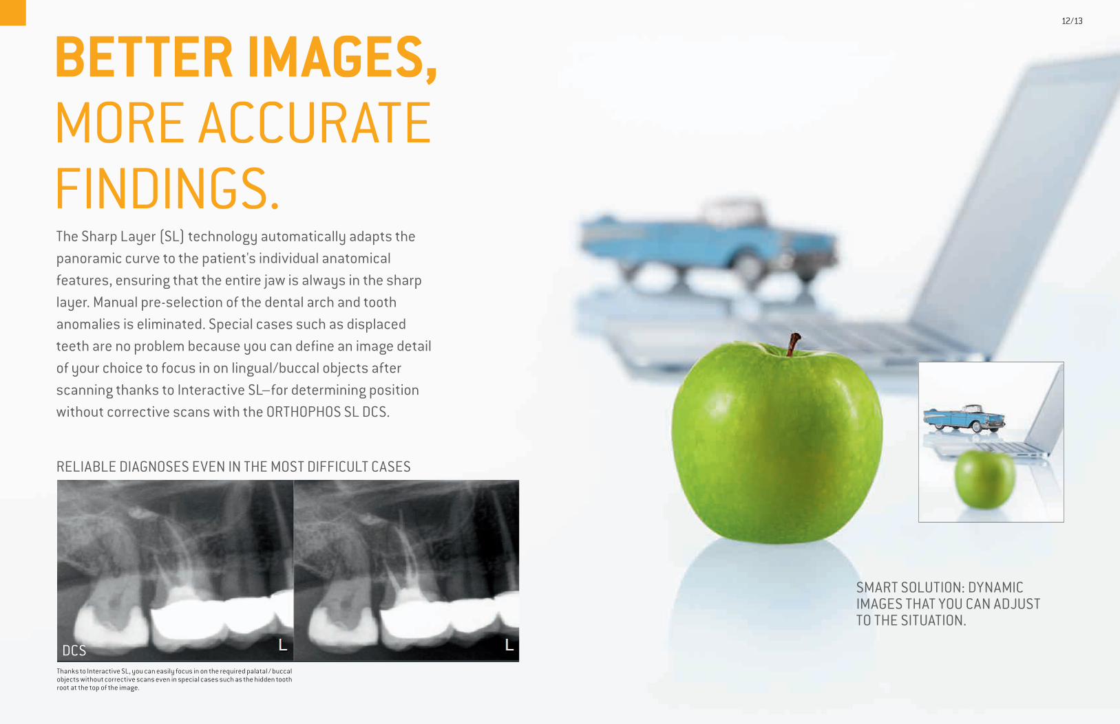
The Sharp Layer (SL) technology automatically adapts the panoramic curve to the patient's individual anatomical features, ensuring that the entire jaw is always in the sharp layer. Manual pre-selection of the dental arch and tooth anomalies is eliminated. Special cases such as displaced teeth are no problem because you can define an image detail of your choice to focus in on lingual/buccal objects after scanning thanks to Interactive SL–for determining position without corrective scans with the ORTHOPHOS SL DCS.
RELIABLE DIAGNOSES EVEN IN THE MOST DIFFICULT CASES
BETTER IMAGES, MORE ACCURATE FINDINGS.
Thanks to Interactive SL, you can easily focus in on the required palatal / buccal objects without corrective scans even in special cases such as the hidden tooth root at the top of the image.
DCS
SMART SOLUTION: DYNAMIC IMAGES THAT YOU CAN ADJUST TO THE SITUATION.
12/13
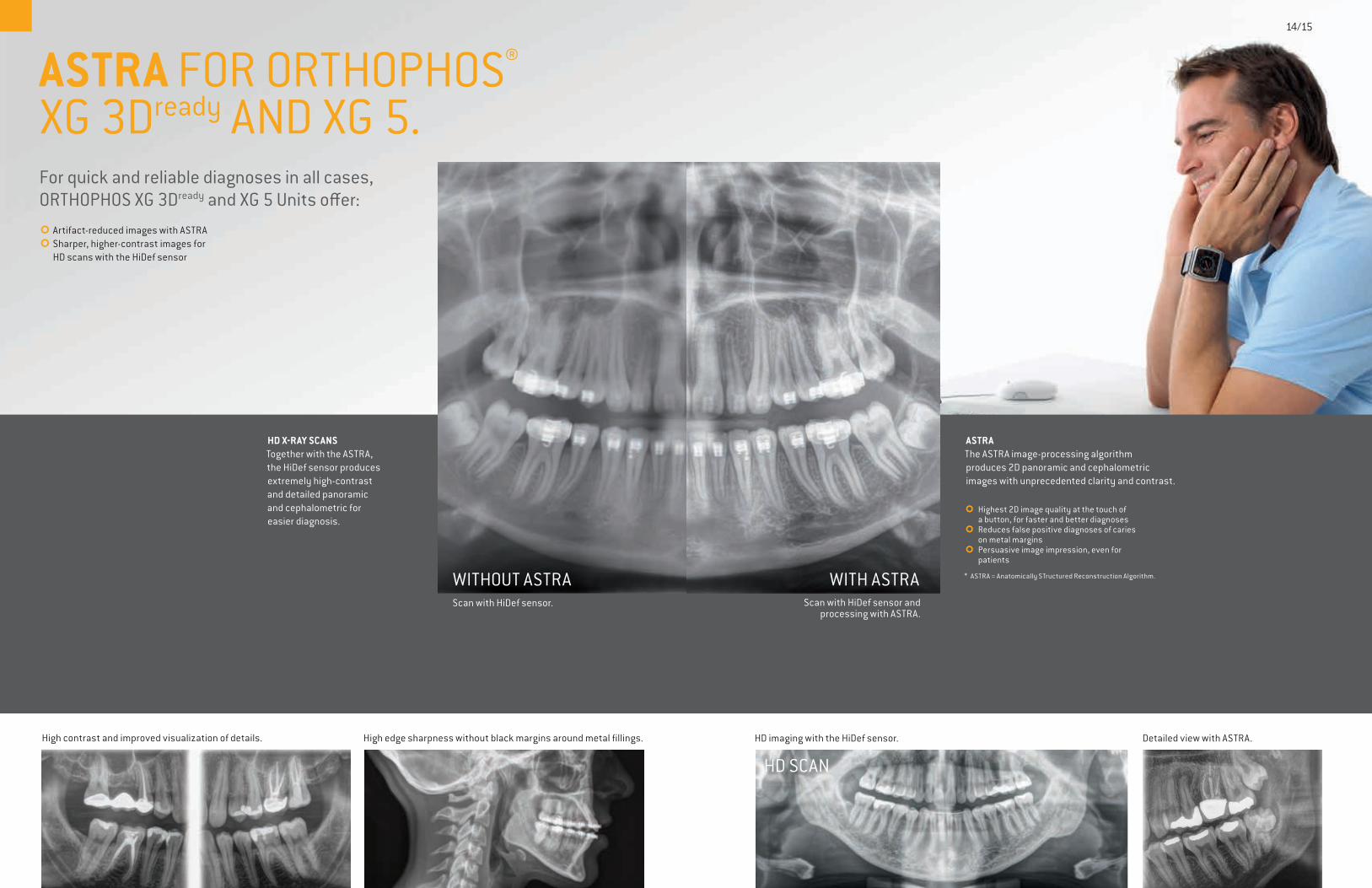
HD X-RAY SCANSTogether with the ASTRA, the HiDef sensor produces extremely high-contrast and detailed panoramic and cephalometric for easier diagnosis.
HD imaging with the HiDef sensor. Detailed view with ASTRA.
HD SCAN
WITHOUT ASTRA WITH ASTRAScan with HiDef sensor. Scan with HiDef sensor and
processing with ASTRA.
High contrast and improved visualization of details. High edge sharpness without black margins around metal fillings.
ASTRAThe ASTRA image-processing algorithm produces 2D panoramic and cephalometric images with unprecedented clarity and contrast.
��Highest 2D image quality at the touch of a button, for faster and better diagnoses��Reduces false positive diagnoses of caries on metal margins��Persuasive image impression, even for patients
* ASTRA = Anatomically STructured Reconstruction Algorithm.
ASTRA FOR ORTHOPHOS® XG 3Dready AND XG 5.
14/15
For quick and reliable diagnoses in all cases, ORTHOPHOS XG 3Dready and XG 5 Units offer: ��Artifact-reduced images with ASTRA��Sharper, higher-contrast images for HD scans with the HiDef sensor
WHICH UNIT IS THE ONE FOR YOU?
WHICH ORTHOPHOS® IS RIGHT FOR YOU?
OVERVIEW OF UNITS
Reliable diagnosis. Superior images. Lowest radiation dose. Gain the trust and confidence of your patients with an ORTHOPHOS by your side. Like all Sirona imaging units, the units in the ORTHOPHOS family have been designed to ensure the best image quality with the lowest dose and a perfect workflow.
ORTHOPHOS XG 5 ORTHOPHOS XG 3D ready ORTHOPHOS SL DCS
Ceph options
ORTHOPHOS XG 5 isnot upgradable
Unit
Target group General dentistry General dentistry,orthodontics, specialistdentistry
General dentistry,orthodontics, specialistdentistry
– �Optional ceph arm right/left
�Optional ceph arm right/left
3D upgrade options �Up to an FoV8 cm x 8 cm
�Up to an FoV11 cm x 10 cm
2D technology HiDef with ASTRA HiDef with ASTRA DCS with Sharp Layer
See for yourself:
ORTHOPHOS SL DCSThe ORTHOPHOS SL DCS offers the highest image quality for demanding diagnoses with the lowest dose. For future-oriented practices, the unit can be equipped with an optional cephalometric arm and a 3D module with a volume up to 11 x 10 cm.
ORTHOPHOS XG 3Dready
ORTHOPHOS XG 3Dready offers sophisticated diagnosis options for endodontics, periodontics, implantology, orthodontics, and surgery. This unit can be equipped with an optional cephalomteric arm and can be upgraded with a 3D module up to 8 x 8 cm and is ideal for future-oriented practices.
ORTHOPHOS XG 5With temporomandibular joint, sinus, bitewing, and pediatric panoramic programs, ORTHOPHOS XG 5 allows for more specific diagnoses.
16/17
Ceph armThe ceph arm can be mounted on the left or right side of the unit and provides detailed, high- contrast cephalometric images perfectly suited for orthodontic analyses and tracings.
Ambient lightThe soothing ambient light with a range of over 100 colors creates a pleasant atmosphere for your patients and fits perfectly into your modern practice look.
Sharp Layer technologyThanks to the SL technology, you not only get high-resolution panoramic images in the Sharp Layer, but can respond interactively within the image to special cases (lingually/buccally) – without additional exposure.
DCS for incomparable image qualityUse the most innovative sensor technology and benefit from high-resolution images with incom-parable definition for a detailed diagnosis.
18/19
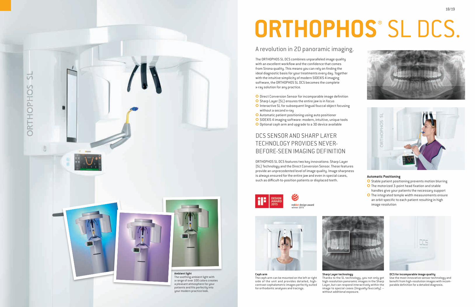
ORTHOPHOS® SL DCS.A revolution in 2D panoramic imaging.The ORTHOPHOS SL DCS combines unparalleled image quality with an excellent workflow and the confidence that comes from Sirona quality. This means you can rely on finding the ideal diagnostic basis for your treatments every day. Together with the intuitive simplicity of modern SIDEXIS 4 imaging software, the ORTHOPHOS SL DCS becomes the complete x-ray solution for any practice.
��Direct Conversion Sensor for incomparable image definition ��Sharp Layer (SL) ensures the entire jaw is in focus ��Interactive SL for subsequent lingual/buccal object focusing without a second x-ray��Automatic patient positioning using auto positioner ��SIDEXIS 4 imaging software: modern, intuitive, unique tools ��Optional ceph arm and upgrade to a 3D device available
DCS SENSOR AND SHARP LAYER TECHNOLOGY PROVIDES NEVER- BEFORE-SEEN IMAGING DEFINITIONORTHOPHOS SL DCS features two key innovations: Sharp Layer (SL) Technology and the Direct Conversion Sensor. These features provide an unprecedented level of image quality. Image sharpness is always ensured for the entire jaw and even in special cases, such as difficult-to-position patients or displaced teeth.
Automatic Positioning���Stable patient positioning prevents motion blurring ��The motorized 3-point head fixation and stable handles give your patients the necessary support ��The integrated temple width measurements ensure an orbit specific to each patient resulting in high image resolution
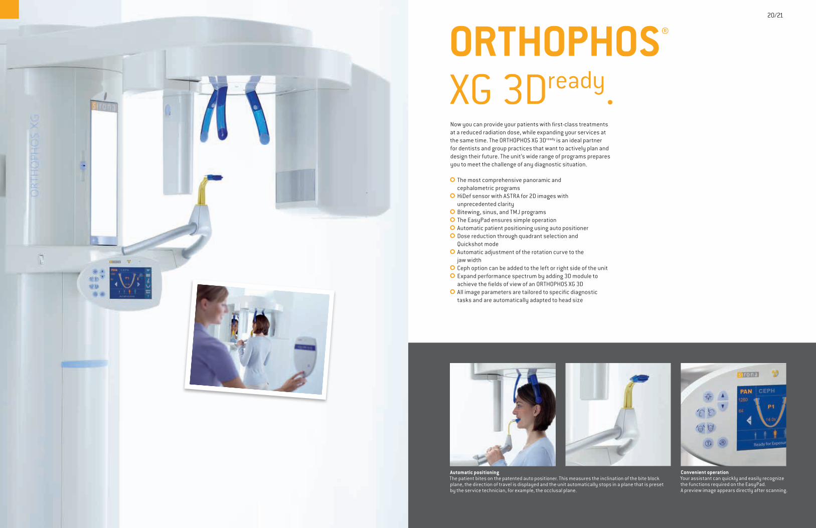
ORTHOPHOS® XG 3Dready.
Automatic positioningThe patient bites on the patented auto positioner. This measures the inclination of the bite block plane, the direction of travel is displayed and the unit automatically stops in a plane that is preset by the service technician, for example, the occlusal plane.
Convenient operation Your assistant can quickly and easily recognize the functions required on the EasyPad. A preview image appears directly after scanning.
20/21
Now you can provide your patients with first-class treatments at a reduced radiation dose, while expanding your services at the same time. The ORTHOPHOS XG 3Dready is an ideal partner for dentists and group practices that want to actively plan and design their future. The unit’s wide range of programs prepares you to meet the challenge of any diagnostic situation.
��The most comprehensive panoramic and cephalometric programs��HiDef sensor with ASTRA for 2D images with unprecedented clarity��Bitewing, sinus, and TMJ programs��The EasyPad ensures simple operation��Automatic patient positioning using auto positioner��Dose reduction through quadrant selection and Quickshot mode��Automatic adjustment of the rotation curve to the jaw width��Ceph option can be added to the left or right side of the unit��Expand performance spectrum by adding 3D module to achieve the fields of view of an ORTHOPHOS XG 3D��All image parameters are tailored to specific diagnostic tasks and are automatically adapted to head size
BE READYFOR THE FUTURE.ORTHOPHOS® SL DCS AND ORTHOPHOS XG 3Dready
ARE DESIGNED TO BE FUTURE-READY, SO YOU DON‘T NEED TO WORRY ABOUT WHAT‘S AROUND THE CORNER.
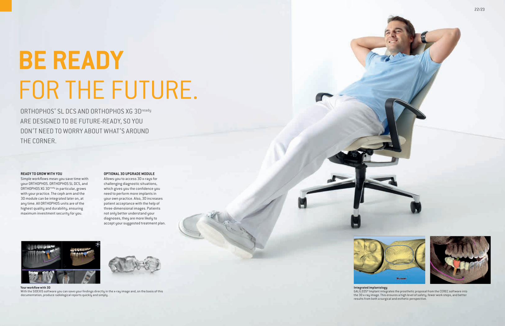
Integrated implantologyGALILEOS® Implant integrates the prosthetic proposal from the CEREC software into the 3D x-ray image. This ensures a high level of safety, fewer work steps, and better results from both a surgical and esthetic perspective.
Your workflow with 3DWith the SIDEXIS software you can save your findings directly in the x-ray image and, on the basis of this documentation, produce radiological reports quickly and simply .
22/23
READY TO GROW WITH YOUSimple workflows mean you save time with your ORTHOPHOS. ORTHOPHOS SL DCS, and ORTHOPHOS XG 3Dready in particular, grows with your practice. The ceph arm and the 3D module can be integrated later on, at any time. All ORTHOPHOS units are of the highest quality and durability, ensuring maximum investment security for you.
OPTIONAL 3D UPGRADE MODULE Allows you to access 3D x-rays for challenging diagnostic situations, which gives you the confidence you need to perform more implants in your own practice. Also, 3D increases patient acceptance with the help of three-dimensional images. Patients not only better understand your diagnoses, they are more likely to accept your suggested treatment plan.
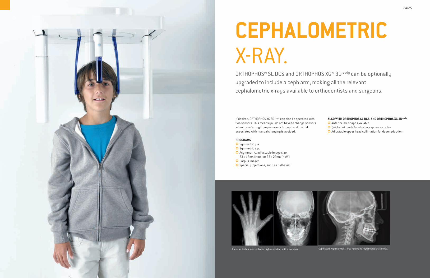
CEPHALOMETRIC X-RAY.ORTHOPHOS® SL DCS and ORTHOPHOS XG® 3Dready can be optionally upgraded to include a ceph arm, making all the relevant cephalometric x-rays available to orthodontists and surgeons.
24/25
The scan technique combines high resolution with a low dose. Ceph scan: High contrast, less noise and high image sharpness.
If desired, ORTHOPHOS XG 3D ready can also be operated with two sensors. This means you do not have to change sensors when transferring from panoramic to ceph and the risk associated with manual changing is avoided.
PROGRAMS��Symmetric p.a.��Symmetric a.p.��Asymmetric, adjustable image size: 23 x 18cm (HxW) or 23 x 29cm (HxW)��Carpus images��Special projections, such as half-axial
ALSO WITH ORTHOPHOS SL DCS AND ORTHOPHOS XG 3Dready
��Anterior jaw shape available��Quickshot mode for shorter exposure cycles��Adjustable upper head collimation for dose reduction

ORTHOPHOS® XG 5.Digital panoramic x-ray made simple.
Pediatric panorama
26/27
HIGH-QUALITY DIAGNOSTIC IMAGES WITH ASTRA��HiDef sensor with ASTRA for 2D images with unprecedented clarity��Automatic beam collimators re-adjust with each new program selection��Spinal column compensation for greater image clarity��Program selections include standard Pan programs for adults and children, bitewing, sinus, and TMJ��All image parameters are tailored to specific diagnostic tasks and are automatically adapted to head size
VERSATILE MULTIPAD��Software-driven program selection��Automated forehead and temple support��Unit height adjustment and patient positioning from a single location
The ORTHOPHOS XG 5 is for dentists and orthodontists who insist on the best technology to meet their most critical clinical requirements.
Excellent image qualityThanks to ASTRA, top image quality for quick and reliable diagnoses is ensured even with the standard sensor.
Bitewing exposure in the posterior tooth region
28/29
Stable floor stand for stand-alone mounting (optional)
Easy control with display of exposure parameters included in the scope of supply
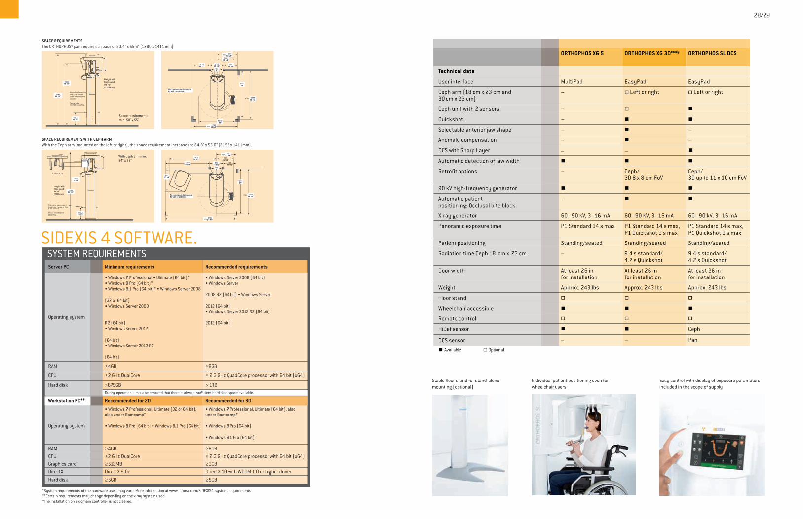
ORTHOPHOS XG 5 ORTHOPHOS XG 3Dready ORTHOPHOS SL DCS
Technical data
User interface MultiPad EasyPad EasyPad
Ceph arm (18 cm x 23 cm and 30 cm x 23 cm)
– � Left or right � Left or right
Ceph unit with 2 sensors – � �
Quickshot – � �
Selectable anterior jaw shape – � –
Anomaly compensation – � –
Automatic detection of jaw width � � �
Retrofit options – Ceph/ 3D 8 x 8 cm FoV
Ceph/ 3D up to 11 x 10 cm FoV
90 kV high-frequency generator � � �
Automatic patientpositioning: Occlusal bite block
– � �
X-ray generator 60–90 kV, 3–16 mA 60–90 kV, 3–16 mA 60–90 kV, 3–16 mA
Panoramic exposure time P1 Standard 14 s max P1 Standard 14 s max, P1 Quickshot 9 s max
P1 Standard 14 s max, P1 Quickshot 9 s max
Patient positioning Standing/seated Standing/seated Standing/seated
Radiation time Ceph 18 cm x 23 cm 9.4 s standard/4.7 s Quickshot
– 9.4 s standard/4.7 s Quickshot
Door width At least 26 in for installation
At least 26 in for installation
At least 26 in for installation
Weight Approx. 243 lbs Approx. 243 lbs Approx. 243 lbs
Floor stand � � �
Wheelchair accessible � � �
Remote control � � �
HiDef sensor � � Ceph
� Available � Optional
DCS sensor – Pan–
DCS with Sharp Layer – – �
Individual patient positioning even for wheelchair users
SIDEXIS 4 SOFTWARE.SYSTEM REQUIREMENTSServer PC Minimum requirements Recommended requirements • Windows 7 Professional • Ultimate (64 bit)* • Windows Server 2008 (64 bit) • Windows 8 Pro (64 bit)* • Windows Server • Windows 8.1 Pro (64 bit)* • Windows Server 2008 2008 R2 (64 bit) • Windows Server (32 or 64 bit) • Windows Server 2008 2012 (64 bit) • Windows Server 2012 R2 (64 bit)Operating system R2 (64 bit) 2012 (64 bit) • Windows Server 2012
(64 bit) • Windows Server 2012 R2
(64 bit)
RAM ≥4GB ≥8GB
CPU ≥2 GHz DualCore ≥ 2.3 GHz QuadCore processor with 64 bit (x64)
Hard disk >675GB > 1TB During operation it must be ensured that there is always sufficient hard disk space available.
Workstation PC** Recommended for 2D Recommended for 3D • Windows 7 Professional, Ultimate (32 or 64 bit), • Windows 7 Professional, Ultimate (64 bit), also also under Bootcamp* under Bootcamp*
Operating system • Windows 8 Pro (64 bit) • Windows 8.1 Pro (64 bit) • Windows 8 Pro (64 bit)
• Windows 8.1 Pro (64 bit)
RAM ≥4GB ≥8GBCPU ≥2 GHz DualCore ≥ 2.3 GHz QuadCore processor with 64 bit (x64)Graphics card† ≥512MB ≥1GBDirectX DirectX 9.0c DirectX 10 with WDDM 1.0 or higher driverHard disk ≥5GB ≥5GB
*System requirements of the hardware used may vary. More information at www.sirona.com/SIDEXIS4-system_requirements**Certain requirements may change depending on the x-ray system used.†The installation on a domain controller is not cleared.
With Ceph arm min. 84“ x 55“
SPACE REQUIREMENTSThe ORTHOPHOS® pan requires a space of 50.4” x 55.6” (1280 x 1411 mm)
SPACE REQUIREMENTS WITH CEPH ARMWith the Ceph arm (mounted on the left or right), the space requirement increases to 84.8” x 55.6” (2155 x 1411mm).
Space requirements min. 50“ x 55“
137154”
104041”
41016 1/2”
52020 1/2“
min.
min.1280
50 3/8”
141155 1/2"
60023 5/8”
min.
1546 ”
39515 1/2“
47518 1/3“
Recommended distances to wall or cabinet.
ORTHOPHOS
402,515 7/8”
224988 1/2”
195076 3/4”
Left CEPH
Height with floor stand: 89.75” (2279mm)
1546”
41016 1/2”
min. 60023 5/8”
min.2155
84 7/8”
143556 1/2” 520
20 1/2“
137154”
141155 1/2"
min.
39515 1/2“
69327 1/4”
135053 1/8”
Recommended distances to wall or cabinet.
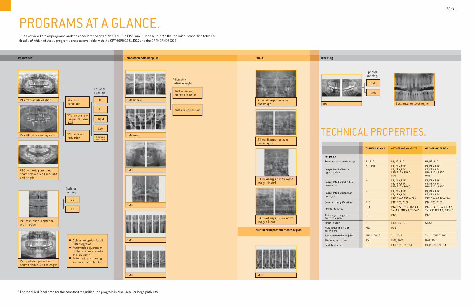
PROGRAMS AT A GLANCE.This overview lists all programs and the associated scans of the ORTHOPHOS® Family. Please refer to the technical properties table for details of which of these programs are also available with the ORTHOPHOS SL DCS and the ORTHOPHOS XG 5.
Panoramic Temporomandibular joint Sinus Bitewing
Multislice in posterior tooth region
Q Quickshot option for all PAN programsQ Automatic adjustment of the rotation curve to the jaw widthQ Automatic positioning with occlusal bite block
Optionalpanning
Left
Individual quadrants
Right
LJ
Left
Right
UJ
With artifact reduction
With a constant magnification of 1.25*
Standard exposure
With a slice position
With open and closed occlusion
P2 without ascending rami
P1 orthoradial radiation
P10 pediatric panorama, beam field reduced in height and length
Optionalpanning
LJ
UJ
Adjustable radiation angle
TM1 lateral
TM3
TM4
TM5
TM6
TM2 axial
MS1
Optionalpanning
BW1 BW2 anterior tooth region
30/31
* The modified focal path for the constant magnification program is also ideal for large patients.
P12 thick slice in anterior tooth region
P20 pediatric panorama, beam field reduced in length
S1 maxillary sinuses in one image
S2 maxillary sinuses in two images
S3 maxillary sinuses in one image (linear)
S4 maxillary sinuses in two images (linear)
TECHNICAL PROPERTIES. ORTHOPHOS XG 5 ORTHOPHOS XG 3D ready ORTHOPHOS SL DCS
Programs
Standard panoramic image P1, P10 P1, P2, P10 P1, P2, P10
Image detail of left orright-hand side
P1L, P1R P1, P1A, P1C P2, P2A, P2CP10, P10A, P10C BW1
P1, P1A, P1C P2, P2A, P2CP10, P10A, P10C BW1
Image detail of individual quadrants
– P1, P1A, P1C P2, P2A, P2CP10, P10A, P10C
P1, P1A, P1C P2, P2A, P2CP10, P10A, P10C
Image detail of upper or lower jaw
– P1, P1A, P1C P2, P2A, P2CP10, P10A, P10C, P12
P1, P1A, P1C P2, P2A, P2CP10, P10A, P10C, P12
Constant magnification P1C P1C, P2C, P10C P1C, P2C, P10C
Artifact-reduced P1A P1A, P2A, P10A, TM1A.1, TM1A.2, TM2A.1, TM2A.2
P1A, P2A, P10A, TM1A.1, TM1A.2, TM2A.1, TM2A.2
Thick-layer images ofanterior region
P12 P12 P12
Sinus images S1 S1, S2, S3, S4 S1, S3
Multi-layer images of pre-molars
MS1 MS1 –
Temporomandibular joint TM1.1, TM1.2 TM1–TM6 TM1.1 /TM1.2, TM3
Bite wing exposure BW1 BW1, BW2 BW1, BW2
Ceph (optional) – C1, C2, C3, C3F, C4 C1, C2, C3, C3F, C4

SIRONA: THE DENTAL TECHNOLOGY LEADERSirona, the technology and innovation leader in dentistry, has served dentists worldwide for more than 130 years. Sirona develops, manufactures, and markets a complete line of dental products, including CAD/CAM restoration systems [CEREC], CBCT imaging systems [GALILEOS], extraoral imaging systems, digital intraoral imaging technology [SCHICK], dental treatment centers, and hand instruments.
INNOVATIVE CHOICES FOR A REWARDING PRACTICE LIFESTYLEAt Sirona, our broad range of compatible solutions is developed entirely around you. Every research study, every R & D investment, and every new innovation is designed for one purpose: to help drive your success.
GALILEOS ComfortPLUS: The best image quality to satisfy any dental specialty
SIROLaser Advance: High-quality powerful laser for treatment and tissue management
Schick 33: The most advanced imaging resolution in the industry
HELIODENTPLUS: Intraoral x-rays made easy
Concept S: Surgery suite designed to optimize your clinical workflow
For more information call 1.800.659.5977 or visit www.sironausa.comJOIN THE CONVERSATION ON FACEBOOK:FACEBOOK.COM/SIRONA3D
SIRONAUSA.COM
CEREC Omnicam: Providing dental restorations in just one session


![Illumination Invariant Imaging: Applications in Robust ...mobile/Papers/2014ICRA_maddern.pdf · invariant point features have been presented [13], but these rely on training images](https://img.pdfslide.net/doc/110x75/60aaa4b0a1d14f79f6448ac8/illumination-invariant-imaging-applications-in-robust-mobilepapers2014icramaddernpdf.jpg)