Embed Size (px)
Citation preview
LUCRĂRI ŞTIINłIFICE MEDICINĂ VETERINARĂ VOL. XL, 2007, TIMIŞOARA
561
THE OSTEOLOGICAL FEATURES OF THE SKELETON IN OSTRICH (STRUTHIO CAMELUS)
C. POP, M. PENTEA
Faculty of Veterinary Medicine Timisoara, Calea Aradului no. 119
In our country in the last years the anatomy of the ostrich has became an important
challenge researcher and for practitioners. From this point of view its been considerate the presentation of the osteological
features due to the similar development of the appendicular and axial skeleton in ostrich and other animals.
Key words: osteology, ostrich
Materials and methods
Have been studied from the morphological point the bones from 3 death adult ostriches, which were coming from two breeding farm from Bihor and Timis County. of view used flat and long bones from 5 dogs, the thoracic appendicular skeleton, scapula, humerus, radius and ulna.
The bones were prepared and according to N.A.V. (1979) the osteological features have been recorded and registered (1, 2, 3, 4, 5).
Results and discussions
The bony head in ostrich is similar with domestic birds, composed by the
same bones. There are no presented the interparietal bone and presphenoidal bone, the frontal bone presents an supraorbytaris foramen or notch.
The shape of skull in ostrich is 2-3 times greater than the head of anseriformes (Fig. 1).
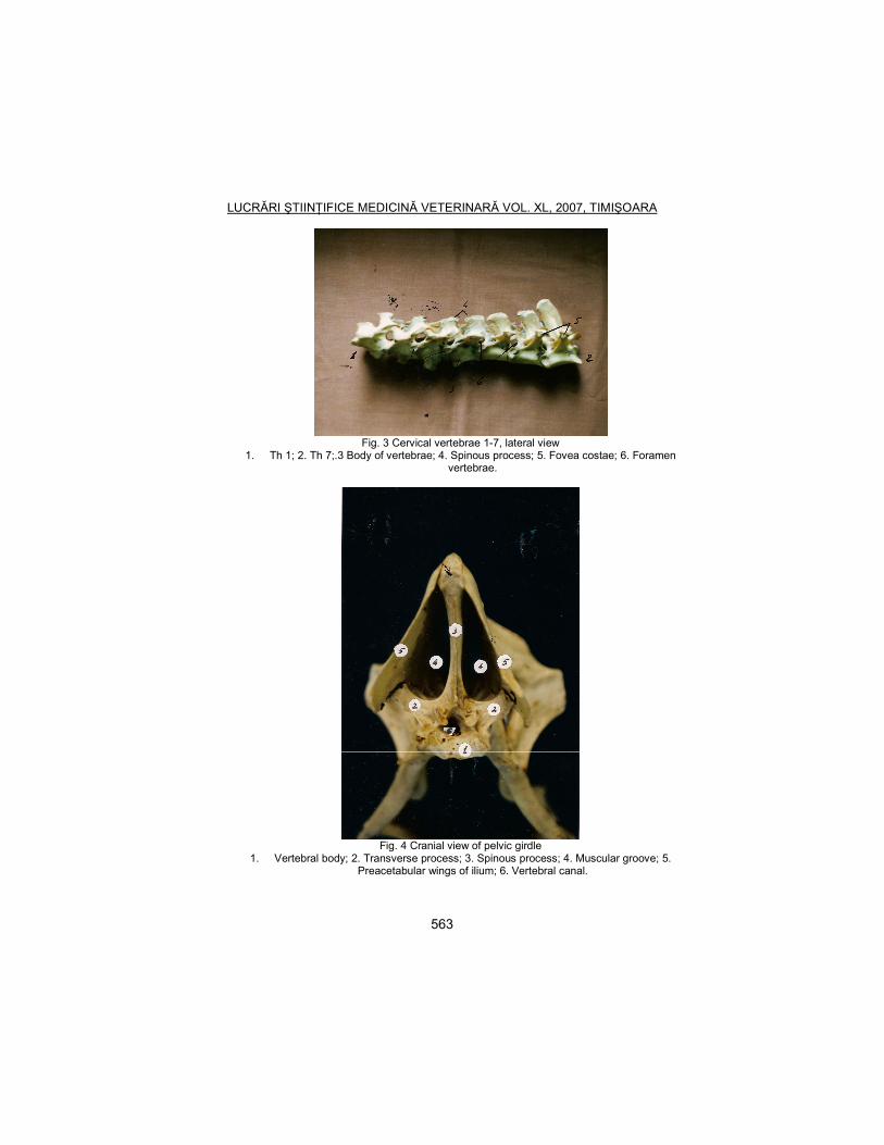
17 cervical vertebrae, 9 thoracic vertebrae, the sinsacrum bone, pygostilus and ribs and sternum compose the axial skeleton.
The cervical vertebrae have the same development with those in domestic birds (Fig. 2).
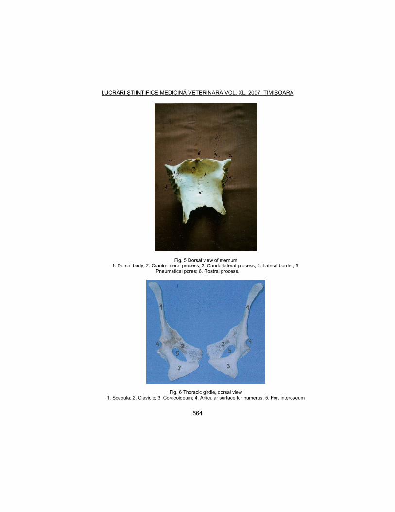
The notarium is compose by 1st to 7th thoracic vertebrae, and the last 2 vertebrae are bound to synsacrum. The vertebrae that forms the notarium are not bound (Fig. 3).
The lumbo-sacral bone presents a prominent dorsal crest that continues the dorsal border of the preacetabular part of ilium. The ventral crest presented in the domestic birds isn’t shown (Fig. 4).
The dorsal divides pelvic cavity in two narrow and deep spaces (Fig. 4). The processus uncinatus is not presented in vertebral ribs.
LUCRĂRI ŞTIINłIFICE MEDICINĂ VETERINARĂ VOL. XL, 2007, TIMIŞOARA
562
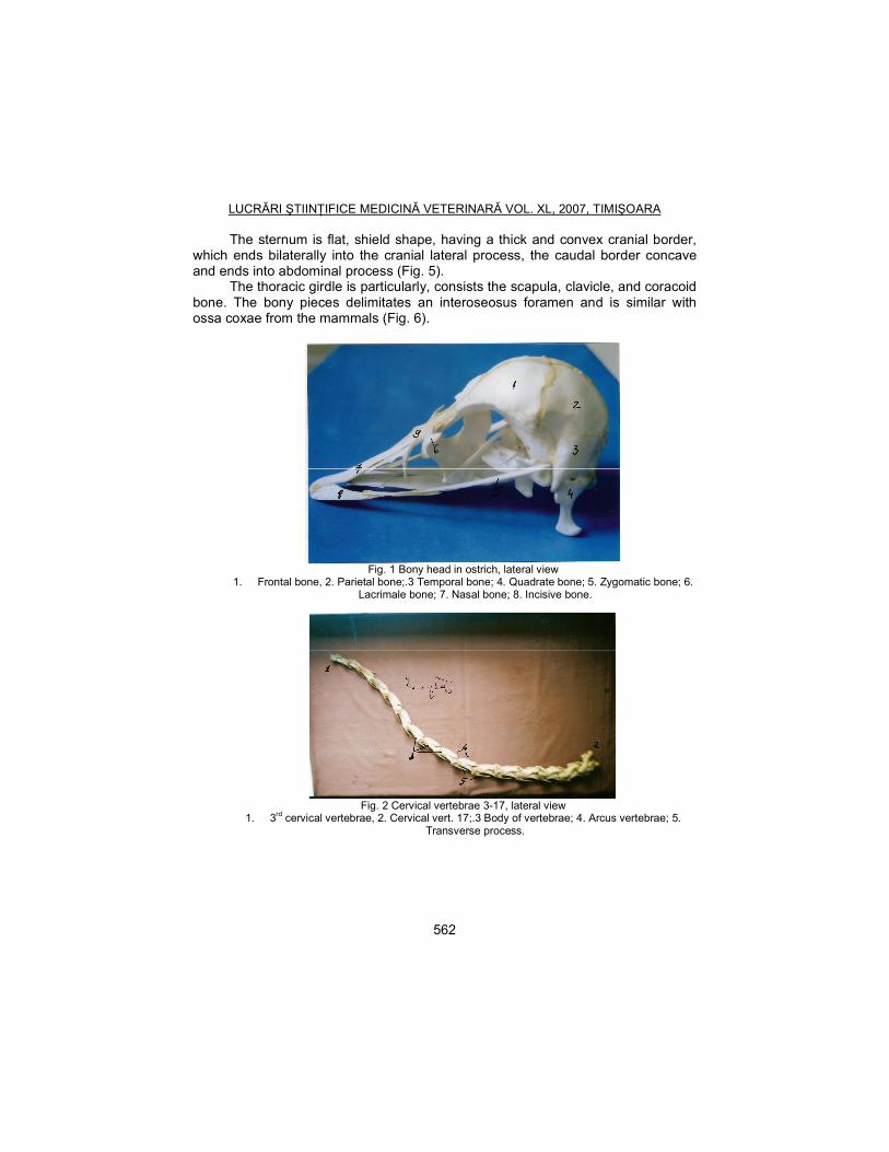
The sternum is flat, shield shape, having a thick and convex cranial border, which ends bilaterally into the cranial lateral process, the caudal border concave and ends into abdominal process (Fig. 5).
The thoracic girdle is particularly, consists the scapula, clavicle, and coracoid bone. The bony pieces delimitates an interoseosus foramen and is similar with ossa coxae from the mammals (Fig. 6).
Fig. 1 Bony head in ostrich, lateral view
1. Frontal bone, 2. Parietal bone;.3 Temporal bone; 4. Quadrate bone; 5. Zygomatic bone; 6. Lacrimale bone; 7. Nasal bone; 8. Incisive bone.
Fig. 2 Cervical vertebrae 3-17, lateral view
1. 3rd cervical vertebrae, 2. Cervical vert. 17;.3 Body of vertebrae; 4. Arcus vertebrae; 5. Transverse process.
LUCRĂRI ŞTIINłIFICE MEDICINĂ VETERINARĂ VOL. XL, 2007, TIMIŞOARA
563
Fig. 3 Cervical vertebrae 1-7, lateral view
1. Th 1; 2. Th 7;.3 Body of vertebrae; 4. Spinous process; 5. Fovea costae; 6. Foramen vertebrae.
Fig. 4 Cranial view of pelvic girdle
1. Vertebral body; 2. Transverse process; 3. Spinous process; 4. Muscular groove; 5. Preacetabular wings of ilium; 6. Vertebral canal.
LUCRĂRI ŞTIINłIFICE MEDICINĂ VETERINARĂ VOL. XL, 2007, TIMIŞOARA
564
Fig. 5 Dorsal view of sternum 1. Dorsal body; 2. Cranio-lateral process; 3. Caudo-lateral process; 4. Lateral border; 5.
Pneumatical pores; 6. Rostral process.
Fig. 6 Thoracic girdle, dorsal view 1. Scapula; 2. Clavicle; 3. Coracoideum; 4. Articular surface for humerus; 5. For. interoseum
LUCRĂRI ŞTIINłIFICE MEDICINĂ VETERINARĂ VOL. XL, 2007, TIMIŞOARA
565
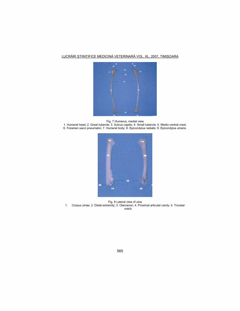
Fig. 7 Humerus, medial view 1. Humeral head; 2. Great tubercle; 3. Sulcus capitis; 4. Small tubercle; 5. Medio-ventral crest; 6. Foramen sacci pneumatici; 7. Humeral body; 8. Epicondylus radialis; 9. Epicondylus ulnaris.
Fig. 8 Lateral view of ulna 1. Corpus ulnae; 2. Distal extremity; 3. Olecranon; 4. Proximal articular cavity; 4. Troclear
notch.
LUCRĂRI ŞTIINłIFICE MEDICINĂ VETERINARĂ VOL. XL, 2007, TIMIŞOARA
566
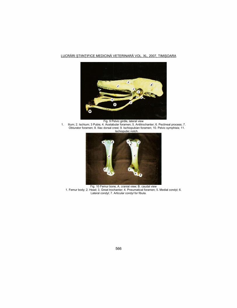
Fig. 9 Pelvic girdle, lateral view
1. Ilium; 2. Ischium; 3 Pubis; 4. Acetabular foramen; 5. Antitrochanter; 6. Pectineal process; 7. Obturator foramen; 8. Iliac dorsal crest; 9. Ischiopubian foramen; 10. Pelvic symphisis; 11.
Ischiopubic notch.
Fig. 10 Femur bone, A. cranial view; B. caudal view
1. Femur body; 2. Head; 3. Great trochanter; 4. Pneumatical foramen; 5. Medial condyl; 6. Lateral condyl; 7. Articular condyl for fibula.
LUCRĂRI ŞTIINłIFICE MEDICINĂ VETERINARĂ VOL. XL, 2007, TIMIŞOARA
567
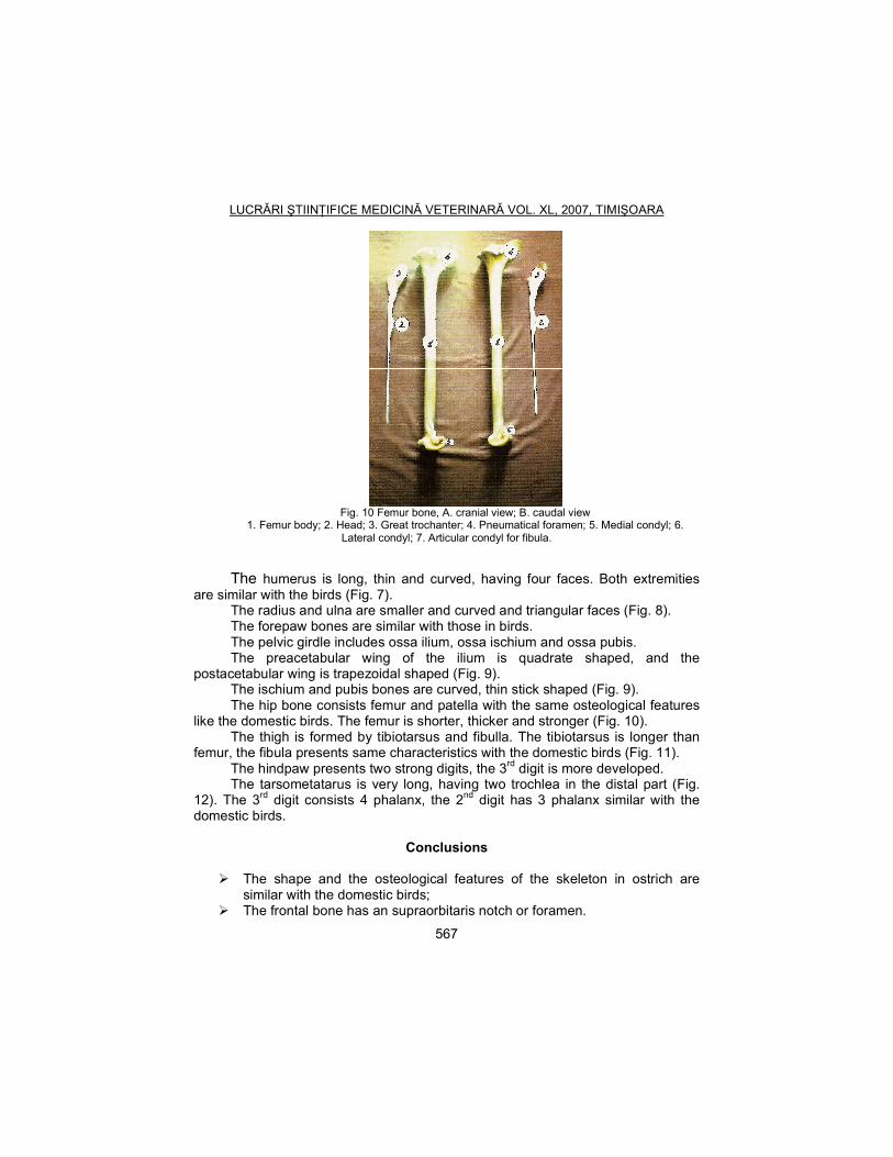
Fig. 10 Femur bone, A. cranial view; B. caudal view
1. Femur body; 2. Head; 3. Great trochanter; 4. Pneumatical foramen; 5. Medial condyl; 6. Lateral condyl; 7. Articular condyl for fibula.
The humerus is long, thin and curved, having four faces. Both extremities are similar with the birds (Fig. 7).
The radius and ulna are smaller and curved and triangular faces (Fig. 8). The forepaw bones are similar with those in birds. The pelvic girdle includes ossa ilium, ossa ischium and ossa pubis. The preacetabular wing of the ilium is quadrate shaped, and the
postacetabular wing is trapezoidal shaped (Fig. 9). The ischium and pubis bones are curved, thin stick shaped (Fig. 9). The hip bone consists femur and patella with the same osteological features
like the domestic birds. The femur is shorter, thicker and stronger (Fig. 10). The thigh is formed by tibiotarsus and fibulla. The tibiotarsus is longer than
femur, the fibula presents same characteristics with the domestic birds (Fig. 11). The hindpaw presents two strong digits, the 3rd digit is more developed. The tarsometatarus is very long, having two trochlea in the distal part (Fig.
12). The 3rd digit consists 4 phalanx, the 2nd digit has 3 phalanx similar with the domestic birds.
Conclusions
� The shape and the osteological features of the skeleton in ostrich are similar with the domestic birds;
� The frontal bone has an supraorbitaris notch or foramen.
LUCRĂRI ŞTIINłIFICE MEDICINĂ VETERINARĂ VOL. XL, 2007, TIMIŞOARA
568
� The notarium is not presented in the axial skeleton, the sternum has an shield shape.
� 3 bound bones compose the thoracic girdle.
References
1. J.J. Baumell (1975) – Nomina Anatomica Avium, Second Ed. Nuttal
Ornithological Club, nr. 23, Cambridge, Massachusetts. 2. Kovacs, GY., Kellz, D. F. –Osteologische untersuchungen an
Vogelknochen, Kozlemenyrk az osszehasanlito elet es Kortan Korebol, Lucr. XXIV, Budapest, 1968.
3. Lucas, A.M., Stetenheim, P.R. – Avian Anatomy, Agricultural Research service, USA Department Of Agriculture.
4. NickeL, K., Schummer, A., Seiferle, E. - Lehrbuch der Anatomie der Haustiere, Band V, Paul Parey in Berlin und Hamburg, 1973.
5. Romer, S. S., Parsons, S.T. - The vertebrate body, Sixt Edition, Saunders College Publishing., Philadelphia, 1986.